Embed Size (px)
Citation preview

Annales Francaises d’Anesthesie et de Reanimation 33 (2014) 65–71
Anarlf meeting
Assessing consciousness in coma and related states using transcranialmagnetic stimulation combined with electroencephalography§,§§
Evaluation de la conscience chez les patients en coma et en etats apparentes par
stimulation magnetique transcranienne combinee a l’electroencephalographie
O. Gosseries a,*,b,c, A. Thibaut a, M. Boly a,b,d, M. Rosanova e, M. Massimini e,f, S. Laureys a
a Coma Science Group, Cyclotron Research Center and Neurology Department, University of Liege and University Hospital of Liege, Sart Tilman B30,
allee du 6-Aout-no 8, 4031 Liege, Belgiumb Center for Sleep and Consciousness, Department of Psychiatry, University of Wisconsin, Madison, WI, USAc Postle Laboratory, Department of Psychology and Psychiatry, University of Wisconsin, Madison, WI, USAd Department of Neurology, University of Wisconsin, Madison, WI, USAe Department of Biomedical and Clinical Sciences ‘‘Luigi Sacco’’, University of Milan, Milan, Italyf Istituto Di Ricovero e Cura a Carattere Scientifico, Fondazione Don Carlo Gnocchi, 20148 Milan, Italy
A R T I C L E I N F O
Keywords:
Disorders of consciousness
Unresponsive wakefulness syndrome
Vegetative state
Minimally conscious state
Locked-in syndrome
Transcranial magnetic stimulation
Electroencephalography
Anesthesia
Sleep
Brain stimulation
Mots cles :
Troubles de la conscience
Syndrome d’eveil non-repondant
Etat vegetatif
Etat de conscience minimale
Locked-in syndrome
Stimulation magnetique transcranienne
Electroencephalographie
Anesthesie
Sommeil
Stimulation cerebrale
A B S T R A C T
Thanks to advances in medical care, an increased number of patients recover from coma. However, some
remain in vegetative/unresponsive wakefulness syndrome or in a minimally conscious state. Detection
of awareness in severely brain-injured patients is challenging because it relies on behavioral
assessments, which can be affected by motor, sensory and cognitive impairments of the patients.
Other means of evaluation are needed to improve the accuracy of the diagnosis in this challenging
population. We will here review the different altered states of consciousness occurring after severe brain
damage, and explain the difficulties associated with behavioral assessment of consciousness. We will
then describe a non-invasive technique, transcranial magnetic stimulation combined with high-density
electroencephalography (TMS-EEG), which has allowed us to detect the presence or absence of
consciousness in different physiological, pathological and pharmacological states. Some potential
underlying mechanisms of the loss of consciousness will then be discussed. In conclusion, TMS-EEG is
highly promising in identifying markers of consciousness at the individual level and might be of great
value for clinicians in the assessment of consciousness.
� 2013 Societe francaise d’anesthesie et de reanimation (Sfar). Published by Elsevier Masson SAS. All
rights reserved.
R E S U M E
Avec les avancees recentes de la medecine, de plus en plus de patients sortent du coma. Cependant,
certains restent en etat vegetatif/syndrome d’eveil non-repondant ou en etat de conscience minimale.
L’evaluation de leur niveau de conscience reste difficile notamment parce qu’elle repose sur des examens
comportementaux qui peuvent etre biaises par des deficits moteurs, sensoriels et cognitifs. D’autres
outils d’evaluation de la conscience sont donc necessaires afin de preciser le diagnostic des patients
severement cerebroleses. Dans cet article, nous exposerons les differents etats de conscience alteree
survenant a la suite d’une grave lesion cerebrale et les difficultes liees a l’evaluation du niveau de
conscience au chevet des patients. Nous decrirons ensuite une technique non invasive, la stimulation
magnetique transcranienne combinee a l’electroencephalographie a haute densite (SMT-EEG) qui
permet de detecter la presence ou l’absence de conscience dans differents etats physiologiques,
§ st nd
French NeuroAnesthesia and Intensive Care society Meeting, Paris, November 2013, 21 and 22 : ‘‘The acutely brain-injured patient: consciousness and neuroethic’’.§§ This article is published under the responsibility of the Scientific Committee of the ‘‘35e Journee de l’Association des neuro-anesthesistes reanimateurs de languefrancaises’’ de la SFAR. The editorial board of the Annales francaises d’anesthesie et de reanimation was not involved in the conception and validation of its content.
* Corresponding author.
E-mail address: [email protected] (O. Gosseries).
0750-7658/$ – see front matter � 2013 Societe francaise d’anesthesie et de reanimation (Sfar). Published by Elsevier Masson SAS. All rights reserved.
http://dx.doi.org/10.1016/j.annfar.2013.11.002
O. Gosseries et al. / Annales Francaises d’Anesthesie et de Reanimation 33 (2014) 65–7166
pathologiques et pharmacologiques. Des mecanismes potentiels pouvant expliquer la perte de
conscience seront ensuite discutes. Dans notre conclusion, nous exposerons les avantages de la SMT-EEG
et en quoi cette technique prometteuse pourrait etre d’une grande utilite pour evaluer le niveau de
conscience au chevet des patients.
� 2013 Societe francaise d’anesthesie et de reanimation (Sfar). Publie par Elsevier Masson SAS. Tous
droits reserves.
1. Disorders of consciousness
Since the invention of mechanical ventilation in the 1950s,many patients survive even after severe brain damage. After thecomatose phase where patients lie with eyes closed (i.e., coma),some patients regain full consciousness while some progress to astate of preserved wakefulness in the absence of awareness (i.e.,unresponsive wakefulness syndrome). Others show fluctuatingsigns of awareness but they remain unable to communicateconsistently (i.e., minimally conscious state). Finally, somepatients fully recover awareness but lack motor output (i.e.,locked-in syndrome).
1.1. Coma
Coma is an acute state of non-responsiveness in which patientscannot be awakened even when intensively stimulated [1].Patients in a coma lack sleep-wake cycles and only show somereflex behaviors [2]. The autonomous functions such as breathingand thermoregulation are reduced and global brain metabolism issignificantly diminished [3]. Coma is the result of diffuse cortical orwhite matter damage and/or an acute lesion in the brainstem [4]. Itlasts at least one hour (to be distinguished from syncope) and up toa few days. The prolonged coma also exists but is rare and can lasttwo to five weeks (e.g., pharmacologically-induced coma).
1.2. Unresponsive wakefulness syndrome
This state was first named apallic syndrome [5] or coma vigil[6], and in 1972 it was termed vegetative state [7]. Newterminology was proposed in 2010 – the unresponsive wakeful-ness syndrome (UWS) [8] – to avoid the strong negativeconnotation with inadvertently risking comparisons betweenpatients and vegetables. The term ‘‘UWS’’ also allows a moreprecise description of the clinical state, referring to patients thatare unable to react to stimuli in a non-reflexive way (henceunresponsive), whilst showing periods of time with eyes opened(hence wakefulness). Clinically, this state is thus defined bywakefulness without awareness, and in which patients are able toopen their eyes but remain unaware of the environment andthemselves [9]. They only show spontaneous or stimulus-inducedreflex behaviors such as grinding teeth, moving eyes, swallowing,chewing, yawning or groaning. This state may be transitory,chronic or permanent.
Although recovery of the sleep-wake cycle is part of the criteriaof UWS, recent studies have demonstrated an absence ofelectrophysiological characteristics of sleep in UWS [10,11]. Brainmetabolism is usually diminished by 40 to 50% with impairedcortico-thalamo-cortical circuits but relatively preserved brain-stem functions [12]. Brain dysfunctions are more specificallylocated in the frontoparietal network (including both medial andlateral networks related to self and environment respectively) andin the thalami [13]. During sensory stimulations, UWS patientsusually show metabolic brain activation that remains isolated inthe primary cortices [14,15]. Finally, top-down processes fromfrontal to temporal cortices have been shown to be impaired inpatients in UWS when measuring the electrical activity duringauditory stimulations [16].
1.3. Minimally conscious state
The minimally conscious state (MCS) is characterized byprimary inconsistent signs of consciousness [17,18]. The criteriaof MCS, introduced in 2002, include reproducible responses toverbal or written commands, visual pursuit, localization to pain,intelligible verbalizations, intentional communication and reach-ing/holding objects [18]. Adapted emotional behaviors such assmiles, laughs or tears can also be observed [18]. This clinical entityhas been recently subcategorized in ‘‘minimally conscious plus’’(MCS+) for patients who present high-order behavioral responsesto stimuli (e.g., response to a command which involves thepreservation of language) and ‘‘minimally conscious minus’’(MCS�) for patients who only show low-level non-reflexiveresponses to stimuli (e.g., visual pursuit or localization tonociception) [19]. This classification is supported by neuroanato-mical data that demonstrate better preservation of language-related networks in MCS+ as compared to MCS� patients [20]. Theoverall cerebral metabolic activity in MCS patients is usuallyreduced but the autonomous functions are preserved, and cortico-thalamo-cortical connections are partly restored [21]. The mainmetabolic dysfunctions appear to be located in the lateral networkand in the thalami [13]. When patients recover the ability tofunctionally communicate or to use objects adequately, this isreferred to ‘‘emergence of the minimally conscious state’’ (EMCS)[18].
1.4. Locked-in syndrome
Locked-in syndrome (LIS), also known as pseudocoma, is not adisorder of consciousness per se but can be mistaken as one. LIS ischaracterized by a complete paralysis of the body resulting from alesion in the brainstem affecting the pyramidal tract, mostfrequently due to an ischemic pontine lesion [17]. If the lesionis only restricted to the brainstem, LIS patients have preservedsensory and cognitive functions [22]. The primary way ofcommunication is through vertical eye movements or blinking[23]. Through the recovery of distal movements, such as the tip of afinger or head movement, chronic LIS patients are often able tocommunicate via a computer and to control their wheelchair.Communication has also been recently made possible by measur-ing electrical brain activity [24] and pupil size [25]. Finally, manychronic LIS patients report having a happy and meaningful life andthe demand for euthanasia, albeit existing, is not so frequent [26].
2. Assessment of the level of consciousness
To date, the level of consciousness is mainly assessed at thepatient’s bedside by searching for response to command or non-reflexive behaviors in response to sensory stimulations. Assessingthe presence or absence of consciousness of non-communicativebrain-damaged patients is however difficult, as consciousness is asubjective first-person experience, and one has necessarily tomake inferences about its presence based on the patient’sbehavior. Currently, the diagnostic decision-making process isextremely challenging leading to a diagnostic error rate up to 40%when not assessed with appropriate standardized scales [27]. TheComa Recovery Scale-Revised (CRS-R) has been shown to be the
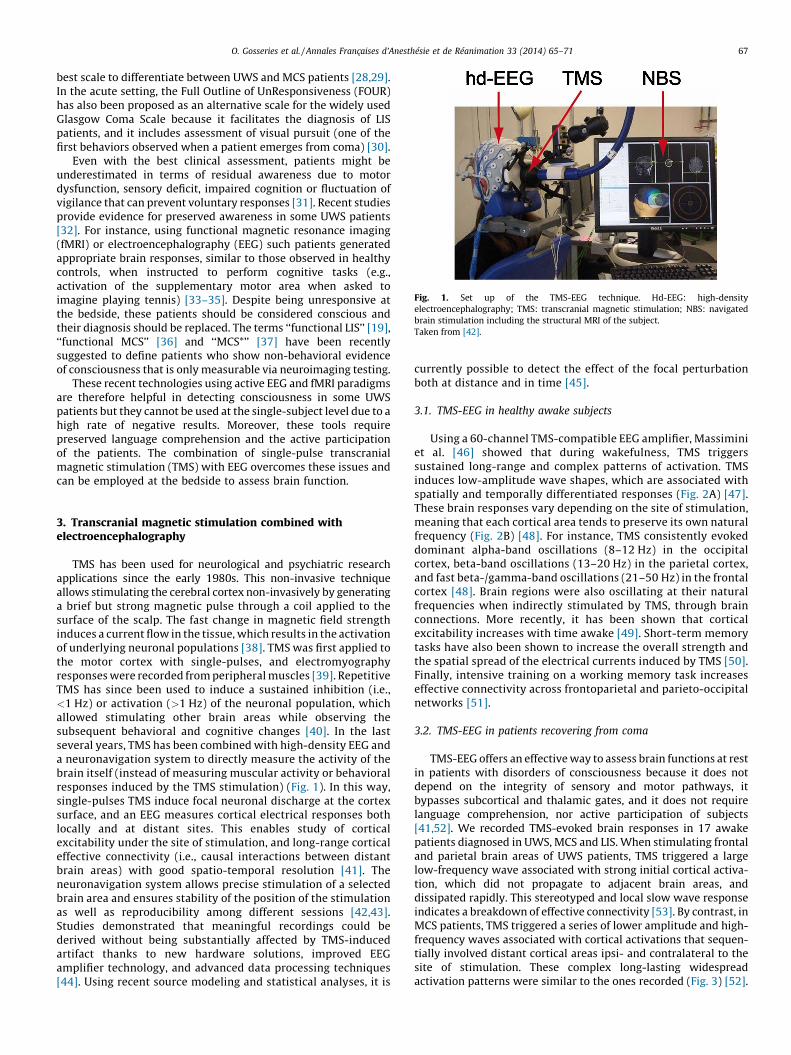
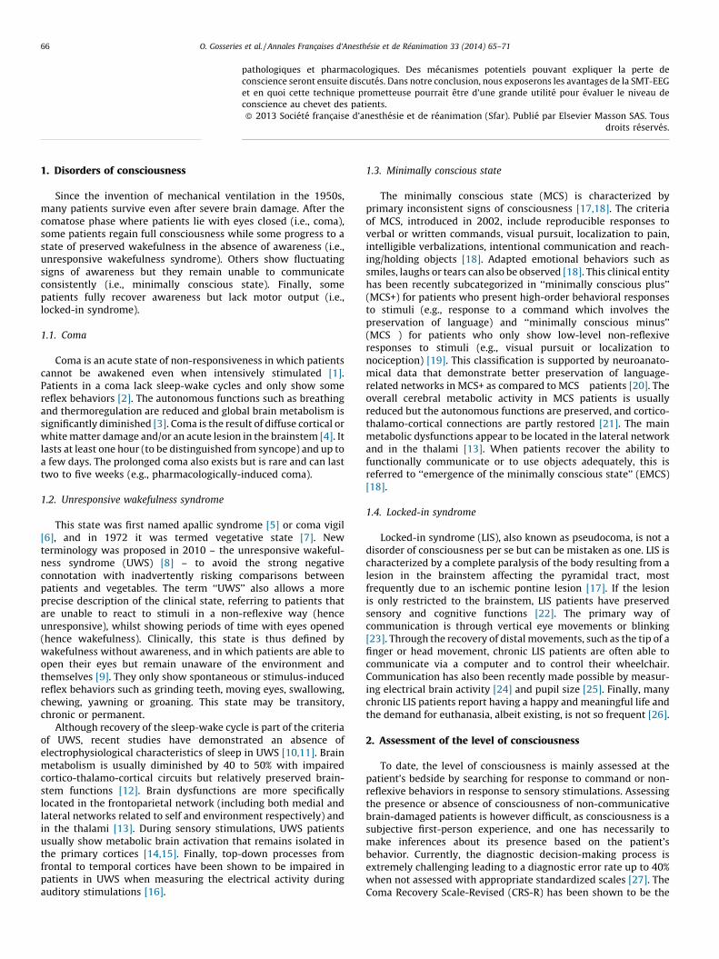
Fig. 1. Set up of the TMS-EEG technique. Hd-EEG: high-density
electroencephalography; TMS: transcranial magnetic stimulation; NBS: navigated
brain stimulation including the structural MRI of the subject.
Taken from [42].
O. Gosseries et al. / Annales Francaises d’Anesthesie et de Reanimation 33 (2014) 65–71 67
best scale to differentiate between UWS and MCS patients [28,29].In the acute setting, the Full Outline of UnResponsiveness (FOUR)has also been proposed as an alternative scale for the widely usedGlasgow Coma Scale because it facilitates the diagnosis of LISpatients, and it includes assessment of visual pursuit (one of thefirst behaviors observed when a patient emerges from coma) [30].
Even with the best clinical assessment, patients might beunderestimated in terms of residual awareness due to motordysfunction, sensory deficit, impaired cognition or fluctuation ofvigilance that can prevent voluntary responses [31]. Recent studiesprovide evidence for preserved awareness in some UWS patients[32]. For instance, using functional magnetic resonance imaging(fMRI) or electroencephalography (EEG) such patients generatedappropriate brain responses, similar to those observed in healthycontrols, when instructed to perform cognitive tasks (e.g.,activation of the supplementary motor area when asked toimagine playing tennis) [33–35]. Despite being unresponsive atthe bedside, these patients should be considered conscious andtheir diagnosis should be replaced. The terms ‘‘functional LIS’’ [19],‘‘functional MCS’’ [36] and ‘‘MCS*’’ [37] have been recentlysuggested to define patients who show non-behavioral evidenceof consciousness that is only measurable via neuroimaging testing.
These recent technologies using active EEG and fMRI paradigmsare therefore helpful in detecting consciousness in some UWSpatients but they cannot be used at the single-subject level due to ahigh rate of negative results. Moreover, these tools requirepreserved language comprehension and the active participationof the patients. The combination of single-pulse transcranialmagnetic stimulation (TMS) with EEG overcomes these issues andcan be employed at the bedside to assess brain function.
3. Transcranial magnetic stimulation combined withelectroencephalography
TMS has been used for neurological and psychiatric researchapplications since the early 1980s. This non-invasive techniqueallows stimulating the cerebral cortex non-invasively by generatinga brief but strong magnetic pulse through a coil applied to thesurface of the scalp. The fast change in magnetic field strengthinduces a current flow in the tissue, which results in the activationof underlying neuronal populations [38]. TMS was first applied tothe motor cortex with single-pulses, and electromyographyresponses were recorded from peripheral muscles [39]. RepetitiveTMS has since been used to induce a sustained inhibition (i.e.,<1 Hz) or activation (>1 Hz) of the neuronal population, whichallowed stimulating other brain areas while observing thesubsequent behavioral and cognitive changes [40]. In the lastseveral years, TMS has been combined with high-density EEG anda neuronavigation system to directly measure the activity of thebrain itself (instead of measuring muscular activity or behavioralresponses induced by the TMS stimulation) (Fig. 1). In this way,single-pulses TMS induce focal neuronal discharge at the cortexsurface, and an EEG measures cortical electrical responses bothlocally and at distant sites. This enables study of corticalexcitability under the site of stimulation, and long-range corticaleffective connectivity (i.e., causal interactions between distantbrain areas) with good spatio-temporal resolution [41]. Theneuronavigation system allows precise stimulation of a selectedbrain area and ensures stability of the position of the stimulationas well as reproducibility among different sessions [42,43].Studies demonstrated that meaningful recordings could bederived without being substantially affected by TMS-inducedartifact thanks to new hardware solutions, improved EEGamplifier technology, and advanced data processing techniques[44]. Using recent source modeling and statistical analyses, it is
currently possible to detect the effect of the focal perturbationboth at distance and in time [45].
3.1. TMS-EEG in healthy awake subjects
Using a 60-channel TMS-compatible EEG amplifier, Massiminiet al. [46] showed that during wakefulness, TMS triggerssustained long-range and complex patterns of activation. TMSinduces low-amplitude wave shapes, which are associated withspatially and temporally differentiated responses (Fig. 2A) [47].These brain responses vary depending on the site of stimulation,meaning that each cortical area tends to preserve its own naturalfrequency (Fig. 2B) [48]. For instance, TMS consistently evokeddominant alpha-band oscillations (8–12 Hz) in the occipitalcortex, beta-band oscillations (13–20 Hz) in the parietal cortex,and fast beta-/gamma-band oscillations (21–50 Hz) in the frontalcortex [48]. Brain regions were also oscillating at their naturalfrequencies when indirectly stimulated by TMS, through brainconnections. More recently, it has been shown that corticalexcitability increases with time awake [49]. Short-term memorytasks have also been shown to increase the overall strength andthe spatial spread of the electrical currents induced by TMS [50].Finally, intensive training on a working memory task increaseseffective connectivity across frontoparietal and parieto-occipitalnetworks [51].
3.2. TMS-EEG in patients recovering from coma
TMS-EEG offers an effective way to assess brain functions at restin patients with disorders of consciousness because it does notdepend on the integrity of sensory and motor pathways, itbypasses subcortical and thalamic gates, and it does not requirelanguage comprehension, nor active participation of subjects[41,52]. We recorded TMS-evoked brain responses in 17 awakepatients diagnosed in UWS, MCS and LIS. When stimulating frontaland parietal brain areas of UWS patients, TMS triggered a largelow-frequency wave associated with strong initial cortical activa-tion, which did not propagate to adjacent brain areas, anddissipated rapidly. This stereotyped and local slow wave responseindicates a breakdown of effective connectivity [53]. By contrast, inMCS patients, TMS triggered a series of lower amplitude and high-frequency waves associated with cortical activations that sequen-tially involved distant cortical areas ipsi- and contralateral to thesite of stimulation. These complex long-lasting widespreadactivation patterns were similar to the ones recorded (Fig. 3) [52].
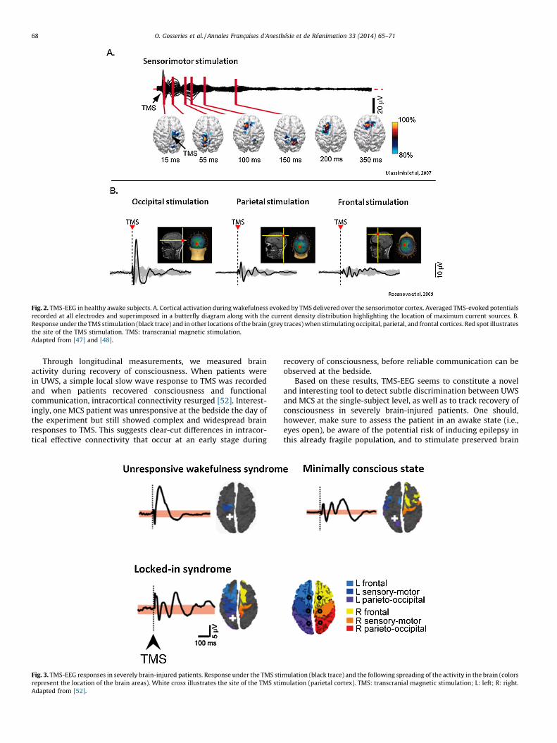
Fig. 2. TMS-EEG in healthy awake subjects. A. Cortical activation during wakefulness evoked by TMS delivered over the sensorimotor cortex. Averaged TMS-evoked potentials
recorded at all electrodes and superimposed in a butterfly diagram along with the current density distribution highlighting the location of maximum current sources. B.
Response under the TMS stimulation (black trace) and in other locations of the brain (grey traces) when stimulating occipital, parietal, and frontal cortices. Red spot illustrates
the site of the TMS stimulation. TMS: transcranial magnetic stimulation.
Adapted from [47] and [48].
O. Gosseries et al. / Annales Francaises d’Anesthesie et de Reanimation 33 (2014) 65–7168
Through longitudinal measurements, we measured brainactivity during recovery of consciousness. When patients werein UWS, a simple local slow wave response to TMS was recordedand when patients recovered consciousness and functionalcommunication, intracortical connectivity resurged [52]. Interest-ingly, one MCS patient was unresponsive at the bedside the day ofthe experiment but still showed complex and widespread brainresponses to TMS. This suggests clear-cut differences in intracor-tical effective connectivity that occur at an early stage during
Fig. 3. TMS-EEG responses in severely brain-injured patients. Response under the TMS sti
represent the location of the brain areas). White cross illustrates the site of the TMS stim
Adapted from [52].
recovery of consciousness, before reliable communication can beobserved at the bedside.
Based on these results, TMS-EEG seems to constitute a noveland interesting tool to detect subtle discrimination between UWSand MCS at the single-subject level, as well as to track recovery ofconsciousness in severely brain-injured patients. One should,however, make sure to assess the patient in an awake state (i.e.,eyes open), be aware of the potential risk of inducing epilepsy inthis already fragile population, and to stimulate preserved brain
mulation (black trace) and the following spreading of the activity in the brain (colors
ulation (parietal cortex). TMS: transcranial magnetic stimulation; L: left; R: right.

O. Gosseries et al. / Annales Francaises d’Anesthesie et de Reanimation 33 (2014) 65–71 69
areas using a neuronavigation system since TMS applied on a brainlesion might induce no response.
Another study recently confirmed the ability of the TMS-EEG todifferentiate UWS from MCS patients and showed the superiorityof the TMS-EEG compared to traditional neurophysiologicalmethods, such as short-latency somatosensory evoked potentialand event-related potentials [54]. In the most recent study, brainresponses were compared between conscious patients with mildbrain injury and healthy awake subjects [55]. Results showedaltered brain reactivity and connectivity in the former compared tothe latter that may be related to compensatory mechanisms ofrecovery. Larger population studies are needed to confirm theseresults and to better understand the neural mechanisms under-lying the functional recovery in post-comatose patients.
3.3. TMS-EEG in sleep and anesthesia
The dynamics of TMS-EEG responses have also been studiedunder physiological and pharmacological unconscious states.During non-rapid eye movement sleep, TMS triggers a simplepositive-negative slow wave, similar to the one observed in UWSpatients [46]. Depending on the intensity of the stimulation, thisslow wave can either remain local or burst into an explosive andstereotypical response. During deep sleep, the thalamo-corticalsystem despite being active and reactive thus tends to break downinto isolated modules, and to lose the ability to producedifferentiated responses [47]. TMS-EEG responses have also beenrecorded during rapid eye movement (REM) sleep during whichthere are dreams. In this paradoxical state, cortical responses toTMS propagate beyond the stimulation site and lasts longer thanduring deep sleep (although still less than during wakefulness),indicating that effective cortical connectivity is largely preserved(Fig. 4) [56].
Finally, TMS-EEG has been employed during general anesthesiainduced by a pharmacological agent, benzodiazepine midazolam[57]. Results showed that responsiveness of a cortical area to TMSwas maintained or even augmented, but the spread of activity toadjacent areas and the reverberatory reactivation of the stimulatedsite were quenched. Similarly in many regards to what occurs inUWS patients and in deep sleep, midazolam anesthesia induces alocal slow wave response and a breakdown of long-range brainconnectivity in response to TMS.
4. Potential mechanisms explaining (the loss of) consciousness
As we have seen above, consciousness involves many differentcortical areas engaging in rapid causal interactions, whilst
Fig. 4. TMS-EEG responses in sleep as compared to wakefulness. Yellow cross illustrat
Taken from [56].
unconsciousness is characterized by stereotypical positive-negative slow wave and decreased large-scale brain connectivity.One common hypothetical neurophysiological mechanism explain-ing unconsciousness is bistability, which could unable corticalneurons to sustain balanced patterns of activation. The resultswould lead to a silent, hyperpolarized downstate after the initialactivation, which could prevent the brain from successfullyintegrating information [58]. Potential causes of this bistabilitymight be alterations in the balance between excitation andinhibition, increased potassium currents, and cortical deaffer-entation [58]. Consciousness thus seems to emerge from bothfunctional integration (i.e., connectivity) and preserved informa-tion capacity in the brain (i.e., differentiation responses betweenbrain regions) [59,60]. To achieve a most accurate estimation ofthe level of consciousness, theoretical approaches such as theIntegrated Information Theory of Consciousness (IITC) aim atdescribing the mechanisms involved in consciousness [60,61].The IITC theory emphasizes the dynamical complexity ofconsciousness, characterized by information being simulta-neously integrated and differentiated, whereas unconsciousnessresults from the loss of the brain’s ability to integrate information.These statements are in line with our TMS-EEG observations.Moreover, we recently designed a reliable theoretically-basedmeasure of consciousness, the so-called Perturbational Complex-ity Index (PCI), in order to quantify in one number the TMS-EEGresponses observed in the aforementioned physiological, patho-logical and pharmacological states.
5. The Perturbational Complexity Index
The PCI is a recent measure that allows estimating braincomplexity, including both the information content and theintegration of brain activations, through algorithmic compres-sibility (‘‘zipping’’) [62]. This index has been shown to success-fully differentiate between consciousness and unconsciousnessat the individual subject level. Indeed, the PCI is invariably highin healthy awake subjects, in patients in MCS, EMCS and LIS aswell as in healthy subjects in REM sleep (i.e., above 0.3). Incontrast, the PCI is always low during deep sleep, in both UWSpatients and in those under general anesthesia using midazolam,propofol or xenon (i.e., below 0.3) (Fig. 5). Further confirmationstudies should test the PCI on a larger cohort of patients toobjectively and quantitatively assess the level of consciousness,but also to determine the efficacy of pharmacological drugs orbrain stimulation procedures in patients with disorders ofconsciousness.
es the site of the TMS stimulation (frontal cortex).
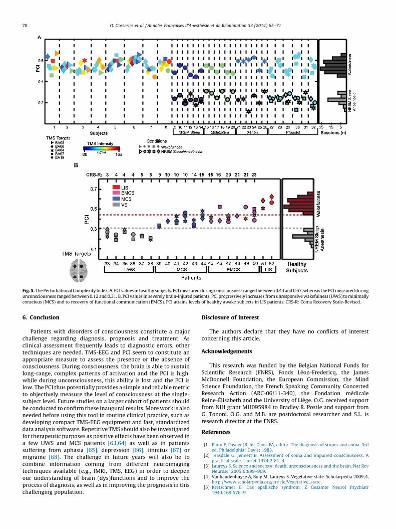
Fig. 5. The Perturbational Complexity Index. A. PCI values in healthy subjects. PCI measured during consciousness ranged between 0.44 and 0.67, whereas the PCI measured during
unconsciousness ranged between 0.12 and 0.31. B. PCI values in severely brain-injured patients. PCI progressively increases from unresponsive wakefulness (UWS) to minimally
conscious (MCS) and to recovery of functional communication (EMCS). PCI attains levels of healthy awake subjects in LIS patients. CRS-R: Coma Recovery Scale-Revised.
O. Gosseries et al. / Annales Francaises d’Anesthesie et de Reanimation 33 (2014) 65–7170
6. Conclusion
Patients with disorders of consciousness constitute a majorchallenge regarding diagnosis, prognosis and treatment. Asclinical assessment frequently leads to diagnostic errors, othertechniques are needed. TMS-EEG and PCI seem to constitute anappropriate measure to assess the presence or the absence ofconsciousness. During consciousness, the brain is able to sustainlong-range, complex patterns of activation and the PCI is high,while during unconsciousness, this ability is lost and the PCI islow. The PCI thus potentially provides a simple and reliable metricto objectively measure the level of consciousness at the single-subject level. Future studies on a larger cohort of patients shouldbe conducted to confirm these inaugural results. More work is alsoneeded before using this tool in routine clinical practice, such asdeveloping compact TMS-EEG equipment and fast, standardizeddata analysis software. Repetitive TMS should also be investigatedfor therapeutic purposes as positive effects have been observed ina few UWS and MCS patients [63,64] as well as in patientssuffering from aphasia [65], depression [66], tinnitus [67] ormigraine [68]. The challenge in future years will also be tocombine information coming from different neuroimagingtechniques available (e.g., fMRI, TMS, EEG) in order to deepenour understanding of brain (dys)functions and to improve theprocess of diagnosis, as well as in improving the prognosis in thischallenging population.
Disclosure of interest
The authors declare that they have no conflicts of interestconcerning this article.
Acknowledgements
This research was funded by the Belgian National Funds forScientific Research (FNRS), Fonds Leon-Fredericq, the JamesMcDonnell Foundation, the European Commission, the MindScience Foundation, the French Speaking Community ConcertedResearch Action (ARC-06/11-340), the Fondation medicaleReine-Elisabeth and the University of Liege. O.G. received supportfrom NIH grant MH095984 to Bradley R. Postle and support fromG. Tononi. O.G. and M.B. are postdoctoral researcher and S.L. isresearch director at the FNRS.
References
[1] Plum F, Posner JB. In: Davis FA, editor. The diagnosis of stupor and coma. 3rded, Philadelphia: Davis; 1983.
[2] Teasdale G, Jennett B. Assessment of coma and impaired consciousness. Apractical scale. Lancet 1974;2:81–4.
[3] Laureys S. Science and society: death, unconsciousness and the brain. Nat RevNeurosci 2005;6:899–909.
[4] Vanhaudenhuyse A, Boly M, Laureys S. Vegetative state. Scholarpedia 2009;4,http://www.scholarpedia.org/article/Vegetative_state.
[5] Kretschmer E. Das apallische syndrom. Z Gesamte Neurol Psychiatr1940;169:576–9.
O. Gosseries et al. / Annales Francaises d’Anesthesie et de Reanimation 33 (2014) 65–71 71
[6] Calvet J, Coll J. Meningitis of sinusoid origin with the form of coma vigil. RevOtoneuroophtalmol 1959;31:443–5.
[7] Jennett B, Plum F. Persistent vegetative state after brain damage. A syndromein search of a name. Lancet 1972;1:734–7.
[8] Laureys S, Celesia G, Cohadon F, Lavrijsen J, Leon-Carrion J, Sannita W, et al.Unresponsivewakefulnesssyndrome:anewnameforthevegetativestateorapallicsyndrome. BMC Med 2010;8:68. http://dx.doi.org/10.1186/1741-7015-8-68.
[9] The Multi-Society Task Force on PVS. Medical aspects of the persistentvegetative state (1). N Engl J Med 1994;330:1499–508.
[10] Cologan V, Drouot X, Parapatics S, Delorme A, Gruber G, Moonen G, et al. Sleepin the unresponsive wakefulness syndrome and minimally conscious state. JNeurotrauma 2013;30:339–46.
[11] Landsness E, Bruno MA, Noirhomme Q, Riedner B, Gosseries O, Schnakers C,et al. Electrophysiological correlates of behavioural changes in vigilance invegetative state and minimally conscious state. Brain 2011;134:2222–32.
[12] Gosseries O, Bruno MA, Chatelle C, Vanhaudenhuyse A, Schnakers C, Soddu A, et al.Disorders of consciousness: what’s in a name? Neurorehabilitation 2011;28:3–14.
[13] Thibaut A, Bruno MA, Chatelle C, Gosseries O, Vanhaudenhuyse A, Demertzi A,et al. Metabolic activity in external and internal awareness networks inseverely brain-damaged patients. J Rehabil Med 2012;44:487–94.
[14] Laureys S, Faymonville ME, Degueldre C, Fiore GD, Damas P, Lambermont B,et al. Auditory processing in the vegetative state. Brain 2000;123:1589–601.
[15] Boly M, Faymonville M, Peigneux P, Lambermont B, Damas F, Luxen A, et al.Cerebral processing of auditory and noxious stimuli in severely brain injuredpatients: differences between VS and MCS. Neuropsychol Rehabil2005;15:283–9.
[16] Boly M, Garrido MI, Gosseries O, Bruno MA, Boveroux P, Schnakers C, et al.Preserved feedforward but impaired top-down processes in the vegetativestate. Science 2011;332:858–62.
[17] American Congress of Rehabilitation Medicine. Recommendations for use ofuniform nomenclature pertinent to patients with severe alterations of con-sciousness. Arch Phys Med Rehabil 1995;76:205–9.
[18] Giacino JT, Ashwal S, Childs N, Cranford R, Jennett B, Katz DI, et al. Theminimally conscious state: definition and diagnostic criteria. Neurology2002;58:349–53.
[19] Bruno MA, Vanhaudenhuyse A, Thibaut A, Moonen G, Laureys S. From unre-sponsive wakefulness to minimally conscious PLUS and functional locked-insyndromes: recent advances in our understanding of disorders of conscious-ness. J Neurol 2011;258:1373–84.
[20] Bruno MA, Majerus S, Boly M, Vanhaudenhuyse A, Schnakers C, Gosseries O,et al. Functional neuroanatomy underlying the clinical subcategorization ofminimally conscious state patients. J Neurol 2012;259:1087–98.
[21] Laureys S, Faymonville ME, Luxen A, Lamy M, Franck G, Maquet P. Restorationof thalamocortical connectivity after recovery from persistent vegetativestate. Lancet 2000;355:1790–1.
[22] Schnakers C, Majerus S, Goldman S, Boly M, Van Eeckhout P, Gay S, et al.Cognitive function in the locked-in syndrome. J Neurol 2008;255:323–30.
[23] Bauby JD. Le scaphandre et le papillon.The diving bell and the butterfly Paris:Laffont Edition; 1997, 139 p.
[24] Lule D, Noirhomme Q, Kleih SC, Chatelle C, Halder S, Demertzi A, et al. Probingcommand following in patients with disorders of consciousness using a brain-computer interface. Clin Neurophysiol 2013;124:101–6.
[25] Stoll J, Chatelle C, Carter O, Koch C, Laureys S, Einhauser W. Pupil responses allowcommunication in locked-in syndrome patients. Curr Biol 2013;23:R647–8.
[26] Bruno MA, Bernheim JL, Ledoux D, Pellas F, Demertzi A, Laureys S. A survey onself-assessed well-being in a cohort of chronic locked-in syndrome patients:happy majority, miserable minority. BMJ Open 2011;1:e000039.
[27] Schnakers C, Vanhaudenhuyse A, Giacino JT, Ventura M, Boly M, Majerus S,et al. Diagnostic accuracy of the vegetative and minimally conscious state:clinical consensus versus standardized neurobehavioral assessment. BMCNeurol 2009;9:35.
[28] Giacino JT, Kalmar K, Whyte J. The JFK coma recovery scale-revised: measurementcharacteristics and diagnostic utility. Arch Phys Med Rehabil 2004;85:2020–9.
[29] Seel RT, Sherer M, Whyte J, Katz DI, Giacino JT, Rosenbaum AM, et al. Assess-ment scales for disorders of consciousness: evidence-based recommendationsfor clinical practice and research. Arch Phys Med Rehabil 2010;91:1795–813.
[30] Bruno MA, Ledoux D, Lambermont B, Damas F, Schnakers C, VanhaudenhuyseA, et al. Comparison of the Full Outline of UnResponsiveness and GlasgowLiege Scale/Glasgow Coma Scale in an intensive care unit population. Neuro-crit Care 2011;15:447–53.
[31] Majerus S, Gill-Thwaites H, Andrews K, Laureys S. Behavioral evaluation ofconsciousness in severe brain damage. Prog Brain Res 2005;150:397–413.
[32] Faugeras F, Rohaut B, Weiss N, Bekinschtein TA, Galanaud D, Puybasset L, et al.Probing consciousness with event-related potentials in the vegetative state.Neurology 2011;77:264–8.
[33] Monti MM, Vanhaudenhuyse A, Coleman MR, Boly M, Pickard JD, Tshibanda L,et al. Willful modulation of brain activity in disorders of consciousness. N EnglJ Med 2010;362:579–89.
[34] Cruse D, Chennu S, Chatelle C, Bekinschtein TA, Fernandez-Espejo D, PickardJD, et al. Bedside detection of awareness in the vegetative state: a cohort study.Lancet 2011;378:2088–94.
[35] Naci L, Owen AM. Making every word count for nonresponsive patients. JAMANeurol 2013. http://dx.doi.org/10.1001/jamaneurol.2013.3686 [Epub aheadof print].
[36] Vogel D, Markl A, Yu T, Kotchoubey B, Lang S, Muller F. Can mentalimagery functional magnetic resonance imaging predict recovery in
patients with disorders of consciousness? Arch Phys Med Rehabil2013;94:1891–8.
[37] Gosseries O, Zasler N, Laureys O. Recent advances in disorders of conscious-ness: focus on the diagnosis. Brain Inj [in press].
[38] Hallett M. Transcranial magnetic stimulation and the human brain. Nature2000;406:147–50.
[39] Lapitskaya N, Coleman M, Nielsen J, Gosseries O, de Noordhout A. Disorders ofconsciousness: further pathophysiological insights using motor cortex tran-scranial magnetic stimulation. Prog Brain Res 2009;177:191–200.
[40] Miniussi C, Rossini PM. Transcranial magnetic stimulation in cognitive reha-bilitation. Neuropsychol Rehabil 2011;21:579–601.
[41] Massimini M, Boly M, Casali A, Rosanova M, Tononi G. A perturbationalapproach for evaluating the brain’s capacity for consciousness. Prog BrainRes 2009;177:201–14.
[42] Rosanova M, Casarotto S, Pigorini A, Canali P, Casali AG, Massimini M.Combining transcranial magnetic stimulation with electroencephalographyto study human cortical excitability and effective connectivity. In: Fellin THM,editor. Neuronal network analysis, Neuromethods. Volume 67, Humana Press,Springer Science+Business, LLC. p. 435–457.
[43] Casarotto S, Romero Lauro LJ, Bellina V, Casali AG, Rosanova M, Pigorini A, et al.EEG responses to TMS are sensitive to changes in the perturbation parametersand repeatable over time. PLoS One 2010;5:e10281.
[44] Rogasch NC, Fitzgerald PB. Assessing cortical network properties using TMS-EEG. Hum Brain Mapp 2012.
[45] Casali AG, Casarotto S, Rosanova M, Mariotti M, Massimini M. General indicesto characterize the electrical response of the cerebral cortex to TMS. Neuro-image 2010;49:1459–68.
[46] Massimini M, Ferrarelli F, Huber R, Esser SK, Singh H, Tononi G. Breakdown ofcortical effective connectivity during sleep. Science 2005;309:2228–32.
[47] Massimini M, Ferrarelli F, Esser SK, Riedner BA, Huber R, Murphy M, et al.Triggering sleep slow waves by transcranial magnetic stimulation. Proc NatlAcad Sci U S A 2007;104:8496–501.
[48] Rosanova M, Casali A, Bellina V, Resta F, Mariotti M, Massimini M. Naturalfrequencies of human corticothalamic circuits. J Neurosci 2009;29:7679–85.
[49] Huber R, Maki H, Rosanova M, Casarotto S, Canali P, Casali A, et al. Humancortical excitability increases with time awake. Cereb Cortex 2013;23:332–8.
[50] Johnson J, Kundu B, Casali A, Postle B. Task-dependent changes in corticalexcitability and effective connectivity: a combined TMS-EEG study. J Neuro-physiol 2012;107:2383–92.
[51] Kundu B, Sutterer DW, Emrich SM, Postle BR. Strengthened effective con-nectivity underlies transfer of working memory training to tests of short-termmemory and attention. J Neurosci 2013;33:8705–15.
[52] Rosanova M, Gosseries O, Casarotto S, Boly M, Casali A, Bruno M, et al. Recoveryof cortical effective connectivity and recovery of consciousness in vegetativepatients. Brain 2012;135(Pt 4):1308–20.
[53] Friston KJ. Functional and effective connectivity: a review. Brain Connect2011;1:13–36.
[54] Ragazzoni A, Pirulli C, Veniero D, Feurra M, Cincotta M, Giovannelli F, et al.Vegetative versus minimally conscious states: a study using TMS-EEG, sensoryand event-related potentials. PLoS One 2013;8:e57069.
[55] Tallus J, Lioumis P, Hamalainen H, Kahkonen S, Tenovuo O. TMS-EEG responsesin recovered and symptomatic mild traumatic brain injury. J Neurotrauma2013;30:1270–7.
[56] Massimini M, Ferrarelli F, Murphy M, Huber R, Riedner B, Casarotto S, et al.Cortical reactivity and effective connectivity during REM sleep in humans.Cogn Neurosci 2010;1:176–83.
[57] Ferrarelli F, Massimini M, Sarasso S, Casali A, Riedner BA, Angelini G, et al.Breakdown in cortical effective connectivity during midazolam-induced lossof consciousness. Proc Natl Acad Sci U S A 2010;107:2681–6.
[58] Massimini M, Ferrarelli F, Sarasso S, Tononi G. Cortical mechanisms of loss ofconsciousness: insight from TMS/EEG studies. Arch Ital Biol 2012;150:44–55.
[59] Boly M, Massimini M, Tononi G. Theoretical approaches to the diagnosis ofaltered states of consciousness. Prog Brain Res 2009;177:383–98.
[60] Tononi G. Consciousness as integrated information: a provisional manifesto.Biol Bull 2008;215:216–42.
[61] Tononi G. Integrated information theory of consciousness: an updatedaccount. Arch Ital Biol 2012;150:56–90.
[62] Casali AG, Gosseries O, Rosanova M, Boly M, Sarasso S, Casali KR, et al. Atheoretically based index of consciousness independent of sensory processingand behavior. Sci Transl Med 2013;5:198ra05.
[63] Louise-Bender Pape T, Rosenow J, Lewis G, Ahmed G, Walker M, Guernon A,et al. Repetitive transcranial magnetic stimulation-associated neurobeha-vioral gains during coma recovery. Brain Stimul 2009;2:22–35.
[64] Manganotti P, Formaggio E, Storti SF, Fiaschi A, Battistin L, Tonin P, et al. Effectof high-frequency repetitive transcranial magnetic stimulation on brain excit-ability in severely brain-injured patients in minimally conscious or vegetativestate. Brain Stimul 2013;6:913–21.
[65] Dammekens E, Vanneste S, Ost J, De Ridder D. Neural correlates of highfrequency repetitive transcranial magnetic stimulation improvement inpost-stroke non-fluent aphasia: a case study. Neurocase 2014;20:1–9.
[66] Dumas R, Padovani R, Richieri R, Lancon C. Repetitive transcranial magneticstimulation in major depression: response factor. Encephale 2012;38:360–8.
[67] De Ridder D, Song J, Vanneste S. Frontal cortex TMS for tinnitus. Brain Stimul2014;20:1–9.
[68] Lipton R, Pearlman S. Transcranial magnetic stimulation in the treatment ofmigraine. Neurotherapeutics 2010;7:204–12.