Embed Size (px)
Citation preview

doi.org/10.26434/chemrxiv.11302478.v1
Magnetically Responsive Horseradish peroxidase@ZIF-8 forBiocatalystsRaffaele Ricco, Heinz Amenitsch, paolo falcaro
Submitted date: 02/12/2019 • Posted date: 18/12/2019Licence: CC BY-NC-ND 4.0Citation information: Ricco, Raffaele; Amenitsch, Heinz; falcaro, paolo (2019): Magnetically ResponsiveHorseradish peroxidase@ZIF-8 for Biocatalysts. ChemRxiv. Preprint.https://doi.org/10.26434/chemrxiv.11302478.v1
Here, we studied a catalytically active and magnetically responsive porous bio-composite obtained from thesynthesis of ZIF-8 in presence of iron oxide magnetic nanoparticles and horseradish peroxidase (HRP)enzyme as guest species. Using a one-pot approach in water the precursors of ZIF-8 (zinc acetate and2-methylimidazole) spontaneously self-assembles around the guest species. We characterized the compositeby means of XRD, SEM, FTIR, AFM, and CLSM. SAXS investigation of the kinetics of crystallization showedhow the presence of the guest species can act as nucleation seeds. Moreover, we found that the bio-catalyticactivity of the HRP/MNP@ZIF-8 biocomposite is 5 times higher than the analoguous composite withoutMNPs.
File list (2)
download fileview on ChemRxivRicco - Supporting Information_chemrxiv.pdf (1.58 MiB)
download fileview on ChemRxivRicco - Manuscript_chemrxiv.pdf (0.91 MiB)

1
Supplementary Information
Magnetically responsive horseradish peroxidase@ZIF-8 for biocatalysts
Raffaele Ricco,a* Heinz Amenitsch,b and Paolo Falcaroa
a. Institute of Physical and Theoretical Chemistry, Graz University of Technology, 8010 Graz,
Austria. E-mail: [email protected]
b. Institute of Inorganic Chemistry, Graz University of Technology, 8010 Graz, Austria.
CONTENTS:
Experimental section
Figure S1. AFM analysis of iron oxide magnetic nanoparticles
Figure S2. SAXS/WAXS growth kinetics for ZIF-8, BSA@ZIF-8, MNP@ZIF-8 and
BSA/HRP@ZIF-8 composites
Figure S3. WAXS profiles of HRP@ZIF-8 and HRP/MNP@ZIF-8 composites
Figure S4. FTIR spectra of ZIF-8, MNP@ZIF-8, HRP@ZIF-8 and HRP/MNP@ZIF-8.
Figure S5. EDX spectrum of HRP-FITC/MNP@ZIF-8
Figure S6. Calibration curves for HRP (and BSA) referred in the Experimental section
Figure S7 Kinetics of free HRP performed in PBS buffer and in DI water
Figure S8. UV-Vis spectra related to Figure S7
Figure S9. Interference of blank and MNPs
Table S10. Data of loading and enzymatic activity of HRP in the composites
References

2
Experimental section
Starting materials and characterization methods
Zinc acetate dihydrate (Zn(CH3COO)2·H2O), 2-methylimidazole (HmIm, C4H6N2), Iron (III)
chloride hexahydrate (FeCl3·6H2O), Iron (II) sulfate heptahydrate (FeSO4·7H2O), trisodium
citrate dihydrate (C6H5O7Na3·2H2O), aqueous ammonia solution (25%), horseradish
peroxidase (HRP), bovine serum albumin (BSA), sodium dodecylsulfate (SDS), and Bradford
Reagent for 0.1-1.4 mg/ml protein were purchased from Sigma Aldrich / Merck with the
highest degree of purity available, and used without further purification. Deionized water
(18 MΩ) was produced from a Millipore Synergy purification system.
Scanning Electron Microscopy (SEM). Sample morphology was assessed using a TESCAN
VEGA scanning electron microscope at 20 kV. Prior to SEM investigation, samples were
sputter coated with gold.
Powder diffraction (XRD) analysis. PXRD patterns were collected on a Rigaku SmartLab at
9 KeV in Bragg-Brentano geometry, with samples deposited on flat silicon wafers and
rotated. Scan speed was 3 deg min–1, step 0.01°, and 2θ was in the range between 5° and
50°.
Small- and Wide- Angle X-Ray Scattering (SAXS/WAXS) analysis. Small- and wide- angle
X-ray scattering (SAXS/WAXS) measurements were performed at the Austrian SAXS
beamline of the electron storage ring Elettra (Trieste, Italy)1 using 8 keV branch
corresponding to a wave-length of 1.54 Å; 1 s exposure time was used to collect the
diffraction image. Data were analysed using Igor Pro software package (WaveMetrics Inc.).
Fourier transform infrared (FTIR) analysis. The measurements were performed using a
Bruker ALPHA FT-IR spectrometer (Bruker Optik GmbH) in transmission mode. 128 scans
were performed with 2 cm-1 intervals.
Confocal Light Scanning Microscopy (CSLM). The images were acquired using a Leica
DM5500 B microscope equipped with a Leica TCS SPE high resolution confocal system,
using an excitation laser (λexc. = 488 nm, 10 mW) and collecting the emission at 580 nm.
Atomic Force Microscopy (AFM). Data was acquired using an Anton Paar Tosca AFM in
non-contact tapping mode. Samples were diluted 1000x in ethanol and drop cast on silicon
substrates.
Synthesis of Iron(II, III) oxide magnetic nanoparticles (MNPs)
The method has been reproduced from literature.2 In a 250 mL, single-neck, round bottom
flask equipped with a magnetic stir bar, FeCl3·6H2O (3.24 g, 0.012 mol) and FeSO4·7H2O
(2.80 g, 0.010 mol) were dissolved in 80 mL of water. Aqueous ammonia solution (10 mL,
25 % w/w) was added in 1 mL steps under vigorous stirring. The mixture was then heated
at 90 °C, then solid trisodium citrate (8.80 g, 0.030 mol) was added in one step and the
stirring continued for additional 30 minutes at the same temperature. After removal of
the stir bar and natural cooling at room temperature, the dark brown mixture was

3
transferred in a cylindrical container, and a strong ND45 magnet (fitted into a glass tube)
was immersed at the approximate centre of the solution for 5 minutes to collect the MNPs.
The liquid part was removed and replaced by DI water, then the magnet extracted. The
suspension was briefly sonicated in a bath for 10 minutes, and the magnetic collection
repeated. The process was repeated with water (3 x 100 mL), ethanol (3 x 100 mL) and
water again (3 x 100 mL), then the suspension diluted to 250 mL and stored at room
temperature in a polythene bottle.
Before use, few millilitres were collected, the water removed and the MNPs dried under
vacuum at 60 °C overnight, then re-suspended to the desired concentration.
Synthesis of ZIF-8 and MNP@ZIF-8
The method has been adapted from literature.3 The procedure involves the preliminary
preparation of three stock solutions, all in DI water: MNP suspension is prepared at a
concentration of 6 mg/mL, and sonicated in a bath for 15 minutes to ensure homogeneous
dispersion; zinc acetate stock solution is prepared with a concentration of 240 mM,
whereas HmIm stock solution is prepared with a concentration of 1.92 M.
The metal-to-ligand ratio is kept as 1:16,4 their final concentrations are 40 mM and 640
mM, respectively. The amount of MNP is varied within a final concentration range of 0 ÷ 1
mg/mL, precisely 0.33, 0.67, and 1.00 mg/mL. For pure ZIF-8, the MNP is omitted and
substituted with an equivalent amount of DI water.
In a typical procedure, HmIm stock (600 µL), MNP stock (0, 100, 200, or 300 µL) and water
(filling up to the 1.5 mL volume mark) where mixed in a 2 mL plastic vial with cap and
briefly vortexed for few seconds. Zn acetate stock (300 µL) is then added: the solution
containing MNPs becomes milky almost instantaneously, whereas for pure ZIF-8 this will
occur in approximately 20 minutes. The mixture is briefly vortexed one more time for few
seconds, and the vial put in a tube rotator (20 rpm, 16 hours).
General workup procedure
The reaction mixture is centrifuged (Eppendorf Minispin with rotor F-45-12-11, 12000 x g,
60 sec) and the supernatant separated for further analysis. An aqueous solution of SDS (1
% w/v, 1 mL) is added and the pellet vortexed until dispersion, with the aid of a brief step
of sonication in bath (3-5 sec) if necessary. The suspension is left in the tube rotator for 2
hours, then centrifuged again (12000 x g, 60 sec). After removing the supernatant, the
pellet is washed with water (3 x 1 mL), vortexed, and centrifuged as previously described.
The pellet is then re-dispersed in water (or a suitable buffer), and stored at room
temperature (or 4 °C if enzyme is present).
A portion (300 µL) of suspension is placed in pre-weighed vials and centrifuged (12000 x g,
5 min), the supernatant discarded and the pellet left to dry at room temperature
4
overnight. The solid is weighed again to determine the amount of material. The final
amount of composite is eventually adjusted to 10 mg/mL.
Synthesis of HRP@ZIF-8
The same procedure for the synthesis of MNP@ZIF-8 is followed, but using a stock solution
of HRP in DI water (6 mg/mL) in place of the MNP suspension, with a final concentration
in the range 0 ÷ 1 mg/mL, precisely 0.33, 0.67, and 1.00 mg/mL (0, 100, 200, 300 µL). The
general workup protocol is then applied.
Synthesis of HRP/MNP@ZIF-8
The same procedure for the synthesis of MNP@ZIF-8 is followed, adding a stock solution
of HRP in DI water (6 mg/mL) along with the MNP suspension, with a final concentration
in the range 0 ÷ 1 mg/mL, precisely 0.33, 0.67, and 1.00 mg/mL (0, 100, 200 300 µL). Before
adding HmIm, the suspension is left standing for few minutes. The general workup
protocol is then applied.
Bradford assay5
A calibration curve with HRP in water was determined in the 0 ÷ 1 mg/mL range. The
resulting equation is:
A = 0.52536 x [p.a.] – 0.00268
where A is the absorbance and [p.a.] the protein amount in mg/mL (see Figure S6).
To perform the assay, the supernatant (20 µL) is mixed with the Bradford Reagent (600 µL)
in a plastic cuvette and the absorbance at 595 nm measured with a Nanodrop OneC
(Thermofisher Scientific Inc.). The quantity is then calculated with the above equation and
the loading obtained from the initial amount of composite used.
Enzymatic activity
The assay performed using pyrogallol as revealing agent6 is modified excluding PBS buffer
(see Figures S7 and S8).7 In a typical procedure, HRP@ZIF-8 (or ZIF-8), is suspended in DI
water (10 mg/mL) and left to mix on a tube rotator (20 rpm, 2 hours) to ensure
homogeneity. 100 µL of the bio-composite suspension (1 mg), is added to 2420 µL of DI
water, 160 µL of hydrogen peroxide (0.5 % v/v), and 320 µL of pyrogallol (50 mg/mL, in DI
water) in a 4 mL cuvette with stirring bar, monitoring the increase of the absorbance at
420 nm within 10 minutes.
Recyclability test
The assay is performed in DI water. The final volume is 1 mL. In a typical procedure, 100
µL of HRP@ZIF-8 or HRP/MNP@ZIF-8 suspension (20 mg/mL) are added to a mixture
containing 420 µL of DI water and 160 µL of hydrogen peroxide (0.5 % w/v). After adding

5
320 µL of pyrogallol (50 mg/mL), the reaction is left for 15 minutes, then the suspension is
centrifuged (12000 x g, 60 sec) or collected with a magnet for 2 minutes, and the
absorbance of the supernatant measured at 420 nm. The material is washed and
centrifuged three times with water before every cycle.
Recirculator setup
The system is illustrated in the image below, along with a photo of the real setup taken at
the Elettra synchrotron:
A 50 mL plastic reactor (1) with magnetic stirring (2) is charged with an aqueous solution
containing HmIm, the enzyme and/or the magnetic nanoparticles. The reactor has an inlet
(3), an outlet drawing out from the bottom of the reactor (4), and an additional inlet (5)
for the injection (6) of the zinc acetate solution at the given time. The reactor is then
connected with plastic tubing to a quartz capillary (7) put on the path of the synchrotron
beam (8), with SAXS (9) and WAXS (10) detectors downstream the beam. The recirculation
of the liquid is ensured by a peristaltic pump (11), withdrawing the liquid from the
capillary. The green arrows illustrate the direction of flow in the system; the peristaltic
pump is set so the mixture takes approx. 10 seconds to reach the capillary. The tubing
volume is ca. 5 mL, the overall reaction volume is 48 mL (40 mL of ligand solution and 8
mL of metal ion solution). Starting concentrations used are [HmIm]: 1.92 M, [Zn(OAc)2]:
240 mM, HRP: 1 mg/mL, MNP: 1 mg/mL

6
Figure S1. AFM maps, line profiles, and histograms including statistics over all measured regions
of Fe3O4 magnetic nanoparticles before (a) and after (b) washing with ethanol to remove the
citrate stabilizer. The average particle size is about 12 nm, with an inorganic core having a
diameter of 10.4 nm.

7
Figure S2. Evolution of the SAXS/WAXS signals for the (011), (112), (022), (013), and (222) crystal
planes (from left to right) during the synthesis of a) ZIF-8, b) BSA@ZIF-8, c) MNP@ZIF-8, and d)
MNP/BSA@ZIF-8 performed at the Elettra synchrotron with the recirculation setup described in
the Experimental section. The (002) plane cannot be shown due to a gap between the SAXS and
WAXS detectors. The data acquisition was started 10 seconds after the injection of the solution
containing the ligand HmIm.
The times when the signals started to appear are summarized in the following table:
Sample Time (sec.)
ZIF-8 20’’
BSA@ZIF-8 13’’
MNP@ZIF-8 13’’
BSA/MNP@ZIF-8 7’’

8
Figure S3. Normalized WAXS profiles of HRP@ZIF-8 and HRP/MNP@ZIF-8 acquired at the SAXS
beamline of Elettra Synchrotron (Trieste, Italy), at different concentrations of enzyme. The
dataset is divided for clarity according to the amounts of magnetic nanoparticles (MNP) in the
reaction mixture: a) 0.33 mg/mL; b) 0.67 mg/mL; c) 1.00 mg/mL.
9
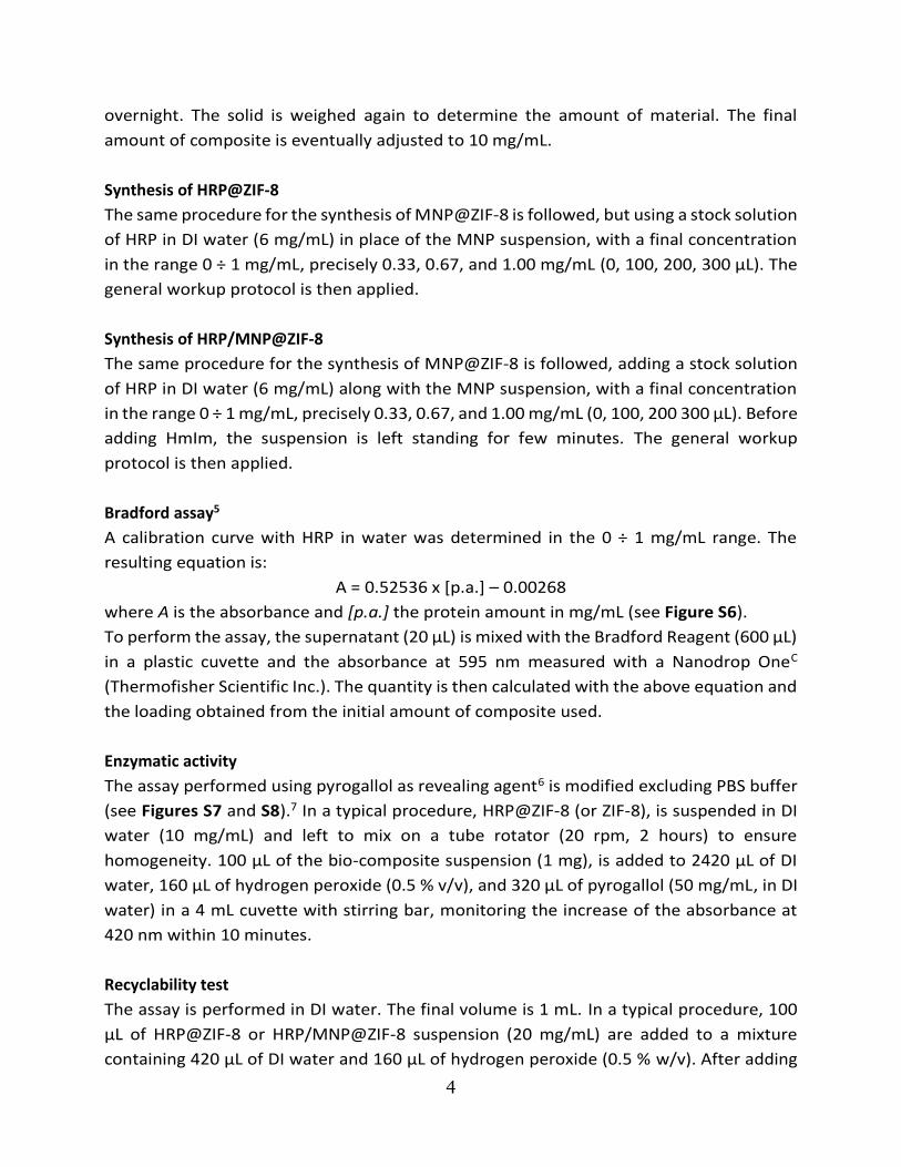
Figure S4. FTIR spectra related to Figure 2c of the main text. The bold spectra refer to the
materials with MNP, the green arrows refer to the location of the Amide bands from HRP at 1650
and 1545 cm-1, the brown arrow to the location of the Fe-O stretching from magnetic
nanoparticles centred at 600 cm-1. Assignments for ZIF-8: 3136 cm-1 (imidazole C-H stretching),
2929 cm-1 (methyl C-H), 1585 cm-1 (C=N stretching), 1446 cm-1 (ring stretching), 1383 cm-1 (ring
bending), 1310 cm-1 (N-H wagging), 1146 cm-1 (C-N stretching), 996 cm-1 (in-plane ring bending),
759 cm-1 (C=N out-of-plane bending), 694 cm-1 (H-C=C-H out-of-plane wagging), 421 cm-1 (Zn-N
stretching). The broad band between 3500 and 2500 cm-1 was attributed to the residual water
due to the mild step of drying at room temperature.

10
Figure S5. EDX spectra of a) ZIF-8, b) MNP@ZIF-8, and c) HRP-FITC/MNP@ZIF-8. The peaks marked
with belong to Si from the substrate (at 1.74 KeV) and to Au from the sputter coating (at 2.12
and 9.71 KeV). The red rectangles refer to the insets zooming on the 0-1 KeV region.

11
Type Value σ
R2 intercept slope intercept slope
BSA (water) 0.00532 0.52848 0.0140 0.0228 0.994445
HRP (water) -0.00268 0.52536 0.0108 0.0176 0.996640
Figure S6. Calibration curves with the Bradford assay for BSA (grey squares) and HRP (blue circles)
in DI water, in the 0 - 1 mg/mL range of protein concentration. The bold data is used in this
manuscript. The table reports the values and relative errors for the intercept and slope of the four
curves.

12
Figure S7. Kinetics of the enzymatic assay on free HRP performed in PBS buffer and in DI water,
corresponding to the intensities at 420 nm referred in the following Figure S8. The outcome in
water (red) is comparable to the one in PBS (black), with a lower background (purple vs blue line
for water and buffer, respectively).

13
Figure S8. Comparison between the enzymatic assay on free HRP over time performed a, b) using
PBS 100 mM, pH 6.0, and c, d) using only DI water. The results obtained with HRP (b and d) are
comparable, however the background using PBS (a) increases more over the time than the one
obtained in water only (c), indicating even a superior stability of free pyrogallol in water rather
than in buffer medium, without appreciable variation of absorption maximum. The red lines refer
to the intensity at 420 nm.

14
Figure S9. Influence of MNPs on the enzymatic assay using pyogallol as revealing dye.

15
Table S10. Data values of the HRP@ZIF-8 and the HRP/MNP@ZIF-8 sets depending on the starting
quantity of the encapsulated species in the reaction mixture. The error is calculated from
standard deviation.
Sample set HRP
(mg/mL)
MNP
(mg/mL)
EE
(%)
Loading
(mg/g)
EA
(U/mg)
EAc
(U/g)
HRP 0.33 0.00 79 ± 5. 6.9 ± 0.4 1.6 ± 0.1 11.0 ± 0.9
@ 0.67 0.00 82 ± 5. 12.0 ± 0.7 1.5 ± 0.1 18.0 ± 1.6
ZIF-8 1.00 0.00 86 ± 2. 13.8 ± 0.4 0.8 ± 0.1 11.0 ± 1.4
HRP 0.33 0.33 84 ± 4. 5.1 ± 0.2 0.3 ± 0.1 1.5 ± 0.5
/ 0.67 0.33 86 ± 6. 4.7 ± 0.3 1.1 ± 0.2 5.2 ± 1.0
MNP 1.00 0.33 88 ± 7. 4.1 ± 0.3 2.2 ± 0.1 9.0 ± 0.8
@ 0.33 0.67 87 ± 3. 8.8 ± 0.3 0.5 ± 0.3 4.4 ± 2.6
ZIF-8 0.67 0.67 92 ± 5. 5.4 ± 0.3 1.5 ± 0.3 8.1 ± 1.7
1.00 0.67 93 ± 6. 4.4 ± 0.3 3.3 ± 0.1 14.5 ± 1.1
0.33 1.00 91 ± 4. 9.1 ± 0.4 1.1 ± 0.4 10.0 ± 3.7
0.67 1.00 94 ± 5. 5.5 ± 0.3 3.0 ± 0.3 16.5 ± 1.9
1.00 1.00 94 ± 6. 5.6 ± 0.3 4.1 ± 0.1 23.0 ± 1.4
Legend:
EE = encapsulation efficiency
EA = Enzymatic activity of the enzyme (encapsulated only)
EAc = Enzymatic activity of the whole composite

16
References
1 H. Amenitsch, M. Rappolt, M. Kriechbaum, H. Mio, P. Laggner and S. Bernstorff, J. Synchrotron Radiat., 1998, 5, 506–508.
2 A. Schejn, T. Mazet, V. Falk, L. Balan, L. Aranda, G. Medjahdi and R. Schneider, Dalton Trans., 2015, 44, 10136–10140.
3 K. Liang, R. Ricco, C. M. Doherty, M. J. Styles, S. Bell, N. Kirby, S. Mudie, D. Haylock, A. J. Hill, C. J. Doonan and P. Falcaro, Nat. Commun., 2015, 6, 7240.
4 W. Liang, R. Ricco, N. K. Maddigan, R. P. Dickinson, H. Xu, Q. Li, C. J. Sumby, S. G. Bell, P. Falcaro and C. J. Doonan, Chem. Mater., , DOI:10.1021/acs.chemmater.7b04977.
5 M. M. Bradford, Anal. Biochem., 1976, 72, 248–254. 6 B. Chance and A. C. Maehly, Methods Enzymol., 1955, 2, 764–775. 7 M. de J. Velásquez-Hernández, R. Ricco, F. Carraro, F. T. Limpoco, M. Linares-Moreau, E. Leitner,
H. Wiltsche, J. Rattenberger, H. Schröttner, P. Frühwirt, E. M. Stadler, G. Gescheidt, H. Amenitsch, C. J. Doonan and P. Falcaro, CrystEngComm, 2019, 21, 4538–4544.

download fileview on ChemRxivRicco - Supporting Information_chemrxiv.pdf (1.58 MiB)

Please do not adjust margins
Please do not adjust margins
Magnetically responsive horseradish peroxidase@ZIF-8 for biocatalysts
Raffaele Ricco,a* Heinz Amenitsch,b and Paolo Falcaroa
A model bioreactor based on the zeolitic imidazolate framework
ZIF-8 is obtained in an one-pot process by directly combining the
enzyme horseradish peroxidase (HRP), iron oxide magnetic
nanoparticles, the ligand and metal ions, in water at room
temperature. The resulting system provides an useful platform for
the next generation of reusable/repositionable biocatalysts.
Enzyme-coated magnetic particles are widely used for
biochemical tests and biocatalysis.1–3 Typically, bio-conjugation
protocols are used to immobilize enzymes on magnetic
particles.4 However, immobilization can compromise the
bioactivity of the protein because of conformational changes at
the bio-interface.5 The final system preserves the native fragility
of the enzyme: the exposure of the grafted and unprotected
biomolecules to inhospitable conditions (e.g. organic solvents
and high temperature) leads to protein denaturation.6
A method that affords remarkable protection to
biomacromolecules, and assembly of thereof, exploits their
encapsulation in porous metal-organic frameworks (MOFs).7–11
For example, zeolitic Imidazolate frameworks (ZIFs) were used
to encapsulate enzymes, forming bioactive composites with
improved robustness to solvents, temperature, and chaotropic
and proteolytic agents with respect to the pure enzyme.10–14 For
ZIF-8, which is composed of 2-methylimidazole (HmIm) and
Zn2+, the framework self-assembles spontaneously in water
around negatively charged proteins. Thus, the synthesis of the
ZIF-8-based biocomposite can be conducted using a one-pot
method, as the enzyme acts as seed for the MOF formation.11
Magnetic particles can also act as seeds for the MOF
crystalization;15 the resulting magnetically active composite
(Magnetic Framework Composites, MFCs)16 can be used for
dynamic localization,17 triggered heating,18 and catalysis.19,20 As
both enzymes and magnetic particles can spontaneously induce
the ZIF-8 formation in aqueous conditions,21 the one-step
synthesis of a magnetically bio-active ZIF composites would
result in a new straightforward procedure to expand the
biotechnological applications of MOFs.
Although a few reports disclose the preparation of
magnetically active ZIF biocomposites, the focus of these
studies was on the use of additives such as polyvinylpyrrolidone
(PVP)22 or cellulose23 to improve the compatibility between the
ZIF matrix and bioactive guests. Alternatively, enzymes were
also immobilised on the surface of magnetic particles@MOF,
exposing the core-shell system to free enzymes in solution.24,25
Here, we show the facile one-pot synthesis that yields the
spontaneous encapsulation of both magnetic nanoparticles
(MNPs) and enzymes in ZIF-8 crystals. The protocol requires an
aqueous solution of enzyme, HmIm and dispersed iron oxide
MNPs.26 The addition of this solution to a second aqueous
solution of Zn2+ ions results in the rapid self-assembly of ZIF-8
(Figure 1) around both MNPs and enzymes.27 As model enzyme
we chose Horseradish peroxidase (HRP) because the catalytic
activity can be monitored following the degradation of the
peroxide (O-O) bond,28 and the enzyme spontaneously triggers
the ZIF-8 formation owing to the appropriate zeta potential.29
Figure 1 Schematic of the a) one-pot synthesis of the magnetite containing ZIF-8 with
Horseradish Peroxidase (HRP) enzyme (HRP/MNP@ZIF-8), and b) its usage as catalyst for
the H2O2-mediated oxidation of pyrogallol to purpurogallin, with the possibility of
magnetic collection.
a. Institute of Physical and Theoretical Chemistry, Graz University of Technology, 8010 Graz, Austria. E-mail: [email protected]
b. Institute of Inorganic Chemistry, Graz University of Technology, 8010 Graz, Austria.
Electronic Supplementary Information (ESI) available: Experimental details, AFM, FTIR, WAXS, EDX, and UV-Vis data

2
Please do not adjust margins
Please do not adjust margins
We fixed the HmIm:Zn2+ ratio to 16:1 to yield protein@ZIF-
8 with sodalite (sod) topology,30 and we investigated the
influence of the amounts of enzyme and MNPs in the final
magnetically active bio-composite, using used Fe3O431 of
approximately 12 nm in diameter, as confirmed by atomic force
microscopy (AFM) measurements (Figure S1, ESI).Both enzyme
and MNPs are studied using 0.33, 0.66, and 1 mg/mL
concentrations in the total volume of solution. The synthesis is
conducted at room temperature for ca. 18 hours, using a low
speed tube rotator (20 rpm) to provide a uniform mixing of the
reagents.13 The workup procedure included an initial washing
with SDS to reduce the amount of proteins attached to the
external surface of the ZIF-8,30 and three steps of centrifugation
at 12000 x g, washings with water. The samples were stored in
water because ZIF-8 is highly hydrophobic in its dried form.32,33
Powder X-ray diffractograms of pure ZIF-8, MNPs@ZIF-8 and
HRP-based ZIF-8 composites with and without magnetic NPs
confirm the presence of pure sod ZIF-834 as crystalline phase
(Figure 2a). From the SEM images reported in Figure 2b, all
systems clearly show the expected rhombic dodecahedron
geometry typical of ZIF-8 in sod phase.35 The pure ZIF-8 particles
(1) synthesised as control have a diameter of 1.5 µm, whereas
all the other composites such as HRP@ZIF-8 (2), MNP@ZIF-8 (3),
and HRP/MNP@ZIF-8 (4) are found as smaller particles (800 nm
in diameter, 46% size reduction). The use of FITC-tagged did not
lead to substantial changes, as shown in Figure 2b.5 and 2b.6.
The smaller size of the composites with respect to the control
(pure ZIF-8) can be explained with the presence of several
nucleation seeds (heterogeneous nucleation process) that
typically reduces the size of the crystals.36 To confirm this
hypothesis, we performed a SAXS/WAXS growth kinetics study
at Elettra Synchrotron. Because of the volumes used in the
experiments and the equally effective biomimetic
mineralization effect, BSA was used as model protein.37 The
results (Figure S2, ESI) showed that both BSA and MNP induce
the crystallization of ZIF-8 in 13 seconds. A slightly faster crystal
growth is noted in presence of both BSA and MNPs: the
formation BSA/MNP@ZIF-8 is detected in 7 seconds. The pure
ZIF-8 shows crystallinity after ca. 20 seconds. This experiment
validates the faster crystallization of ZIF-8 in presence of
heterogeneous nucleation seeds.
The biocomposites were studied with FTIR spectroscopy to
investigate the composition (Figure 2c). The spectra in the
fingerprint region (Figure S4, ESI) show typical ZIF-8 modes,22,38
such as the band at 421 cm-1 related to the Zn-N stretching. The
presence of iron oxide MNPs is confirmed by the band right
below 600 cm-1 attributed to the Fe-O stretching,22,39 and the
presence of HRP is ascertained by the typical amide bands at
about 1545 (C=O stretching) and 1650 (N-H stretching) cm-1.40
To further confirm the presence of MNPs in the ZIF crystals,
we collected energy-dispersive X-ray spectroscopy (EDX) maps
of HRP-FITC/MNP@ZIF-8. The EDX maps in Figure 3a show an
overlap of Zn and Fe, associated with ZIF-8 and MNPs, along
with the C, O and N signals (Figure S5, ESI). We verified the
presence of the FITC-tagged HRP enzyme in the composite using
confocal scanning microscopy (CSLM). In Figure 3b and 3c the
fluorescein emission at 580 nm (excitation laser: 488 nm, 10
mW) of the FITC- HRP arises from the crystalline biocomposites
with and without magnetic nanoparticles (HRP-FITC/MNP@ZIF-
8 and HRP-FITC@ZIF-8, respectively).
The evaluation of the encapsulation efficiency EE% was
conducted by monitoring the absorbance at 595 nm of the
Bradford assay, carried before and after reaction, against a
calibration curve (Figure S6, ESI). Figure 4a shows that the mean
EE% is ca. 82 % in absence of MNPs (grey bars), and increases
from ca. 86 % (blue bars) to ca. 94 % (red bars) when adding
0.33 and 1.00 mg/mL of MNPs, respectively. Also, the quantity
of enzyme per g of composites increases from 6.9 mg/g when
starting with 0.33 mg/mL of HRP, up to 13.8 mg/g for an initial
enzyme concentration of 1.00 mg/mL (see also Table S10, ESI).
To verify the enzyme functionality after encapsulation, we
tested the catalytic activity of HRP@ZIF-8 and HRP/MNP@ZIF-8
using the classic pyrogallol assay in presence of diluted
H2O2.41,42 However, this assay is generally performed in
phosphate buffer, a condition recently proven detrimental to
ZIF-8 stability.43 After confirming that the results are fully
comparable with the assay performed in phosphate buffer
(Figure S7 and S8, ESI), we used pure water to run the assay.
Figure 2 XRD (a), SEM (b), and FTIR (c) analysis of ZIF-8 (black), HRP@ZIF-8 (blue) HRP-FITC@ZIF-8 (orange), and their analogues with magnetic nanoparticles (thick borders and
lines in graphs). WAXS profiles for HRP@ZIF-8 and HRP/MNP@ZIF-8 with different amounts of enzyme and magnetic nanoparticles, and full FTIR spectra, are reported respectively
in Figure S3 and S4 of the Supplementary Information.

3
Please do not adjust margins
Please do not adjust margins
Figure 3 a) EDX of HRP-FITC/MNP@ZIF-8 synthesized with 1 mg/mL of HRP-FITC and 1
mg/mL of magnetic nanoparticles; CSLM images of (b) HRP-FITC/MNP@ZIF-8, and (c)
HRP-FITC@ZIF-8, without magnetic nanoparticles, for comparison (scale bar = 10 µm).
The role of pure MNPs in the enzymatic response was also
studied, finding that they do not interfere with the assay, i.e.
their impact is analogous to the slow self-oxidation of pyrogallol
(Figure S9, ESI). When combined with HRP for the preparation
of the magnetically active biocomposite, the presence of MNPs
improves the enzymatic activity (Figure 4b). For example,
HRP@ZIF-8 shows a decrease in the catalytic activity from 1.6
U/mg (HRP = 0.33 mg/mL) to 0.8 U/mg (HRP = 1.00 mg/mL).
Remarkably, in presence of MNPs, the activity increases in all
biocomposites. For example, in the 1.00 mg/mL MNP set, the
activity of the biocomposite increases from 1.1 U/mg to 4.1
U/mg (ca. 380% increase) when the concentration of HRP in the
reaction mixture rises from 0.33 mg/mL to 1.00 mg/mL.
Previous studies, using nanoparticles for the preparation of
MOF composites44 led to improved properties for hydrogen
storage,45 catalysis,46,47 and photothermal conversion.48,49 In
this case we believe that the magnetic nanoparticles, which act
as large nucleation seeds, could enhance structural defects on
nanoparticles and MOFs50 leading to an improved mass transfer
through the framework.51
Being the enzyme the more expensive component of the
MOF bio-composite, we focused our attention on the
recyclability of our system, testing the activities of HRP@ZIF-8
and HRP/MNP@ZIF-8 composites (MNP 1 mg/mL) for 10 cycles
(Figure 4c). In all cycles, the HRP/MNP@ZIF-8 composite shows
superior product conversion over the composite without
magnetic nanoparticles (about 4 times higher). This was
justified considering two relevant aspects: the higher EE% of
HRP/MNP@ZIF-8 and the potential presence of defects that
could facilitate mass transfer processes.
In summary, we prepared a MOF composite based on ZIF-8
encapsulating both HRP and iron MNPs (HRP/MNP@ZIF-8). The
one-pot synthesis was conducted at room temperature in water
without the need for additives or organic solvents. The
composite was characterized by SEM, XRD, FTIR, and CSLM to
ascertain the morphology, crystallinity, and presence of the
enzyme and the MNPs within the ZIF-8 matrix. Starting with 1
mg/mL of both enzyme and MNPs, it was possible to generate a
magnetic porous carrier with an enzymatic loading of about 5.6
mg/g per gram of composite. The activity of the HRP/MNP@ZIF-
8 was 5 times higher than the corresponding composite
obtained via the encapsulation of HRP in ZIF-8 (HRP@ZIF-8)
without MNPs.
The combination of enzyme, magnetic nanoparticles, and
porous metal-organic frameworks, along with the possibility
offered by the dynamic localization,17,20,22 holds much promise
for the progress of MOF biocomposites for application to
biocatalysts. This work was supported by the European Union’s
Horizon 2020 research and innovation programme under the
Marie Skłodowska-Curie grant agreement #748649 (project
“MNEMONIC”), and under the European Union's Horizon 2020
Programme (FP/2014-2020)/ERC Grant Agreement no. 771834
– POPCRYSTAL. The authors acknowledge support from the
European Union's Horizon 2020 FETOPEN-1-2016-2017
research and innovation program under grant agreement
801464. Marcello Solomon, Mercedes Linares-Moreau, and
Peter Wied are acknowledged for their support in the
acquisition of XRD, AFM, and CLSM data. We gratefully thank
Varta Microtechnologies GmbH for the use of SEM and EDX
instruments, and CERIC-ERIC Consortium for the travel support
and access to the SAXS facility.
Conflicts of interest
There are no conflicts to declare.
Figure 4 a) Encapsulation efficiency (EE%) and b) enzymatic activity (in units per mg of enzyme), for HRP@ZIF-8 (grey) and HRP/MNP@ZIF-8 composites containing different amounts
of magnetic nanoparticles used during the synthesis (blue = 0.33 mg/mL; green = 0.67 mg/mL; red = 1.00 mg/mL). c) Product (purpurogallin) obtained after 10 cycles of recyclability
performed on HRP@ZIF-8 (grey) and HRP/MNP@ZIF-8 composite (red); every cycle was performed for 15 minutes. All data values are reported in Table S10 of the supplementary
Information. For clarity, the amount of HRP in mg/mL refers to the concentration of enzyme in the reaction mixture, not the starting concentration of the stock solution.
4
Please do not adjust margins
Please do not adjust margins
References
1 W. L. Shelver, L. M. Kamp, J. L. Church and F. M. Rubio, J. Agric. Food Chem., 2007, 55, 3758–3763.
2 C. S. Hottenstein, F. M. Rubio, D. P. Herzog, J. R. Fleeker and T. S. Lawruk, J. Agric. Food Chem., 1996, 44, 3576–3581.
3 T. S. Lawruk, C. S. Hottenstein, D. P. Herzog and F. M. Rubio, Bull. Environ. Contam. Toxicol., 1992, 48, 643–650.
4 L. H. Reddy, J. L. Arias, J. Nicolas and P. Couvreur, Chem. Rev., 2012, 112, 5818–5878.
5 J. Xu, J. Sun, Y. Wang, J. Sheng, F. Wang and M. Sun, Molecules, 2014, 19, 11465–11486.
6 D. Shortle, FASEB J., 1996, 10, 27–34. 7 R. Ricco, C. Pfeiffer, K. Sumida, C. J. Sumby, P. Falcaro, S.
Furukawa, N. R. Champness and C. J. Doonan, CrystEngComm, 2016, 18, 6532–6542.
8 W. Liang, H. Xu, F. Carraro, N. K. Maddigan, Q. Li, S. G. Bell, D. M. Huang, A. Tarzia, M. B. Solomon, H. Amenitsch, L. Vaccari, C. J. Sumby, P. Falcaro and C. J. Doonan, J. Am. Chem. Soc., 2019, 141, 2348–2355.
9 F.-S. Liao, W.-S. Lo, Y.-S. Hsu, C.-C. Wu, S.-C. Wang, F.-K. Shieh, J. V. Morabito, L.-Y. Chou, K. C.-W. Wu and C.-K. Tsung, J. Am. Chem. Soc., 2017, 139, 6530–6533.
10 G. Lu, S. Li, Z. Guo, O. K. Farha, B. G. Hauser, X. Qi, Y. Wang, X. Wang, S. Han, X. Liu, J. S. DuChene, H. Zhang, Q. Zhang, X. Chen, J. Ma, S. C. J. Loo, W. D. Wei, Y. Yang, J. T. Hupp and F. Huo, Nat. Chem., 2012, 4, 310–316.
11 K. Liang, R. Ricco, C. M. Doherty, M. J. Styles, S. Bell, N. Kirby, S. Mudie, D. Haylock, A. J. Hill, C. J. Doonan and P. Falcaro, Nat. Commun., 2015, 6, 7240.
12 Y. Feng, H. Wang, S. Zhang, Y. Zhao, J. Gao, Y. Zheng, P. Zhao, Z. Zhang, M. J. Zaworotko, P. Cheng, S. Ma and Y. Chen, Adv. Mater., 2019, 31, 1805148.
13 E. Astria, M. Thonhofer, R. Ricco, W. Liang, A. Chemelli, A. Tarzia, K. Alt, C. E. Hagemeyer, J. Rattenberger, H. Schroettner, T. Wrodnigg, H. Amenitsch, D. M. Huang, C. J. Doonan and P. Falcaro, Mater. Horiz., 2019, 6, 969–977.
14 S. Li, M. Dharmarwardana, R. P. Welch, Y. Ren, C. M. Thompson, R. A. Smaldone and J. J. Gassensmith, Angew. Chem., 2016, 128, 10849–10854.
15 T. Zhang, X. Zhang, X. Yan, L. Kong, G. Zhang, H. Liu, J. Qiu and K. L. Yeung, Chem. Eng. J., 2013, 228, 398–404.
16 R. Ricco, L. Malfatti, M. Takahashi, A. J. Hill and P. Falcaro, J. Mater. Chem. A, 2013, 1, 13033–13045.
17 P. Falcaro, F. Normandin, M. Takahashi, P. Scopece, H. Amenitsch, S. Costacurta, C. M. Doherty, J. S. Laird, M. D. H. Lay, F. Lisi, A. J. Hill and D. Buso, Adv. Mater., 2011, 23, 3901–3906.
18 M. R. Lohe, K. Gedrich, T. Freudenberg, E. Kockrick, T. Dellmann and S. Kaskel, Chem. Commun., 2011, 47, 3075–3077.
19 M. Faustini, J. Kim, G.-Y. Jeong, J. Y. Kim, H. R. Moon, W.-S. Ahn and D.-P. Kim, J. Am. Chem. Soc., 2013, 135, 14619–14626.
20 T. Toyao, M. J. Styles, T. Yago, M. M. Sadiq, R. Ricco, K. Suzuki, Y. Horiuchi, M. Takahashi, M. Matsuoka and P. Falcaro, CrystEngComm, 2017, 19, 4201–4210.
21 F. Pang, M. He and J. Ge, Chem. – Eur. J., 2015, 21, 6879–6887. 22 C. Hou, Y. Wang, Q. Ding, L. Jiang, M. Li, W. Zhu, D. Pan, H. Zhu
and M. Liu, Nanoscale, 2015, 7, 18770–18779. 23 S.-L. Cao, H. Xu, L.-H. Lai, W.-M. Gu, P. Xu, J. Xiong, H. Yin, X.-H. Li,
Y.-Z. Ma, J. Zhou, M.-H. Zong and W.-Y. Lou, Bioresour. Bioprocess., 2017, 4, 56.
24 M. Zhao, X. Zhang and C. Deng, Chem. Commun., 2015, 51, 8116–8119.
25 A. Samui, A. R. Chowdhuri, T. K. Mahto and S. K. Sahu, RSC Adv., 2016, 6, 66385–66393.
26 C. Hui, C. Shen, T. Yang, L. Bao, J. Tian, H. Ding, C. Li and H.-J. Gao, J. Phys. Chem. C, 2008, 112, 11336–11339.
27 K. S. Park, Z. Ni, A. P. Côté, J. Y. Choi, R. Huang, F. J. Uribe-Romo, H. K. Chae, M. O’Keeffe and O. M. Yaghi, Proc. Natl. Acad. Sci. U. S. A., 2006, 103, 10186–91.
28 N. C. Veitch, Phytochemistry, 2004, 65, 249–259. 29 N. K. Maddigan, A. Tarzia, D. M. Huang, C. J. Sumby, S. G. Bell, P.
Falcaro and C. J. Doonan, Chem. Sci., 2018, 9, 4217–4223. 30 W. Liang, R. Ricco, N. K. Maddigan, R. P. Dickinson, H. Xu, Q. Li, C.
J. Sumby, S. G. Bell, P. Falcaro and C. J. Doonan, Chem. Mater., 2018, 30, 1069–1077.
31 A. Schejn, T. Mazet, V. Falk, L. Balan, L. Aranda, G. Medjahdi and R. Schneider, Dalton Trans., 2015, 44, 10136–10140.
32 A. U. Ortiz, A. P. Freitas, A. Boutin, A. H. Fuchs and F.-X. Coudert, Phys. Chem. Chem. Phys., 2014, 16, 9940–9949.
33 P. Küsgens, M. Rose, I. Senkovska, H. Fröde, A. Henschel, S. Siegle and S. Kaskel, Microporous Mesoporous Mater., 2009, 120, 325–330.
34 W. Morris, C. J. Stevens, R. E. Taylor, C. Dybowski, O. M. Yaghi and M. A. Garcia-Garibay, J. Phys. Chem. C, 2012, 116, 13307–13312.
35 C. Avci, J. Ariñez‐Soriano, A. Carné‐Sánchez, V. Guillerm, C. Carbonell, I. Imaz and D. Maspoch, Angew. Chem. Int. Ed., 2015, 54, 14417–14421.
36 S. Yang, Y. Hou, B. Zhang, X. H. Yang, H. Zhang, H. J. Zhao and H. G. Yang, CrystEngComm, 2014, 16, 7502–7506.
37 A. S. Determan, B. G. Trewyn, V. S.-Y. Lin, M. Nilsen-Hamilton and B. Narasimhan, J. Controlled Release, 2004, 100, 97–109.
38 Y. Hu, H. Kazemian, S. Rohani, Y. Huang and Y. Song, Chem. Commun., 2011, 47, 12694.
39 T. J. Daou, J. M. Grenèche, G. Pourroy, S. Buathong, A. Derory, C. Ulhaq-Bouillet, B. Donnio, D. Guillon and S. Begin-Colin, Chem. Mater., 2008, 20, 5869–5875.
40 A. Barth and C. Zscherp, Q. Rev. Biophys., 2002, 35, 369–430. 41 B. Chance and A. C. Maehly, Methods Enzymol., 1955, 2, 764–775. 42 L. M. Shannon, E. Kay and J. Y. Lew, J. Biol. Chem., 1966, 241,
2166–2172. 43 M. de J. Velásquez-Hernández, R. Ricco, F. Carraro, F. T. Limpoco,
M. Linares-Moreau, E. Leitner, H. Wiltsche, J. Rattenberger, H. Schröttner, P. Frühwirt, E. M. Stadler, G. Gescheidt, H. Amenitsch, C. J. Doonan and P. Falcaro, CrystEngComm, 2019, 21, 4538–4544.
44 Q. Yang, Q. Xu and H.-L. Jiang, Chem. Soc. Rev., 2017, 46, 4774–4808.
45 G. Li, H. Kobayashi, J. M. Taylor, R. Ikeda, Y. Kubota, K. Kato, M. Takata, T. Yamamoto, S. Toh, S. Matsumura and H. Kitagawa, Nat. Mater., 2014, 13, 802–806.
46 Y. C. Tan and H. C. Zeng, Nat. Commun., 2018, 9, 4326. 47 Z. Li and H. C. Zeng, Chem. Mater., 2013, 25, 1761–1768. 48 Y.-Z. Chen, Z. U. Wang, H. Wang, J. Lu, S.-H. Yu and H.-L. Jiang, J.
Am. Chem. Soc., 2017, 139, 2035–2044. 49 Q. Yang, Q. Xu, S.-H. Yu and H.-L. Jiang, Angew. Chem. Int. Ed.,
2016, 55, 3685–3689. 50 Z. Fang, B. Bueken, D. E. De Vos and R. A. Fischer, Angew. Chem.
Int. Ed., 2015, 54, 7234–7254. 51 N. A. Khan, Z. Hasan and S. H. Jhung, Coord. Chem. Rev., 2018,
376, 20–45.

download fileview on ChemRxivRicco - Manuscript_chemrxiv.pdf (0.91 MiB)


![A Glucose Biosensor based on Horseradish Peroxidase and ... · According to previous results [8-10], the horseradish peroxidase (HRP) can catalyze the oxidation of H 2 O 2 into O](https://img.pdfslide.net/doc/110x75/60bb4bc8eaf70c137a426ecc/a-glucose-biosensor-based-on-horseradish-peroxidase-and-according-to-previous.jpg)