Embed Size (px)
Citation preview
American Journal of Clinical Medicine® • Winter 2011 • Volume Eight, Number One40
Malignant and Benign Eyelid Lesions in San Francisco
Malignant and Benign Eyelid Lesions in San Francisco: Study of a Diverse Urban PopulationSean Paul, MDDat T. Vo, BSRona Z. Silkiss, MD, FACS
PurposeTo describe the incidence and epidemiology of primary eye-lid tumors diagnosed in a diverse urban medical center in San Francisco from 2004-2007.
DesignRetrospective eyelid pathology data review.
ParticipantsA total of 855 primary eyelid tumors retrieved from the Cali-fornia Pacific Medical Center Surgical Pathology database be-tween January 2004 and December 2007. All 855 were histo-pathologically verified and used for analysis.
MethodsThe histopathological diagnosis of benign and malignant eye-lid tumors was stratified based on age, gender, ethnicity, and tumor location.
Main Outcome MeasureIncidence of eyelid neoplasia.
ResultsThe mean age of patients treated for an eyelid lesion was 60.1 years. Of the eyelid lesions 24.1% were malignant and 75.9% were benign. The most common eyelid malignancy was basal cell carcinoma (71.8%), followed by squamous cell carcino-
ma (9.7%), melanoma (9.2%), and sebaceous cell carcinoma (7.3%). The most common benign lesions were seborrheic kera-tosis (19.7%), followed by lipogranuloma (13.7%), intradermal nevus (12.2%), and hidrocystoma and fibroepithelial polyps (each with 8.6%).
ConclusionsEyelid tumors affecting an ethnically diverse population were studied. The data demonstrate an increased number of basal cell carcinomas among male patients relative to previous stud-ies.1-4 This may be secondary to the increased incidence of HIV/AIDS in the San Francisco area. The data also corroborate pre-vious reports that Asian ancestry, though increased in the San Francisco area as well relative to other populations studied, is not a risk factor for developing sebaceous carcinoma outside of Asia.5
IntroductionEyelid lesions are common concerns amongst patients. Inter-nists, family practitioners, dermatologists, and ophthalmolo-gists are often requested to determine if a lesion is benign or malignant.
Histopathological diagnosis and clinical correlation have been studied in published case reviews. Studies overseas have also examined the incidence in specific populations.1-3,6,7 Benign tu-mors greatly outnumber malignant tumors.1-5 Basal cell carci-noma has been shown to have the highest incidence among ma-lignancies.8 Lin et al. described a significantly higher incidence of sebaceous gland cell carcinoma in Taiwan.1
American Journal of Clinical Medicine® • Winter 2011 • Volume Eight, Number One 41
Malignant and Benign Eyelid Lesions in San Francisco
In this case series review, the authors examined the incidence and histopathology of eyelid tumors in an American urban medical center with a diverse patient population. Eyelid tumor pathology data were gathered from the California Pacific Med-ical Center in San Francisco, California, from 2004 to 2007. The authors had full access to all data in the study and take responsibility for the integrity of the data and the accuracy of the data analysis.
While previous studies regarding eyelid tumors have relied on national tumor registries in various countries and regions within the United States, this study focused solely on eyelid tumors at a specific hospital in an urban setting with a known diverse population.
Materials and MethodsCalifornia Pacific Medical Center (CPMC) provides pathology services for its four-hospital network and referring community physicians. Pathologists working at CPMC collect, process, and interpret pathology specimens. The reports are placed into an electronic database available to CPMC physicians. Items registered include the patient’s name, gender, age, location of tumor, and histopathogical diagnosis.
The investigators of this study reviewed the data specific to eyelid tumors. Duplications in data entry were eliminated to aid in the accuracy of analysis. For this analysis, the authors examined eyelid tumor data between January 2004 and Decem-ber 2007 from the CPMC pathology database under the search entry ‘eyelid.’
Individual state tumor registries do not specifically track eyelid tumors and malignancies. The use of the CPMC registry al-lowed us a unique opportunity to study the spectrum of eyelid tumors in an urban population.
This study received approval of the Institutional Review Board of California Pacific Medical Center.
ResultsA total of 855 people with eyelid lesions diagnosed from Janu-ary 2004 to December 2007 were identified from the CPMC tumor database. There were 453 females (53.0 %), 400 males (46.8 %), and 2 (0.2 %) unspecified gender. Neither the ethnic-ity nor HIV status of the patients was specifically identified.
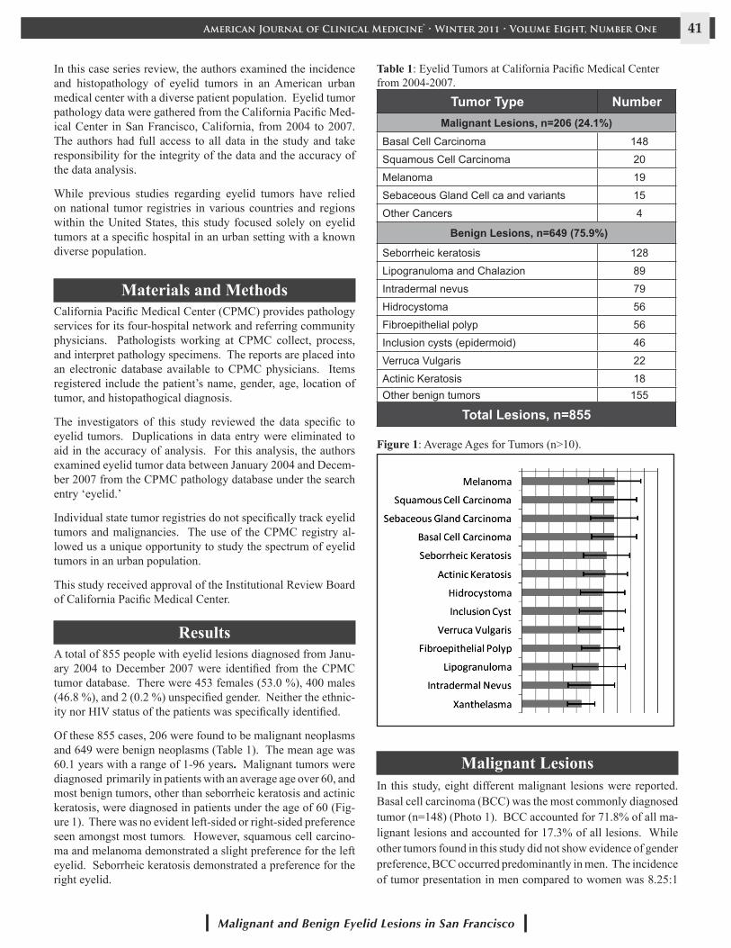
Of these 855 cases, 206 were found to be malignant neoplasms and 649 were benign neoplasms (Table 1). The mean age was 60.1 years with a range of 1-96 years. Malignant tumors were diagnosed primarily in patients with an average age over 60, and most benign tumors, other than seborrheic keratosis and actinic keratosis, were diagnosed in patients under the age of 60 (Fig-ure 1). There was no evident left-sided or right-sided preference seen amongst most tumors. However, squamous cell carcino-ma and melanoma demonstrated a slight preference for the left eyelid. Seborrheic keratosis demonstrated a preference for the right eyelid.
Tumor Type NumberMalignant Lesions, n=206 (24.1%)
Basal Cell Carcinoma 148
Squamous Cell Carcinoma 20
Melanoma 19
Sebaceous Gland Cell ca and variants 15
Other Cancers 4
Benign Lesions, n=649 (75.9%)
Seborrheic keratosis 128
Lipogranuloma and Chalazion 89
Intradermal nevus 79
Hidrocystoma 56
Fibroepithelial polyp 56
Inclusion cysts (epidermoid) 46
Verruca Vulgaris 22
Actinic Keratosis 18Other benign tumors 155
Total Lesions, n=855
Table 1: Eyelid Tumors at California Pacific Medical Center from 2004-2007.
Figure 1: Average Ages for Tumors (n>10).
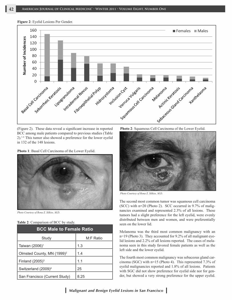
Malignant LesionsIn this study, eight different malignant lesions were reported.Basal cell carcinoma (BCC) was the most commonly diagnosed tumor (n=148) (Photo 1). BCC accounted for 71.8% of all ma-lignant lesions and accounted for 17.3% of all lesions. While other tumors found in this study did not show evidence of gender preference, BCC occurred predominantly in men. The incidence of tumor presentation in men compared to women was 8.25:1
American Journal of Clinical Medicine® • Winter 2011 • Volume Eight, Number One42
Malignant and Benign Eyelid Lesions in San Francisco
(Figure 2). These data reveal a significant increase in reported BCC among male patients compared to previous studies (Table 2).1-4 This tumor also showed a preference for the lower eyelid in 132 of the 148 lesions.
Photo 1: Basal Cell Carcinoma of the Lower Eyelid.
Photo Courtesy of Rona Z. Silkiss, M.D.
BCC Male to Female Ratio
Study M:F Ratio
Taiwan (2006)1 1.3
Olmsted County, MN (1999)2 1.4
Finland (2005)3 1.1
Switzerland (2009)4 25
San Francisco (Current Study) 8.25
Table 2: Comparison of BCC by study.
Photo 2: Squamous Cell Carcinoma of the Lower Eyelid.
Photo Courtesy of Rona Z. Silkiss, M.D.
The second most common tumor was squamous cell carcinoma (SCC) with n=20 (Photo 2). SCC occurred in 9.7% of malig-nancies examined and represented 2.3% of all lesions. These tumors had a slight preference for the left eyelid, were evenly distributed between men and women, and were preferentially seen on the lower lid.
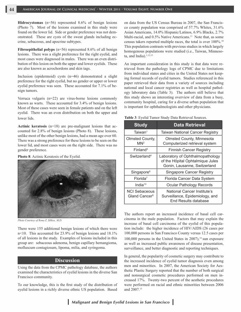
Melanoma was the third most common malignancy with an n=19 (Photo 3). They accounted for 9.2% of all malignant eye-lid lesions and 2.2% of all lesions reported. The cases of mela-noma seen in this study favored female patients as well as the left side and the lower eyelid.
The fourth most common malignancy was sebaceous gland car-cinoma (SGC) with n=15 (Photo 4). This represented 7.3% of eyelid malignancies reported and 1.8% of all lesions. Patients with SGC did not show preference for eyelid side nor for gen-der, but showed a very strong preference for the upper eyelid.
Figure 2: Eyelid Lesions Per Gender.
American Journal of Clinical Medicine® • Winter 2011 • Volume Eight, Number One 43
Malignant and Benign Eyelid Lesions in San Francisco
Out of the 15 clinical samples, 86.7% of the tumors (n = 13) were located on the upper lid while only two, or 13.3%, were located on the lower lid. The preference for the upper lid is linked to an increased number of meibomian glands located in the upper lid (30) versus the lower lid (20).9, 10
Other cancers of note in this study included malignant B cell lymphoma n=2, Merkel cell carcinoma n=1, and an adnexal malignant tumor n=1.
Photo 3: Melanoma of the Lower Eyelid.
Photo Courtesy of Rona Z. Silkiss, M.D.
Photo 4: Sebaceous Gland Carcinoma of the Upper Eyelid
Photo Courtesy of Rona Z. Silkiss, M.D.
Benign LesionsBenign lesions accounted for 75.9% of all tumors in this study. The average age of individuals diagnosed with a benign lesion was less than 60 years. There were numerous types of benign le-sions; those with over ten occurrences are described in this study.
Seborrheic keratosis was the most common benign neoplasm of the eyelid with n=128, representing 19.7% of benign lesions. These lesions were seen equally in male and female patients, demonstrated a preference for the upper lid and location on the right side. These have been reported in multiple studies as the most common benign tumor in older individuals. This was sub-stantiated by the CPMC experience.
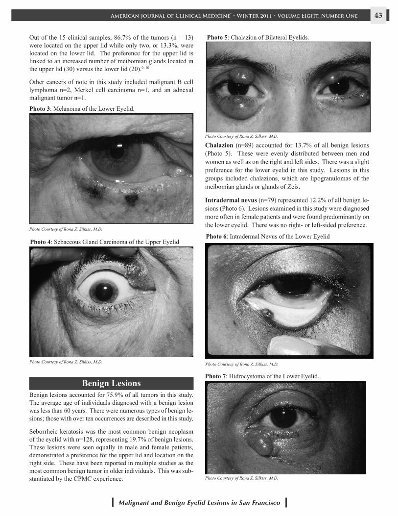
Photo 5: Chalazion of Bilateral Eyelids.
Photo Courtesy of Rona Z. Silkiss, M.D.
Chalazion (n=89) accounted for 13.7% of all benign lesions (Photo 5). These were evenly distributed between men and women as well as on the right and left sides. There was a slight preference for the lower eyelid in this study. Lesions in this groups included chalazions, which are lipogranulomas of the meibomian glands or glands of Zeis.
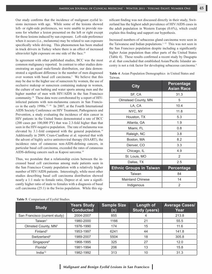
Intradermal nevus (n=79) represented 12.2% of all benign le-sions (Photo 6). Lesions examined in this study were diagnosed more often in female patients and were found predominantly on the lower eyelid. There was no right- or left-sided preference.
Photo 6: Intradermal Nevus of the Lower Eyelid
Photo Courtesy of Rona Z. Silkiss, M.D.
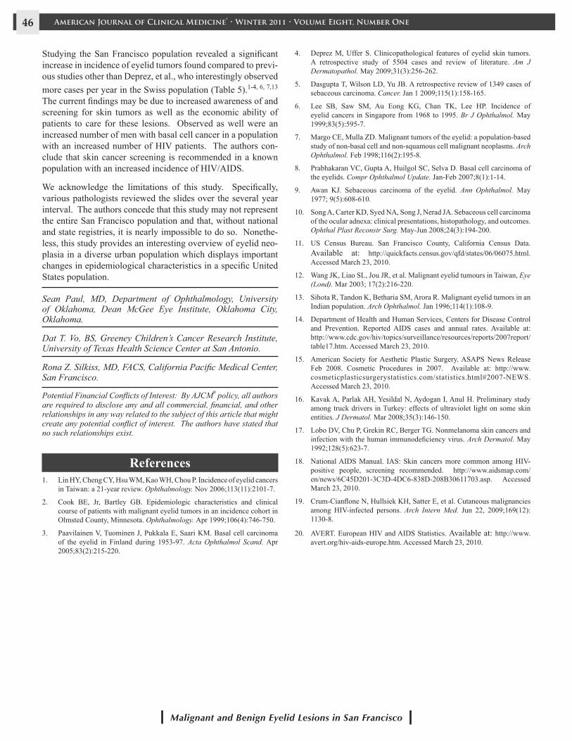
Photo 7: Hidrocystoma of the Lower Eyelid.
Photo Courtesy of Rona Z. Silkiss, M.D.
American Journal of Clinical Medicine® • Winter 2011 • Volume Eight, Number One44
Malignant and Benign Eyelid Lesions in San Francisco
Hidrocystomas (n=56) represented 8.6% of benign lesions (Photo 7). Most of the lesions examined in this study were found on the lower lid. Side or gender preference was not dem-onstrated. These are cysts of the sweat glands including ec-crine, sebaceous, and apocrine subtypes.
Fibroepithelial polyps (n=56) represented 8.6% of all benign lesions. There was a slight preference for the right eyelid, and most cases were diagnosed in males. There was an even distri-bution of this lesion on both the upper and lower eyelids. These are also known as acrochordons and skin tags.
Inclusion (epidermoid) cysts (n=46) demonstrated a slight preference for the right eyelid, but no gender or upper or lower eyelid preference was seen. These accounted for 7.1% of be-nign tumors.
Verruca vulgaris (n=22) are virus-borne lesions commonly known as warts. These accounted for 3.4% of benign lesions. Most of these cases were seen in female patients and on the left eyelid. There was an even distribution on both the upper and lower lids.
Actinic keratosis (n=18) are pre-malignant lesions that ac-counted for 2.8% of benign lesions (Photo 8). These lesions, unlike most of the other benign lesions, had a mean age over 60. There was a strong preference for these lesions to be seen on the lower lid, and most cases were on the right side. There was no gender preference.
Photo 8: Actinic Keratosis of the Eyelid.
Photo Courtesy of Rona Z. Silkiss, M.D.
There were 155 additional benign lesions of which there were n<10. This accounted for 23.9% of benign lesions and 18.1% of all lesions in the study. Examples of lesions included in this group are: sebaceous adenoma, benign capillary hemangioma, molluscum contagiosum, lipoma, milia, and syringoma.
DiscussionUsing the data from the CPMC pathology database, the authors examined the characteristics of eyelid lesions in the diverse San Francisco community.
To our knowledge, this is the first study of the distribution of eyelid lesions in a richly diverse ethnic US population. Based
on data from the US Census Bureau in 2007, the San Francis-co county population was comprised of 57.7% Whites, 31.6% Asian Americans, 14.0% Hispanic/Latinos, 6.9% Blacks, 2.7% Multi-racial, and 0.5% Native Americans.11 Note that, as some census takers reported multiple races, the total is over 100%.10 This population contrasts with previous studies in which largely homogenous populations were studied (i.e., Taiwan, Minneso-ta, and India).1,12,13
An important consideration in this study is that data were re-trieved from the pathology logs of CPMC due to limitations from individual states and cities in the United States not keep-ing formal records of eyelid tumors. Studies referenced in this paper retrieved their data from a variety of sources including national and local cancer registries as well as hospital pathol-ogy laboratory data (Table 3). The authors still believe that this study shows an interesting overview of data from a busy, community hospital, caring for a diverse urban population that is important for ophthalmologists and other physicians.
Study Data RetrievalTaiwan1 Taiwan National Cancer Registry
Olmsted County, MN2
Olmsted County, Minnesota Computerized retrieval system
Finland3 Finnish Cancer RegistrySwitzerland4 Laboratory of Ophthalmopathology
of the Hôpital Ophtalmique Jules Gonin, Lausanne, Switzerland
Singapore6 Singapore Cancer Registry Florida7 Florida Cancer Data SystemIndia13 Ocular Pathology Records
NCI Sebaceous Gland Cancer5
National Cancer Institute’s Surveillance, Epidemiology, and
End Results database
Table 3: Eyelid Tumor Study Data Retrieval Sources.
The authors report an increased incidence of basal cell car-cinoma in the male population. Factors that may explain the increase of basal cell carcinoma of the eyelid of this popula-tion include: the higher incidence of HIV/AIDS (26 cases per 100,000 persons in San Francisco County versus 12.5 cases per 100,000 persons in the United States in 2007),14 sun exposure as well as increased public awareness of disease presentation, surveillance, and better diagnostic and reporting techniques.
In general, the popularity of cosmetic surgery may contribute to the increased incidence of eyelid tumor diagnosis even among men and minorities. In 2007, the American Society for Aes-thetic Plastic Surgery reported that the number of both surgical and nonsurgical cosmetic procedures performed on men in-creased 17%. Twenty-two percent of the aesthetic procedures were performed on racial and ethnic minorities between 2006 and 2007.15
American Journal of Clinical Medicine® • Winter 2011 • Volume Eight, Number One 45
Malignant and Benign Eyelid Lesions in San Francisco
Our study confirms that the incidence of malignant eyelid le-sions increases with age. While some of the lesions showed left- or right-side preferences, we were unable to provide rea-sons for whether a lesion presented on the left or right except for those lesions induced by sun exposure. Left-side preference when it occurs (i.e., melanoma) may be related to sun exposure specifically while driving. This phenomenon has been studied in truck drivers in Turkey where there is an effect of increased ultraviolet light exposure on one side of the face.16
In agreement with other published studies, BCC was the most common malignancy reported. In contrast to other studies dem-onstrating an equal male/female distribution, our data demon-strated a significant difference in the number of men diagnosed over women with basal cell carcinoma.2 We believe that this may be due to the higher use of sunscreen by women, the use of occlusive makeup or sunscreen containing makeup in women, the culture of sun bathing and water sports among men and the higher number of men with HIV/AIDS in the San Francisco community.14 These data were corroborated by a report of HIV-infected patients with non-melanoma cancers in San Francis-co in the early 1990s.17, 18 In 2007, at the Fourth International AIDS Society Conference on HIV Treatment, Pathogenesis and Prevention, a study evaluating the incidence of skin cancer in HIV patients in the United States demonstrated a rate of BCC (200 cases per 100,000 PY) that was 2.3-fold higher than that seen in the HIV-negative population. The rate of melanoma was elevated by 3.1-fold compared with the general population.14 Additionally in 2009, Crum-Cianflone et al. reported that with the advent of highly active antiretroviral therapy (HAART), the incidence rates of cutaneous non-AIDS-defining cancers, in particular basal cell carcinoma, exceeded the rates of cutaneous AIDS-defining cancers such as Kaposi sarcoma.19
Thus, we postulate that a relationship exists between the in-creased basal cell carcinomas among male patients seen in the San Francisco County population with a relatively higher number of HIV/AIDS patients. Interestingly, while most other studies describing basal cell carcinoma distribution showed nearly a 1:1 male to female ratio, Deprez et al. saw a signifi-cantly higher ratio of male to females with a diagnosis of basal cell carcinoma (25:1) in the Swiss population. While this sig-
nificant finding was not discussed directly in their study, Swit-zerland has the highest adult prevalence of HIV/AIDS cases in the adult population in Western Europe (0.6%), which could explain this finding and support our hypothesis.
Increased numbers of sebaceous gland carcinoma were seen in the Taiwanese and Indian populations.1, 12 This was not seen in the San Francisco population despite including a significantly higher Asian population than other parts of the United States (Table 4). These results confirmed a recent study by Dasgupta et al. that concluded that established Asian/Pacific Islander an-cestry is not a risk factor for developing sebaceous carcinoma.5
City Percentage Asian Race
SF, CA 31.3Olmstead County, MN 5
LA, CA 10.4NYC, NY 11.8
Houston, TX 5.3Atlanta, GA 1.9Miami, FL 0.8
Raleigh, NC 3.8Boston, MA 8.2Denver, CO 3.3Chicago, IL 4.9
St. Louis, MO 2Dallas, TX 2.5
Ethnic Groups in Taiwan PercentageTaiwan 84
Mainland Chinese 14Indigenous 2
Table 4: Asian Population Demographics in United States and Taiwan.
Study Years Study Conducted
Sample Size (n)
Length of Study (years)
Average Cases/Year
San Francisco (current study) 2004-2007 855 4 213.8Taiwan1 1980-2000 1166 21 55.5
Olmsted County, MN2 1976-1990 174 15 11.6Finland3 1953-1997 6241 44 141.8
Switzerland4 1989-2007 5504 18 305.8Singapore6 1968-1995 325 27 12.0
Florida7 1981-1994 206 13 15.8India13 1982-1992 313 10 31.3
Table 5: Comparison of Eyelid Studies.
American Journal of Clinical Medicine® • Winter 2011 • Volume Eight, Number One46
Malignant and Benign Eyelid Lesions in San Francisco
Studying the San Francisco population revealed a significant increase in incidence of eyelid tumors found compared to previ-ous studies other than Deprez, et al., who interestingly observed more cases per year in the Swiss population (Table 5).1-4, 6, 7,13 The current findings may be due to increased awareness of and screening for skin tumors as well as the economic ability of patients to care for these lesions. Observed as well were an increased number of men with basal cell cancer in a population with an increased number of HIV patients. The authors con-clude that skin cancer screening is recommended in a known population with an increased incidence of HIV/AIDS.
We acknowledge the limitations of this study. Specifically, various pathologists reviewed the slides over the several year interval. The authors concede that this study may not represent the entire San Francisco population and that, without national and state registries, it is nearly impossible to do so. Nonethe-less, this study provides an interesting overview of eyelid neo-plasia in a diverse urban population which displays important changes in epidemiological characteristics in a specific United States population.
Sean Paul, MD, Department of Ophthalmology, University of Oklahoma, Dean McGee Eye Institute, Oklahoma City, Oklahoma.
Dat T. Vo, BS, Greeney Children’s Cancer Research Institute, University of Texas Health Science Center at San Antonio.
Rona Z. Silkiss, MD, FACS, California Pacific Medical Center, San Francisco.
Potential Financial Conflicts of Interest: By AJCM® policy, all authors are required to disclose any and all commercial, financial, and other relationships in any way related to the subject of this article that might create any potential conflict of interest. The authors have stated that no such relationships exist.
References1. Lin HY, Cheng CY, Hsu WM, Kao WH, Chou P. Incidence of eyelid cancers
in Taiwan: a 21-year review. Ophthalmology. Nov 2006;113(11):2101-7.
2. Cook BE, Jr, Bartley GB. Epidemiologic characteristics and clinical course of patients with malignant eyelid tumors in an incidence cohort in Olmsted County, Minnesota. Ophthalmology. Apr 1999;106(4):746-750.
3. Paavilainen V, Tuominen J, Pukkala E, Saari KM. Basal cell carcinoma of the eyelid in Finland during 1953-97. Acta Ophthalmol Scand. Apr 2005;83(2):215-220.
4. Deprez M, Uffer S. Clinicopathological features of eyelid skin tumors. A retrospective study of 5504 cases and review of literature. Am J Dermatopathol. May 2009;31(3):256-262.
5. Dasgupta T, Wilson LD, Yu JB. A retrospective review of 1349 cases of sebaceous carcinoma. Cancer. Jan 1 2009;115(1):158-165.
6. Lee SB, Saw SM, Au Eong KG, Chan TK, Lee HP. Incidence of eyelid cancers in Singapore from 1968 to 1995. Br J Ophthalmol. May 1999;83(5):595-7.
7. Margo CE, Mulla ZD. Malignant tumors of the eyelid: a population-based study of non-basal cell and non-squamous cell malignant neoplasms. Arch Ophthalmol. Feb 1998;116(2):195-8.
8. Prabhakaran VC, Gupta A, Huilgol SC, Selva D. Basal cell carcinoma of the eyelids. Compr Ophthalmol Update. Jan-Feb 2007;8(1):1-14.
9. Awan KJ. Sebaceous carcinoma of the eyelid. Ann Ophthalmol. May 1977; 9(5):608-610.
10. Song A, Carter KD, Syed NA, Song J, Nerad JA. Sebaceous cell carcinoma of the ocular adnexa: clinical presentations, histopathology, and outcomes. Ophthal Plast Reconstr Surg. May-Jun 2008;24(3):194-200.
11. US Census Bureau. San Francisco County, California Census Data. Available at: http://quickfacts.census.gov/qfd/states/06/06075.html. Accessed March 23, 2010.
12. Wang JK, Liao SL, Jou JR, et al. Malignant eyelid tumours in Taiwan, Eye (Lond). Mar 2003; 17(2):216-220.
13. Sihota R, Tandon K, Betharia SM, Arora R. Malignant eyelid tumors in an Indian population. Arch Ophthalmol. Jan 1996;114(1):108-9.
14. Department of Health and Human Services, Centers for Disease Control and Prevention. Reported AIDS cases and annual rates. Available at: http://www.cdc.gov/hiv/topics/surveillance/resources/reports/2007report/table17.htm. Accessed March 23, 2010.
15. American Society for Aesthetic Plastic Surgery. ASAPS News Release Feb 2008. Cosmetic Procedures in 2007. Available at: http://www.cosmeticplasticsurgerystatistics.com/statistics.html#2007-NEWS. Accessed March 23, 2010.
16. Kavak A, Parlak AH, Yesildal N, Aydogan I, Anul H. Preliminary study among truck drivers in Turkey: effects of ultraviolet light on some skin entities. J Dermatol. Mar 2008;35(3):146-150.
17. Lobo DV, Chu P, Grekin RC, Berger TG. Nonmelanoma skin cancers and infection with the human immunodeficiency virus. Arch Dermatol. May 1992;128(5):623-7.
18. National AIDS Manual. IAS: Skin cancers more common among HIV-positive people, screening recommended. http://www.aidsmap.com/en/news/6C45D201-3C3D-4DC6-838D-208B30611703.asp. Accessed March 23, 2010.
19. Crum-Cianflone N, Hullsiek KH, Satter E, et al. Cutaneous malignancies among HIV-infected persons. Arch Intern Med. Jun 22, 2009;169(12): 1130-8.
20. AVERT. European HIV and AIDS Statistics. Available at: http://www.avert.org/hiv-aids-europe.htm. Accessed March 23, 2010.