Embed Size (px)
Citation preview

Malignant tumor of the
respiratory system
Nasopharygeal carcinomaLung cancer

Lung cancer
Pulmonary Carcinoma

Introduction No doubt it is the leading
cause of cancer-related deaths.
The incidence is increasing at a
fast rate for both male and
female. So it is the commonest
cancer in the world.

Etiology:1. Smoking and atmospheric pollution2. Oncogenes and suppressor genes over express or/and mutation3. Others: virus infection, asbestosis, radioactive substances inhalation et, al.

PathologyGross types: 1. Central type (hilar type):


2. Peripheral type (nodular
type):
single or multiple nodules
arise
in one of the small bronchi
or
bronchioles.
3. Diffuse type: rare

Peripheral type


Carcinoma in situ in bronchial muc
osa or only invade the wall of bronchi,
mass<2cm, no LN metastasis.
Early lung cancer:

Occult lung cancer
Exam of sputum(+)
Clinical feature (-)
X-ray exam (-)
Pathology: carcinoma
in situ or early
invasive carcinoma

Histologic types:1. Squamous cell carcinoma: The commonest type and most closely associated with cigarette smoking.
•Well- differentiated•Poorly-differentiated•Undifferentiated


2. Adenocarcinoma Usually shown as peripheral type, grow rapidly; hematogenous metastasis may happen early and widely spread. Special types:
Bronchiolo-alveolar carcinomaColloid carcinomaScar cancer


Bronchiolo-alveolar carcinoma
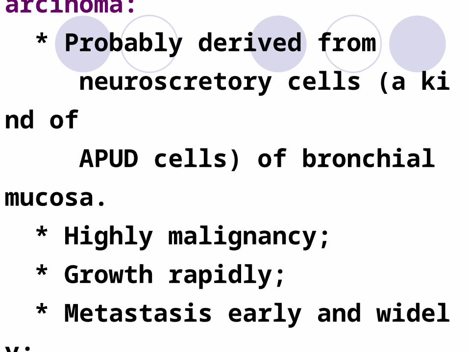
3. Small cell (or oat cell) carcinoma: * Probably derived from neuroscretory cells (a kind of APUD cells) of bronchial mucosa. * Highly malignancy; * Growth rapidly; * Metastasis early and widely; * Radiosensitive.


4. Large cell carcinoma
5. Adeno-squamous carcinoma

Large cell lung
cancer


Large cell lung cancer

Patterns of spread and complications: 1. Direct extention (1). Obstruction of airway (2). Pleurisy with effusion, often hemorrhagic in nature.

(3). Extension of apical lung cancers may involve the lower cords of bronchial plexus and cervical sympathetic plexus ( Horner’s Syndrome: ptosis, miosis, anhydrosis)

2. Lymphatic metastasis
3. Hematogenous metastasis

Clinical manifestation:
Methods for lung cancer diagnosis:1. Sputum cytology, pleural effusion
cytology
2. Fiberbronchoscope examination
and biopsy
3. X-ray examination and CT
4. Fine-needle aspiration biopsy

NASOPHARYNGE
AL CARCINOMA

IntroductionEtiology: The major risk factors are follows:1. Smoking2. Food with high carcinogen contents3. Virus infection: EB Virus4. Genetic and family history

Pathology
Location:
Nasopharyngeal roof,
lateral wall and
pharyngeal recess

Gross type: Nodular type Cauliflower type Submucosa type Ulcerative type Unclassified type.


Histologic type:1. Keratinizing squamous cell carcinoma
2. Non-keratinizing carcinoma
Differentiated carcinoma
Undifferentiated carcinoma
Vesicular nuclear cell carcinoma
3. Adenocarcinoma


1. Direct extension Upward to base of skull Laterally to auditory tube and middle ear Forward to nasal cavity, orbit
Spread and metastasis:

2. Lymphatic metastasis: Retropharyngeal LN
Upper deep LN Internal jugular vein LN Superior cervical LN
Important: Cervical LN enlargement may be the first scene of NPC
3. Hematogenous metastasis