Embed Size (px)
Citation preview
Abstract:
Occupational crash injuries of the foot present a significant reconstructive challenge due to the superficial location of
structures such as tendon, joint and neurovascular network essential for proper acceptance of weight bearing and shear
forces associated with bipedal gait. We discussed here a rare case in which an extensive degloving injury of the foot and
combined with knee traumatic dislocation and proximal fibula tip fracture that underwent amputation. The authors
suggested that none of treatment modalities of degloving are entirely satisfactory, so that each case must be considered
independently with cons and pros.
Key words: Foot, Injuries, Emergency Service
Öz
Ayağın iş kazalarıyla ilgili parçalı yaralanmalarının rekonstrüksiyonu iki ayaklı yürüme ile ilişkili düzgün
kabul edilebilir yük taşıma ve kesme kuvvetleri için gereken tendon, eklem ve nörovasküler ağ yapılarının
yüzeysel yerleşimi nedeniyle çok zordur. Biz burada travmatik diz eklemi dislokasyonu ve fibula proximal
uç fraktürünün eşlik ettiği ayak amputasyunu ile sonuçlanan ayağın nadir görülen geniş eldiven tarzı bir
yaralanmasını tartıştık. Sonuç olarak yazarlar eldiven tarzı yaralanmalarda hiçbir tedavi yöntemi tümüyle
tatmin edici olmadığından her vaka bağımsız olarak artı ve eksileriyle birlikte değerlendirilmesi gerektiğini
önermektedir.
Anahtar Kelimeler: Ayak, Yaralanmalar, Acil Servis
Management of a Sever Degloving Foot Injury: A Rare Presentation of food injury in Emergency Department
Geniş Eldiven-parmak Tarzı Ayak Yaralanmasına Bir Yaklaşım; Acil Serviste Nadir Görülen Bir Olgu Sunumu
1 2 1 1 1Uğur Lok , Hasan Buyukaslan , Umut Gulactı , Fatih Dogan , İrfan Aydın1 Adiyaman University Faculty of Medicine, Department of Emergency Medicine, Adiyaman
2 Harran University Faculty of Medicine, Department of Emergency Medicine, Şanlıurfa
Yazışma adresi: Hasan Büyükaslan, Department of Emergency Medicine, Harran University Faculty of
Medicine, 63200 Şanlıurfa, Turkey GSM: ++90 5306446845, E-mail: [email protected]
Geliş tarihi / Received:31.01.2016� � Kabul tarihi / Accepted: 23.02.2016
Olgu Sunumu
Introduction
Occupational and industrial injuries are important
problems in public health due to fatality and
destabilizing outcomes in particularly new-
developing countries. More than 350,000 workers
lose their lives each year due to unintentional
occupational injuries at global level. The injury
results from accidents at work place and more than
half of this injury burden occurs among workers, and
were one of the most common surgical consultations
in the outpatient or emergency room setting. Workers
with persistent disabilities had a significantly higher
incidence of occupational injuries and higher
medical costs compared with workers without
Harran Üniversitesi Tıp Fakültesi Dergisi (Journal of Harran University Medical Faculty) Cilt 13. Sayı 1, 2016 113
persistent disabilities (1, 2). Trauma of the foot and
knee are commonly encountered in the emergency
departments. The most common mechanisms of
injuries are motor vehicle accidents, falls,
recreational and sports activity, and direct injuries
from striking objects (3). Fractures and soft tissue
injuries are common encountered settings in all
age groups. Swelling of soft tissue, smoking and
co-morbidities such as diabetes mellitus and
peripheral vascular disease should be considered
when planning management schedule. Careful
attention should be paid to neurovascular status
and the soft tissue envelope to effective
management of these injuries especially where
crush injuries have occurred. The core principles
of management consist of to maintain the soft
tissue envelope; to obtain appropriate alignment;
restoration of joint surfaces; and rehabilitation to
obtain optimum function (4). Amputation is one of
the treatment options in acute trauma settings
when the potential risks of the salvage efforts
outweigh the potential benefits. A free-flap
application can be planned as an alternative
method to amputation. Nevertheless, the long-
term outcomes of this protective procedure have
been controversial yet (5).
A degloving injury is a type of avulsion of soft
tissue in which shearing forces that lead to
separation of the skin and soft tissues from the
underlying bone and cause degloving injuries.
Friquently the skin is disconnected and the injury
is readily diagnosed. A large portion of skin is
completely separated from underlying structures,
cutting of its blood supply and exposing cartilage,
the bone, tendon, or nerve (6, 7). In this study, we
presented a case of total degloving injury of the
foot to characterize this unusual and potentially
very serious injury of the skin.
Case
A 45-years-old male was presented to emergency
room after sustaining isolated left lower extremity
crash injury related to occupational trauma that was
occurred via falling down an blunt object heighted
approximately 9 meter and weighted 4000 kilogram
leading to vertical loading and shearing forces.
Patient past medical history revaluated one pack per
day smoking history. The patient had no surgeries,
and any procedure in past, any medications usage,
and had no known drug allergies. He was coopered,
oriented and agitated. Vital signs were as follows;
blood pressure 125/76 mmHg, pulse rate 88 bpm and
regular, auxiliary body temperature 37.6 Celsius and
breathing rate 18/pm respectively. On physical
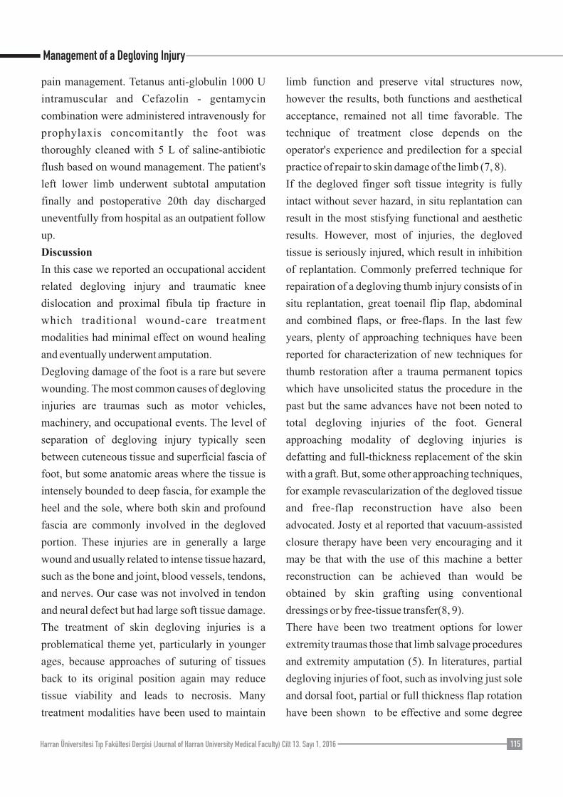
examination revaluated large soft tissue laseration,
and all fingers had been degloved; left food dorsal
and plantar skin cover encircle, glove shaped full
thickness had been avulsed to medially that lead to
uncovering of metatarsal and phalanxes periosteum
(image 1a, 1b and 1c) The plantar fat pad was also
sheared laterally and medial margin of the foot
displaced medially. Extensor tendons, dorsal nerves,
and bones were uncovered because the dorsal foot
soft tissues was seriously degloved from distal to the
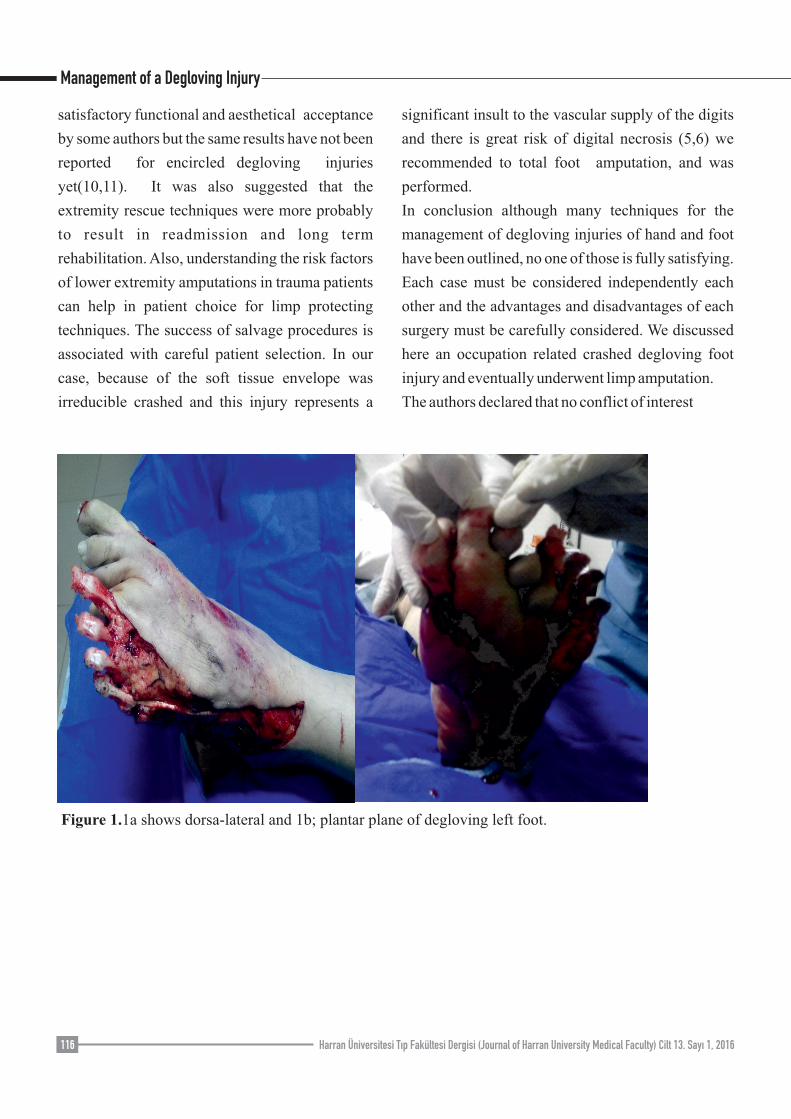
mid foot level. The great toe was fully degloved and
envelope was putted on second toe phalanges (image
2). Left knee distorted and eventually dislocated
(image 3). Patient's Arteria dorsalis pedis and Arteria
tibialis posterior pulses were noted strong by
douppler ultrasound method but venous return and
tissue viability was compromised. Laboratory test
results were all normal limits. Radiographic
examination showed left knee dislocation and
proximal fibula tip fracture ( image 4) and left foot
5th metatarsus fracture and 2th digit distal phalanx
dislocation (Image 5).
Intravenous phentanyl was administered for severe
Management of a Degloving Injury
114 Harran Üniversitesi Tıp Fakültesi Dergisi (Journal of Harran University Medical Faculty) Cilt 13. Sayı 1, 2016
pain management. Tetanus anti-globulin 1000 U
intramuscular and Cefazolin - gentamycin
combination were administered intravenously for
prophylaxis concomitantly the foot was
thoroughly cleaned with 5 L of saline-antibiotic
flush based on wound management. The patient's
left lower limb underwent subtotal amputation
finally and postoperative 20th day discharged
uneventfully from hospital as an outpatient follow
up.
Discussion
In this case we reported an occupational accident
related degloving injury and traumatic knee
dislocation and proximal fibula tip fracture in
which traditional wound-care treatment
modalities had minimal effect on wound healing
and eventually underwent amputation.
Degloving damage of the foot is a rare but severe
wounding. The most common causes of degloving
injuries are traumas such as motor vehicles,
machinery, and occupational events. The level of
separation of degloving injury typically seen
between cuteneous tissue and superficial fascia of
foot, but some anatomic areas where the tissue is
intensely bounded to deep fascia, for example the
heel and the sole, where both skin and profound
fascia are commonly involved in the degloved
portion. These injuries are in generally a large
wound and usually related to intense tissue hazard,
such as the bone and joint, blood vessels, tendons,
and nerves. Our case was not involved in tendon
and neural defect but had large soft tissue damage.
The treatment of skin degloving injuries is a
problematical theme yet, particularly in younger
ages, because approaches of suturing of tissues
back to its original position again may reduce
tissue viability and leads to necrosis. Many
treatment modalities have been used to maintain
limb function and preserve vital structures now,
however the results, both functions and aesthetical
acceptance, remained not all time favorable. The
technique of treatment close depends on the
operator's experience and predilection for a special
practice of repair to skin damage of the limb (7, 8).
If the degloved finger soft tissue integrity is fully
intact without sever hazard, in situ replantation can
result in the most stisfying functional and aesthetic
results. However, most of injuries, the degloved
tissue is seriously injured, which result in inhibition
of replantation. Commonly preferred technique for
repairation of a degloving thumb injury consists of in
situ replantation, great toenail flip flap, abdominal
and combined flaps, or free-flaps. In the last few
years, plenty of approaching techniques have been
reported for characterization of new techniques for
thumb restoration after a trauma permanent topics
which have unsolicited status the procedure in the
past but the same advances have not been noted to
total degloving injuries of the foot. General
approaching modality of degloving injuries is
defatting and full-thickness replacement of the skin
with a graft. But, some other approaching techniques,
for example revascularization of the degloved tissue
and free-flap reconstruction have also been
advocated. Josty et al reported that vacuum-assisted
closure therapy have been very encouraging and it
may be that with the use of this machine a better
reconstruction can be achieved than would be
obtained by skin grafting using conventional
dressings or by free-tissue transfer(8, 9).
There have been two treatment options for lower
extremity traumas those that limb salvage procedures
and extremity amputation (5). In literatures, partial
degloving injuries of foot, such as involving just sole
and dorsal foot, partial or full thickness flap rotation
have been shown to be effective and some degree
Management of a Degloving Injury
Harran Üniversitesi Tıp Fakültesi Dergisi (Journal of Harran University Medical Faculty) Cilt 13. Sayı 1, 2016 115
satisfactory functional and aesthetical acceptance
by some authors but the same results have not been
reported for encircled degloving injuries
yet(10,11). It was also suggested that the
extremity rescue techniques were more probably
to result in readmission and long term
rehabilitation. Also, understanding the risk factors
of lower extremity amputations in trauma patients
can help in patient choice for limp protecting
techniques. The success of salvage procedures is
associated with careful patient selection. In our
case, because of the soft tissue envelope was
irreducible crashed and this injury represents a
significant insult to the vascular supply of the digits
and there is great risk of digital necrosis (5,6) we
recommended to total foot amputation, and was
performed.
In conclusion although many techniques for the
management of degloving injuries of hand and foot
have been outlined, no one of those is fully satisfying.
Each case must be considered independently each
other and the advantages and disadvantages of each
surgery must be carefully considered. We discussed
here an occupation related crashed degloving foot
injury and eventually underwent limp amputation.
The authors declared that no conflict of interest
Figure 1.1a shows dorsa-lateral and 1b; plantar plane of degloving left foot.
Management of a Degloving Injury
116 Harran Üniversitesi Tıp Fakültesi Dergisi (Journal of Harran University Medical Faculty) Cilt 13. Sayı 1, 2016
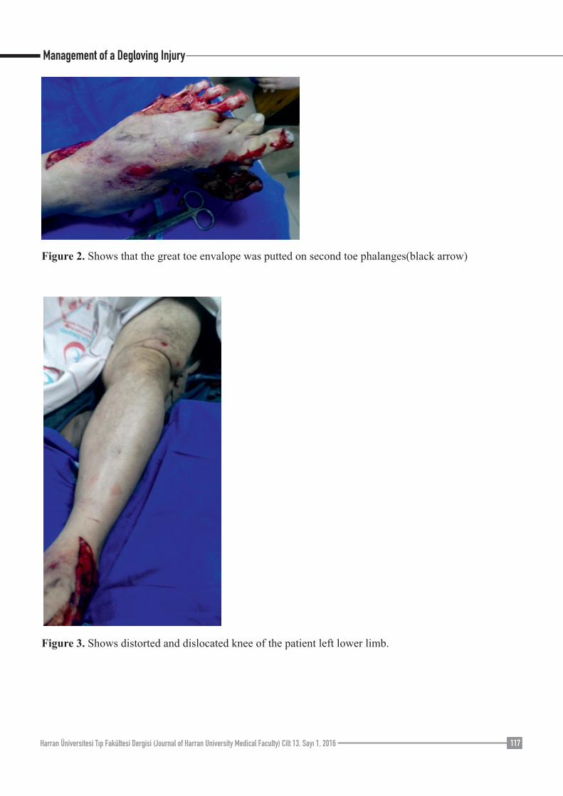
Figure 2. Shows that the great toe envalope was putted on second toe phalanges(black arrow)
Figure 3. Shows distorted and dislocated knee of the patient left lower limb.
Management of a Degloving Injury
Harran Üniversitesi Tıp Fakültesi Dergisi (Journal of Harran University Medical Faculty) Cilt 13. Sayı 1, 2016 117
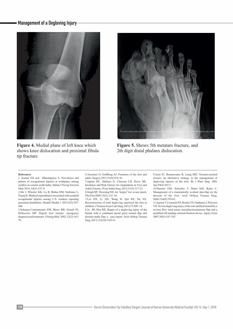
Figure 4. Medial plane of left knee which shows knee dislocation and proximal fibula tip fracture
Figure 5. Shows 5th metatars fracture, and 2th digit distal phalanx dislocation.
References
1. Kumar GS and Dharanipriya A. Prevalence and
pattern of occupational injuries at workplace among
welders in coastal south India, Indian J Occup Environ
Med. 2014 ;18(3):135–9.
2.Shi J, Wheeler KK, Lu B, Bishai DM, Stallones L,
Xiang H. Medical expenditures associated with nonfatal
occupational injuries among U.S. workers reporting
persistent disabilities. Disabil Health J. 2015;8(3):397-
406.
3.Schnaue-Constantouris EM, Birrer RB, Grisafi PJ,
Dellacorte MP. Digital foot trauma: emergency
diagnosis and treatment. J Emerg Med. 2002 ;22(2):163-
70.
4.Aiyenuro O, Goldberg AJ. Fractures of the foot and
ankle. Surgery 2013;31(9):474–81.
5.Jupiter DC, Shibuya N, Clawson LD, Davis ML.
Incidence and Risk Factors for Amputation in Foot and
Ankle Trauma. J Foot Ankle Surg. 2012;51(3):317-22.
6.Singh HP, Downing ND. An “empty” toe: a case report,
The Foot 2005;15(2): 114–16.
7.Liu DX, Li XD, Wang H, Qiu KF, Du SX.
Reconstruction of total degloving injuriesof the foot in
children. J Trauma Acute Care Surg. 2012;73:209–14
8.Ju JH, Hou RX, Repair of a degloving injury of the
thumb with a combined dorsal great toenail flap and
dorsalis pedis flap: a case report. Arch Orthop Trauma
Surg. 2013;133(10):1455-8.
9.Josty IC, Ramaswamy R, Laing JHE. Vacuum-assisted
closure: an alternative strategy in the management of
degloving injuries of the foot. Br J Plast Surg. 2001
Jun;54(4):363-5.
10.Huemer GM, Schoeller T, Dunst KM, Raine C.
Management of a traumatically avulsed skin-flap on the
dorsum of the foot. Arch Orthop Trauma Surg.
2004;124(8):559-62
11.Zgonis T, Cromack DT, Roukis TS, Orphanos J, Polyzois
VD. Severe degloving injury of the sole and heel treated by a
reverse flow sural artery neurofasciocutaneous flap and a
modified off-loading external fixation device. Injury Extra
2007;38(5):187-192
Management of a Degloving Injury
118 Harran Üniversitesi Tıp Fakültesi Dergisi (Journal of Harran University Medical Faculty) Cilt 13. Sayı 1, 2016