Embed Size (px)
Citation preview

Management of Patients With
Urinary Disorders
Management of Patients With
Urinary Disorders

Urinary Tract Infections
• The second most common reason persons seek health care• A common site of health acquired infection• Lower UTI
– Cystitis– Prostatitis– Urethritis
• Upper UTI– Pyelonephritis: acute and chronic– Interstitial nephritis– Renal abscess and perirenal abscess

Factors Contributing to UTI
• Function of glycosaminoglycan (GAG)• Urethrovesical reflux• Uretherovescal reflux• Uropathogenic bacteria• Shorter urethra in women • Risk factors

Routes of infection:
1. Up the urethra: ascending infection ( most common route)2. Through the blood stream (hematogenous spread).3. By means of a fistula from the intestine ( direct extension)• Risk factors:1. Inability or failure to empty the bladder completely 2. Obstructed urinary flow3. Decrease natural host defense or immunosuppression4. Instrumentation of the urinary tract5. Inflammation or abrasion of the urethral mucosa6. Contributing conditions : DM, pregnancy, neurological disorders,
gout.

Urethrovesical Reflux and Uretherovesical Reflux
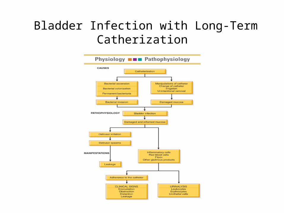
Bladder Infection with Long-Term Catherization

Lower Urinary tract infections: …
• Pathophysiology: for infection to occur 1. bacteria must gain access to the bladder,2. attach to and colonize the epithelium of the urinary tract
to avoid being washed out with voiding, 3. evade host defense mechanisms, and initiate inflammation• Most UTI’s results from 1. fecal organism2. Reflux: Urethrovesical reflux ( backward flow of urine from
the urethra into the bladder

Clinical manifestations:
• about half patient with Bacteriuria have no symptoms.• Uncomplicated: 1. pain and burning on urination, 2. frequency, 3. urgency, nocturia, incontinence, 4. Suprapubic or pelvic pain, 5. and Hematuria with low back pain may presented • Complicated UTI: manifestations may range from asymptomatic
bacteriuria to a gram-negative sepsis with shock

Assessment and Diagnostic findings:
1. Colony count: at least 100,000 colony per ml of urine on a clean catch midstream or cathetarized specimen is a major criterion for infection
2. Cellular studies: microscopic hematuria ( greater than 4 RBC’s per high power field, Pyuria ( greater than 4 WBC’s per high power field)
3. Urine culture: urine culture remains the gold standard in documenting a UTI and can Identify the specific organism present

Medical management:
1. A cute pharmacologic therapy: • single dose administration, short course (3-4 days)
medication regimen, or 7-10 day therapeutic course used in treating uncomplicated lower UTI.
2. Long term pharmacologic therapy:• If infection reoccurs within 2 weeks after completing
antimicrobial therapy, another short course of full-dose antimicrobial therapy, followed by a regular bedtime dose of an antimicrobial agent be prescribed
• If there is no recurrence, medication may taken every other night for 6-7 months

Cont…
• Patient education include: 1. Hygiene (shower rather than bathe tube 2. Fluid intake: drink enough fluid, avoid coffee, tea, colas,
alcohol 3. Voiding Habits: void every 2-3 hours, void immediately
after sexual intercourse 4. therapy: take medication exactly as prescribed, if
recurrence take long term treatment

Upper UTI
1. Acute pyelonephritis: is bacterial infection of the renal pelvis, tubules, and interstitial tissue of one or both kidneys
• Upper UTI is associated with the antibody coating of the bacteria in the urine
• Pathophysiology:1. Ascending of bacteria from the urethra, then to bladder to reach the
kidney2. Rarely from the blood ( less than 3%)3. Ureterovesical reflux4. Urinary tract obstruction, bladder tumor, strictures, benign prostatic
hyperplasia, and urinary stones

Cont…..
• Usually these pt has enlarged kidneys with interstitial infiltration of inflammatory cells which may lead to destruction and atrophy of the kidney
• Clinical manifestation: 1. Acutely ill with chills and fever, 2. leukocytosis, 3. Bacteriuria and Pyuria, 4. Flank pain. 5. Dysuria and frequency may associated.• Assessment and Diagnostic findings: US, CT scan to locate
any obstruction, urine culture and sensitivity may performed

Medical management:
• patient usually treated as outpatient if they are not dehydrated, not experiencing nausea or vomiting and not showing S/S of sepsis
• For outpatient, a 2-weeks course of antibiotic is recommended , 6 weeks therapy may needed if relapse is seen, follow up urine culture is done 2 weeks after completion of antibiotic therapy

Nursing Process: The Care of the Patient with a UTI—Assessment
• Symptoms may include pain and burning upon urination; frequency; nocturia; incontinence; suprapubic, pelvic, or back pain; hematuria; and change in urine or urinary pattern
• About half are asymptomatic• Assess voiding patterns, association of symptoms with
sexual intercourse, contraceptive practices, and personal hygiene
• Gerontologic considerations• Assessment of urine, urinalysis, and urine cultures• Other diagnostic tests

Nursing Process: The Care of the Patient with a UTI—Diagnoses
• Acute pain• Deficient knowledge

Collaborative Problems/Potential Complications
• Sepsis• Renal failure
Nursing Process: The Care of the Patient with a UTI—Planning
• Major goals may include relief of pain and discomfort, increased knowledge of preventive measures and treatment modalities, and absence of complications.

Interventions
• Prevention: avoid indwelling catheters, care of catheters • Personal hygiene • Medications as prescribed: antibiotics, analgesics, and
antispasmodics • Application of heat to the perineum to relieve pan and
spasm• Increased fluid intake• Avoidance of urinary tract irritants such as coffee, tea,
citrus, spices, cola, and alcohol• Frequent voiding • Patient education

2. Chronic pyelonephritis:
• Repeated of a cute pyelonephritis may lead to chronic pyelonephritis
• Clinical manifestations: usually no symptoms of infection, S/S may include fatigue, headache, poor appetite, polyuria, excessive thirst, and weight loss
• Persistent and recurring infection may produce progressive scaring of the kidney, with renal failure as the end result
• Assessment and diagnostic findings: Intravenous urogram, Measurement of creatinine clearance, BUN and creatinine levels, and urine culture

Complication:• ESRF, hypertension, and formation of kidney stones• Medical management: Antibiotics depends on U/C,
careful monitoring of renal function is important while giving medication due to the alteration of kidney function
• Nursing Management: Monitor I&O, encourage fluid(3-4 L/day) unless contraindicated, Assess Temp. every 4 hrs, administer antibiotic as prescribed,Teach the pt the preventive measures of UTI
Urinary Incontinence
• An underdiagnosed and underreported problem that can have significant impact on the quality of life and decrease independence, and which may lead to compromise of the upper urinary system
• Urinary incontinence is not a normal consequence of aging
• Risk factors

Types of Urinary Incontinence
• Stress• Urge• Reflex• Overflow• Functional• Iatrogenic• Mixed incontinence
Patient Teaching• Urinary incontinence is not inevitable and is treatable• Management takes time (provide encouragement and
support)• Develop and use a voiding log or diary • Behavioral interventions• Medication teaching related to pharmacologic
therapy• Strategies for promoting continence

Cont…
II. Pharmacological therapy:1. Anticholinergic agents: (oxybutynin, dicyclomic) which inhibit
bladder contraction, first line medication for urge incontinence2. Tricyclic antidepressant (impramine): decrease bladder contraction
as well as strengthen bladder neck resistance 3. Estrogen: restoring the mucosal integrity, vascular, and muscular
integrity of the urethra
III.Surgical management: surgical correction of the bladder and urethra if the patient not responding to the previous management

III. Neurogenic Bladder:
• Is a dysfunction of the bladder due to a lesion of the nervous system caused by spinal injury, spinal tumor, herniated vertebral disk, multiple sclerosis, infection, congenital anomalies, and DM.
• Pathophysiology:1. Spastic (or reflex) bladder: is the most common type and is caused
by any spinal cord injury above the voiding reflex arc ( Upper motor neuron lesion).
• The result is a loss of conscious sensation and cerebral motor control.• A spastic bladder empties on reflex, with minimal or no controlling
influence to regulate its activity

Cont..
2. Flaccid bladder: caused by lower motor neuron lesion, commonly result from trauma.
• Mainly recognized in DM Pt.. • The bladder continues to fill and becomes greatly
distended, and overflow incontinence occurs. The bladder is not contracted forcefully at any time. Because of sensory loss the patient feels no discomfort.

Medical management:
• Prevention of overdistention of the bladder• Emptying the bladder frequently and completely• Maintaining urine sterility with no stone formation• Maintain adequate bladder capacity without reflux• Pharmacological therapy: Parasympathomimetic medication
(Urecholine)• Surgical management: to correct bladder neck contractures or
vesicoureteral reflux, perfoming some type of urinary diversions procedures

Urinary Retention• Inability of the bladder to empty completely• Residual urine: amount of urine left in the
bladder after voiding• Causes include age (50–100 mL in adults older
than age 60 due to decreased detrusor muscle activity), diabetes, prostate enlargement, pregnancy, neurologic disorders, medications
• Assessment• Nursing measures to promote voiding

• Complication:
1. may lead to chronic infection which may lead to calculi formation,
2. polynephritis, 3. sepsis, 4. back flow of urine lead to deterioration of
the kidney, 5. leakage of the urine may lead to peripheral
skin damage
Nursing Management:
1. Promote normal urinary elimination:• Provide privacy, ensure the environment and position is conducive
to voiding, assisting the patient to use bathroom, and offering reassurance
• Applying warmth to relax sphincter• Simple trigger techniques, such as turning on the water while
voiding attempt, stroking the abd or inner thigh, tapping above the pubic area
• After surgery the prescribed analgesia should be given

Cont…2. Promote urinary elimination: Catheterization is used to
prevent overdistention of the bladder3. Promote home and community-based care:• Provide easy, safe access to the bathroom• Installing support bars in the bathroom• Placing a bedpan or urinal within easy reach• Leaving a light on the bedroom, and bathroom• Wearing clothing that is easy to remove

Chart 45-8 (Strategies for promoting Urinary Continence:

Catheterization (1585)
• Is the introduction of the catheter through the urethra into the bladder for the purpose of withdrawing urine.
• Indications:1. relieve urinary tract retention,2. monitor accurate urine output in critically ill patients, 3. promote urinary drainage, 4. prevent urinary leakage in patient with advance pressure ulcer, 5. obtain a sterile urine specimen, 6. emptying the bladder before, during, after surgery and before
certain diagnostic procedure.

Types of catheters:
1. Indwelling urethral catheter (Folly’s catheter) is remains in the place for continuous drainage . Types (Double and triple lumen catheter).
2. Intermittent catheter: is used to drain the bladder for short time (5-10 min)
3. Suprapubic catheter: it is surgical inserted into the bladder through a small incision above the pubic area.

Nursing Management during catheterization:
1. Assessing the patient and the system:2. Assessing for age-related complication: infection,
elderly patient doesn’t exhibit the S/S of infection but any physical and mental changes should be considered and reported.
3. Minimizing trauma: using proper size, use lubricate, proper technique, and securing the catheter

Cont….4. Bladder retraining after indwelling catheterization: chart 45-
10).• place patient on timed voiding schedule usually every 2-3
hours• the patient instructed to void as scheduled• scan the bladder for residual urine• if more equal or more than100 ml straight catheter may
inserted for complete bladder emptying.5. Assisting with intermittent self catheterization every 4-6
hours and at bed time (or when ever needed)

Cont…..
5. Prevent infection in the catheterized patient:• Use aseptic technique during insertion of the catheter• Use sterile closed urinary drainage system• Prevent contamination of the closed system: never disconnect the
tubing, the drainage bag should not touch the floor• The bag and collecting tubing are changed if contamination occurs, if
urine flow become obstructed, if tubing start to leak.• Clamp the urine drainage if you raised the system above the kidneys
level• Ensure free flow of urine

Cont…• Empty the collection bag frequently• Never irrigate the catheter routinely• Never disconnect the tubing to collect urine sample• Avoid routine catheter changes• Wash the perineal area with soap and water at least twice a day• Monitor the patient’s voiding when the catheter is removed. The
patient must void within 8 hours• Instruct the patient to drink measure fluid fro 8 am- 10 pm and stop
drinking after 10pm

Urolithiasis and Nephrolithiasis
• Calculi (stones) in the urinary tract or kidney• Pathophysiology• Causes; may be unknown• Manifestations– Depend upon location and presence of obstruction
or infection– Pain and hematuria
• Diagnosis: x-ray, blood chemistries, and stone analysis; strain all urine and save stones
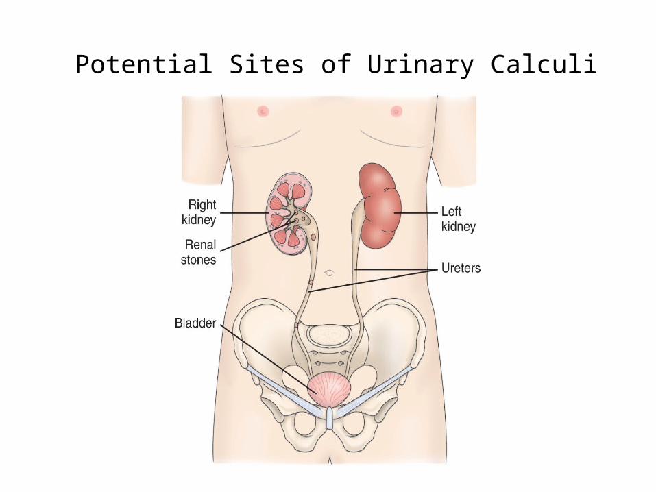
Potential Sites of Urinary Calculi
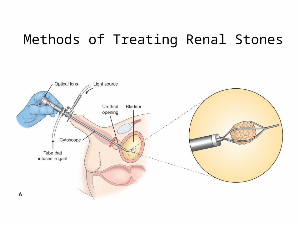
Methods of Treating Renal Stones
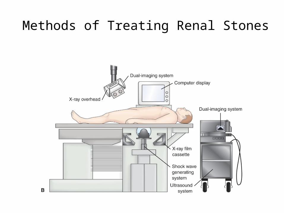
Methods of Treating Renal Stones
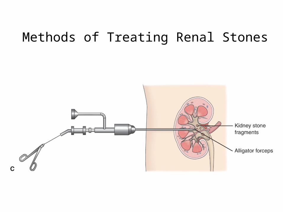
Methods of Treating Renal Stones

Patient Teaching• Signs and symptoms to report• Follow-up care• Urine pH monitoring• Measures to prevent recurrent stones• Importance of fluid intake • Dietary teaching • Medication teaching as needed

Urinary Diversion
• Reasons: bladder cancer or other pelvic malignancies, birth defects, trauma, strictures, neurogenic bladder, chronic infection or intractable cystitis; used as a last resort for incontinence
• Types:– Cutaneous urinary diversion: ileal conduit, cutaneous
ureterostomy, vesicostomy, nephrostomy– Continent urinary diversion: Indiana pouch, Kock
pouch, uretherosigmoidostomy

Cutaneous Urinary Diversions

Continent Urinary Diversions

Nursing Diagnoses: Preoperative
• Anxiety • Imbalanced nutrition• Deficient knowledge
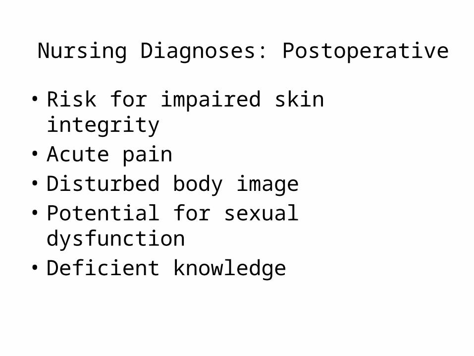
Nursing Diagnoses: Postoperative
• Risk for impaired skin integrity• Acute pain• Disturbed body image• Potential for sexual dysfunction• Deficient knowledge