Embed Size (px)
Citation preview
MANAGEMENT OF MANAGEMENT OF PULMONARY EDEMA PULMONARY EDEMA
Dr. M.O. BOJUWOYEDr. M.O. BOJUWOYE
INTRODUCTIONINTRODUCTION
DEFINITION:PULMONARY EDEMA DEFINITION:PULMONARY EDEMA REFERS TO THE EXTRAVASATION OF REFERS TO THE EXTRAVASATION OF FLUID FROM THE PULMONARY FLUID FROM THE PULMONARY VASCULATURE INTO THE INTERSTITIUM VASCULATURE INTO THE INTERSTITIUM AND ALVEOLI OF THE LUNGS.AND ALVEOLI OF THE LUNGS.
IT IS THE MOST SEVERE IT IS THE MOST SEVERE MANIFESTATION OF CONGESTIVE MANIFESTATION OF CONGESTIVE HEART FAILUREHEART FAILURE
FORMATION IS CAUSED BY 4 MAJOR FORMATION IS CAUSED BY 4 MAJOR PATHOPHYSIOLOGIC MECHANISMS:PATHOPHYSIOLOGIC MECHANISMS:
1)1) IMBALANCE OF STARLING FORCES IMBALANCE OF STARLING FORCES 2)2) DAMAGE TO THE ALVEOLAR-DAMAGE TO THE ALVEOLAR-
CAPILLARY BARRIERCAPILLARY BARRIER3)3) LYMPATHIC OBSTRUCTION OR LYMPATHIC OBSTRUCTION OR
LYMPHATIC INSUFFICIENCYLYMPHATIC INSUFFICIENCY4)4) IDIOPATHIC OR UNKNOWN OR MIXED IDIOPATHIC OR UNKNOWN OR MIXED
CAUSESCAUSES
TYPESTYPES
A)A) CARDIOGENIC PULMONARY EDEMACARDIOGENIC PULMONARY EDEMA
B)B) NON-CARDIOGENIC PULMONARY NON-CARDIOGENIC PULMONARY EDEMAEDEMA
CARDIOGENIC PULMONARY CARDIOGENIC PULMONARY EDEMA (CPE)EDEMA (CPE)
PULMONARY EDEMA DUE TO INCREASE PULMONARY EDEMA DUE TO INCREASE CAPILLARY HYDROSTATIC PRESSURE CAPILLARY HYDROSTATIC PRESSURE 2200 TO ELEVATED PULMONARY VENOUS TO ELEVATED PULMONARY VENOUS PRESSUREPRESSURECPE RELFECTS THE ACCUMULATION OF CPE RELFECTS THE ACCUMULATION OF FLUID WITH A LOW PROTEIN CONTENT FLUID WITH A LOW PROTEIN CONTENT IN THE LUNG INTERSTITIUM AND IN THE LUNG INTERSTITIUM AND ALVEOLIALVEOLI
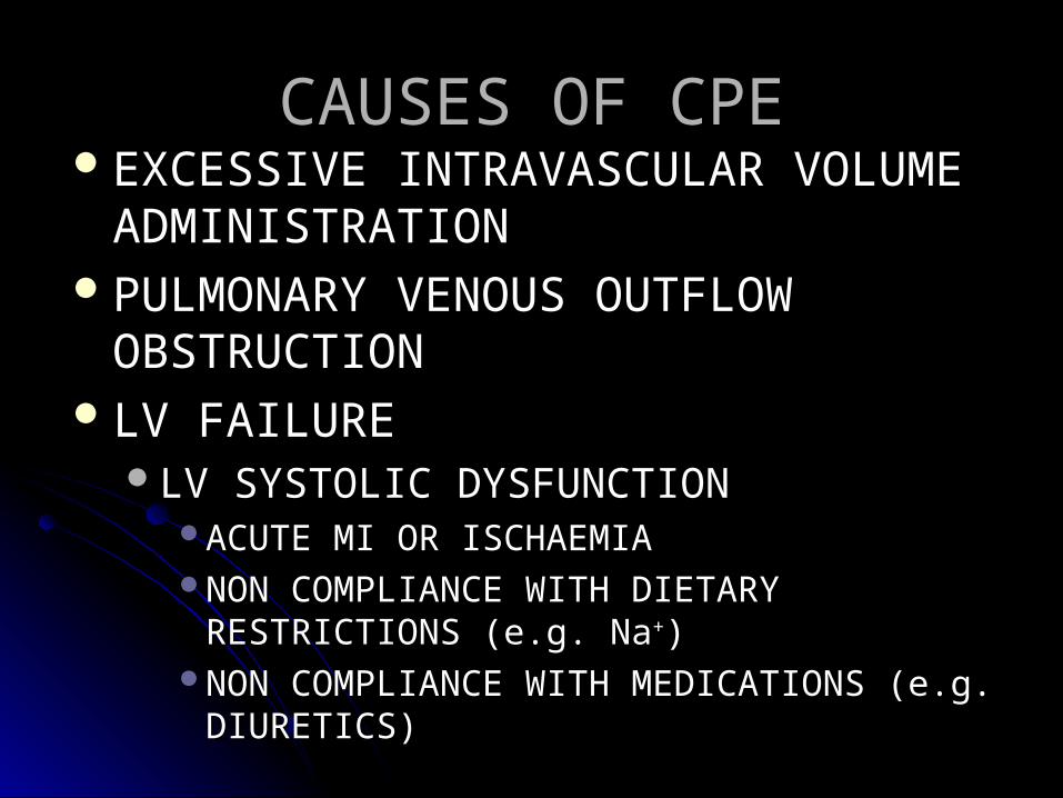
CAUSES OF CPECAUSES OF CPEEXCESSIVE INTRAVASCULAR VOLUME EXCESSIVE INTRAVASCULAR VOLUME
ADMINISTRATIONADMINISTRATIONPULMONARY VENOUS OUTFLOW PULMONARY VENOUS OUTFLOW
OBSTRUCTIONOBSTRUCTIONLV FAILURE LV FAILURE
LV SYSTOLIC DYSFUNCTIONLV SYSTOLIC DYSFUNCTIONACUTE MI OR ISCHAEMIAACUTE MI OR ISCHAEMIANON COMPLIANCE WITH DIETARY NON COMPLIANCE WITH DIETARY
RESTRICTIONS (e.g. NaRESTRICTIONS (e.g. Na++))NON COMPLIANCE WITH MEDICATIONS (e.g. NON COMPLIANCE WITH MEDICATIONS (e.g.
DIURETICS)DIURETICS)
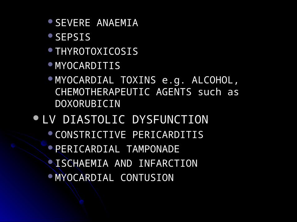
SEVERE ANAEMIASEVERE ANAEMIASEPSISSEPSISTHYROTOXICOSISTHYROTOXICOSISMYOCARDITISMYOCARDITISMYOCARDIAL TOXINS e.g. ALCOHOL, MYOCARDIAL TOXINS e.g. ALCOHOL,
CHEMOTHERAPEUTIC AGENTS such as CHEMOTHERAPEUTIC AGENTS such as DOXORUBICINDOXORUBICIN
LV DIASTOLIC DYSFUNCTIONLV DIASTOLIC DYSFUNCTIONCONSTRICTIVE PERICARDITISCONSTRICTIVE PERICARDITISPERICARDIAL TAMPONADEPERICARDIAL TAMPONADEISCHAEMIA AND INFARCTIONISCHAEMIA AND INFARCTIONMYOCARDIAL CONTUSIONMYOCARDIAL CONTUSION
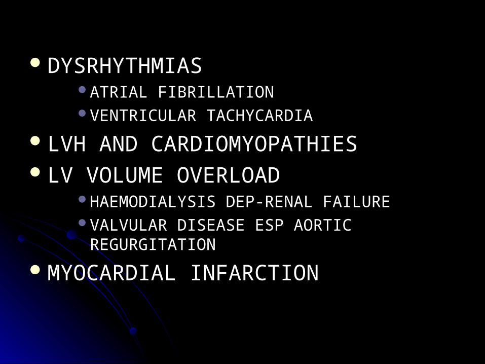
DYSRHYTHMIASDYSRHYTHMIASATRIAL FIBRILLATIONATRIAL FIBRILLATIONVENTRICULAR TACHYCARDIAVENTRICULAR TACHYCARDIA
LVH AND CARDIOMYOPATHIESLVH AND CARDIOMYOPATHIESLV VOLUME OVERLOADLV VOLUME OVERLOAD
HAEMODIALYSIS DEP-RENAL FAILUREHAEMODIALYSIS DEP-RENAL FAILUREVALVULAR DISEASE ESP AORTIC VALVULAR DISEASE ESP AORTIC
REGURGITATIONREGURGITATIONMYOCARDIAL INFARCTIONMYOCARDIAL INFARCTION
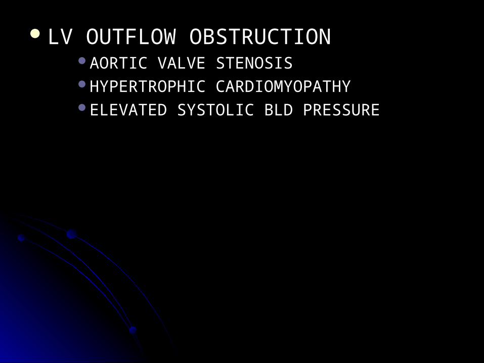
LV OUTFLOW OBSTRUCTIONLV OUTFLOW OBSTRUCTIONAORTIC VALVE STENOSISAORTIC VALVE STENOSISHYPERTROPHIC CARDIOMYOPATHYHYPERTROPHIC CARDIOMYOPATHYELEVATED SYSTOLIC BLD PRESSUREELEVATED SYSTOLIC BLD PRESSURE
PATHOPYSIOLOGY OF CPEPATHOPYSIOLOGY OF CPECPE is caused by elevated pulmonary CPE is caused by elevated pulmonary capillary hydrostatic pressure leading to capillary hydrostatic pressure leading to transudation of fluid into the pulmonary transudation of fluid into the pulmonary interstitium and alveoli. Increased left interstitium and alveoli. Increased left atrial pressure increases pulmonary atrial pressure increases pulmonary venous pressure and pressure in the venous pressure and pressure in the lung microvasculature, resulting in lung microvasculature, resulting in pulmonary edema. pulmonary edema.
MECHANISM OF CPEMECHANISM OF CPE Pulmonary capillary blood and alveolar gas Pulmonary capillary blood and alveolar gas are separated by the alveolar-capillary are separated by the alveolar-capillary membrane, which consists of 3 anatomically membrane, which consists of 3 anatomically different layers: different layers: (1) the capillary endothelium; (1) the capillary endothelium; (2) the interstitial space, which may contain (2) the interstitial space, which may contain connective tissue, fibroblasts, and connective tissue, fibroblasts, and macrophagesmacrophages 3) the alveolar epithelium. 3) the alveolar epithelium.
The Starling relationship determines the fluid The Starling relationship determines the fluid balance between the alveoli and the vascular bed. balance between the alveoli and the vascular bed. Net flow of fluid across a membrane is determined Net flow of fluid across a membrane is determined by applying the following equation: by applying the following equation:
QQ = = KK (P(Pcap - cap - PPis) - is) - λλ((ππcap - cap - ππis)is)
where where QQ is net fluid filtration is net fluid filtration KK is a constant called the filtration coefficient is a constant called the filtration coefficient PPcap is capillary hydrostatic pressurecap is capillary hydrostatic pressurePPis is hydrostatic pressure in the interstitial fluid is is hydrostatic pressure in the interstitial fluid λλ is the reflection coefficient, which indicates the is the reflection coefficient, which indicates the effectiveness of the capillary wall in preventing effectiveness of the capillary wall in preventing protein filtrationprotein filtrationππcap is the colloid osmotic pressure of plasmacap is the colloid osmotic pressure of plasmaππis is the colloid osmotic pressure in the interstitial is is the colloid osmotic pressure in the interstitial fluidfluid
The net filtration of fluid may increase with The net filtration of fluid may increase with changes in different parameters of the changes in different parameters of the Starling equation. Starling equation.
For CPE to develop 2For CPE to develop 200 to increased to increased pulmonary capillary pressure, the pulmonary capillary pressure, the PPcap must cap must rise to a level higher than the rise to a level higher than the ππcap. cap. PPcap is normally 8-12 mm Hg, cap is normally 8-12 mm Hg, and and ππcap is 28 mm Hg. cap is 28 mm Hg.
LYMPHATICS LYMPHATICS The lymphatics play an important role in The lymphatics play an important role in maintaining an adequate fluid balance in the lungs maintaining an adequate fluid balance in the lungs by removing solutes, colloid, and liquid from the by removing solutes, colloid, and liquid from the interstitial space at a rate of approximately 10-20 interstitial space at a rate of approximately 10-20 mL/h. mL/h. An acute rise in pulmonary arterial capillary An acute rise in pulmonary arterial capillary pressure (ie, to >18 mm Hg) may increase filtration pressure (ie, to >18 mm Hg) may increase filtration of fluid into the lung interstitium, but the lymphatic of fluid into the lung interstitium, but the lymphatic removal does not increase correspondingly. removal does not increase correspondingly. In contrast, in the presence of chronically elevated In contrast, in the presence of chronically elevated left atrial pressure, the rate of lymphatic removal left atrial pressure, the rate of lymphatic removal can be as high as 200 mL/h, which protects the can be as high as 200 mL/h, which protects the lungs from pulmonary edema. lungs from pulmonary edema.
STAGESSTAGESSTAGE 1:STAGE 1:
Elevated left atrial pressureElevated left atrial pressureDistention & opening of small pulmonary Distention & opening of small pulmonary
vesselsvesselsNo change in blood gas exchangeNo change in blood gas exchange
STAGE 2:STAGE 2:Continuing filtration of fluid overwhelms the Continuing filtration of fluid overwhelms the
pumping capacity of the lymphaticspumping capacity of the lymphatics
Hypoxaemia is rarely of sufficient magnitude Hypoxaemia is rarely of sufficient magnitude to stimulate tachypneato stimulate tachypnea
Tachypnea is mainly due to the stimulation of Tachypnea is mainly due to the stimulation of stretch receptors of the J-typestretch receptors of the J-type
STAGE THREESTAGE THREEWith further accumulation, fluid crosses the With further accumulation, fluid crosses the
alveoli epithelium into the alveoli alveoli epithelium into the alveoli alveolar alveolar floodingflooding
Abnormalities in gas exchanges are notableAbnormalities in gas exchanges are notableSevere hypoxaemia is usually ass with Severe hypoxaemia is usually ass with
hypocapnia hypocapnia
PATHOPHYSIOLOGIC PATHOPHYSIOLOGIC CONSIDERATIONSCONSIDERATIONS
CPE usually occurs secondary to left atrial outflow CPE usually occurs secondary to left atrial outflow impairment or LV dysfunction. Left atrial outflow impairment or LV dysfunction. Left atrial outflow impairment may be acute or chronic. Causes of impairment may be acute or chronic. Causes of chronic impairment include mitral stenosis or left chronic impairment include mitral stenosis or left atrial tumors. Increased heart rate, which may atrial tumors. Increased heart rate, which may occur secondary to atrial fibrillation, leads to occur secondary to atrial fibrillation, leads to
pulmonary edema because of reduced LV filling. pulmonary edema because of reduced LV filling. Acute mitral-valve regurgitation secondary to Acute mitral-valve regurgitation secondary to
papillary muscle dysfunction or ruptured chordae papillary muscle dysfunction or ruptured chordae tendineae increases LV end-diastolic pressure and tendineae increases LV end-diastolic pressure and
is another cause of pulmonary edema. is another cause of pulmonary edema.
LV dysfunction can be systolic or diastolic or LV dysfunction can be systolic or diastolic or both types. It can also be associated with LV both types. It can also be associated with LV volume overload or LV outflow obstruction. volume overload or LV outflow obstruction. Systolic dysfunction, a common cause of Systolic dysfunction, a common cause of CPE, is defined as decreased myocardial CPE, is defined as decreased myocardial contractility that reduces cardiac output. The contractility that reduces cardiac output. The fall in cardiac output stimulates sympathetic fall in cardiac output stimulates sympathetic activity and blood volume expansion by activity and blood volume expansion by activating the renin-angiotensin-aldosterone activating the renin-angiotensin-aldosterone system, which deteriorate the condition by system, which deteriorate the condition by decreasing LV filling time and increasing decreasing LV filling time and increasing capillary hydrostatic pressure, respectively. capillary hydrostatic pressure, respectively.
NON-CARDIOGENIC NON-CARDIOGENIC PULMONARY EDEMAPULMONARY EDEMA
INJURY TO THE ALVEOLAR-INJURY TO THE ALVEOLAR-CAPILLARY MEMBRANECAPILLARY MEMBRANE
DIRECT INJURY DIRECT INJURY PNEUMONIAPNEUMONIAASPIRATION PNEUMONITISASPIRATION PNEUMONITISTOXIN INHALATION (PHOSGENE)TOXIN INHALATION (PHOSGENE)PULMONARY CONTUSIONPULMONARY CONTUSIONRADIATION/THERMAL LUNG INJURYRADIATION/THERMAL LUNG INJURY
DROWNINGDROWNINGFAT EMBOLIFAT EMBOLIDICDIC
INDIRECT INJURYINDIRECT INJURYSEPSISSEPSISSHOCKSHOCKMULTIPLE TRANSFUSIONSMULTIPLE TRANSFUSIONSACUTE HAEMORRHAGIC PANCREATITISACUTE HAEMORRHAGIC PANCREATITISANAPHYLACTIC SHOCKANAPHYLACTIC SHOCK
NON-CARDIOGENIC CONDITIONS NON-CARDIOGENIC CONDITIONS THAT 1THAT 100LY AFFECT STARLING LY AFFECT STARLING FORCES RATHER THAN FORCES RATHER THAN ALVEOLAR-CAPILLARY BARRIERALVEOLAR-CAPILLARY BARRIERDECREASED PLASMA ONCOTIC DECREASED PLASMA ONCOTIC
PRESSURE e.g. HYPOALBUMINEMIAPRESSURE e.g. HYPOALBUMINEMIAINCREASED NEGATIVITY OF INCREASED NEGATIVITY OF
INTERSTITIAL PRESSURE e.g. RAPID INTERSTITIAL PRESSURE e.g. RAPID REMOVAL OF PNEUMOTHORAXREMOVAL OF PNEUMOTHORAX
LYMPHATIC INSUFFICIENCYLYMPHATIC INSUFFICIENCYLYMPHANGITIC CARCINOMATOSISLYMPHANGITIC CARCINOMATOSISFIBROSING LYMPHANGITISFIBROSING LYMPHANGITISLUNG TRANSPLANTATIONLUNG TRANSPLANTATION
UNKNOWN MECHANISMSUNKNOWN MECHANISMSHIGH-ALTITUDE PULMONARY EDEMAHIGH-ALTITUDE PULMONARY EDEMAACUTE CNS DISORDERS/NEUROGENIC ACUTE CNS DISORDERS/NEUROGENIC
EDEMAEDEMANARCOTIC OVERDOSENARCOTIC OVERDOSEFOLLOWING CARDIOPULMONARY FOLLOWING CARDIOPULMONARY
BYPASSBYPASS
Several features may differentiate CPE from Several features may differentiate CPE from NCPE. NCPE. In CPE, a history of an acute cardiac event is In CPE, a history of an acute cardiac event is usually present. Physical examination shows a usually present. Physical examination shows a low-flow state, an S3 gallop, jugular venous low-flow state, an S3 gallop, jugular venous distention, and crackles on auscultation. distention, and crackles on auscultation. Patients with NCPE have a warm periphery, a Patients with NCPE have a warm periphery, a bounding pulse, and no S3 gallop or jugular bounding pulse, and no S3 gallop or jugular venous distention. venous distention. Definite differentiation is based on PCWP Definite differentiation is based on PCWP measurements. The PCWP is generally >18 mm measurements. The PCWP is generally >18 mm Hg in CPE and <18 mm Hg in NCPE, but Hg in CPE and <18 mm Hg in NCPE, but superimposition of chronic pulmonary vascular superimposition of chronic pulmonary vascular disease can make this distinction difficult. disease can make this distinction difficult.
CLINICAL PRESENTATIONCLINICAL PRESENTATIONHISTORY: PRESENTS WITH THE MOST HISTORY: PRESENTS WITH THE MOST
DRAMATIC CLINICAL FEATURES OF DRAMATIC CLINICAL FEATURES OF LEFT HEART FAILURELEFT HEART FAILURESUDDEN ONSET OF EXTREME SUDDEN ONSET OF EXTREME
BREATHLESSNESS, ANXIETY AND BREATHLESSNESS, ANXIETY AND FEELINGS OF DROWNINGFEELINGS OF DROWNING
PXS COMMONLY COMPLAIN OF PXS COMMONLY COMPLAIN OF SHORTNESS OF BREATHSHORTNESS OF BREATH
HX OF DYSPNEA ON EXERTION; HX OF DYSPNEA ON EXERTION; ORTHOPNEA & PNDORTHOPNEA & PND
COUGHCOUGHCHEST PAIN SHOULD ALERT THE CHEST PAIN SHOULD ALERT THE
PHYSICIAN TO THE POSSIBILITY OF PHYSICIAN TO THE POSSIBILITY OF ACUTE MYOCARDIAL ISCHAEMIA, ACUTE MYOCARDIAL ISCHAEMIA, INFARCTION OR AORTIC DISSECTION INFARCTION OR AORTIC DISSECTION WITH ACUTE AORTIC REGURGITATION WITH ACUTE AORTIC REGURGITATION AS THE PRECIPITANT OF PULMONARY AS THE PRECIPITANT OF PULMONARY EDEMAEDEMA
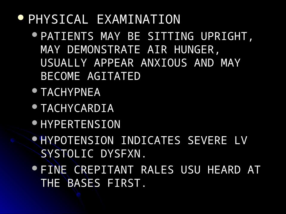
PHYSICAL EXAMINATIONPHYSICAL EXAMINATIONPATIENTS MAY BE SITTING UPRIGHT, PATIENTS MAY BE SITTING UPRIGHT,
MAY DEMONSTRATE AIR HUNGER, MAY DEMONSTRATE AIR HUNGER, USUALLY APPEAR ANXIOUS AND MAY USUALLY APPEAR ANXIOUS AND MAY BECOME AGITATEDBECOME AGITATED
TACHYPNEATACHYPNEATACHYCARDIATACHYCARDIAHYPERTENSIONHYPERTENSIONHYPOTENSION INDICATES SEVERE LV HYPOTENSION INDICATES SEVERE LV
SYSTOLIC DYSFXN.SYSTOLIC DYSFXN.FINE CREPITANT RALES USU HEARD AT FINE CREPITANT RALES USU HEARD AT
THE BASES FIRST.THE BASES FIRST.
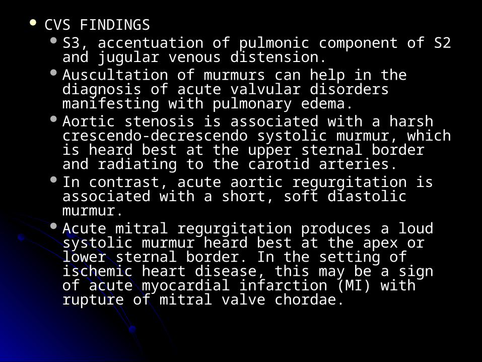
CVS FINDINGSCVS FINDINGS S3, accentuation of pulmonic component of S2 S3, accentuation of pulmonic component of S2
and jugular venous distension. and jugular venous distension. Auscultation of murmurs can help in the diagnosis Auscultation of murmurs can help in the diagnosis
of acute valvular disorders manifesting with of acute valvular disorders manifesting with pulmonary edema. pulmonary edema.
Aortic stenosis is associated with a harsh Aortic stenosis is associated with a harsh crescendo-decrescendo systolic murmur, which is crescendo-decrescendo systolic murmur, which is heard best at the upper sternal border and heard best at the upper sternal border and radiating to the carotid arteries. radiating to the carotid arteries.
In contrast, acute aortic regurgitation is associated In contrast, acute aortic regurgitation is associated with a short, soft diastolic murmur. with a short, soft diastolic murmur.
Acute mitral regurgitation produces a loud systolic Acute mitral regurgitation produces a loud systolic murmur heard best at the apex or lower sternal murmur heard best at the apex or lower sternal border. In the setting of ischemic heart disease, border. In the setting of ischemic heart disease, this may be a sign of acute myocardial infarction this may be a sign of acute myocardial infarction (MI) with rupture of mitral valve chordae.(MI) with rupture of mitral valve chordae.
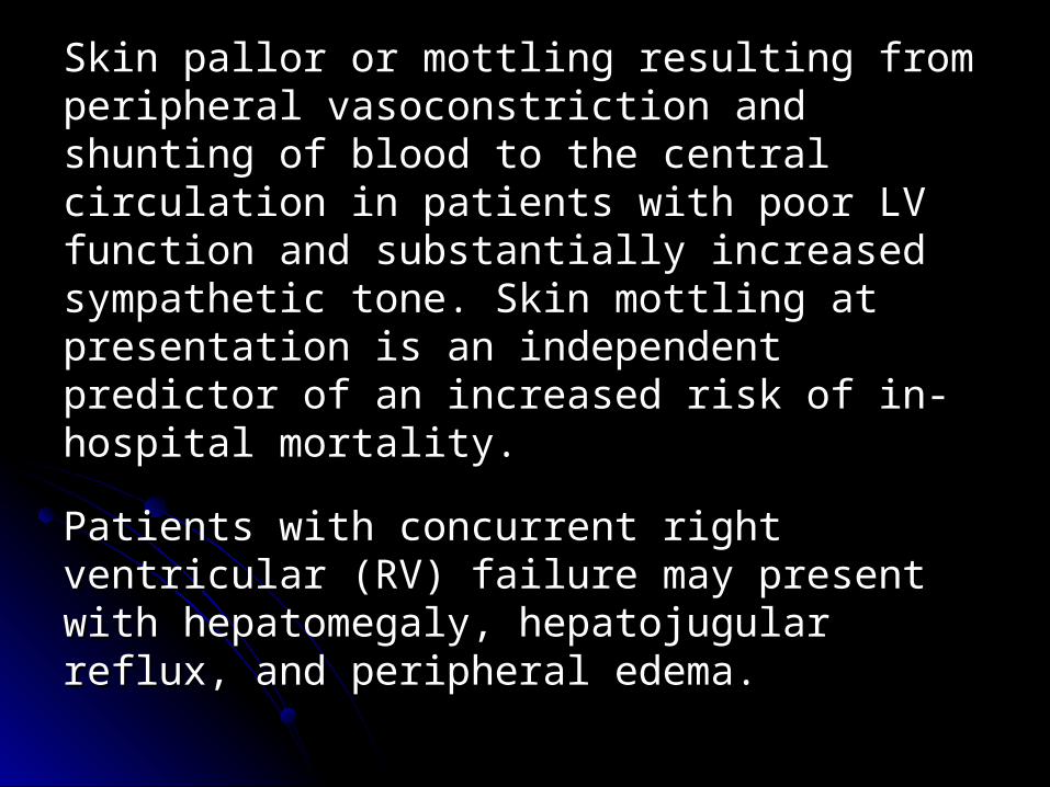
Skin pallor or mottling resulting from Skin pallor or mottling resulting from peripheral vasoconstriction and shunting of peripheral vasoconstriction and shunting of blood to the central circulation in patients blood to the central circulation in patients with poor LV function and substantially with poor LV function and substantially increased sympathetic tone. Skin mottling at increased sympathetic tone. Skin mottling at presentation is an independent predictor of presentation is an independent predictor of an increased risk of in-hospital mortality.an increased risk of in-hospital mortality.
Patients with concurrent right ventricular Patients with concurrent right ventricular (RV) failure may present with hepatomegaly, (RV) failure may present with hepatomegaly, hepatojugular reflux, and peripheral edema.hepatojugular reflux, and peripheral edema.
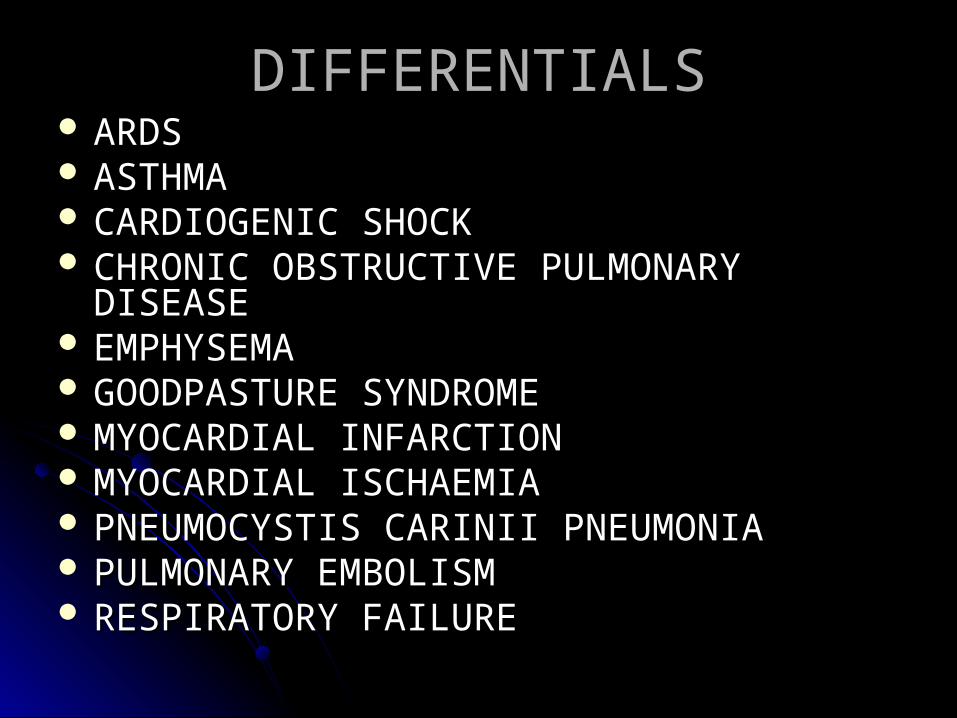
DIFFERENTIALSDIFFERENTIALS ARDSARDS ASTHMAASTHMA CARDIOGENIC SHOCKCARDIOGENIC SHOCK CHRONIC OBSTRUCTIVE PULMONARY CHRONIC OBSTRUCTIVE PULMONARY
DISEASEDISEASE EMPHYSEMAEMPHYSEMA GOODPASTURE SYNDROMEGOODPASTURE SYNDROME MYOCARDIAL INFARCTIONMYOCARDIAL INFARCTION MYOCARDIAL ISCHAEMIAMYOCARDIAL ISCHAEMIA PNEUMOCYSTIS CARINII PNEUMONIAPNEUMOCYSTIS CARINII PNEUMONIA PULMONARY EMBOLISMPULMONARY EMBOLISM RESPIRATORY FAILURERESPIRATORY FAILURE
INVESTIGATIONSINVESTIGATIONS Lab Studies: Lab Studies: Blood count: The FBC with differential helps in Blood count: The FBC with differential helps in
assessing for severe anemia and may suggest assessing for severe anemia and may suggest sepsis or infection if a markedly elevated WBC sepsis or infection if a markedly elevated WBC count or bandemia is present.count or bandemia is present.
Serum electrolyte measurementsSerum electrolyte measurementsPatients with chronic CHF often use diuretics. Patients with chronic CHF often use diuretics.
Therefore, they are predisposed to have Therefore, they are predisposed to have electrolyte abnormalities, especially electrolyte abnormalities, especially hypokalemia and hypomagnesemia.hypokalemia and hypomagnesemia.
Patients with chronic renal failure are at high Patients with chronic renal failure are at high risk for hyperkalemia, especially when they risk for hyperkalemia, especially when they are noncompliant with hemodialysis sessions.are noncompliant with hemodialysis sessions.
BUN and creatinine determinations: These tests BUN and creatinine determinations: These tests help in assessing for renal failure and the help in assessing for renal failure and the anticipated response to diuretics. In low-output anticipated response to diuretics. In low-output states, such as systolic dysfunction, decreased states, such as systolic dysfunction, decreased BUN and creatinine levels may be secondary to BUN and creatinine levels may be secondary to hypoperfusion of the kidneys.hypoperfusion of the kidneys.
Imaging Studies:Imaging Studies: Chest radiography is helpful in distinguishing Chest radiography is helpful in distinguishing
CPE from other pulmonary causes of severe CPE from other pulmonary causes of severe dyspnea.dyspnea.
An enlarged heart, inverted blood flow, Kerley An enlarged heart, inverted blood flow, Kerley lines, basilar edema (vs diffuse edema), lines, basilar edema (vs diffuse edema), absence of air bronchograms, and presence of absence of air bronchograms, and presence of pleural effusion (particularly bilateral and pleural effusion (particularly bilateral and symmetrical pleural effusions) are features thatsymmetrical pleural effusions) are features that
suggest CPE versus NCPE and other lung suggest CPE versus NCPE and other lung pathologies.pathologies.
Chest radiography is somewhat limited in Chest radiography is somewhat limited in patients with CPE of abrupt onset because the patients with CPE of abrupt onset because the classic radiographic abnormalities may not classic radiographic abnormalities may not appear for as long as 12 hours after dyspnea appear for as long as 12 hours after dyspnea begins begins
Other Tests: Other Tests: Arterial blood gas analysisArterial blood gas analysis
This test is more accurate than pulse oximetry This test is more accurate than pulse oximetry for measuring oxygen saturation.for measuring oxygen saturation.
Arterial blood gases help in assessing for Arterial blood gases help in assessing for hypercapnia, a potential early marker of hypercapnia, a potential early marker of impending respiratory failure. Hypoxemia and impending respiratory failure. Hypoxemia and hypocapnia occur in stage 1 and 2 of hypocapnia occur in stage 1 and 2 of pulmonary edema due to ventilation-perfusion pulmonary edema due to ventilation-perfusion mismatch. In stage 3 CPE, a right-to-left mismatch. In stage 3 CPE, a right-to-left intrapulmonary shunt develops secondary to intrapulmonary shunt develops secondary to alveolar flooding and further contributes to alveolar flooding and further contributes to hypoxemia. In severe cases, hypercapnia and hypoxemia. In severe cases, hypercapnia and respiratory acidosis are usually observed.respiratory acidosis are usually observed.
The decision to start mechanical ventilation is The decision to start mechanical ventilation is based on clinical findings and rarely arterial based on clinical findings and rarely arterial blood gas results.blood gas results.
Pulse oximetryPulse oximetryPulse oximetry is highly accurate in assessing Pulse oximetry is highly accurate in assessing
hypoxia and, therefore, the severity of CPE.hypoxia and, therefore, the severity of CPE. It is useful for monitoring the patient's It is useful for monitoring the patient's
response to supplemental oxygenation and response to supplemental oxygenation and other therapies.other therapies.
ElectrocardiographyElectrocardiographyLeft atrial enlargement and LV hypertrophy Left atrial enlargement and LV hypertrophy
are sensitive, though nonspecific, indicators of are sensitive, though nonspecific, indicators of chronic LV dysfunction.chronic LV dysfunction.
The ECG may suggest an acute The ECG may suggest an acute tachydysrhythmia or bradydysrhythmia as the tachydysrhythmia or bradydysrhythmia as the cause of CPE.cause of CPE.
The ECG may suggest acute myocardial The ECG may suggest acute myocardial ischaemia or infarction as the cause of CPE.ischaemia or infarction as the cause of CPE.
ECG is helpful when an acute valvular ECG is helpful when an acute valvular abnormality or LV systolic dysfunction is the abnormality or LV systolic dysfunction is the suspected cause of CPE.suspected cause of CPE.
Large and diffuse inverted T waves and QT Large and diffuse inverted T waves and QT prolongation may be seen within 24 hours and prolongation may be seen within 24 hours and may resolve within 1 week after the patient's may resolve within 1 week after the patient's condition is stabilized. Several condition is stabilized. Several pathophysiologies, including increased pathophysiologies, including increased sympathetic tone, subendocardial ischemia, sympathetic tone, subendocardial ischemia, and metabolic derangements, may be and metabolic derangements, may be involved in causing these ECG changes in involved in causing these ECG changes in CPE.CPE.

Plasma brain natriuretic peptide (BNP) Plasma brain natriuretic peptide (BNP) testing testing
Pro–brain natriuretic peptide (proBNP) Pro–brain natriuretic peptide (proBNP) testing testing
TREATMENTTREATMENTMedical Care: Medical Care: Initial management of Initial management of
patients with CPE should address the patients with CPE should address the ABCs of resuscitationABCs of resuscitation
Oxygen should be administered to all Oxygen should be administered to all patients to keep oxygen saturation >90%. patients to keep oxygen saturation >90%.
In case of persistent hypoxemia, acidosis In case of persistent hypoxemia, acidosis or altered mental status, intubation and or altered mental status, intubation and mechanical ventilation may become mechanical ventilation may become necessary. necessary.
Any associated arrhythmia or myocardial Any associated arrhythmia or myocardial infarction should be treated appropriately. infarction should be treated appropriately.
Medical therapy of CPE focuses on 3 main Medical therapy of CPE focuses on 3 main goals: goals: (1) reduction of pulmonary venous return (1) reduction of pulmonary venous return (preload reduction)(preload reduction)(2) reduction of systemic vascular (2) reduction of systemic vascular resistance (afterload reduction)resistance (afterload reduction)(3) inotropic support. (3) inotropic support.
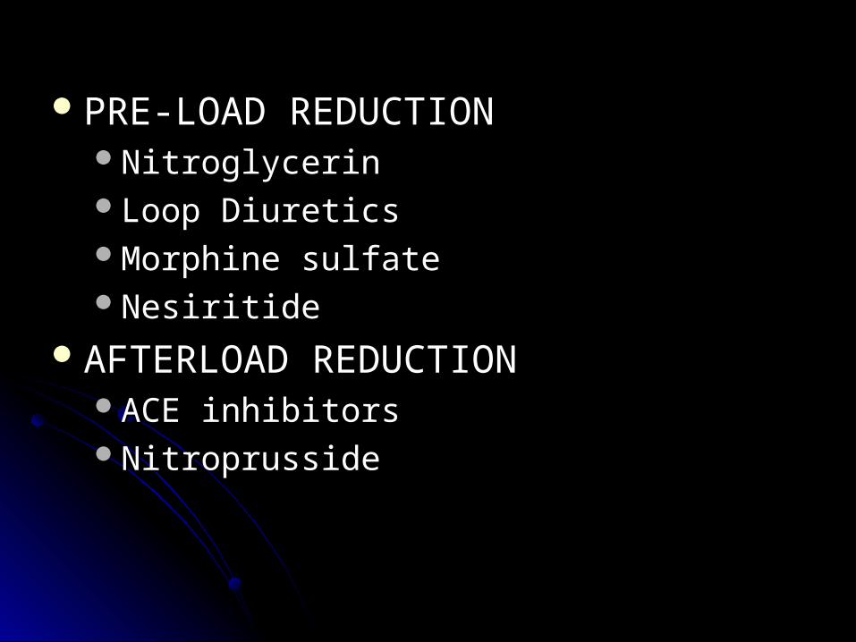
PRE-LOAD REDUCTIONPRE-LOAD REDUCTIONNitroglycerin Nitroglycerin Loop DiureticsLoop DiureticsMorphine sulfate Morphine sulfate Nesiritide Nesiritide
AFTERLOAD REDUCTION AFTERLOAD REDUCTION ACE inhibitors ACE inhibitors NitroprussideNitroprusside
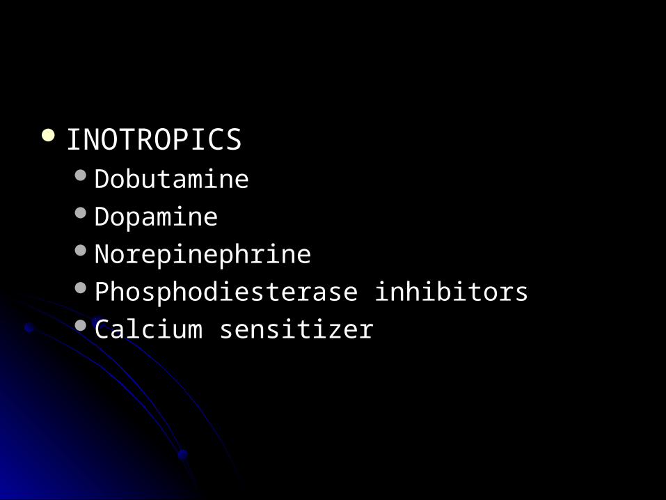
INOTROPICSINOTROPICSDobutamineDobutamineDopamineDopamineNorepinephrine Norepinephrine Phosphodiesterase inhibitors Phosphodiesterase inhibitors Calcium sensitizer Calcium sensitizer
Surgical Care: Surgical Care: Kantrowitz initially described Kantrowitz initially described intra-aortic balloon pumping (IABP) in 1953, but intra-aortic balloon pumping (IABP) in 1953, but IABP was first used clinically in 1969 in a patient IABP was first used clinically in 1969 in a patient with cardiogenic shock. Since the 1980s, IABP with cardiogenic shock. Since the 1980s, IABP has been increasingly applied in various clinical has been increasingly applied in various clinical situations as a life-saving intervention to achieve situations as a life-saving intervention to achieve hemodynamic stabilization before definite hemodynamic stabilization before definite therapy. The intra-aortic balloon pump therapy. The intra-aortic balloon pump decreases afterload as the pump deflates and decreases afterload as the pump deflates and inflates it during diastole to improve coronary inflates it during diastole to improve coronary blood flow. blood flow.
Diet: Diet: Patients admitted with heart failure Patients admitted with heart failure or pulmonary edema should be given a or pulmonary edema should be given a low-salt diet to minimize fluid retention. low-salt diet to minimize fluid retention. Closely monitor their fluid balance. Closely monitor their fluid balance.