Embed Size (px)
Citation preview
MANAGEMENT OF
UPPER GI BLEEDING
M K ALAM MS; FRCSEd
ILOsAt the end of this presentation students will be able to:
Define upper GI haemorrhage.
Describe the resuscitative measures.
Enumerate the causes of upper GI bleeding.
Describe the symptoms & signs of UGI bleeding.
Describe diagnostic work up.
Describe the non-surgical management and
indications for surgical intervention.
Introduction
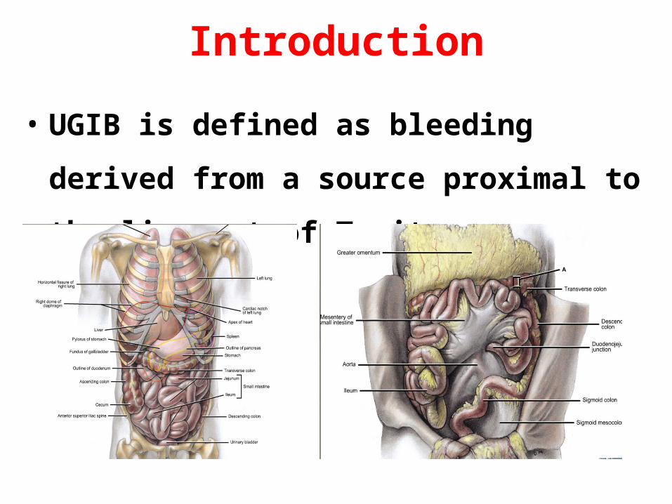
• UGIB is defined as bleeding derived from a
source proximal to the ligament of Treitz.
UGIB
• A potentially life-threatening emergency.
• A common cause of hospitalization
• More common in male.
• 4 times more common than lower GI bleeding.
• Mortality 6-10%
Mortality in UGIB
• Comorbid illness (72%) rather than actual
bleeding, is the major cause of death.
• Comorbid illness- 51% of patients.
• Rebleeding or continued bleeding- associated
with increased mortality
Causes of UGIB
• Peptic ulcer disease (duodenal & gastric ulcer)
• Oesophageal varices (portal hypertension)
• Mallory-Weiss syndrome- mucosal tears of the esophagus.
• Erosive gastritis /esophagitis.
• Dieulafoy lesion.
• Gastric cancer.
• Ulcerated gastric stromal tumor (GIST)
• Aortoenteric fistula- erosion of the aortic graft into the bowel.
• Angiodysplasia- dilated, thin-walled vessels appearing as cherry spots
Pathophysiology
• Arterial hemorrhage- ulcer disease, mucosal tears
as in Mallory-Weiss syndrome.
• Low-pressure venous hemorrhage, as in
telangiectasias.
• Variceal hemorrhage is due to elevated portal
pressure (>12 mmHg) transmitted to esophageal and
gastric varices and resulting in rupture of varices.
Mucosal ulceration can be a bleeding source.
Peptic ulcer disease (PUD)
• The most common cause of UGIB.
• High-risk for PUD: Alcohol abuse, chronic renal failure, and/or
nonsteroidal anti-inflammatory drug (NSAID) use.
• Duodenal ulcers are more common than gastric ulcers
• Ulcer burrows deeper into the mucosa, causes weakening and
necrosis of the arterial wall, leading to a pseudoaneurysm. The
weakened wall ruptures, producing hemorrhage.
• Approximately in 80% bleeding from PUD stops spontaneously.
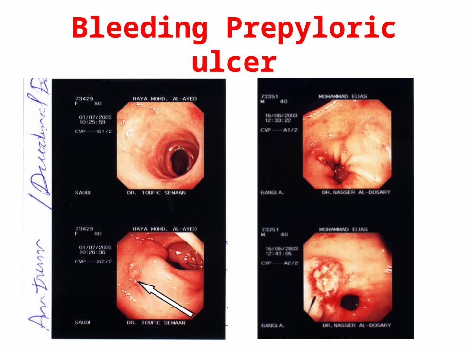
Bleeding Prepyloric ulcer
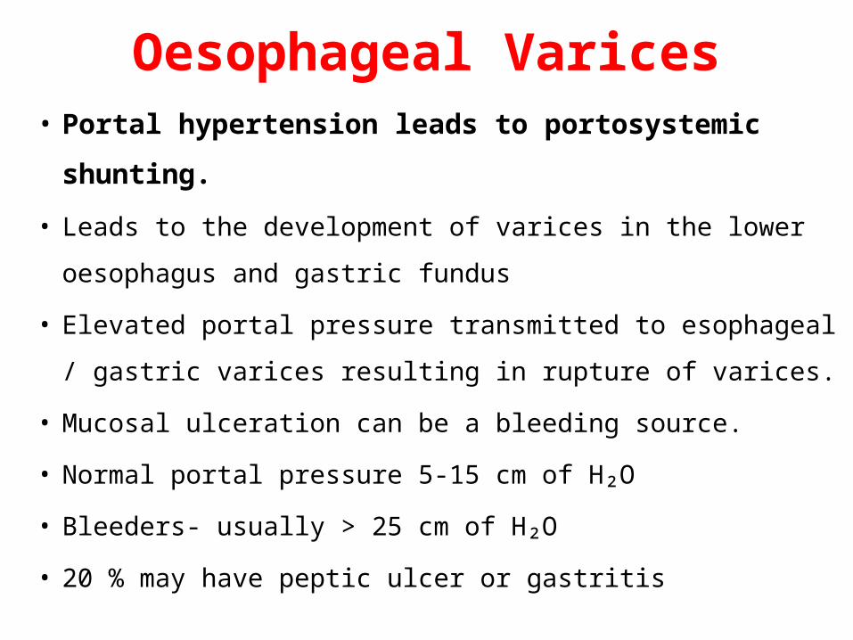
Oesophageal Varices• Portal hypertension leads to portosystemic shunting.
• Leads to the development of varices in the lower oesophagus
and gastric fundus
• Elevated portal pressure transmitted to esophageal / gastric
varices resulting in rupture of varices.
• Mucosal ulceration can be a bleeding source.
• Normal portal pressure 5-15 cm of H₂O
• Bleeders- usually > 25 cm of H₂O
• 20 % may have peptic ulcer or gastritis
Causes of portal hypertension
• Pre-hepatic: Congenital atresia of PV, PV thrombosis, Compression of PV (tumours)
• Intrahepatic: Pre-sinusoidal- Schistosomiasis Sinusoidal- Cirrhosis
• Post-hepatic (Post-sinusoidal): Budd-Chiari syndrome,
Constrictive pericarditis
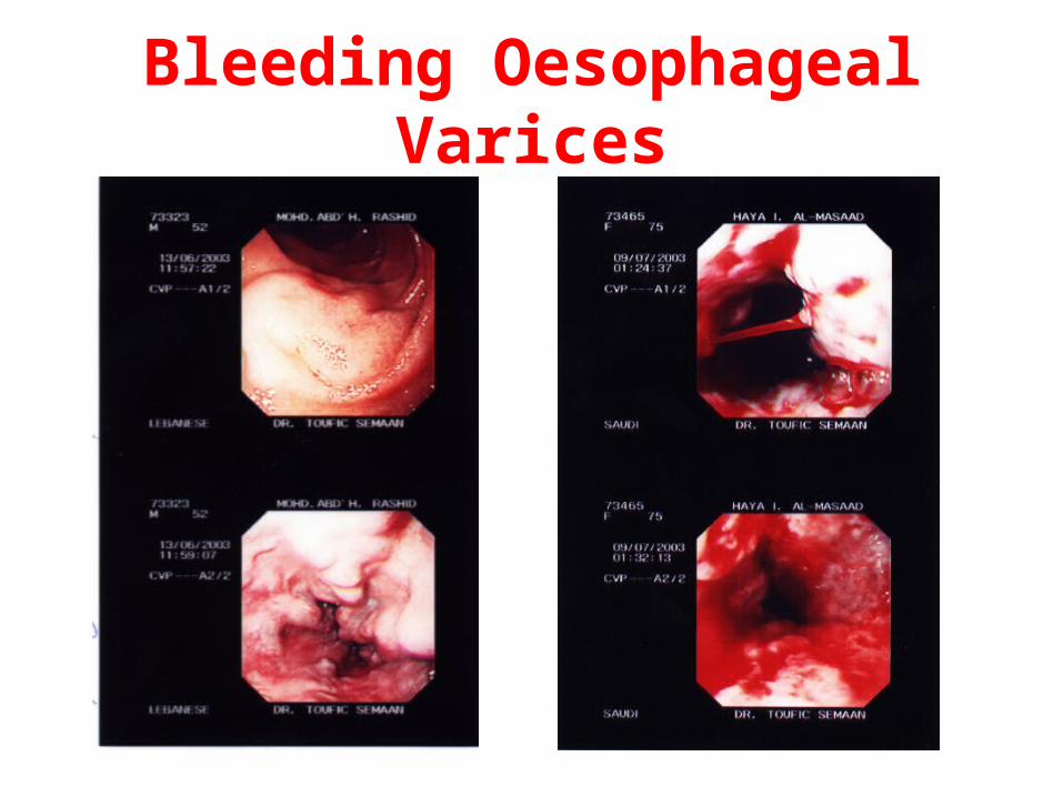
Bleeding Oesophageal Varices
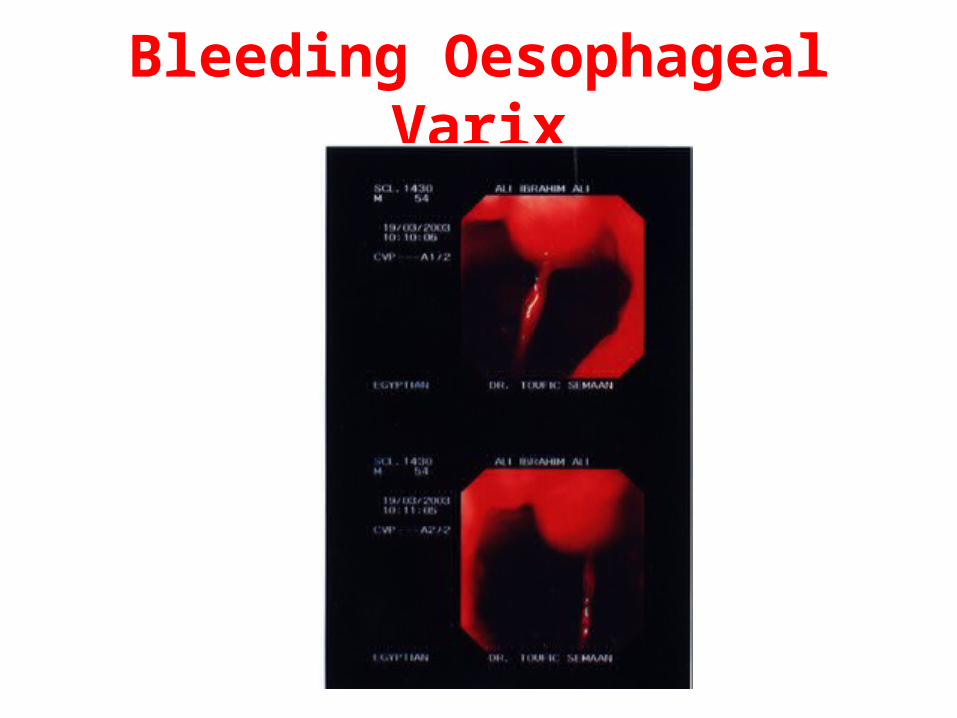
Bleeding Oesophageal Varix
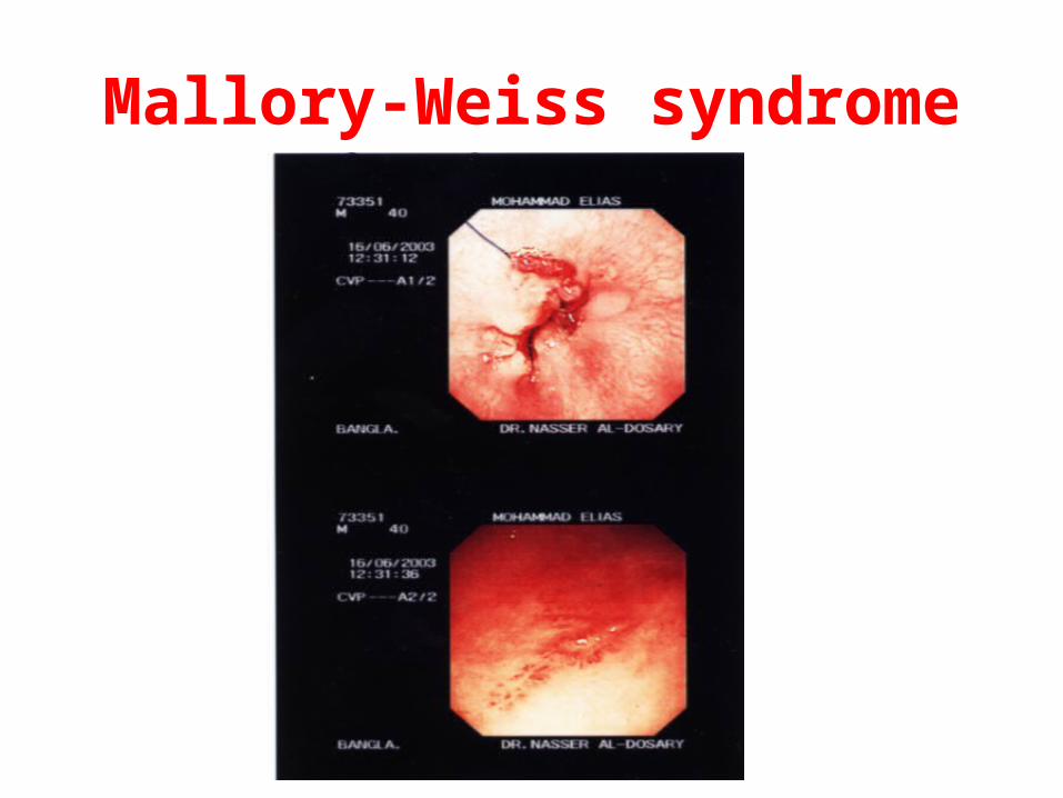
Mallory-Weiss syndrome
• Mallory-Weiss tears -15% of acute upper UGIB
• Mucosal laceration- result of forceful vomiting
• 80-90%- tear along the lesser curve of the stomach just
distal to the gastro-esophageal junction
Mallory-Weiss syndrome
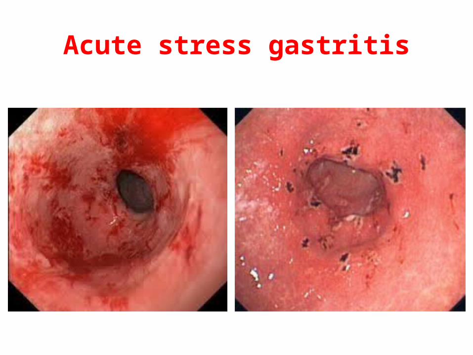
Acute stress gastritis
• Seen in shock, multiple trauma, acute respiratory distress syndrome, systemic respiratory distress syndrome, acute renal failure, and sepsis patients.
• Predisposing conditions alter local mucosal protective barriers, such as mucus, bicarbonate, blood flow, and prostaglandin synthesis.
• Disruption of balance of these factors results in diffuse gastric mucosal erosions.
• The principal mechanisms- decreased splanchnic mucosal blood flow and altered gastric luminal acidity.
Acute stress gastritis
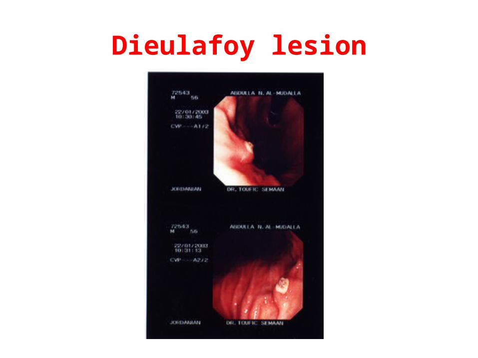
Dieulafoy lesion
• A vascular malformation of the proximal stomach.
• 2-5% of acute UGIB episodes.
• Endoscopic appearance: large ulcerated submucosal vessel.
• Bleeding can be massive and brisk.
• Vessel rupture occurs in the setting of chr. gastritis
• Alcohol use is associated with the Dieulafoy lesion.
• Mostly- men in their third to tenth decade.
• Can occur anywhere along the GI tract
Dieulafoy lesion
GIST (gastrointestinal stromal tumour)
• Mesenchymal tumour, submucosal lesions
• 50-60%- stomach
• 20-30%- small intestine
• 10%- rectum
• Benign or malignant (positive for c-Kit oncogene)
• Pacemaker cells in smooth muscle
• Asymptomatic, bleeding or obstruction
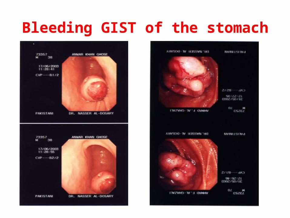
Bleeding GIST of the stomach
Gastric carcinoma
• Common- chronic blood loss (anaemia)
• Haematemesis- uncommon
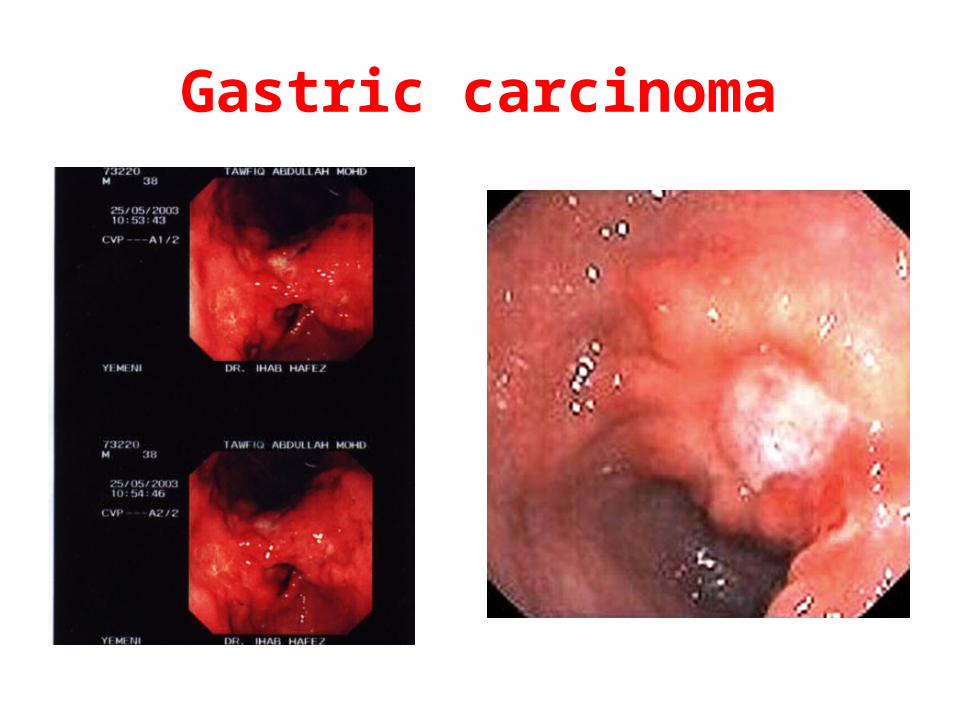
Gastric carcinoma
NSAID in UGIB
• Cause gastric and duodenal ulcers by inhibiting
cyclooxygenase - ↓ mucosal prostaglandin
synthesis- results in impaired mucosal defenses.
• Daily NSAID: 40-fold increase in gastric ulcer &
8-fold increase in duodenal ulcer creation.
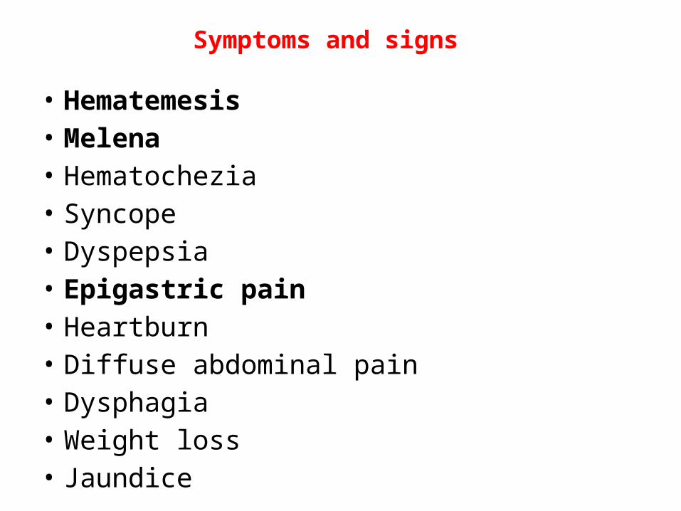
Symptoms and signs
• Hematemesis• Melena• Hematochezia • Syncope• Dyspepsia• Epigastric pain• Heartburn• Diffuse abdominal pain• Dysphagia• Weight loss• Jaundice
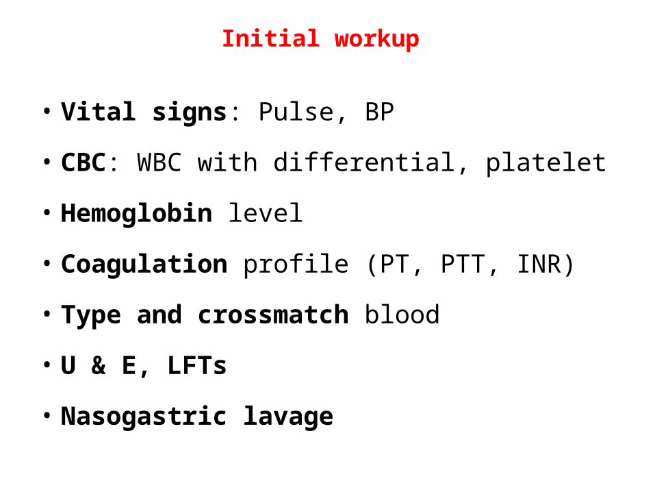
Initial workup
• Vital signs: Pulse, BP
• CBC: WBC with differential, platelet
• Hemoglobin level
• Coagulation profile (PT, PTT, INR)
• Type and crossmatch blood
• U & E, LFTs
• Nasogastric lavage
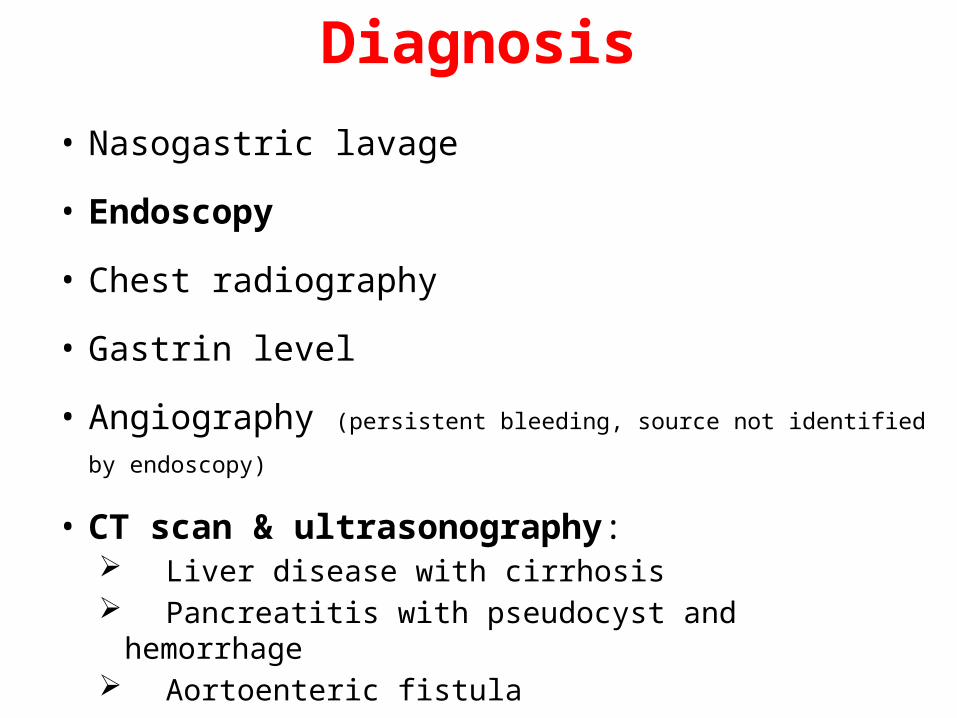
Diagnosis
• Nasogastric lavage
• Endoscopy
• Chest radiography
• Gastrin level
• Angiography (persistent bleeding, source not identified by endoscopy)
• CT scan & ultrasonography: Liver disease with cirrhosis Pancreatitis with pseudocyst and hemorrhage Aortoenteric fistula
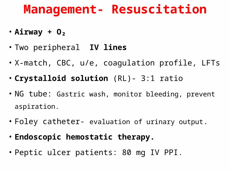
Management- Resuscitation
• Airway + O₂
• Two peripheral IV lines
• X-match, CBC, u/e, coagulation profile, LFTs
• Crystalloid solution (RL)- 3:1 ratio
• NG tube: Gastric wash, monitor bleeding, prevent aspiration.
• Foley catheter- evaluation of urinary output.
• Endoscopic hemostatic therapy.
• Peptic ulcer patients: 80 mg IV PPI.
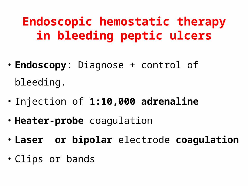
Endoscopic hemostatic therapy in bleeding peptic ulcers
• Endoscopy: Diagnose + control of bleeding.
• Injection of 1:10,000 adrenaline
• Heater-probe coagulation
• Laser or bipolar electrode coagulation
• Clips or bands
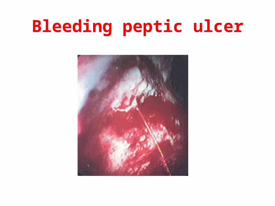
Bleeding peptic ulcer
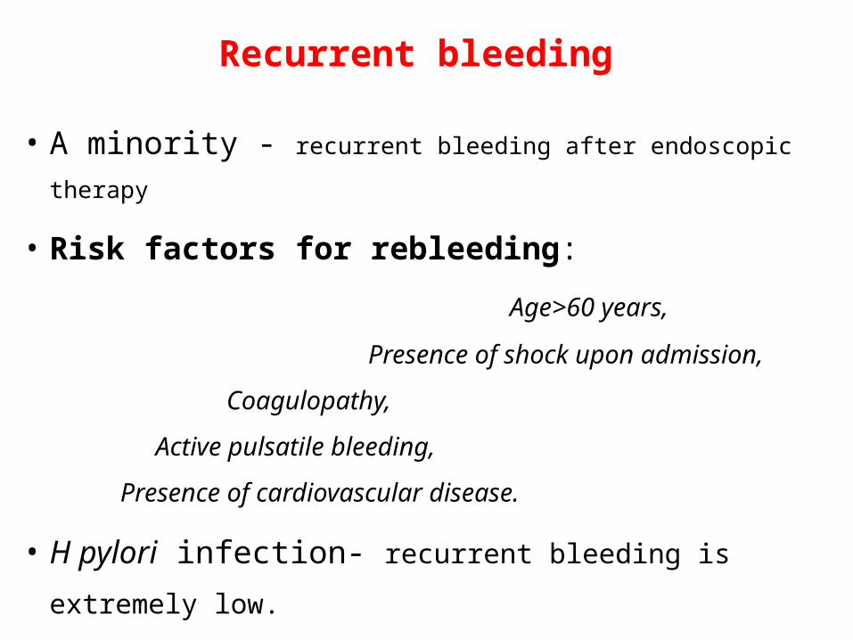
Recurrent bleeding
• A minority - recurrent bleeding after endoscopic therapy
• Risk factors for rebleeding: Age>60 years,
Presence of shock upon admission,
Coagulopathy,
Active pulsatile bleeding,
Presence of cardiovascular disease.
• H pylori infection- recurrent bleeding is extremely low.
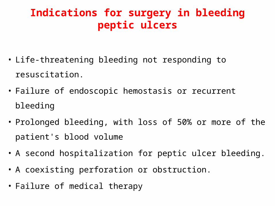
Indications for surgery in bleeding peptic ulcers
• Life-threatening bleeding not responding to resuscitation.
• Failure of endoscopic hemostasis or recurrent bleeding
• Prolonged bleeding, with loss of 50% or more of the
patient's blood volume
• A second hospitalization for peptic ulcer bleeding.
• A coexisting perforation or obstruction.
• Failure of medical therapy
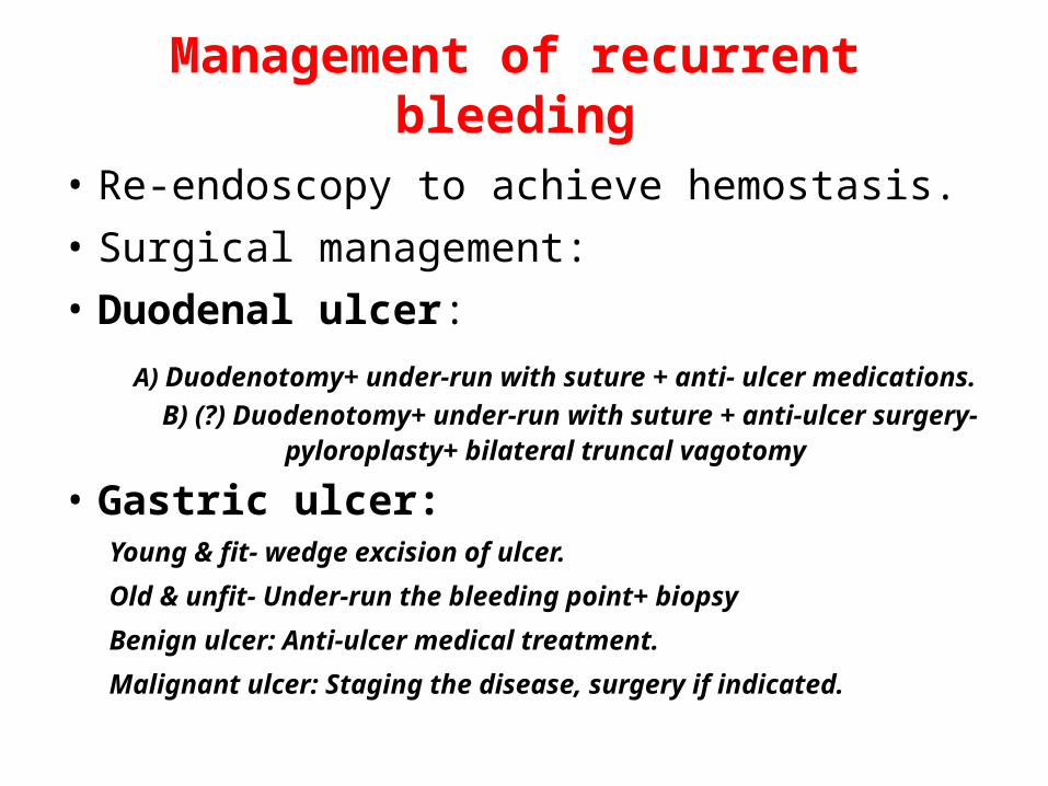
Management of recurrent bleeding
• Re-endoscopy to achieve hemostasis.• Surgical management: • Duodenal ulcer:
A) Duodenotomy+ under-run with suture + anti- ulcer medications. B) (?) Duodenotomy+ under-run with suture + anti-ulcer surgery- pyloroplasty+ bilateral truncal vagotomy
• Gastric ulcer:Young & fit- wedge excision of ulcer.
Old & unfit- Under-run the bleeding point+ biopsy
Benign ulcer: Anti-ulcer medical treatment.
Malignant ulcer: Staging the disease, surgery if indicated.
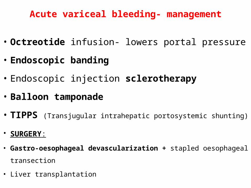
Acute variceal bleeding- management
• Octreotide infusion- lowers portal pressure
• Endoscopic banding
• Endoscopic injection sclerotherapy
• Balloon tamponade
• TIPPS (Transjugular intrahepatic portosystemic shunting)
• SURGERY: • Gastro-oesophageal devascularization + stapled oesophageal transection
• Liver transplantation
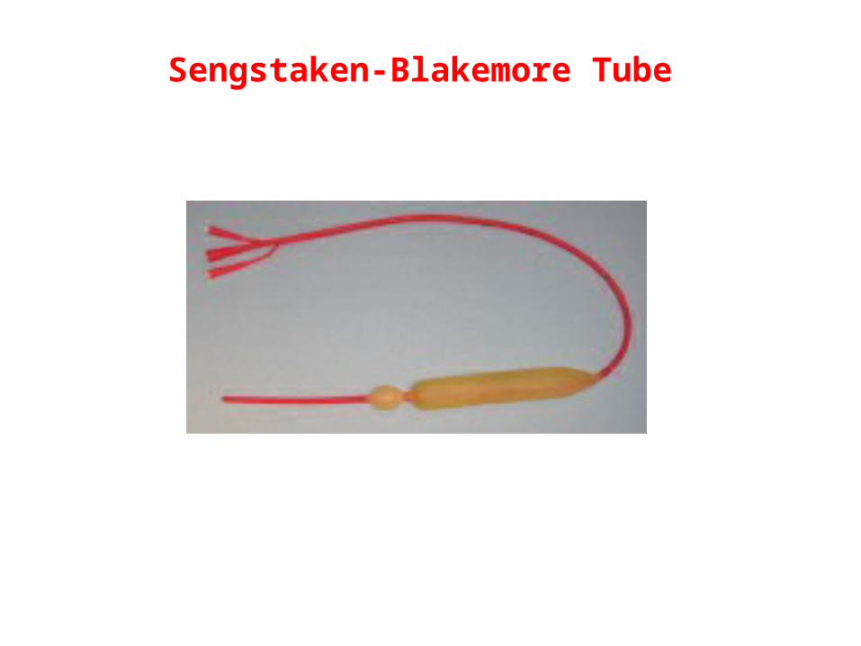
Sengstaken-Blakemore Tube
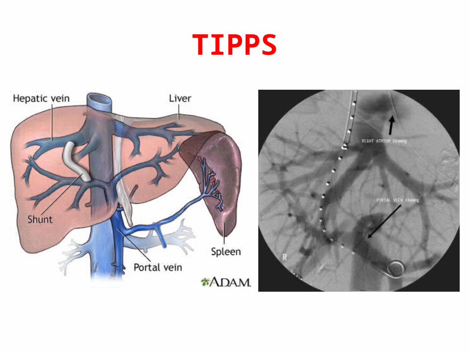
TIPPS
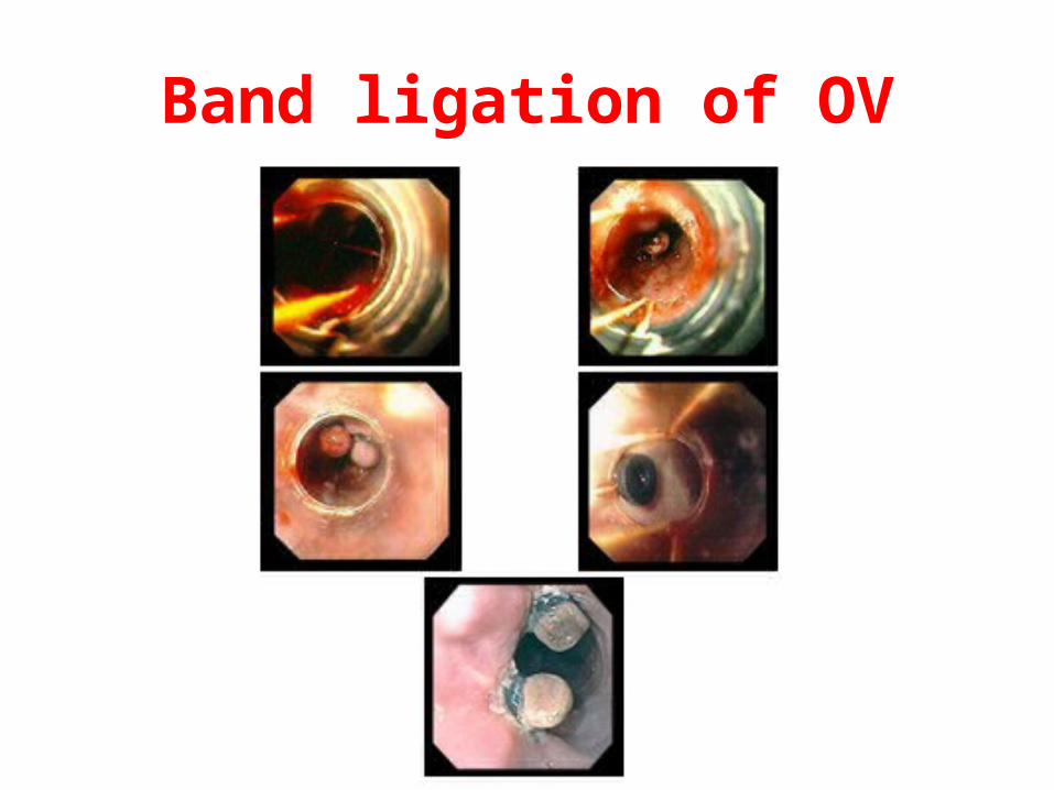
Band ligation of OV
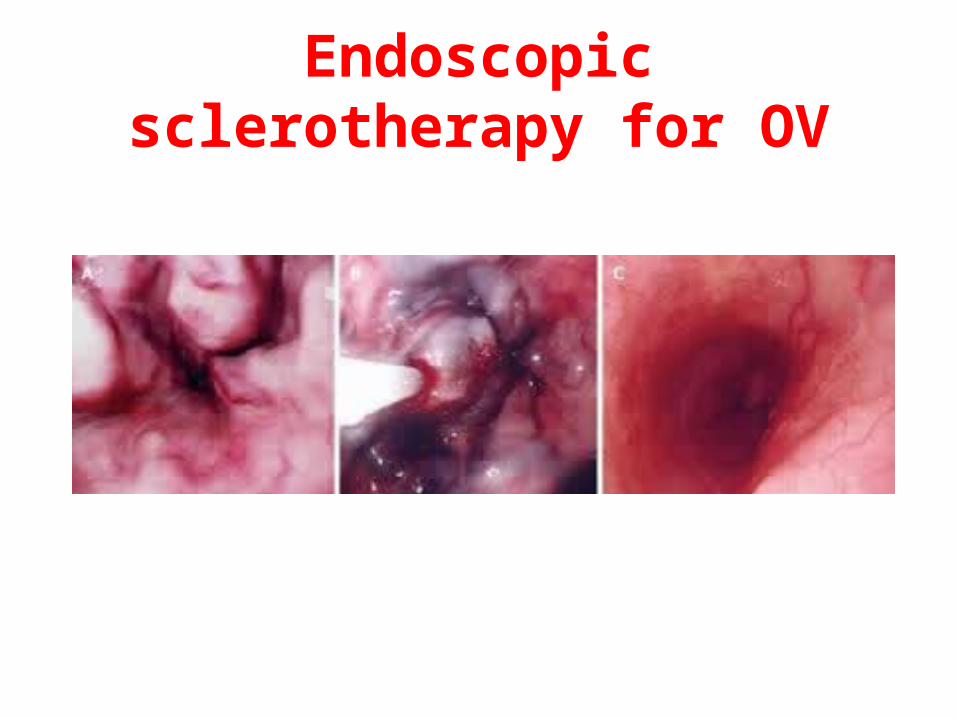
Endoscopic sclerotherapy for OV
Prognosis• Risk factors associated with:
Increased mortality, recurrent bleeding, the need for endoscopic hemostasis, or surgery :
• Age >60 years• Severe comorbidity• Active bleeding (witnessed hematemesis, blood in nasogastric tube,
fresh blood per rectum)
• Hypotension• Blood transfusion ≥ 6 units• Inpatient at time of bleed• Severe coagulopathy
Management of uncommon causes of UGI bleeding
• Conservative/ endoscopic management:
Mallory-Weiss syndrome-
Erosive gastritis /esophagitis.
Dieulafoy lesion.
• Surgical management after stabilization and diagnosis:
Gastric cancer.
Ulcerated gastric stromal tumor (GIST)

Thank you!