Embed Size (px)
Citation preview
Manual
IDE
XX
Ref
eren
ce L
abo
rato
ries
M
anua
l
IDEXX Reference LaboratoriesDivision of IDEXX Laboratories
Mörikestraße 28/3 D-71636 Ludwigsburg
Druckereistraße 4 D-04159 Leipzig
Tel.: 00800 1234 3399 (toll free)
www.idexx.eu
Nordic374-0913
IDEXX Reference Laboratories
Manual IDEXX Reference Laboratories

IDEXX Reference Laboratories5th Edition • April 2013
IDEXX Reference LaboratoryDivisin of IDEXX Laboratory
Mörikestraße 28/3 D-71636 Ludwigsburg
Druckereistraße 4 D-04159 Leipzig
Tel.: 00800 1234 3399 (toll free)Tel.: 07141 6483 0Fax: 07141 6483 555
[email protected] www.idexx.eu

Dear colleague,
We make it our aim always to offer you the best possible service. To that end, we are constantly developing new methods and improving existing ones. In 2011 alone, 58 million euros were invested in in-house research and development. We also have numerous cooperative arrangements with research institutions and universities that allow us access to the latest technologies. The tests for pancreas-specific lipase, Spec cPL®, Spec fPL® and Cardiopet® proBNP, are just some of the examples that are exclusively available to our clients.
A substantial contribution to our success is made by our highly-qualified laboratory staff. Each department is under veterinary supervision. In microbiology, for example, two veterinary specialists, a microbiologist and nine MTAs are on hand to guarantee quick processing. In histopathology, the samples you submit are assessed by 16 veterinary specialists.
Many new tests have been added to the IDEXX test menu. In PCR diagnostics, for example, we can offer a quantitative PCR for a series of parameters. It is also worth taking a look at our ever-expanding range of profiles: general profiles can be customised by adding on a selection of attractively-priced profiles and tests, to help address your patients' symptoms in a targeted way.
This is the first Nordic edition of our Directory of Services, which provides a comprehensive overview of all tests available from us together with important information about the tests and required sample material. Updates to this Directory have been made necessary by the development of new tests and improvements to existing ones: We are delighted to inform you about our free hotline concerning changes to particular tests. This number can also be used to reach our accounts department, courier service and specialist advisers.On behalf of myself and my colleagues, I would like to thank you for your confidence in us and look forward to continuing our excellent cooperation.
Yours faithfully,
Dr. med. vet. Ulrich Brandenburg
(Laboratory Manager)
April 2013

Contents description
1 Index I
2 General Information 1
2.1 General Advice . . . . . . . . . . . . . . . . . . . . . . . . . . . . . . . . . . . . . . . . . . . . . . . . . . . . . 12.2 General Advice on blood sampling and sample preparation . . . . . . . . . . . . . . . . 102.3 General Advice on microbiology tests . . . . . . . . . . . . . . . . . . . . . . . . . . . . . . . . . . 162.4 General Advice on molecular biology tests . . . . . . . . . . . . . . . . . . . . . . . . . . . . . 182.5 General Advice on histopathology and cytology tests . . . . . . . . . . . . . . . . . . . . . 212.6 General Advice on parasitology tests . . . . . . . . . . . . . . . . . . . . . . . . . . . . . . . . . . 232.7 Quality management . . . . . . . . . . . . . . . . . . . . . . . . . . . . . . . . . . . . . . . . . . . . . . . 242.8 Abbreviations/Legend . . . . . . . . . . . . . . . . . . . . . . . . . . . . . . . . . . . . . . . . . . . . . . 252.9 Conversion table . . . . . . . . . . . . . . . . . . . . . . . . . . . . . . . . . . . . . . . . . . . . . . . . . . 27
3 Screening profile 29
3.1 General profile cats and dogs . . . . . . . . . . . . . . . . . . . . . . . . . . . . . . . . . . . . . . . 293.2 Add-on tests (dogs and cats) at a reduced price . . . . . . . . . . . . . . . . . . . . . . . . 313.3 Profile dogs, cats (in alphabetical order) . . . . . . . . . . . . . . . . . . . . . . . . . . . . . . . . 343.4 Profiles for horses (in alphabetical order). . . . . . . . . . . . . . . . . . . . . . . . . . . . . . . . 393.5 Profiles bovine (in alphabetical order) . . . . . . . . . . . . . . . . . . . . . . . . . . . . . . . . . 423.6 Profile porcine . . . . . . . . . . . . . . . . . . . . . . . . . . . . . . . . . . . . . . . . . . . . . . . . . . . . 443.7 Profile camelid . . . . . . . . . . . . . . . . . . . . . . . . . . . . . . . . . . . . . . . . . . . . . . . . . . . . 453.8 Profile rabbit/rodent/reptile (in alphabetical order) . . . . . . . . . . . . . . . . . . . . . . . . 463.9 Multi-species Profiles (in alphabetical order). . . . . . . . . . . . . . . . . . . . . . . . . . . . . 48
4 Hematology 51
4.1 Hematology . . . . . . . . . . . . . . . . . . . . . . . . . . . . . . . . . . . . . . . . . . . . . . . . . . . . . . 514.2 Coagulation parameters . . . . . . . . . . . . . . . . . . . . . . . . . . . . . . . . . . . . . . . . . . . . 534.3 Blood groups . . . . . . . . . . . . . . . . . . . . . . . . . . . . . . . . . . . . . . . . . . . . . . . . . . . . . 564.4 Blood parasites and haemotropic bacteria . . . . . . . . . . . . . . . . . . . . . . . . . . . . . . 57
5 Biochemistry 58
Contents description
6 Toxicology and Drug detection 95
6.1 Medication . . . . . . . . . . . . . . . . . . . . . . . . . . . . . . . . . . . . . . . . . . . . . . . . . . . . . . . 956.2 Toxicology . . . . . . . . . . . . . . . . . . . . . . . . . . . . . . . . . . . . . . . . . . . . . . . . . . . . . . . 966.3 Medical substance detection . . . . . . . . . . . . . . . . . . . . . . . . . . . . . . . . . . . . . . . . 97
7 Gastrointestinal tract diseases, liver, pancreas 99
7.1 Gastrointestinal Diseases . . . . . . . . . . . . . . . . . . . . . . . . . . . . . . . . . . . . . . . . . . . 997.2 Diseases of the Liver . . . . . . . . . . . . . . . . . . . . . . . . . . . . . . . . . . . . . . . . . . . . . . 1037.3 Diseases of exocrine pancreas . . . . . . . . . . . . . . . . . . . . . . . . . . . . . . . . . . . . . . 104
8 Kidneys and urinary tract organs 106
8.1 Blood tests . . . . . . . . . . . . . . . . . . . . . . . . . . . . . . . . . . . . . . . . . . . . . . . . . . . . . 1068.2 Urine analysis . . . . . . . . . . . . . . . . . . . . . . . . . . . . . . . . . . . . . . . . . . . . . . . . . . . 107
9 Muscles, bones, joint 110
9.1 Infectious muscle diseases . . . . . . . . . . . . . . . . . . . . . . . . . . . . . . . . . . . . . . . . 1109.2 Non-infectious muscle diseases . . . . . . . . . . . . . . . . . . . . . . . . . . . . . . . . . . . . 1119.3 Non-infectious bone diseases . . . . . . . . . . . . . . . . . . . . . . . . . . . . . . . . . . . . . . 1119.4 Infectious joint diseases . . . . . . . . . . . . . . . . . . . . . . . . . . . . . . . . . . . . . . . . . . . 112 9.5 Non-infectious joint diseases . . . . . . . . . . . . . . . . . . . . . . . . . . . . . . . . . . . . . . . 113
10 CNS 114
10.1 Infectious CNS diseases . . . . . . . . . . . . . . . . . . . . . . . . . . . . . . . . . . . . . . . . . . 11410.2 Non-infectious CNS diseases . . . . . . . . . . . . . . . . . . . . . . . . . . . . . . . . . . . . . . 119
11 Skin diseases 120
11.1 Allergic/Infectious skin diseases . . . . . . . . . . . . . . . . . . . . . . . . . . . . . . . . . . . . 12011.2 Non-infectious skin diseases . . . . . . . . . . . . . . . . . . . . . . . . . . . . . . . . . . . . . . 122
12 Endocrinology 123
12.1 Hormonal disturbances/diseases of adrenal glands . . . . . . . . . . . . . . . . . . . . 12312.2 Hormonal disturbances/diseases of thyroid gland . . . . . . . . . . . . . . . . . . . . . . 13812.3 Sex hormones/Pregnancy . . . . . . . . . . . . . . . . . . . . . . . . . . . . . . . . . . . . . . . . 14812.4 Special hormones . . . . . . . . . . . . . . . . . . . . . . . . . . . . . . . . . . . . . . . . . . . . . . . 155
Contents description
13 Infectious diseases 156
14 Immunology and Allergy 232
14.1 Autoimmune diseases . . . . . . . . . . . . . . . . . . . . . . . . . . . . . . . . . . . . . . . . . . . . 23214.2 Allergy Diagnostics . . . . . . . . . . . . . . . . . . . . . . . . . . . . . . . . . . . . . . . . . . . . . . 236
15 Molecular Biology tests 241
15.1 General Information on PCR . . . . . . . . . . . . . . . . . . . . . . . . . . . . . . . . . . . . . . . 24115.2 Pathogen detection with PCR (in alphabetical order). . . . . . . . . . . . . . . . . . . . 24315.3 Hereditary diseases . . . . . . . . . . . . . . . . . . . . . . . . . . . . . . . . . . . . . . . . . . . . . 27015.4 Avian Sex Identification . . . . . . . . . . . . . . . . . . . . . . . . . . . . . . . . . . . . . . . . . . . 29615.5.Parentage verification/ genetic fingerprint . . . . . . . . . . . . . . . . . . . . . . . . . . . . . 297
16 Microbiology 300
16.1 Bacteriology . . . . . . . . . . . . . . . . . . . . . . . . . . . . . . . . . . . . . . . . . . . . . . . . . . . . 300 16.1.1 Testing Times and Charges. . . . . . . . . . . . . . . . . . . . . . . . . . . . . . . . . . . . . . 301 16.1.2 General bacteriology tests . . . . . . . . . . . . . . . . . . . . . . . . . . . . . . . . . . . . . . 30216.2 Faecal tests . . . . . . . . . . . . . . . . . . . . . . . . . . . . . . . . . . . . . . . . . . . . . . . . . . . . 30416.3 Mycology tests . . . . . . . . . . . . . . . . . . . . . . . . . . . . . . . . . . . . . . . . . . . . . . . . . 308 16.3.1 Testing Times and Charges . . . . . . . . . . . . . . . . . . . . . . . . . . . . . . . . . . . . . 309 16.3.2 General mycology tests . . . . . . . . . . . . . . . . . . . . . . . . . . . . . . . . . . . . . . . . 310
17 Parasitology 313
17.1 Endoparasites . . . . . . . . . . . . . . . . . . . . . . . . . . . . . . . . . . . . . . . . . . . . . . . . . . 31317.2 Ectoparasites . . . . . . . . . . . . . . . . . . . . . . . . . . . . . . . . . . . . . . . . . . . . . . . . . . . 316
18 Histopathology 317
18.1 Histopathology and Cytology . . . . . . . . . . . . . . . . . . . . . . . . . . . . . . . . . . . . . . 31718.2 Biological Fluids . . . . . . . . . . . . . . . . . . . . . . . . . . . . . . . . . . . . . . . . . . . . . . . . . 318

I
Index
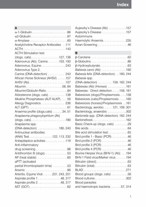
A a-1-Globulin . . . . . . . . . . . . . . . . . . . . .86 a2-Globulin . . . . . . . . . . . . . . . . . . . . . .87a-Amylase . . . . . . . . . . . . . . . . . . . . . . .60 Acetylcholine Receptor Antibodies . .111ACTH. . . . . . . . . . . . . . . . . . . . . . . . . .143 ACTH Stimulation test (dogs, cats). . . . . . . . . . . . . . . . .127, 138Adenovirus (Ab), Canine. . . . . . .103, 193Adenovirus, Equine. . . . . . . . . . . . .. . 243 Adenovirus Type 2, Canine (DNA-detection) . . . . . . . . . . 243 African Horse Sickness (AHSV) . . . . .157AHSV (Ab) . . . . . . . . . . . . . . . . . . . . . .157Albumin . . . . . . . . . . . . . . . . . . . . . .58, 84 Albumin/Globulin-Ratio . . . . . . . . . . . . .84 Aldosterone (dogs, cats) . . . . . . . . . .138 Alkaline Phosphatase (ALP, ALKP) . . . 59 Allergy Diagnostics . . . . . . . . . . . . . . 236ALT (GPT) . . . . . . . . . . . . . . . . . . . . . . 61 Anaemia profile (dogs,cats) . . . . . .34, 51 Anaplasma phagocytophilum (Ak) (dogs, cats) . . . . . . . . . . . . . . . . . . . . .180 Anaplasma spp. (DNA-detection) . . . . . . . . . . . . .180, 243 Antinuclear antibodies (ANA) Test . . . . . . . . . . . . . .122, 113, 232Antiepileptics activities . . . . . . . . . . . .119Anti-inflammatory drug screening . . . . . . . . . . . . . . . . . . .98Antithrombin III (dogs) . . . . . . . . . . . . . .53 AP (heat stable) . . . . . . . . . . . . . . . . . .60 aPTT (activated partial thromboplastin time) . . . . . . . . .53Arsenic . . . . . . . . . . . . . . . . . . . . . . . . .96 Arteritis, Equine Viral . . . . . .231, 243, 251 Aspirate profile 1 . . . . . . . . . . . . . .48, 317 Aspirate profile 2 . . . . . . . . . . . . . .48, 317 AST (GOT) . . . . . . . . . . . . . . . . . . . . . . .62
Aujeszky’s Disease (Ab) . . . . . . . . . . .157Aujeszky’s Disease . . . . . . . . . . . . . . .157Autoimmune Haemolytic Anaemia . . . . . . . . . . . . . .235Avian-Screening . . . . . . . . . . . . . . . . . .46
Bb-Carotene . . . . . . . . . . . . . . . . . . . . . .62 b-Globulins . . . . . . . . . . . . . . . . . . . . . .86 b-Hydroxybutyrate . . . . . . . . . . . . . . . . .63 Babesia canis (Ab) . . . . . . . . . . . . . . .160 Babesia felis (DNA-detection) . . .160, 244Babesia spp. (DNA-detection) . . . . . . . . .159, 162, 244Babesias (Ab) (Horses) . . . . . . . . . . .161 Babesias - Direct detection . . . . .159, 161Babesiosis (dogs)/Piroplasmosis . . . .158Babesiosis (cats)/Piroplasmosis . . . . .160Babesiosis (horses)/Piroplasmosis . .161Bacteriology, aerobic . . . . .121, 109, 301Bacteriology, anaerobic . . . . . . . . . . .302 Bartonella spp. (DNA-detection) .162, 244Bartonellosis . . . . . . . . . . . . . . . . . . . .162 Basic Check-up (dogs, cats) . . . . . . . .29 Bile acids . . . . . . . . . . . . . . . . . . . . . . . .64Bile acid stimulation test . . . . . . . . . . . .65Bird profile 1 - Basic (PCR) . . . . . . . . . .46 Bird profile 2 (PCR) . . . . . . . . . . . . . . . .46 Bird profile 3 (PCR) . . . . . . . . . . . . . . . .46 Bird profile 4 (PCR) . . . . . . . . . . . . . . . .46 Bovine Herpes Virus (BHV-1) (Ab) . . .194BHV-1 Field virus/Marker virus . . . . . .194Bilirubin (direct) . . . . . . . . . . . . . . . . . . .63Bilirubin (total) . . . . . . . . . . . . . . . . . . . .63BLAD . . . . . . . . . . . . . . . . . . . . . . . . . .271 Blood groups (dogs, cats) . . . . . . . . . .56 Blood cultures . . . . . . . . . . . . . . . . . . .302 Blood parasites and haemotropic bacteria . . . . . . .57, 314
II
Index
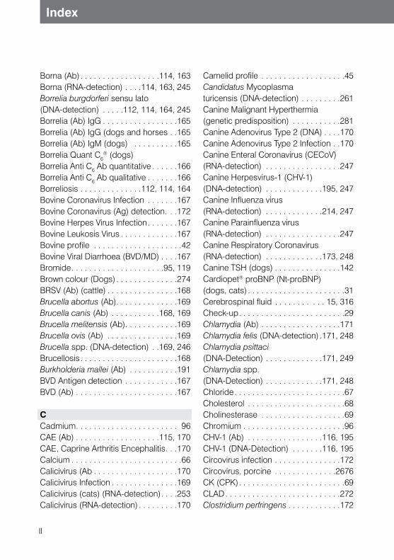
Borna (Ab) . . . . . . . . . . . . . . . . . .114, 163Borna (RNA-detection) . . . .114, 163, 245Borrelia burgdorferi sensu lato (DNA-detection) . . . . .112, 114, 164, 245Borrelia (Ab) IgG . . . . . . . . . . . . . . . . .165Borrelia (Ab) IgG (dogs and horses . .165Borrelia (Ab) IgM (dogs) . . . . . . . . . .165Borrelia Quant C6
® (dogs) Borrelia Anti C6 Ab quantitative . . . . . .166Borrelia Anti C6 Ab qualitative . . . . . . .166Borreliosis . . . . . . . . . . . . . .112, 114, 164Bovine Coronavirus Infection . . . . . . .167 Bovine Coronavirus (Ag) detection. . .172Bovine Herpes Virus Infection . . . . . . .167 Bovine Leukosis Virus . . . . . . . . . . . . .167 Bovine profile . . . . . . . . . . . . . . . . . . . .42Bovine Viral Diarrhoea (BVD/MD) . . . .167 Bromide . . . . . . . . . . . . . . . . . . . . .95, 119Brown colour (Dogs) . . . . . . . . . . . . . .274BRSV (Ab) (cattle) . . . . . . . . . . . . . . . .168Brucella abortus (Ab) . . . . . . . . . . . . . .169 Brucella canis (Ab) . . . . . . . . . . .168, 169Brucella melitensis (Ab) . . . . . . . . . . . .169 Brucella ovis (Ab) . . . . . . . . . . . . . . . .169 Brucella spp. (DNA-detection) . .169, 246Brucellosis . . . . . . . . . . . . . . . . . . . . . .168 Burkholderia mallei (Ab) . . . . . . . . . . .191BVD Antigen detection . . . . . . . . . . . .167BVD (Ab) . . . . . . . . . . . . . . . . . . . . . . .167
CCadmium . . . . . . . . . . . . . . . . . . . . . . . 96 CAE (Ab) . . . . . . . . . . . . . . . . . . .115, 170CAE, Caprine Arthritis Encephalitis . . .170Calcium . . . . . . . . . . . . . . . . . . . . . . . . .66Calicivirus (Ab . . . . . . . . . . . . . . . . . . .170Calicivirus Infection . . . . . . . . . . . . . . .169Calicivirus (cats) (RNA-detection) . . . .253 Calicivirus (RNA-detection) . . . . . . . . .170
Camelid profile . . . . . . . . . . . . . . . . . . .45Candidatus Mycoplasma turicensis (DNA-detection) . . . . . . . . .261Canine Malignant Hyperthermia (genetic predisposition) . . . . . . . . . . .281 Canine Adenovirus Type 2 (DNA) . . . .170Canine Adenovirus Type 2 Infection . .170Canine Enteral Coronavirus (CECoV) (RNA-detection) . . . . . . . . . . . . . . . . .247Canine Herpesvirus-1 (CHV-1) (DNA-detection) . . . . . . . . . . . . .195, 247Canine Influenza virus (RNA-detection) . . . . . . . . . . . . .214, 247Canine Parainfluenza virus (RNA-detection) . . . . . . . . . . . . . . . . .247Canine Respiratory Coronavirus (RNA-detection) . . . . . . . . . . . . .173, 248 Canine TSH (dogs) . . . . . . . . . . . . . . .142Cardiopet® proBNP (Nt-proBNP) (dogs, cats) . . . . . . . . . . . . . . . . . . . . . .31Cerebrospinal fluid . . . . . . . . . . . 15, 316 Check-up . . . . . . . . . . . . . . . . . . . . . . . .29Chlamydia (Ab) . . . . . . . . . . . . . . . . . .171Chlamydia felis (DNA-detection) .171, 248Chlamydia psittaci (DNA-Detection) . . . . . . . . . . . . .171, 249Chlamydia spp. (DNA-Detection) . . . . . . . . . . . . .171, 248Chloride . . . . . . . . . . . . . . . . . . . . . . . . .67 Cholesterol . . . . . . . . . . . . . . . . . . . . . .68Cholinesterase . . . . . . . . . . . . . . . . . . .69Chromium . . . . . . . . . . . . . . . . . . . . . . .96 CHV-1 (Ab) . . . . . . . . . . . . . . . . .116, 195CHV-1 (DNA-Detection) . . . . . . .116, 195Circovirus infection . . . . . . . . . . . . . . .172Circovirus, porcine . . . . . . . . . . . . . .2676CK (CPK) . . . . . . . . . . . . . . . . . . . . . . . .69CLAD . . . . . . . . . . . . . . . . . . . . . . . . . .272Clostridium perfringens . . . . . . . . . . . .172
III
Index
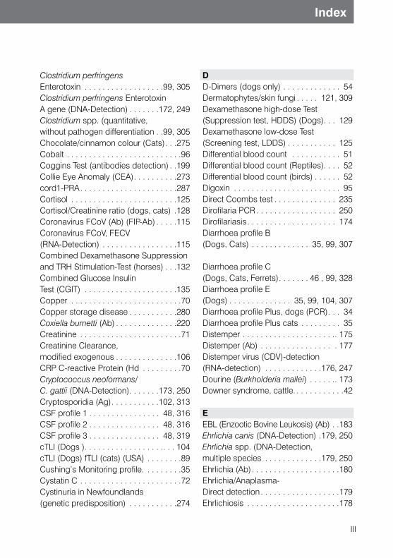
Clostridium perfringens Enterotoxin . . . . . . . . . . . . . . . . . .99, 305 Clostridium perfringens Enterotoxin A gene (DNA-Detection) . . . . . . .172, 249Clostridium spp. (quantitative, without pathogen differentiation . .99, 305Chocolate/cinnamon colour (Cats) . . .275Cobalt . . . . . . . . . . . . . . . . . . . . . . . . . .96Coggins Test (antibodies detection) . .199 Collie Eye Anomaly (CEA) . . . . . . . . . .273cord1-PRA . . . . . . . . . . . . . . . . . . . . . .287Cortisol . . . . . . . . . . . . . . . . . . . . . . . .125 Cortisol/Creatinine ratio (dogs, cats) .128 Coronavirus FCoV (Ab) (FIP-Ab) . . . . .115Coronavirus FCoV, FECV (RNA-Detection) . . . . . . . . . . . . . . . . .115 Combined Dexamethasone Suppression and TRH Stimulation-Test (horses) . . .132 Combined Glucose Insulin Test (CGIT) . . . . . . . . . . . . . . . . . . . . .135 Copper . . . . . . . . . . . . . . . . . . . . . . . . .70 Copper storage disease . . . . . . . . . . .280Coxiella burnetti (Ab) . . . . . . . . . . . . . .220 Creatinine . . . . . . . . . . . . . . . . . . . . . . .71 Creatinine Clearance, modified exogenous . . . . . . . . . . . . . .106 CRP C-reactive Protein (Hd . . . . . . . . .70Cryptococcus neoformans/ C. gattii (DNA-Detection). . . . . . .173, 250Cryptosporidia (Ag) . . . . . . . . . . .102, 313CSF profile 1 . . . . . . . . . . . . . . . . 48, 316 CSF profile 2 . . . . . . . . . . . . . . . . 48, 316 CSF profile 3 . . . . . . . . . . . . . . . . 48, 319cTLI (Dogs ) . . . . . . . . . . . . . . . . . .. . . 104cTLI (Dogs) fTLI (cats) (USA) . . . . . . . .89 Cushing’s Monitoring profile. . . . . . . . .35 Cystatin C . . . . . . . . . . . . . . . . . . . . . . .72 Cystinuria in Newfoundlands (genetic predisposition) . . . . . . . . . . .274
D D-Dimers (dogs only) . . . . . . . . . . . . . 54 Dermatophytes/skin fungi . . . . . 121, 309 Dexamethasone high-dose Test (Suppression test, HDDS) (Dogs) . . . 129 Dexamethasone low-dose Test (Screening test, LDDS) . . . . . . . . . . . 125Differential blood count . . . . . . . . . . . 51Differential blood count (Reptiles) . . . . 52Differential blood count (birds) . . . . . . 52 Digoxin . . . . . . . . . . . . . . . . . . . . . . . . 95 Direct Coombs test . . . . . . . . . . . . . . 235 Dirofilaria PCR . . . . . . . . . . . . . . . . . . 250 Dirofilariasis . . . . . . . . . . . . . . . . . . . . 174 Diarrhoea profile B (Dogs, Cats) . . . . . . . . . . . . . 35, 99, 307
Diarrhoea profile C (Dogs, Cats, Ferrets) . . . . . . . 46 , 99, 328 Diarrhoea profile E (Dogs) . . . . . . . . . . . . . . 35, 99, 104, 307 Diarrhoea profile Plus, dogs (PCR) . . . 34 Diarrhoea profile Plus cats . . . . . . . . . 35Distemper . . . . . . . . . . . . . . . . . . . . .. 175 Distemper (Ab) . . . . . . . . . . . . . . . . . 177 Distemper virus (CDV)-detection (RNA-detection) . . . . . . . . . . . . .176, 247 Dourine (Burkholderia mallei) . . . . . .. 173Downer syndrome, cattle. . . . . . . . . . . .42
EEBL (Enzootic Bovine Leukosis) (Ab) . .183 Ehrlichia canis (DNA-Detection) .179, 250Ehrlichia spp. (DNA-Detection, multiple species . . . . . . . . . . . . .179, 250 Ehrlichia (Ab) . . . . . . . . . . . . . . . . . . . .180 Ehrlichia/Anaplasma- Direct detection . . . . . . . . . . . . . . . . . .179 Ehrlichiosis . . . . . . . . . . . . . . . . . . . . .178
IV
Index
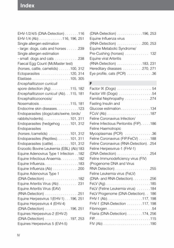
EHV-1/2/4/5 (DNA-Detection) . . . . . . .116 EHV-1/4 (Ab) . . . . . . . . . . . .116, 196, 251 Single allergen estimation - large: dogs, cats and horses . . . . . .239 Single allergen estimation - small: dogs and cats . . . . . . . . . . . .238 Faecal Egg Count (McMaster test) (horses, cattle, camelids) . . . . . .100, 312 Ectoparasites . . . . . . . . . . . . . . .120, 314 Elastase . . . . . . . . . . . . . . . . . . . .105, 305 Encephalitozoon cuniculi spore detection (Ag) . . . . . . . . . .115, 182 Encephalitozoon cuniculi (Ab) . . .115, 181 Encephalitozoonosis/ Nosematosis . . . . . . . . . . . . . . . .115, 181 Endocrine skin diseases . . . . . . . . . . .123Endoparasites (dogs/cats/swine, birds/ rabbits/rodents) . . . . . . . . . . . . . .101, 311 Endoparasites (hedgehog . . . . .101, 312Endoparasites (horses /camelids) . . . . . . . . . . .101, 312 Endoparasites (Reptiles) . . . . . . .101, 311 Endoparasites (cattle) . . . . . . . . .101, 312 Enzootic Bovine Leukemia (EBL) (Ab) 183Equine Adenovirus Type 1 Infection . .182 Equine Infectious Anaemia. . . . . . . . .182 Equine Influenza. . . . . . . . . . . . . . . . . .183 Equine Influenza (Ab) . . . . . . . . . . . . .200Equine Adenovirus Type 1 (DNA-Detection) . . . . . . . . . . . . . . . . .182 Equine Arteritis Virus (Ab) . . . . . . . . . 231 Equine Arteritis Virus (EAV) (RNA-Detection) . . . . . . . . . . . . . . . . .251 Equine Herpesvirus 1(EHV-1) . . .196, 251 Equine Herpesvirus 4 (EHV-4) (DNA-Detection) . . . . . . . . . . . . .196, 251 Equines Herpesvirus-2 (EHV-2) (DNA-Detection) . . . . . . . . . . . . .197, 253 Equines Herpesvirus 5 (EVH-5)
(DNA-Detection) . . . . . . . . . . . . .196, 253Equine Influenza virus (RNA-Detection) . . . . . . . . . . . . .200, 253 Equine Metabolic Syndrome/ Pre-Cushing (horses) . . . . . . . . . . . . 132 Equine viral Arteritis (RNA-Detection) . . . . . . . . . . . . .183, 231 Hereditary diseases . . . . . . . . . .270, 271Eye profile, cats (PCR) . . . . . . . . . . . . .36 FFactor IX (Dogs) . . . . . . . . . . . . . . . . .. 54 Factor VIII (Dogs) . . . . . . . . . . . . . . . . .54 Familial Nephropathy . . . . . . . . . . . . .274 Fasting Insulin and Glucose estimation . . . . . . . . . . . . . . .134 FCoV (Ab) . . . . . . . . . . . . . . . . . . . . . .187 Feline Coronavirus Infection/ Feline Infectious Peritonitis (FIP) . . . . .186 Feline Haemotropic Mycoplasmas (PCR) . . . . . . . . . . . . . .36 Feline Coronavirus (FIP/FeCV) . . . . . .188 Feline Coronavirus (RNA-Detection) . .254 Feline Herpesvirus-1 (FHV-1) (DNA-Detection) . . . . . . . . . . . . . . . . .254 Feline Immunodeficiency virus (FIV) (Progenome DNA and Virus RNA Detection) . . . . . . . . . . . . . . . . . .255 Feline Leukemia virus (FeLV) (DNA- and RNA-Detection) . . . . . . . . .256 FeLV (Ag) . . . . . . . . . . . . . . . . . . . . . . .185 FeLV (Feline Leukemia virus) . . . . . . .184FeLV Progenome (DNA-Detection) . . 186 FHV-1 (Ab) . . . . . . . . . . . . . . . . . .117, 198 FHV-1 (DNA-Detection . . . . . . . .117, 198 Fibrinogen . . . . . . . . . . . . . . . . . . . . . . .54 Filaria (DNA-Detection) . . . . . . . .174, 256 FIP. . . . . . . . . . . . . . . . . . . . . . . . . . . . .115 FIV (Ab) . . . . . . . . . . . . . . . . . . . . . . . .190
V
Index
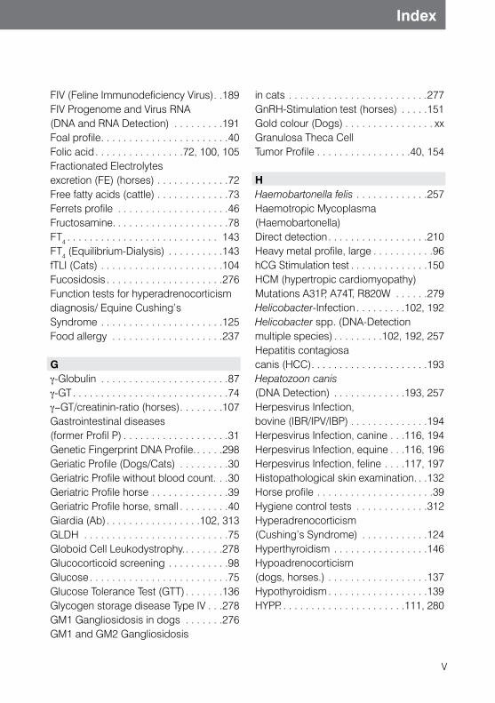
FIV (Feline Immunodeficiency Virus) . .189 FIV Progenome and Virus RNA (DNA and RNA Detection) . . . . . . . . .191 Foal profile . . . . . . . . . . . . . . . . . . . . . . .40 Folic acid . . . . . . . . . . . . . . . .72, 100, 105 Fractionated Electrolytes excretion (FE) (horses) . . . . . . . . . . . . .72 Free fatty acids (cattle) . . . . . . . . . . . . .73 Ferrets profile . . . . . . . . . . . . . . . . . . . .46 Fructosamine. . . . . . . . . . . . . . . . . . . . .78 FT4 . . . . . . . . . . . . . . . . . . . . . . . . . . . 143 FT4 (Equilibrium-Dialysis) . . . . . . . . . .143 fTLI (Cats) . . . . . . . . . . . . . . . . . . . . . .104 Fucosidosis . . . . . . . . . . . . . . . . . . . . .276 Function tests for hyperadrenocorticism diagnosis/ Equine Cushing’s Syndrome . . . . . . . . . . . . . . . . . . . . . .125 Food allergy . . . . . . . . . . . . . . . . . . . .237 Gγ-Globulin . . . . . . . . . . . . . . . . . . . . . . .87 γ-GT . . . . . . . . . . . . . . . . . . . . . . . . . . . .74 γ-GT/creatinin-ratio (horses) . . . . . . . .107 Gastrointestinal diseases (former Profil P) . . . . . . . . . . . . . . . . . . .31Genetic Fingerprint DNA Profile. . . . . .298 Geriatic Profile (Dogs/Cats) . . . . . . . . .30 Geriatric Profile without blood count. . .30 Geriatric Profile horse . . . . . . . . . . . . . .39 Geriatric Profile horse, small . . . . . . . . .40 Giardia (Ab) . . . . . . . . . . . . . . . . .102, 313GLDH . . . . . . . . . . . . . . . . . . . . . . . . . .75 Globoid Cell Leukodystrophy. . . . . . . .278Glucocorticoid screening . . . . . . . . . . .98 Glucose . . . . . . . . . . . . . . . . . . . . . . . . .75 Glucose Tolerance Test (GTT) . . . . . . .136 Glycogen storage disease Type IV . . .278 GM1 Gangliosidosis in dogs . . . . . . .276 GM1 and GM2 Gangliosidosis
in cats . . . . . . . . . . . . . . . . . . . . . . . . .277 GnRH-Stimulation test (horses) . . . . .151 Gold colour (Dogs) . . . . . . . . . . . . . . . . xx Granulosa Theca Cell Tumor Profile . . . . . . . . . . . . . . . . .40, 154
HHaemobartonella felis . . . . . . . . . . . . .257 Haemotropic Mycoplasma (Haemobartonella) Direct detection . . . . . . . . . . . . . . . . . .210 Heavy metal profile, large . . . . . . . . . . .96hCG Stimulation test . . . . . . . . . . . . . .150 HCM (hypertropic cardiomyopathy) Mutations A31P, A74T, R820W . . . . . .279 Helicobacter-Infection . . . . . . . . .102, 192 Helicobacter spp. (DNA-Detection multiple species) . . . . . . . . .102, 192, 257 Hepatitis contagiosa canis (HCC) . . . . . . . . . . . . . . . . . . . . .193 Hepatozoon canis (DNA Detection) . . . . . . . . . . . . .193, 257 Herpesvirus Infection, bovine (IBR/IPV/IBP) . . . . . . . . . . . . . .194 Herpesvirus Infection, canine . . .116, 194 Herpesvirus Infection, equine . . .116, 196 Herpesvirus Infection, feline . . . .117, 197 Histopathological skin examination. . .132 Horse profile . . . . . . . . . . . . . . . . . . . . .39 Hygiene control tests . . . . . . . . . . . . .312 Hyperadrenocorticism (Cushing’s Syndrome) . . . . . . . . . . . .124 Hyperthyroidism . . . . . . . . . . . . . . . . .146 Hypoadrenocorticism (dogs, horses.) . . . . . . . . . . . . . . . . . .137 Hypothyroidism . . . . . . . . . . . . . . . . . .139 HYPP. . . . . . . . . . . . . . . . . . . . . . .111, 280
VI
Index
IIBR/IPV . . . . . . . . . . . . . . . . . . . . . . . .198 Identification of ectoparasites . . . . . . .314 IGF I (Insulin-Like Growth Factor) . . . .155 Immunoglobulin status/IgG (foals) . . . .76 Immunotherapy Solution (dogs, cats, horses) . . . . . . . . . . . . . .240Infectious Anaemia, equine . . . . . . . . .199 Influenza, equine . . . . . . . . . . . . . . . . .200 Influenza virus Infection . . . . . . . . . . . .200 Insects - Allergy screening for horses . . . . . . . . . . . . . . . . . . . . . .240 Insulin . . . . . . . . . . . . . . . . . . . . . . . . .155 Intestinal pathogens . . . . . . . . . 300, 304Interpretation of T4- and cTSH-results . . . . . . . . . . . . .142 Iron . . . . . . . . . . . . . . . . . . . . . . . . . . . .77
KK-value (FT4/Cholesterol) (dogs) . . . .143Kidney profile . . . . . . . . . . . . . . . .48, 106
LL-2-HGA (L-2-Hydroxyglutaracidurie) 281 Lactate . . . . . . . . . . . . . . . . . . . . . 77, 111 Large Blood count . . . . . . . . . . . . . . .. 51 Large Blood count (Reptiles) . . . . . . .. 52 Large Blood count (birds) . . . . . . . . . .. 52 Large Bovine profile . . . . . . . . . . . . . . .43 Large Check-up . . . . . . . . . . . . . . . . . .29 Large Copper profile for cattle . . . . . . .43 Large Equine profile . . . . . . . . . . . . . . .39 Large Feline profile . . . . . . . . . . . . . . . .30 Large Porcine profile . . . . . . . . . . . . . . .44 Large Coagulation Profile (Dogs) . . . . .54 Large Reptile profile . . . . . . . . . . . . . . .47Lawsonia intracellularis (DNA-detection) . . . . . . . . . . . . 201, 258 Lawsonia intracellularis
(Equine Proliferative Enteropathy) . . . 201 Lead . . . . . . . . . . . . . . . . . . . . . . . .27, 96 LDH . . . . . . . . . . . . . . . . . . . . . . . . . . . 78Liver profile 1 . . . . . . . . . . . . . . . . 36, 103 Liver profile 2 (dogs, cats) . . . . . 36, 103 Leishmania spp. (DNA-detection, quantitative) . . . . . . . . . . . . .121, 203, 259 Leishmania (Ab) . . . . . . . . . . . . 121, 204 Leishmania Direct detection . . . . . . . 203 Leishmaniasis . . . . . . . . . . . . . . 121, 202 Leptospira spp. (DNA-detection, many species) . . . . . . 103, 106, 206, 260 Leptospira (Ab) . . . . . . . . . 103, 106, 205 Leptospirosis . . . . . . . . . . . . . . . . . . 204 Leukemia, bovine . . . . . . . . . . . . . . . 207 Leukemia virus Infection, feline . . . . . 207 Lipase . . . . . . . . . . . . . . . . . . . . . . . . . 78 Listeria monocytogenes (DNA-detection) . . . . . . . . . . . . 207, 261 Listerias (Ab) . . . . . . . . . . . . . . . . . . . 207 Listeriosis. . . . . . . . . . . . . . . . . . . . . . 207 Local Anaesthetic Screening. . . . . . . . 98 Lungworms . . . . . . . . . . . . . . . . 101, 313
MMaedi/Visna (Ab) . . . . . . . . . . . . .117, 208 Magnesium . . . . . . . . . . . . . . . . . . . . . .79 Macrofilaria (Ag) (Dirofilaria immitis) . . . . . . . . . . . . .57, 174 Maintenance Solution . . . . . . . . . . . . .240Malignant Hyperthermia, canine . . . .281 Malignant Hyperthermia, porcine . . . .281 Manganese . . . . . . . . . . . . . . . . . . . . . .80Mating time estimation. . . . . . . . . . . . .148Medical substance detection . . . . . . . .97 Megabacteria Direct detection . . . . . .208 Megabacteria Infection . . . . . . . . . . . 208 Microbiology . . . . . . . . . . . . . . . . . . . .121 Microfilarias-Direct detection . . . . . . .174
VII
Index
Molybdenum . . . . . . . . . . . . . . . . . . . . .96 Mucopolysaccharidosis VII . . . . . . . . .282 Muscle profile . . . . . . . . . . . . . . . 49, 111 Myasthenia gravis . . . . . . . . . . . .111, 233 Mycoplasma felis (DNA-detection) . . . . . . . . . . . . .212, 261 Mycoplasma haemocanis, Candidatus Mycoplasma haematoparvum (DNA-detection) . . . . . . . . . . . . .211, 261 Mycoplasma haemofelis, Candidatus Mycoplasma haemominutum, Candidatus Mycoplasma turicensis, Mycoplasma haemocanis und Candidatus Mycoplasma haematoparvum . . . . . .209 Mycoplasma haemofelis, Candidatus Mycoplasma haemominutum (DNA-detection) . . . . . . . . . . . . . . . . .209 Mycoplasma spp. (DNA-detection, multiple species) . . . . . . . . . . . . .212, 262 Myotonia congenita In miniature schnauzers . . . . . . . . . . .283
NNight blindness in Briards . . . . . . . . . .283 Neospora caninum (Ab) . . .110, 117, 214 Neospora Infection . . . . . . .110, 117, 213 Neospora spp. (dogs) .110, 117, 214, 262 Neurology Profile, dogs (PCR) . . . . . . .34 Non-infectious joint diseases . . . . . . .113Nonspecific Parameters for Cushing’s Disease diagnostics . . . . . . . . . . . . . .136 Nickel. . . . . . . . . . . . . . . . . . . . . . . . . . .96 NSAID Screening . . . . . . . . . . . . . . . . .98 Nt-proBNP (Cardiopet® proBNP) (dogs/cats). . . . . . . . . . . . . . . . . . . . . . .31
OObductions . . . . . . . . . . . . . . . . . . . . .315
Occult Blood . . . . . . . . . . . . . . . .100, 306 OLWS . . . . . . . . . . . . . . . . . . . . . . . . .284 Oestradiol (17b-) . . . . . . . . . . . . . . . . .149 Oestrone sulfate (horses, male) . . . .. 151 Oestrone sulfate (horses, female . . . .153 Ovarian tumors in horses . . . . . . . . . .154
PPancreas specific Lipase, canine (Spec cPL®) . . . . . . . . . . . . . . . . .82, 104 Pancreas specific Lipase, feline (Spec fPL®) . . . . . . . . . . . . . . . . .82, 104 Parainfluenza virus (Ab) (cattle) . . . .. 214 Parainfluenza virus Infection . . . . . . . .214 Parasites In faeces . . . . . . . . . . . . . . .311 Paratuberculosis . . . . . . . . . . . . . . . . .215 Paratuberculosis (Ab) (Cattle) . . . . . . .215 Parvovirosis/Panleukopenia . . . .102, 216 Parvovirus (Ag) (dogs, cats) . . . .102, 216 Parvovirus (Ab) (dogs, cats) . . . .102, 218 Parvovirus FPV, CPV (DNA-detection) . . . . . . . . . . . . .217, 264 PBFD-Virus (DNA-detection . . . .219, 265 PCR (Polymerase Chain Reaction) . . .241 Performance profile, horses. . . . . . . . . 41Pregnancy diagnostics, horses . . . . .153 Phenobarbital . . . . . . . . . . . . . . . . . . . .95 Phosphate. . . . . . . . . . . . . . . . . . . . . .. 80 Phosphofructokinase deficiency. . . . 285 PKD (Polycystic Kidney Disease) . . . .286 PMSG/eCG . . . . . . . . . . . . . . . . . . . . .153 Polyomavirus, avian (BFD-Virus) (DNA-detection) . . . . . . . . . . . . .218, 265 Polyuria/Polydipsia Profile (dogs, cats) . . . . . . . . . . . . . . . . . 36, 106 Porcine Circovirus 2 (PCV-2) (DNA-detection) . . . . . . . . . . . . .218, 266 Porcine Influenza virus (Ab) . . . . . . . .219 Porcine Malignant Hyperthermia
VIII
Index
Syndrome (genetic predisposition). .. 281 Potassium . . . . . . . . . . . . . . . . . . . . .. . 79PRA . . . . . . . . . . . . . . . . . . . . . . . . . . .287 prcd-PRA. . . . . . . . . . . . . . . . . . . . . . .288 Profile respiratory diseases, foals (PCR) . . . . . . . . . . . . . . . . . . . . . .40 Profile respiratory diseases, horses (PCR). . . . . . . . . . . . . . . . . . . . .40 Profile EMS/Cushing 1 . . . . . . . . . . . .133 Profile EMS/Cushing 2 . . . . . . . . . . . .133 Profile Feline Haemotropic Mycoplasms (DNA-detection) . . . . . . . . . . . . . .36, 263 Profile S (electrolytes + trace elements) . . . . . . .49 Progesterone . . . . . . . . . . . . . . . . . . . .148 Protein/Creatinine ratio . . . . . . . . . . . .107 PRRS (Ab) (porcine) . . . . . . . . . . . . . .219 PRRS (Porcine Reproductive and Respiratory Syndrome) . . . . . . .. 219 PT (Quick-Test) (Thromboplastin time, Prothrombin time) . . . . . . . . . . . . . . . . .53 PU/PD Profile (Polyuria/Polydipsia) . . .106 Pyruvate kinase deficiency . . . . . . . . .290
QQ-Fever . . . . . . . . . . . . . . . . . . . . . . . .220
RRabies virus (Ab) (NT) . . . . . . . . . . . . .222 Rabies virus antibody detection for travel. . . . . . . . . . . . . . . 222 Rrcd1-PRA . . . . . . . . . . . . . . . . . . . . . 289 rcd2-PRA, PRA in Collies . . . . . . . . . .289 rdAc-PRA . . . . . . . . . . . . . . . . . . . . . . .289 Respiratory Profile, dogs (PCR) . . . . . .34 Respiratory Profile, cats (PCR) . . . . . . .34 Reticulocytes (Dogs, Cats) . . . . . . . . .. 51 Rheumatoid factors . . . . . . . . . . .113, 235 Rheumatoid Polyarthritis . . . . . . .113, 234
Rhodococcus equi (DNA-detection) . . . . . . . . . . . . .222, 267 Rickettsias (Ab) (Dogs) . . . . . . . . . . . .222 Rocky Mountain Rotavirus (Ag) . . . . . . . . . . . . . . . . . . .223 Rotavirus (Ag)-detection. . . . . . . . . . .223 Rotavirus-Infection . . . . . . . . . . .102, 223 SSalmonella abortus equi (Ak) . . . . . . .223 Salmonellas detection . . . . . . . . . .99, 304 Sarcoptes . . . . . . . . . . . . . . . . . . . . . .120 Sarcoptes (Ab) (dogs) . . . .120, 122, 224 SCID in Arabians . . . . . . . . . . . . . . . .291 Scrapie (TGF) (genetic predisposition) . . . . . . . . . . .293 Screening for foreign substances . . . . .97 Screening Test . . . . . . . . . . . . . . . . . . .238 SDS-Page Electrophoresis(Urine protein electrophoresis) . . . . . .108 Sedative/Tranquilizer Screening . . . . . .97 Selenium . . . . . . . . . . . . . . . . . . . .83, 111 Sequence analysis . . . . . . . . . . . . . . .299 Serum electrophoresis (Agarose-Gel) .83 Sex hormones . . . . . . . . . . . . . . . . . . .149 Sex identification in birds . . . . . . . . . .296Spec cPL®, Canine pancrase specific Lipase . . . . . . . . . . . . . . . . . . . . . .82, 104 Spec fPL®, Feline pancrease specific Lipase . . . . . . . . . . . . . . . . . . . . . .82, 104 Stone analysis . . . . . . . . . . . . . . . . . . .109 Stimulant Screening. . . . . . . . . . . . . . . .98 Stomatitis vesicularis (Ab) (horses) . .230 Screening Fertility disorders 1 . .. . . . . . 43 Screening Fertility disorders 2 . . . . . . . .43 Screening Fertility disorders 3 . .. . . . . . 43 Skin profile 1 . . . . . . . . . . . . . . . . . . . . 49 Skin profile 2 . . . . . . . . . . . . . . . . . . . . .49 Skin profile 3 . . . . . . . . . . . . . . . . . . . . .49
IX
Index
Skin profile 4 (Dogs) . . . . . . . . . . . . . . .49 Skin profile 7 (Dogs, Cats) . . . . . . . . . .49Small blood count . . . . . . . . . . . . . . . . .51 Small blood count (Reptiles) . . . . . . . . .52 Small blood count (birds) . . . . . . . . . . .52Small copper profile for cattle . . . . . . . .43Sodium . . . . . . . . . . . . . . . . . . . . . . . . .81Spotted Fever (RMSF) . . . . . .. . . . . . 233 Synovia . . . . . . . . . . . . . . . . .50, 112, 318 Synovia Profile 1 . . . . . . . . . .50, 112, 318 Synovia Profile 2 . . . . . . . . . .50, 112, 318 Synovia Profile 3 . . . . . . . . . .50, 112, 318 Systemic Lupus erythosus (SLE) 113, 232
TT3 . . . . . . . . . . . . . . . . . . . . . . . . . . . .141 T3-Suppression test . . . . . . . . . . . . . . .146 T4 . . . . . . . . . . . . . . . . . . . . . . . . .140, 146 T4-Antibodies (dogs) . . . . . . . . . . . . . .144 Testosterone . . . . . . . . . . . . . . . . . . . .150 Tickborne Encephalitisvirus . . . . . . .116Ticks profile 1 (serology) . . . . . . . . . . . .37 Ticks profile 2 (serology) . . . . . . . . . . . .37 Ticks profile 3 (PCR blood). . . . . . . . . .37 Ticks profile 4 (PCR ticks) . . . . . . . . . . .37 Thallium . . . . . . . . . . . . . . . . . . . . . . . . .96 Thallium (Hair) . . . . . . . . . . . . . . . .96, 122 Thallium (Urine . . . . . . . . . . . . . . .96, 122 Thrombin time . . . . . . . . . . . . . . . . . . . .54 Thyreoglobulin (Anti-thyroid Antibodies) (dogs) . . . . . . . . . . . . . . . . . . . . . . . . .143 Thyroid hormones - Function tests . . . . . . . . . . . . . . .144, 146 Thyroid profile 1 . . . . . . . . . . . . . . .39, 141 Thyroid profile 2 (dogs . . . . . . . . .39, 141 Thyroid hormones - single estimation . . . . . . . . . . . . .139, 146 Total protein . . . . . . . . . . . . . . . . . . . . . .87
Toxoplasma gondii (DNA-detection . . . . . .110, 118, 227, 168 Toxoplasma (Ab) . . . . . . . . . . . . .110, 227 Toxoplasma - Direct detection . . . . . . . . . .110, 118, 227 Toxoplasmosis. . . . . . . . . . .110, 118, 227 Transmissible Gastroenteritis Virus (TGV) (RNA-detection) . . . . . . . .228, 257 Transmissible Gastroenteritis Virus, porcine . . . . . . . . . . . . . . . . . . .228 Travel diseases Profile 1 - early (dogs) . . . . . . . . . . . . . .37 Travel diseases Profile 2 - late (dogs) . . . . . . . . . . . . . . .37 Travel diseases Profile 3 - acute (dogs) . . . . . . . . . . . . .37 Trematode eggs. . . . . . . . . . . . . .102, 313 TRH-Stimulation test . . . . . . . . . . . . . .147 TRH-Stimulation test (Dogs) . . . . . . . 145 TRH-Stimulation test (Horses) . . . . . .145 Trichomonas Direct detection . . . . . . .229 Trichomonas-Infection . . . . . . . . . . . . .229 Triglycerides . . . . . . . . . . . . . . . . . . . . .88 Tritrichomonas foetus (DNA-detection) . . . . . . . . . . . . .229, 269 Tritrichomonas Infection . . . . . . . . . . .228 Tricyclic Antidepressives Screening . . .98 Troponin I . . . . . . . . . . . . . . . . . . . . . . . .89 Trypanosoma equiperdum-Ab . . . . . . .230 Trypanosoma-Infections . . . . . . . . . . .229 Trypanosomes Direct detection . . . . .229 TSH-Stimulation test (Dogs) with rhTSH (human recombinant TSH . . . .144 T-cell Carcinoma Screening (TCC) (dogs) . . . . . . . . . . . . . . . . . . . .109
UUric acid . . . . . . . . . . . . . . . . . . . . . . . .91Urine sediment . . . . . . . . . . . . . . . . . .107
X
Index
Urine analysis . . . . . . . . . . . . . . . . . .. 107Urea (BUN) . . . . . . . . . . . . . . . . . . . . . .90
VVaginal cytology (dogs, cats) . . . . . . .152 Virology faecal examination . . . .100, 306 Virus arteritis, equine (EVA) . . . . . . . . 231 Viral Diarrhea, bovine . . . . . . . . . . . . .231 Vitamin A . . . . . . . . . . . . . . . . . . . . . . . .99 Vitamin B1 (Thiamin) . . . . . . . . . . . . . . 99 Vitamin B2 (Riboflavine) . . . . . . . . . . .100 Vitamin B6 (Pyridoxine) . . . . . . . . . . . .100 Vitamin B12 (Cobalamine) . .93, 100, 105 Vitamin D3 (1,25-di-OH) Vitamin D3 (25-OH) . . . . . . . . . . . .93, 111 Vitamin E (Tocopherol) . . . . . . . . .94, 111 Vitamin H (Biotin). . . . . . . . . . . . . .94, 122
Von Willebrand disease (vWF) . . . . . .294 Von Willebrand Factor 1 - 3. . . . . .55, 294 Von Willebrand Factor Antigen (vWF: Ag) (dogs) . . . . . . . . . . 55
XX-SCID . . . . . . . . . . . . . . . . . . . . . . . . 295
YYeasts in faecal sample (quantitative) . . . . . . . . . . . . . . . . . . . .310 Yeasts and moulds . . . . . . . . . . .121, 310
ZZinc . . . . . . . . . . . . . . . . . . . . . . . . . . . .94 Zinc (Serum, hair) . . . . . . . . . . . . . . . .122
1
2.1 General information
2 General Information
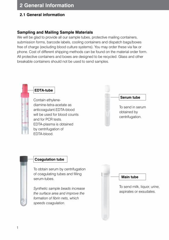
Sampling and Mailing Sample MaterialsWe will be glad to provide all our sample tubes, protective mailing containers, submission forms, barcode labels, cooling containers and dispatch bags/boxes free of charge (excluding blood culture systems). You may order these via fax or phone. Cost of different shipping methods can be found on the material order form. All protective containers and boxes are designed to be recycled. Glass and other breakable containers should not be used to send samples.
EDTA-tube
Contain ethylene-diamine-tetra-acetate as anticoagulant.EDTA-blood will be used for blood counts and for PCR tests. EDTA-plasma is obtained by centrifugation ofEDTA-blood.
Coagulation tube
To obtain serum by centrifugation of coagulating tubes and filling serum-tubes.
Synthetic sample beads increase the surface area and improve the formation of fibrin nets, which speeds coagulation.
Main tube
To send milk, liquor, urine, aspirates or exsudates.
Serum tube
To send in serum obtained by centrifugation.
2
2 General Information
2.1 General information
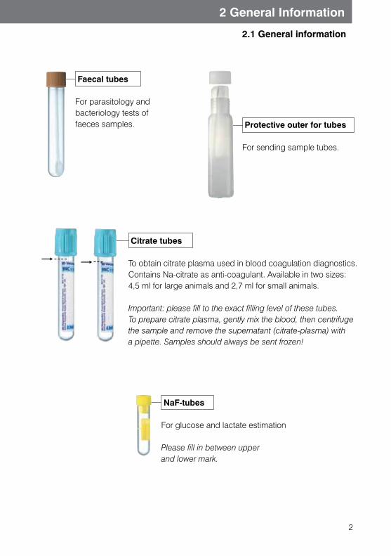
NaF-tubes
For glucose and lactate estimation
Please fill in between upper and lower mark.
Faecal tubes
For parasitology and bacteriology tests of faeces samples.
Citrate tubes
To obtain citrate plasma used in blood coagulation diagnostics. Contains Na-citrate as anti-coagulant. Available in two sizes: 4,5 ml for large animals and 2,7 ml for small animals. Important: please fill to the exact filling level of these tubes. To prepare citrate plasma, gently mix the blood, then centrifuge the sample and remove the supernatant (citrate-plasma) with a pipette. Samples should always be sent frozen!
Protective outer for tubes
For sending sample tubes.

3
2 General Information
Faecal pot for large and small animals
Container for collective samples(red cap) with protective container.
Please fill inner container almost to the rim.
Blood culture bottle
Special bottles for blood sample culture. For prices, please see our pricelist.
Cyto-Brush
For sample collection or molecular diagnostic tests, for example conjunctiva- or mucosal membrane Swabs.
Slide protection incl. two glass slides
Used for sending blood smears for differential blood count and for blood parasites, haemotropic bacteria and cytology.
Barcode labels
For safe marking of your samples.
Histology pot
Container with formalin, available in two sizes (60 ml, 120 ml). The 120 ml size is subject to a cost.
2.1 General information
4
2 General Information
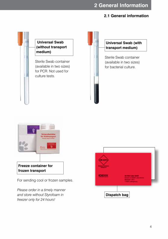
Dispatch bag
Freeze container for frozen transport
For sending cool or frozen samples.
Please order in a timely manner and store without Styrofoam infreezer only for 24 hours!
Universal Swab (with transport medium)
Sterile Swab container (available in two sizes) for bacterial culture.
Universal Swab (without transport medium)
Sterile Swab container (available in two sizes) for PCR. Not used for culture tests.
2.1 General information
5
2 General Information
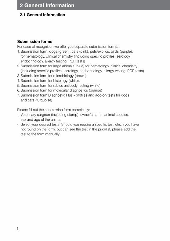
Submission formsFor ease of recognition we offer you separate submission forms:1. Submission form: dogs (green), cats (pink), pets/exotics, birds (purple):
for hematology, clinical chemistry (including specific profiles, serology, endocrinology, allergy testing, PCR tests)
2. Submission form for large animals (blue) for hematology, clinical chemistry (including specific profiles , serology, endocrinology, allergy testing, PCR tests)
3. Submission form for microbiology (brown).4. Submission form for histology (white).5. Submission form for rabies antibody testing (white)6. Submission form for molecular diagnostics (orange)7. Submission form Diagnostic Plus - profiles and add-on tests for dogs
and cats (turquoise)
Please fill out the submission form completely:- Veterinary surgeon (including stamp), owner’s name, animal species,
sex and age of the animal- Select your desired tests. Should you require a specific test which you have
not found on the form, but can see the test in the pricelist, please add the test to the form manually.
2.1 General information
6
2 General Information
2.1 General information
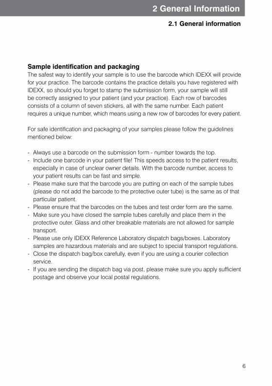
Sample identification and packagingThe safest way to identify your sample is to use the barcode which IDEXX will provide for your practice. The barcode contains the practice details you have registered with IDEXX, so should you forget to stamp the submission form, your sample will still be correctly assigned to your patient (and your practice). Each row of barcodes consists of a column of seven stickers, all with the same number. Each patient requires a unique number, which means using a new row of barcodes for every patient.
For safe identification and packaging of your samples please follow the guidelines mentioned below:
- Always use a barcode on the submission form - number towards the top.- Include one barcode in your patient file! This speeds access to the patient results,
especially in case of unclear owner details. With the barcode number, access to your patient results can be fast and simple.
- Please make sure that the barcode you are putting on each of the sample tubes (please do not add the barcode to the protective outer tube) is the same as of that particular patient.
- Please ensure that the barcodes on the tubes and test order form are the same.- Make sure you have closed the sample tubes carefully and place them in the
protective outer. Glass and other breakable materials are not allowed for sample transport.
- Please use only IDEXX Reference Laboratory dispatch bags/boxes. Laboratory samples are hazardous materials and are subject to special transport regulations.
- Close the dispatch bag/box carefully, even if you are using a courier collection service.
- If you are sending the dispatch bag via post, please make sure you apply sufficient postage and observe your local postal regulations.
7
2 General Information
Courier collection serviceCourier services permit quick and efficient transport of your samples to our lab. Please contact the IDEXX Reference Laboratories Hotline or your local Sales Representative to get more information about the options in your region.
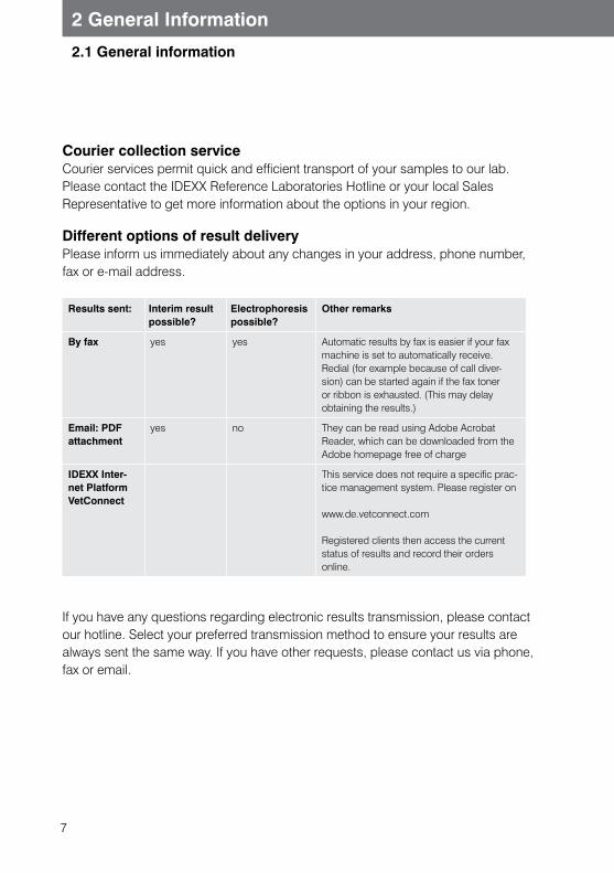
Different options of result deliveryPlease inform us immediately about any changes in your address, phone number, fax or e-mail address.
Results sent: Interim result possible?
Electrophoresis possible?
Other remarks
By fax yes yes Automatic results by fax is easier if your fax machine is set to automatically receive. Redial (for example because of call diver-sion) can be started again if the fax toner or ribbon is exhausted. (This may delay obtaining the results.)
Email: PDF attachment
yes no They can be read using Adobe Acrobat Reader, which can be downloaded from the Adobe homepage free of charge
IDEXX Inter-net Platform VetConnect
This service does not require a specific prac-tice management system. Please register on
www.de.vetconnect.com
Registered clients then access the current status of results and record their orders online.
If you have any questions regarding electronic results transmission, please contact our hotline. Select your preferred transmission method to ensure your results are always sent the same way. If you have other requests, please contact us via phone, fax or email.
2.1 General information
8
2 General Information
Telephone enquiriesPlease contact us with any questions or requests for information.Denmark: 80347618Finland: 0800 98458Norway: 800 31026Sweden: 020 160 58 90The Netherlands: 023 5587 001
Additional testing requestsSubmitted sample material will be stored at IDEXX for 5-7 days, depending on storage availability. (Note: faecal samples are only kept for a maximum of 2-3 days.) During this time, provided that sufficient sample is available, additional tests and profiles can be requested. If bacteriology/mycology cultures are required, additional charges will be made.
Please note: PCR testing should be done from samples that were prepared from the start for PCR testing. Using samples that were harvested for other testing methods may cause false positive results due to contamination of the sample.
2.1 General information
9
2 General Information
2.1 General information
1. Invoice
Invoice receiver is the submitting veterinarian (summary invoice):You receive a monthly invoice. If you wish, you may receive a price breakdown with your results, which will be list prices only. Any discounts will only be shown on your periodic invoice.
2. CancellationCancellation is only possible if you inform us before the requested test is performed. Please let us know as soon as possible if you intend to cancel a test, as you will be charged for tests which have already been peformed.
3. PricesPlease check our price list for our current prices.
10
2 General Information
Sample collection
1. Preparing the patientSatisfactory blood results depend on good preparation of the patient. If possibile the patient should be starved 10-12 hours prior to blood sampling, if the state of the animal permits it. Otherwise many blood parameters may be inaccurate. The animal must be starved before TLI, ammonia and bile acid tests. The patient should not have been heavily exercised immediately before sampling, and the procedure should be carried out quickly and calmly. Agitation and exertion may lead to increased CK, LDH, lactate, glucose and cortisol levels as well as a rise in circulating lymphocytes.
2. Blood sampling techniqueTo avoid haemolysis, blood should be taken immediately after the vein has been raised. ‘Pumping’ blood from the vein can affect results. Avoid high negative pressure in the syringe, as this may cause erythrocytes to rupture. Do not squirt the blood forcefully into the tube. Instead, let it run down the tube wall. Do not try to get the last remaining blood drops that are left in the needle. When using a tube with an anticoagulant, do not shake the contents - instead, you should gently invert the sample tube several times after sampling is completed. Please remember to remove the needle before mailing your sample. (Note: sharp objects should not be transported by post, as there is a risk of personal injury during transportation or unwrapping at the laboratory.)
3. Which type of blood for which test?Our manual explains whether serum or whole blood is required for every parameter we test. Generally, most laboratory tests can be carried out on either serum or plasma. Exceptions are mentioned below. The type and amount of sample needed is also stated on our submission forms.
2.2 General advice on blood collection and sample preparation
11
2 General Information
2.2 General advice on blood collection and sample preparation
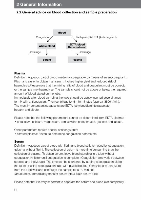
PlasmaDefinition: Aqueous part of blood made noncoagulable by means of an anticoagulant.Plasma is easier to obtain than serum. It gives higher yield and reduced risk of haemolysis.Please note that the mixing ratio of blood and coagulant must be correct, or the sample may haemolyse. The sample should not be above or below the required amount of blood stated on the tube.Immediately after blood sampling the tube should be gently inverted several times to mix with anticoagulant. Then centrifuge for 5 - 10 minutes (approx. 3500 r/min). The most important anticoagulants are EDTA (ethylenediamintetraacetate),heparin and citrate.
Please note that the following parameters cannot be determined from EDTA-plasma:• potassium, calcium, magnesium, iron, alkaline phosphatase, glucose and lactate.
Other parameters require special anticoagulants:• citrated plasma: frozen, to determine coagulation parameters.
SerumDefinition: Aqueous part of blood with fibrin and blood cells removed by coagulation. (plasma without fibrin). The collection of serum is more time consuming than the collection of plasma. To obtain serum, leave blood standing in a tube without coagulation inhibitor until coagulation is complete. (Coagulation time varies between species and individuals. The time can be shortened by adding a coagulation aid to the tube; or using a coagulation tube with plastic beads). Gently loosen coagulate from the tube wall and centrifuge the sample for 5-10 minutes(3500 r/min). Immediately transfer serum into a plain serum tube.
Please note that it is very important to separate the serum and blood clot completely,
Blood
EDTA-bloodHeparin-blood
Plasma
Whole blood
Serum
Coagulation
Centrifuge Centrifuge
Li-Heparin, K-EDTA (Anticoagulant)
12
2 General Information
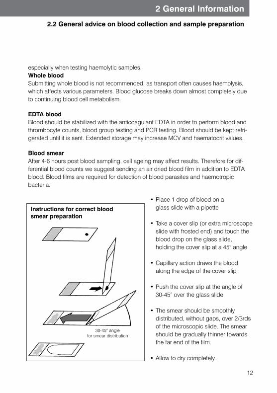
Instructions for correct blood smear preparation
30-45° angle for smear distribution
2.2 General advice on blood collection and sample preparation
especially when testing haemolytic samples. Whole bloodSubmitting whole blood is not recommended, as transport often causes haemolysis,which affects various parameters. Blood glucose breaks down almost completely due to continuing blood cell metabolism.
EDTA bloodBlood should be stabilized with the anticoagulant EDTA in order to perform blood and thrombocyte counts, blood group testing and PCR testing. Blood should be kept refri-gerated until it is sent. Extended storage may increase MCV and haematocrit values.
Blood smearAfter 4-6 hours post blood sampling, cell ageing may affect results. Therefore for dif-ferential blood counts we suggest sending an air dried blood film in addition to EDTA blood. Blood films are required for detection of blood parasites and haemotropic bacteria.
• Place 1 drop of blood on a glass slide with a pipette
• Take a cover slip (or extra microscope slide with frosted end) and touch the blood drop on the glass slide, holding the cover slip at a 45° angle
• Capillary action draws the blood along the edge of the cover slip
• Push the cover slip at the angle of 30-45° over the glass slide
• The smear should be smoothly distributed, without gaps, over 2/3rds of the microscopic slide. The smear should be gradually thinner towards the far end of the film.
• Allow to dry completely.
13
2 General Information
2.2 General advice on blood collection and sample preparation
Blood smear technique 4. Sample volumeThe required sample volumes will differ depending on your desired test. The required volumes are provided in the individual test descriptions and in our alphabetical price list.
5. Factors that can affect results
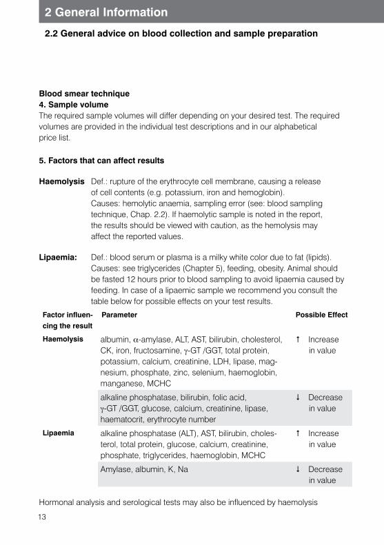
Haemolysis Def.: rupture of the erythrocyte cell membrane, causing a release of cell contents (e.g. potassium, iron and hemoglobin). Causes: hemolytic anaemia, sampling error (see: blood sampling technique, Chap. 2.2). If haemolytic sample is noted in the report, the results should be viewed with caution, as the hemolysis may affect the reported values.
Lipaemia: Def.: blood serum or plasma is a milky white color due to fat (lipids). Causes: see triglycerides (Chapter 5), feeding, obesity. Animal should be fasted 12 hours prior to blood sampling to avoid lipaemia caused by feeding. In case of a lipaemic sample we recommend you consult the table below for possible effects on your test results.
Factor influen-cing the result
Parameter Possible Effect
Haemolysis albumin, α-amylase, ALT, AST, bilirubin, cholesterol, CK, iron, fructosamine, γ-GT /GGT, total protein, potassium, calcium, creatinine, LDH, lipase, mag-nesium, phosphate, zinc, selenium, haemoglobin, manganese, MCHC
# Increase in value
alkaline phosphatase, bilirubin, folic acid, γ-GT /GGT, glucose, calcium, creatinine, lipase, haematocrit, erythrocyte number
$ Decrease in value
Lipaemia alkaline phosphatase (ALT), AST, bilirubin, choles-terol, total protein, glucose, calcium, creatinine, phosphate, triglycerides, haemoglobin, MCHC
# Increase in value
Amylase, albumin, K, Na $ Decrease in value
Hormonal analysis and serological tests may also be influenced by haemolysis
14
2 General Information
2.2 General advice on blood collection and sample preparation
and lipaemia.
6. Deeply frozen samplesFor some special tests it is necessary to send a frozen sample.
Coagulation factors: citrate plasmaADH, ammonia, ACTH, parathormone EDTA plasmaInsulin: serum, plasma (No Serum gel tubes)
These samples should be sent in a special frozen container, which is available from IDEXX on request. Before transport the container should be frozen separately overnight (without Styrofoam insulation). To ensure that submitted samples remain frozen until testing, avoid sending samples at the end of the week. Samples frozen to -20 degrees Celsius in the frozen containers will stay frozen up to 12 hours in outside temperatures of 18-20 degrees Celsius. In case of higher outside temperatures this time is shorter. Alternatively, samples may be sent in dry ice.
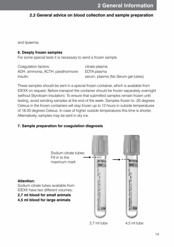
7. Sample preparation for coagulation diagnosis
Attention:Sodium citrate tubes available from IDEXX have two different volumes:2,7 ml blood for small animals4,5 ml blood for large animals
Sodium citrate tubes:Fill in to the maximum mark
2,7 ml tube 4,5 ml tube
15
2 General Information
2.2 General advice on blood collection and sample preparation
Sample preparation for coagulation diagnosis1. Raise the vein carefully and briefly (less than 30 seconds).2. The initial blood drops should be discarded, or can be used to obtain serum.3. Sodium citrate tubes should be filled up to top of the label to achieve 1 part
citrate per 9 parts blood (1:10 dilution). 4. Invert the tubes quickly. 5. Check the blood sample: if it is clotted, the sample is not suitable for testing.6. Centrifuge blood immediately after sampling, or a maximum 2 hours later
(5 minutes @ 3500 RPM).7. Remove the supernatant (citrate plasma) with a pipette and place into a plain tube.
Do not use EDTA, heparin or other citrate tubes.8. As we do not test coagulation factors every day, if you request screening tests and
coagulation factors at the same time, the submitted serum should be divided into two separate tubes.
9. Samples for coagulation testing should be frozen and kept in the freezer (-20 degrees C) until transport.
10. Shipment must be in deep freeze boxes ordered from IDEXX Reference Laboratories. These boxes should be kept in the freezer without Styrofoam pa-ckaging for 24 hours after arrival at your practice. Samples must arrive at the laboratory frozen. Please follow the directions for deep freeze samples.
8. Cerebrospinal fluid and aspirate testsCerebrospinal fluid (CSF) is physiologically clear. When sampling, do not add any anticoagulants or preservatives. CSF and other aspirates should be collected into sterile tubes. If you desire multiple tests (bacteriology and cytology) it is better to send samples in separate tubes to enable us to perform both tests simultaneously.
CSF and other aspirates are very unstable biological samples. The sample material may degrade as soon as 30 minutes after sampling, and by 4 hours after sample collection, the results of the test may be significantly affected. Therefore cytological examination of aspirated fluid and examination of the number of cells in cerebrospinal fluid is possible only during this time period. To enable us to perform a cytology examination, please prepare a sediment smear as soon as possible after sample collection (after centrifugation for 3-5 minutes at 1000 rounds/min; prepare smear as in case of blood and air dry).
16
2 General Information
2.3 General advice on sample collection for microbiology tests
Sample collection for bacteriology tests
Collection timeIf possible, samples should be taken before antibiotic therapy. In case of treatment monitoring it is advised to allow sufficient time interval after antibiotic administration. Sample collection from a necropsy should be performed immediately post mortem.Collection site Sample collection is best done at sites on the border of healthy and inflamed tissue. The best are sites that are likely to contain pathogenic microorganisms. Useful locacions include purulent lesions, areas of inflammatory changes in the ear, and absceses (Please note that it is usually impossible to grow bacteria from pus).Sampling techniqueWhen collecting samples for bacteriology you should avoid contamination by foreign substances (e.g. dirt or other contaminants.) Also, after collection you should avoid contamination during sample preparation or packaging for transport.
Samples for bacteriology tests:• Swabs:
For sample collection from different sites we use cotton Swabs. If possible you should use Swabs with transport medium. With dry Swabs there is a risk that fragile or sensitive microorganisms may not be cultured in the laboratory. If the surface we want to collect from is very dry, the Swab may be moistened with sterile fluid.
• Urine: Please send urine samples in uncoated tubes. It is preferable to collect urine by cystocentesis or by catheter. Naturally voided (free catch) urine may contain microorganisms from body surfaces or from the environment. Urine samples from the environment (from litter box or examination tables) are not suitable for tests. Urine can also can be sent in a culture system (Uricult) instead of sample tubes.
• Biopsies and organ fragments: Send in uncoated sterile tubes. If transport time is expected to be prolonged, organs should be sent deeply frozen, without any chance for thawing and refreezing. Please clearly indicate on the packaging that frozen samples are being sent.
• Biological fluids: (synovial fluid, cerebrospinal fluid, organ aspirate, milk etc.) should be sent in sterile uncoated tubes. If you require an anaerobic culture, please limit sample contact with atmospheric oxygen (use our special container).
17
2 General Information
2.3 General advice on sample collection for microbiology tests
• Faeces: (synovial fluid, cerebrospinal fluid, organ aspirate, milk etc.) should be sent in sterile uncoated tubes. If you require an anaerobic culture, please limit sample contact with atmospheric oxygen (use our special container).
•Blood culture: Proper bacterial culture from blood requires special culture bottles, available from our laboratory. It is impossible to culture blood from the routinely used blood tubes. Samples should be collected in completely sterile conditions. Bottles containing blood samples should be kept in room temperature (unrefrigerated) and sent to the laboratory as soon as possible.
Mycology sample collection
Collection technique:With sample collection for yeast and mould culture we use the same advice as for bacteriology tests. Swabs with transport medium are most suitable for sending. During sample collection from mucosal membranes you should pay attention to membrane-ous and purulent deposits, from which organisms are best cultured.
To isolate dermatophytes it is best to disinfect the site with 70 % alcohol; this prevents incidental bacteria from overwhelming any mycological culture. The sample should be collected from a site on the border of the lesion and sent in a dry tube.
If a lesion needs to be tested for both bacteriology and mycology, the bacteriology sample should be collected first and put in transport medium. Following this, disinfect the site with 70% alcohol and collect sample for the mycology test, placing the collec-ted material into a sterile tube).
Sample for mycology test:The best samples are deep skin scrapings or plucked hair that includes the root. Hairs clipped with scissors are not suitable for mycological testing. Fungal culture medium incubated in your own clinic may also be sent for identification. For fungal testing of fa-eces you must send a faecal sample, as a faecal Swab is not suitable for this purpose.
18
2 General Information
2.4 General advice on molecular biology tests
Sample material for molecular pathogen diagnostics Samples used for PCR testing should be those with the highest possible content of the organism in question. Therefore, prior to taking a sample, take the following into account:• whether the animal is currently in a viraemic/bacteraemic stage.• whether the organism may have reached its final target organ and if so, where
it is likely to be found when considering the clinical symptoms.• whether there is a latent organ where the organism may be hiding during the
subclinical phase (e.g. EHV-1 in leukocytes).
Possible test sample:• Swabs:
For Swabs, please use a sterile, dry Swab without transport medium and send in uncoated tubes.
Note: these samples are not suitable for bacteriological tests!If requesting simultaneous bacteriological and molecular biology tests, please collect and send two separate Swabs.
• Biological fluids : (Synovia, cerebrospinal fluid, body cavity aspirate, aqueous humor, urine, etc.) Send in sterile uncoated tubes. 0,5-2 ml of material is normally needed. For urine samples 5 ml of sample is needed, depending on requested test. If sample will be sent on the next day after collection, please store the sample between + 2° C to + 8° C and send unfrozen. If sending is expected to be delayed, freeze the sample and send without breaking the cold chain (e.g. usage of coldpacks and styrofoam packane, or sending in dry ice). For intracellular organism testing (e.g. Listeria), freezing should be avoided, so for these samples we recommend storage between + 2° C to + 8° C. Please indicate clearly on the packaging that these are samples in deep freeze, and so thawing and refreezing should be avoided.
• Biopsies, organ parts, aborted material: Send in sterile uncoated tubes. Add sufficient sterile saline to cover the sample. If sample sending by the next day is not possible, please send sample material in deep freeze, without adding saline. Ensure that the cold chain is not broken. Please indicate clearly on the packaging that these are samples in deep freeze, and so thawing and refreezing should be avoided.
19
2 General Information
2.4 General advice on molecular biology tests
• EDTA-blood, Citrate blood: The required amount of sample depends on test parameters and disease phase. Please do not send frozen EDTA or citrate blood under any circumstances. Please do not send heparinised blood!
• Faeces: Send in uncoated, sterile tubes.
Sample material for molecular genetic diagnostics (hereditary diseases, parentage verification) Standard sample for animal genetic tests is 0,5 - 2 ml EDTA-blood. Transport is not time sensitive. The standard sample for genetic identification (especially parental verification) is a minimum of 0,5 ml EDTA-blood, or Swabs (two preferable) from the buccal mucosal membrane. A separate submission form can be ordered.
Guidelines for buccal mucosal membrane Swabs 1. The patient should not receive any food or fluids (except for water) for at least
30 minutes before sample collection.2. Using a sterile cotton Swab (or ideally a ”cytobrush”) strongly rub each buccal
cavity a minimum of 10 times. Following this, rub the cotton Swabs on each other.3. Transport containers should be clearly labeled to avoid loss or confusion!4. Air dry the Swab for a minimum 1-2 hours at room temperature. Place the Swab
a few centimeters into the protective outer tube and leave it. 5. After the sample is completely dry, place the Swab deep into the the protective
outer tube. 6. Store the sample in a cold (5 - 8 °C) and dry place or immediately send it to the
laboratory.
Do not touch the cotton Swab under any circumstances, as test results may be affected.
20
2 General Information
2.4 General advice on molecular biology tests
Precautions during sample preparation Because of high sensitivity of PCR method, please obey the following sample collection:• To avoid contamination, always wear gloves during sample collection.• Separate samples should be collected for this type of test.• Sterile tubes and instruments should be used to avoid contamination during sample
manipulation (e.g. when filling or packaging the sample)!• Do not send the samples chilled, unless sample will not be sent immediately. If the
sample will be sent within 24 hours, sample material should be cooled to between + 2 °C and + 8 °C.
• When longer transport time or delay is unavoidable, send deeply frozen samples (except for EDTA/Citrate-blood), ensuring an unbroken cold chain. (Suggestions include usage of coldpacks and stryrofoam packages, or sending on dry ice)! If this is not possible, send unfrozen samples. Thawing and refreezing of the sample should be avoided.
Additional requirementPlease note: If you request an additional test for molecular biology pathogen PCR testing from a test sample that was not prepared for this purpose (and was used for other diagnostic tests) there is a risk of contamination, which can lead to false positive results.
21
2 General Information
2.5 General advice on histopathology and cytology tests
IDEXX Reference Laboratories will perform the following tissue tests:• histopathology of neoplasias, skin punches, skin and organ biopsies, fine needle
aspirates, as well as from any nonspecific changes found in tissues. Also from tissue or organs or parts of organs collected during surgery or autopsy/post mortem examination.
• cytology of fine needle aspirates collected from body fluids (e.g. joint or pleural fluids, ascites, urine) or from organs (e.g. mammary gland, kidney, liver, thyroid gland or lymph nodes)
• cytology of vaginal smear (vaginal cytology)
Important guidelines for optimal sample preparation: • clearly fill out the histopathology submission form.• remember to complete the reverse side of the submission form when submitting
dermatological samples.• all samples should be submitted completely covered in fixative. Please avoid
crushing the sample. Make sure your submission pot is large enough, or your sample may not be entirely covered by fixative. The autolytic processes will then continue.
• use pots with a large opening. The sample hardens due to the action of the fixative. If the opening is too small, artefacts due to crushing can occur when removing the sample from the tube.
Tru-cut-Biopsy Purpose-designed biopsy systems are available on the market of 0,3 mm and 1 mm diameter. Such tissue cylinders collected with wide lumen needles can be put directly into formaldehyde solution and mailed. Fixated tissue cylinders have the advantage over aspirates in that tissue morphology is preserved. An additional advantage is that you may be able to obtain more information on tumour origin and structure. If a neo-plasia reachs a larger diameter, larger organ samples may be biopsied with a larger needle diameter.If lymphoma is suspected, Tru Cut and cytology should not be performed in mandibu-lar lymph nodes, as strong reactive activity/hyperplasia is often seen here, which can hide neoplastic processes.
22
2 General Information
2.5 General advice on histopathology and cytology tests
Fine needle aspiration from masses and fluids In order to perform a fine needle biopsy you may use a 0.8-2 mm (18-22G) needle of sufficient length. The use of an aspiration guide is often helpful. This enables you to safely collect several aspirates. A 5 or 10 ml syringe is recommended.
Sample collection is completed after a brief, single aspiration. If possible, when collecting several aspirates, use a new needle for each new biopsy. Try not to collect your aspirates by poking the needle around in the tissue. Also, try not to collect your sample by lengthly aspiration of the tissue. This will lead to excessive mixing of blood into the sample. It can also increase the risk of metastasis of neoplastic or purulent infectious processes.The collected aspirate can be treated like a blood smear and can be transferred to a glass slide.
Liquid aspirates should be centrifuged at 1500 RPM for 5-10 minutes. Discard the supernatant fluid and transfer the sediment onto a glass slide. Make a smear and allow to dry naturally, then place in a slide protection box and send to the lab. Remember to inform the lab about the site of collection
Price information A higher fee is charged for very large samples, samples containing several tumours or several samples from the same animal, as well as for more than six skin biopsies from a single animal. This is due to the increase in time required for processing the samples, as well as the larger number of sections and possibly diagnoses that have to be made.
23
2 General Information
2.6 General advice on parasitology tests
Samples collection and sendingFaecal samples should ideally be taken directly from the rectum. If you are unable to collect a rectal sample, ensure you collect fresh faeces. Faeces collected from the ground can be contaminated by free living nematodes in a short time.
For reliable results a minimal amount of faeces is needed (amount is specified with each test description). Samples should be put into a tightly closed and damage resis-tant package, cooled and sent to laboratory directly after collection.If sending sample is delayed, it should be stored in the refrigerator. Parasite larvae are not damaged, but oocyst and egg development is inhibited.
Parasites or parasite parts shed with faeces or should be sent in a plain tube (without formalin) or in physiological salt solution, separate from the faecal sample.
Parasitology tests results estimation.Each diagnostic procedure has its limitations. A positive result (direct parasite confirmation) confirms infestation, but a negative result does not exclude parasitic infestation. Multiple tests may be necessary to confirm the presence of parasites.
Because the various development stages of the parasite are not excreted continuously, it is advised to test faecal samples collected over 3 days. In animal herds (apart from fattening swine and poultry) a representative number of randomly taken samples should be collected (not a collective sample from multiple animals)!
Showing various development stages of parasites is possible only in patent phase (prepatent or postpatent infections in are not detected this way). It is important in some parasitic infestations, as clinical signs may be present in the prepatent period.
24
2 General Information
2.7 Quality management
Quality management at IDEXX Reference LaboratoriesThe quality of diagnostics at IDEXX Vet Med Labor is subject to continuous and extensive monitoring. Since June 2003, the high quality standards at our Ludwigsburg facility have been confirmed by accreditation in accordance with DIN EN ISO 17025. The German accreditation body DAkkS grants this recognition after thorough scrutiny. Accredited tests carried out at this facility are marked (1) in the following pages; non-accredited tests are identified by (2).Our new Leipzig facility is committed to the same quality requirements. However, the tests carried out there have not yet been accredited.
Our quality management does not start with the diagnostic machines - it begins by giving information and advice to our clients on all pre-testing questions. We make special efforts to ensure correct and reliable laboratory results.
To be able to process the wide range of submitted samples, our methods are specifically calibrated to different animal species. All our diagnostic procedures are validated and the reliability or our results are frequently monitored. By participating in numerous national and international research groups, the quality of our analytic methods are constantly revised and improved.
Even with the greatest care in diagnostic procedures, test results and parameters may have some errors. We aim to minimise any deviation from the actual result. On your request we can give information according Information on our expected margins within our validation methods is available on request.
In the interests of clarity, we show the results of our tests in the most readable way. In order to achieve this, we present a summary of the test results. A more detailed description of the diagnostic method or procedure will be sent if requested.
An important element for quality improvement is a careful analysis of feedback from our clients. We are always keen to hear any comments or criticism from our clients. Your feedback is always welcome.
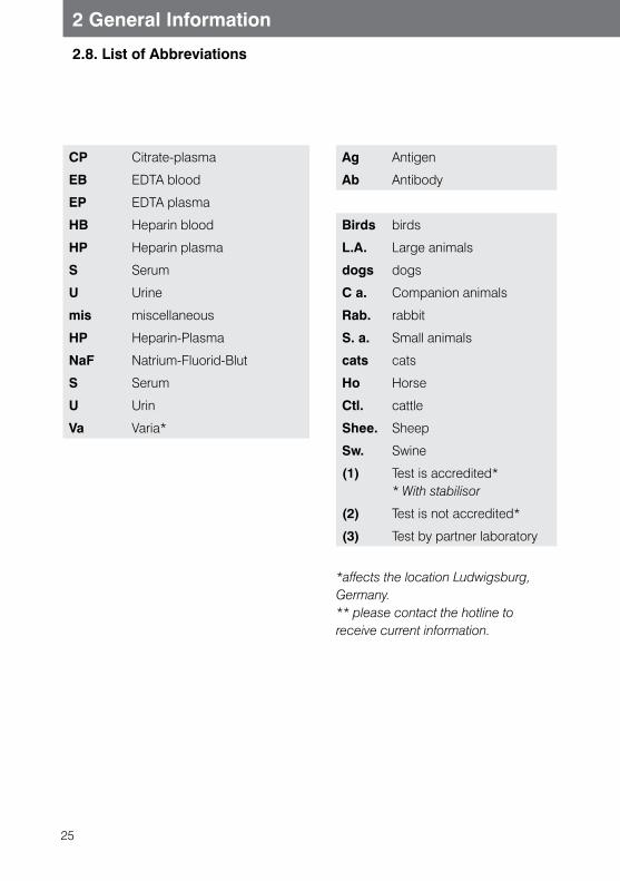
25
2 General Information
2.8. List of Abbreviations
CP Citrate-plasma
EB EDTA blood
EP EDTA plasma
HB Heparin blood
HP Heparin plasma
S Serum
U Urine
mis miscellaneous
HP Heparin-Plasma
NaF Natrium-Fluorid-Blut
S Serum
U Urin
Va Varia*
Ag Antigen
Ab Antibody
Birds birds
L.A. Large animals
dogs dogs
C a. Companion animals
Rab. rabbit
S. a. Small animals
cats cats
Ho Horse
Ctl. cattle
Shee. Sheep
Sw. Swine
(1) Test is accredited* * With stabilisor
(2) Test is not accredited*
(3) Test by partner laboratory
*affects the location Ludwigsburg, Germany. ** please contact the hotline to receive current information.
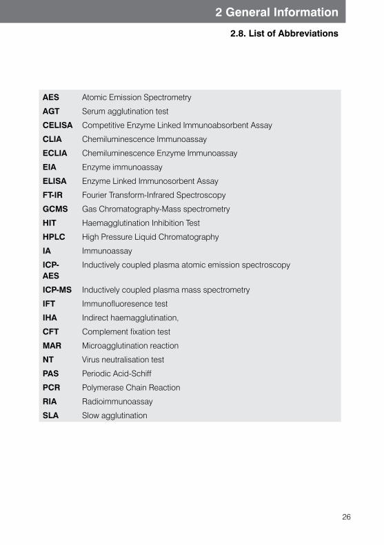
26
2 General Information
2.8. List of Abbreviations
AES Atomic Emission Spectrometry
AGT Serum agglutination test
CELISA Competitive Enzyme Linked Immunoabsorbent Assay
CLIA Chemiluminescence Immunoassay
ECLIA Chemiluminescence Enzyme Immunoassay
EIA Enzyme immunoassay
ELISA Enzyme Linked Immunosorbent Assay
FT-IR Fourier Transform-Infrared Spectroscopy
GCMS Gas Chromatography-Mass spectrometry
HIT Haemagglutination Inhibition Test
HPLC High Pressure Liquid Chromatography
IA Immunoassay
ICP-AES
Inductively coupled plasma atomic emission spectroscopy
ICP-MS Inductively coupled plasma mass spectrometry
IFT Immunofluoresence test
IHA Indirect haemagglutination,
CFT Complement fixation test
MAR Microagglutination reaction
NT Virus neutralisation test
PAS Periodic Acid-Schiff
PCR Polymerase Chain Reaction
RIA Radioimmunoassay
SLA Slow agglutination

27
2 General Information
2.9 Conversion Table
SI units conventional units
Multiply with divide by
SI units
ACTH pg/ml 0.2202 pmol/l
Albumin g/dl 10 g/l
Aldosterone g/ml 2.77 pmol/l
Ammonia μg/dl 0.587 μmol/l
Bilirubin mg/dl 17.104 μmol/l
Calcium mg/dl 0.2495 mmol/l
Cholesterol mg/dl 0.02586 mmol/l
Copper μg/dl 0.157 μmol/l
Cortisol μg/dl 27.6 nmol/l
Creatinine mg/dl 88.4 μmol/l
Digoxin μg/l 1.28 nmol/l
Fibrinogen mg/dl 0.01 g/l
Folic acid ng/ml 2.27 nmol/l
FT3 ng/l 1.54 pmol/l
FT4 ng/dl 12.87 pmol/l
Glucose mg/dl 0.0555 mmol/l
Haemoglobin g/dl 0.621 mmol/l
Insulin μU/ml 7.18 pmol/l
Iron μg/dl 0.1791 μmol/l
Lactate mg/dl 0.11 mmol/l
Lead μg/l 0.00483 μmol/l
Magnesium mg/dl 0,411 mmol/l
Kreatinin mg/dl 88,4 μmol/l
Kupfer μg/dl 0,157 μmol/l
Laktat mg/dl 0,11 mmol/l
Magnesium mg/dl 0,411 mmol/l
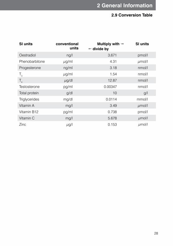
28
2 General Information
2.9 Conversion Table
SI units conventional units
Multiply with divide by
SI units
Oestradiol ng/l 3,671 pmol/l
Phenobarbitone μg/ml 4.31 μmol/l
Progesterone ng/ml 3.18 nmol/l
T3 μg/ml 1.54 nmol/l
T4 μg/dl 12.87 nmol/l
Testosterone pg/ml 0.00347 nmol/l
Total protein g/dl 10 g/l
Triglycerides mg/dl 0.0114 mmol/l
Vitamin A mg/l 3.49 μmol/l
Vitamin B12 pg/ml 0.738 pmol/l
Vitamin C mg/l 5.678 μmol/l
Zinc μg/l 0.153 μmol/l
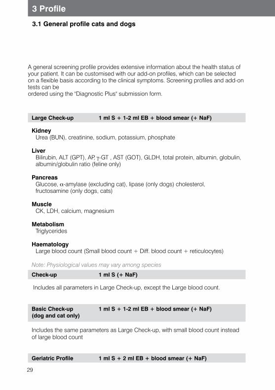
29
3.1 General profile cats and dogs
3 Profile
Large Check-up 1 ml S + 1-2 ml EB + blood smear (+ NaF)
Kidney Urea (BUN), creatinine, sodium, potassium, phosphate
Liver Bilirubin, ALT (GPT), AP, γ-GT , AST (GOT), GLDH, total protein, albumin, globulin,
albumin/globulin ratio (feline only)
Pancreas Glucose, α-amylase (excluding cat), lipase (only dogs) cholesterol, fructosamine (only dogs, cats)
Muscle CK, LDH, calcium, magnesium
Metabolism Triglycerides
Haematology Large blood count (Small blood count + Diff. blood count + reticulocytes)
Note: Physiological values may vary among species
Check-up 1 ml S (+ NaF)
Includes all parameters in Large Check-up, except the Large blood count.
Basic Check-up(dog and cat only)
1 ml S + 1-2 ml EB + blood smear (+ NaF)
Includes the same parameters as Large Check-up, with small blood count instead of large blood count
Geriatric Profile 1 ml S + 2 ml EB + blood smear (+ NaF)
A general screening profile provides extensive information about the health status of your patient. It can be customised with our add-on profiles, which can be selected on a flexible basis according to the clinical symptoms. Screening profiles and add-on tests can be ordered using the "Diagnostic Plus" submission form.
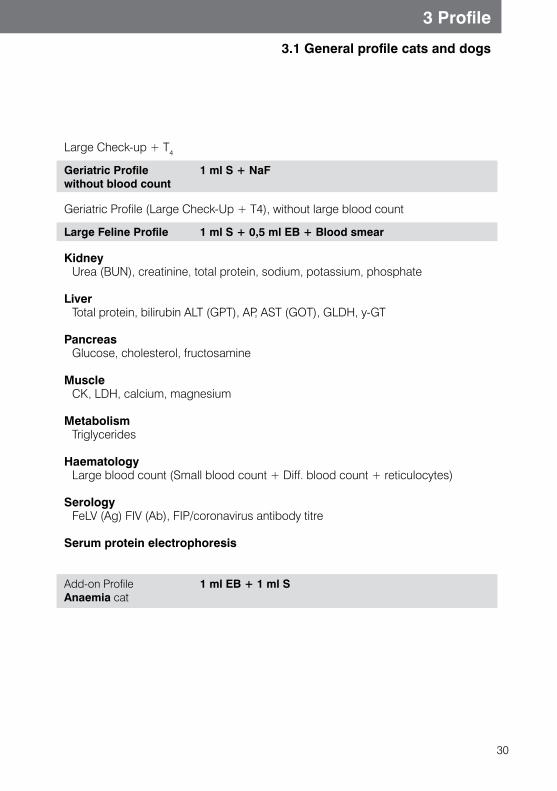
30
3.1 General profile cats and dogs
3 Profile
Large Check-up + T4
Geriatric Profile without blood count
1 ml S + NaF
Geriatric Profile (Large Check-Up + T4), without large blood count
Large Feline Profile 1 ml S + 0,5 ml EB + Blood smear
Kidney Urea (BUN), creatinine, total protein, sodium, potassium, phosphate
Liver Total protein, bilirubin ALT (GPT), AP, AST (GOT), GLDH,Υy-GT
Pancreas Glucose, cholesterol, fructosamine
Muscle CK, LDH, calcium, magnesium
Metabolism Triglycerides
Haematology Large blood count (Small blood count + Diff. blood count + reticulocytes)
Serology FeLV (Ag) FIV (Ab), FIP/coronavirus antibody titre
Serum protein electrophoresis
Add-on Profile Anaemia cat
1 ml EB + 1 ml S
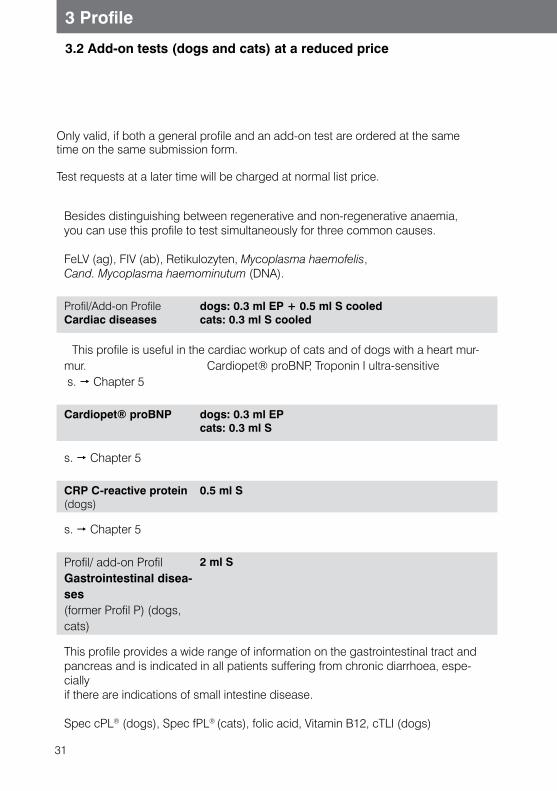
31
3.2 Add-on tests (dogs and cats) at a reduced price
3 Profile
Besides distinguishing between regenerative and non-regenerative anaemia, you can use this profile to test simultaneously for three common causes.
FeLV (ag), FIV (ab), Retikulozyten, Mycoplasma haemofelis, Cand. Mycoplasma haemominutum (DNA).
Profil/Add-on Profile Cardiac diseases
dogs: 0.3 ml EP + 0.5 ml S cooledcats: 0.3 ml S cooled
This profile is useful in the cardiac workup of cats and of dogs with a heart mur-mur. Cardiopet® proBNP, Troponin I ultra-sensitive s. Chapter 5
Cardiopet® proBNP dogs: 0.3 ml EPcats: 0.3 ml S
s. Chapter 5
CRP C-reactive protein (dogs)
0.5 ml S
s. Chapter 5
Profil/ add-on Profil Gastrointestinal disea-ses(former Profil P) (dogs, cats)
2 ml S
This profile provides a wide range of information on the gastrointestinal tract and pancreas and is indicated in all patients suffering from chronic diarrhoea, espe-cially if there are indications of small intestine disease.
Spec cPL® (dogs), Spec fPL® (cats), folic acid, Vitamin B12, cTLI (dogs)
Only valid, if both a general profile and an add-on test are ordered at the same time on the same submission form.
Test requests at a later time will be charged at normal list price.
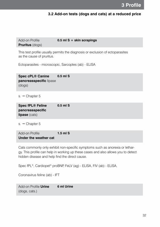
32
3.2 Add-on tests (dogs and cats) at a reduced price
Add-on Profile Pruritus (dogs)
0.5 ml S + skin scrapings
This test profile usually permits the diagnosis or exclusion of ectoparasites as the cause of pruritus.
Ectoparasites - microscopic, Sarcoptes (ab) - ELISA
Spec cPL® Canine pancreasspecific lipase (dogs)
0.5 ml S
s. Chapter 5
Spec fPL® Feline pancreasspecific lipase (cats)
0.5 ml S
s. Chapter 5
Add-on Profile Under the weather cat
1.5 ml S
Cats commonly only exhibit non-specific symptoms such as anorexia or lethar-gy. This profile can help in working up these cases and also allows you to detect hidden disease and help find the direct cause.
Spec fPL®, Cardiopet® proBNP, FeLV (ag) - ELISA, FIV (ab) - ELISA,
Coronavirus feline (ab) - IFT
Add-on Profile Urine (dogs, cats.)
6 ml Urine
3 Profile
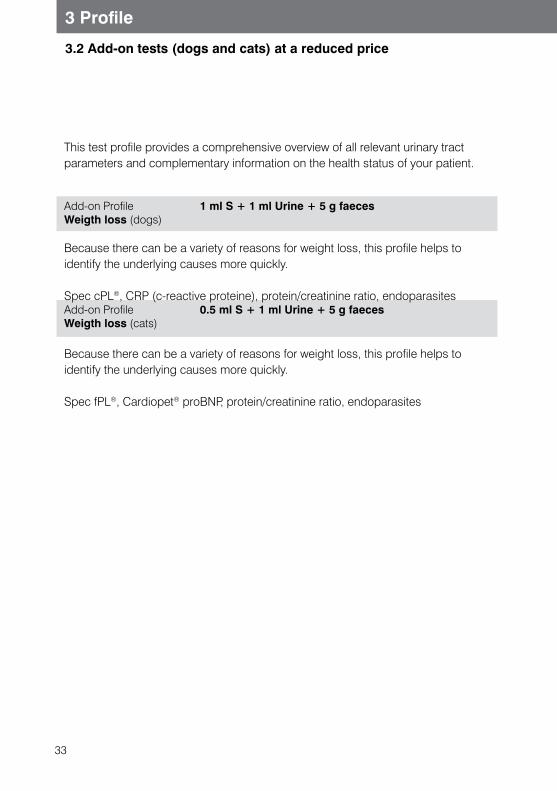
33
3 Profile
3.2 Add-on tests (dogs and cats) at a reduced price
This test profile provides a comprehensive overview of all relevant urinary tract parameters and complementary information on the health status of your patient.
Add-on Profile Weigth loss (dogs)
1 ml S + 1 ml Urine + 5 g faeces
Because there can be a variety of reasons for weight loss, this profile helps to identify the underlying causes more quickly.
Spec cPL®, CRP (c-reactive proteine), protein/creatinine ratio, endoparasitesAdd-on Profile Weigth loss (cats)
0.5 ml S + 1 ml Urine + 5 g faeces
Because there can be a variety of reasons for weight loss, this profile helps to identify the underlying causes more quickly.
Spec fPL®, Cardiopet® proBNP, protein/creatinine ratio, endoparasites
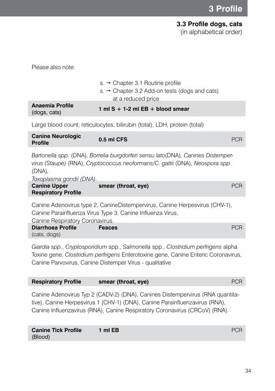
34
3 Profile
3.3 Profile dogs, cats (in alphabetical order)
Please also note:
s. Chapter 3.1 Routine profile s. Chapter 3.2 Add-on tests (dogs and cats)
at a reduced priceAnaemia Profile (dogs, cats)
1 ml S + 1-2 ml EB + blood smear
Large blood count, reticulocytes, bilirubin (total), LDH, protein (total)
Canine Neurologic Profile 0.5 ml CFS PCR
Bartonella spp. (DNA), Borrelia burgdorferi sensu lato(DNA), Canines Distemper-virus (Staupe) (RNA), Cryptococcus neoformans/C. gattii (DNA), Neospora spp. (DNA), Toxoplasma gondii (DNA).Canine Upper Respiratory Profile
smear (throat, eye) PCR
Canine Adenovirus type 2, CanineDistempervirus, Canine Herpesvirus (CHV-1), Canine Parainfluenza Virus Type 3, Canine Influenza Virus, Canine Respiratory Coronavirus.Diarrhoea Profile(cats, dogs)
Feaces PCR
Giardia spp., Cryptosporidium spp., Salmonella spp., Clostridium perfrigens alpha Toxine gene, Clostridium perfrigens Enterotoxine gene, Canine Enteric Coronavirus, Canine Parvovirus, Canine Distemper Virus - qualitative
Respiratory Profile smear (throat, eye) PCR
Canine Adenovirus Typ 2 (CADV-2) (DNA), Canines Distempervirus (RNA quantita-tive), Canine Herpesvirus 1 (CHV-1) (DNA), Canine Parainfluenzavirus (RNA), Canine Influenzavirus (RNA), Canine Respiratory Coronavirus (CRCoV) (RNA).
Canine Tick Profile (Blood)
1 ml EB PCR
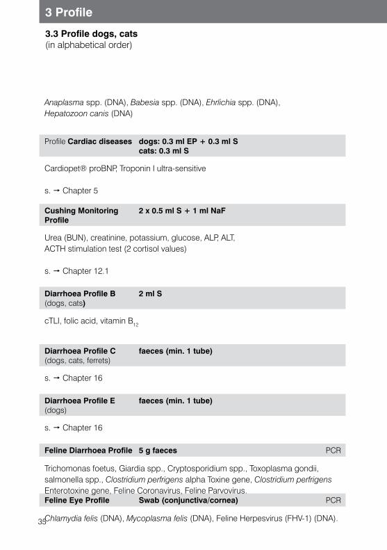
35
3 Profile
3.3 Profile dogs, cats (in alphabetical order)
Anaplasma spp. (DNA), Babesia spp. (DNA), Ehrlichia spp. (DNA), Hepatozoon canis (DNA)
Profile Cardiac diseases dogs: 0.3 ml EP + 0.3 ml Scats: 0.3 ml S
Cardiopet® proBNP, Troponin I ultra-sensitive
s. Chapter 5
Cushing Monitoring Profile
2 x 0.5 ml S + 1 ml NaF
Urea (BUN), creatinine, potassium, glucose, ALP, ALT,ACTH stimulation test (2 cortisol values)
s. Chapter 12.1
Diarrhoea Profile B (dogs, cats)
2 ml S
cTLI, folic acid, vitamin B12
Diarrhoea Profile C (dogs, cats, ferrets)
faeces (min. 1 tube)
s. Chapter 16
Diarrhoea Profile E (dogs)
faeces (min. 1 tube)
s. Chapter 16
Feline Diarrhoea Profile 5 g faeces PCR
Trichomonas foetus, Giardia spp., Cryptosporidium spp., Toxoplasma gondii, salmonella spp., Clostridium perfrigens alpha Toxine gene, Clostridium perfrigens Enterotoxine gene, Feline Coronavirus, Feline Parvovirus.Feline Eye Profile Swab (conjunctiva/cornea) PCR
Chlamydia felis (DNA), Mycoplasma felis (DNA), Feline Herpesvirus (FHV-1) (DNA).
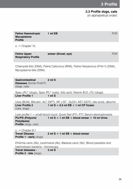
36
3 Profile
3.3 Profile dogs, cats (in alphabetical order)
Feline Haemotropic Mycoplasma Profile
1 ml EB PCR
s. Chapter 15
Feline Upper Respiratory Profile
smear (throat, eye) PCR
Chlamydia felis (DNA), Feline Calicivirus (RNA), Feline Herpesvirus (FHV-1) (DNA), Mycoplasma felis (DNA).
Gastrointestinal Diseases (former Profil P) (dogs, cats)
2 ml S
Spec cPL® (dogs), Spec fPL® (cats), folic acid, Vitamin B12, cTLI (dogs)Liver Profile 1 1 ml S
Urea (BUN), Bilirubin, ALT (GPT), AP, γ-GT , GLDH, AST (GOT), bile acids, albumin Liver Profile 2 (cats, dogs)
1 ml S + 0.5 ml EB + 1 ml CP frozen
Liver profile 1 + small blood count, Quick-Test (PT), PTT, Serum electrophoresisPU/PD (Polyuria/Polydipsia) Profile (dogs, cats)
1 ml S + 1 ml EB + blood smear + 10 ml Urine
s. Chapter 8.1Travel Disease Profile 1 - early (dogs)
2 ml S + 1 ml EB + blood smear
Ehrlichia canis (Ab), Leishmania (Ab), Babesia canis (Ab), Blood parasites and haemotropic bacteria - microscopyTravel diseases - Profile 2 - late (dogs)
3 ml S
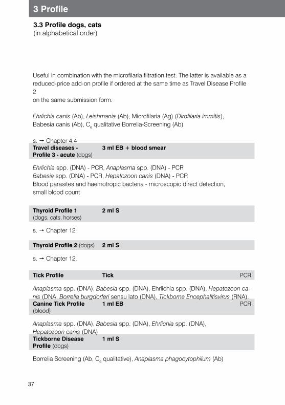
37
3 Profile
3.3 Profile dogs, cats (in alphabetical order)
Useful in combination with the microfilaria filtration test. The latter is available as a reduced-price add-on profile if ordered at the same time as Travel Disease Profile 2 on the same submission form.
Ehrlichia canis (Ab), Leishmania (Ab), Microfilaria (Ag) (Dirofilaria immitis), Babesia canis (Ab), C6 qualitative Borrelia-Screening (Ab)
s. Chapter 4.4Travel diseases - Profile 3 - acute (dogs)
3 ml EB + blood smear
Ehrlichia spp. (DNA) - PCR, Anaplasma spp. (DNA) - PCRBabesia spp. (DNA) - PCR, Hepatozoon canis (DNA) - PCRBlood parasites and haemotropic bacteria - microscopic direct detection, small blood count
Thyroid Profile 1 (dogs, cats, horses)
2 ml S
s. Chapter 12
Thyroid Profile 2 (dogs) 2 ml S
s. Chapter 12.
Tick Profile Tick PCR
Anaplasma spp. (DNA), Babesia spp. (DNA), Ehrlichia spp. (DNA), Hepatozoon ca-nis (DNA, Borrelia burgdorferi sensu lato (DNA), Tickborne Encephalitisvirus (RNA).Canine Tick Profile (blood)
1 ml EB PCR
Anaplasma spp. (DNA), Babesia spp. (DNA), Ehrlichia spp. (DNA), Hepatozoon canis (DNA)Tickborne Disease Profile (dogs)
1 ml S
Borrelia Screening (Ab, C6 qualitative), Anaplasma phagocytophilum (Ab)

38
3 Profile
3.3 Profile dogs, cats (in alphabetical order)
Large Tickborne Disease Profile (serological) (dogs)
2 ml S
Borrelia Screening (Ab, C6 qualitative), Anaplasma phagocytophilum (Ab)Ehrlichia canis (Ab), Babesia canis (Ab)
Cushing Monitoring Profile
1 ml EB
Urea(BUN), crea, K, Na, glucose, ALP, ALT,ACTH stimulation test (2 cortisol values)
Equine Profile, large 1 ml S + 1 ml EB + blood smear + NaF
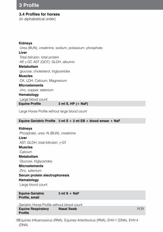
39
3 Profile
3.4 Profiles for horses (in alphabetical order)
Kidneys Urea (BUN), creatinine, sodium, potassium, phosphateLiver Total bilirubin, total protein AP, γ-GT, AST (GOT), GLDH, albumin Metabolism glucose, cholesterol, triglyceridesMuscles CK, LDH, Calcium, Magnesium Microelements zinc, copper, selenium Hematology Large blood count Equine Profile 3 ml S, HP (+ NaF)
Large Horse Profile without large blood count
Equine Geriatric Profile 3 ml S + 2 ml EB + blood smear + NaF
Kidneys Phosphate, urea -N (BUN), creatinine Liver AST, GLDH, total bilirubin, γ-GT Muscles Calcium Metabolism Glucose, triglyceridesMicroelements Zinc, seleniumSerum protein electrophoresis Hematology Large blood count
Equine Geriatric Profile, small
3 ml S + NaF
Geriatric Horse Profile without blood count Equine Respiratory Profile
Nasal Swab PCR
Equines Influenzavirus (RNA), Equines Arteritisvirus (RNA), EHV-1 (DNA), EHV-4 (DNA)
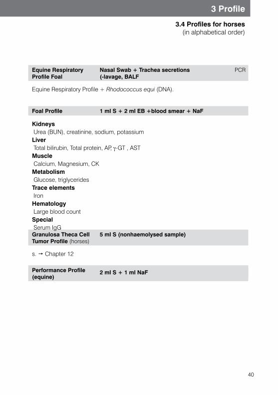
40
3 Profile
3.4 Profiles for horses (in alphabetical order)
Equine Respiratory Profile Foal
Nasal Swab + Trachea secretions PCR(-lavage, BALF
Equine Respiratory Profile + Rhodococcus equi (DNA).
Foal Profile 1 ml S + 2 ml EB +blood smear + NaF
Kidneys Urea (BUN), creatinine, sodium, potassiumLiver Total bilirubin, Total protein, AP, γ-GT , AST Muscle Calcium, Magnesium, CK Metabolism Glucose, triglyceridesTrace elements IronHematology Large blood count Special Serum IgGGranulosa Theca Cell Tumor Profile (horses)
5 ml S (nonhaemolysed sample)
s. Chapter 12
Performance Profile (equine)
2 ml S + 1 ml NaF
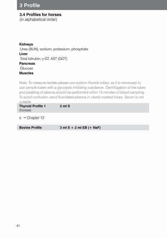
41
3 Profile
3.4 Profiles for horses (in alphabetical order)
Kidneys Urea (BUN), sodium, potassium, phosphateLiver Total bilirubin, γ-GT, AST (GOT) Pancreas GlucoseMuscles
Note: To measure lactate please use sodium fluoride tubes, as it is necessary to use sample tubes with a glycolysis inhibiting substance. Centrifugation of the tubes and pipetting of plasma should be performed within 15 minutes of blood sampling. To avoid confusion, send fluoridated plasma in clearly marked tubes. Serum is not suitable.Thyroid Profile 1(horses)
2 ml S
s. Chapter 12
Bovine Profile 3 ml S + 2 ml EB (+ NaF)
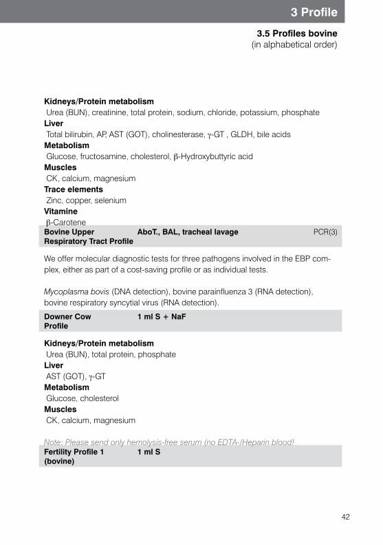
42
3 Profile
3.5 Profiles bovine(in alphabetical order)
Kidneys/Protein metabolism Urea (BUN), creatinine, total protein, sodium, chloride, potassium, phosphateLiver Total bilirubin, AP, AST (GOT), cholinesterase, γ-GT , GLDH, bile acids Metabolism Glucose, fructosamine, cholesterol, β-Hydroxybuttyric acidMuscles CK, calcium, magnesium Trace elements Zinc, copper, seleniumVitamine β-CaroteneBovine Upper Respiratory Tract Profile
AboT., BAL, tracheal lavage PCR(3)
We offer molecular diagnostic tests for three pathogens involved in the EBP com-plex, either as part of a cost-saving profile or as individual tests.
Mycoplasma bovis (DNA detection), bovine parainfluenza 3 (RNA detection), bovine respiratory syncytial virus (RNA detection).
Downer Cow Profile
1 ml S + NaF
Kidneys/Protein metabolism Urea (BUN), total protein, phosphate Liver AST (GOT), γ-GT Metabolism Glucose, cholesterolMuscles CK, calcium, magnesium
Note: Please send only hemolysis-free serum (no EDTA-/Heparin blood!Fertility Profile 1 (bovine)
1 ml S
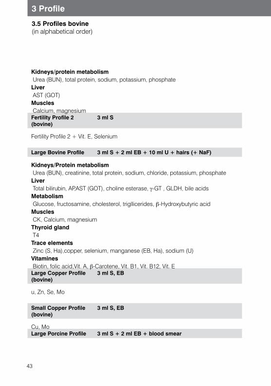
43
3 Profile
3.5 Profiles bovine(in alphabetical order)
Kidneys/protein metabolism Urea (BUN), total protein, sodium, potassium, phosphate Liver AST (GOT) Muscles Calcium, magnesiumFertility Profile 2 (bovine)
3 ml S
Fertility Profile 2 + Vit. E, Selenium
Large Bovine Profile 3 ml S + 2 ml EB + 10 ml U + hairs (+ NaF)
Kidneys/Protein metabolism Urea (BUN), creatinine, total protein, sodium, chloride, potassium, phosphateLiver Total bilirubin, AP,AST (GOT), choline esterase, γ-GT , GLDH, bile acids Metabolism Glucose, fructosamine, cholesterol, trigllicerides, β-Hydroxybutyric acid Muscles CK, Calcium, magnesium Thyroid gland T4 Trace elements Zinc (S, Ha),copper, selenium, manganese (EB, Ha), sodium (U) Vitamines Biotin, folic acid,Vit. A, β-Carotene, Vit. B1, Vit. B12, Vit. ELarge Copper Profile (bovine)
3 ml S, EB
u, Zn, Se, Mo
Small Copper Profile (bovine)
3 ml S, EB
Cu, MoLarge Porcine Profile 3 ml S + 2 ml EB + blood smear
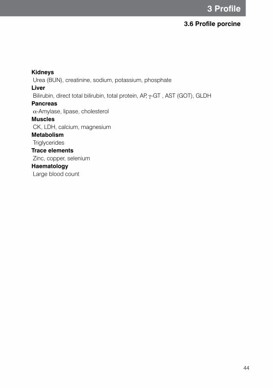
44
3 Profile
3.6 Profile porcine
Kidneys Urea (BUN), creatinine, sodium, potassium, phosphate Liver Bilirubin, direct total bilirubin, total protein, AP, γ-GT , AST (GOT), GLDH Pancreas α-Amylase, lipase, cholesterol Muscles CK, LDH, calcium, magnesium Metabolism TriglyceridesTrace elements Zinc, copper, selenium Haematology Large blood count

45
3 Profile
3.7 Profile camelid
Camelid Profile 2 ml S + 2 ml EB + blood smear
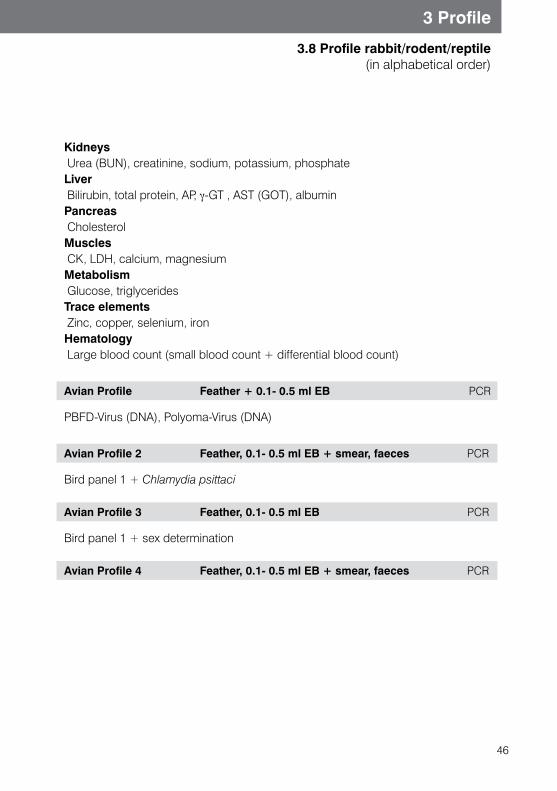
46
3 Profile
3.8 Profile rabbit/rodent/reptile (in alphabetical order)
Kidneys Urea (BUN), creatinine, sodium, potassium, phosphateLiver Bilirubin, total protein, AP, γ-GT , AST (GOT), albumin Pancreas Cholesterol Muscles CK, LDH, calcium, magnesium Metabolism Glucose, triglycerides Trace elements Zinc, copper, selenium, iron Hematology Large blood count (small blood count + differential blood count)
Avian Profile Feather + 0.1- 0.5 ml EB PCR
PBFD-Virus (DNA), Polyoma-Virus (DNA)
Avian Profile 2 Feather, 0.1- 0.5 ml EB + smear, faeces PCR
Bird panel 1 + Chlamydia psittaci
Avian Profile 3 Feather, 0.1- 0.5 ml EB PCR
Bird panel 1 + sex determination
Avian Profile 4 Feather, 0.1- 0.5 ml EB + smear, faeces PCR
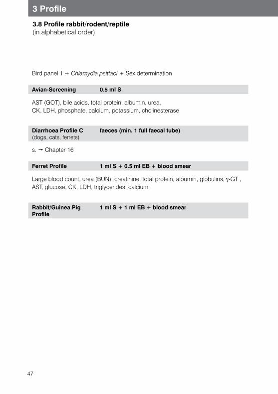
47
3 Profile
3.8 Profile rabbit/rodent/reptile (in alphabetical order)
Bird panel 1 + Chlamydia psittaci + Sex determination
Avian-Screening 0.5 ml S
AST (GOT), bile acids, total protein, albumin, urea, CK, LDH, phosphate, calcium, potassium, cholinesterase
Diarrhoea Profile C (dogs, cats, ferrets)
faeces (min. 1 full faecal tube)
s. Chapter 16
Ferret Profile 1 ml S + 0.5 ml EB + blood smear
Large blood count, urea (BUN), creatinine, total protein, albumin, globulins, γ-GT , AST, glucose, CK, LDH, triglycerides, calcium
Rabbit/Guinea Pig Profile
1 ml S + 1 ml EB + blood smear
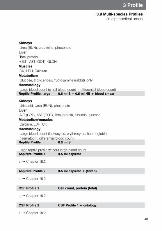
48
3 Profile
3.9 Multi-species Profiles (in alphabetical order)
Kidneys Urea (BUN), creatinine, phosphateLiver Total protein, γ-GT , AST (GOT), GLDH Muscles CK, LDH, Calcium Metabolism Glucose, triglycerides, fructosamine (rabbits only) Haematology Large blood count (small blood count + differential blood count)Reptile Profile, large 0.5 ml S + 0.5 ml HB + blood smear
Kidneys Uric acid, Urea (BUN), phosphateLiver ALT (GPT), AST (GOT), Total protein, albumin, glucose Metabolism/muscles Calcium, LDH, CK Haematology Large blood count (leukocytes, erythrocytes, haemoglobin, haematocrit, differential blood count)Reptile Profile 0.5 ml S
Large reptile profile without large blood count Aspirate Profile 1 3-5 ml aspirate
s. Chapter 18.2
Aspirate Profile 2 3-5 ml aspirate + (Swab)
s. Chapter 18.2
CSF Profile 1 Cell count, protein (total)
s. Chapter 18.2
CSF Profile 2 CSF Profile 1 + cytology
s. Chapter 18.2
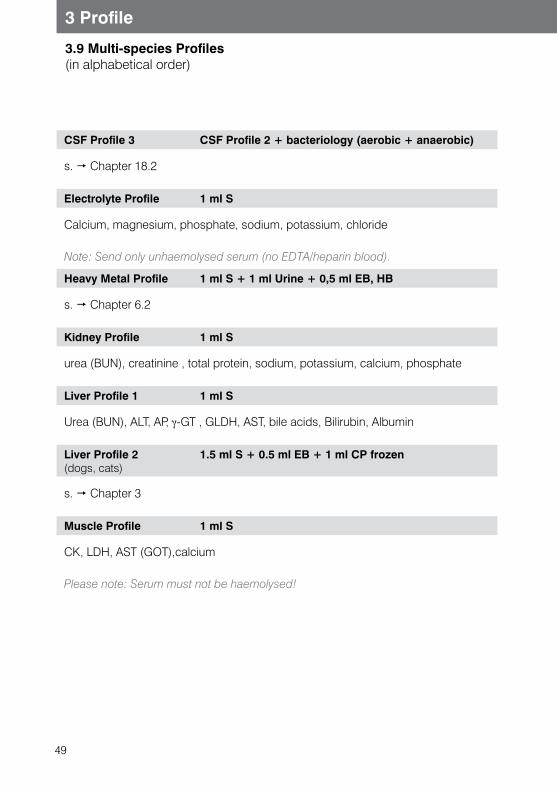
49
3 Profile
3.9 Multi-species Profiles (in alphabetical order)
CSF Profile 3 CSF Profile 2 + bacteriology (aerobic + anaerobic)
s. Chapter 18.2
Electrolyte Profile 1 ml S
Calcium, magnesium, phosphate, sodium, potassium, chloride
Note: Send only unhaemolysed serum (no EDTA/heparin blood).
Heavy Metal Profile 1 ml S + 1 ml Urine + 0,5 ml EB, HB
s. Chapter 6.2
Kidney Profile 1 ml S
urea (BUN), creatinine , total protein, sodium, potassium, calcium, phosphate
Liver Profile 1 1 ml S
Urea (BUN), ALT, AP, γ-GT , GLDH, AST, bile acids, Bilirubin, Albumin
Liver Profile 2 (dogs, cats)
1.5 ml S + 0.5 ml EB + 1 ml CP frozen
s. Chapter 3
Muscle Profile 1 ml S
CK, LDH, AST (GOT),calcium
Please note: Serum must not be haemolysed!
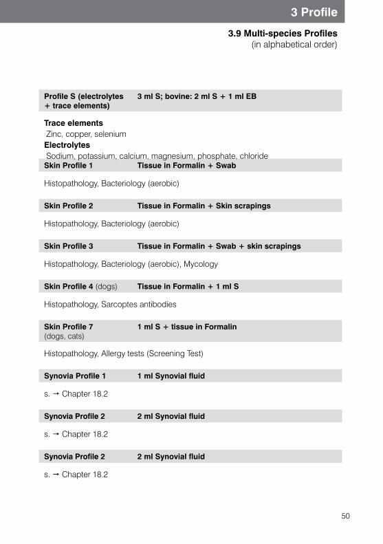
50
3 Profile
3.9 Multi-species Profiles (in alphabetical order)
Profile S (electrolytes + trace elements)
3 ml S; bovine: 2 ml S + 1 ml EB
Trace elements Zinc, copper, selenium Electrolytes Sodium, potassium, calcium, magnesium, phosphate, chlorideSkin Profile 1 Tissue in Formalin + Swab
Histopathology, Bacteriology (aerobic)
Skin Profile 2 Tissue in Formalin + Skin scrapings
Histopathology, Bacteriology (aerobic)
Skin Profile 3 Tissue in Formalin + Swab + skin scrapings
Histopathology, Bacteriology (aerobic), Mycology
Skin Profile 4 (dogs) Tissue in Formalin + 1 ml S
Histopathology, Sarcoptes antibodies
Skin Profile 7 (dogs, cats)
1 ml S + tissue in Formalin
Histopathology, Allergy tests (Screening Test)
Synovia Profile 1 1 ml Synovial fluid
s. Chapter 18.2
Synovia Profile 2 2 ml Synovial fluid
s. Chapter 18.2
Synovia Profile 2 2 ml Synovial fluid
s. Chapter 18.2
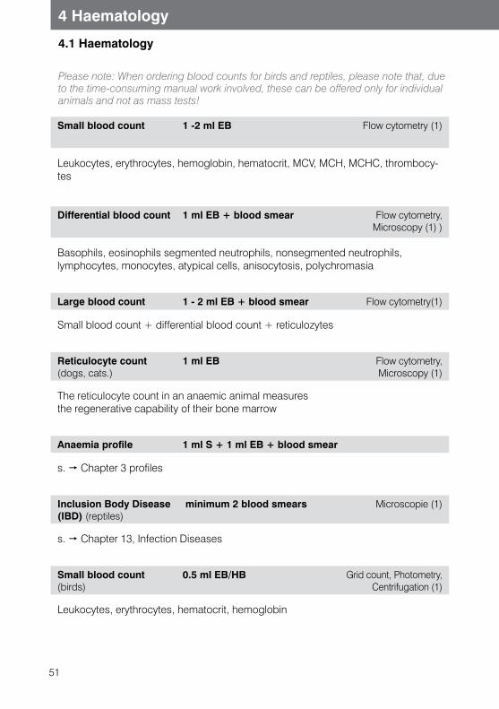
51
4.1 Haematology
4 Haematology
Small blood count 1 -2 ml EB Flow cytometry (1)
Leukocytes, erythrocytes, hemoglobin, hematocrit, MCV, MCH, MCHC, thrombocy-tes
Differential blood count 1 ml EB + blood smear Flow cytometry, Microscopy (1) )
Basophils, eosinophils segmented neutrophils, nonsegmented neutrophils,lymphocytes, monocytes, atypical cells, anisocytosis, polychromasia
Large blood count 1 - 2 ml EB + blood smear Flow cytometry(1)
Small blood count + differential blood count + reticulozytes
Reticulocyte count(dogs, cats.)
1 ml EB Flow cytometry, Microscopy (1)
The reticulocyte count in an anaemic animal measures the regenerative capability of their bone marrow
Anaemia profile 1 ml S + 1 ml EB + blood smear
s. Chapter 3 profiles
Inclusion Body Disease (IBD) (reptiles)
minimum 2 blood smears Microscopie (1)
s. Chapter 13, Infection Diseases
Small blood count (birds)
0.5 ml EB/HB Grid count, Photometry, Centrifugation (1)
Leukocytes, erythrocytes, hematocrit, hemoglobin
Please note: When ordering blood counts for birds and reptiles, please note that, due to the time-consuming manual work involved, these can be offered only for individual animals and not as mass tests!
52
4 Haematology
4.1 Haematology
Differential blood count(birds)
0.5 ml EB,HB + blood smear Microscopy (1)
Basophils, eosinophils, heterophils, lymphocytes, monocytes, anisocytes, polichro-masia
Large blood count (birds)
0.5 ml EB, HB+ blood smear Microscopy (1)
Small blood count + differential blood count
Small blood count (reptiles)
1 ml HB + blood smear Grid count, Photometry, centrifugation(1)
Leukocytes, erythrocytes, hematocrit, hemoglobin
Differential blood count(Reptiles)
0.5 ml HB + blood smear Mikroskopie (1)
Basophils, eosinophils, heterophils, lymphocytes, monocytes, azurophils, anisocy-tes, polychromasia.
Note: Bird and reptile erthrocytes and thrombocytes contain cell nuclei. For this reason automatic cell count is not possibile. In addition, do not use EDTA with reptile blood, as it may cause haemolysis. Please use heparin as the preferred anticoagu-lant.
Large blood count (Reptiles)
0.5 ml HB + blood smear Grid count photometry, centrifugation (1)
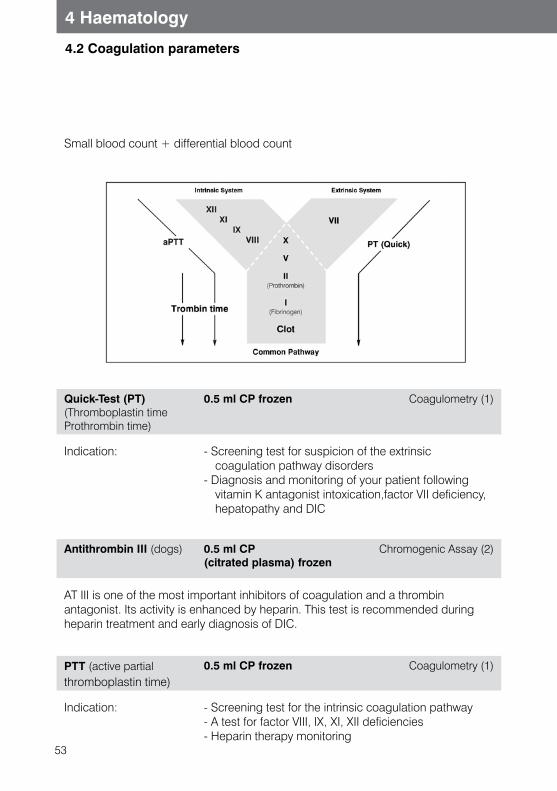
53
4 Haematology
4.2 Coagulation parameters
Small blood count + differential blood count
Quick-Test (PT)(Thromboplastin time Prothrombin time)
0.5 ml CP frozen Coagulometry (1)
Indication: - Screening test for suspicion of the extrinsic coagulation pathway disorders
- Diagnosis and monitoring of your patient following vitamin K antagonist intoxication,factor VII deficiency, hepatopathy and DIC
Antithrombin III (dogs) 0.5 ml CP(citrated plasma) frozen
Chromogenic Assay (2)
AT III is one of the most important inhibitors of coagulation and a thrombin antagonist. Its activity is enhanced by heparin. This test is recommended during heparin treatment and early diagnosis of DIC.
PTT (active partial thromboplastin time)
0.5 ml CP frozen Coagulometry (1)
Indication:
- Screening test for the intrinsic coagulation pathway - A test for factor VIII, IX, XI, XII deficiencies- Heparin therapy monitoring
54
4 Haematology
4.2 Coagulation parameters
Thrombin time 0.5 ml CP frozen Coagulometry (1)
Indication: - Suspected fibrinogen deficiency or disturbance of- Fibrinogen production- Fibrinolysis therapy monitoring- Heparin therapy monitoring
D-Dimer (dogs only) 0.5 ml CP frozen Immunogy disorders Test (2)
D-Dimer is a fibrin degradation product. This screening is used to test fibrinolytic activation.Increased D-dimer concentration is seen in dogs with DIC, thromboembolism and also in acute kidney failure, neoplasia, immune-mediated anaemia and other diseases.
Fibrinogen 1 ml CP frozen Coagulometry (1)
Indications: DIC, hepatopathy, fibrinogen deficiency, consumptive coagulopathy or hyperfibrinolysis
- As an acute phase protein marker during inflammation.
Large Coagulation Screening
1 ml CP frozen Coagulometry (1)
Fibrinogen, PTT, Quick-Test, thrombin time
Coagulation Screening (dogs)
1 - 2 ml CP frozen Coagulometry(1)
Total coagulation status, D-Dimer, Antithrombin III
Factor VIII (dogs) 0.5 ml CP frozen Coagulometry(1)
Indications: Diagnosis of hemophilia A (Factor VIII deficiency)
Factor IX (Hd.) 0.5 ml CP frozen Coagulometry(1)
Indications: Diagnosis of hemophilia B (Factor IX deficiency )
55
4 Haematology
4.2 Coagulation parameters
Von Willebrand-Factor- Antigen (vWF: Ag) (dogs)
1 ml EB Immunologic disorder test (1)
Von Willebrand factor mediates the adhesion of thrombocytes to the endothelial wall of blood vessels and acts as a carrier protein for factor VIII. Von Willebrand syndrome has been described in numerous dog breeds, but is most frequently seen in Dobermanns and Scottish Terriers.Test is indicated if PTT or skin/mucosal membrane bleeding time is increasd.
Von-Willebrand-Factor 1 - 3
1 ml EB PCR (3)
s. Chapter 15, Molecular biology tests
56
4 Haematology
4.3 Blood groups
Blood group typing (dogs, cats)
0.5 ml EB, HB + blood smear immmunochromato-graphic test (1)
Dogs There are currently 13 known blood groups described in dogs.These are known as DEA (dog erythrocyte antigen) 1.1,1.2,etc. Dogs do not have any clinically significant endogenous antibodies towards other blood groups. This is why transfusion reaction haemolysis is not expected following the first blood transfusion. We test for blood group DEA1.1, as this is the group with the strongest antigenic potential, and may cause significant antibody production leading to delayed hemolysis.An animal which has never had a blood transfusion has no naturally occurring clinically relevant antibodies. However once a DEA 1.1-negative animal has ben sensitized by a DEA 1.1-positive transfusion, a further DEA 1.1-positive transfusion may cause acute hemolytic transfusion reaction.
Cats Cats have blood groups A, B and AB. The most common one is blood group A (96%). Type B varies according to the breed, with an increased prevalence in Devon Rex or British Shorthair cats (20-45%).Blood group AB is extremely rare. Cats have naturallyoccurring antibodies towards other blood groups, so the blood group of both the donor and recipient should be checked before any transfusion.Blood group testing reduces the chance of the occurrence of neonatal erythrolysis when both parents are tested prior to breeding, as there is a great risk of neonatal erythrolysis if kittens of A (or AB) blood group are born from B blood group mother.
57
4 Haematology
4.4 Blood parasites and haemotropic bacteria
Blood parasites and haemotropic bacteria
0.5 ml EB + blood smear Microscopy (1)
Microscopy of Giemsa stained blood smear, looking for Babesia, Ehrlichia, Anaplasma, Hepatozoon or other pathogens. Direct pathogen detection is possible only in parasitic or bacteriemic phase; therefore multiple tests may be required to rule out disease.
s. Travel Profile 1 + 3 s. Chapter 13, Infectious diseases
Microfilaria - Knott Test 1 - 2 ml EB Microscopy (1)
s. Chapter 13, Infectious diseases
Dirofilaria immitis (Macrofilaria) (Ag)
1 ml S, EP, HP Filtration test, Microscopy (1)
s. Chapter 13, Infectious diseases
58
5 Biochemistry
Albumin 0.3 ml S, EP, HP Photometry (1) )
Indications: HepatopathiesNephropathiesDetermination of albumin/globulin ratio (FIP diagnostics)
Occurrence: Albumin is synthesizedin the liver
Decreased in: - Protein deficiency (nutritional)- Anorexia- Malassimilation- Hepatopathies- Renal, glomerular loss (nephritis, nephrotic syndrome)- Protein-losing enteropathy- FIP- Burns- Blood loss- Body cavity effusions- Hypoadrenocorticism- CNS disease- Relative deficiency due to overhydration- Hypergammaglobulinaemia
Increased in: - Dehydration
59
5 Biochemistry
Alkaline Phosphatase (AP)
0.3 ml S, HP Enzyme kinetics, Photometry (1)
Indications: HepatopathiesHyperadrenocorticismOsteopathies
Occurrence: Alkaline phosphatase is found in the liver (membrane-bound in biliary duct epithelium), small intestinal mucosa, bones, kidneys, placenta, spleen, leukocytes, and erythrocytes
Increased in physiologically- Growthliver specific increase- Hepatopathies with intra- or extrahepatic
cholestasis (cats/ruminants show a very slow reaction)- Liver neoplasia- Hepatotoxicity- Pancreatitisnon-specific increase- Hyperadrenocorticism (esp. dogs)- Hyperthyroidism- Diabetes mellitus- Hyperparathyroidism- Bone healing- Osteopathies- Neoplasias- Pregnancy (esp. cats)- Medication (e.g. glucocorticoids, anticonvulsant drugs,
barbiturates, certain antibiotics)
Results affected by: Haemolysis, EDTA, severe lipaemia and bilirubinaemia
Please note: Young animals show considerably higher alkaline phosphatase values than adults.
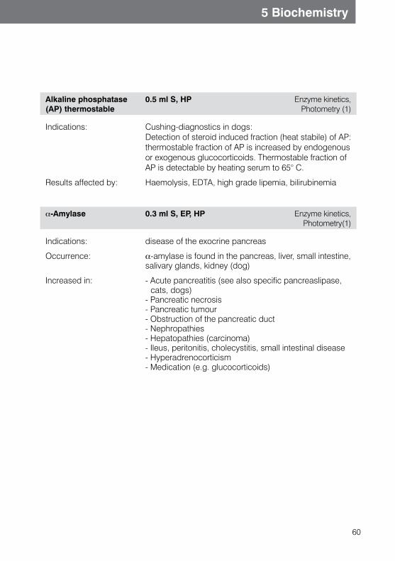
60
5 Biochemistry
Alkaline phosphatase (AP) thermostable
0.5 ml S, HP Enzyme kinetics, Photometry (1)
Indications: Cushing-diagnostics in dogs: Detection of steroid induced fraction (heat stabile) of AP: thermostable fraction of AP is increased by endogenous or exogenous glucocorticoids. Thermostable fraction of AP is detectable by heating serum to 65° C.
Results affected by: Haemolysis, EDTA, high grade lipemia, bilirubinemia
a-Amylase 0.3 ml S, EP, HP Enzyme kinetics, Photometry(1)
Indications: disease of the exocrine pancreas
Occurrence: a-amylase is found in the pancreas, liver, small intestine,salivary glands, kidney (dog)
Increased in: - Acute pancreatitis (see also specific pancreaslipase, cats, dogs)
- Pancreatic necrosis- Pancreatic tumour- Obstruction of the pancreatic duct- Nephropathies- Hepatopathies (carcinoma)- Ileus, peritonitis, cholecystitis, small intestinal disease- Hyperadrenocorticism- Medication (e.g. glucocorticoids)
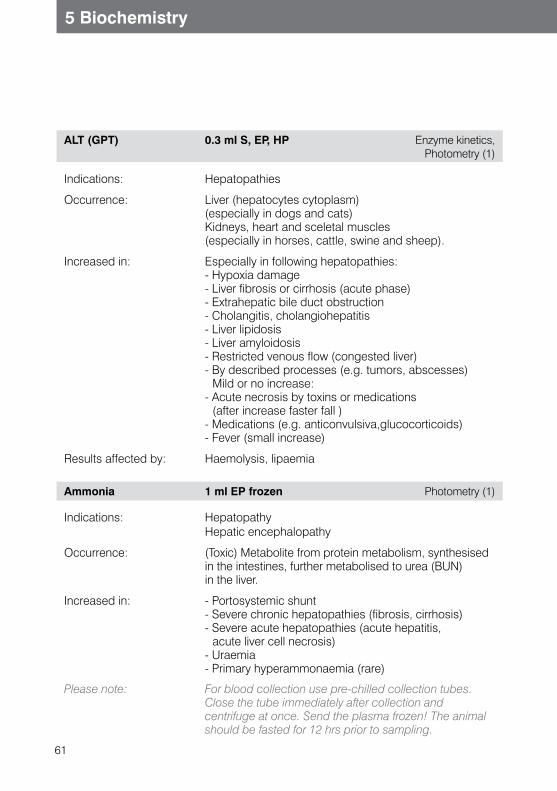
61
5 Biochemistry
ALT (GPT) 0.3 ml S, EP, HP Enzyme kinetics, Photometry (1)
Indications: Hepatopathies
Occurrence: Liver (hepatocytes cytoplasm) (especially in dogs and cats) Kidneys, heart and sceletal muscles (especially in horses, cattle, swine and sheep).
Increased in: Especially in following hepatopathies: - Hypoxia damage - Liver fibrosis or cirrhosis (acute phase) - Extrahepatic bile duct obstruction- Cholangitis, cholangiohepatitis - Liver lipidosis- Liver amyloidosis - Restricted venous flow (congested liver)- By described processes (e.g. tumors, abscesses)
Mild or no increase: - Acute necrosis by toxins or medications
(after increase faster fall ) - Medications (e.g. anticonvulsiva,glucocorticoids) - Fever (small increase)
Results affected by: Haemolysis, lipaemia
Ammonia 1 ml EP frozen Photometry (1)
Indications: HepatopathyHepatic encephalopathy
Occurrence: (Toxic) Metabolite from protein metabolism, synthesised in the intestines, further metabolised to urea (BUN) in the liver.
Increased in: - Portosystemic shunt- Severe chronic hepatopathies (fibrosis, cirrhosis)- Severe acute hepatopathies (acute hepatitis,
acute liver cell necrosis)- Uraemia- Primary hyperammonaemia (rare)
Please note: For blood collection use pre-chilled collection tubes. Close the tube immediately after collection and centrifuge at once. Send the plasma frozen! The animal should be fasted for 12 hrs prior to sampling.
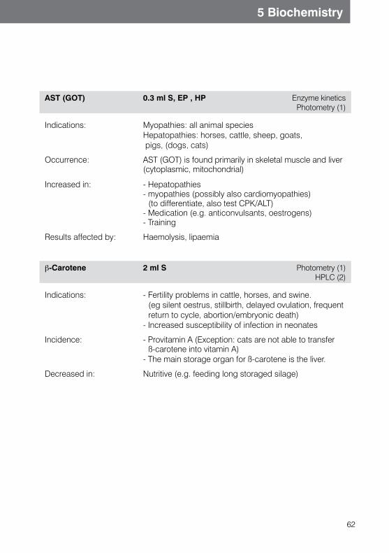
62
5 Biochemistry
AST (GOT) 0.3 ml S, EP , HP Enzyme kinetics Photometry (1)
Indications: Myopathies: all animal speciesHepatopathies: horses, cattle, sheep, goats, pigs, (dogs, cats)
Occurrence:
AST (GOT) is found primarily in skeletal muscle and liver(cytoplasmic, mitochondrial)
Increased in: - Hepatopathies- myopathies (possibly also cardiomyopathies)
(to differentiate, also test CPK/ALT)- Medication (e.g. anticonvulsants, oestrogens)- Training
Results affected by: Haemolysis, lipaemia
b-Carotene 2 ml S Photometry (1) HPLC (2)
Indications: - Fertility problems in cattle, horses, and swine. (eg silent oestrus, stillbirth, delayed ovulation, frequent return to cycle, abortion/embryonic death)
- Increased susceptibility of infection in neonates
Incidence: - Provitamin A (Exception: cats are not able to transfer ß-carotene into vitamin A)
- The main storage organ for ß-carotene is the liver.
Decreased in: Nutritive (e.g. feeding long storaged silage)
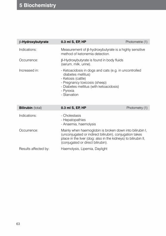
63
5 Biochemistry
b-Hydroxybutyrate 0.3 ml S, EP, HP Photometrie (1)
Indications: Measurement of b-hydroxybutyrate is a highly sensitive method of ketonemia detection.
Occurrence: b-Hydroxybutyrate is found in body fluids (serum, milk, urine).
Increased in: - Ketoacidosis in dogs and cats (e.g. in uncontrolled diabetes mellitus)
- Ketosis (cattle)- Pregnancy toxicosis (sheep)- Diabetes mellitus (with ketoacidosis)- Pyrexia- Starvation
Bilirubin (total) 0.3 ml S, EP, HP Photometry (1)
Indications: - Cholestasis - Hepatopathies- Anaemia, haemolysis
Occurrence: Mainly when haemoglobin is broken down into bilirubin I,(unconjugated or indirect bilirubin), conjugation takes place in the liver (dog: also in the kidneys) to bilirubin II, (conjugated or direct bilirubin).
Results affected by: Haemolysis, Lipemia, Daylight
64
5 Biochemistry
Bile acids 0.3 ml S, EP, HP Photometry (1)
Indications: Hepatopathies
Occurrence: Bile acids are synthesised in the liver from cholesterol. They are responsible for digestion and absorption of lipids in the intestines (bile acids reach the intestines in bile and a small amount is passed with the faeces. A larger amount is reabsorbed and transported back to the liver).
Hepatopathies lead to disturbances in bile acid secretion. The accumulation of bile acids then leads tofunctional disorders due to their toxic properties.
Increased in: specific increaseLiver and bile duct disease with intra- or post-hepatic cholestasis, e.g.- Hepatitis- Chronic hepatitis- Portosystemic shunt
non specific increase- An increase is physiologically normal up to 24 hrs
following a fatty meal- Hyperthyroidism- Hyperadrenocorticism- Diabetes mellitus
Please note Please note: Animal must be fasted 12 hrsprior to sampling!
65
5 Biochemistry
Bile AcidStimulation Test
2 x 0.3 ml S, EP, HP Photometry (1)
Test principle: - Testing for liver function disorders- Suspected portosystemic shunt
Test method: Under normal conditions blood bile acid concentration will increase following a fatty meal.If the liver function is disturbed, or in the case of ashunt this increase will be abnormally high.1. the first blood sample measures bile acid basal level
(starved animal)2. stress feeding (small fatty meal or recovery diet)3. second blood sample 2 hrs following the
meal measures the postprandial (post-feeding) level
or
1. The first blood sample measures bile acid basal level (starved animal)
2. Injection of Ceruletid (Takus®, Pharmacia) 0.3 μg/kg i.m.
3. Second blood sample 20 minutes post-injection mea-sures the bile acid stimulation value
Interpretation: - Basal level < 20 μmol/l and postprandial level < 40 μmol/l = normal
- Basal level > 20 μmol/l and postprandial level 20-40 μmol/l = borderline
- Postprandial level > 40 μmol/l = pathological
66
5 Biochemistry
Calcium 0.3 ml S, HP Photometry (1)
Occurrence: Mainly in bones
Increased in: - Primary/tertiary hyperparathyroidism- Hypervitaminosis D- Hypoadrenocorticism- Acidosis- Neoplasia (lymphoma, adenocarcinoma)- Osteolytic tumours- Osteomyelitis- Osteoporosis- Nephropathies- Hyperalbuminaemia (increase of the protein bound
part)- malignant hypercalcaemia
Decreased in: - Hypoparathyroidism- Secondary (renal) hyperparathyroidism- Nephropathies- Hypoalbuminaemia- Hypovitaminosis D- (Necrotic) pancreatitis- Tetanus- Puerperal tetany- Milk fever (parturient paresis)- Malabsorption- Hypercalcitonism- Ethylene glycol intoxication (e.g. antifreeze)
Results affected by: Lipaemia, Haemolysis, EDTA
Cardiopet® proBNP (Nt-proBNP)
dogs: 3.0 ml EPcats: 0.3 ml S
ELISA (1)
Dogs with heart murmur: In dogs with a heart murmur and clinical symptoms such as respiratory symptoms and/or exercise intolerance, this test determines the probability of these symptoms being caused by heart disease.The test helps to assess the risk of a dog developing congestive heart failure over the next 12 months, when presented with a heart murmur due to degenerative mitral valve disease, but which does not currently exhibit clinical symptoms.
67
5 Biochemistry
Indications in cats: The Cardiopet® proBNP Test can also be used in all cats as a screening test (e.g. pre-anaesthesia, as part of preventive testing, in predisposed breeds) or in animals with suspected heart disease and indicates the probability of cardiomyopathy. The result makes it easier to decide whether a further cardiac workup of the patient is necessary.
Results affected by: - Haemolysis - Lipemia
Chloride 0.2 ml S, EP, HP Ion selective electrode (1)
Indications:
Electrolyte disturbances
Occurrence:
Chloride is the most important extracellular anion in the living organism. Under normal physiologicalconditions of acid/base balance the serum chloride concentration equals the sodium concentration.
Increased in
- Dehydration (fluid loss, reduced fluid intake)- Increased intake of sodium chloride- Diabetes insipidus- Diabetes mellitus (following insulin therapy)- Mineralocorticoids (retention of sodium)- Nephropathy- Acidosis- Small intestinal diarrhoea
Decreased in: - Increased loss of sodium chloride (vomiting, diarrhoea, sweating)
- Insufficient intake of sodium chloride- Increased intake of water- Hypoadrenocorticism- Osmotic diuresis (e.g. diabetes mellitus)- Congestive heart failure (oedema)- Nephropathy- Loop diuretics (e.g. furosemide), - Aldosterone antagonists (e.g. spironolactone)- Reduced colloid osmotic pressure
(hypoalbuminaemia)- Metabolic alkalosis
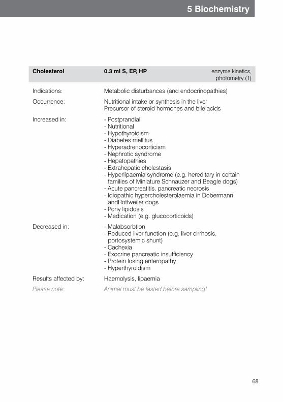
68
5 Biochemistry
Cholesterol 0.3 ml S, EP, HP enzyme kinetics, photometry (1)
Indications: Metabolic disturbances (and endocrinopathies)
Occurrence:
Nutritional intake or synthesis in the liverPrecursor of steroid hormones and bile acids
Increased in:
- Postprandial- Nutritional- Hypothyroidism- Diabetes mellitus- Hyperadrenocorticism- Nephrotic syndrome- Hepatopathies- Extrahepatic cholestasis- Hyperlipaemia syndrome (e.g. hereditary in certain
families of Miniature Schnauzer and Beagle dogs)- Acute pancreatitis, pancreatic necrosis- Idiopathic hypercholesterolaemia in Dobermann
andRottweiler dogs- Pony lipidosis- Medication (e.g. glucocorticoids)
Decreased in: - Malabsorbtion- Reduced liver function (e.g. liver cirrhosis,
portosystemic shunt)- Cachexia- Exocrine pancreatic insufficiency- Protein losing enteropathy- Hyperthyroidism
Results affected by: Haemolysis, lipaemia
Please note: Animal must be fasted before sampling!
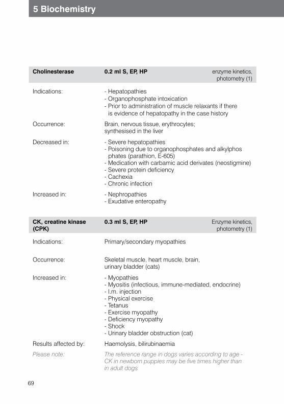
69
5 Biochemistry
Cholinesterase 0.2 ml S, EP, HP enzyme kinetics, photometry (1)
Indications:
- Hepatopathies- Organophosphate intoxication- Prior to administration of muscle relaxants if there
is evidence of hepatopathy in the case history
Occurrence:
Brain, nervous tissue, erythrocytes;synthesised in the liver
Decreased in:
- Severe hepatopathies- Poisoning due to organophosphates and alkylphos
phates (parathion, E-605)- Medication with carbamic acid derivates (neostigmine)- Severe protein deficiency- Cachexia- Chronic infection
Increased in: - Nephropathies- Exudative enteropathy
CK, creatine kinase (CPK)
0.3 ml S, EP, HP Enzyme kinetics, photometry (1)
Indications:
Primary/secondary myopathies
Occurrence:
Skeletal muscle, heart muscle, brain, urinary bladder (cats)
Increased in:
- Myopathies- Myositis (infectious, immune-mediated, endocrine)- I.m. injection- Physical exercise- Tetanus- Exercise myopathy- Deficiency myopathy- Shock- Urinary bladder obstruction (cat)
Results affected by: Haemolysis, bilirubinaemia
Please note: The reference range in dogs varies according to age - CK in newborn puppies may be five times higher than in adult dogs
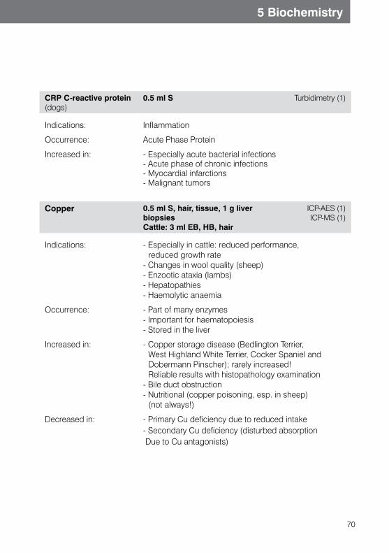
70
5 Biochemistry
CRP C-reactive protein(dogs)
0.5 ml S Turbidimetry (1)
Indications: Inflammation
Occurrence: Acute Phase Protein
Increased in: - Especially acute bacterial infections- Acute phase of chronic infections- Myocardial infarctions - Malignant tumors
Copper 0.5 ml S, hair, tissue, 1 g liver biopsiesCattle: 3 ml EB, HB, hair
ICP-AES (1) ICP-MS (1)
Indications: - Especially in cattle: reduced performance, reduced growth rate
- Changes in wool quality (sheep)- Enzootic ataxia (lambs)- Hepatopathies- Haemolytic anaemia
Occurrence: - Part of many enzymes- Important for haematopoiesis- Stored in the liver
Increased in: - Copper storage disease (Bedlington Terrier, West Highland White Terrier, Cocker Spaniel and Dobermann Pinscher); rarely increased! Reliable results with histopathology examination
- Bile duct obstruction- Nutritional (copper poisoning, esp. in sheep)
(not always!)
Decreased in: - Primary Cu deficiency due to reduced intake- Secondary Cu deficiency (disturbed absorption Due to Cu antagonists)
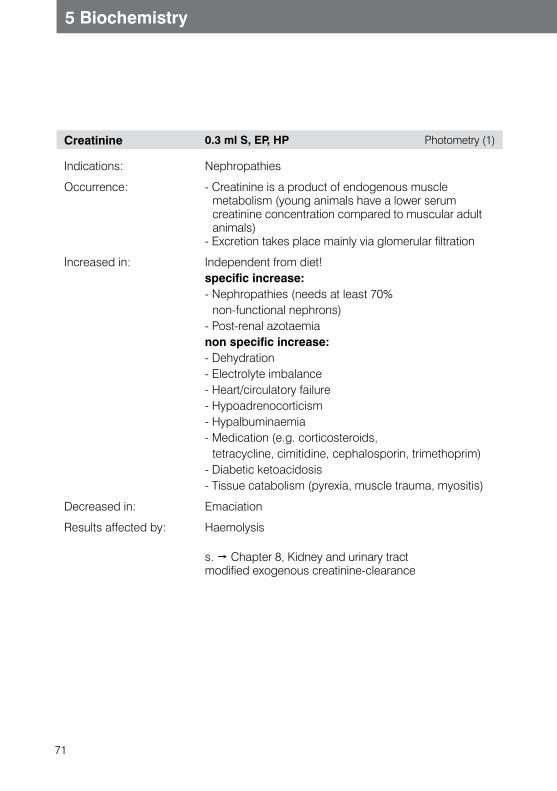
71
5 Biochemistry
Creatinine 0.3 ml S, EP, HP Photometry (1)
Indications: Nephropathies
Occurrence: - Creatinine is a product of endogenous muscle metabolism (young animals have a lower serum creatinine concentration compared to muscular adult animals)
- Excretion takes place mainly via glomerular filtration
Increased in: Independent from diet!specific increase:- Nephropathies (needs at least 70%
non-functional nephrons)- Post-renal azotaemianon specific increase:- Dehydration- Electrolyte imbalance- Heart/circulatory failure- Hypoadrenocorticism- Hypalbuminaemia- Medication (e.g. corticosteroids,
tetracycline, cimitidine, cephalosporin, trimethoprim)- Diabetic ketoacidosis- Tissue catabolism (pyrexia, muscle trauma, myositis)
Decreased in: Emaciation
Results affected by: Haemolysis
s. Chapter 8, Kidney and urinary tractmodified exogenous creatinine-clearance
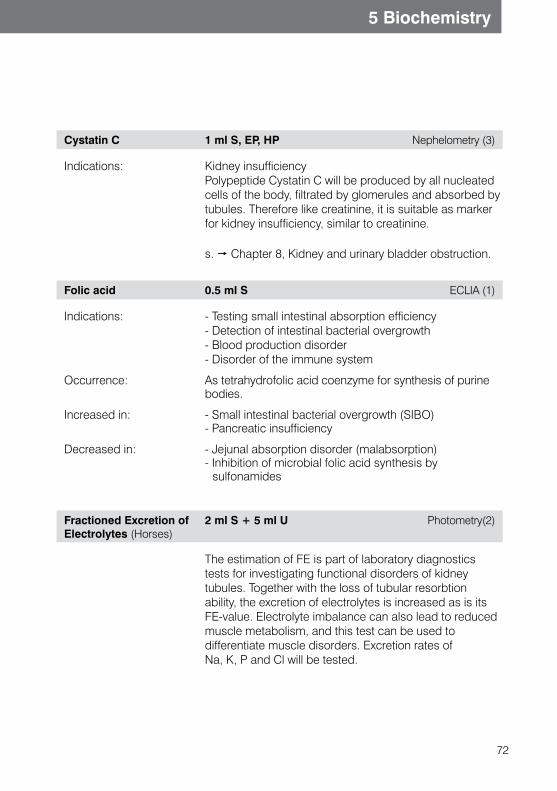
72
5 Biochemistry
Cystatin C 1 ml S, EP, HP Nephelometry (3)
Indications: Kidney insufficiencyPolypeptide Cystatin C will be produced by all nucleated cells of the body, filtrated by glomerules and absorbed by tubules. Therefore like creatinine, it is suitable as marker for kidney insufficiency, similar to creatinine.
s. Chapter 8, Kidney and urinary bladder obstruction.
Folic acid 0.5 ml S ECLIA (1)
Indications: - Testing small intestinal absorption efficiency- Detection of intestinal bacterial overgrowth- Blood production disorder- Disorder of the immune system
Occurrence: As tetrahydrofolic acid coenzyme for synthesis of purine bodies.
Increased in: - Small intestinal bacterial overgrowth (SIBO)- Pancreatic insufficiency
Decreased in: - Jejunal absorption disorder (malabsorption)- Inhibition of microbial folic acid synthesis by
sulfonamides
Fractioned Excretion of Electrolytes (Horses)
2 ml S + 5 ml U Photometry(2)
The estimation of FE is part of laboratory diagnostics tests for investigating functional disorders of kidney tubules. Together with the loss of tubular resorbtion ability, the excretion of electrolytes is increased as is its FE-value. Electrolyte imbalance can also lead to reduced muscle metabolism, and this test can be used to differentiate muscle disorders. Excretion rates of Na, K, P and Cl will be tested.
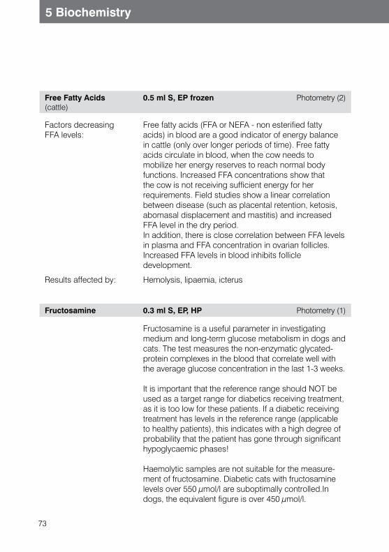
73
5 Biochemistry
Free Fatty Acids(cattle)
0.5 ml S, EP frozen Photometry (2)
Factors decreasing FFA levels:
Free fatty acids (FFA or NEFA - non esterified fatty acids) in blood are a good indicator of energy balance in cattle (only over longer periods of time). Free fatty acids circulate in blood, when the cow needs to mobilize her energy reserves to reach normal body functions. Increased FFA concentrations show that the cow is not receiving sufficient energy for her requirements. Field studies show a linear correlation between disease (such as placental retention, ketosis, abomasal displacement and mastitis) and increased FFA level in the dry period.In addition, there is close correlation between FFA levels in plasma and FFA concentration in ovarian follicles. Increased FFA levels in blood inhibits follicle development.
Results affected by: Hemolysis, lipaemia, icterus
Fructosamine 0.3 ml S, EP, HP Photometry (1)
Fructosamine is a useful parameter in investigating medium and long-term glucose metabolism in dogs and cats. The test measures the non-enzymatic glycated-protein complexes in the blood that correlate well with the average glucose concentration in the last 1-3 weeks.
It is important that the reference range should NOT be used as a target range for diabetics receiving treatment, as it is too low for these patients. If a diabetic receiving treatment has levels in the reference range (applicable to healthy patients), this indicates with a high degree of probability that the patient has gone through significant hypoglycaemic phases!
Haemolytic samples are not suitable for the measure-ment of fructosamine. Diabetic cats with fructosamine levels over 550 μmol/l are suboptimally controlled.In dogs, the equivalent figure is over 450 μmol/l.
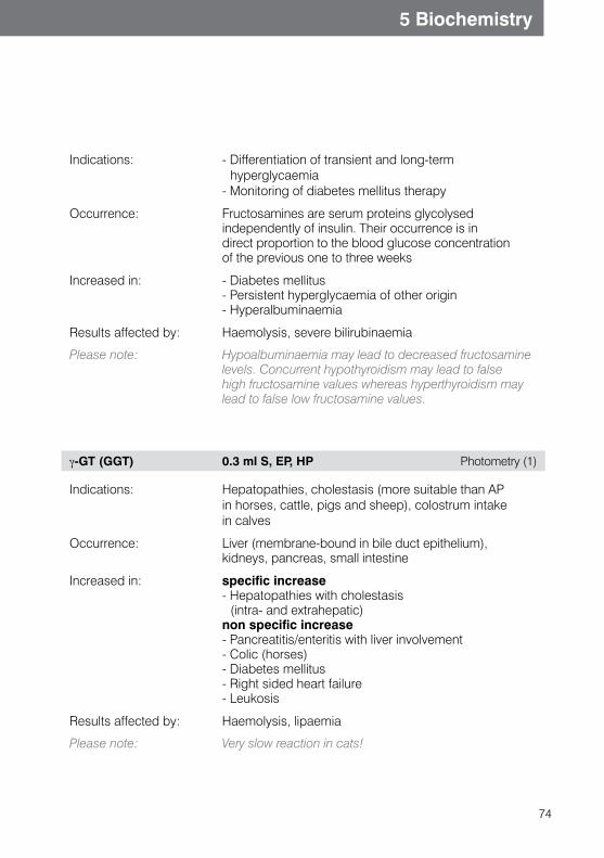
74
5 Biochemistry
Indications: - Differentiation of transient and long-term hyperglycaemia
- Monitoring of diabetes mellitus therapy
Occurrence: Fructosamines are serum proteins glycolysedindependently of insulin. Their occurrence is indirect proportion to the blood glucose concentrationof the previous one to three weeks
Increased in: - Diabetes mellitus- Persistent hyperglycaemia of other origin- Hyperalbuminaemia
Results affected by: Haemolysis, severe bilirubinaemia
Please note: Hypoalbuminaemia may lead to decreased fructosamine levels. Concurrent hypothyroidism may lead to falsehigh fructosamine values whereas hyperthyroidism may lead to false low fructosamine values.
g-GT (GGT) 0.3 ml S, EP, HP Photometry (1)
Indications: Hepatopathies, cholestasis (more suitable than AP in horses, cattle, pigs and sheep), colostrum intake in calves
Occurrence: Liver (membrane-bound in bile duct epithelium),kidneys, pancreas, small intestine
Increased in: specific increase- Hepatopathies with cholestasis
(intra- and extrahepatic)non specific increase- Pancreatitis/enteritis with liver involvement- Colic (horses)- Diabetes mellitus- Right sided heart failure- Leukosis
Results affected by: Haemolysis, lipaemia
Please note: Very slow reaction in cats!
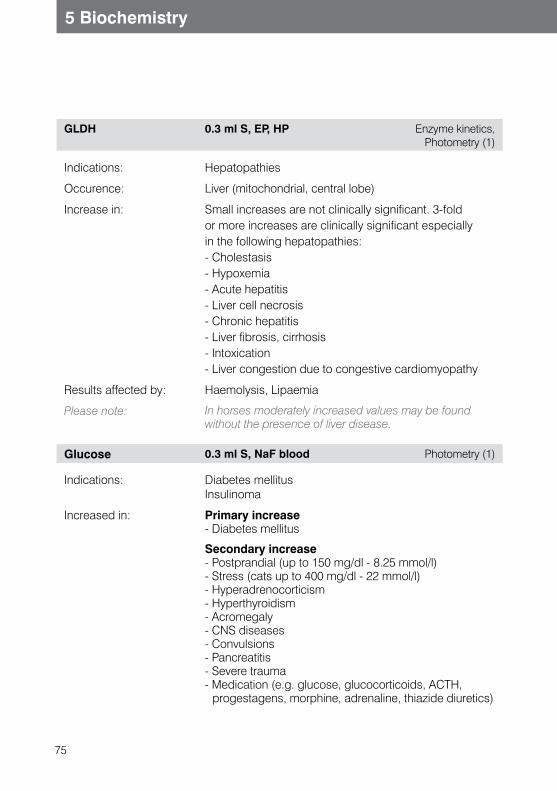
75
5 Biochemistry
GLDH 0.3 ml S, EP, HP Enzyme kinetics, Photometry (1)
Indications: Hepatopathies
Occurence: Liver (mitochondrial, central lobe)
Increase in: Small increases are not clinically significant. 3-foldor more increases are clinically significant especiallyin the following hepatopathies:- Cholestasis- Hypoxemia- Acute hepatitis- Liver cell necrosis- Chronic hepatitis- Liver fibrosis, cirrhosis- Intoxication- Liver congestion due to congestive cardiomyopathy
Results affected by: Haemolysis, Lipaemia
Please note: In horses moderately increased values may be found without the presence of liver disease.
Glucose 0.3 ml S, NaF blood Photometry (1)
Indications: Diabetes mellitusInsulinoma
Increased in: Primary increase- Diabetes mellitus
Secondary increase- Postprandial (up to 150 mg/dl - 8.25 mmol/l)- Stress (cats up to 400 mg/dl - 22 mmol/l)- Hyperadrenocorticism- Hyperthyroidism- Acromegaly- CNS diseases- Convulsions- Pancreatitis- Severe trauma- Medication (e.g. glucose, glucocorticoids, ACTH,
progestagens, morphine, adrenaline, thiazide diuretics)
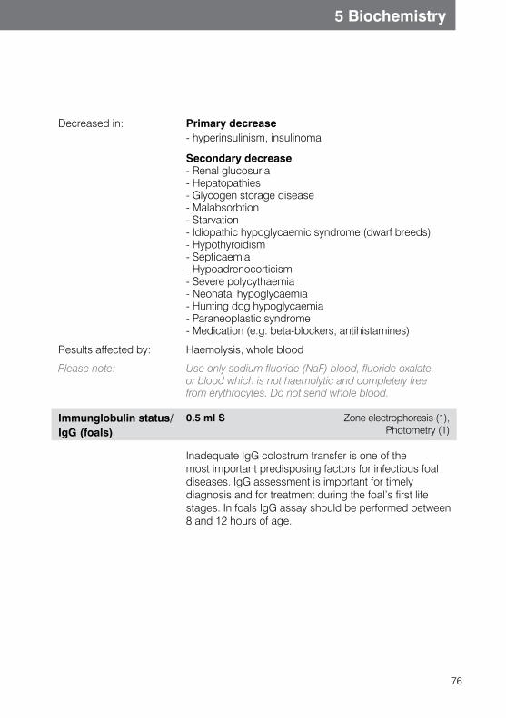
76
5 Biochemistry
Decreased in: Primary decrease- hyperinsulinism, insulinoma
Secondary decrease- Renal glucosuria- Hepatopathies- Glycogen storage disease- Malabsorbtion- Starvation- Idiopathic hypoglycaemic syndrome (dwarf breeds)- Hypothyroidism- Septicaemia- Hypoadrenocorticism- Severe polycythaemia- Neonatal hypoglycaemia- Hunting dog hypoglycaemia- Paraneoplastic syndrome- Medication (e.g. beta-blockers, antihistamines)
Results affected by: Haemolysis, whole blood
Please note: Use only sodium fluoride (NaF) blood, fluoride oxalate, or blood which is not haemolytic and completely free from erythrocytes. Do not send whole blood.
Immunglobulin status/IgG (foals)
0.5 ml S Zone electrophoresis (1), Photometry (1)
Inadequate IgG colostrum transfer is one of themost important predisposing factors for infectious foal diseases. IgG assessment is important for timelydiagnosis and for treatment during the foal’s first life stages. In foals IgG assay should be performed between 8 and 12 hours of age.
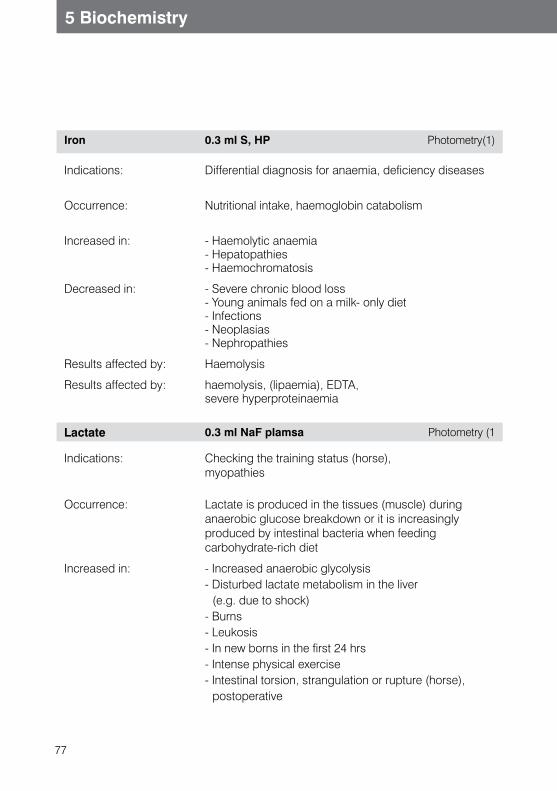
77
5 Biochemistry
Iron 0.3 ml S, HP Photometry(1)
Indications:
Differential diagnosis for anaemia, deficiency diseases
Occurrence:
Nutritional intake, haemoglobin catabolism
Increased in:
- Haemolytic anaemia- Hepatopathies- Haemochromatosis
Decreased in:
- Severe chronic blood loss- Young animals fed on a milk- only diet- Infections- Neoplasias- Nephropathies
Results affected by: Haemolysis
Results affected by: haemolysis, (lipaemia), EDTA,severe hyperproteinaemia
Lactate 0.3 ml NaF plamsa Photometry (1
Indications: Checking the training status (horse),myopathies
Occurrence: Lactate is produced in the tissues (muscle) during anaerobic glucose breakdown or it is increasingly produced by intestinal bacteria when feeding carbohydrate-rich diet
Increased in: - Increased anaerobic glycolysis- Disturbed lactate metabolism in the liver
(e.g. due to shock)- Burns- Leukosis- In new borns in the first 24 hrs- Intense physical exercise- Intestinal torsion, strangulation or rupture (horse),
postoperative
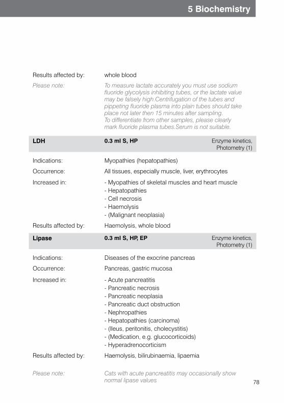
78
5 Biochemistry
Results affected by: whole blood
Please note: To measure lactate accurately you must use sodium fluoride glycolysis inhibiting tubes, or the lactate value may be falsely high.Centrifugation of the tubes and pippeting fluoride plasma into plain tubes should take place not later then 15 minutes after sampling. To differentiate from other samples, please clearly mark fluoride plasma tubes.Serum is not suitable.
LDH 0.3 ml S, HP Enzyme kinetics, Photometry (1)
Indications: Myopathies (hepatopathies)
Occurrence: All tissues, especially muscle, liver, erythrocytes
Increased in: - Myopathies of skeletal muscles and heart muscle- Hepatopathies- Cell necrosis- Haemolysis- (Malignant neoplasia)
Results affected by: Haemolysis, whole blood
Lipase 0.3 ml S, HP, EP Enzyme kinetics, Photometry (1)
Indications: Diseases of the exocrine pancreas
Occurrence: Pancreas, gastric mucosa
Increased in: - Acute pancreatitis- Pancreatic necrosis- Pancreatic neoplasia- Pancreatic duct obstruction- Nephropathies- Hepatopathies (carcinoma)- (Ileus, peritonitis, cholecystitis)- (Medication, e.g. glucocorticoids)- Hyperadrenocorticism
Results affected by: Haemolysis, bilirubinaemia, lipaemia
Please note: Cats with acute pancreatitis may occasionally show normal lipase values
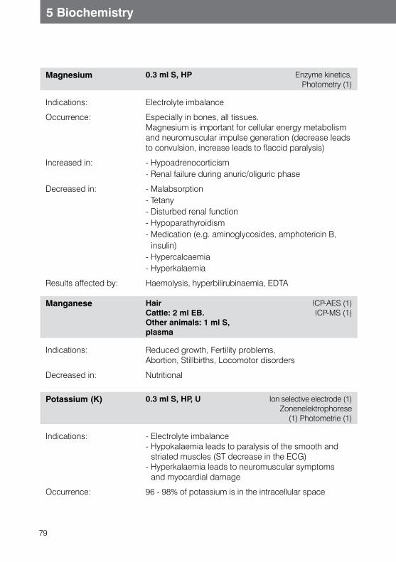
79
5 Biochemistry
Magnesium 0.3 ml S, HP Enzyme kinetics, Photometry (1)
Indications: Electrolyte imbalance
Occurrence: Especially in bones, all tissues.Magnesium is important for cellular energy metabolism and neuromuscular impulse generation (decrease leads to convulsion, increase leads to flaccid paralysis)
Increased in: - Hypoadrenocorticism- Renal failure during anuric/oliguric phase
Decreased in: - Malabsorption- Tetany- Disturbed renal function- Hypoparathyroidism- Medication (e.g. aminoglycosides, amphotericin B,
insulin)- Hypercalcaemia- Hyperkalaemia
Results affected by: Haemolysis, hyperbilirubinaemia, EDTA
Manganese Hair Cattle: 2 ml EB. Other animals: 1 ml S, plasma
ICP-AES (1)ICP-MS (1)
Indications: Reduced growth, Fertility problems,Abortion, Stillbirths, Locomotor disorders
Decreased in: Nutritional
Potassium (K) 0.3 ml S, HP, U Ion selective electrode (1)Zonenelektrophorese
(1) Photometrie (1)
Indications: - Electrolyte imbalance- Hypokalaemia leads to paralysis of the smooth and
striated muscles (ST decrease in the ECG)- Hyperkalaemia leads to neuromuscular symptoms
and myocardial damage
Occurrence: 96 - 98% of potassium is in the intracellular space
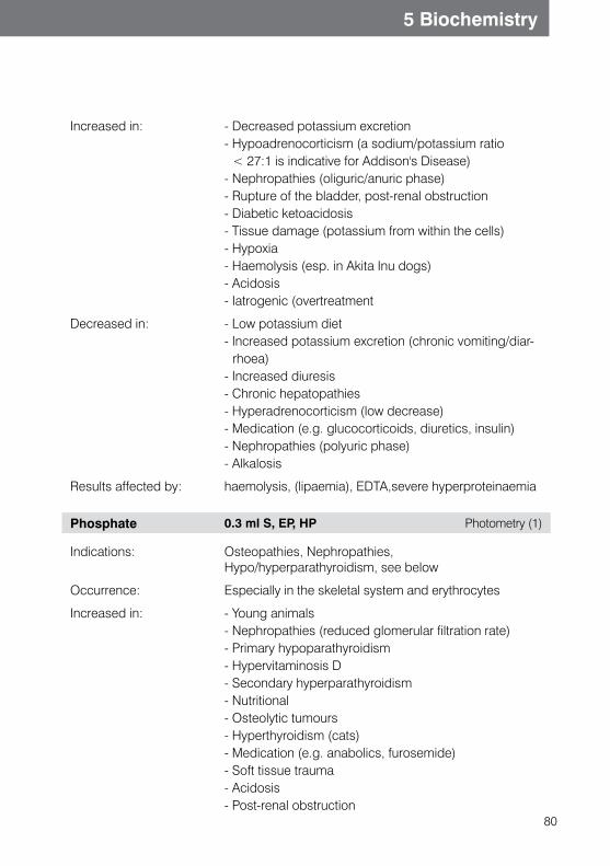
80
5 Biochemistry
Increased in: - Decreased potassium excretion- Hypoadrenocorticism (a sodium/potassium ratio
< 27:1 is indicative for Addison's Disease)- Nephropathies (oliguric/anuric phase)- Rupture of the bladder, post-renal obstruction- Diabetic ketoacidosis- Tissue damage (potassium from within the cells)- Hypoxia- Haemolysis (esp. in Akita Inu dogs)- Acidosis- Iatrogenic (overtreatment
Decreased in: - Low potassium diet- Increased potassium excretion (chronic vomiting/diar-
rhoea)- Increased diuresis- Chronic hepatopathies- Hyperadrenocorticism (low decrease)- Medication (e.g. glucocorticoids, diuretics, insulin)- Nephropathies (polyuric phase)- Alkalosis
Results affected by: haemolysis, (lipaemia), EDTA,severe hyperproteinaemia
Phosphate 0.3 ml S, EP, HP Photometry (1)
Indications: Osteopathies, Nephropathies,Hypo/hyperparathyroidism, see below
Occurrence: Especially in the skeletal system and erythrocytes
Increased in: - Young animals- Nephropathies (reduced glomerular filtration rate)- Primary hypoparathyroidism- Hypervitaminosis D- Secondary hyperparathyroidism- Nutritional- Osteolytic tumours- Hyperthyroidism (cats)- Medication (e.g. anabolics, furosemide)- Soft tissue trauma- Acidosis- Post-renal obstruction
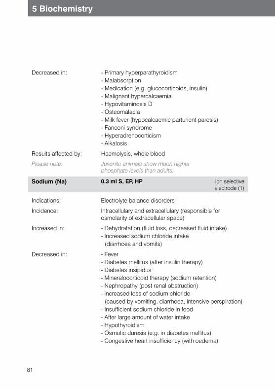
81
5 Biochemistry
Decreased in: - Primary hyperparathyroidism- Malabsorption- Medication (e.g. glucocorticoids, insulin)- Malignant hypercalcaemia- Hypovitaminosis D- Osteomalacia- Milk fever (hypocalcaemic parturient paresis)- Fanconi syndrome- Hyperadrenocorticism- Alkalosis
Results affected by: Haemolysis, whole blood
Please note: Juvenile animals show much higher phosphate levels than adults.
Sodium (Na) 0.3 ml S, EP, HP Ion selective electrode (1)
Indications: Electrolyte balance disorders
Incidence: Intracellulary and extracellulary (responsible for osmolarity of extracellular space)
Increased in: - Dehydratation (fluid loss, decreased fluid intake)- Increased sodium chloride intake
(diarrhoea and vomits)
Decreased in: - Fever- Diabetes mellitus (after insulin therapy)- Diabetes insipidus- Mineralocorticoid therapy (sodium retention)- Nephropathy (post renal obstruction)- increased loss of sodium chloride
(caused by vomiting, diarrhoea, intensive perspiration)- Insufficient sodium chloride in food- After large amount of water intake- Hypothyroidism- Osmotic duresis (e.g. in diabetes mellitus)- Congestive heart insufficiency (with oedema)
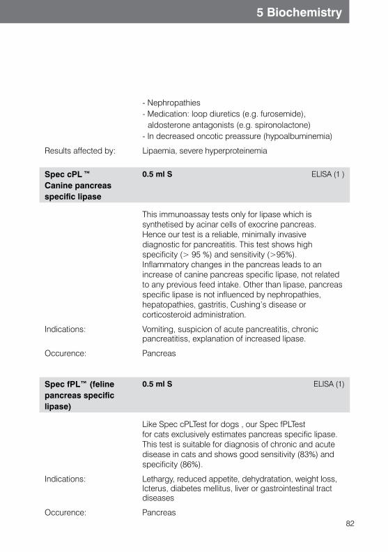
82
5 Biochemistry
- Nephropathies- Medication: loop diuretics (e.g. furosemide),
aldosterone antagonists (e.g. spironolactone)- In decreased oncotic preassure (hypoalbuminemia)
Results affected by: Lipaemia, severe hyperproteinemia
Spec cPL ™
Canine pancreas specific lipase
0.5 ml S ELISA (1 )
This immunoassay tests only for lipase which is synthetised by acinar cells of exocrine pancreas. Hence our test is a reliable, minimally invasive diagnostic for pancreatitis. This test shows high specificity (> 95 %) and sensitivity (>95%).Inflammatory changes in the pancreas leads to an increase of canine pancreas specific lipase, not related to any previous feed intake. Other than lipase, pancreas specific lipase is not influenced by nephropathies, hepatopathies, gastritis, Cushing’s disease or corticosteroid administration.
Indications: Vomiting, suspicion of acute pancreatitis, chronic pancreatitiss, explanation of increased lipase.
Occurence: Pancreas
Spec fPL™ (felinepancreas specific lipase)
0.5 ml S ELISA (1)
Like Spec cPLTest for dogs , our Spec fPLTest for cats exclusively estimates pancreas specific lipase. This test is suitable for diagnosis of chronic and acute disease in cats and shows good sensitivity (83%) and specificity (86%).
Indications: Lethargy, reduced appetite, dehydratation, weight loss, Icterus, diabetes mellitus, liver or gastrointestinal tract diseases
Occurence: Pancreas
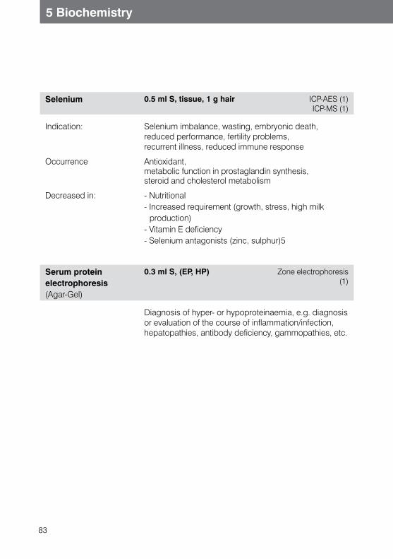
83
5 Biochemistry
Selenium 0.5 ml S, tissue, 1 g hair ICP-AES (1)ICP-MS (1)
Indication: Selenium imbalance, wasting, embryonic death,reduced performance, fertility problems,recurrent illness, reduced immune response
Occurrence Antioxidant,metabolic function in prostaglandin synthesis,steroid and cholesterol metabolism
Decreased in: - Nutritional- Increased requirement (growth, stress, high milk
production)- Vitamin E deficiency- Selenium antagonists (zinc, sulphur)5
Serum protein electrophoresis (Agar-Gel)
0.3 ml S, (EP, HP) Zone electrophoresis (1)
Diagnosis of hyper- or hypoproteinaemia, e.g. diagnosis or evaluation of the course of inflammation/infection,hepatopathies, antibody deficiency, gammopathies, etc.
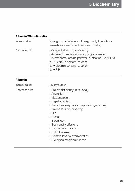
84
5 Biochemistry
Albumin/Globulin-ratio
Increased in:
Hypogammaglobulinaemia (e.g. rarely in newborn animals with insufficient colostrum intake)
Decreased in:
- Congenital immunodeficiency- Acquired immunodeficiency (e.g. distemper
in newborns, canine parvovirus infection, FeLV, FIV)s. Globulin content increase s. albumin content reduction s. FIP
Albumin
Increased in: - Dehydration
Decreased in: - Protein deficiency (nutritional) - Anorexia - Malabsorption - Hepatopathies- Renal loss (nephrosis, nephrotic syndrome) - Protein loss nephropathy - FIP - Burns - Blood loss- Body cavity effusions- Hypoadrenocorticism - CNS diseases- Relative loss by overhydration - Hypergammaglobulinaemia
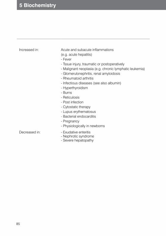
85
5 Biochemistry
Increased in:
Acute and subacute inflammations (e.g. acute hepatitis) - Fever- Tssue injury, traumatic or postoperatively- Malignant neoplasia (e.g. chronic lymphatic leukemia)- Glomerulonephritis, renal amyloidosis - Rheumatoid arthritis - Infectious diseases (see also albumin) - Hyperthyroidism - Burns- Reticulosis - Post infection - Cytostatic therapy - Lupus erythematosus - Bacterial endocarditis- Pregnancy - Physiologically in newborns
Decreased in: - Exudative enteritis - Nephrotic syndrome - Severe hepatopathy
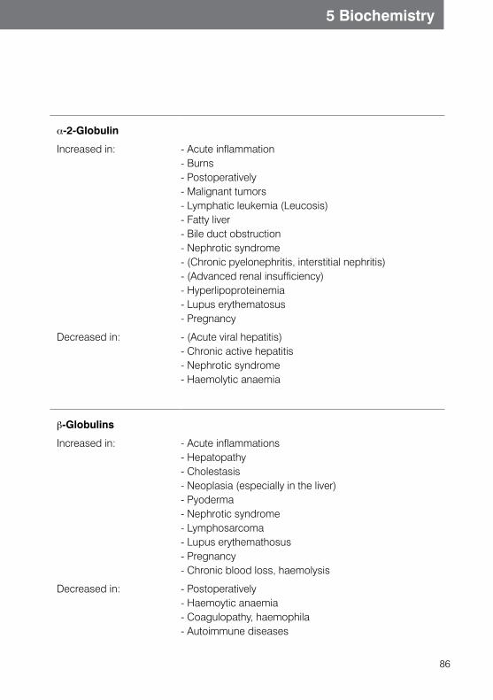
86
5 Biochemistry
a-2-Globulin
Increased in: - Acute inflammation- Burns- Postoperatively - Malignant tumors - Lymphatic leukemia (Leucosis) - Fatty liver- Bile duct obstruction - Nephrotic syndrome - (Chronic pyelonephritis, interstitial nephritis) - (Advanced renal insufficiency) - Hyperlipoproteinemia - Lupus erythematosus- Pregnancy
Decreased in: - (Acute viral hepatitis)- Chronic active hepatitis- Nephrotic syndrome- Haemolytic anaemia
b-Globulins
Increased in: - Acute inflammations- Hepatopathy - Cholestasis - Neoplasia (especially in the liver) - Pyoderma - Nephrotic syndrome- Lymphosarcoma- Lupus erythemathosus- Pregnancy- Chronic blood loss, haemolysis
Decreased in: - Postoperatively- Haemoytic anaemia - Coagulopathy, haemophila- Autoimmune diseases
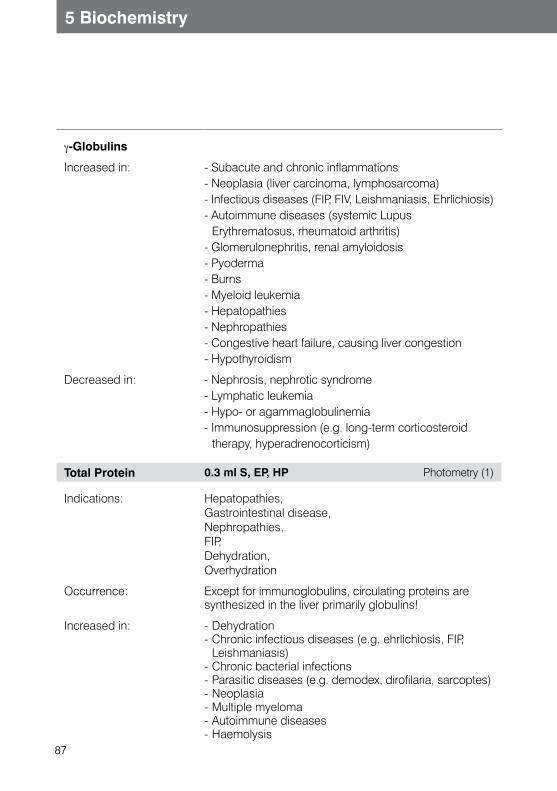
87
5 Biochemistry
g-Globulins
Increased in: - Subacute and chronic inflammations- Neoplasia (liver carcinoma, lymphosarcoma) - Infectious diseases (FIP, FIV, Leishmaniasis, Ehrlichiosis) - Autoimmune diseases (systemic Lupus
Erythrematosus, rheumatoid arthritis) - Glomerulonephritis, renal amyloidosis - Pyoderma - Burns- Myeloid leukemia - Hepatopathies - Nephropathies - Congestive heart failure, causing liver congestion- Hypothyroidism
Decreased in: - Nephrosis, nephrotic syndrome- Lymphatic leukemia - Hypo- or agammaglobulinemia - Immunosuppression (e.g. long-term corticosteroid
therapy, hyperadrenocorticism)
Total Protein 0.3 ml S, EP, HP Photometry (1)
Indications: Hepatopathies,Gastrointestinal disease,Nephropathies,FIP,Dehydration,Overhydration
Occurrence: Except for immunoglobulins, circulating proteins aresynthesized in the liver primarily globulins!
Increased in: - Dehydration- Chronic infectious diseases (e.g. ehrlichiosis, FIP,
Leishmaniasis)- Chronic bacterial infections- Parasitic diseases (e.g. demodex, dirofilaria, sarcoptes)- Neoplasia- Multiple myeloma- Autoimmune diseases- Haemolysis
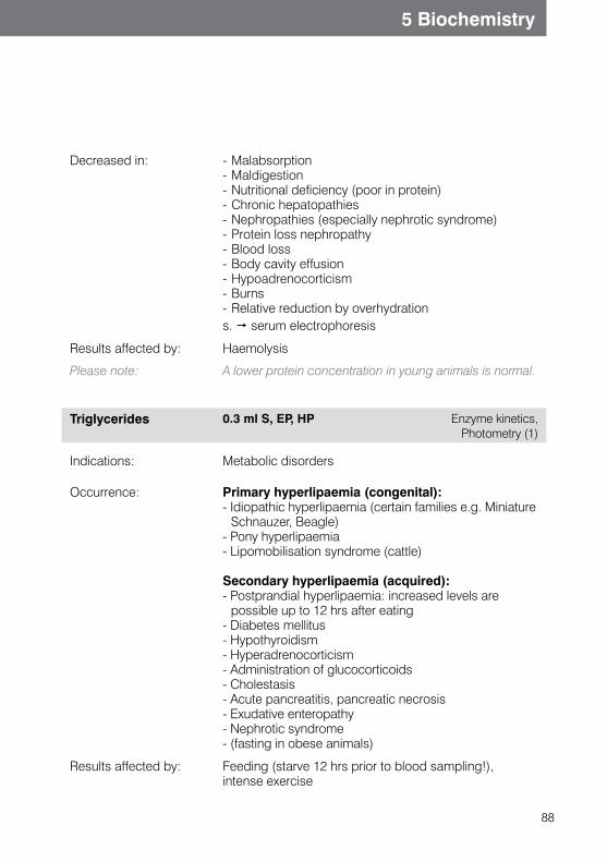
88
5 Biochemistry
Decreased in: - Malabsorption- Maldigestion- Nutritional deficiency (poor in protein)- Chronic hepatopathies- Nephropathies (especially nephrotic syndrome)- Protein loss nephropathy- Blood loss- Body cavity effusion- Hypoadrenocorticism- Burns- Relative reduction by overhydrations. serum electrophoresis
Results affected by: Haemolysis
Please note: A lower protein concentration in young animals is normal.
Triglycerides 0.3 ml S, EP, HP Enzyme kinetics, Photometry (1)
Indications: Metabolic disorders
Occurrence: Primary hyperlipaemia (congenital):- Idiopathic hyperlipaemia (certain families e.g. Miniature
Schnauzer, Beagle)- Pony hyperlipaemia- Lipomobilisation syndrome (cattle)
Secondary hyperlipaemia (acquired):- Postprandial hyperlipaemia: increased levels are
possible up to 12 hrs after eating- Diabetes mellitus- Hypothyroidism- Hyperadrenocorticism- Administration of glucocorticoids- Cholestasis- Acute pancreatitis, pancreatic necrosis- Exudative enteropathy- Nephrotic syndrome- (fasting in obese animals)
Results affected by: Feeding (starve 12 hrs prior to blood sampling!),intense exercise
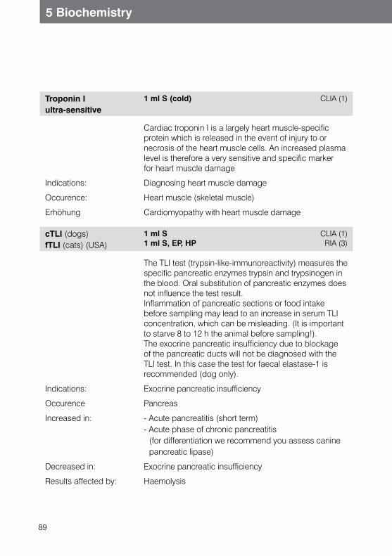
89
5 Biochemistry
Troponin I ultra-sensitive
1 ml S (cold) CLIA (1)
Cardiac troponin I is a largely heart muscle-specific protein which is released in the event of injury to or necrosis of the heart muscle cells. An increased plasma level is therefore a very sensitive and specific marker for heart muscle damage
Indications: Diagnosing heart muscle damage
Occurence: Heart muscle (skeletal muscle)
Erhöhung Cardiomyopathy with heart muscle damage
cTLI (dogs) fTLI (cats) (USA)
1 ml S1 ml S, EP, HP
CLIA (1) RIA (3)
The TLI test (trypsin-like-immunoreactivity) measures thespecific pancreatic enzymes trypsin and trypsinogen in the blood. Oral substitution of pancreatic enzymes does not influence the test result.Inflammation of pancreatic sections or food intake before sampling may lead to an increase in serum TLI concentration, which can be misleading. (It is important to starve 8 to 12 h the animal before sampling!). The exocrine pancreatic insufficiency due to blockage of the pancreatic ducts will not be diagnosed with the TLI test. In this case the test for faecal elastase-1 is recommended (dog only).
Indications: Exocrine pancreatic insufficiency
Occurence Pancreas
Increased in: - Acute pancreatitis (short term) - Acute phase of chronic pancreatitis
(for differentiation we recommend you assess canine pancreatic lipase)
Decreased in: Exocrine pancreatic insufficiency
Results affected by: Haemolysis
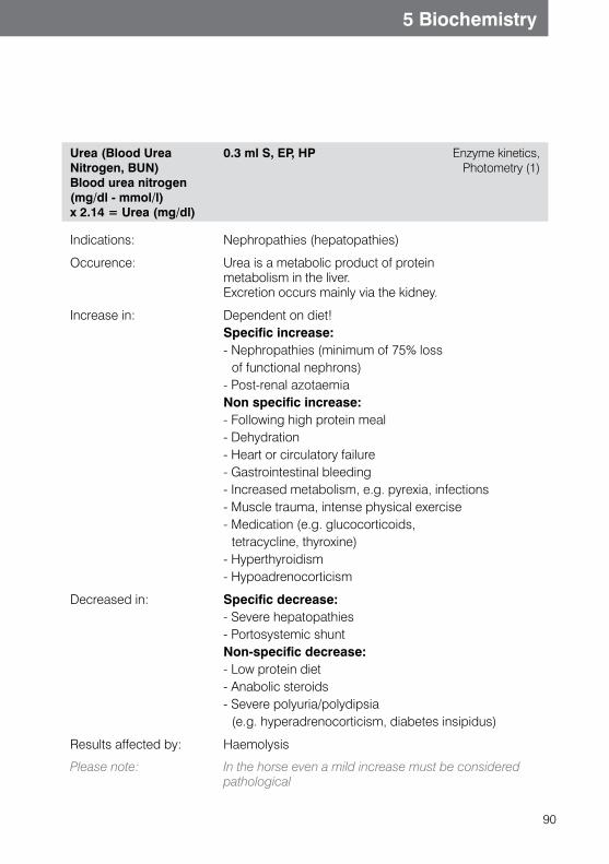
90
5 Biochemistry
Urea (Blood Urea Nitrogen, BUN) Blood urea nitrogen (mg/dl - mmol/l) x 2.14 = Urea (mg/dl)
0.3 ml S, EP, HP Enzyme kinetics, Photometry (1)
Indications: Nephropathies (hepatopathies)
Occurence: Urea is a metabolic product of protein metabolism in the liver.Excretion occurs mainly via the kidney.
Increase in: Dependent on diet!Specific increase:- Nephropathies (minimum of 75% loss
of functional nephrons)- Post-renal azotaemiaNon specific increase:- Following high protein meal- Dehydration- Heart or circulatory failure- Gastrointestinal bleeding- Increased metabolism, e.g. pyrexia, infections- Muscle trauma, intense physical exercise- Medication (e.g. glucocorticoids,
tetracycline, thyroxine)- Hyperthyroidism- Hypoadrenocorticism
Decreased in: Specific decrease:- Severe hepatopathies- Portosystemic shuntNon-specific decrease:- Low protein diet- Anabolic steroids- Severe polyuria/polydipsia
(e.g. hyperadrenocorticism, diabetes insipidus)
Results affected by: Haemolysis
Please note: In the horse even a mild increase must be considered pathological
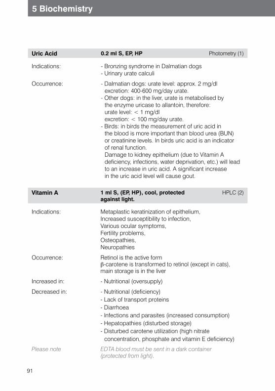
91
5 Biochemistry
Uric Acid 0.2 ml S, EP, HP Photometry (1)
Indications: - Bronzing syndrome in Dalmatian dogs- Urinary urate calculi
Occurrence: - Dalmatian dogs: urate level: approx. 2 mg/dl excretion: 400-600 mg/day urate.
- Other dogs: in the liver, urate is metabolised by the enzyme uricase to allantoin, therefore: urate level: < 1 mg/dl excretion: < 100 mg/day urate.
- Birds: in birds the measurement of uric acid in the blood is more important than blood urea (BUN) or creatinine levels. In birds uric acid is an indicator of renal function. Damage to kidney epithelium (due to Vitamin A deficiency, infections, water deprivation, etc.) will lead to an increase in uric acid. A significant increase in the uric acid level will cause gout.
Vitamin A 1 ml S, (EP, HP), cool, protected against light.
HPLC (2)
Indications: Metaplastic keratinization of epithelium,Increased susceptibility to infection,Various ocular symptoms,Fertility problems,Osteopathies,Neuropathies
Occurrence: Retinol is the active formb-carotene is transformed to retinol (except in cats),main storage is in the liver
Increased in: - Nutritional (oversupply)
Decreased in:
- Nutritional (deficiency)- Lack of transport proteins- Diarrhoea- Infections and parasites (increased consumption)- Hepatopathies (disturbed storage)- Disturbed carotene utilization (high nitrate
concentration, phosphate and vitamin E deficiency)
Please note EDTA blood must be sent in a dark container(protected from light).
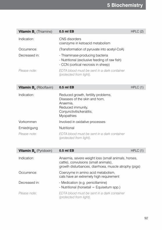
92
5 Biochemistry
Vitamin B1 (Thiamine) 0.5 ml EB HPLC (2)
Indication: CNS disorderscoenzyme in ketoacid metabolism
Occurrence: (Transformation of pyruvate into acetyl-CoA)
Decreased in:
- Thiaminase-producing bacteria- Nutritional (exclusive feeding of raw fish)- CCN (cortical necrosis in sheep)
Please note: EDTA blood must be sent in a dark container(protected from light).
Vitamin B2 (Riboflavin) 0.5 ml EB HPLC (1)
Indication: Reduced growth, fertility problems,Diseases of the skin and horn,Anaemia,Reduced immunity,Conjunctivitis/keratitis,Myopathies
Vorkommen Involved in oxidative processes
Erniedrigung Nutritional
Please note: EDTA blood must be sent in a dark container(protected from light).
Vitamin B6 (Pyridoxin) 0.5 ml EB HPLC (1)
Indication: Anaemia, severe weight loss (small animals, horses, cattle), convulsions (small animals),growth disturbances, diarrhoea, muscle atrophy (pigs)
Occurrence: Coenzyme in amino acid metabolism,cats have an extremely high requirement
Decreased in: - Medication (e.g. penicillamine)- Nutritional (horsetail = Equisetum spp.)
Please note: EDTA blood must be sent in a dark container(protected from light).
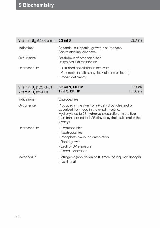
93
5 Biochemistry
Vitamin B12 (Cobalamin) 0.3 ml S CLIA (1)
Indication: Anaemia, leukopenia, growth disturbancesGastrointestinal diseases
Occurrence: Breakdown of proprionic acid,Resynthesis of methionine
Decreased in: - Disturbed absorbtion in the ileum. Pancreatic insufficiency (lack of intrinsic factor)
- Cobalt deficiency
Vitamin D3 (1,25-di-OH)Vitamin D3 (25-OH)
0.5 ml S, EP, HP 1 ml S, EP, HP
RIA (3)HPLC (1)
Indications: Osteopathies
Occurrence: Produced in the skin from 7-dehydrocholesterol or absorbed from food in the small intestine. Hydroxylated to 25-hydroxycholecalciferol in the liver, then transformed to 1.25-dihydroxycholecalciferol in the kidneys
Decreased in: - Hepatopathies- Nephropathies- Phosphate oversupplementation- Rapid growth- Lack of UV exposure- Chronic diarrhoea
Increased in - Iatrogenic (application of 10 times the required dosage)- Nutritional
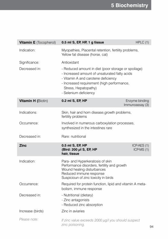
94
5 Biochemistry
Vitamin E (Tocopherol) 0.5 ml S, EP, HP, 1 g tissue HPLC (1)
Indication: Myopathies, Placental retention, fertility problems,Yellow fat disease (horse, cat)
Significance: Antioxidant
Decreased in: - Reduced amount in diet (poor storage or spoilage)- Increased amount of unsaturated fatty acids- Vitamin A and carotene deficiency- Increased requirement (high performance,
Stress, Hepatopathy)- Selenium deficiency
Vitamin H (Biotin) 0.2 ml S, EP, HP Enzyme binding Immunoassay (3)
Indications:
Skin, hair and horn disease,growth problems,fertility problems
Occurrence: Involved in numerous carboxylation processes,synthesized in the intestines rare
Decreased in: Rare: nutritional
Zinc 0.5 ml S, EP, HP(Bird: 200 µl S, EP, HPhair, tissue
ICP-AES (1) ICP-MS (1)
Indication: Para- and Hyperkeratosis of skin Performance disorders, fertility and growthWound healing disturbancesReduced immune responseSuspicioun of zinc toxicity in birds
Occurrence: Required for protein function, lipid and vitamin A meta-bolism, immune response
Decreased in: - Nutritional (dietary)- Zinc antagonists - Reduced zinc absorption
Increase (birds)
Please note:
Zinc in aviaries
If zinc value exceeds 2000 μg/l you should suspectzinc poisoning.
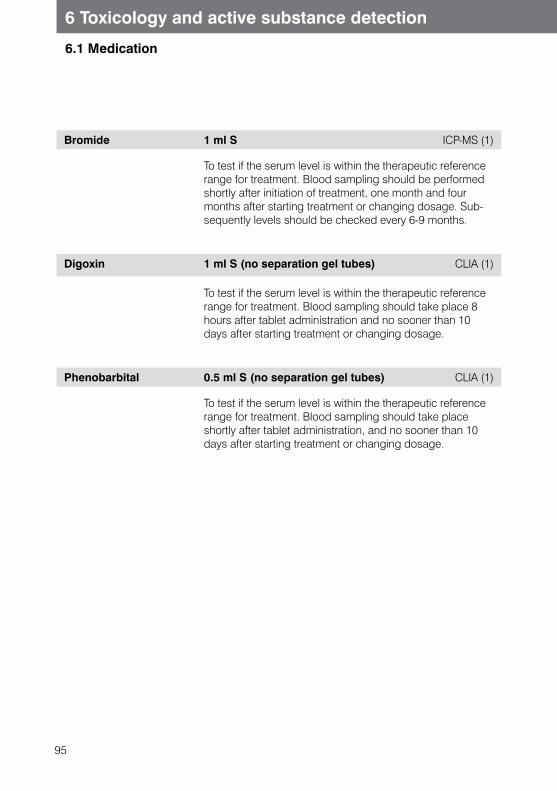
95
6.1 Medication
6 Toxicology and active substance detection
Bromide 1 ml S ICP-MS (1)
To test if the serum level is within the therapeutic reference range for treatment. Blood sampling should be performed shortly after initiation of treatment, one month and four months after starting treatment or changing dosage. Sub-sequently levels should be checked every 6-9 months.
Digoxin 1 ml S (no separation gel tubes) CLIA (1)
To test if the serum level is within the therapeutic reference range for treatment. Blood sampling should take place 8 hours after tablet administration and no sooner than 10 days after starting treatment or changing dosage.
Phenobarbital 0.5 ml S (no separation gel tubes) CLIA (1)
To test if the serum level is within the therapeutic reference range for treatment. Blood sampling should take place shortly after tablet administration, and no sooner than 10 days after starting treatment or changing dosage.
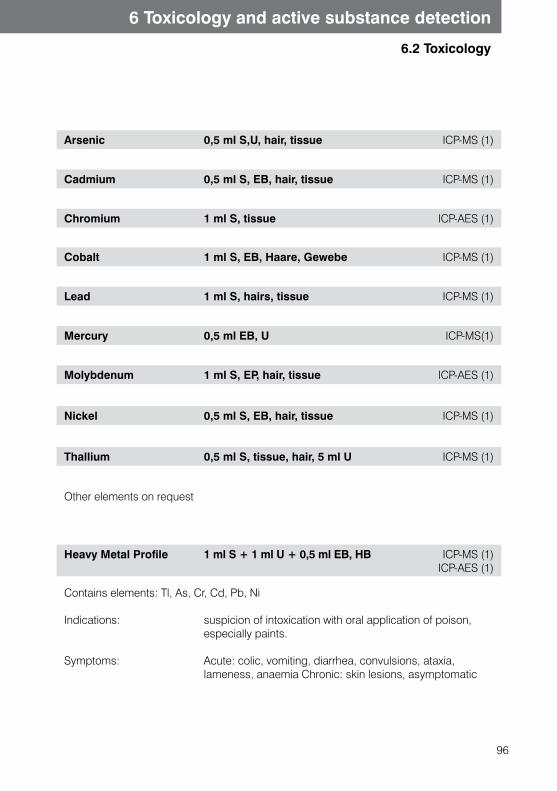
96
6 Toxicology and active substance detection
6.2 Toxicology
Arsenic 0,5 ml S,U, hair, tissue ICP-MS (1)
Cadmium 0,5 ml S, EB, hair, tissue ICP-MS (1)
Chromium 1 ml S, tissue ICP-AES (1)
Cobalt 1 ml S, EB, Haare, Gewebe ICP-MS (1)
Lead 1 ml S, hairs, tissue ICP-MS (1)
Mercury 0,5 ml EB, U ICP-MS(1)
Molybdenum 1 ml S, EP, hair, tissue ICP-AES (1)
Nickel 0,5 ml S, EB, hair, tissue ICP-MS (1)
Thallium 0,5 ml S, tissue, hair, 5 ml U ICP-MS (1)
Other elements on request
Heavy Metal Profile 1 ml S + 1 ml U + 0,5 ml EB, HB ICP-MS (1)ICP-AES (1)
Contains elements: Tl, As, Cr, Cd, Pb, Ni
Indications: suspicion of intoxication with oral application of poison, especially paints.
Symptoms: Acute: colic, vomiting, diarrhea, convulsions, ataxia, lameness, anaemia Chronic: skin lesions, asymptomatic
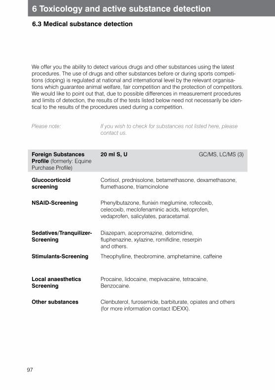
97
6 Toxicology and active substance detection
6.3 Medical substance detection
We offer you the ability to detect various drugs and other substances using the latest procedures. The use of drugs and other substances before or during sports competi-tions (doping) is regulated at national and international level by the relevant organisa-tions which guarantee animal welfare, fair competition and the protection of competitors. We would like to point out that, due to possible differences in measurement procedures and limits of detection, the results of the tests listed below need not necessarily be iden-tical to the results of the procedures used during a competition.
Please note: If you wish to check for substances not listed here, please contact us.
Foreign Substances Profile (formerly: Equine Purchase Profile)
20 ml S, U GC/MS, LC/MS (3)
Glucocorticoid screening
Cortisol, prednisolone, betamethasone, dexamethasone, flumethasone, triamcinolone
NSAID-Screening Phenylbutazone, flunixin meglumine, rofecoxib, celecoxib, meclofenaminic acids, ketoprofen, vedaprofen, salicylates, paracetamal.
Sedatives/Tranquilizer-Screening
Diazepam, acepromazine, detomidine, fluphenazine, xylazine, romifidine, reserpin and others.
Stimulants-Screening Theophylline, theobromine, amphetamine, caffeine
Local anaesthetics Screening
Procaine, lidocaine, mepivacaine, tetracaine,Benzocaine.
Other substances Clenbuterol, furosemide, barbiturate, opiates and others (for more information contact IDEXX).
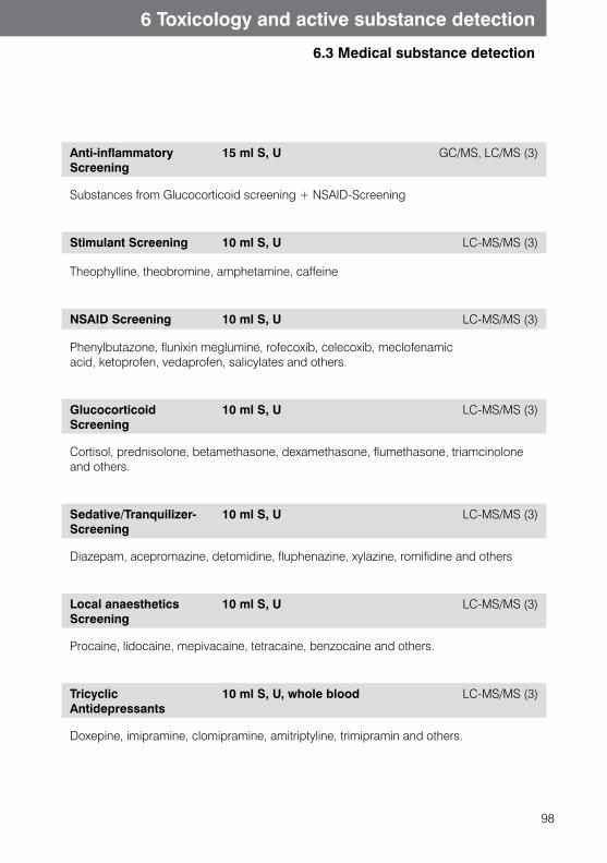
98
6 Toxicology and active substance detection
6.3 Medical substance detection
Anti-inflammatoryScreening
15 ml S, U GC/MS, LC/MS (3)
Substances from Glucocorticoid screening + NSAID-Screening
Stimulant Screening 10 ml S, U LC-MS/MS (3)
Theophylline, theobromine, amphetamine, caffeine
NSAID Screening 10 ml S, U LC-MS/MS (3)
Phenylbutazone, flunixin meglumine, rofecoxib, celecoxib, meclofenamicacid, ketoprofen, vedaprofen, salicylates and others.
Glucocorticoid Screening
10 ml S, U LC-MS/MS (3)
Cortisol, prednisolone, betamethasone, dexamethasone, flumethasone, triamcinolone and others.
Sedative/Tranquilizer-Screening
10 ml S, U LC-MS/MS (3)
Diazepam, acepromazine, detomidine, fluphenazine, xylazine, romifidine and others
Local anaesthetics Screening
10 ml S, U LC-MS/MS (3)
Procaine, lidocaine, mepivacaine, tetracaine, benzocaine and others.
Tricyclic Antidepressants
10 ml S, U, whole blood LC-MS/MS (3)
Doxepine, imipramine, clomipramine, amitriptyline, trimipramin and others.
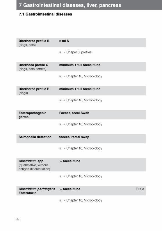
99
7.1 Gastrointestinal diseases
7 Gastrointestinal diseases, liver, pancreas
Diarrhorea profile B (dogs, cats)
2 ml S
s. Chaper 3, profiles
Diarrhoea profile C(dogs, cats, ferrets)
minimum 1 full faecal tube
s. Chapter 16, Microbiology
Diarrhorea profile E (dogs)
minimum 1 full faecal tube
s. Chapter 16, Microbiology
Enteropathogenic germs
Faeces, fecal Swab
s. Chapter 16, Microbiology
Salmonella detection faeces, rectal swap
s. Chapter 16, Microbiology
Clostridium spp. (quantitative, without antigen differentiation)
¼ faecal tube
s. Chapter 16, Microbiology
Clostridium perfringens Enterotoxin
¼ faecal tube ELISA
s. Chapter 16, Microbiology

100
7.1 Gastrointestinal diseases
7 Gastrointestinal diseases, liver, pancreas
Folic acid 0.5 S
s. Chapter 5, Biochemistry
Vitamin B12 (Cobalamine)
0.3 ml S, EP, HP
s. Chapter 5, Biochemistry
Occult Blood Faeces (minimum 1/3 faecal tube)
s. Chapter 16, Microbiology
General Virology (Viral faecal test)
minimum ½ faecal tube Electron microsco
Any viruses shed in faeces are detected and identified with electron microscopy.s. Chapter 16.2s. Chapter 13, Corona-, Rota- and Parvovirus detection
Mc-Master Egg Counting Technique(horse, ruminants, camelids)
20 g faeces Quantitative detection of eggs grid count,
flotation (1)
s. Chapter 17, Parasitology
Gastrointestinal Profile (formerly Profile P) (dogs, cats)
1-2 ml S Chromatography (2)
Spec cPL®/Spec fPL®, folic acid, Vitamin B12, cTLI (dogs)
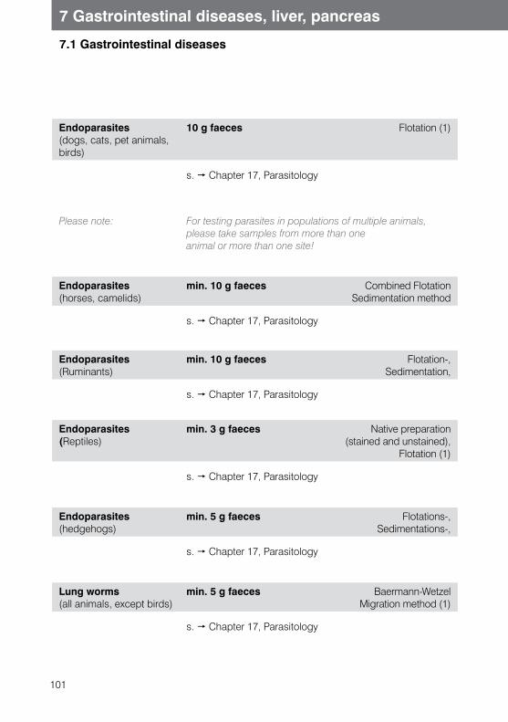
101
7 Gastrointestinal diseases, liver, pancreas
7.1 Gastrointestinal diseases
Endoparasites(dogs, cats, pet animals, birds)
10 g faeces Flotation (1)
s. Chapter 17, Parasitology
Please note: For testing parasites in populations of multiple animals,please take samples from more than one animal or more than one site!
Endoparasites(horses, camelids)
min. 10 g faeces Combined FlotationSedimentation method
s. Chapter 17, Parasitology
Endoparasites (Ruminants)
min. 10 g faeces Flotation-, Sedimentation,
s. Chapter 17, Parasitology
Endoparasites (Reptiles)
min. 3 g faeces Native preparation (stained and unstained),
Flotation (1)
s. Chapter 17, Parasitology
Endoparasites (hedgehogs)
min. 5 g faeces Flotations-, Sedimentations-,
s. Chapter 17, Parasitology
Lung worms(all animals, except birds)
min. 5 g faeces Baermann-WetzelMigration method (1)
s. Chapter 17, Parasitology
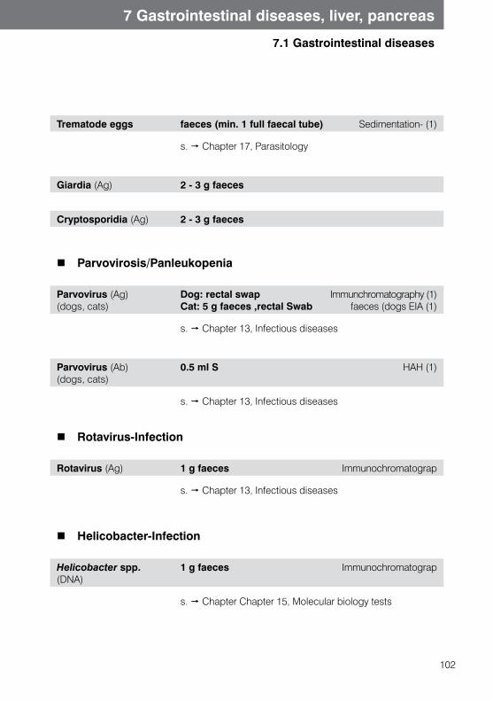
102
7 Gastrointestinal diseases, liver, pancreas
7.1 Gastrointestinal diseases
Trematode eggs faeces (min. 1 full faecal tube) Sedimentation- (1)
s. Chapter 17, Parasitology
Giardia (Ag) 2 - 3 g faeces
Cryptosporidia (Ag) 2 - 3 g faeces
Parvovirosis/Panleukopenia
Parvovirus (Ag) (dogs, cats)
Dog: rectal swapCat: 5 g faeces ,rectal Swab
Immunchromatography (1) faeces (dogs EIA (1)
s. Chapter 13, Infectious diseases
Parvovirus (Ab) (dogs, cats)
0.5 ml S HAH (1)
s. Chapter 13, Infectious diseases
Rotavirus-Infection
Rotavirus (Ag) 1 g faeces Immunochromatograp
s. Chapter 13, Infectious diseases
Helicobacter-Infection
Helicobacter spp. (DNA)
1 g faeces Immunochromatograp
s. Chapter Chapter 15, Molecular biology tests
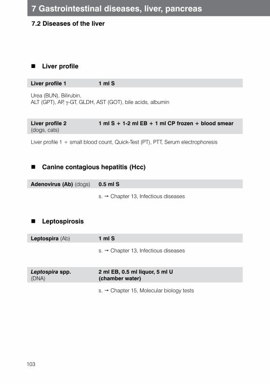
103
7 Gastrointestinal diseases, liver, pancreas
7.2 Diseases of the liver
Liver profile
Liver profile 1 1 ml S
Urea (BUN), Bilirubin, ALT (GPT), AP, γ-GT, GLDH, AST (GOT), bile acids, albumin
Liver profile 2 (dogs, cats)
1 ml S + 1-2 ml EB + 1 ml CP frozen + blood smear
Liver profile 1 + small blood count, Quick-Test (PT), PTT, Serum electrophoresis
Canine contagious hepatitis (Hcc)
Adenovirus (Ab) (dogs) 0.5 ml S
s. Chapter 13, Infectious diseases
Leptospirosis
Leptospira (Ab) 1 ml S
s. Chapter 13, Infectious diseases
Leptospira spp. (DNA)
2 ml EB, 0.5 ml liquor, 5 ml U (chamber water)
s. Chapter 15, Molecular biology tests
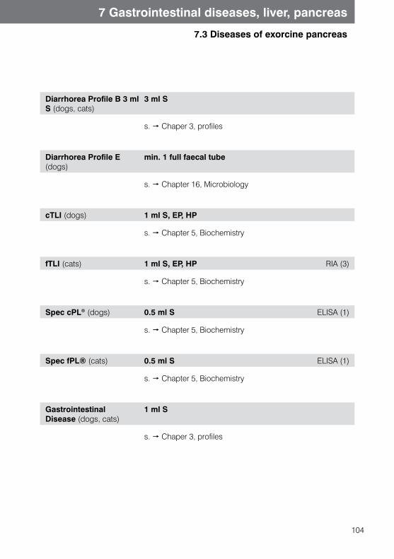
104
7 Gastrointestinal diseases, liver, pancreas
7.3 Diseases of exorcine pancreas
Diarrhorea Profile B 3 ml S (dogs, cats)
3 ml S
s. Chaper 3, profiles
Diarrhorea Profile E (dogs)
min. 1 full faecal tube
s. Chapter 16, Microbiology
cTLI (dogs) 1 ml S, EP, HP
s. Chapter 5, Biochemistry
fTLI (cats) 1 ml S, EP, HP RIA (3)
s. Chapter 5, Biochemistry
Spec cPL® (dogs) 0.5 ml S ELISA (1)
s. Chapter 5, Biochemistry
Spec fPL® (cats) 0.5 ml S ELISA (1)
s. Chapter 5, Biochemistry
Gastrointestinal Disease (dogs, cats)
1 ml S
s. Chaper 3, profiles
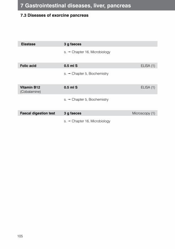
105
7.3 Diseases of exorcine pancreas
7 Gastrointestinal diseases, liver, pancreas
Elastase 3 g faeces
s. Chapter 16, Microbiology
Folic acid 0.5 ml S ELISA (1)
s. Chapter 5, Biochemistry
Vitamin B12 (Cobalamine)
0.5 ml S ELISA (1)
s. Chapter 5, Biochemistry
Faecal digestion test 3 g faeces Microscopy (1)
s. Chapter 16, Microbiology
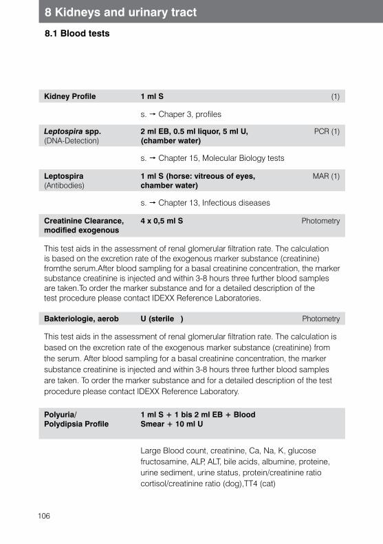
106
8.1 Blood tests
8 Kidneys and urinary tract
Kidney Profile 1 ml S (1)
s. Chaper 3, profiles
Leptospira spp. (DNA-Detection)
2 ml EB, 0.5 ml liquor, 5 ml U, (chamber water)
PCR (1)
s. Chapter 15, Molecular Biology tests
Leptospira (Antibodies)
1 ml S (horse: vitreous of eyes, chamber water)
MAR (1)
s. Chapter 13, Infectious diseases
Creatinine Clearance, modified exogenous
4 x 0,5 ml S Photometry
This test aids in the assessment of renal glomerular filtration rate. The calculation is based on the excretion rate of the exogenous marker substance (creatinine) fromthe serum.After blood sampling for a basal creatinine concentration, the marker substance creatinine is injected and within 3-8 hours three further blood samples are taken.To order the marker substance and for a detailed description of thetest procedure please contact IDEXX Reference Laboratories.
Bakteriologie, aerob U (sterile ) Photometry
This test aids in the assessment of renal glomerular filtration rate. The calculation is based on the excretion rate of the exogenous marker substance (creatinine) from the serum. After blood sampling for a basal creatinine concentration, the marker substance creatinine is injected and within 3-8 hours three further blood samples are taken. To order the marker substance and for a detailed description of the test procedure please contact IDEXX Reference Laboratory.
Polyuria/Polydipsia Profile
1 ml S + 1 bis 2 ml EB + Blood Smear + 10 ml U
Large Blood count, creatinine, Ca, Na, K, glucosefructosamine, ALP, ALT, bile acids, albumine, proteine,urine sediment, urine status, protein/creatinine ratiocortisol/creatinine ratio (dog),TT4 (cat)
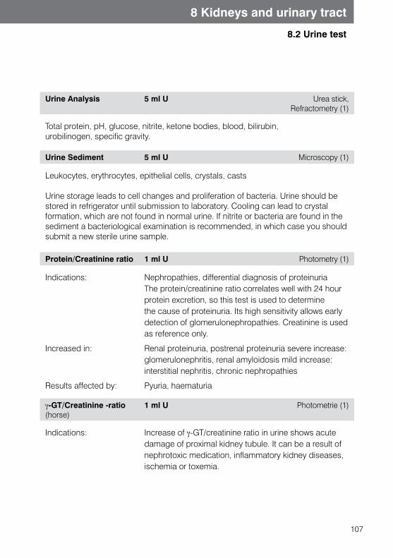
107
8.2 Urine test
8 Kidneys and urinary tract
Urine Analysis 5 ml U Urea stick, Refractometry (1)
Total protein, pH, glucose, nitrite, ketone bodies, blood, bilirubin, urobilinogen, specific gravity.
Urine Sediment 5 ml U Microscopy (1)
Leukocytes, erythrocytes, epithelial cells, crystals, casts
Urine storage leads to cell changes and proliferation of bacteria. Urine should be stored in refrigerator until submission to laboratory. Cooling can lead to crystal formation, which are not found in normal urine. If nitrite or bacteria are found in the sediment a bacteriological examination is recommended, in which case you should submit a new sterile urine sample.
Protein/Creatinine ratio 1 ml U Photometry (1)
Indications: Nephropathies, differential diagnosis of proteinuriaThe protein/creatinine ratio correlates well with 24 hourprotein excretion, so this test is used to determinethe cause of proteinuria. Its high sensitivity allows earlydetection of glomerulonephropathies. Creatinine is used as reference only.
Increased in: Renal proteinuria, postrenal proteinuria severe increase: glomerulonephritis, renal amyloidosis mild increase: interstitial nephritis, chronic nephropathies
Results affected by: Pyuria, haematuria
γ-GT/Creatinine -ratio (horse)
1 ml U Photometrie (1)
Indications: Increase of γ-GT/creatinine ratio in urine shows acute damage of proximal kidney tubule. It can be a result of nephrotoxic medication, inflammatory kidney diseases, ischemia or toxemia.
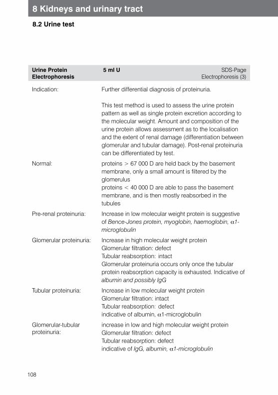
108
8 Kidneys and urinary tract
8.2 Urine test
Urine Protein Electrophoresis
5 ml U SDS-Page Electrophoresis (3)
Indication: Further differential diagnosis of proteinuria.
This test method is used to assess the urine protein pattern as well as single protein excretion according to the molecular weight. Amount and composition of the urine protein allows assessment as to the localisation and the extent of renal damage (differentiation between glomerular and tubular damage). Post-renal proteinuria can be differentiated by test.
Normal: proteins > 67 000 D are held back by the basement membrane, only a small amount is filtered by the glomerulusproteins < 40 000 D are able to pass the basement membrane, and is then mostly reabsorbed in the tubules
Pre-renal proteinuria: Increase in low molecular weight protein is suggestive of Bence-Jones protein, myoglobin, haemoglobin, α1-microglobulin
Glomerular proteinuria: Increase in high molecular weight proteinGlomerular filtration: defectTubular reabsorption: intactGlomerular proteinuria occurs only once the tubular protein reabsorption capacity is exhausted. Indicative of albumin and possibly IgG
Tubular proteinuria: Increase in low molecular weight proteinGlomerular filtration: intactTubular reabsorption: defectindicative of albumin, α1-microglobulin
Glomerular-tubular proteinuria:
increase in low and high molecular weight proteinGlomerular filtration: defectTubular reabsorption: defectindicative of IgG, albumin, α1-microglobulin
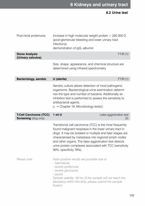
109
8 Kidneys and urinary tract
8.2 Urine test
Post-renal proteinuria: Increase in high molecular weight protein > 250 000 D(post-glomerular bleeding and lower urinary tract infections)demonstration of IgG, albumin
Stone Analysis(Urinary calculus)
FT-IR (1)
Size, shape, appearance, and chemical structure are determined using infrared spectrometry.
Bacteriology, aerobic U (sterile) FT-IR (1)
Aerobic culture allows detection of most pathogenic organisms. Bacteriological urine examination determi-nes the type and number of bacteria. Additionally an inhibition test is performed to assess the sensitivity to antibacterial agents. s. Chapter 16, Microbiology testsU
T-Cell Carcinoma (TCC)Screening (dog only)
1 ml U Latex-agglutination test (2)
Transitional cell carcinoma (TCC) is the most frequently found malignant neoplasia in the lower urinary tract in dogs. It may be isolated or multiple and later stages arecharacterised by metastasis into regional lymph nodes and other organs. The latex agglutination test detects urine protein complexes associated with TCC (sensitivity 90%, specificity 78%).
Please note False positive results are possible due to- haematuria- severe proteinuria- severe glucosuria- pyuriaSample stability: 48 hrs (if the sample will not reach thelaboratory within this time, please submit the samplefrozen).
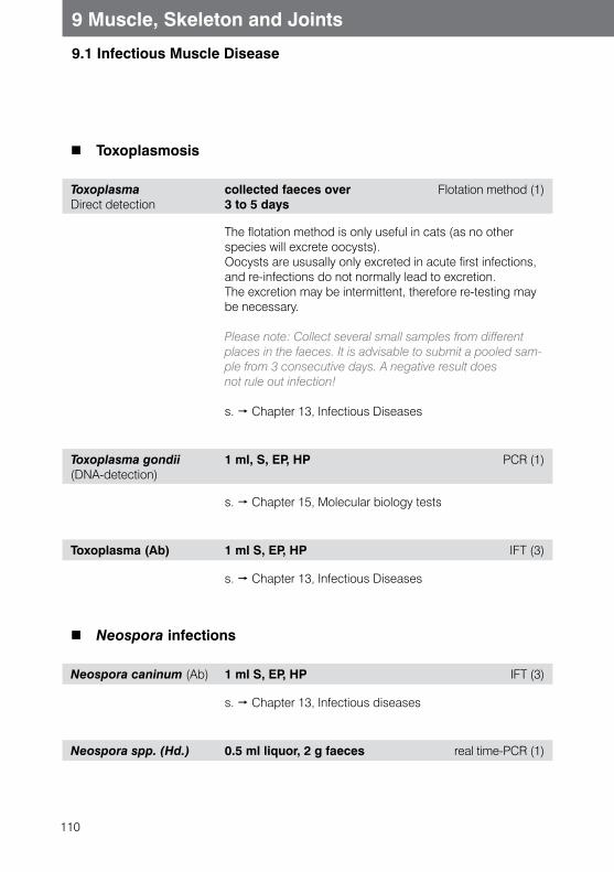
110
9.1 Infectious Muscle Disease
9 Muscle, Skeleton and Joints
Toxoplasmosis
ToxoplasmaDirect detection
collected faeces over 3 to 5 days
Flotation method (1)
The flotation method is only useful in cats (as no other species will excrete oocysts).Oocysts are ususally only excreted in acute first infections, and re-infections do not normally lead to excretion. The excretion may be intermittent, therefore re-testing may be necessary.
Please note: Collect several small samples from different places in the faeces. It is advisable to submit a pooled sam-ple from 3 consecutive days. A negative result does not rule out infection!
s. Chapter 13, Infectious Diseases
Toxoplasma gondii (DNA-detection)
1 ml, S, EP, HP PCR (1)
s. Chapter 15, Molecular biology tests
Toxoplasma (Ab) 1 ml S, EP, HP IFT (3)
s. Chapter 13, Infectious Diseases
Neospora infections
Neospora caninum (Ab) 1 ml S, EP, HP IFT (3)
s. Chapter 13, Infectious diseases
Neospora spp. (Hd.) 0.5 ml liquor, 2 g faeces real time-PCR (1)
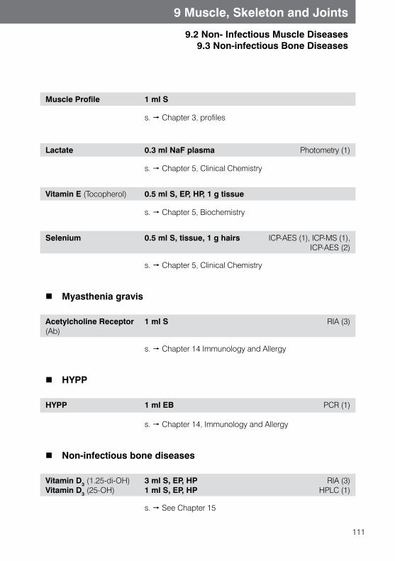
111
9.2 Non- Infectious Muscle Diseases9.3 Non-infectious Bone Diseases
9 Muscle, Skeleton and Joints
Muscle Profile 1 ml S
s. Chapter 3, profiles
Lactate 0.3 ml NaF plasma Photometry (1)
s. Chapter 5, Clinical Chemistry
Vitamin E (Tocopherol) 0.5 ml S, EP, HP, 1 g tissue
s. Chapter 5, Biochemistry
Selenium 0.5 ml S, tissue, 1 g hairs ICP-AES (1), ICP-MS (1), ICP-AES (2)
s. Chapter 5, Clinical Chemistry
Myasthenia gravis
Acetylcholine Receptor (Ab)
1 ml S RIA (3)
s. Chapter 14 Immunology and Allergy
HYPP
HYPP 1 ml EB PCR (1)
s. Chapter 14, Immunology and Allergy
Non-infectious bone diseases
Vitamin D3 (1.25-di-OH) Vitamin D3 (25-OH)
3 ml S, EP, HP 1 ml S, EP, HP
RIA (3) HPLC (1)
s. See Chapter 15
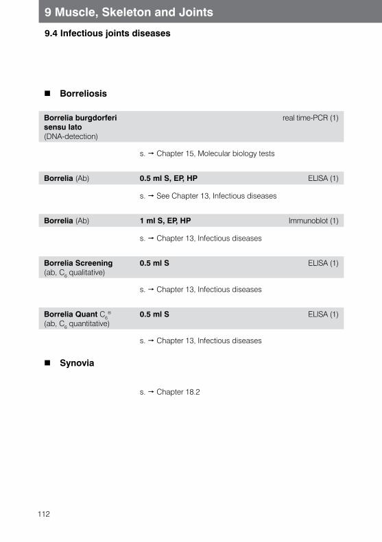
112
9 Muscle, Skeleton and Joints
9.4 Infectious joints diseases
Borreliosis
Borrelia burgdorferi sensu lato (DNA-detection)
real time-PCR (1)
s. Chapter 15, Molecular biology tests
Borrelia (Ab) 0.5 ml S, EP, HP ELISA (1)
s. See Chapter 13, Infectious diseases
Borrelia (Ab) 1 ml S, EP, HP Immunoblot (1)
s. Chapter 13, Infectious diseases
Borrelia Screening (ab, C6 qualitative)
0.5 ml S ELISA (1)
s. Chapter 13, Infectious diseases
Borrelia Quant C6®
(ab, C6 quantitative)0.5 ml S ELISA (1)
s. Chapter 13, Infectious diseases
Synovia
s. Chapter 18.2
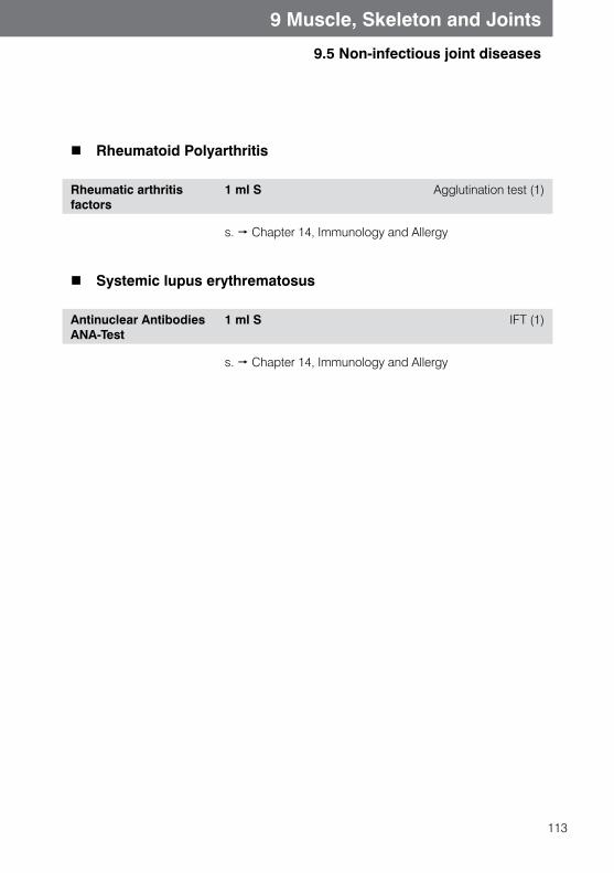
113
9 Muscle, Skeleton and Joints
9.5 Non-infectious joint diseases
Rheumatoid Polyarthritis
Rheumatic arthritis factors
1 ml S Agglutination test (1)
s. Chapter 14, Immunology and Allergy
Systemic lupus erythrematosus
Antinuclear AntibodiesANA-Test
1 ml S IFT (1)
s. Chapter 14, Immunology and Allergy
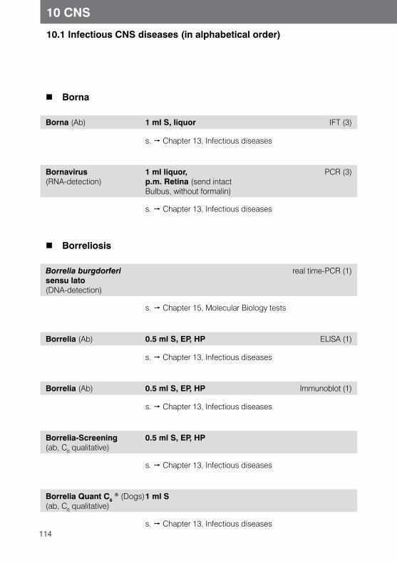
114
10.1 Infectious CNS diseases (in alphabetical order)
10 CNS
Borna
Borna (Ab) 1 ml S, liquor IFT (3)
s. Chapter 13, Infectious diseases
Bornavirus (RNA-detection)
1 ml liquor, p.m. Retina (send intact Bulbus, without formalin)
PCR (3)
s. Chapter 13, Infectious diseases
Borreliosis
Borrelia burgdorferi sensu lato (DNA-detection)
real time-PCR (1)
s. Chapter 15, Molecular Biology tests
Borrelia (Ab) 0.5 ml S, EP, HP ELISA (1)
s. Chapter 13, Infectious diseases
Borrelia (Ab) 0.5 ml S, EP, HP Immunoblot (1)
s. Chapter 13, Infectious diseases
Borrelia-Screening (ab, C6 qualitative)
0.5 ml S, EP, HP
s. Chapter 13, Infectious diseases
Borrelia Quant C6 ® (Dogs)
(ab, C6 qualitative)1 ml S
s. Chapter 13, Infectious diseases
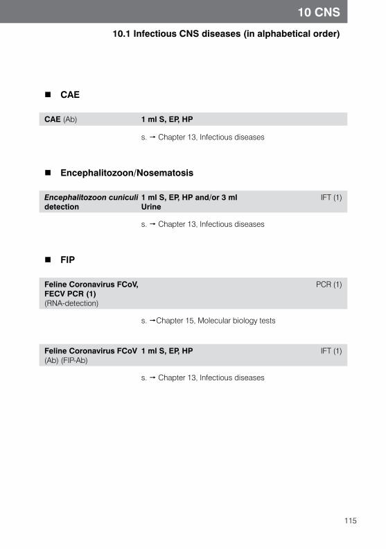
115
10.1 Infectious CNS diseases (in alphabetical order)
10 CNS
CAE
CAE (Ab) 1 ml S, EP, HP
s. Chapter 13, Infectious diseases
Encephalitozoon/Nosematosis
Encephalitozoon cuniculi detection
1 ml S, EP, HP and/or 3 ml Urine
IFT (1)
s. Chapter 13, Infectious diseases
FIP
Feline Coronavirus FCoV, FECV PCR (1)(RNA-detection)
PCR (1)
s. Chapter 15, Molecular biology tests
Feline Coronavirus FCoV (Ab) (FIP-Ab)
1 ml S, EP, HP IFT (1)
s. Chapter 13, Infectious diseases
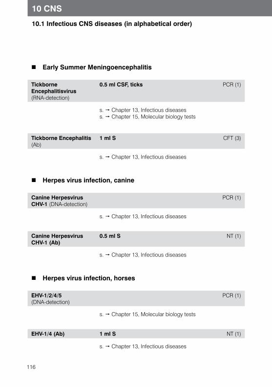
116
10 CNS
10.1 Infectious CNS diseases (in alphabetical order)
Early Summer Meningoencephalitis
Tickborne Encephalitisvirus (RNA-detection)
0.5 ml CSF, ticks PCR (1)
s. Chapter 13, Infectious diseases s. Chapter 15, Molecular biology tests
Tickborne Encephalitis (Ab)
1 ml S CFT (3)
s. Chapter 13, Infectious diseases
Herpes virus infection, canine
Canine Herpesvirus CHV-1 (DNA-detection)
PCR (1)
s. Chapter 13, Infectious diseases
Canine Herpesvirus CHV-1 (Ab)
0.5 ml S NT (1)
s. Chapter 13, Infectious diseases
Herpes virus infection, horses
EHV-1/2/4/5 (DNA-detection)
PCR (1)
s. Chapter 15, Molecular biology tests
EHV-1/4 (Ab) 1 ml S NT (1)
s. Chapter 13, Infectious diseases
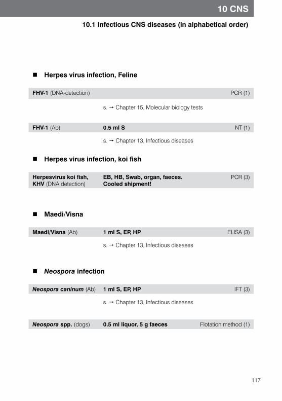
117
10 CNS
10.1 Infectious CNS diseases (in alphabetical order)
Herpes virus infection, Feline
FHV-1 (DNA-detection) PCR (1)
s. Chapter 15, Molecular biology tests
FHV-1 (Ab) 0.5 ml S NT (1)
s. Chapter 13, Infectious diseases
Herpes virus infection, koi fish
Herpesvirus koi fish, KHV (DNA detection)
EB, HB, Swab, organ, faeces.Cooled shipment!
PCR (3)
Maedi/Visna
Maedi/Visna (Ab) 1 ml S, EP, HP ELISA (3)
s. Chapter 13, Infectious diseases
Neospora infection
Neospora caninum (Ab) 1 ml S, EP, HP IFT (3)
s. Chapter 13, Infectious diseases
Neospora spp. (dogs) 0.5 ml liquor, 5 g faeces Flotation method (1)
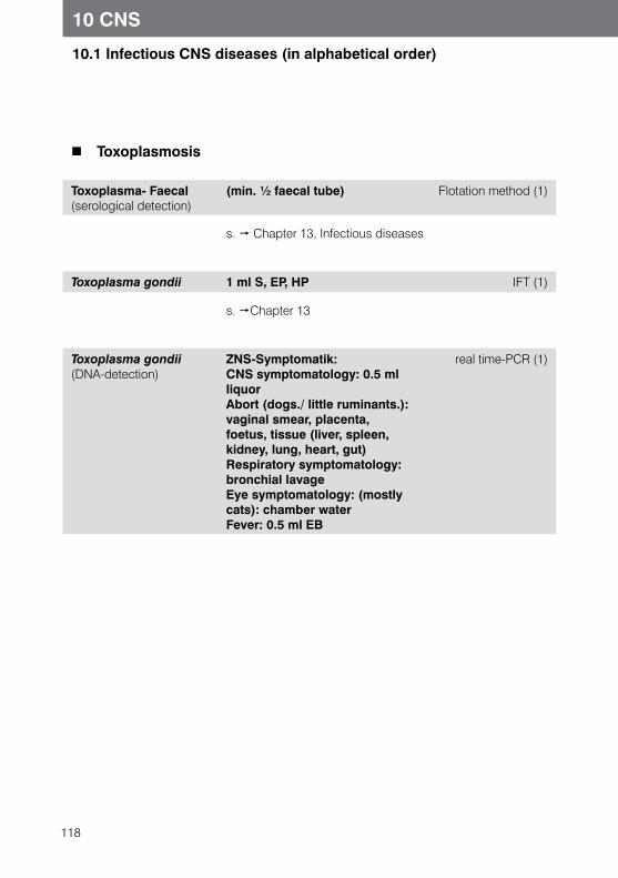
118
10 CNS
10.1 Infectious CNS diseases (in alphabetical order)
Toxoplasmosis
Toxoplasma- Faecal(serological detection)
(min. ½ faecal tube) Flotation method (1)
s. Chapter 13, Infectious diseases
Toxoplasma gondii 1 ml S, EP, HP IFT (1)
s. Chapter 13
Toxoplasma gondii(DNA-detection)
ZNS-Symptomatik: CNS symptomatology: 0.5 ml liquorAbort (dogs./ little ruminants.): vaginal smear, placenta, foetus, tissue (liver, spleen, kidney, lung, heart, gut)Respiratory symptomatology: bronchial lavageEye symptomatology: (mostly cats): chamber waterFever: 0.5 ml EB
real time-PCR (1)
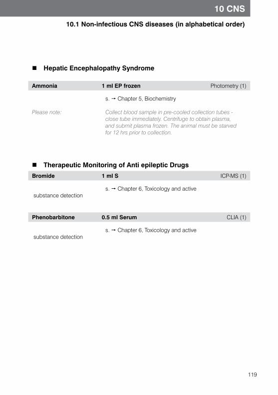
119
10 CNS
10.1 Non-infectious CNS diseases (in alphabetical order)
Hepatic Encephalopathy Syndrome
Ammonia 1 ml EP frozen Photometry (1)
s. Chapter 5, Biochemistry
Please note: Collect blood sample in pre-cooled collection tubes - close tube immediately. Centrifuge to obtain plasma, and submit plasma frozen. The animal must be starved for 12 hrs prior to collection.
Therapeutic Monitoring of Anti epileptic Drugs
Bromide 1 ml S ICP-MS (1)
s. Chapter 6, Toxicology and active substance detection
Phenobarbitone 0.5 ml Serum CLIA (1)
s. Chapter 6, Toxicology and active substance detection
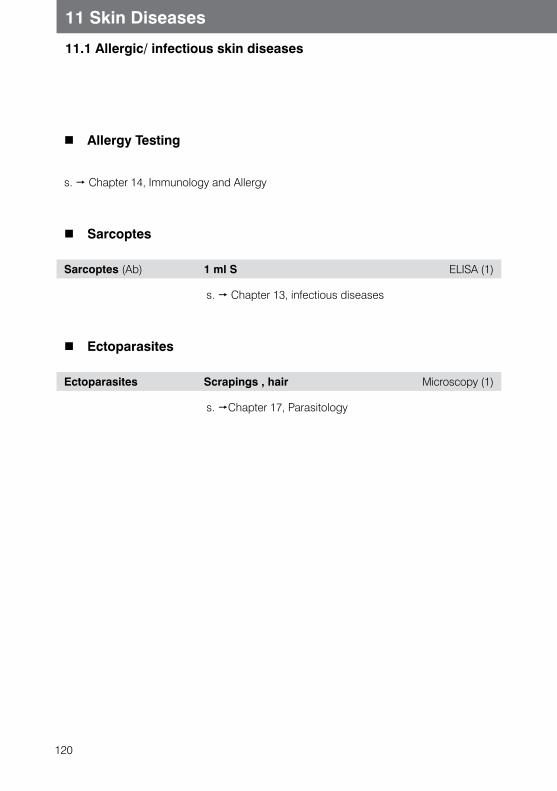
120
11.1 Allergic/ infectious skin diseases
11 Skin Diseases
Allergy Testing
s. Chapter 14, Immunology and Allergy
Sarcoptes
Sarcoptes (Ab) 1 ml S ELISA (1)
s. Chapter 13, infectious diseases
Ectoparasites
Ectoparasites Scrapings , hair Microscopy (1)
s. Chapter 17, Parasitology
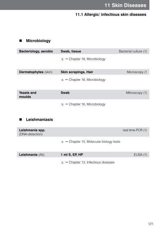
121
11 Skin Diseases
11.1 Allergic/ infectious skin diseases
Microbiology
Bacteriology, aerobic Swab, tissue Bacterial culture (1)
s. Chapter 16, Microbiology
Dermatophytes (skin) Skin scrapings, Hair Microscopy (1
s. Chapter 16, Microbiology
Yeasts and moulds
Swab Mikroscopy (1)
s. Chapter 16, Microbiology
Leishmaniasis
Leishmania spp. (DNA-detection)
real time-PCR (1)
s. Chapter 15, Molecular biology tests
Leishmania (Ab) 1 ml S, EP, HP ELISA (1)
s. Chapter 13, Infectious diseases
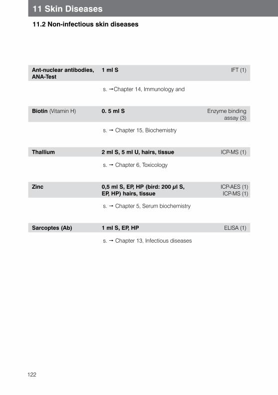
122
11 Skin Diseases
11.2 Non-infectious skin diseases
Ant-nuclear antibodies, ANA-Test
1 ml S IFT (1)
s. Chapter 14, Immunology and
Biotin (Vitamin H) 0. 5 ml S Enzyme bindingassay (3)
s. Chapter 15, Biochemistry
Thallium 2 ml S, 5 ml U, hairs, tissue ICP-MS (1)
s. Chapter 6, Toxicology
Zinc 0,5 ml S, EP, HP (bird: 200 µl S, EP, HP) hairs, tissue
ICP-AES (1) ICP-MS (1)
s. Chapter 5, Serum biochemistry
Sarcoptes (Ab) 1 ml S, EP, HP ELISA (1)
s. Chapter 13, Infectious diseases
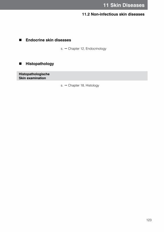
123
11 Skin Diseases
11.2 Non-infectious skin diseases
Endocrine skin diseases
s. Chapter 12, Endocrinology
Histopathology
HistopathologischeSkin examination
s. Chapter 18, Histology
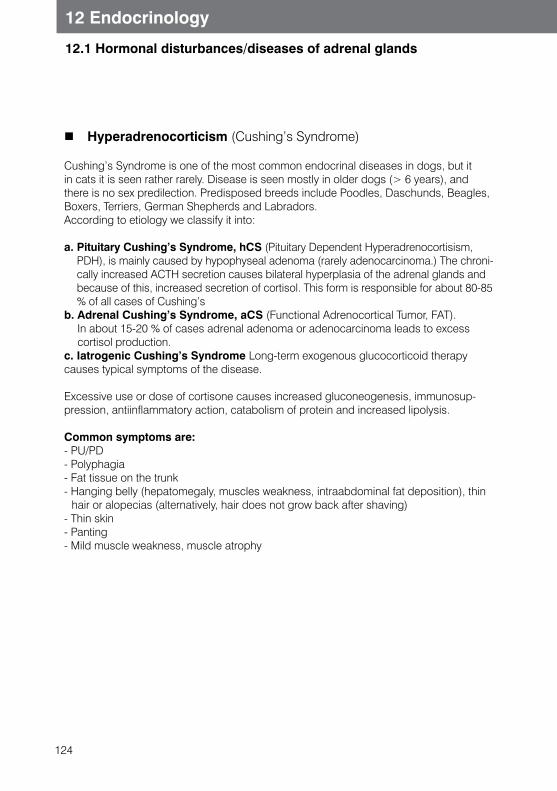
124
12.1 Hormonal disturbances/diseases of adrenal glands
12 Endocrinology
Hyperadrenocorticism (Cushing’s Syndrome)
Cushing’s Syndrome is one of the most common endocrinal diseases in dogs, but it in cats it is seen rather rarely. Disease is seen mostly in older dogs (> 6 years), and there is no sex predilection. Predisposed breeds include Poodles, Daschunds, Beagles, Boxers, Terriers, German Shepherds and Labradors.According to etiology we classify it into:
a. Pituitary Cushing’s Syndrome, hCS (Pituitary Dependent Hyperadrenocortisism, PDH), is mainly caused by hypophyseal adenoma (rarely adenocarcinoma.) The chroni-cally increased ACTH secretion causes bilateral hyperplasia of the adrenal glands and because of this, increased secretion of cortisol. This form is responsible for about 80-85 % of all cases of Cushing’s
b. Adrenal Cushing’s Syndrome, aCS (Functional Adrenocortical Tumor, FAT). In about 15-20 % of cases adrenal adenoma or adenocarcinoma leads to excess cortisol production.
c. Iatrogenic Cushing’s Syndrome Long-term exogenous glucocorticoid therapy causes typical symptoms of the disease.
Excessive use or dose of cortisone causes increased gluconeogenesis, immunosup-pression, antiinflammatory action, catabolism of protein and increased lipolysis.
Common symptoms are:- PU/PD- Polyphagia- Fat tissue on the trunk- Hanging belly (hepatomegaly, muscles weakness, intraabdominal fat deposition), thin
hair or alopecias (alternatively, hair does not grow back after shaving)- Thin skin- Panting- Mild muscle weakness, muscle atrophy
125
12 Endocrinology
12.1 Hormonal disturbances/diseases of adrenal glands
In horses Cushing syndrome is a significant and frequent endocrinopathy of horses and ponies with an age over 15 years. It is caused by dysfunction of the pars intermedia of hypophysis. Typical clinical signs are hirsuthism (excessive hair growth), muscle weak-ness, abnormal deposition of fat tissue in the body, weakness after exercise, PU/PD and recurring laminitis. For all the diagnostic tests, horses need to be calm and pain free. Pain (due to laminitis, for example) or increased stress before or during sampling can lead to false positive results. Seasonal changes of the hypophysis and adrenal glands in autumn can lead to false positive test results in healthy horses. Negative test results in autumn exclude Equine Cushing’s Syndrome (ECS) with great probability, while positive results in horses with inconclusive clinical symptoms should be checked again between January and August.
Various non specific parameters as well as endocrinological function tests may be used to diagnose Cushing’s syndrome.
Cortisol 0.5 ml S ECLIA(1)
The determination of a single cortisol value is not useful for diagnosing Cushing’s disease because of the episodic secretion of cortisol in dogs, and it is extremely stress- de-pendant in cats. In horses, resting cortisol levels are not or very slightly elevated, but the circadian rhythm is lost.
Functional tests for diagnosing hyperadrenocortisism/ Equine Cushing Syndrome
Low dose of dexamethasone test (screening test, LDDS)2 cortisol values 3 cortisol values
2 x 0.5 ml S3 x 0.5 ml S
CLIA (1) CLIA (1)
Test principles: ACTH from the pituitary gland, controlled by the hypothala-mus, stimulates the adrenal cortex to produce cortisol. The rising cortisol level leads to a reduced ACTH secretion via a negative feedback mechanism. This also happens when dexamethasone is administered exogenously
Physiological mechanism: After approx. 2-3 hours ACTH secretion is suppressed due to the negative feedback mechanism. The suppression lasts for approx. 24-48 hours. The adrenal cortex produces less cortisol, causing the cortisol level to drop.
126
12 Endocrinology
12.1 Hormonal disturbances/diseases of adrenal glands
Adrenal Cushing’s Syndrome (aCS)
Tumors of Functional Adrenal Tissue (FAT) produce auto-nomic cortisol. Dexamethasone inhibits secretion of ACTH, but does not lead to reduced cortisol secretion, so the level of cortisol does not fall or falls only very slightly.
Pituitary Cushing’s Syndrome(hCS)
The pituitary gland in sick animals will show little or no res-ponse to dexamethasone. The ACTH secretion will not be suppressed at all or only for a short period of time, then it resumes and therefore the cortisol secretion in the adrenal cortex remains stimulated. The cortisol level is unaffected, drops only very little, or only for a very short period of time.
Approx. 15-20% of the animals with PDH do not react with a significant suppression of cortisol levels,
even in the high- dose test
Sensitivity of these tests ranges about 85-95%, specificity will be about 70-75%..
Performing the test in dogs, cats:
1. First blood sample, for basal cortisol value2. Injection of dexamethasone 0,01 mg/kg body weight
i.v. (dogs) Injection of dexamethasone 0,1 mg/kg b.w. i.v. (cats)
3. Second blood sample is taken 8 hours post injection, for suppression value (an additional blood sample may be taken 4 hours post injection)
Assessment dogs, cats - 4 hour value and 8 hour value <1,0μg/dl: negative result, physiologically normal. - 4 hour value and 8 hour value >1,4 μg/dl: suspicion of CS (hCS or aCS)- 4 hours value <1,4 μg/dl and 8 hour value >1,4 μg/dl or - 4 hour value < 50 % of basal value and 8 hour value >
1,4 μg/dl: suspicion of CS (hCS more likely, but aCS not excluded) - 8 hour value < 50 % of basal value, but > 1,4 μg/dl:
suspicion of CS (hCS more probably, but aCS not excluded)
127
12 Endocrinology
12.1 Hormonal disturbances/diseases of adrenal glands
Performing the test in Horses
1. first blood sampling for basal cortisol value2. Injection of dexamethasone 0,04 mg/kg (4 mg/100 kg)
b.w. i.m. or i.v.3. next day: second blood sampling 19 to 24 hours post inj.
for suppression value (an additional blood sample may be taken 15 hours post injection)
Please note: Note: Sampling tubes should be labeled sample 1 and sample 2 (and sample 3, if applicable), test interpretation for horses.
In healthy horses, corticosteroids act due to negative feedback and cause reduction of endogenous cortisol distribution. This leads to post suppression values of 0.5 to 1 μg/dl. In horses with ECS, dexamethasone induces no negative feedback and causes no significant reduction of cortisol concentration after dexamethasone administration.
Dexamethasone suppression test is the test of choice in dogs, cats and horses to diagnose hyperadrenocorticism.
ACTH-Stimulation test(dogs, cats)2 cortisol assessments
2 x 0.5 ml S ECLIA (1)
Test Principle: With this test you can assess the secretory ability of th adrenal cortex.Dogs, cats: ACTH-Stimulation test is the test of choice for diagnosing iatrogenic Cushing’s Syndrome, for treatment control in dogs with hyperadrenocorticism and also for diagnosing hypoadrenocorticism.
Test method: 1.First blood sampling, for basal value of cortisol2. Injection of ACTH (i.e. Synachten) i.v./i.m. Cats: 0,125 mg/animal Dogs: 0,25 mg/animal (0,125 mg= 12,5 IU, 0,25 mg=25 IU)
Interpretation: -dogs - Basal value <0.5-2μg/dl and stimulation value <0.5-2μg/dl: Iatrogenic Cushing’s Syndrome or suspicion of Addison’s disease
Treatment monitoring During treatment of CS with trilostane please follow the manufacturer’s recommended protocol.
128
12 Endocrinology
12.1 Hormonal disturbances/diseases of adrenal glands
Cortisol/creatinine ratio (dogs, cats)1 determination 1 x 3 ml urine CLIA (1)
Test principle: Animals with Cushing’s syndrome show an elevated serum cortisol level and elevated cortisol excretion. Creatinine is used as reference value only, since the cortisol level may also be elevated in a physiological metabolic state. The test is highly sensitive (95-99%) and therefore sui-table for ruling out Cushing’s syndrome. On the other hand, it has a low specificity (pathological levels may be found in diabetes mellitus, diabetes insipidus, pyometra, hypercalcaemia, renal disease, liver disease, etc). Patho-logical results should therefore be confirmed with further function tests (e.g. dexamethasone low dose test). The urine should always be collected in a stress-free envi-ronment (preferably at home and not in the practice).
Test method: Day 1: collect the morning urine for the first sampleDay 2: collect the morning urine for the second sample
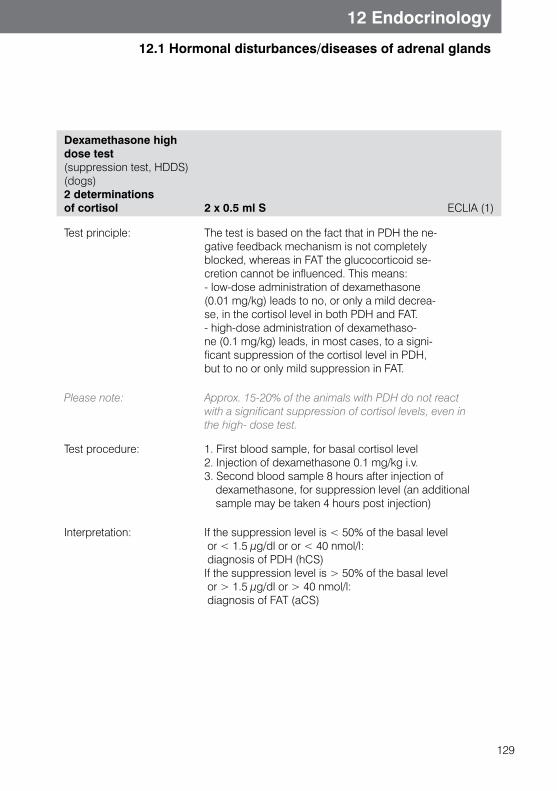
129
12 Endocrinology
12.1 Hormonal disturbances/diseases of adrenal glands
Dexamethasone high dose test (suppression test, HDDS)(dogs)2 determinations of cortisol 2 x 0.5 ml S ECLIA (1)
Test principle: The test is based on the fact that in PDH the ne-gative feedback mechanism is not completely blocked, whereas in FAT the glucocorticoid se-cretion cannot be influenced. This means:- low-dose administration of dexamethasone (0.01 mg/kg) leads to no, or only a mild decrea-se, in the cortisol level in both PDH and FAT.- high-dose administration of dexamethaso-ne (0.1 mg/kg) leads, in most cases, to a signi-ficant suppression of the cortisol level in PDH, but to no or only mild suppression in FAT.
Please note: Approx. 15-20% of the animals with PDH do not react with a significant suppression of cortisol levels, even in the high- dose test.
Test procedure: 1. First blood sample, for basal cortisol level2. Injection of dexamethasone 0.1 mg/kg i.v. 3. Second blood sample 8 hours after injection of
dexamethasone, for suppression level (an additional sample may be taken 4 hours post injection)
Interpretation: If the suppression level is < 50% of the basal level or < 1.5 μg/dl or or < 40 nmol/l: diagnosis of PDH (hCS)If the suppression level is > 50% of the basal level or > 1.5 μg/dl or > 40 nmol/l: diagnosis of FAT (aCS)
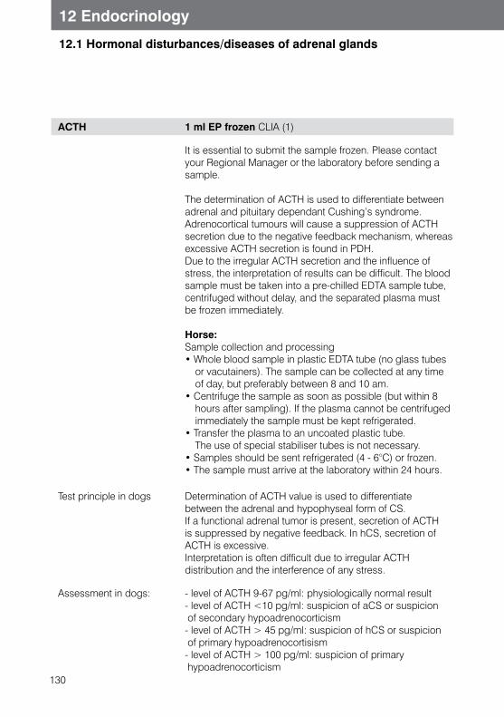
130
12 Endocrinology
12.1 Hormonal disturbances/diseases of adrenal glands
ACTH 1 ml EP frozen CLIA (1)
It is essential to submit the sample frozen. Please contact your Regional Manager or the laboratory before sending a sample.
The determination of ACTH is used to differentiate between adrenal and pituitary dependant Cushing’s syndrome. Adrenocortical tumours will cause a suppression of ACTH secretion due to the negative feedback mechanism, whereas excessive ACTH secretion is found in PDH.Due to the irregular ACTH secretion and the influence of stress, the interpretation of results can be difficult. The blood sample must be taken into a pre-chilled EDTA sample tube, centrifuged without delay, and the separated plasma must be frozen immediately.
Horse:Sample collection and processing• Whole blood sample in plastic EDTA tube (no glass tubes
or vacutainers). The sample can be collected at any time of day, but preferably between 8 and 10 am.
• Centrifuge the sample as soon as possible (but within 8 hours after sampling). If the plasma cannot be centrifuged immediately the sample must be kept refrigerated.
• Transfer the plasma to an uncoated plastic tube. The use of special stabiliser tubes is not necessary.
• Samples should be sent refrigerated (4 - 6°C) or frozen.• The sample must arrive at the laboratory within 24 hours.
Test principle in dogs Determination of ACTH value is used to differentiate between the adrenal and hypophyseal form of CS. If a functional adrenal tumor is present, secretion of ACTH is suppressed by negative feedback. In hCS, secretion of ACTH is excessive.Interpretation is often difficult due to irregular ACTH distribution and the interference of any stress.
Assessment in dogs: - level of ACTH 9-67 pg/ml: physiologically normal result- level of ACTH <10 pg/ml: suspicion of aCS or suspicion of secondary hypoadrenocorticism- level of ACTH > 45 pg/ml: suspicion of hCS or suspicion of primary hypoadrenocortisism- level of ACTH > 100 pg/ml: suspicion of primary hypoadrenocorticism
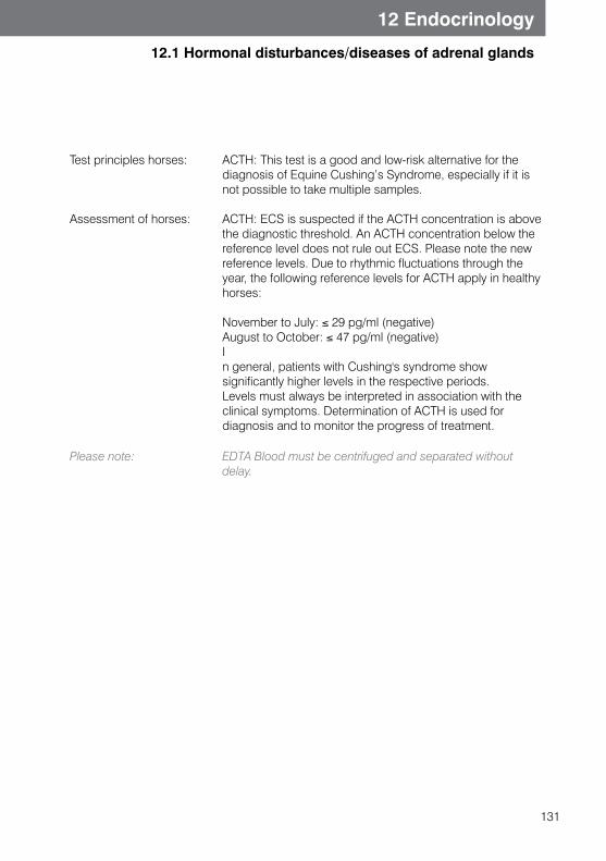
131
12 Endocrinology
12.1 Hormonal disturbances/diseases of adrenal glands
Test principles horses:
Assessment of horses:
ACTH: This test is a good and low-risk alternative for the diagnosis of Equine Cushing’s Syndrome, especially if it is not possible to take multiple samples.
ACTH: ECS is suspected if the ACTH concentration is above the diagnostic threshold. An ACTH concentration below the reference level does not rule out ECS. Please note the new reference levels. Due to rhythmic fluctuations through the year, the following reference levels for ACTH apply in healthy horses: November to July: ≤ 29 pg/ml (negative)August to October: ≤ 47 pg/ml (negative)In general, patients with Cushing's syndrome show significantly higher levels in the respective periods. Levels must always be interpreted in association with the clinical symptoms. Determination of ACTH is used for diagnosis and to monitor the progress of treatment.
Please note: EDTA Blood must be centrifuged and separated without delay.
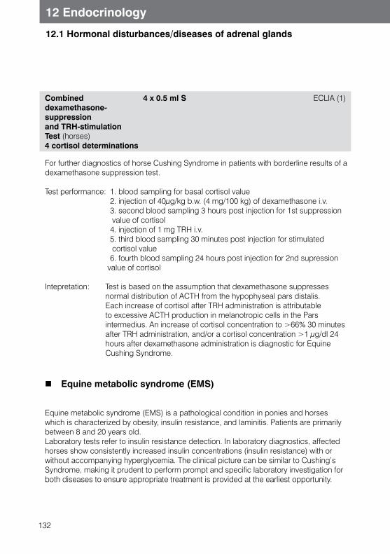
132
12 Endocrinology
12.1 Hormonal disturbances/diseases of adrenal glands
Combined dexamethasone- suppression and TRH-stimulation Test (horses)4 cortisol determinations
4 x 0.5 ml S ECLIA (1)
For further diagnostics of horse Cushing Syndrome in patients with borderline results of a dexamethasone suppression test.
Test performance: 1. blood sampling for basal cortisol value 2. injection of 40μg/kg b.w. (4 mg/100 kg) of dexamethasone i.v. 3. second blood sampling 3 hours post injection for 1st suppression
value of cortisol 4. injection of 1 mg TRH i.v. 5. third blood sampling 30 minutes post injection for stimulated
cortisol value 6. fourth blood sampling 24 hours post injection for 2nd supression value of cortisol
Intepretation: Test is based on the assumption that dexamethasone suppresses normal distribution of ACTH from the hypophyseal pars distalis. Each increase of cortisol after TRH administration is attributable to excessive ACTH production in melanotropic cells in the Pars intermedius. An increase of cortisol concentration to >66% 30 minutes after TRH administration, and/or a cortisol concentration >1 μg/dl 24 hours after dexamethasone administration is diagnostic for Equine Cushing Syndrome.
Equine metabolic syndrome (EMS)
Equine metabolic syndrome (EMS) is a pathological condition in ponies and horses which is characterized by obesity, insulin resistance, and laminitis. Patients are primarily between 8 and 20 years old.Laboratory tests refer to insulin resistance detection. In laboratory diagnostics, affected horses show consistently increased insulin concentrations (insulin resistance) with or without accompanying hyperglycemia. The clinical picture can be similar to Cushing’s Syndrome, making it prudent to perform prompt and specific laboratory investigation for both diseases to ensure appropriate treatment is provided at the earliest opportunity.
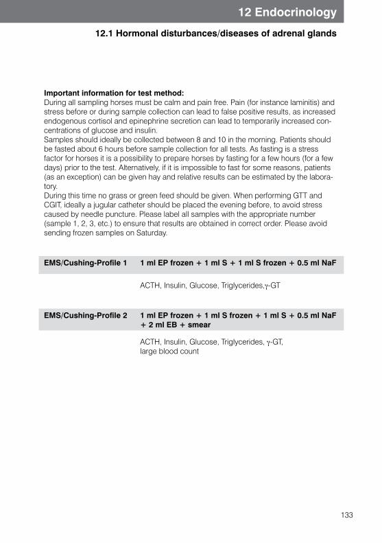
133
12 Endocrinology
12.1 Hormonal disturbances/diseases of adrenal glands
Important information for test method: During all sampling horses must be calm and pain free. Pain (for instance laminitis) and stress before or during sample collection can lead to false positive results, as increased endogenous cortisol and epinephrine secretion can lead to temporarily increased con-centrations of glucose and insulin.Samples should ideally be collected between 8 and 10 in the morning. Patients should be fasted about 6 hours before sample collection for all tests. As fasting is a stress factor for horses it is a possibility to prepare horses by fasting for a few hours (for a few days) prior to the test. Alternatively, if it is impossible to fast for some reasons, patients (as an exception) can be given hay and relative results can be estimated by the labora-tory.During this time no grass or green feed should be given. When performing GTT and CGIT, ideally a jugular catheter should be placed the evening before, to avoid stress caused by needle puncture. Please label all samples with the appropriate number (sample 1, 2, 3, etc.) to ensure that results are obtained in correct order. Please avoid sending frozen samples on Saturday.
EMS/Cushing-Profile 1 1 ml EP frozen + 1 ml S + 1 ml S frozen + 0.5 ml NaF
ACTH, Insulin, Glucose, Triglycerides,γ-GT
EMS/Cushing-Profile 2 1 ml EP frozen + 1 ml S frozen + 1 ml S + 0.5 ml NaF + 2 ml EB + smear
ACTH, Insulin, Glucose, Triglycerides, γ-GT, large blood count
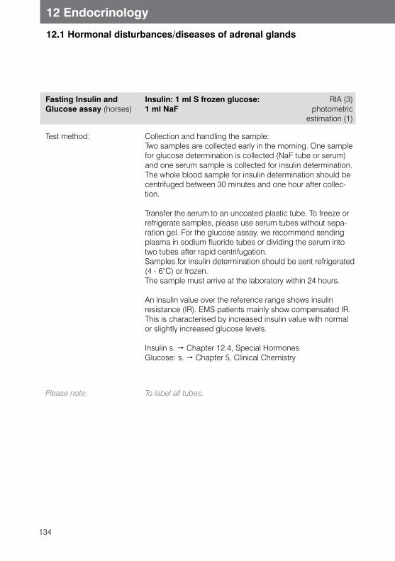
134
12 Endocrinology
12.1 Hormonal disturbances/diseases of adrenal glands
Fasting Insulin and Glucose assay (horses)
Insulin: 1 ml S frozen glucose: 1 ml NaF
RIA (3) photometric
estimation (1)
Test method: Collection and handling the sample:Two samples are collected early in the morning. One sample for glucose determination is collected (NaF tube or serum) and one serum sample is collected for insulin determination.The whole blood sample for insulin determination should be centrifuged between 30 minutes and one hour after collec-tion.
Transfer the serum to an uncoated plastic tube. To freeze or refrigerate samples, please use serum tubes without sepa-ration gel. For the glucose assay, we recommend sending plasma in sodium fluoride tubes or dividing the serum into two tubes after rapid centrifugation.Samples for insulin determination should be sent refrigerated (4 - 6°C) or frozen.The sample must arrive at the laboratory within 24 hours.
An insulin value over the reference range shows insulin resistance (IR). EMS patients mainly show compensated IR. This is characterised by increased insulin value with normal or slightly increased glucose levels.
Insulin s. Chapter 12.4, Special HormonesGlucose: s. Chapter 5, Clinical Chemistry
Please note: To label all tubes.
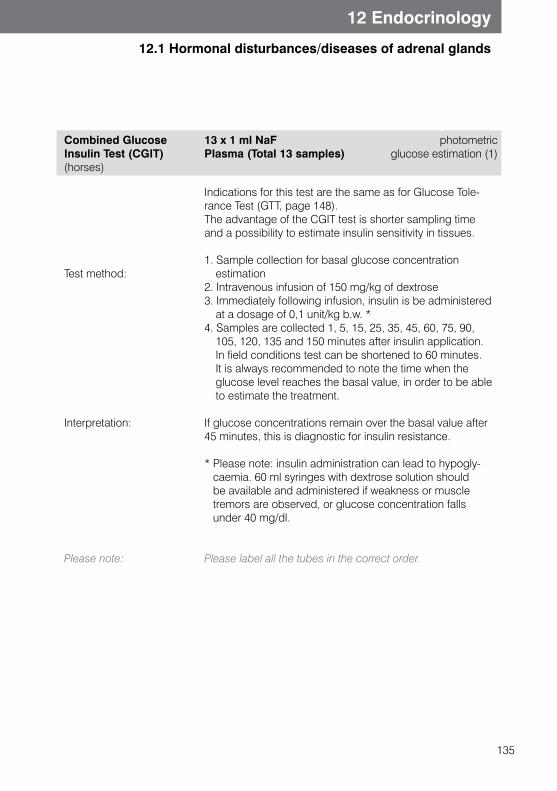
135
12 Endocrinology
12.1 Hormonal disturbances/diseases of adrenal glands
Combined GlucoseInsulin Test (CGIT) (horses)
13 x 1 ml NaF Plasma (Total 13 samples)
photometric glucose estimation (1)
Test method:
Interpretation:
Please note:
Indications for this test are the same as for Glucose Tole-rance Test (GTT, page 148).The advantage of the CGIT test is shorter sampling time and a possibility to estimate insulin sensitivity in tissues.
1. Sample collection for basal glucose concentration estimation
2. Intravenous infusion of 150 mg/kg of dextrose3. Immediately following infusion, insulin is be administered
at a dosage of 0,1 unit/kg b.w. *4. Samples are collected 1, 5, 15, 25, 35, 45, 60, 75, 90,
105, 120, 135 and 150 minutes after insulin application. In field conditions test can be shortened to 60 minutes. It is always recommended to note the time when the glucose level reaches the basal value, in order to be able to estimate the treatment.
If glucose concentrations remain over the basal value after 45 minutes, this is diagnostic for insulin resistance.
* Please note: insulin administration can lead to hypogly-caemia. 60 ml syringes with dextrose solution should be available and administered if weakness or muscle tremors are observed, or glucose concentration falls under 40 mg/dl.
Please label all the tubes in the correct order.
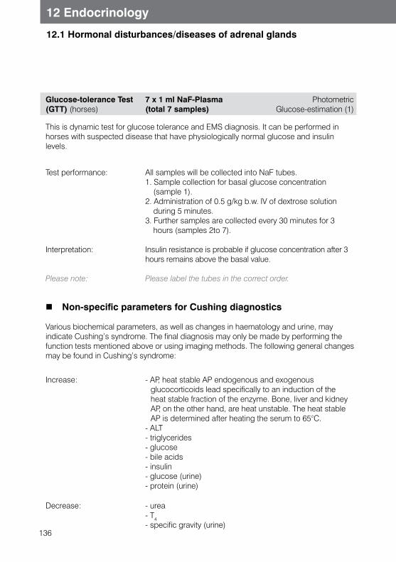
136
12 Endocrinology
12.1 Hormonal disturbances/diseases of adrenal glands
Glucose-tolerance Test (GTT) (horses)
7 x 1 ml NaF-Plasma(total 7 samples)
Photometric Glucose-estimation (1)
This is dynamic test for glucose tolerance and EMS diagnosis. It can be performed in horses with suspected disease that have physiologically normal glucose and insulin levels.
Test performance:
Interpretation:
Please note:
All samples will be collected into NaF tubes.1. Sample collection for basal glucose concentration
(sample 1). 2. Administration of 0.5 g/kg b.w. IV of dextrose solution
during 5 minutes.3. Further samples are collected every 30 minutes for 3
hours (samples 2to 7).
Insulin resistance is probable if glucose concentration after 3 hours remains above the basal value.
Please label the tubes in the correct order.
Non-specific parameters for Cushing diagnostics
Various biochemical parameters, as well as changes in haematology and urine, may indicate Cushing’s syndrome. The final diagnosis may only be made by performing the function tests mentioned above or using imaging methods. The following general changes may be found in Cushing’s syndrome:
Increase: - AP, heat stable AP endogenous and exogenous glucocorticoids lead specifically to an induction of the heat stable fraction of the enzyme. Bone, liver and kidney AP, on the other hand, are heat unstable. The heat stable AP is determined after heating the serum to 65°C.
- ALT- triglycerides- glucose- bile acids- insulin- glucose (urine)- protein (urine)
Decrease: - urea- T4- specific gravity (urine)
137
12 Endocrinology
12.1 Hormonal disturbances/diseases of adrenal glands
Hypoadrenocorticism (dogs, horses)
Hypoadrenocorticism is a relatively rarely seen endocrine disorder, which can be caused by the adrenal gland cortex disease (primary hypoadrenocorticism, Addison’s disease) or reduced secretion of ACTH (secondary hypoadrenocorticism).Primary hypoadrenocorticism usually affects both glucocorticoid and mineralocorticoid synthesis, while secondary hypoadrenocorticism usually affects glucocorticoid synthesis only. There is a gender predisposition in females (70%). In the majority of cases the affected animals are medium to large breeds and middle aged.The most common form in veterinary medicine is iatrogenic hypoadrenocorticism caused by long term exogenous glucocorticoid administration or administration of o,p´-DDD (Mitotane) in Cushing’s treatment.Various non-specific changes may or may not be found in laboratory diagnostics, such as mild anaemia, azotaemia, hypercalcaemia or hypoglycaemia, whereas a change of the sodium/potassium ratio is seen only if mineralocorticoid synthesis is affected. The normal ratio is 27:1 - 40:1, whereas in hypoadrenocorticism, a ratio below 27:1 is often found.A single test for cortisol is appropriate only to exclude hypoadrenocorticism, as healthy animals can show cortisol levels < 0.5 μg/dl.
Horse: As in dogs and cats, hypoadrenocorticism is relatively rarely seen endocrine disorder based on function restriction of adrenal glands (primary hypoadrenocorticism, similar to Addison’s disease) or reduced secretion of ACTH or CRH (secondary hypoadrenocorticism). The most common form in veterinary medicine is iatrogenic hypoadrenocor-ticism caused by long term exogenous glucocorticoids application. A single test for cortisol is not appropriate for diagnosis. ACTH stimulation test and single ACTH estimation can give important diagnostic clues, but diagnosis should be made only in combination with history, clinical symptoms and diagnostic tests.
138
12 Endocrinology
12.2 Hormonal disturbances/diseases of thyroid gland
ACTH-Stimulation test2 Cortisol assay
2 x 0.5 ml S CLIA (1)
Test method:(dogs, cats)
s. Chapter 12.1
Interpretation: Basal value mostly < 0.5 - 2 μg/dl andStimulation value < 0.5 - 2 μg/dl
Test performance (horses)
Interpretation (horses)
1. first blood sampling for cortisol basal value at 9 o’clock2. Injection of 100 IU ACTH i.v. (e.g. Synacthen®)3. second blood sampling 2 hours post injection for
stimulated cortisol value
In healthy horses cortisol value increases by about 80 %. Horses with hypoadrenocorticism have mostly very low cortisol basal value and only low or no cortisol increase after stimulation.
Aldosterone (dogs, cats) 0.5 ml S cold RIA (3)
A single aldosterone determination is of little diagnostic value. Interpretation should be made after performing an ACTH stimulation test (Follow the protocol used for cortisol level determination in the diagnosis of Cushing’s syndrome. s. ACTH stimulation test)
Indications: - selective aldosterone deficiency (hyponatraemia and hyperkalaemia with normal cortisol basal level or physio-logical cortisol value after ACTH stimulation test)
- primary hyperaldosteronism
Occurence: Produced in the Zona glomerulosa of the adrenal glands,regulated by the renin-angiotensin-aldosterone-system andserum postassium concentration
Increased in: excessive stimulation: primary hyperaldosteronism: hyperactivity of adrenal glands (secondary hyperaldosteronism: disturbances in aldosterone removal)
Decreased in: little or no stimulation:Hypoaldosteronism
139
12 Endocrinology
12.2 Hormonal disturbances/diseases of thyroid gland
Hypothyroidism
Primary canine hypothyroidism (T4 deficiency) is caused by lymphocytic thyreoditis, idiopathic follicular atrophy or (rarely) neoplasia of thyroid gland. Secondary (TSH deficiency caused by pituitary tumor) has been very rarely described and tertiary (TRH deficiency) hypothyroidism has not been reported in veterinary medicine.
Clinical symptoms are caused by a low concentration of thyroid hormone circulating in blood. Large and medium breed dogs are predisposed.
The nonspecific parameters listed below, described in laboratory diagnostics, can sug-gest hypothyroidism:- Increased levels of cholesterol in serum- mild to moderate anaemia (usually normochromic, normocytic,
or rarely hypochromic or microcytic)- Increased fructosamine levels- Insignificant increase of creatinine kinase.
In cats hypothyroidism is very rare.In horses, primary thyroid diseases are rare in adult horses, but cases have been de-scribed. Often it is overdiagnosed in overweight or animals with hair loss, and therefore tests for hypothyroidism should be accompanied by tests for Cushing’s Syndrome or Equine Metabolic Syndrome. Note: Foals can physiologically show increased thyroid hormone values.
Thyroid hormones: individual hormone sssay
A common diagnostic challengs is dogs and cats with “Euthyroid Sick Syndrome”.Euthyroid Sick Syndrome occurs when thyroid hormone levels in blood are decreased due to different diseases, but the patient has few or no symptoms of hypothyroidism. Interpretation can be made more difficult by the presence of medication.
s. also Thyroid Profile in Horses above.
140
12 Endocrinology
12.2 Hormonal disturbances/diseases of thyroid gland
Note: Thyroid values may be decreased due to non-thyroidal diseases (NTI).NTI (Non-thyroidal illness):Diabetes mellitus, hyperadrenocorticism, hypoadrenocor-ticism, renal diseases, hepatic diseases, acute infections, neuro-muscular diseases, pyodermatitis, hypoproteinemia, congestive heart failure and others. Dogs suffering from one of these diseases should be tested with caution. If hyperadrenocorticism is suspected, it should be diagnosed and treated first.MedicationNSAIDs, glucocorticoids, anti-convulsive drugs, and especially sulphonamides.These medications should be withdrawn around 4-6 weeks before performing the test.
Total T4 0.5 ml S, EP, HP Enzyme immunoassay (1)
Total T4 is composed of a free and protein bound fraction. Both fractions are measured in this test. Endogenous T4 is produced only in thyroid gland and is an important indicator for diagnosing hyperthyroidism in cats and excluding hypothyroidism in dogs - as only a few dogs with hypothyroidism have T4 concentrations within reference ranges. (Normal values of T4 in hypothyroid dogs are possible in the early stages of hypothyroidism.) In addition, there are a few hypothyroid dogs (c. 1,5%) which develop T4 antibodies. Confusingly, these antibodies can “cause” falsely high T4 laboratory results. In these dogs we recommend testing Free T4 (FT4) values equilibrium dialysis and/or anti-T4 antibody detection.
Please note: NTI and medications can influence T4 values. (See above).
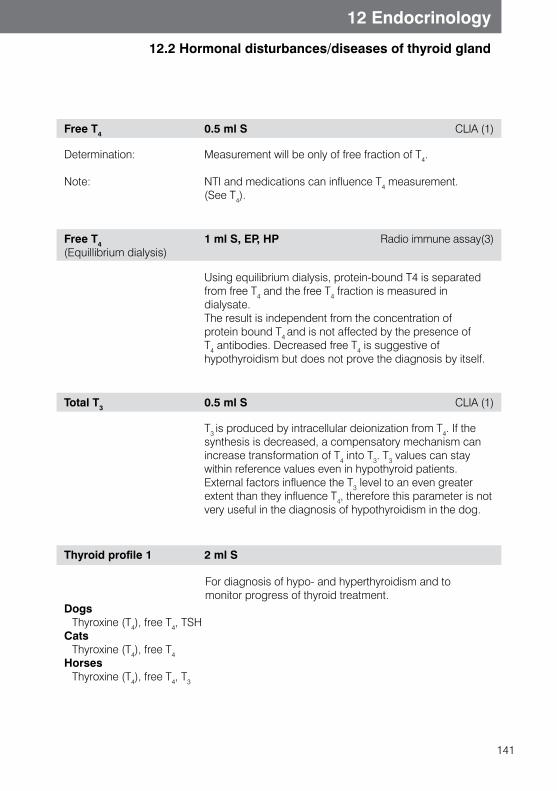
141
12 Endocrinology
12.2 Hormonal disturbances/diseases of thyroid gland
Free T4 0.5 ml S CLIA (1)
Determination:
Note:
Measurement will be only of free fraction of T4. NTI and medications can influence T4 measurement. (See T4).
Free T4 (Equillibrium dialysis)
1 ml S, EP, HP Radio immune assay(3)
Using equilibrium dialysis, protein-bound T4 is separated from free T4 and the free T4 fraction is measured in dialysate.The result is independent from the concentration of protein bound T4 and is not affected by the presence of T4 antibodies. Decreased free T4 is suggestive of hypothyroidism but does not prove the diagnosis by itself.
Total T3 0.5 ml S CLIA (1)
T3 is produced by intracellular deionization from T4. If the synthesis is decreased, a compensatory mechanism can increase transformation of T4 into T3. T3 values can stay within reference values even in hypothyroid patients. External factors influence the T3 level to an even greater extent than they influence T4, therefore this parameter is not very useful in the diagnosis of hypothyroidism in the dog.
Thyroid profile 1 2 ml S
For diagnosis of hypo- and hyperthyroidism and to monitor progress of thyroid treatment.Dogs Thyroxine (T4), free T4, TSHCats Thyroxine (T4), free T4Horses Thyroxine (T4), free T4, T3
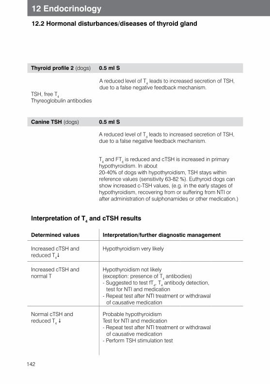
142
12 Endocrinology
12.2 Hormonal disturbances/diseases of thyroid gland
Thyroid profile 2 (dogs) 0.5 ml S
A reduced level of T4 leads to increased secretion of TSH, due to a false negative feedback mechanism.
TSH, free T4Thyreoglobulin antibodies
Canine TSH (dogs) 0.5 ml S
A reduced level of T4 leads to increased secretion of TSH, due to a false negative feedback mechanism.
T4 and FT4 is reduced and cTSH is increased in primary hypothyroidism. In about 20-40% of dogs with hypothyroidism, TSH stays within reference values (sensitivity 63-82 %). Euthyroid dogs can show increased c-TSH values, (e.g. in the early stages of hypothyroidism, recovering from or suffering from NTI or after administration of sulphonamides or other medication.)
Interpretation of T4 and cTSH results
Determined values Interpretation/further diagnostic management
Increased cTSH and reduced T4$
Hypothyroidism very likely
Increased cTSH and normal T
Hypothyroidism not likely(exception: presence of T4 antibodies) - Suggested to test fT4, T4 antibody detection,
test for NTI and medication- Repeat test after NTI treatment or withdrawal
of causative medication
Normal cTSH and reduced T4 $
Probable hypothyroidismTest for NTI and medication- Repeat test after NTI treatment or withdrawal
of causative medication - Perform TSH stimulation test
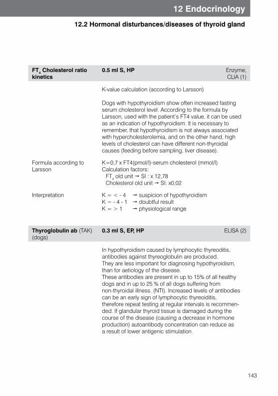
143
12 Endocrinology
12.2 Hormonal disturbances/diseases of thyroid gland
FT4 Cholesterol ratio kinetics
0.5 ml S, HP Enzyme,CLIA (1)
K-value calculation (according to Larsson)
Dogs with hypothyroidism show often increased fasting serum cholesterol level. According to the formula by Larsson, used with the patient’s FT4 value, it can be used as an indication of hypothyroidism. It is necessary to remember, that hypothyroidism is not always associated with hypercholesterolemia, and on the other hand, high levels of cholesterol can have different non-thyroidal causes (feeding before sampling, liver disease).
Formula according to Larsson
K=0,7 x FT4(pmol/l)-serum cholesterol (mmol/l)Calculation factors: FT4 old unit SI : x 12,78 Cholesterol old unit SI: x0,02
Interpretation K = < - 4 suspicion of hypothyroidismK = - 4 - 1 doubtful resultK = > 1 physiological range
Thyroglobulin ab (TAK) (dogs)
0.3 ml S, EP, HP ELISA (2)
In hypothyroidism caused by lymphocytic thyreoditis, antibodies against thyreoglobulin are produced. They are less important for diagnosing hypothyroidism, than for aetiology of the disease. These antibodies are present in up to 15% of all healthy dogs and in up to 25 % of all dogs suffering from non-thyroidal illness. (NTI). Increased levels of antibodies can be an early sign of lymphocytic thyreoiditis, therefore repeat testing at regular intervals is recommen-ded. If glandular thyroid tissue is damaged during the course of the disease (causing a decrease in hormone production) autoantibody concentration can reduce as a result of lower antigenic stimulation.
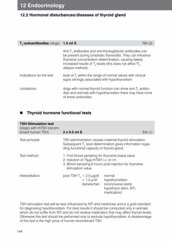
144
12 Endocrinology
12.2 Hormonal disturbances/diseases of thyroid gland
T4-autoantibodies (dogs) 1.5 ml S RIA (3)
Anti-T4 antibodies and anti-thyreoglobulin antibodies can be present during lymphatic thyreoditis. They can influence thyroxine concentration determination, causing falsely increased results of T4 levels (this does not affect fT4 dialysis method).
Indications for the test: level of T4 within the range of normal values with clinical signs strongly associated with hypothyroidism.
Limitations: dogs with normal thyroid function can show anti-T4 antibo-dies and animals with hypothyroidism there may have none of these antibodies.
Thyroid hormone functional tests
TSH-Stimulation test (dogs) with rhTSH (recom-binant human TSH) 2 x 0.5 ml S EIA (1)
Test principle: TSH administration causes maximal thyroid stimulation. Subsequent T4 level determination gives information regar-ding functional capacity of thyroid gland.
Test method: 1. First blood sampling for thyroxine basal value.2. Injection of 75μg rhTSH i.v. or i.m.3. Blood sampling 6 hours post injection for thyroxine
stimulation value
Interpretation: post TSH T4 > 2.5 μg/dl normal < 1,5 μ/dl hypothyroidism dazwischen inconclusive (early hypothyroi dism, NTI,
medication)
TSH stimulation test will be less influenced by NTI and medicines and is a gold standard for diagnosing hypothyroidism. For best results it should be conducted only in animals which do not suffer from NTI and do not receive medication that may affect thyroid levels.Otherwise this test should be performed only to exclude hypothyroidism. A disadvantage of this test is the high price of human recombinant TSH.
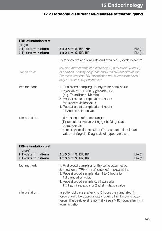
145
12 Endocrinology
12.2 Hormonal disturbances/diseases of thyroid gland
TRH-stimulation test(dogs)2 T4-determinations 3 T4-determinations
2 x 0.5 ml S, EP; HP 3 x 0.5 ml S, EP, HP
EIA (1) EIA (1)
Please note:
By this test we can stimulate and evaluate T4 levels in serum.
NTI and medications can influence T4 stimulation. (See T4).In addition, healthy dogs can show insufficient stimulation. For these reasons TRH stimulation test is recommended only to exclude hypothyroidism.
Test method: 1. First blood sampling, for thyroxine basal value2. Injection of TRH (200 μg/animal) i.v.
(e.g. Thyroliberin (Merck))3. Repeat blood sample after 2 hours
for 1st stimulation value4. Repeat blood sample after 4 hours
for 2nd stimulation value
Interpretation: - stimulation in reference range (T4-stimulation value >1,5 μg/dl): Diagnosis
of euthyroidism- no or only small stimulation (T4-basal and stimulation
value <1,5μg/dl): Diagnosis of hypothyroidism
TRH-stimulation test(horses)2 T4-determinations3 T4-determinations
2 x 0.5 ml S, EP, HP 3 x 0.5 ml S, EP, HP
EIA (1) EIA (1)
Test method: 1. First blood sampling for thyroxine basal value2. Injection of TRH (1 mg/horse, 0.5 mg/pony) i.v.3. Repeat blood sample after 4 to 5 hours for
1st stimulation value.4. Repeat blood sample c. 8 hours after
TRH administration for 2nd stimulation value
Interpretation: in euthyroid cases, after 4 to 5 hours the stimulated T4 value should be approximately double the thyroxine basal value. The peak level is normally seen 4-10 hours after TRH administration.
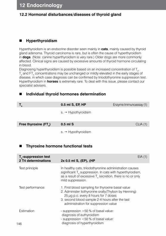
146
12 Endocrinology
12.2 Hormonal disturbances/diseases of thyroid gland
Hyperthyroidism
Hyperthyroidism is an endocrine disorder seen mainly in cats, mainly caused by thyroid gland adenoma. Thyroid carcinoma is rare, but is often the cause of hyperthyroidism in dogs. (Note: canine hyperthyroidism is very rare.) Older dogs are more commonly affected. Clinical signs are caused by excessive amounts of thyroid hormone circulating in blood.Diagnosing hyperthyroidism is possible based on an increased concentration of T4. T4 and FT4 concentrations may be unchanged or mildly elevated in the early stages of disease, in which case diagnosis can be confirmed by triiodothyronine suppression test.Hyperthyroidism in horses is extremely rare. To deal with this issue, please contact our specialist advisers.
Individual thyroid hormones determination
T4 0.5 ml S, EP, HP Enzyme Immunoassay (1)
s. Hypothyroidism
Free thyroxine (FT4) 0.5 ml S CLIA (1)
s. Hypothyroidism
Thyroxine hormone functional tests
T3-suppression test 2 T4 determinations 2x 0.5 ml S, (EP), (HP
EIA (1)
Test principle In healthy cats, triiodothyronine administration causes significant T4 suppression. In cats with hyperthyroidism, as a result of excessive T4 secretion, there is no or only mild suppression.
Test performance: 1. First blood sampling for thyroxine basal value2. Administer liothyronine orally(Thybon by Henning)
25 μg p.o. every 8 hours for 7 doses3. second blood sample 2-4 hours after the last
administration for suppression value
Estimation - suppression >50 % of basal value: diagnosis of euthyroidism- suppression <50 % of basal value: diagnosis of hyperthyroidism
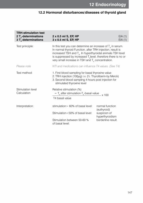
147
12 Endocrinology
12.2 Hormonal disturbances/diseases of thyroid gland
TRH-stimulation test2 T4-determinations3 T4-determinations
2 x 0.5 ml S, EP, HP 3 x 0.5 ml S, EP, HP
EIA (1) EIA (1)
Test principle: In this test you can determine an increase of T4 in serum. In normal thyroid Function, after TRH injection, result is increased TSH and T4. In hyperthyroidal animals TSH level is suppressed by increased T4level, therefore there is no or very small increase in TSH and T4 concentration.
Please note NTI and medications can influence T4 values. (See T4)
Test method: 1. First blood sampling for basal thyroxine value2. TRH injection (100μg) i.v. (f.i. Thyroliberin-by Merck)3. Second blood sampling 4 hours post injection for
stimulated thyroxine level
Stimulation level Calculation
Relative stimulation (%) = T4 after stimulation-T4-basal value__________________________________ x 100 T4 basal value
Interpretation: stimulation> 60% of basal level: normal function (euthyroid)Stimulation<50% of basal level: suspicion of hyperthyroidism Stimulation between 50-60 % borderline result. of basal level:
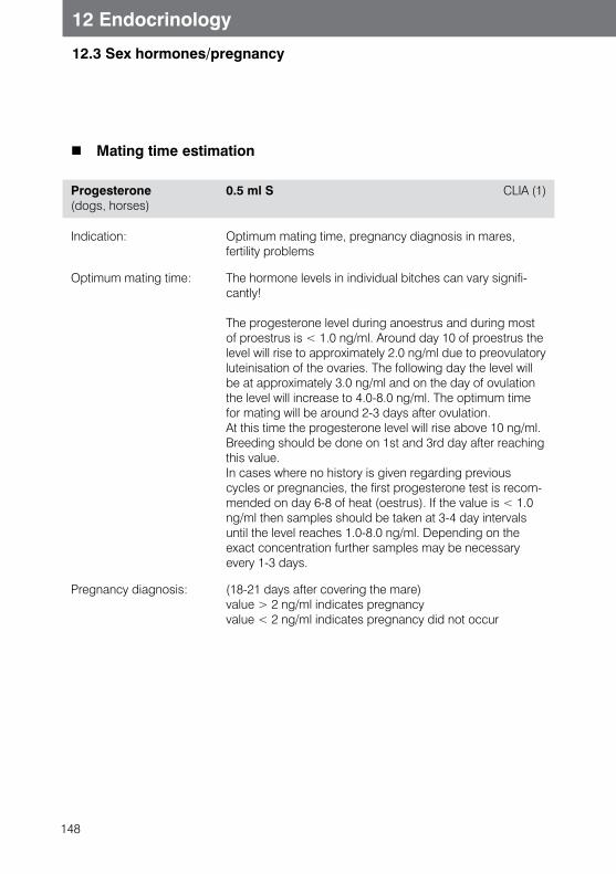
148
12.3 Sex hormones/pregnancy
12 Endocrinology
Mating time estimation
Progesterone (dogs, horses)
0.5 ml S CLIA (1)
Indication: Optimum mating time, pregnancy diagnosis in mares, fertility problems
Optimum mating time: The hormone levels in individual bitches can vary signifi-cantly!
The progesterone level during anoestrus and during most of proestrus is < 1.0 ng/ml. Around day 10 of proestrus the level will rise to approximately 2.0 ng/ml due to preovulatory luteinisation of the ovaries. The following day the level will be at approximately 3.0 ng/ml and on the day of ovulation the level will increase to 4.0-8.0 ng/ml. The optimum time for mating will be around 2-3 days after ovulation. At this time the progesterone level will rise above 10 ng/ml. Breeding should be done on 1st and 3rd day after reaching this value.In cases where no history is given regarding previous cycles or pregnancies, the first progesterone test is recom-mended on day 6-8 of heat (oestrus). If the value is < 1.0 ng/ml then samples should be taken at 3-4 day intervals until the level reaches 1.0-8.0 ng/ml. Depending on the exact concentration further samples may be necessary every 1-3 days.
Pregnancy diagnosis: (18-21 days after covering the mare)value > 2 ng/ml indicates pregnancyvalue < 2 ng/ml indicates pregnancy did not occur
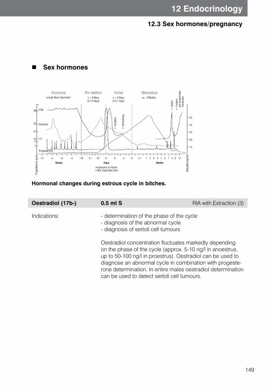
149
12.3 Sex hormones/pregnancy
12 Endocrinology
Sex hormones
Hormonal changes during estrous cycle in bitches.
Oestradiol (17b-) 0.5 ml S RIA with Extraction (3)
Indications: - determination of the phase of the cycle- diagnosis of the abnormal cycle- diagnosis of sertoli cell tumours
Oestradiol concentration fluctuates markedly depending on the phase of the cycle (approx. 5-10 ng/l in anoestrus, up to 50-100 ng/l in proestrus). Oestradiol can be used to diagnose an abnormal cycle in combination with progeste-rone determination. In entire males oestradiol determination can be used to detect sertoli cell tumours.
150
12.3 Sex hormones/pregnancy
12 Endocrinology
Testosterone 1 ml S, (EP), (HP) RIA with Extraction (3)
Indications: Differentiating between castrated and cryptorchid animals, testing androgen levels.Simple testosterone estimation is often not enough to confirm cryptorchidism. To confirm the diagnosis an hCG-stimulation test should be performed. For information on granulosa theca tumors in mares see Chapter 12.3.
HCG Stimulation Test2 Testosterone values3 Testosterone values
2 x 0.5 ml S, EP, HP 3 x 0.5 ml S, EP, HP
RIA with Extraction (3)RIA with Extraction (3)
Test performance(dogs, cats)
1. Blood sampling for basal testosterone value2. Injection of 50 IU hCG/kg body weight i.v.3. Blood sample 1 hour post injection for stimulation value
Test performance (horses) 1. Blood sampling for basal testosterone value2. Injection of 5,000 - 10,000 IU HCG per animal i.v.3. Second blood sampling 1 hour post
injection for first stimulation value4. Optional third blood sampling 24 hours post
injection for second stimulation value
Interpretation: - mild or no stimulation indicates there is no functioning testicular tissue.
- a marked stimulation indicates there is functioning testicular tissue.
In horses, a significant increase in testosterone levels following hCG administration proves that testicular tissue is present. Please note that in some horses it may take up to 120 minutes for hCG stimulation to take effect.A further peak 24 hours post hCG application will be obser-ved. Cryptorchidism cannot be excluded by a significant in-crease of testosterone concentration post hCG application.In case of inconclusive hCG stimulation test, an oestrone sulfate test can be performed.This test is not reliable in horses younger then three years old and in donkeys.
151
12.3 Sex hormones/pregnancy
12 Endocrinology
Oestrone Sulfate (horses,male)
1 ml S RIA (3)
Interpretation:
Please note:
In case of an inconclusive hCG stimulation test result, an oestrone sulfate test can be performed. (serum test). If the hCG stimulation test is performed, ideally the oestrone sulphate assay should take place immediately after the hCG administration.
Concentration of hormones over threshold value is considered suspicious for cryptorchidism.
This test is not reliable in horses younger then three years old and in donkeys.
GnRH-Stimulation test(horses)2 Testosterone estimations
2 x 0.5 ml S, (EP), (HP) RIA (3)
This test stimulates the hypothalamus and tests the function of the hypothalamic-hypophyseal axis. It is often used for diagnosis of infertile stalions.
Test performance: 1. Blood sampling (in the morning) for basal testosterone value
2. Injection of 0,04 mg GnRH/ horse i. v. (Receptal®)3. Blood sampling 1 hour post injection for stimulation
testosterone value
Estimation: No or minimal stimulation is due to a lack of active testicular tissue, whereas significant stimulation is caused by active testicular tissue.
152
12 Endocrinology
12.3 Sex hormones/pregnancy
Vaginal cytology (dogs, cats )
Vaginal smear Microscopy (1)
Increased estrogen concentration causes massive thickening of vaginal epithelium. During anestrus the vaginal mucosal membrane is composed of 4-6 cell layers and is relatively fragile. In proestrus it gains 20 to 30 layers. By gaining cell layers, cells which are closest to the vaginal lumen are further from blood supply, which leads to cell death. In addition keratinisation of cells takes place.These processes are understandable in vaginal cytology.
Range of applications: • cycle diagnostics in bitches• mating time estimation• diagnosis of cycle disorders diagnostics• diagnosis of vaginitis • confirmation if mating has occurred• differentiate between castrated /intact bitch/queen • vaginal tumor diagnostics (limited)• estimation of suspected delivery date (daily vaginal
cytology for estimation of metestrus, as delivery date is approximately 57 days after start of metestrus)
Technique of preparation With moisturized smear (NaCl) cytology will be taken from cranial region of vaginal space. Smear will be rolled two or three times on the microscopic slide and driedTwo or three preparations can be produced from the Swab.
Please note: Diagnostics of cycle disorders; Mating time estimation; Mating/vaginal monitoring; can only be used together with clinical symptoms or additional tests/results. In some cases there is a need for multiple tests.
153
12 Endocrinology
12.3 Sex hormones/pregnancy
Horse pregnancy diagnostics
PMSG 3 ml S, EP, HP ELISA (3)
This pregnancy specific hormone will be produced by endometrial cups between day 45 and 90 of pregnancy. Peak point of hormone secretion lies between day 60 and 75. If the pregnancy resorbs, endometrial cups stay active for a few weeks and PMSG test is false positive. In case of positive test results we recommend oestrone sulphate test after 100 days.
Oestrone sulphate(horses, female)
1 ml S, 5 ml U RIA (3)
Oestrone sulphate is a pregnancy specific hormone, which is produced by the intact placenta. Significantly high estrone sulphate level is indicative of a living foetus. Testing is possible from day 100 of pregnancy. As not all pregnant mares show high estrone sulphate level on day 100 after last mating, we recommend another test 2-4 weeks later in case of inconclusive results.If the test is negative after 120 days of pregnancy, it can be caused by damage to the fetus. We recommend rectal palpation and/or ultrasound examination in these cases.
Cattle pregnancy diagnostics
Oestrone sulphate (cattle, sheep, goats, female)
1 ml S, 5 ml U RIA (3)
Oestrone sulphate is produced by the intact foeto-placental tissue in pregnant ruminants and is therefore a sensitive and specific parameter for the presence of an ongoing pregnancy. In small ruminants, it can be detected from around day 50 of pregnancy in some cases. However, measurements from day 110 of pregnancy are more reliable. In cows of European breeds, a clear positive result can be expected from around day 120.
154
12.3 Sex hormones/pregnancy
12 Endocrinology
Pregnancy associated glycoproteins (PAGs) (bovines)
0.5 ml S, EP ELISA (1)
Ovarian tumors in horses
Granulosa Theca cell Tumor Profile (horse)
5 ml S(nonhaemolysed sample)
Inhibin: RIA (3)Testosterone: RIA (3)
Progesterone: EIA (3)
Granulosa theca-cell-tumors (GCT) are the most frequent ovarian in the mare. The tumor is mostly unilateral. Mares with GCT show aggressiveness or masculine behaviour, nymphomania, irregular cycle, anoestrus or infertility.
Diagnosis is based on clinical symptoms, ultrasonogra-phy of ovaries and laboratory endocrine tests. Ultrasound shows an enlarged ovary with multicystic or honeycomb structure. The affected ovary can also appear as solid tissue or a large fluid filled ovarian cyst. The contralateral healthy ovary is usually very small and shows very few or no follicles. Possible differential diagnosis are anovulatory follicles (transitional phase), ovarian haematoma, mature teratoma or cystadenoma.
Hormonal estimation is a very good method for granulosa theca tumors detection. GCT are hormonally active and testosterone is increasesd in 50 % of cases. Because of variations in testosterone concentration, multiple samples are necessary to detect high testosterone levels. Mares with GCT often demonstrate low progesterone levels.
The glycoprotein hormone inhibin will be produced in large amounts in GCT and is increased in about 90 % affected mares. Screening for inhibin, progesterone and testosterone in range of Granulose theca cell tumor Profile show a diag-nostic sensitivity of 95% and is the best laboratory diag-nostic possibillity.
155
12.4 Special hormones
12 Endocrinology
Special hormones
IGF 1 (Insulin-Like Growth Factor)
0.3 ml S RIA (3)
Indications: DwarfismAcromegaly
Occurence: IGF I (Somatomedin C) is synthetised in the liverSecretion depends strongly on growth hormone secretion. As IGF-I is secreted very steadily, IGF I is better for Growth hormone diagnostics, than GH assay alone.
Decrease: - proportional dwarfism (congenital growth hormone deficiency)
Increase: - Acromegaly
Please note: As reference values depend on breed, we can offer several reference ranges to you. Please ensure that patient breed details are sent to the laboratory.
Insulin 0.5 ml S cooled. (4-6 °C) RIA (3)
Indications: Insulinoma (dogsInsulin resistence (Equine Metabolic Syndrome/Equine Cushing Syndrome) (horses)Consistent blood glucose value < 60 mg/dl in association with insulin concentration in upper reference range or more can indicate insulinoma in dogs.
Horse See Chapter 12.1 Hyperadrenocorticism Equine Cushing Syndrome / Equine Metabolic Syndrome
Please note: Dogs: Patient should be fasted until the moment of blood collection.Glucose value should be < 60 mg/dl.When performing glucose assay at the same time as this test, we recommend dividing serum into twotubes; one should be sent deeply frozen for insulin estimation.Please use serum tubes without separation gel.
156
12 Endocrinology
12.4 Special hormones
Adenovirus-Infection (reptiles)
Adenoviruses are found in various lizards and snakes, and frequently also in species of bearded dragon (Amphibolurus barbatus, Pogona vitticeps, Pogona henrylawsoni). Affected animals show non-specific disease symptoms such as anorexia, diarrhoea, regurgitation and opisthotonus. Post mortem reveals mainly intranuclear inclusion bo-dies, primarily in the liver and intestines. In the live animal, direct detection is by testing cloacal Swabs or faeces.
Adenovirus (DNA detection)
sweab (cloaca), faeces PCR (3)
See Chapter 15, Molecular Diagnostics
157
13 Infectious diseases (in alphabetical order)
13 Infectious diseases
African horse sickness (AHSV)
African horse sickness is a severe, highly contagious and usually fatal viral disease in of horses and equids in southern Africa. Sporadically some cases are observed in Nort-hern Africa, Southern Europe and Middle East.Disease is transfered by insects. Clinically you can distinguish 4 forms:Peracute or lungform (dunkop), subacute oedema or heart form (dikkop), acute or mixed form and „Horse Sickness Fever“ or abortive form. Symptoms depend from the form and you can find fever, dyspnea, or oedema (lungs, conjunctiva, abdomen). Death can occur within 3-5 days. Test is performed mainly for the export of horses from Africa. Direct transmission from one infected horse to another horse has not been proven.
AHSV (Ab) 1 ml S CELISA (3)
Anaplasmosis
s. Ehrlichiosis
Aujeszky’s Disease
Aujeszky’s Disease (pseudorabies) is an acute viral disease caused by herpesvirus, mainly found in swine. Depending on the age of the animal, the virus attacks the central nervous system, respiratory tract or reproductive tract. Other animal species, (not hu-man beings) may be hosts.CNS infection is fatal, with dogs being especially susceptible. (Sudden death of the house dog may be an indication of Aujeszky’s Disease in a swine farm).
Clinical signs: - Fever, - CNS-disorders, - Nasal discharge - Cough (fattening pigs) - Abortion
Aujeszky’s Disease (Ab) 1-2 ml S ELISA (3)
Aujeszky’s Disease (Ab) 1-2 ml S NT (3)
Virus neutralisation for Aujeszky’s Disease antibody determination will be performed for an export of dogs.

158
13 Infectious diseases (in alphabetical order)
13 Infectious diseases
Babesiosis (dogs)/Piroplasmosis
Piroplasmosis in dogs in Europe are mainly caused by large babesias, from the Babesia canis group. The most important species are mainly B. canis canis (Syn. Babesia canis) and B. canis vogeli (Syn. Babesia vogeli). Single strains are of various pathogenicity. B. canis rossi (Syn. Babesia rossi) is found mainly in South Africa and causes highly pathogenic infections. Infections with “small” babesias (B. gibsoni , Babesia annae (Syn. Babesia microti-like, Theileria annae) or B. conradae (so far only found in USA) are rarely found in Europe, , but there has been a tendency for it to spread in recent years.In North West Spain there are stories about “small” babesias resulting in highly pathogenic infections.Probably they are caused by one of the human-like pathogens of babesia species, B. annae (Syn.B. microti-like orTheileria annae). The difference between large and small babesias can have therapeutic indications, as small babesias are not killed by medications that work against B. canis.Transmission of Babesias in Europe is caused by ticks of Rhipicephalus and Dermacentor species. The pathogen may be found from Mediterranean Sea to Hungary and the Baltic countries. Canine Babesiosis was a typical traveler’s disease associated with Mediterranean travel, but today it is more and more frequently seen in Germany, Austria and Switzerland.
Clinical symptoms: Depending on pathogenicity of the pathogen, the age and immunity of the dog; disease course can range from chronic to peracute. Incubation can last from 3 days to 5 weeks, and the patient develops typical disease symptoms: - Fever (over 40° C) - Haemolytic anaemia, Hemoglobinemia, Hemoglobinuria - Icterus, Bilirubinuria- Hepato- and Splenomegaly- DIC, Coagulopathy- Anorexia, Lethargy
159
13 Infectious diseases
13 Infectious diseases (in alphabetical order)
Babesia direct detection blood smear + 0.5 ml EB Microscopy (1)
Detection of intra red blood cell merozoits is possible from Giemsa-stained blood smear under light microscopy, ideally taken from capillary blood. This way “large” and “small” Babesias can be differentiated; PCR is indicated for diffe-rentiation, particularly in atypical forms. Parasitemia shows about 4-21 days post infection. In chronic Babesia infections, direct pathogen detection is difficult, as quiet phase and parasitemic phase can alternate. In B. canis infections in particular, there are often low levels of pathogen in the blood, so microscopic detection is not always successful.
Please note: Direct pathogen detection is therefore not always possible!
Babesia spp (DNA-detection).
1 ml EB real time-PCR (1)
Both large and small Babesias are found in PCR testing. Differentiation between Babesia canis canis, B. canis vogeli, B. canis rossi, B. gibsonii and B. conradae will be possible in 1-3 work days without additional costs.Comparing to light microscopy detection from blood smear, PCR is much more sensitive. Parasitemia comes about 4-21 days post infection. In chronic infections, direct patho-gen detection is not always possible. Serological detection is therefore recommended.
160
13 Infectious diseases
13 Infectious diseases (in alphabetical order)
Babesia canis (Ab) 1 ml S, EP, HP ELISA (3)
In chronic infections, direct pathogen detection is not always possible. Serological detection is therefore recommended.
The earliest detection of Babesia antibodies is possible 10-14 days post infection. Younger animals (under 8 months) develop often low antibody titer and should not be tested by serology below the age of 3 months, as maternal antibodies can be available, are protective in puppies up to 2 months of age. Cross-reactions between B. canis and B. gibsoni are possible and can be distingu-ished by means of the travel history and a species-specific PCR.This method does not detect antibodies against Babesia canis. If antibodies against Babesia gibsoni are needed (e.g. for export), please contact the laboratory, as this has to be noted specifically on the order form.
Please note the Following tests and Profiles
Blood parasites and haemotrophic bacterial microscopy. Traveler’s Disease Profile, Tick Profile
Babesiosis (cats )/ Piroplasmosis
Babesia felis DNA-Detection)
1 ml EB real time-PCR (1)
161
13 Infectious diseases
13 Infectious diseases (in alphabetical order)
Babesiosis (horse)/Piroplasmosis
Theileria (vorm. Babesia) equi and Babesia caballi Piroplasmosis is a parasitic disease of horse blood transmited by ticks. It is spread in North- and South America, and South and East Europe. Because of increased horse transport and increased spread of vectors (Dermacentor/Hyalomma spp.) it is also possible to find clinical cases or seropositive animals in Germany.Disease can be chronic, subclinical, acute, and peracute.In acute cases clinically fever, Agathy, oedema, ecchymosis (swelling) of the third eylid, colic, icterus and hemoglobinuria are observed. Death is possible. Laboratory tests show anaemia, leucopaenia, increased bilirubin and prolonged clotting time. In chronic cases weight loss and performance decreases with mild anaemia and increased or normal bilirubin concentration can be observed.T. equi can also be transmitted by placenta and cause abortion and neonatal piroplasmosis. Infected animals can be long term (even lifelong) pathogen carriers and act as an infection source for ticks.
Babesia (Ab) (Horses) 1 ml S, EP, HP IFT (3)
Antibody detection with titre determination by IFT.
Babesia (Ab) (horses) 1 ml S CBR (3)
Antibody detection with titre determination by CBR. CBR for Babesia-antibodies detection will be mainly performed for export of horses.
Babesia (Ab) (horses) 1 ml S cELISA (3)
Qualitative antibody detection by competitive ELISA. For export to the United States.
Babesiea- Direct detection
Blood smear + 0.5 ml EB Microscopy (1)
s. Babesiosis (dogs)
Microscopic detection of intraerythrocytes stadium.
162
13 Infectious diseases
13 Infectious diseases (in alphabetical order)
Babesia spp. (DNA-test)
1 ml EB PCR (1)
Differentiation between T. equi and B. caballi is possible by sequencing. These can be requested in case of positive results, in which case additional costs will be incurred.
Bartonellosis
Bartonella spp.(DNA-detection)
0.5 ml EB, lymphnode aspirate, eye smear
real time-PCR (1)
This PCR test detects Bartonella henselae, B. vinsonii, B. Quintana and B. clarridgeiae. Companion animals represent a large reservoir for human Bartonella infections, as most Bartonella-species posess zoonotic potential. Cats are the main reservoir for Bartonella henselae, B. clarridgeiae and B. koehlerar. Dogs can be infected with B. vinsonii subsp. berkhoffii, B. henselae, B. clarridgeiae, B. washoensis, B. elizabethae and B. quintana. Infections with Bartonella henselae are asymptomatic in cats. The connection with regional or generalised lymphadeno-pathy will be discussed. Infected cats can be bacteriemic for months or years, where the amount of bacteria in blood fluctuates. It is interesting to diagnose the pathogen in the cat when the contact person is suspected of “cat scratch” disease. It is in benign in over 90 % of cases in people, causing a self-limiting lymphadenopathy. Rarely in immunosuppresed people it can cause severe complica-tions, for instance encephalopathy, arthritis and pneumonia.
163
13 Infectious diseases
13 Infectious diseases (in alphabetical order)
Borna Disease, BD
Borna Disease Virus (BDV) is a pathogen of non purulent meningoencephalitis, which leads to neurological changes and behavioral disorders in peracute, acute or subacute disease. Most infections show no symptoms. If clinical signs are seen, disease is mostly lethal. Time of incubation is not known, but reports range from 2 weeks to many months. Seasonal occurrence of clinical cases is more often observed during spring and autumn. Most clinical cases occur in horses and sheep with increased numbers in some regions of Germany, Austria, Lichtenstein and Switzerland. Recent studies show that BDV can be found outside endemic regions. Direct virus transmission from horse to horse is not yet proven.
Borna (Ab) 1 ml S, liquor IFT (3)
A single positive IFT result for antibodies in blood does not confirm disease. Active infection can be proven by two serum examinations with a 10-14 day interval between tests. In a genuinely infected animal, seroconversion or antibody titers will triple by the second test, proving a continued contact with the pathogen. Antibody detection in CSF is possible only in clinically sick animals
Bornavirus (RNA-detection)
0.3 ml liquor (send intact eyeball, not retina in formalin)
PCR (3)
When testing CSF it is sensible to simultaneously test with both IFT and PCR. Positive result in CSF confirms Borna infection.
164
13 Infectious diseases
13 Infectious diseases (in alphabetical order)
Borreliosis
In Europe until now 6 of the 13 species of Borrelia burgdorferi sensu lato have been confirmed (B.burgdorferi sensu stricto, B. afzelii, B. garini,B. lusitaniae, B. bissetti I B. valaisiana). In addition to that we have heard lately many re-ports about the incidence of a new species of probable human pathogen, B. spielmanii.For most of these species there are no findings about meaning of pathogens for the ani-mals. Transmission in our area is mainly by the tick Ixodes ricinus. Incidence of Borrelia covers most extensively Ixodes, so that infection is possible in all of Germany.to count. Next to humans, the most susceptible to infection are dogs. Other animals appear to be less infected. Clinical infection has been discussed in horses and cats.In humans, disease can be divided into three phases. First we have a localized infec-tion, mainly in the form of erythrema migrans. Dissemination into the organism follows this, with a great spectrum of clinical manifestations. Borreliosis is often associated with neurological symptoms (i.e. lymphocytic meningoradiculitis, lymphocytic meningitis.) In the third chronic stadium, arthritis and chronic dermatitis dominate. Chronic neurological symptoms are rarely seen. In dogs we do not see this kind of clini-cal classification or it is not significant.
Symptoms: Affected animals show depending on the size of infection: - Fever, - Loss of appetite, Apathy, - Intermittent lameness, mono- or oligoarthritis (5-10%).
The following symptoms are also discussed in association with Borreliosis:
- Myocarditis - Uveitis, Chorioretinitis, Conjuntivitis - Nephritis, Glomerulonephritis and renal failure Predisposed dog breeds: (i.e. Bernese Mountain Dog, Labrador, Golden Retriever, Sheltie)
Borrelia burgdorferisensu lato (DNA-detection)
real time-PCR (1)
s. Chapter 15, Molecular biology tests
165
13 Infectious diseases
13 Infectious diseases (in alphabetical order)
Borrelia (Ak) IgG(dogs and horses)
0.5 ml S, EP, HP ELISA (1)
Detection of IgG is possible about 4-6 weeks post infection. The rate of spread of infection in dogs is relatively high, so a positive result does not inevitably mean active disease.False positive results are possible, due to cross reaction by infection with other Spirochetes and antibodies in connec-tion with vaccination.Any positive or borderline antibody detection should be confirmed with Immunoblot (two levels diagnostics).High IgG titer can remain high in spite of clinically successful therapy for a very long time. This means that treatment success monitoring is not possible with this method.
Borrelia (Ab) IgM (dogs) 0.5 ml S, EP, HP ELISA (1)
In humans anti-Borrelia IgM antibodies are usually detecta-ble 3 weeks post infection and mean acute disease. IgM is not important in dogs. In dogs, IgM antibodies are detectable only temporarily in the first 60-90 days. Cross-reactions occur and detection does not necessarily indicate acute borreliosis. The clinical symptoms in dogs generally occur at a time when the IgM peak has already passed.
Borrelia (Ab) IgG 1 ml S, EP, HP Immunoblot (1)
Immunoblot should be performed as confirmation test in connection with positive or borderline Anti-Borrelia antibodies detection in IgG/IgM-ELISA.
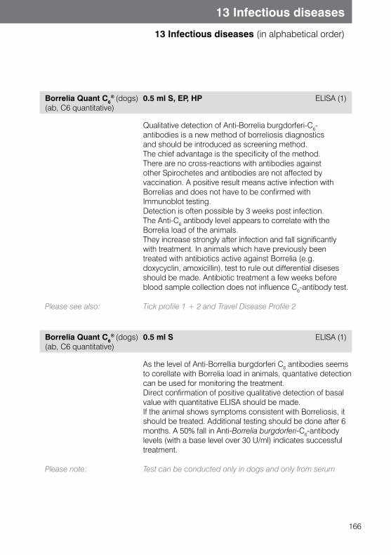
166
13 Infectious diseases
13 Infectious diseases (in alphabetical order)
Borrelia Quant C6® (dogs)
(ab, C6 quantitative)0.5 ml S, EP, HP ELISA (1)
Qualitative detection of Anti-Borrelia burgdorferi-C6-antibodies is a new method of borreliosis diagnostics and should be introduced as screening method.The chief advantage is the specificity of the method. There are no cross-reactions with antibodies against other Spirochetes and antibodies are not affected by vaccination. A positive result means active infection with Borrelias and does not have to be confirmed with Immunoblot testing.Detection is often possible by 3 weeks post infection. The Anti-C6 antibody level appears to correlate with the Borrelia load of the animals. They increase strongly after infection and fall significantly with treatment. In animals which have previously been treated with antibiotics active against Borrelia (e.g. doxycyclin, amoxicillin), test to rule out differential disesesshould be made. Antibiotic treatment a few weeks before blood sample collection does not influence C6-antibody test.
Please see also: Tick profile 1 + 2 and Travel Disease Profile 2
Borrelia Quant C6® (dogs)
(ab, C6 quantitative)0.5 ml S ELISA (1)
As the level of Anti-Borrellia burgdorferi C6 antibodies seems to corellate with Borrelia load in animals, quantative detection can be used for monitoring the treatment.Direct confirmation of positive qualitative detection of basal value with quantitative ELISA should be made.If the animal shows symptoms consistent with Borreliosis, it should be treated. Additional testing should be done after 6 months. A 50% fall in Anti-Borrelia burgdorferi-C6-antibody levels (with a base level over 30 U/ml) indicates successful treatment.
Please note: Test can be conducted only in dogs and only from serum
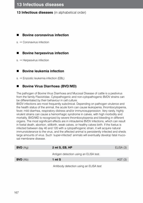
167
13 Infectious diseases
13 Infectious diseases (in alphabetical order)
Bovine coronavirus infection
s. Coronavirus infection
Bovine herpesvirus infection
s. Herpesvirus infection
Bovine leukemia infection
s. Enzootic leukemia infection (EBL)
Bovine Virus Diarrhoea (BVD/MD)
The pathogen of Bovine Virus Diarrhoea and Mucosal Disease of cattle is a pestivirus from the family Flaviviridae. Cytopathogenic and non-cytopathogenic BVDV strains can be differentiated by their behaviour in cell culture. BVDV infections are most frequently subclinical. Depending on pathogen virulence and the health status of the animal, the acute form can cause leukopenia, thrombocytopenia, fever, mild diarrhea, respiratory distress and/or immunosuppression. Very rarely, highly virulent strains can cause a hemorrhagic syndrome in calves, with high morbidity and mortality. BVD/MD is recognized by severe thrombocytopenia and bleeding in different organs. The most significant effects are in intrauterine BVDV infections, which can result in foetal death, abortion, stillbirth, weak calves, or healthy calves birth. If the foetus is infected between day 40 and 120 with a cytopathogenic strain, it will acquire natural immunotolerance to the virus, and the affected animal is persistently infected and sheds large amounts of virus. Such ‘super-infected’ animals will eventually develop fatal muco-sal membrane disease.
BVD (Ag) 2 ml S, EB, HP ELISA (3)
Antigen detection using an ELISA test.
BVD (Ab) 1 ml S AGT (3)
Antibody detection using an ELISA test
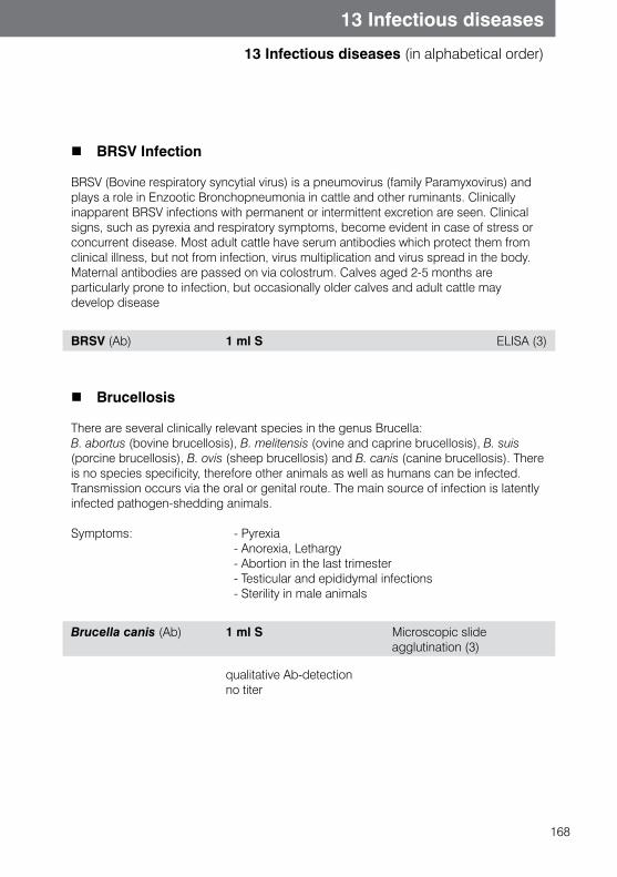
168
13 Infectious diseases
13 Infectious diseases (in alphabetical order)
BRSV Infection
BRSV (Bovine respiratory syncytial virus) is a pneumovirus (family Paramyxovirus) and plays a role in Enzootic Bronchopneumonia in cattle and other ruminants. Clinically inapparent BRSV infections with permanent or intermittent excretion are seen. Clinical signs, such as pyrexia and respiratory symptoms, become evident in case of stress or concurrent disease. Most adult cattle have serum antibodies which protect them from clinical illness, but not from infection, virus multiplication and virus spread in the body. Maternal antibodies are passed on via colostrum. Calves aged 2-5 months are particularly prone to infection, but occasionally older calves and adult cattle may develop disease
BRSV (Ab) 1 ml S ELISA (3)
Brucellosis
There are several clinically relevant species in the genus Brucella:B. abortus (bovine brucellosis), B. melitensis (ovine and caprine brucellosis), B. suis(porcine brucellosis), B. ovis (sheep brucellosis) and B. canis (canine brucellosis). There is no species specificity, therefore other animals as well as humans can be infected. Transmission occurs via the oral or genital route. The main source of infection is latently infected pathogen-shedding animals.
Symptoms: - Pyrexia - Anorexia, Lethargy - Abortion in the last trimester - Testicular and epididymal infections - Sterility in male animals
Brucella canis (Ab) 1 ml S Microscopic slide agglutination (3)
qualitative Ab-detection no titer
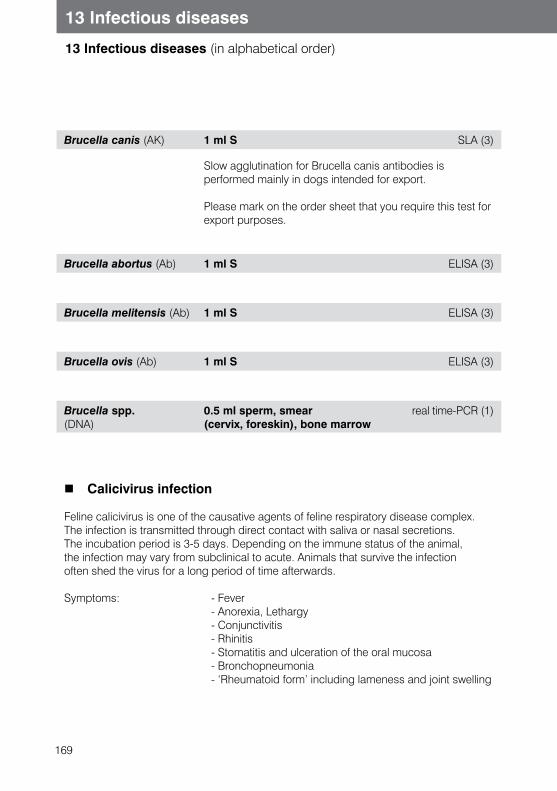
169
13 Infectious diseases
13 Infectious diseases (in alphabetical order)
Brucella canis (AK) 1 ml S SLA (3)
Slow agglutination for Brucella canis antibodies is performed mainly in dogs intended for export.
Please mark on the order sheet that you require this test for export purposes.
Brucella abortus (Ab) 1 ml S ELISA (3)
Brucella melitensis (Ab) 1 ml S ELISA (3)
Brucella ovis (Ab) 1 ml S ELISA (3)
Brucella spp. (DNA)
0.5 ml sperm, smear (cervix, foreskin), bone marrow
real time-PCR (1)
Calicivirus infection
Feline calicivirus is one of the causative agents of feline respiratory disease complex.The infection is transmitted through direct contact with saliva or nasal secretions. The incubation period is 3-5 days. Depending on the immune status of the animal, the infection may vary from subclinical to acute. Animals that survive the infection often shed the virus for a long period of time afterwards.
Symptoms: - Fever - Anorexia, Lethargy - Conjunctivitis - Rhinitis - Stomatitis and ulceration of the oral mucosa - Bronchopneumonia - ‘Rheumatoid form’ including lameness and joint swelling
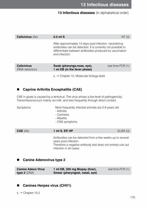
170
13 Infectious diseases
13 Infectious diseases (in alphabetical order)
Calicivirus (Ab) 0.5 ml S NT (3)
After approximately 14 days post-infection, neutralizing antibodies can be detected. It is currently not possible to differentiate between antibodies produced by vaccination and infection.
Calicivirus(RNA detection)
Swab (pharyngs,nose, eye), 1 ml EB (in the fever phase)
real time-PCR (1)
s. Chapter 15, Molecular biology tests
Caprine Arthritis Encephalitis (CAE)
CAE in goats is caused by a lentivirus. The virus shows a low level of pathogenicity. Transmissionoccurs mainly via milk, and less frequently through direct contact.
Symptoms: Most frequently infected animals are 2-9 years old - Arthritis - Cachexia - Mastitis - CNS symptoms
CAE (Ab) 1 ml S, EP, HP ELISA (3)
Antibodies can be detected from a few weeks up to several years post infection. Therefore a negative antibody test does not entirely rule out infection in all cases.
Canine Adenovirus type 2
Canine Adeno Virus type 2 (DNA)
1 ml EB, 200 mg Biopsy (liver), Smear (pharyngeal, nasal, eye)
real time-PCR (1)
Canines Herpes virus (CHV1)
s. Chapter 15.2
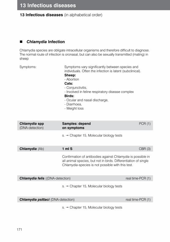
171
13 Infectious diseases
13 Infectious diseases (in alphabetical order)
Chlamydia Infection
Chlamydia species are obligate intracellular organisms and therefore difficult to diagnose.The normal route of infection is oronasal, but can also be sexually transmitted (mating) in sheep
Symptoms: Symptoms vary significantly between species and individuals. Often the infection is latent (subclinical). Sheep: - Abortion Cats: - Conjunctivitis, - Involved in feline respiratory disease complex Birds: - Ocular and nasal discharge, - Diarrhoea, - Weight loss
Chlamydia spp(DNA-detection)
Samples: depend on symptoms
PCR (1)
s. Chapter 15, Molecular biology tests
Chlamydia (Ab) 1 ml S CBR (3)
Confirmation of antibodies against Chlamydia is possible in all animal species, but not in birds. Differentiation of single Chlamydia-species is not possible with this test.
Chlamydia felis ((DNA-detection) real time-PCR (1)
s. Chapter 15, Molecular biology tests
Chlamydia psittaci (DNA-detection) real time-PCR (1)
s. Chapter 15, Molecular biology tests
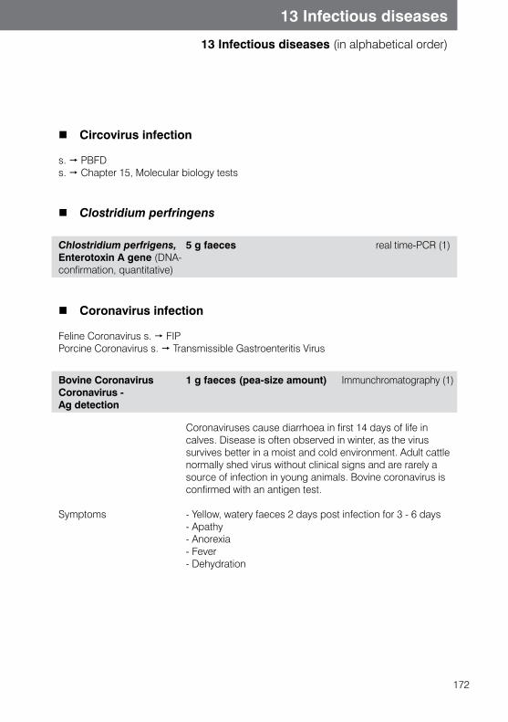
172
13 Infectious diseases
13 Infectious diseases (in alphabetical order)
Circovirus infection
s. PBFDs. Chapter 15, Molecular biology tests
Clostridium perfringens
Chlostridium perfrigens, Enterotoxin A gene (DNA-confirmation, quantitative)
5 g faeces real time-PCR (1)
Coronavirus infection
Feline Coronavirus s. FIPPorcine Coronavirus s. Transmissible Gastroenteritis Virus
Bovine Coronavirus Coronavirus -Ag detection
1 g faeces (pea-size amount) Immunchromatography (1)
Symptoms
Coronaviruses cause diarrhoea in first 14 days of life in calves. Disease is often observed in winter, as the virus survives better in a moist and cold environment. Adult cattle normally shed virus without clinical signs and are rarely a source of infection in young animals. Bovine coronavirus is confirmed with an antigen test.
- Yellow, watery faeces 2 days post infection for 3 - 6 days- Apathy - Anorexia - Fever- Dehydration
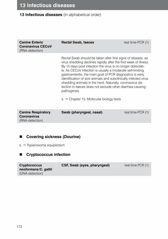
173
13 Infectious diseases
13 Infectious diseases (in alphabetical order)
Canine Enteric Coronavirus CECoV (RNA-detection)
Rectal Swab, faeces real time-PCR (1)
Rectal Swab should be taken after first signs of disease, as virus shedding declines rapidly after the first week of illness. By 15 days post infection the virus is no longer detectab-le. As CECoV infection is usually a moderate self-limiting gastroenteritis, the main goal of PCR diagnostics is early identification of sick animals and subclinically infected virus shedding animals in the herd. Naturally, coronavirus de-tection in faeces does not exclude other diarrhea-causing pathogenes.
s. Chapter 15, Molecular biology tests
Canine Respiratory Coronavirus (RNA-detection)
Swab (pharyngeal, nasal) real time-PCR (1)
Covering sickness (Dourine)
s. Trypanosoma equiperdum
Cryptococcus infection
Cryptococcus neoformans/C. gattii (DNA-detection)
CSF, Swab (eyes, pharyngeal) real time PCR (1)
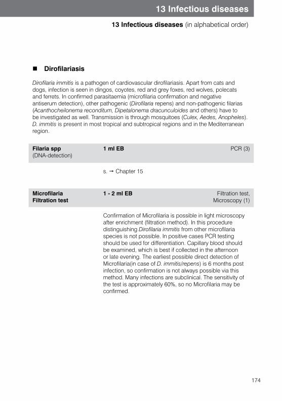
174
13 Infectious diseases
13 Infectious diseases (in alphabetical order)
Dirofilariasis
Dirofilaria immitis is a pathogen of cardiovascular dirofilariasis. Apart from cats and dogs, infection is seen in dingos, coyotes, red and grey foxes, red wolves, polecats and ferrets. In confirmed parasitaemia (microfilaria confirmation and negative antiserum detection), other pathogenic (Dirofilaria repens) and non-pathogenic filarias (Acanthocheilonema reconditum, Dipetalonema dracunculoides and others) have to be investigated as well. Transmission is through mosquitoes (Culex, Aedes, Anopheles). D. immitis is present in most tropical and subtropical regions and in the Mediterranean region.
Filaria spp (DNA-detection)
1 ml EB PCR (3)
s. Chapter 15
Microfilaria Filtration test
1 - 2 ml EB Filtration test, Microscopy (1)
Confirmation of Microfilaria is possible in light microscopy after enrichment (filtration method). In this procedure distinguishing Dirofilaria immitis from other microfilaria species is not possible. In positive cases PCR testing should be used for differentiation. Capillary blood should be examined, which is best if collected in the afternoon or late evening. The earliest possible direct detection of Microfilaria(in case of D. immitis/repens) is 6 months post infection, so confirmation is not always possible via this method. Many infections are subclinical. The sensitivity of the test is approximately 60%, so no Microfilaria may be confirmed.
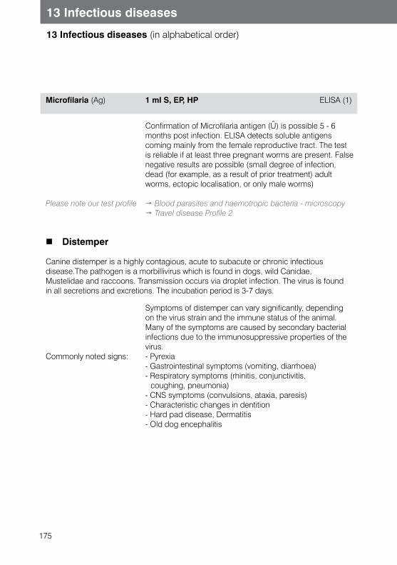
175
13 Infectious diseases
13 Infectious diseases (in alphabetical order)
Microfilaria (Ag) 1 ml S, EP, HP ELISA (1)
Please note our test profile
Confirmation of Microfilaria antigen (Û) is possible 5 - 6 months post infection. ELISA detects soluble antigens coming mainly from the female reproductive tract. The test is reliable if at least three pregnant worms are present. False negative results are possible (small degree of infection, dead (for example, as a result of prior treatment) adult worms, ectopic localisation, or only male worms)
Blood parasites and haemotropic bacteria - microscopy Travel disease Profile 2
Distemper
Canine distemper is a highly contagious, acute to subacute or chronic infectious disease.The pathogen is a morbillivirus which is found in dogs, wild Canidae, Mustelidae and raccoons. Transmission occurs via droplet infection. The virus is found in all secretions and excretions. The incubation period is 3-7 days.
Commonly noted signs:
Symptoms of distemper can vary significantly, depending on the virus strain and the immune status of the animal. Many of the symptoms are caused by secondary bacterial infections due to the immunosuppressive properties of the virus.- Pyrexia- Gastrointestinal symptoms (vomiting, diarrhoea)- Respiratory symptoms (rhinitis, conjunctivitis,
coughing, pneumonia)- CNS symptoms (convulsions, ataxia, paresis)- Characteristic changes in dentition- Hard pad disease, Dermatitis- Old dog encephalitis
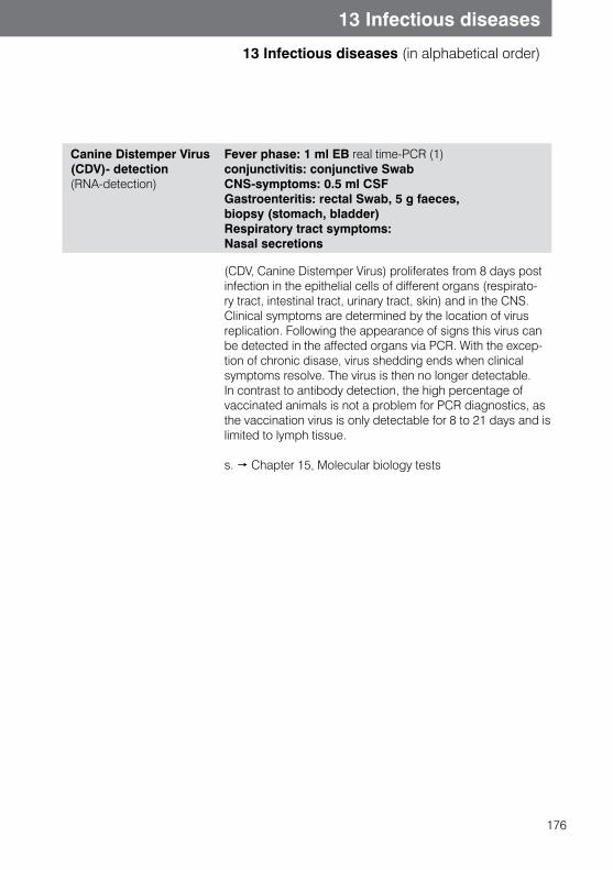
176
13 Infectious diseases
13 Infectious diseases (in alphabetical order)
Canine Distemper Virus (CDV)- detection(RNA-detection)
Fever phase: 1 ml EB real time-PCR (1) conjunctivitis: conjunctive SwabCNS-symptoms: 0.5 ml CSFGastroenteritis: rectal Swab, 5 g faeces, biopsy (stomach, bladder)Respiratory tract symptoms: Nasal secretions
(CDV, Canine Distemper Virus) proliferates from 8 days post infection in the epithelial cells of different organs (respirato-ry tract, intestinal tract, urinary tract, skin) and in the CNS. Clinical symptoms are determined by the location of virus replication. Following the appearance of signs this virus can be detected in the affected organs via PCR. With the excep-tion of chronic disase, virus shedding ends when clinical symptoms resolve. The virus is then no longer detectable. In contrast to antibody detection, the high percentage of vaccinated animals is not a problem for PCR diagnostics, as the vaccination virus is only detectable for 8 to 21 days and is limited to lymph tissue.
s. Chapter 15, Molecular biology tests
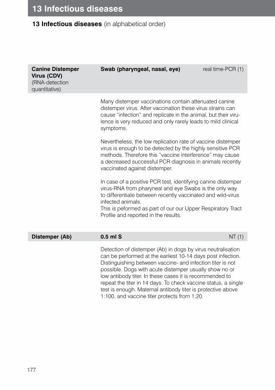
177
13 Infectious diseases
13 Infectious diseases (in alphabetical order)
Canine Distemper Virus (CDV) (RNA-detection quantitative)
Swab (pharyngeal, nasal, eye) real time-PCR (1)
Many distemper vaccinations contain attenuated canine distemper virus. After vaccination these virus strains can cause “infection” and replicate in the animal, but their viru-lence is very reduced and only rarely leads to mild clinical symptoms.
Nevertheless, the low replication rate of vaccine distemper virus is enough to be detected by the highly sensitive PCR methods. Therefore this “vaccine interference” may cause a decreased successful PCR diagnosis in animals recently vaccinated against distemper.
In case of a positive PCR test, identifying canine distemper virus-RNA from pharyneal and eye Swabs is the only way to differentiate between recently vaccinated and wild-virus infected animals.This is peformed as part of our our Upper Respiratory Tract Profile and reported in the results.
Distemper (Ab) 0.5 ml S NT (1)
Detection of distemper (Ab) in dogs by virus neutralisation can be performed at the earliest 10-14 days post infection. Distinguishing between vaccine- and infection titer is not possible. Dogs with acute distemper usually show no or low antibody titer. In these cases it is recommended to repeat the titer in 14 days. To check vaccine status, a single test is enough. Maternal antibody titer is protective above 1:100, and vaccine titer protects from 1:20.
178
13 Infectious diseases
13 Infectious diseases (in alphabetical order)
Ehrlichiosis /Anaplasmosis
In order to detect ehrlichiosis or anaplasmosis, it is necessary to distinguish between infections indigenous to tropical and subtropical areas, such as canine monocytic or canine thrombocytic ‘ehrlichiosis’ (Anaplasma platys, Ehrlichia canis and other Ehrlichia spp.), and the granulocytic disease form (Anaplasma phagocytophilum, formerly called Ehrlichia phagocytophila) that is prevalent in more northern regions.E. canis plays a major role as an agent of canine monocytic ehrlichiosis. It is transmitted in Europe by the tick Rhipicephalus sanguineus. E. canis is widespread in tropical and subtropical regions and occurs throughout the Mediterranean region. Isolated cases of infection may also occur in Germany. Other monocytic infections, e.g. with E. chaffeensis, occur predominantly in the USA.
In Southern Europe, infections with Anaplasma platys are also found that can cause so-called canine cyclic thrombocytopenia. Infections with Anaplasma phagocytophilum, the agent of infectious canine granulocytic anaplasmosis (ehrlichiosis), are becoming increasingly important. This form is primarily found in Northern and Central Europe. Transmission occurs through the tick Ixodes ricinus.Canine monocytic ehrlichiosis (caused by E. canis) can be manifested by a broad spectrum of clinical symptoms. An incubation period of 3 weeks can be followed by 2-4 weeks of acute disease. Ehrlichiosis is usually known for mild nonspecific clinical symptoms, although serious life threatening disease can also happen.
Affected dogs may show signs of fever, anorexia, lethargy, lymphadenopathy and spleno-megaly. Increased bleeding is also possible. Ophthalmic and neurological symptoms may also be seen. Laboratory diagnostics will show thrombocytopeania and mild anaemia, leucopaenia and hypergammaglobulinaemia. ALT and AP values are also increased.
Following the acute disease period, a subclinical phase of varying length may develop, after which the disease may progress into a chronic state. (Not all animals develop the chronic state.) Findings range from oedema, anorexia, chronic weight loss, neurological symptoms and generalised lymph node enlargement, to CNS disorders (meningitis), polymyositis and polyarthritis.
In horses, granulocytic ehrlichiosis caused by A. phagocytophilum (formerly Ehrlichia equi) and transmitted by ticks of the genus Ixodes is to the forefront in Europe. Iatrogenic transmission by contaminated vehicles is possible. After entering the bloodstream, the agent spreads in the blood and lymph system. It displays a cytotropism for neutrophilic and eosinophilic granulocytes, in which it multiplies within cytoplasmic vacuoles. The clinical symptoms include fever, mild apathy, petechiae, weakness, limb oedema and ataxia. There have been no reports to date of abortion or laminitis in connection with this infection. The disease is normally self-limiting but affected horses may be more susceptible to secondary bacterial or viral infections. Persistent infections have not so far been detected in the horse.

179
13 Infectious diseases
13 Infectious diseases (in alphabetical order)
Ehrlichia/Anaplasma - Direct detection
Blood smear + 1 ml EB Microscopy (1)
Please note:
Direct pathogen detection in blood smear is possible only during acute disease phase of disease. Light microscopy is peformed on a Giemsa stained blood smear, ideally from capillary blood. The probability of finding A. phagocytophilum is significantly higher than that of finding E. canis.
Negative direct pathogen detection does not exclude infection!
Ehrlichia spp.(DNA-detection)
2 ml EB, spleen, bone marrow, 0.5 ml liquor, tick
real time-PCR (1)
PCR testing is more sensitive than light microscopy of a blood smear, as negative direct pathogen detection does not exclude infection. Differentiation of Ehrlichia canis, E. ewingii and E. chaffeensis is possible on request, with real time PCR.
s. Chapter 15, Molecular biology tests
Ehrlichia canis(DNA-detection)
2 ml EB, spleen-, bone marrow, 0.5 ml liquor, tick
real time-PCR (1)
Direct pathogen detection with PCR can be performed 4-10 days post infection. It is usually more sensitive then light microscopy of a blood smear. Therefore it is recommen-ded in acute phase of disease, as in later stages often no pathogen is found in the blood and a negative result does not exclude infection. To some extent, treatment monitoring is possible with PCR.
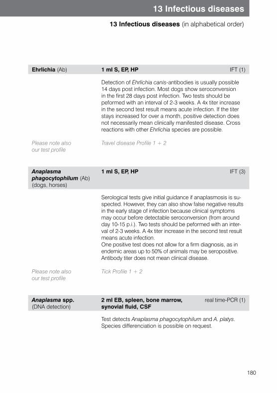
180
13 Infectious diseases
13 Infectious diseases (in alphabetical order)
Ehrlichia (Ab) 1 ml S, EP, HP IFT (1)
Please note also our test profile
Detection of Ehrlichia canis-antibodies is usually possible 14 days post infection. Most dogs show seroconversion in the first 28 days post infection. Two tests should be peformed with an interval of 2-3 weeks. A 4x titer increase in the second test result means acute infection. If the titer stays increased for over a month, positive detection does not necessarily mean clinically manifested disease. Cross reactions with other Ehrlichia species are possible.
�Travel disease Profile 1 + 2
Anaplasma phagocytophilum (Ab)(dogs, horses)
1 ml S, EP, HP IFT (3)
Please note also our test profile
Serological tests give initial guidance if anaplasmosis is su-spected. However, they can also show false negative results in the early stage of infection because clinical symptoms may occur before detectable seroconversion (from around day 10-15 p.i.). Two tests should be peformed with an inter-val of 2-3 weeks. A 4x titer increase in the second test result means acute infection.One positive test does not allow for a firm diagnosis, as in endemic areas up to 50% of animals may be seropositive. Antibody titer does not mean clinical disease.
Tick Profile 1 + 2
Anaplasma spp. (DNA detection)
2 ml EB, spleen, bone marrow, synovial fluid, CSF
real time-PCR (1)
Test detects Anaplasma phagocytophilum and A. platys. Species differenciation is possible on request.
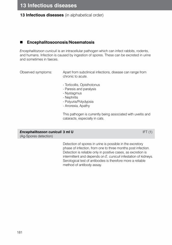
181
13 Infectious diseases
13 Infectious diseases (in alphabetical order)
Encephalitosoonosis/Nosematosis
Encephalitozoon cuniculi is an intracellular pathogen which can infect rabbits, rodents, and humans. Infection is caused by ingestion of spores. These can be excreted in urine and sometimes in faeces.
Observed symptoms: Apart from subclinical infections, disease can range from chronic to acute.
- Torticollis, Opisthotonus - Paresis and paralysis - Nystagmus - Nephritis - Polyuria/Polydypsia - Anorexia, Apathy
This pathogen is currently being associated with uveitis and cataracts, especially in cats.
Encephalitozoon cuniculi(Ag-Spores detection)
3 ml U IFT (1)
Detection of spores in urine is possible in the excretory phase of infection, from one to three months post infection. Detection is reliable only in positive cases, as excretion is intermittent and depends on E. cuniculi infestation of kidneys. Serological test of antibodies is therefore more a reliable method of antibody assay.
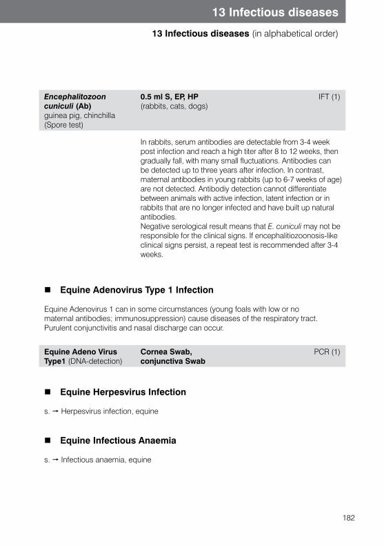
182
13 Infectious diseases
13 Infectious diseases (in alphabetical order)
Encephalitozoon cuniculi (Ab)guinea pig, chinchilla (Spore test)
0.5 ml S, EP, HP(rabbits, cats, dogs)
IFT (1)
In rabbits, serum antibodies are detectable from 3-4 week post infection and reach a high titer after 8 to 12 weeks, then gradually fall, with many small fluctuations. Antibodies can be detected up to three years after infection. In contrast, maternal antibodies in young rabbits (up to 6-7 weeks of age) are not detected. Antibodiy detection cannot differentiate between animals with active infection, latent infection or in rabbits that are no longer infected and have built up natural antibodies. Negative serological result means that E. cuniculi may not be responsible for the clinical signs. If encephalitiozoonosis-like clinical signs persist, a repeat test is recommended after 3-4 weeks.
Equine Adenovirus Type 1 Infection
Equine Adenovirus 1 can in some circumstances (young foals with low or no maternal antibodies; immunosuppression) cause diseases of the respiratory tract. Purulent conjunctivitis and nasal discharge can occur.
Equine Adeno Virus Type1 (DNA-detection)
Cornea Swab, conjunctiva Swab
PCR (1)
Equine Herpesvirus Infection
s. Herpesvirus infection, equine
Equine Infectious Anaemia
s. Infectious anaemia, equine
183
13 Infectious diseases
13 Infectious diseases (in alphabetical order)
Equine Influenza
s. Influenza, equine
Equine Viral Arteritis
s. Viral Arteritis, equine
Enzootic Bovine Leukemia
Bovine Leukemia complex can be classed into four clinical forms. Unlike skin leukemia, juvenile leukemia and mast cell reticulosis (which all occur spontaneously), enzootic (lymphatic) bovine leukemia is caused by a retrovirus. Transmission is usually possible shortly after birth through colostrum and milk.Horizontal transmission is always possible.
Symptoms: - Apathy, anorexia- Oedema- Anaemia, lymphocytosis- Lymphadenopathy- Splenomegaly
Bovine Leukosis Virus, EBL-Antibodies
1 ml S ELISA (3)
184
13 Infectious diseases
13 Infectious diseases (in alphabetical order)
FeLV (Feline Leukemia Virus)
FeLV belongs to the family Retroviridae. Different FeLV groups exist, which are described as FeLV-A, B and C. Infections with FeLV-B and C occur only together with FeLV-A. Prevalence in the European feline population ranges (with regional variation) between 1 and 8%.
Transmission is both horizontal with saliva or other body fluids (urine, blood) and also vertical, though the placenta or mother’s milk. The course of infection varies, depending on the immune status of the animal and also infectious exposure and virulence of pathogen. Only a small part of FeLV infected cats show FeLV-associated diseases.Most affected animals limit the infection or suppress it. So a large part of infected cats show good immune reaction and can eliminate the pathogen before viremia is seen (regressor cats/abortive infection). Detection of the FeLV antigen in blood is not possible in these cats. Some affected animals develops transient viremia, which can last up to 16 weeks. During this time the virus is shed and extracellular antigen can be detected in blood. Depending on host defences, virus proliferation may stop, may proceed to to persistent viremia, or may be completely eliminated.
When virus replication begins, integrated viral DNA can stay in infected cells in the form of provirus (progenome), leaving animals latently infected. Detection of extracellular or intracellular FeLV antigen is not possible in the blood at this time. Depending on the number of infected cells, progenome can be detected by PCR in bone marrow or blood. Reactivation with viremia is possible. In some animals it is possible that the pathogen can be (eventually) completely eliminated.
If the infected cat is unable to build enough neutralizing antibodies, productive virus proliferation (permanent persistant viremia) takes place. In about one third of affected cats the infection shows this progressive course. These cats have a poor prognosis and often die in 3-5 years from FeLV-associated diseases. They are normally heavy virus shedders and threaten other cats with infection risk. In a small percentage of infected animals there is an atypical form of infection with limited virus proliferation in the bladder, eyes and milk ducts. This form of disease cannot be detected by routine diagnostic tests.
185
13 Infectious diseases
13 Infectious diseases (in alphabetical order)
Depending on severity and stage of disease, the following symptoms can occur:
Tumoren: Lymphoma, Leukemia, Myeloid tumors, Fibrosarcoma
FeLV-associated disease: Fever, Anorexia, Apathy, Stomatitis, Gingivitis, Abscesses, Respiratory symptoms, Gastrointestinal symptoms
Bone marrow: Leukopenia, Neutropenia, Non-regenerative anaemia, thrombocytopenia
Immune mediated diseases: Autoimmune haemolytic anaemia, Glomerulonephritis, Uveitis, polyarthritis
Reproduction disorders: Abortion, Stillbirths, Fading Kitten Syndrome
FeLV (Ag) 0.5 ml S, EP, HP ELISA (1)
The detection of free extracellular FeLV-p27 antigen is possible from approximately 3 weeks post infection. Latently infected cats may show false negative results. A positive result may indicate a transient or persistent viraemia, so the test should be repeated after 6 weeks. If this second test is positive, a third test should be performed after another 10 weeks. If it is positive, persistent viraemia must be assumed. Negative repeat tests indicate virus elimination or a transition into the latently infected stage. In around half of all cats which appear to be recovering from FeLV infection, there is a latent infection of the bone marrow. Detection via conventional tests for free p27 antigen from blood samples is not possible in these cats, due to the low virus release. However, PCR can be used to detect the virus progenome in the blood. Vaccination does not lead to viraemia, therefore false positive results are not possible.
Please note also our test profile and combinations
Large cat profile FeLV/FIV/FIP, FeLV/FIP, FeLV/FIV, FeLV/FIV
186
13 Infectious diseases
13 Infectious diseases (in alphabetical order)
FeLV Progenome (DNA-detection)
2 ml EB, bone marrow real time-PCR (1)
Please note:
Viral DNA integrated in the host cell genome is known as progenome or provirus, which can be detected using PCR. This test is highly specific and can therefore be used to confirm doubtful results with other methods.Latent infections can be diagnosed to some extent, if other test results have shown negative results. The sensitivity of PCR is highly dependent on the number of infected cells (provirus load), which is why a negative result does not entirely rule out infection.
This method does not measure virus replication ability.
Feline Coronavirus Infection (Feline Infectious Peritionitis)
Infection with Feline Coronavirus (FCoV) is widespread in cat populations. Approximately 50% of the animals are carriers of antibodies against FCoV. In catteries and animal shelters, 100% of cats are positive.
Transmission is through faeces, and can be via direct or indirect oronasal infection. Distinguishing between FCoV and the FIP causing mutant is not possible as the genetic similarity is over 99%. The theory of harmless enteral coronaviruses (with ony pathogenic mutants in the rest of the body) has been disproved. As copy errors can develop with every virus replication, pathogenic variants can theoretically develop from every coronavirus. Therefore next to the immune status of the cat, a high feline population density (shelters and kennels) is one of the most important factors for FIP development.This is because constant reinfections lead to enrichment of coronaviruses in such a population. With increased virus load in a single animal, there is always an increased danger of mutations. The occurrence of pathogenic variants and immune suppressing factors favour strong virus proliferation in macrophages and pathogen spread in all organs. Antibody production cannot eliminate pathogen and as a result of increasing numbers of antigen-antibody immune complexes, FIP symptoms develop.
187
13 Infectious diseases
13 Infectious diseases (in alphabetical order)
Symptoms: Antigen-antibody complexes cause vasculitis and polyserositis (exudative form) and/or granulomatous inflammations (dry form)
Symptoms are therefore very various:
- Fluctuating, treatment resistant fever- Apathy, anorexia - Ascites, thoracic- and pericardial fluid accumulation - Dyspnoea - Glomerulonephritis - Liver damage- CNS symptoms - Uveitis
Diagnosis of FIP is difficult and in living animals is usually a diagnosis of exclusion. The probability of FIP diagnosis is increased when a combination of different diagnostic possibilities is used.
FCoV (Ab) 0.5 ml S, EP, HP IFT (1)
Please note our test profile and combinations
Detection of antibodies against FCoV is problematic, because of high endemic infection. A positive titer is only proof that the animal has had contact with coronavirus. In addition, canine coronaviruses and in some cases also FIP-vaccination can cause seroconversion in some cases. A single detection of antibodies in suspiciousl clinical cases is therefore not enough for diagnosis in any case. Hyperproteinemia, hypergammaglobulinemia, reduced albumin/globulin ratio, increased liver values, lymphopenia, neutrophiles and anaemia are common clinical changes in FIP. In addition, a negative result does not exclude FIP-disease, as massive virus proliferation can cause a significant excess of antigen, leaving no free antibodies to be detected. In healthy animals, antibody detection can be helpful in identifying seropositive animals and hence potential shedders, although this should be verified by virus detection in faecal samples (4 at 1-week intervals).Coronavirus testing is always recommended prior to FIP vaccination.
Large cat profile FeLV/FIV/ FIP, FIV/FIP and FeLV/FIP; FeLV/FIV
188
13 Infectious diseases
13 Infectious diseases (in alphabetical order)
Feline Coronavirus (FIP/FCoV) (RNA-detection)
5 g faeces, 1 ml EB (viremic phase), real time-PCR (1) 0.5 ml liquor, aspirate
Distinguishing between Feline Infectious Peritonitis Virus (FIPV) and Feline (FCoV, which can mutate to FIP) is yet not possible with PCR.
Detection of Feline Coronavirus (FCoV) in aspirate or CSF suggests FIP as a diagnosis when clinical symptoms and other laboratory diagnostics (serology, biochemistry) are indicative of disease. In rare cases, because of tumors, especially inflammatory processes enteral coronaviruses can be found in body haemorrhages. Therefore positive result is not always a guarantee of FIP.
Qualitative detection of FCoV in faeces confirms infection with FCoV and is not proof of FIP. It serves as identification of virus shedders, so case of a negative result, the test should be repeated, as virus shedding can be intermittent ( 4 probes within 1 weeks). Quantifying virus shedding in faeces by PCR (in development) may in future serve as valuable diagnostics for identification of high viral excretion in cat populations, as they a high risk for other animals and with their high virus load have a higher chance of FIP development.
Infection of monocytes and macrophages is an important element of pathogenesis of FIP infection. Hence detection of FCoV in the monocytic/macrophagic fraction of EDTA blood (Buffy coat) is very specific for FIP infection.
189
13 Infectious diseases
13 Infectious diseases (in alphabetical order)
FIV (Feline immune deficiency virus)
FIV is a lente viurs in the Retroviridae family. The prevalence in cats population varies by region, ranging between between 0,7 and 11%. Transmission is a result of bite wounds, but infections by maternal milk are described and transmission by mating or via placen-ta is possible. Similarly, as with HIV infection in human, despite neutralizing antibody production, there is no virus elimination. The virus causes an increase of mainly CD4+ lymphocytes, which (along with other factors) leads to significant immunosuppression.
Symptoms: FIV infection can be divided into 4 phases. However, the individual phases are not always distinct and the transitions are fluid.
Acute Phase: Lasting weeks to a month- Fever- Neutropenia - Lymphadenopathy
Asymptomatic Phase Lasting for 3 - 7 years
Phase of nonspecific symptoms
Variable length- Fever- Lymphadenopathy- Leukopenia, anaemia, thrombocytopenia- Apathy, anorexia, cachexia - Stomatitis, gingivitis, rhinitis, enteritis - Behavioural changes
AIDS-like Phase Lasting about a year - Opportunistic infections- Neoplasia - CNS Symptoms
190
13 Infectious diseases
13 Infectious diseases (in alphabetical order)
FIV (Ab) 0.5 ml S, EP, HP ELISA (1)
As screening test for routine diagnostics, ELISA FIV detection is the method of choice to diagnose FIV antibodies. This test detects antibodies against core protein p24 and transmembrane protein gp40. About 95% of infected cats show seroconversion after 2-4 weeks.Some animals build antibodies later in the course of infection (due to immune complex formation and immunodeficiency, antibodies can fall below the threshold value of the test). In the final stage of infection no antibodies are detected. In cats under 6 months of age maternal antibodies can exist. In animals with a positive antibody result we recommend detection by PCR or additional test with ELISA, if animal is older than 6 months. Positive result in ELISA screening test should be confirmed on immunoblot. Confirmed positive result indicates an infection.Distinguishing between infection and vaccination-caused antibodies is not possible.
Please note our test profile and combinations
Large cat profile FeLV/FIV/FIP, FIV/FIP and FeLV/FIV, FeLV/FIV
FIV (Ab) 0.1 ml S, EP, HP Immunoblot (3)
This method is used because of high specificity to confirm positive antibodies result in ELISA. Distinguishing between infections and vaccination-caused antibodies is not possible.
191
13 Infectious diseases
13 Infectious diseases (in alphabetical order)
FIV-Progenome and Virus-RNA (DNA and RNA-detection)
2 ml EB real time-PCR (1)
Detection of viral RNA or proviral DNA is highly specific and in case of progenome is possible from 5 day post infection. Sensitivity is dependent on the number of infected lymphocytes. Because of this, it is possible that not all FIV strain types will be recognized. In addition, because of high mutation rates, not all subtypes may be recognized.A negative result does not exclude infection, but a positive result is highly indicative. This test is used as a confirmation test in animals that have had a positive antibody test. (Maternal antibodies can be excluded using this test.)
s. Chapter 15, Molecular biology tests
Glanders (Burkholderia mallei)
Glanders has been eliminated in Europe and occurs only in some countries in Asia, Africa and South America. The disease is acute (mainly in donkeys and mules) or chronic (mainly in horses) with lumps and absceses in mucosal membranes (nasal form), skin (skin form), lungs (lung form) or other organs. Glanders can be transmitted to humans.
Burkholderia mallei (Ab) 1 ml S CBR (3)
Haemobartonellosis /haemotropic Mycoplasms
s. Mycoplasma haemofelis, Candidatus Mycoplasma haematoparvum, Candidatus Mycoplasma haemominutum, Candidatus Mycoplasma turicensis, Mycoplasma haemocanis
192
13 Infectious diseases
13 Infectious diseases (in alphabetical order)
Helicobacter-infection
Significant contradictory data exists for Helicobacter infection in animals. In dogs and cats with gastritis, chronic vomiting or enteritis, Helicobacter spp. can be isolated from stomach mucosal membrane. Nevertheless Helicobacter is possible also in healthy animals, and prevalence in the dog and cat population is between 40-100%. Apart from H. pylori, the following helicobacter species can be found in dogs and cats: H. heilmanni, H. felis, H. canis or H. mustelae. Genome sequencing is the only way to differentiate between the species. Whether companion animals are a source of infection for humans is currently under increasing debate.
H. bizzozeronii and H. felis are the main species seen in cats. H. pylori is only rarely detected in cats.
Symptoms: bearing in mind the above mentioned issues, helicobacter positive animals may show the following symptoms:- Vomiting- Diarrhoea- Stomach ulcers- Stomach carcinoma
Positive Helicobacter DNA detection in rodents (laboratory animals) can be further differentiated into H. bilis, H. hepaticus and H. muridarum (separate test order).
Helicobacter spp. (DNA-detectionMany species)
Faeces, Stomach biopsy PCR (1)
s. Chapter 15, Molecular biology tests
193
13 Infectious diseases
13 Infectious diseases (in alphabetical order)
Hepatitis contagiosa canis (HCC)
Canine Adenovirus I (CAV I) causes HCC in dogs. It is strongly associated with CAV II serotype, which is part of the Kennel cough complex. The virus will be mainly excreted in urine for up to 6 months.
Symptoms: Clinical symptoms occur after an incubation period of 2-7 days and depend on the degree of cell damage by virus replication: - Fever- Anorexia, Apathy- Tonsillitis, Pharyngitis- Hepatomegaly- Oedema, Ascites- Haemorrhagic diathesis- Corneal clouding, Uveitis
Adenoviruses (Ab) (dogs)
0.5 ml S CBR (3)
The earliest possible positive antibody detection is 10-14 days post infection. Differentiation between CAV I and CAV II antibodies and between vaccination and infection titer is not possible. For infection confirmation the test should be repeated. A titer increase after 10-14 days is considered a positive result.
Hepatozoon infection
Hepatozoon canis (DNA-detection)
1 ml EB, Zecke real time-PCR (1)
s. Chapter 15, Molecular biology tests
194
13 Infectious diseases
13 Infectious diseases (in alphabetical order)
Herpes virus infection, bovine (IBR, IPV, IBP)
Bovine herpesvirus 1 leads to two different disease complexes in cattle, a respiratory form and a genital form. As in all herpesvirus infections, the infected animal remains a lifelong carrier and the virus may be excreted intermittently via secretions or faeces.
Symptoms: - Pyrexia- Salivation, Nasal discharge- Coughing- Meningoencephalitis (calves)- Vaginitis, Balanoposthitis, Abortion
BHV-1 (Ab) 4 ml S, EP, heparin plasma
ELISA (3)
BHV-1 field virus/marker virus
2ml S ELISA (3)
Distinguishes between field and marker virus in animals vaccinated with marker vaccination.
Herpesvirus infection, canine
Canine herpesvirus 1 leads to a generally lethal general infection in puppies. Older animals usually only show mild respiratory signs or genital infection, which can influence fertility. Animals may be asymptomatic, but these subclinical individuals play an important role as virus shedders. CHV-1 can also be detected in kennel cough. The route of infection is oronasal, mostly frequently at birth. The incubation period is 4-6 days. Animals who survive the infection remain lifelong carriers
Symptoms: - Anorexia, lethargy- Salivation, nasal discharge- Crying- Diarrhoea- CNS symptoms- Abortion
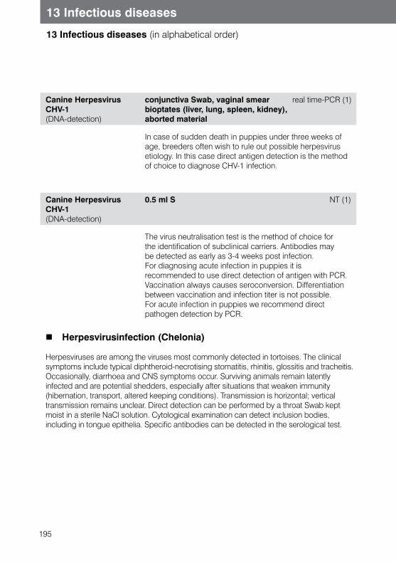
195
13 Infectious diseases
13 Infectious diseases (in alphabetical order)
Canine Herpesvirus CHV-1 (DNA-detection)
conjunctiva Swab, vaginal smear real time-PCR (1)bioptates (liver, lung, spleen, kidney), aborted material
In case of sudden death in puppies under three weeks of age, breeders often wish to rule out possible herpesvirus etiology. In this case direct antigen detection is the method of choice to diagnose CHV-1 infection.
Canine Herpesvirus CHV-1 (DNA-detection)
0.5 ml S NT (1)
The virus neutralisation test is the method of choice for the identification of subclinical carriers. Antibodies may be detected as early as 3-4 weeks post infection. For diagnosing acute infection in puppies it is recommended to use direct detection of antigen with PCR.Vaccination always causes seroconversion. Differentiation between vaccination and infection titer is not possible. For acute infection in puppies we recommend direct pathogen detection by PCR.
Herpesvirusinfection (Chelonia)
Herpesviruses are among the viruses most commonly detected in tortoises. The clinical symptoms include typical diphtheroid-necrotising stomatitis, rhinitis, glossitis and tracheitis. Occasionally, diarrhoea and CNS symptoms occur. Surviving animals remain latently infected and are potential shedders, especially after situations that weaken immunity (hibernation, transport, altered keeping conditions). Transmission is horizontal; vertical transmission remains unclear. Direct detection can be performed by a throat Swab kept moist in a sterile NaCl solution. Cytological examination can detect inclusion bodies, including in tongue epithelia. Specific antibodies can be detected in the serological test.
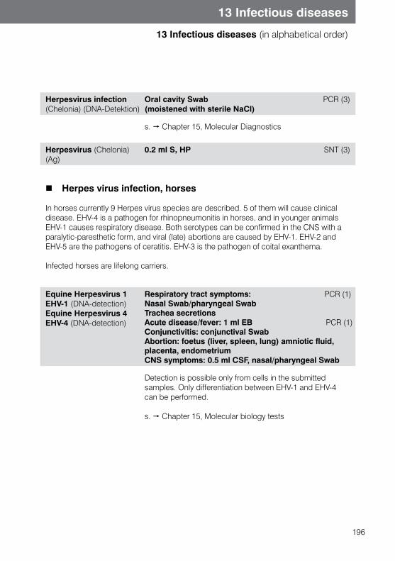
196
13 Infectious diseases
13 Infectious diseases (in alphabetical order)
Herpesvirus infection (Chelonia) (DNA-Detektion)
Oral cavity Swab (moistened with sterile NaCl)
PCR (3)
s. Chapter 15, Molecular Diagnostics
Herpesvirus (Chelonia) (Ag)
0.2 ml S, HP SNT (3)
Herpes virus infection, horses
In horses currently 9 Herpes virus species are described. 5 of them will cause clinical disease. EHV-4 is a pathogen for rhinopneumonitis in horses, and in younger animals EHV-1 causes respiratory disease. Both serotypes can be confirmed in the CNS with a paralytic-paresthetic form, and viral (late) abortions are caused by EHV-1. EHV-2 and EHV-5 are the pathogens of ceratitis. EHV-3 is the pathogen of coital exanthema.
Infected horses are lifelong carriers.
Equine Herpesvirus 1EHV-1 (DNA-detection) Equine Herpesvirus 4EHV-4 (DNA-detection)
Respiratory tract symptoms: PCR (1) Nasal Swab/pharyngeal SwabTrachea secretionsAcute disease/fever: 1 ml EB PCR (1) Conjunctivitis: conjunctival SwabAbortion: foetus (liver, spleen, lung) amniotic fluid, placenta, endometrium CNS symptoms: 0.5 ml CSF, nasal/pharyngeal Swab
Detection is possible only from cells in the submitted samples. Only differentiation between EHV-1 and EHV-4 can be performed.
s. Chapter 15, Molecular biology tests
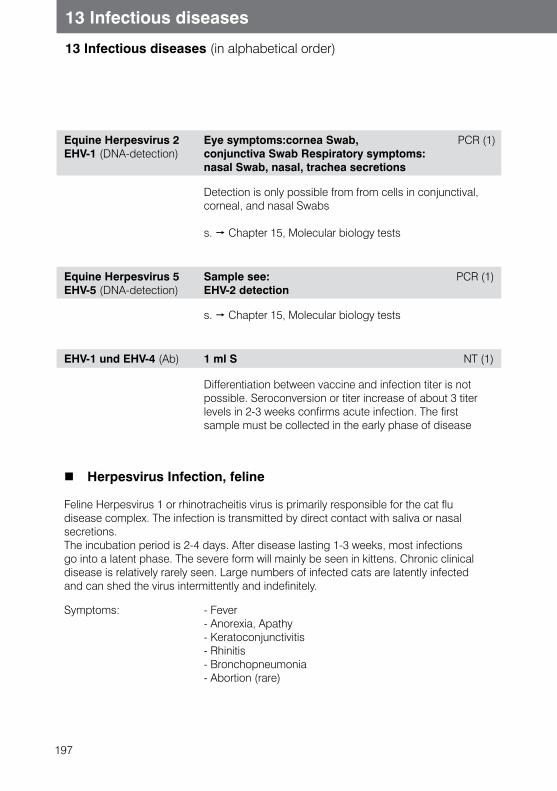
197
13 Infectious diseases
13 Infectious diseases (in alphabetical order)
Equine Herpesvirus 2EHV-1 (DNA-detection)
Eye symptoms:cornea Swab, PCR (1) conjunctiva Swab Respiratory symptoms: nasal Swab, nasal, trachea secretions
Detection is only possible from from cells in conjunctival, corneal, and nasal Swabs
s. Chapter 15, Molecular biology tests
Equine Herpesvirus 5EHV-5 (DNA-detection)
Sample see: PCR (1)EHV-2 detection
s. Chapter 15, Molecular biology tests
EHV-1 und EHV-4 (Ab) 1 ml S NT (1)
Differentiation between vaccine and infection titer is not possible. Seroconversion or titer increase of about 3 titer levels in 2-3 weeks confirms acute infection. The first sample must be collected in the early phase of disease
Herpesvirus Infection, feline
Feline Herpesvirus 1 or rhinotracheitis virus is primarily responsible for the cat fludisease complex. The infection is transmitted by direct contact with saliva or nasal secretions.The incubation period is 2-4 days. After disease lasting 1-3 weeks, most infections go into a latent phase. The severe form will mainly be seen in kittens. Chronic clinical disease is relatively rarely seen. Large numbers of infected cats are latently infected and can shed the virus intermittently and indefinitely.
Symptoms: - Fever- Anorexia, Apathy- Keratoconjunctivitis- Rhinitis- Bronchopneumonia- Abortion (rare)
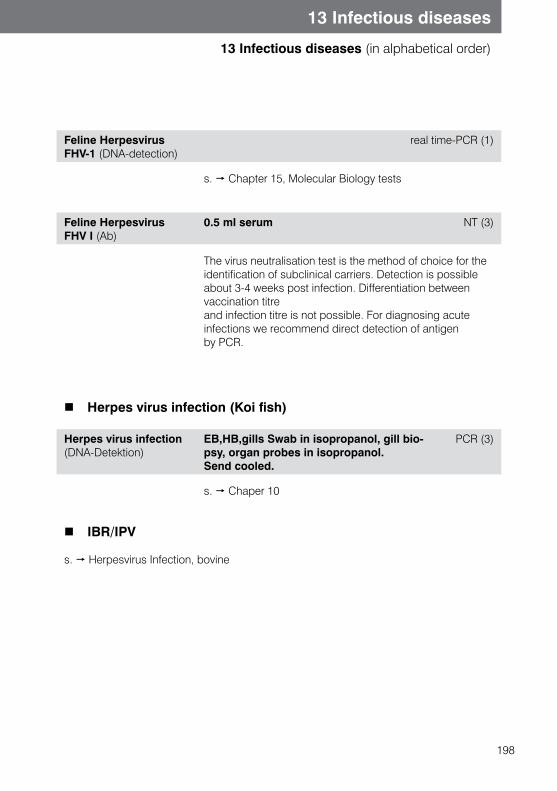
198
13 Infectious diseases
13 Infectious diseases (in alphabetical order)
Feline Herpesvirus FHV-1 (DNA-detection)
real time-PCR (1)
s. Chapter 15, Molecular Biology tests
Feline Herpesvirus FHV I (Ab)
0.5 ml serum NT (3)
The virus neutralisation test is the method of choice for the identification of subclinical carriers. Detection is possible about 3-4 weeks post infection. Differentiation between vaccination titre and infection titre is not possible. For diagnosing acute infections we recommend direct detection of antigen by PCR.
Herpes virus infection (Koi fish)
Herpes virus infection(DNA-Detektion)
EB,HB,gills Swab in isopropanol, gill bio-psy, organ probes in isopropanol. Send cooled.
PCR (3)
s. Chaper 10
IBR/IPV
s. Herpesvirus Infection, bovine
199
13 Infectious diseases
13 Infectious diseases (in alphabetical order)
Inclusion body disease of boids (IBD) (Reptiles)
Inclusion body disease, or IBD, is observed mainly in snakes of the Boidae and Pythonidae families. It is characterised by the occurrence of intracytoplasmic inclusion bodies in the liver, pancreas, kidneys, gastrointestinal mucosa cells and blood cells. The aetiology of this disease has not yet been sufficiently determined. The suspected cause is a retrovirus. Infection is by direct contact, indirect (via contaminated objects), airborne, intrauterine and probably also by a mite vector (Ophionyssus natricis). The rate of infection is increasing in boas but falling in pythons. Affected animals can show general symptoms (regurgitation, lethargy, anorexia, weight loss), respiratory symptoms (pneumonia, mouth breathing, stomatitis) and neurological disorders (tremor, absence of turning reflex, opisthotonus, torticollis, disorientation). A paramyxovirus infection is cli-nically indistinguishable from IBD. The disease is usually fatal but asymptomatic carriers are possible. Ante mortem detection is performed microscopically in blood smears or organ biopsies (e.g. ultrasound-guided percutaneous liver biopsies). There is no PCR test for detection to date.
IBD minimum 2 smears Microscopy (1)
Infectious anaemia, equine
Equine Infectious Anaemia (EIA) is a lentivirus affecting equids, occurring worldwide. Transmission can be via infected blood, blood sucking insects, iatrogenic (contaminated instruments) or intrauterine. Clinically, horses show recurring fever, thrombocytopenia, anaemia, rapid weight loss and peripheral oedema.The infection has several forms, ranging from acute (lethal) to chronic (relapsing). The blood of infected horses remains permanently infectious, even if the patient appears to have recovered.
Equine infectious anaemia/Coggins-Test (Antibodies detection)
0.5 ml S Agar gel diffusion test (1)
In the first 2 to 3 weeks after infection sometimes no antibodies are detectable. Most horses show seroconver-sion by 45 days post infection. Because of this, suspected horses should be retested 4 weeks later. In rare cases seroconversion may not occur until 90 days post infection.
200
13 Infectious diseases
13 Infectious diseases (in alphabetical order)
Influenza virus Infection
Canine Influenza virus (RNA-detection)
Nasal/pharyngeal Swab real time-PCR (1)
Influenza, equine
Equine influenza is an acute, highly contagious viral disease of the respiratory tract caused by the influenza A/equi-1 (H7N7) and A/equi-2 (H3N8) viruses. Equine influenza subtype H7N7 (A/equi-1/Prague/1/56) has not been observed in clinical cases in Western Europe for several decades and is deemed to have been eradicated, but is contained in many vaccines.Transmission occurs via aerosol droplet infection. The symptoms generally consist of fever, nasal discharge, inappetite, dry cough, bronchopneumonia and myalgia. Infected horses can continue to shed the virus for around 10 days p.i. There are no asymptomatic carriers, in contrast to EHV-1 and EHV-4 ..
Equine Influenza (Ab) 1 ml S NT (3)
The following strains have been identified:
Information about active infection can be given by two serum tests with a 2 to 3 week interval. Seroconversion or significant titer increase prove fresh contact with pathogen. First sample must be taken in the early phase of disease.
Prague, Miami, Fontainbleau, Kentucky and Solvalla. Differentiation between vaccination and infection titer is not possible.
Equine Influenza virus (RNA-detection)
Nasal-/pharyngeal Swab, Tracheal secretion(-lavage, BAL)
real time-PCR (1)
s. Chapter 15, Molecular biology tests
201
13 Infectious diseases
13 Infectious diseases (in alphabetical order)
Iridovirus, Reptiles
Iridovirus, Reptiles (DNA-Detektion)
mouth Swab without medium PCR (3)
Lawsonia intracellularis (Equine Proliferative Enteropathy)
Lawsonia intracellularis is a pathogen of proliferative enteropathies that may affect a wide range of mammals and birds. Foals up to 12 months old are affected by Equine Proliferative Enteropathy, but the most common age is between four and six months. Oral transmission is suspected, as clinically inapparent young carriers can excrete the pathogen with faeces.The obligatory intracellular bacteria proliferates in cytoplasm of erythrocytes (mainly in the middle and distal part of gastrointestinal tract) and influences cell proliferation, usually without causing inflammatory reaction. Therefore progressive intestinal cell proliferation is caused with poor cell differentiation and so causes decreased enzymatic and absorptive features (proliferative enteropathy). Pathogenesis is not yet clear. The most important clinical signs are lethargy, anorexia, weight loss, oedema (hypogastrium, prepuce, legs and head). Colic and diarrhoea often occur, due to intestinal malabsorption and the increased permeability of the gastrointestinal tract. The pathogen is intermittently excreted in faeces. In case of negative result, a new test with fresh sample is recommended. L. intracellularis is found worldwide and the disease has been described in foals in North America, Australia and Europe. There is currently no direct evidence that L. intracellularis infects humans.
Lawsonia intracellularis (DNA-detection)
5 g faeces real-time PCR (1)
202
13 Infectious diseases
13 Infectious diseases (in alphabetical order)
Leishmaniasis
Canine leishmaniasis is caused by Leishmania infantum (syn. L. chagasi in South- and Central America). Dogs are also (rarely) infected by L. tropica. Transmission occurs by the moth fly Phlebotomus (L. chagasi is transmitted by Lutzomyia sp.). The pathogen range in Europe is the Mediterranean region and is mainly offshore and on the larger islands.
Symptoms: Incubation time lasts months to years. The following symptoms are found:- Weight loss - Anorexia, Apathy, Enteritis - Hyperkeratosis, Alopecia (beginning periorbitally),
Dermatitis, Pad fissures - Claw lenghthening, Claw bed inflammation- Pancytopenia - Lymph node oedema - Hyperproteinaemia, Hypoalbuminemia,
Hypergammaglobulinemia - Hepato- and Splenomegaly- Glomerulonephritis - Polyarthritis- Keratoconjunctivitis, Uveitis,Iritis, Blindness- Epistaxis
Leishmania- Direct detection
Smear Microscopy (1)
Please note:
Direct detection of Leishmania is reliable only from lymph node and bone marrow aspirate or skin biosy(sensitivity 30-50 %). Detection in a blood smear is usually not successful.
Negative direct pathogen detection does not exclude infection.
203
13 Infectious diseases
13 Infectious diseases (in alphabetical order)
Leishmania spp. (DNA-detection, quantitative)
Bone marrow, 1 ml EB real time-PCR (1)
With help of real-time PCR it is possible to quantify the number of Leishmania in test sample. Knowing the parasite concentration allows exact knowledge of infection status in cases where:- ELISA-results were not reliable, - Dogs showing clinical symptoms, but without
seroconversion- Dogs without clinical symptoms, but originating from
endemic regions;
Studies have shown that dogs with medium to high concentrations of Leishmania in bone marrow or blood are either already sick or have a high probability to be affected with clinical leishmaniasis.Leishmania quantifying offers very good conditions for treatment monitoring (one month after beginning of treatment).
s. Chapter 15, Molecular biology tests
Leishmania spp.(DNA-detection, qualitative)
3 ml U, 0.5 ml Synovia, biopsy (liver, spleen), eye-, noise-, tissue Swab
.
204
13 Infectious diseases
13 Infectious diseases (in alphabetical order)
Leishmanien (Ab) 1 ml S, EP, HP dogs ELISA (1) cats IFT (1)
Asymptomatically infected animals often show no specific or only borderline/low antibody titres (cellular immunity: Th1 cells). In clinically infected animals, antibodies are detectable in the majority of cases (Th2 immune response with production of non-protective antibodies). As a rule, seroconversion does not occur until several months post-infection: 1-22 (Ø 5) months in the case of natural infections and around 1-6 (Ø 3) following experimental infection.
Please note our test profile Blood parasites and haemotropic bacteria - microscopy Travel Profie 1 + 2
Leptospirosis
Leptospirosis is caused by the following serotypes: L. australis (bratislava), L. autumna-lis, L. canicola, L. copenhageni (icterohaemorrhagiae), L. grippotyphosa, L. saxkoebing,L. sejroe and L. tarassovi.Transmission occurs directly via contact with infected urine or indirectly via contaminatedwater. The pathogen is transported via the bloodstream into the body, and especially into the liver and kidneys.
Symptoms: following an incubation period of 4-12 days the followingsymptoms can occur:- Pyrexia- Anorexia,Vomiting, Enteritis- Polyuria/polydipsia- Haemolysis, Icterus- Haemorrhagic diathesis- Chronic liver and kidney disease- Uveitis, Retinitis
205
13 Infectious diseases
13 Infectious diseases (in alphabetical order)
Equine Recurrent Uveitis (ERU) Intraocular persistent Leptospira infection as the aetiology of ERU is regarded in Europe as highly probable. Only Ab or Ag detection in the aqueous humor or vitreous sample is diagnostically relevant. Increased serum (Ab) detection does not confirm Leptospira in eye disease.
Leptospira (Ab) 1 ml S MAR (1)
Detection of Leptospira (Ab) by Microagglutination reaction (MAR) is usually the method of choice for confirming suspected infection. The test should be performed at the earliest 14 days after infection.In dogs 9 serovars are tested. In horses only L. australis, L. autumnalis, L. bratislava, L. copenhageni, L. grippotyphosa and L. pomona are tested. In other animal species other relevant serovars are tested. Differentiation between vaccination and infection titer is only limitedly possible (depending on titer height). Vaccination in dogs is only with L. canicola and L. copenhageni (icterohaemorrhagiae), but crossreactions with other serovars is also possible.
In horses: to detect active infection one can use two serum tests with an interval of 2 to 3 weeks. Seroconversion, 2 levels of titer increase, or a 4x increase in antibody titer confirm fresh contact with pathogen. The first sample has to be collected in the early phase of the disease. Positive antibody detection with a titer of 1:800 together with appropriate clinical symptoms is diagnostic for acute Leptospira infection.
s. Chapter 15, Molecular biology Tests
206
13 Infectious diseases
13 Infectious diseases (in alphabetical order)
Leptospira spp. (DNA-detection)
2 ml EB, CSF, 5 ml U, aqueous humour, vitrous body sample Abortion: placenta, umbilicus, foetus (kidneys and liver)
real time-PCR (1)
The pathogenic Leptospira include:
The non-pathogenic Leptospira include:
Opportunistic/intermediate pathogens are:
Direct detection of Leptospira is possible in blood only a short time after pathogen exposure. Pathogen excretion in urine starts about 7 days post infection and can continue for months to years. Detection from aqueous humour is possible in horses. The test system detects only pathogenic Leptospira strains (differentiation is not possible):
L. interrogansL. kirschneriL. santarosaiL. weiliiL. alexanderiL. borgpeterseniiL. genomospecies 1L. noguchii
L. biflexaL. meyeriL. wolbachiiL. genomospecies 3L. genomospecies 4L. genomospecies 5
L. broomiiL. faineiL. inadai
(The above classification is based on the publications of Slack et al., 2006, and Perolat et al., 1998).
s. Chapter 15, Molecular biology tests
Negative direct pathogen detection does not exclude infection!
207
13 Infectious diseases
13 Infectious diseases (in alphabetical order)
Leukemia, bovine
s. Enzootic bovine leukemia
Leukemia virus infection, feline
s. FeLv
Listeriosis
Listeria is a bacteria occuring worldwide, which is spread by subclinically infected rodents. Very high pathogen amounts are required for infection, which accumulate especially in the edge and superficial layers of contaminated silage, but also occur in other feed.Listeria monocytogenes is a facultative intracellular bacteria (gram positive rods). It can penetrate into different animal cell types and proliferates in macrophages, epithelial cells or fibroblasts.
Cytolytic toxin listeriolysine is an essential virulence factor, which L. monocytogenes needs for escape from phagosomes in the cytoplasm. In the clinical manifestation, horses, cattle and sheep show mainly CNS-symptoms, fever, restlessness, coordination disorders and other signs of encephalitis. A metrogenic form, which leads to late abortion, early births or wasting of foals/calves/lambs on delivery is also described.
Listeria (Ab) 1 ml S CFT (3
Listeria monocytogenes(DNA-detection)
0.5 ml liquor, 1 ml EB, 5 g faeces, aborted sample
PCR (1)
s. Chapter 15, Molecular biology tests
208
13 Infectious diseases
13 Infectious diseases (in alphabetical order)
Maedi/Visna
The Maedi/Visna virus leads to interstitial pneumonia or demyelinating encephalitis in sheep.
Symptoms: - Dyspnoea, Coughing- Ataxia, lameness- Decreased milk production- Emaciation- Splenomegaly- Possibly hepatomegaly
Maedi/Visna (Ab) 1 ml S, EP, HP ELISA (3)
Antibody detection using ELISA. Antibodies occur several weeks to years post infection. Therefore a negative result does not rule out infection
Megabacteria Infection
Megabacteria (Syn. Macrorhabdus ornithogaster, Avian gastric yeast) are fungi that can cause inflammatory changes in the glandular stomach of birds. They are found in different bird species, such as parrots, sparrows, chickens, geese and storks.In psittacidae, this disease is described as “Going light syndrome”. It is amultifactoral disease. Other infections, parasitosis and tumors should be excluded from differential diagnosis list.
Symptoms: - Vomiting/regurgitation - Diarrhoea- Lethargy- Slimming- Ruffled feathers
Megabacteria- Direct detection
2 g faeces Microscopy (1) PAS-staining
Please note:
Pathogen will be intermittently excreted, therefore we recommend you collect faeces over 5 days before submis-sion for testing.
Negative result in faeces Swab does not exclude Megabacteria Infection.
209
13 Infectious diseases
13 Infectious diseases (in alphabetical order)
Mycoplasma agassizii-Infektion
Mycoplasma agassizii is an agent of multifactorial Upper Respiratory Tract Disease (URTD) in tortoises. The infection is characterised by serous, mucous or purulent nasal discharge, as well as eye discharge, conjunctivitis and eyelid oedema. The differential diagnosis should rule out herpesvirus infection. Detection is by nasal lavage using sterile NaCl solution or by throat Swab.
Mycoplasma agassizii 0.5 ml nasal lavage, Swab (throat)
PCR (3)
s. Chapter 15
Mycoplasma haemofelis, Candidatus Mycoplasma haemominutum, Candidatus Mycoplasma turicensis, Mycoplasma haemocanis and Candidatus Mycoplasma haematoparvum
Pathogens earlier described as Haemobartonellas have now been reclassified and assigned to genus Mycoplasma. Isolate Ohio of Haemobartonella felis is named Mycoplasma Haemofelis and California isolate is now called Candidatus Mycoplasma haemominutum. Haemobartonella canis is Mycoplasma haemocanis ebenfalls of genus Mycoplasma. Mycoplasma haemofelis seems more pathogenic then Candidatus Mycoplasma haemominutum and can cause a disease in immunocompetent cats. Infection with Candidatus Mycoplasma haemominutum is mostly mild or subclinic.With simultaneous immunosuppression (for instance FeLV infection) infected animals develop more severe disease. Clinical disease is only observed in dogs with in immunosuppression, splenectomy, or simultaneous infection with other pathogens. The transmission method is not fully clear, but ticks, lice, fleas, blood transfusions, and bite traumas are likely to be important. Vertical transmission is probable.
210
13 Infectious diseases
13 Infectious diseases (in alphabetical order)
Symptoms: Depending on pathogenicity and immune status, disease types ranges from subclinical to chronic (latent) to acute. - Fever (over 40° C) - Haemolytic anaemia - Icterus, bilirubinuria- Hepato-, Splenomegaly - Anorexia, Apathy
Haemotropic Mycoplasma(Haemobartonellas)- Direct detection
0.5 ml EB + blood smear Microscopy (1)
Please note:
The epicellular organism is found in a Giemsa stained blood smear using a light microscope. In the chronic course of the disease asymptomatic and parasitaemic phases will alternate.Direct detection of the organism is therefore not always possible!
The pathogen can be confused with with Howell-Jolly, Heinz bodies or artefacts, therefore our test of choice is a PCR test from EDTA blood.
Please note our profiles Travel disease profile (1 and 2) Blood parasites and haemotropic bacteria - microscopy
Feline Mycoplasmsa Profile
1 ml EB real time-PCR (1)
s. Chapter 15
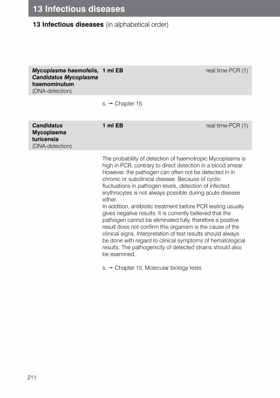
211
13 Infectious diseases
13 Infectious diseases (in alphabetical order)
Mycoplasma haemofelis, Candidatus Mycoplasma haemominutum (DNA-detection)
1 ml EB real time-PCR (1)
s. Chapter 15
Candidatus Mycoplasma turicensis (DNA-detection)
1 ml EB real time-PCR (1)
The probability of detection of haemotropic Mycoplasms is high in PCR, contrary to direct detection in a blood smear.However, the pathogen can often not be detected in in chronic or subclinical disease. Because of cyclic fluctuations in pathogen levels, detection of infected erythrocytes is not always possible during acute disease either. In addition, antibiotic treatment before PCR testing usually gives negative results. It is currently believed that the pathogen cannot be eliminated fully, therefore a positive result does not confirm this organism is the cause of the clinical signs. Interpretation of test results should always be done with regard to clinical symptoms of hematological results. The pathogenicity of detected strains should also be examined.
s. Chapter 15, Molecular biology tests
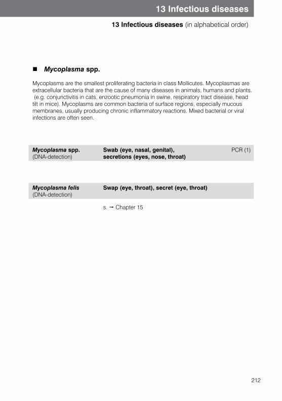
212
13 Infectious diseases
13 Infectious diseases (in alphabetical order)
Mycoplasma spp.
Mycoplasms are the smallest proliferating bacteria in class Mollicutes. Mycoplasmas are extracellular bacteria that are the cause of many diseases in animals, humans and plants. (e.g. conjunctivitis in cats, enzootic pneumonia in swine, respiratory tract disease, head tilt in mice). Mycoplasms are common bacteria of surface regions, especially mucous membranes, usually producing chronic inflammatory reactions. Mixed bacterial or viral infections are often seen.
Mycoplasma spp. (DNA-detection)
Swab (eye, nasal, genital), PCR (1) secretions (eyes, nose, throat)
Mycoplasma felis (DNA-detection)
Swap (eye, throat), secret (eye, throat)
s. Chapter 15
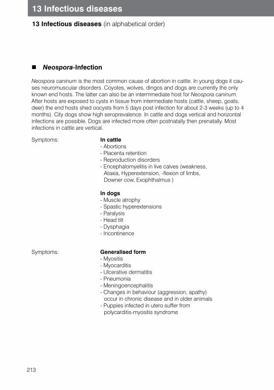
213
13 Infectious diseases
13 Infectious diseases (in alphabetical order)
Neospora-Infection
Neospora caninum is the most common cause of abortion in cattle. In young dogs it cau-ses neuromuscular disorders. Coyotes, wolves, dingos and dogs are currently the only known end hosts. The latter can also be an intermmediate host for Neospora caninum. After hosts are exposed to cysts in tissue from intermediate hosts (cattle, sheep, goats, deer) the end hosts shed oocysts from 5 days post infection for about 2-3 weeks (up to 4 months). City dogs show high seroprevalence. In cattle and dogs vertical and horizontal infections are possible. Dogs are infected more often postnatally then prenatally. Most infections in cattle are vertical.
Symptoms: In cattle- Abortions - Placenta retention - Reproduction disorders - Encephalomyelitis in live calves (weakness,
Ataxia, Hyperextension, -flexion of limbs, Downer cow, Exophthalmus )
In dogs- Muscle atrophy- Spastic hyperextensions - Paralysis - Head tilt- Dysphagia- Incontinence
Symptoms: Generalised form - Myositis - Myocarditis - Ulcerative dermatitis - Pneumonia- Meningoencephalitis - Changes in behaviour (aggression, apathy)
occur in chronic disease and in older animals- Puppies infected in utero suffer from
polycarditis-myositis syndrome
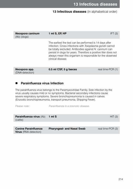
214
13 Infectious diseases
13 Infectious diseases (in alphabetical order)
Neospora caninum (Ab) (dogs)
1 ml S, EP, HP IFT (3)
The earliest the test can be performed is 14 days after infection. Cross infections with Toxoplasma gondii cannot be totally excluded. Antibodies against N. caninum can persist in dogs for years. Therefore a positive titer does not always mean this organism is responsible for the observed clinical disease.
Neospora spp. (DNA-detection)
0.5 ml CSF, 5 g faeces real time-PCR (1)
Parainfluenza virus Infection
The parainfluenza virus belongs to the Paramyxoviridae Family. Sole infection by the virus usually causes mild or no symptoms. Bacterial secondary infections cause severe respiratory symptoms. Severe bronchopneumonia is caused in calves. (Enzootic bronchopneumonia, transport pneumonia, Shipping Fever). Please note: Parainfluenza is a zoonotic disease.
Parainfluenza virus (Ab) (cattle)
1 ml S HIT (3)
Canine Parainfluenza Virus (RNA-detection)
Pharyngeal- and Nasal Swab real time-PCR (3)
215
13 Infectious diseases
13 Infectious diseases (in alphabetical order)
Paramyxovirus-Infection (OPMV) (Reptile)
Ophidian paramyxoviruses (OPMV) are characterised by a broad host spectrum and primarily affect the Viperidae, Colubridae, Elapidae and Boidae (and, more rarely, lizards or Chelonia). They lead to peracute mortality or protracted respiratory disease with CNS involvement. Typical symptoms are an open mouth, bloody exudate in the oral cavity, rales, head tremor and opisthotonus. Depending on the pathogenicity of the virus strain, mortality can be up to 100%. In Boidae, the differential diagnosis should rule out IBD (inclusion body disease). Virus transmission can be faecal-oral or by aerosol droplet infection. Throat Swabs are suitable for direct RNA detection. Specific antibodies can be detected in serological testing.
OPMV (Reptiles) (DNA-detection)
Swap (throat) PCR (3)
s. Chapter 15, Molecular Diagnostics
OPMV (Reptiles) (Ab) 0.2 ml S, HP SNT (3)
Paratuberculosis General Information
The infection with the acid-fast bacillus Mycobacterium avium subsp. paratuberculosisoccurs in ruminants and is also called Johne’s Disease. After a long incubation period of2 to 6 years the affected animals begin to suffer from chronic enteritis and weight loss, with eventually fatal consequences.
Paratuberculosis (Ab) (cattle)
1 ml S, EP, HP ELISA (3)
216
13 Infectious diseases
13 Infectious diseases (in alphabetical order)
Parvovirus/Panleukopenia
The pathogens causing canine parvovirus enteritis (CPV) and feline parvovirus enteritis(FPV) are very closely related. More recent strains of CPV may also cause clinical disease in cats. Transmission occurs oronasally through contact with infected faeces or contaminated objects. The course of disease varies from subclinical to peracute, depending on the age and immune status of the animal. Virus replication takes place in all tissues with a high cell multiplication rate, especially intestinal mucosa, bone marrow, lymphatic tissue and myocardium. In cats replication may also take place in the retina and cerebellum.
Symptoms: animal carriers:- Abortion, Mummification
In puppies we usually observe following symptoms:- Pyrexia/hypothermia- Anorexia, Lethargy- Vomiting, (haemorrhagic) Diarrhoea - Dehydration- Leukopenia- Dyspnoea, Cardiac symptoms
Cerebellar hypoplasia (kittens)- Lymphopenia
Parvovirus (Ag) (dogs, cats )
Dogs: rectab SwabCats: 5 g faeces, rectal Swab
Immunochromatography (1) EIA (1)
217
13 Infectious diseases
13 Infectious diseases (in alphabetical order)
Please note:
Direct detection of parvovirus antigen in faeces is possible in cats and dogs. Excretion happens 3-4 days post infection and lasts for about 7-10 days. In some cases it can last also longer. Using modified live vaccine can lead to virus excretion in the first 4 weeks after vaccination; distinguishing between vaccination and field virus is not possible.
Negative direct pathogen detection does not exclude infection!
Parvovirus FPV, CPV (DNA-detection)
5 g faeces, rectal Swab real time-PCR (1)
Please note:
Direct PCR pathogen detection from faeces or rectal Swab is possible in dogs and cats. It is important to specify the animal species being tested. In dogs vaccination strain CPV2 and wild strain CPV 2a/CPV 2b can be differentiated. It has diagnostic value, as vaccination virus can shed for 2-12 days after vaccination. Shedding of field virus starts 3-4 days post infection and usually lasts 7-10 days. In certain cases longer shedding is possible.
A negative PCR result does not exclude infection.
218
13 Infectious diseases
13 Infectious diseases (in alphabetical order)
Parvovirus (Ab) (dogs, cats)
0.5 ml S HI (1)
Parvovirus antibodies can be detected with the haemagglutionation inhibition test (HIT) from 4-6 days post infection in cats and dogs. Seroconversion in unvaccinated animals is proof of infection. Distinguishing between vaccination virus, field virus, and maternal antibodies is not possible. Since vaccination is widely used, we recommend confirming suspected infection with direct detection of parvovirus from faeces.Low maternal antibody titer (usually up to 1:40) does not protect from infection, but can interfere with vaccination (immunologic gap). Early vaccination may not be be effective, as attenuated vaccination virus is neutralised by maternal antibodies. The half life of maternal antibodies is approximately 10 days. Maternal antibody titer of single litter puppies is usually high, so testing puppies allows you to determine the ideal time for first vaccination.
Please note also our test profile
Viral faeces test
Polyomavirus, avian
s. Chapter 15, Molecular biology tests
Porcine Circovirus-2
s. Chapter 15, Molecular biology tests
219
13 Infectious diseases
13 Infectious diseases (in alphabetical order)
Porcine Influenza virus
Swine influenza is caused by porcine Influenza virus A (Orthomyxovirus). This virus contains two different forms of superficial antigens (H and N), which are the basis for classification of different subtypes. A wide range of subtypes enables infection in human, swine, birds and even horses. Clinical diagnosis is not easy. A successful virus culture from a nasal or pharyngeal Swab is required for a reliable diagnosis, or alternatively, detection of a subtype specific antibody increase in two blood samples with a 3 week interval.
Porcine Influenza virus (Ab)
2 ml S Haemagglutination (3)
PRRS (Porcine Reproductive and Respiratory Syndrome)
The causative agent of PRRS is a highly infectious arterivirus. The disease is associatedwith abortion and reproductive disorders. Male pigs can also be affected, often showinggeneral symptoms such as inappetence, and can shed the virus with sperm. Subclinicaldisease (without any clinical symptoms) is possible.
PRRS (Ab) (porcine) 1 ml S ELISA (3)
Detection of antibodies using an ELISA test. Serum antibo-dies are detectable one week post infection, with maximum titres after 3-5 weeks. Virus neutralizing antibodies do not develop until 4-8 weeks post infection. It is recommended to test at least 5-10 animals per herd or population.
PBFD
s. Chapter 15, Molecular biology tests
220
13 Infectious diseases
13 Infectious diseases (in alphabetical order)
Q Fever
Q fever is a zoonotic disease caused by a species of bacteria called Coxiella burnetii. It does not play a major role in animals, but affected animals constitute a risk of infection for humans. Ruminants, horses, dogs and cats are susceptible.
Symptoms: - Pyrexia, lethargy, inappetence- Conjunctivitis- Bronchopneumonia- Arthritis- Abortion
Coxiella burnetti (Ab) 1 ml S CFT (3)
221
13 Infectious diseases
13 Infectious diseases (in alphabetical order)
Rabies virus antibody detection for travel reasons
When entering some countries in the EU and non EU (e.g. Japan) it is compulsory to show proof of antibody titres against the rabies virus. This can only be done at laboratories certified by the EU commission; IDEXX Vet Med Lab is one of these laboratories.Travelling pets require an EU pet passport. The regulations for different countries may vary, so it is very important to inquire well ahead of time what the exact requirements are. Information can be found on the internet from national internet portals, from the country’s embassy, or their ministry responsible for animal import and export.
For the test to be performed, a few points must be followed. Please use only special order form for rabies antibodies detection. You can download these from www.idexx.de or order directly from IDEXX Vet·Med·Lab. Please fill in the form completely, correctly, and legibly. If the form is illegible the results cannot be sent. Only good quality, non-haemolytic and non-lipaemic serum is used as sample. (EDTA, citrate- and heparin blood can lead to false results, and therefore are not tested).
Sample tubes must be clearly marked and must match the details on the special form. Results will be sent as a hard copy certificate by post. Please note that additional analysis is not possible with the same sample material. Please remember that national entry regulations in can vary from country to country, so please check for the required tests before any travel is planned.
Note: This test is not used to diagnose animals with suspected rabies infection. Please do not send samples from suspected animals!
Rabies virus (Ab) (NT): Please use a separate form
0.5 ml S. Please label unmistakably
FAVN (1)
Test is peformed by fluorescent antibody virus neutralisation (FAVN) The test is performed according to O.I.E. regulations.
222
13 Infectious diseases
13 Infectious diseases (in alphabetical order)
Rhodococcus equi-Infection
Rhodococcus equi (DNA-detection)
Tracheal secretions (fluid, BAL), Synovial membrane, tissue (lungs), faeces
real time-PCR (1)
s. Chapter 15, Molecular biology tests
Rocky Mountain Spotted Fever (RMSF)
Rocky Mountain Spotted Fever is a significant zoonosis. The pathogen is Rickettsia rickettsii, which is transmitted by ticks. Disease is found in North, Central and South America. The infection is usually mild in dogs, but a severe, lethal course is also possible. Chronic disease has not been described. Incubation time is from 2 to 14 days.
Symptoms: - Sudden high fever - Anorexia- Vomiting, Diarrhorea - Petechiae- Oedema (mainly scrotal)- Joint swelling - Myalgia- Dyspnoea - Beeding in the eye- Neurological disorders frequently: - Thrombocytopenia
In Southern Europe Rickettsia conorii is the pathogen of Mediterranean typhus in humans, but dogs can also be infected. Affected animals show seroconversion. Clinical disease in dogs can occur in regions where the tick responsible for transmission (Rhipicephalus sanguineus) is also found; the clinical expression is similar to granulocytic anaplasmosis.
Rickettsia (Ab) (dogs) 1 ml S IFT (3)
If accompanied by appropriate clinical signs, a 4x titer increase in the second of two tests with a 3 week interval is confirmation of RMSF infection.
223
13 Infectious diseases
13 Infectious diseases (in alphabetical order)
Rotavirus
Rotaviruses are found in almost all animal species. The virus has a high affinity for small intestinal epithelium. Virus replication leads to massive destruction of epithelial villi, leading to malabsorption and hypersecretion. Severe watery diarrhoea is found, especially in young animals. The route of infection is oral, with older animals acting as a virus reservoir.
Symptoms: clinical symptoms occur after an incubation periodof 1-2 days:- Watery diarrhoea- Vomiting- Dehydration
Rotavirus (Ag) 1 g faeces (pea-sized amount) Immunchromatography(1)
Virus excretion via the faeces usually lasts 3-10 days. Using immunochromatography the superficial antigen of the virus will be detected.
Please note:
Please note also our test profile:
A single negative test result with simultaneous clinical suspicion should be confirmed by testing a second faecal sample.
Virological examination using electron microscopy
Salmonella abortus equi
Transmission of the pathogen is mainly oral, but is also possible through mating. In Germany Salmonella abortus equi is no longer believed to play a role in abortions.
Salmonella abortus equi (Ab)
1 ml S Slow agglutination (3)
224
13 Infectious diseases
13 Infectious diseases (in alphabetical order)
Sarcoptes
Canine mange is caused by Sarcoptes scabiei var. canis. Severe pruritus is characteristic for this disease. There is little or no response to treatment with glucocorticoids. Initially the changes in the skin are seen on the abdomen, sternum, lateral limbs and ears, before they generalise. The detection of mites in skin scrapes is often unsuccessful in chronic cases, as due to sensitization even very low numbers of mites continue to cause clinical symptoms. The sensitivity of skin scrapes is approx. 30-50%. Microscopic detection of the mites is based on a deep skin scrape (1 mite constitutes proof). Several scrapes should be taken from different sites and always at the edges of the lesion.
Sarcoptes (Ab) (dogs) 0.5 ml S ELISA (1)
Please note also our test
The ELISA method for detecting sarcoptes antibodies in dogs is highly specific (94.6%) and highly sensitive (92.1%). No cross reactions occur with storage mites, demodex mites or cheyletiella mites. The antibody test can be performed approx. 3-4 weeks post infection. A negativeresult does not rule out infection, since 5-10% of dogs will not produce antibodies. Antibody titres persist over a long period of time; therefore they are not very useful in treatment monitoring.
Ectoparasites in the skin scraping
225
13 Infectious diseases
13 Infectious diseases (in alphabetical order)
Tickborn Encephalitis
The pathogen of the Tickborne Encephalitisvirus is a Flavivirus, which is mainly transmitted by Ixodes ricinus in Middle Europe. Clinical disease in companion animals has been mainly described in dogs. Isolated cases are described in horses and small ruminants. Endemic areas are usually locally limited in different European countries, as for instance Switzerland, Austria, France, Hungary, Czech Republic, Poland, Russia and Slovenia. Sweden and Finland are also affected. Baden-Württemberg, Bavaria and Südhessen are where the disease is most frequently seen in Germany. Tickborne encephalitis is mostly acute, with a progressive course. Peracute, acute, subacute and chronic forms are al possible. Fever, apathy, anorexia, behavior changes such as nervousness, aggressivness, seizures, paresis, ataxia, hyperaesthesia and hyperalgesia are often described.
Tickborne Encephalitisvirus(RNA-detection)
0.5 ml CSF, ticks PCR (1)
In case of significant clinical symptoms we recommend direct pathogen detection in cerebrospinal fluid.
s. Chapter 15, Molecular Biology Tests
Tickborne Encephalitisvirus (Ab)
1 ml S CBR (3)
A complement binding reaction is used for detection of seropositive animals. In endemic areas up to 30% of dogs may be infected without showing any clinical signs.Therefore a positive antibody detection is not a confirmation of clinical disease.Two serum tests should be peformed, and a significant titer increase in the second test test proves acute infection. Complement binding antibodies are detectable for a long time after exposure. If there is a suspicion of clinical CNS disease, a CSF test is always recommended.
226
13 Infectious diseases
13 Infectious diseases (in alphabetical order)
Toxoplasmosis
Toxoplasma gondii, the causative agent of toxoplasmosis, is prevalent worldwide. Only cats and related felidae act as final hosts, while almost all warm-blooded animals (including humans) may act as intermediate hosts.Clinical disease in cats is rare and is usually only seen in very young or immunosup-pressed animals. Infection in cats occurs via ingestion of cyst-containing meat of intermediate hosts or via feline faeces containing infective oocysts. Almost every organ will be colonized and in cats the parasite can multiply in the intestinal epithelium. Oocyst excretion may begin approximately 3 to 9 days after infection with sporulated oocysts. Approximately 20% of cats excrete them for 18-35 days. Infection of other warm-blooded animals and humans occurs via ingestion of inadequately cooked, cyst-containing meat of intermediate hosts or via contact with infective oocysts originating from feline faeces. A short parasitaemia is observed as in cats and the parasites then colonize in all organs, but there is no excretion in these non-feline species.
Symptoms: the infection is usually subclinical, but thefollowing symptoms may be observed:
- Pyrexia - Anorexia, Apathy- Pneumonia- Enteritis- Retinopathies- Abortion (humans, sheep, goats)- Encephalitis- Pneumonia- Lymph node enlargement
Toxoplasma DirectDetection
faeces, collected over 3-5 days flotation method (1)
Direct detection of toxoplasma oocysts in faeces using the flotation method is only useful in cats. As excretion is intermittent, not permanent, repeating testing on faecal samples collected over 3 to 5 days is recommended. Negative direct pathogen detection does not rule out infection!
227
13 Infectious diseases
13 Infectious diseases (in alphabetical order)
Toxoplasma gondii(DNA-detection)
CNS-Symptms: 0.5 ml CSF real time-PCR (1) Abortion (dogs/small ruminants.): Vaginal Swab, placenta, foetus, tissue (liver, spleen, kidney, lung, heart, gut) Respiratory signs: Bronchial lavage Eye symptoms (mostly cats) aqueous humor Fever: 0.5 ml EB
Detection with PCR is not possible in faeces. Using the other samples listed above, PCR can confirm existing disease. It should be noted that positive PCR result does not always confirm acute infection with T. gondii. The patho-gen can be detected in CSF and in the aqueous humor in clinically healthy animals. Therefore positive result should always be interpreted together with clinical symptoms, and negative result does not exclude infection.
s. Chapter 15, Molecular biology tests
Toxoplasms IgM, IgG (Ab)
1 ml S, EP, HP (dogs, cats) IFT (1) Exotic animals: IHA
Detection of toxoplasma (ab) in cats and in dogs by IFT is usually the method of choice for confirmation of suspected infection. IgG antibodies are usually detected from 2 weeks post infection and can persist for many years. Therefore to diagnose active toxoplasmosis, increasing IgG titer (in a repeat test) should be measured. IgM antibodies can be found from 1-2 weeks post infection and reach maximum levels 3-6 weeks post infection. In most cats they decrease under the detection limit about 12 weeks post infection. A high IgM titer is diagnostic for active infection
228
13 Infectious diseases
13 Infectious diseases (in alphabetical order)
Transmissible gastroenteritis (TGE) in swine
The pathogen of TGE is a porcine coronavirus (TGEV), which can causa diarrhorea in swine of all ages, but especially in nursing piglets. Virus proliferation takes place in the epithelial villi of the intestines. Normally the virus is shed in faeces by nursing piglets from 1 to 7 days post infection, and in fatteners from 3 to 7 days post infectionAnimals may excrete the virus in the faeces intermittently for up to 18 months.
Transmissible Gastroenteritis Virus (TGEV) (RNA-detection)
2 g faeces, rectal Swab, intestinal mucosal membrane
real time-PCR (1)
Pathogen detection by PCR facilitates clinical identification of subclinical excretion. As virus shedding occurs intermittently, in case of a suspicious clinical picture and a negative PCR result, the test should be repeated. With this test protocol we can detect porcine respiratory coronavirus (PRCV) and TGEV mutants which cause mild or subclinical respiratory tract infections.
Trichomonas Infection
Trichomonas (Trichomonas gallinae) is seen in pigeons and other bird species (chicken, falcons, parrots) in the pharynx, esophageus and crop. In addition the liver, heart and other organs can be affected. The disease occurs in young animals, which are infected from older animals. Trichomonas species (e. g. Tetratrichomonas gallinarum) found in the distal part of chicken and waterfowl intestine are harmless.
Symptoms: - Yellow, cheese-like covering on the beak and pharynx (“yellow button”)
- Loss of appetite- Weight loss- Difficulties in drinking and feeding
229
13 Infectious diseases
13 Infectious diseases (in alphabetical order)
Trichomonas- Direct detection
Swap in NaCl, feacal sample, crop Swab
Microscopy(1)
Please note:
Crop Swabs should be taken from fasted animals.A mucosal membrane sample should be taken by a Swab moistened with saline solution.The s should be sent in a tube without transport medium, with saline solution.
Negative result does not exclude infection.
Tritrichomonas-Infection
Tritrichomonas foetus (DNA-detection)
5 g faeces, NO rectal swap real time-PCR (1)
s. Chapter 15
Trypanosoma-Infections
Trypanosoma does not play a significant role in companion animal disease in our area.
Trypanosoma- Direct detection
Blood smear Microscopy (1)
Please note: Direct pathogen detection is not always possible!
230
13 Infectious diseases
13 Infectious diseases (in alphabetical order)
Trypanosoma equiperdum (Ab)
1 ml S CFT (3)
Trypanosoma is a specific chronic or acute contagious infection in the family Equidae.Transmission occurs through mating. The first clinical sign is oedema of the external genitals about 2-4 weeks post infection. The disease progresses with the formation of characteristic round skin lesions with depigmentation on the throat, flank, and abdomen. The third stage of the disease is characterized by peripheral neural disorders. The disease can may be lethal. Trypanosoma is widespread, mainly in Asia and in North and South Africa. Central Europe is free from T. equiperdum.
Vesicular Stomatitis
This is a highly contagious virus infection in equids, cattle and swine. In rare cases infection can be transmitted into humans. The main clinical symptom is vesicles in the mouth, tongue, udder and hoof crown. Transmission occurs by skin or mucosal membrane contact and probably also by insects. Main ranges are the USA and Central America.
Vesicular Stomatitis (Ab) (horses)
1 ml S, EP, HP NT (1)
231
13 Infectious diseases
13 Infectious diseases (in alphabetical order)
Viral arteritis, equine (EVA)
EVA is a contagious virus disease of horses caused by equine arteritis virus (EVA). EVA is present worldwide in the horse population. EVA occurrence has increased in recent years, mainly due to increasingly common horse transport and the widespread use of transported semen. Virus transmission is possible mainly through semen, but may also be passed on via aerosol transmission, urine and aborted material. Most frequently the infection is subclinical, and is only diagnosed by seroconversion.
Clinical symptoms: - Fever - Depression, anorexia - Limb, scrotum and prepuce oedema - Conjunctivitis (“pinkeye“) - Urticaria-like skin reactions - Abortion (especially between 3 - 10 months)
Rarely in young foals - Pneumonia or enteritis - Pneumonien oder Enteritiden
In infected stallions the virus stays in accessory sex glands and shed in genital secretions, while mares, geldings and immature stallions are not longstanding virus carriers.
Equine Viral Arteritis (Ab) 1 ml S NT (1)
Infection with EVA can be diagnosed indirectly by detection of EVA antibodies. A neutralizing antibody titer from 1:4 or higher is internationally accepted as positive. Titer increase over 2 titer levels in an interval of 3-4 weeks (serum pair) confirms acute infection.
Equine Viral Arteritis (RNA-detection)
Sample depends on symptoms (see PCR-leaflet)
real time-PCR (1)
s. Chapter 15, Molecular biology tests
Viral Diarrhoea, bovine
s. Bovine Viral diarrhoea
232
14.1 Autoimmune diseases
14 Immunology and allergy
Systemic Lupus Erythematosus (SLE)
Systemic lupus erythematosus is characterized by the production of autoantibodies against many cell structures, mainly against nuclear structures. Erythrocytes, coagulation factors and immunoglobulins may also be affected. In dogs, the disease most commonly affects German Shepherds, Poodles, Shelties and Collies. In dogs SLE can cause illness at any age. In cats there is a breed predisposition in Siamese, Persian and Himalayan cats.
Symptoms: In cats (like humans) we usually observe several symptom complexes, whereas in most dogs only one symptom predominates- Pyrexia- Polyarthritis- Haemolytic anaemia, icterus, haemoglobinuria- Thrombocytopenia, neutropenia- Glomerulonephritis- Hydropic degeneration of the skin and hyperkeratosis- (Discoid lupus)
Anti nuclear antibodies ANA-test
1 ml S IFT (1)
The immunofluorescence test for ANA can be performed in both dogs and cats. It detects IgG antibodies, but only about 70% of animals develop clear antibody levels. A positive test only proves lupus erythematosus in conjunction with corresponding clinical symptoms, because clinically asymptomatic animals may also show antibodies, and autoantibodies may be produced in the course of other diseases as well. The blood sample should be collected during an acute phase of disease. For the diagnosis of discoid lupus and other immune mediated skin diseases, a test for circulating antibodies is not very useful. In these cases it is recommended to submit a skin biopsy for histological examination.
233
14.1 Autoimmune diseases
14 Immunology and allergy
Myasthenia gravis
Myasthenia gravis is caused by a disturbance in the transmission of nervous signals at the neuromuscular end-plate, triggered by a reduction of acetylcholine receptors. Two different types are found in dogs and cats:
1. Congenital type: Inherited lack of acetylcholine receptors. This type is mostly found in Jack Russell Terriers, Fox Terriers, Springer Spaniels and Siamese cats. Symptoms often show as early as 6-8 weeks of age.
2. Acquired type: Production of autoantibodies against acetylcholine receptors. Dog breeds most frequently affected are German Shepherds, Akita Inu, Labrador Retrievers, Golden Retrievers, Dachshund, German shorthaired and Chihuahua. In cats, the Abyssinian and Somali breeds are predisposed. An onset of disease is often seen at the age of 2-3 years or 7-9 years. The cause for the production of autoantibodies is not yet known. Concurrent presence of myasthenia with neoplasia (especially thymoma) has been described.
Focal form: Three courses of the disease can be differentiated:
Focal form:- Dysphagia- Megaesophagus- Regurgitation- Aspiration pneumonia
Acute form:- Acute muscle weakness- Dyspnoea
Chronic form:- Progressive weakness- Megoesophagus- Regurgitation- Aspiration pneumonia
(Megoesophagus is not observed in congenital disease)
234
14 Immunology and allergy
14.1 Autoimmune diseases
Acetylcholine ReceptorAntibodies (USA)
1 ml S RIA (3)
The detection of circulating autoantibodies using immunoprecipitation (radioimmunoassay) is themethod of choice for diagnosing inherited myasthenia gravis. At present it is only performed at San Diego University, California, USA. In cases of inherited, generalized myasthenia gravis sensitivity is about 98 %. Regarding sensitivity in the focal form, no precise test is currently available. In the congenital form there are no or few detectable autoantibodies. In these cases (or unclear cases) a Tensilon® or Mestinon® test is recommended.
Rheumatoid Polyarthritis
Rheumatoid polyarthritis is an immunoreactive polyarthritis. Immunemediated arthritic diseases are the most commonly found inflammatory joint diseases insmall animal practice. Common factors are that multiple joints are affected (minimum 2-6)and that generalised symptoms are present.Rheumatoid arthritis is characterised by erosive damage to the joints. It commonly affectsdogs aged 5-6 years, mostly dwarf and toy breed dogs. The disease is caused bythe production of abnormal antibodies against endogenous immunoglobulins, which arethen deposited in the joints.
Symptoms: - Inappetence, apathy- Pyrexia- Stiff gait, lameness- Increased amount of synovial fluid
(especially carpal and tarsal joints)- Joint deformation in chronic cases
In veterinary medicine, the detection of rheumatoid factors is characteristic, but not specific to this disease. Rheumatoid factors may also occur in other diseases, such as SLE, dirofilariasis, leishmaniasis, pyometra, and others. A positive result is only reliable with appriopriate clinical signs, radiological changes and synovial fluid analysis. Sensitivity is under 90 %, so false negative results are possible. Blood sampling should be done in every case of acute disease.
235
14 Immunology and allergy
14.1 Autoimmune diseases
Rheumatoid arthritis factors (dogs)
1 ml S Agglutination test (1)
Please note alsoour test profiles
s. Synovial profile 1 - 3s. Chapter 3, Profiles
Autoimmune Haemolytic Anaemia (AIHA)
Autoimmune processes are the most frequent cause of haemolytic anaemia in dogs. A differentiation is made between a primary, idiopathic form and a secondary form. The secondary form is caused by an underlying infectious disease, for instance babesiosis, ehrlichiosis, dirofilariasis, and viral or bacterial infection. Neoplasia and SLE or drugs such as penicillin, sulfonamides and vaccines may also cause AIHA.Cats rarely have immune-mediated anaemia, and if seen is most frequently due toFeLV infection or haemotropic Mycoplasmas infection. Mainly young and middle aged animals are affected. In dogs, breed predispositions are described in the American Cocker Spaniel, Springer Spaniel, Irish Setter and Poodle.
Symptoms: - Inappetence, apathy, weakness- Pyrexia, dyspnoea- Anaemia, icterus, haemoglobinuria- Splenomegaly, possibly hepatomegaly
Direct Coombs Test 1 ml EB Agglutination test (1)
The direct Coombs test or direct antiglobulin test is used to detect antibodies or complement on the erythrocyte surface. Low antibody titres may lead to false negative results. Secondary autoimmune haemolytic anaemias (see above) may lead to positive results. More definite signs of AIHA include evidence of spherocytes in the blood smear, and occasionally microscopic or even macroscopic autoagglutination.
236
14 Immunology and allergy
14.2 Allergy diagnostics
Allergy
Allergies are inherited or acquired specific changes of the immune system’s ability torespond towards external, intrinsically harmless substances. These substances are then recognized as allergens, causing an allergic reaction. Allergies are always preceded by a phase of sensitization, during which there is repeated contact with one or several allergens. A differentiation is made between four types of hypersensitivity reactions. In veterinary medicine only type I (immediate type) and type IV (cell mediated type) is important.
The following allergy forms can be differentiated in animals, based on the cause:- fFea bite or flea saliva allergy- Atopy- Allergic skin reactions to food components- Allergic contact dermatitis- Allergic skin reactions to staphyloccocus or malassezia- Allergic reactions to insect allergens
Flea bite and flea saliva allergies are one of the most common allergies in dogs and cats.Sensitization occurs against saliva allergen and probably against excretion products. The allergic reaction is not necessarily limited to the site of the flea bite, but may be found all over the body. Fleas cannot always be found. In sensitized animals one single flea bite every 10-14 days is sufficient to maintain the symptoms. Similar mechanisms seem to play a role in sarcoptes infestation (see Sarcoptes).
Atopy (canine atopic dermatitis) is an allergic hyperreaction (immediate type) towards different environmental allergens. In most cases there is believed to be a genetic predisposition. The allergens are mostly taken up by airways or percutaneously. In dogs allergen intake by skin is dominating. Once in the skin these allergens become recognised by the immune system by ‘antigen presenting cells’. This leads to the production of specific IgE antibodies which bind to the surface of mast cells. (The role of IgE in atopy is controversial, as some atopic canine patients do not appear to have elevated IgE levels.) In the event of a new contact with the allergen, bridge-forming of the IgE antibodies occurs which leads to the release of histamine and other biogenic amines from the mast cells, which leads to the typical symptoms of pruritus, erythema and alopecia. The allergy usually starts between one and three years of age. Serological IgE tests are not sufficient to diagnose atopy - this is a disease of exclusion.
237
14 Immunology and allergy
14.2 Allergy diagnostics
Certain breeds show a genetic predisposition for atopic disease: West Highland White Terrier, Bull terrier, Chow Chow, Boxer, and German Shepherd. Cats, horses, and very rarely dogs may develop asthma-like symptoms or allergic rhinitis and conjunctivitis.
In food allergies, the immediate allergic reaction with the production of IgE antibodiesplays also a role. Trigger mechanism can also be an allergy of type II, III or IV. Here neutrophils and eosinophils migrate into the skin where they release inflammatory mediators. Symptoms are similar to those found in atopic dermatitis. Gastrointestinal tract symptoms may occur. Serological IgE tests are not sufficient to diagnose food allergy - an elimination diet must be performed.If dietary allergies are suspected it is advisable to perform an elimination diet (based on serological results) over 8-10 weeks, followed by provocation (food challenge) testing. The owners may prepare the diet themselves.. The diet should consist of a single protein source (duck, egg, venison, or chicken) and a single carbohydrate source (potato).
Delayed allergic reactions are seen in contact dermatitis. The symptoms are primarily found in body areas where contact with the allergen has occurred (ventral abdomen, head etc.). Testing for IgE is not very useful, but the suspected allergens should be eliminated from the animal’s immediate environment.
Allergic reactions against staphylococcus and malassezia antigens appear to be common in animals. Both organisms belong to the normal skin flora and are primarily non-pathogenic. When the environment of the skin changes due to other diseases, massive profliferation of staphylococcus and malassezia may occur, leading to sensitization. Staphylococcus allergy cannot be diagnosed serologically. Bacterial culture is recommended.
Allergic reactions to insect allergens play only a small role in cats and dogs. In horses, on the other hand, insect allergens play a role in the development of summer eczema.
238
14 Immunology and allergy
14.2 Allergy diagnostics
Screening Test (Greer®)
1 ml S (dogs, cats)2 ml S (horses)
ELISA (1)
Screening Test for dogs, cats and horses facilitates cheap analysis and if needed, a single allergen test can be performed. It contains three (companion animals) or four (horses) allergen test groups:
Dogs and cats1. Flea saliva, mites2. Trees3. Grasses and herbs
With / without flea
Horse1. Mites and moulds 2. Trees3. Grasses and herbs 4. Insects (excluding Stomoxys. In suspected cases we
recommend IDEXX Insect Allergy Screening)
Single allergens determination -SMALL (GREER®):dogs and cats
1 ml S (per group) ELISA (1)
Mites/moulds without flea (6 allergens) • Alternaria + Aspergillus• Cladosporium + Penicillium• Dermatophagoides farinae (House dust mite)• Dermatophagoides pteronyssinus (House dust mite)• Tyrophagus putrescentiae (storage mite)• Acarus siro (storage mite)
Trees/grasses/herbs (8 Allergens)• 6 grasses mix - - Cock's foot (Dactylis glomerata) - Meadow fescue (Festuca pratensis) - Kentucky bluegrass (Poa pratensis) - Perennial Ryegrass (Lolium perenne) - Timothy grass (Phleum pratense) - Velvet grass (Holcus lanatus)• Rye (Secale cereale)• Artemisia (Artemisia spp.)• Ribwort plantain (Plantago lanceolata)• Birch (Betula)• Willow (Salix)• Stinging nettle (Urtica dioica)• Curly dock (Rumex crispus)
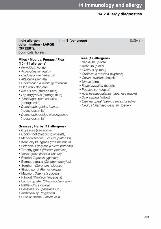
239
14 Immunology and allergy
14.2 Allergy diagnostics
ingle allergen determination - LARGE(GREER®): dogs, cats, horses.
1 ml S (per group) ELISA (1)
Mites / Moulds, Fungus / Flea (10 - 11 allergens)• Penicillium notatum• Aspergillus fumigatus• Cladosporium herbarum• Alternaria alternata• Cockcroach (Blatella germanica)• Flea (only dog/cat)• Acarus siro (storage mite)• Lepidoglyphus (storage mite)• Tyrophagus putrescentiae (storage mite)• Dermatophagoides farinae (house dust mite)• Dermatophagoides pteronyssinus (house dust mite)
Grasses / Herbs (12 allergens)• 6 grasses (see above)• Cock's foot (Dactylis glomerata) • Meadow fescue (Festuca pratensis) • Kentucky bluegrass (Poa pratensis) • Perennial Ryegrass (Lolium perenne) • Timothy grass (Phleum pratense) • Velvet grass (Holcus lanatus)• Redtop (Agrostis gigantea) • Bermuda grass (Cynodon dactylon)• Sorghum (Sorghum halpense)• Sheep sorrel (Rumex crispus)• Mugwort (Artemisia vulgaris)• Ribwort (Plantago lanceolata)• Lambs quarter (Chenopodium spp.) • Nettle (Urtica dioica)• Parietaria sp. (parietaria jud.)• Ambrosia sp. (ragweed)• Russian thistle (Salsola kali)
Trees (12 allergens)• Betula sp. (birch)• Alnus sp (alder)• Quercus sp (oak)• Cupressus avellana (cypress)• Corylus avellana (hazel)• Ulmus (elm)• Fagus sylvatica (beech)• Populus sp. (poplar)• Acer pseudoplatanus (japanese maple)• Salix caprea (willow)• Olea europea/ Fraxinus excelsior (olive)• Cedrus Chamaecyparis sp. (ceder)
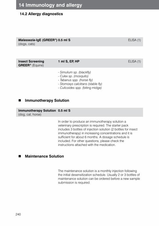
240
14 Immunology and allergy
14.2 Allergy diagnostics
Malassezia-IgE (GREER®)(dogs, cats)
0.5 ml S ELISA (1)
Insect Screening GREER® (Equine)
1 ml S, EP, HP ELISA (1)
- Simulium sp. (blackfly)- Culex sp. (mosquito)- Tabanus spp. (horse fly)- Stomoxys calcitrans (stable fly)- Culicoides spp. (biting midge)
Immunotherapy Solution
Immunotherapy Solution (dog, cat, horse)
0.5 ml S
In order to produce an immunotherapy solution a veterinary prescription is required. The starter pack includes 3 bottles of injection solution (2 bottles for insect immunotherapy) in increasing concentrations and it is sufficient for about 6 months. A dosage schedule is included. For other questions, please check the instructions attached with the medication.
Maintenance Solution
The maintenance solution is a monthly injection following the initial desensitization schedule. Usually 2 or 3 bottles of maintenance solution can be ordered before a new sample submission is required.
241
15.1 General advice on PCR
15 Molecular biology tests
PCR (Polymerase Chain Reaction)
The diagnostic advantage of PCR (polymerase chain reaction) is based on the principlethat specific segments of the various nucleic acids (DNA and RNA) contained in a sample may be increased (amplified), so that they become measurable or identifiable (e.g. sequenced). The amplified nucleic acid is usually derived from pathogen specific DNA or RNA, or in the case of hereditary diseases, from gene sections where changes(mutations) are located. For sex identification, the specific genome sequence of the male or female gene sequence is amplified and tested.
PCR techniquePCR runs in three reaction steps:In the first reaction step the sample DNA is heated to the high temperature (e.g. to 94 °C), which denaturates it and splits it into two complementary single strands.The temperature is reduced in the second reaction step, so that a specific, complementary oligonucleotide (primer) can attach to each single sample strand of DNA (template DNA) The region of the template DNA between the two primers is the region that will be amplified (duplicated). Specificity of the primer for detected genome segment will be based on similarity with sequence information saved in a databank (GenBank/EMBL database)The primers serve as contacts for the heat-stable DNA polymerase (e.g taq-polymerase).
In the third reaction step, the template primer is exposed to a high molar concentration of deoxyribonucleotide triphosphate (dNTP), aided by template-specific DNA Polymerase. This then produces two new (complete) DNA double strands.This amplified DNA serves as a template for further high concentrations of oligonucleotide primer.The cycle of denaturation, hybridization and amplification is repeated many times to gain a large number of identical copies of the original DNA segment.
Several modifications to the test protocol expand the range of use of PCR:- Amplification of RNA to detect RNA viruses or gene expression products- Increased specificity and sensitivity by using an additional specific primer pair in what
is called ‘nested PCR’ which is a multiplication of the original DNA/RNA by using proprietary methods with real time PCR
242
15.1 General advice on PCR
15 Molecular biology tests
Test interpretation in pathogen diagnosticsA positive PCR result indicates the presence of the target nucleic acid in the testedsample. However it is not possible to distinguish whether or not the pathogen of the nucleic acid is viable and able to multiply. Conventional PCR techniques also do not allow us to definitively define the amount of nucleic acid in the tube.Suitable methods for quantitative PCR are currently available at our laboratory for relevant parameters.Please note that due to the high sensitivity false positive PCR results are possible ifthe sample is even slightly contaminated with the target nucleic acid.
A negative PCR result indicates that at the time of sampling the target nucleic acid could not be amplified, either because there was no such nucleic acid present in the sample or the amount was insufficient to be amplified.
False negative results are possible when using unsuitable sample, e.g. sample material containing inhibitory substances (such as heparin) or inappropriate handling of the sample before or during transport (e.g. repeated freezing and thawing). However, inhibitory substances will be detected during PCR analysis and removed, or if not possible, commented on. Therefore, false negative PCR results due to inhibition can be avoided.
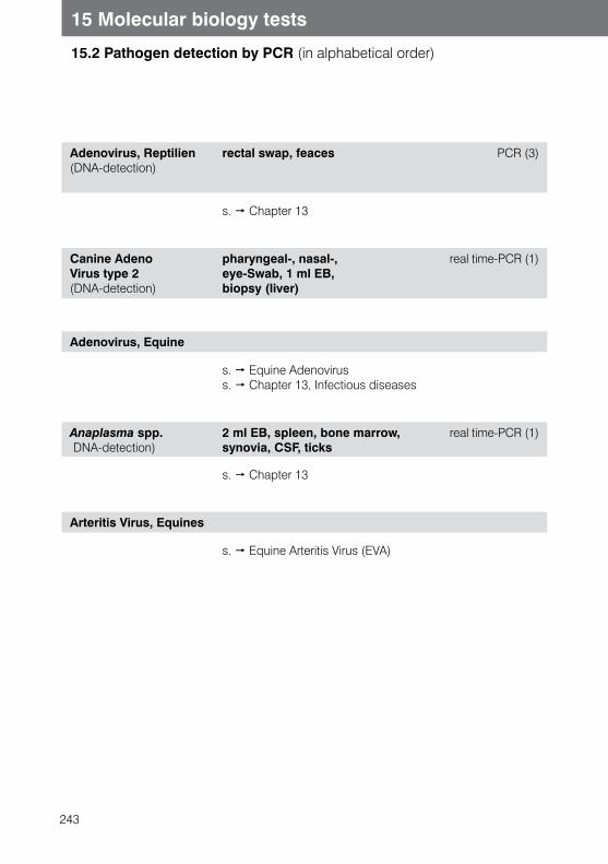
243
15 Molecular biology tests
15.2 Pathogen detection by PCR (in alphabetical order)
Adenovirus, Reptilien (DNA-detection)
rectal swap, feaces PCR (3)
s. Chapter 13
Canine Adeno Virus type 2 (DNA-detection)
pharyngeal-, nasal-, eye-Swab, 1 ml EB, biopsy (liver)
real time-PCR (1)
Adenovirus, Equine
s. Equine Adenovirus s. Chapter 13, Infectious diseases
Anaplasma spp. DNA-detection)
2 ml EB, spleen, bone marrow, synovia, CSF, ticks
real time-PCR (1)
s. Chapter 13
Arteritis Virus, Equines
s. Equine Arteritis Virus (EVA)
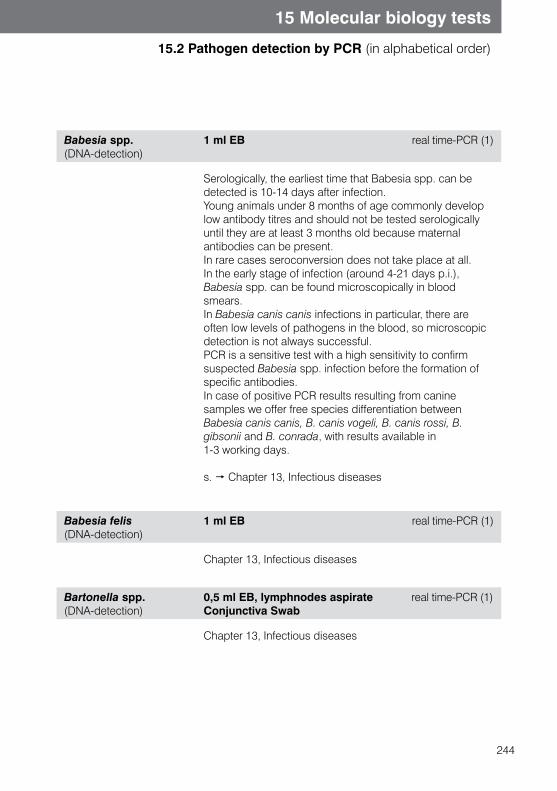
244
15 Molecular biology tests
15.2 Pathogen detection by PCR (in alphabetical order)
Babesia spp.(DNA-detection)
1 ml EB real time-PCR (1)
Serologically, the earliest time that Babesia spp. can be detected is 10-14 days after infection. Young animals under 8 months of age commonly develop low antibody titres and should not be tested serologically until they are at least 3 months old because maternal antibodies can be present.In rare cases seroconversion does not take place at all. In the early stage of infection (around 4-21 days p.i.), Babesia spp. can be found microscopically in blood smears. In Babesia canis canis infections in particular, there are often low levels of pathogens in the blood, so microscopic detection is not always successful.PCR is a sensitive test with a high sensitivity to confirm suspected Babesia spp. infection before the formation of specific antibodies.In case of positive PCR results resulting from canine samples we offer free species differentiation between Babesia canis canis, B. canis vogeli, B. canis rossi, B. gibsonii and B. conrada, with results available in 1-3 working days.
s. Chapter 13, Infectious diseases
Babesia felis (DNA-detection)
1 ml EB real time-PCR (1)
Chapter 13, Infectious diseases
Bartonella spp.(DNA-detection)
0,5 ml EB, lymphnodes aspirate real time-PCR (1) Conjunctiva Swab
Chapter 13, Infectious diseases
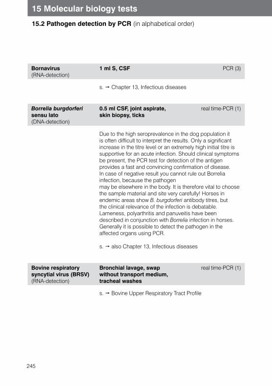
245
15 Molecular biology tests
15.2 Pathogen detection by PCR (in alphabetical order)
Bornavirus(RNA-detection)
1 ml S, CSF PCR (3)
s. Chapter 13, Infectious diseases
Borrelia burgdorferi sensu lato (DNA-detection)
0.5 ml CSF, joint aspirate, skin biopsy, ticks
real time-PCR (1)
Due to the high seroprevalence in the dog population it is often difficult to interpret the results. Only a significant increase in the titre level or an extremely high initial titre is supportive for an acute infection. Should clinical symptoms be present, the PCR test for detection of the antigen provides a fast and convincing confirmation of disease.In case of negative result you cannot rule out Borrelia infection, because the pathogenmay be elsewhere in the body. It is therefore vital to choose the sample material and site very carefully! Horses in endemic areas show B. burgdorferi antibody titres, but the clinical relevance of the infection is debatable. Lameness, polyarthritis and panuveitis have been described in conjunction with Borrelia infection in horses. Generally it is possible to detect the pathogen in the affected organs using PCR.
s. also Chapter 13, Infectious diseases
Bovine respiratory syncytial virus (BRSV)(RNA-detection)
Bronchial lavage, swap without transport medium, tracheal washes
real time-PCR (1)
s. Bovine Upper Respiratory Tract Profile
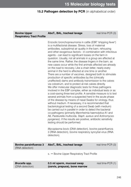
246
15 Molecular biology tests
15.2 Pathogen detection by PCR (in alphabetical order)
Bovine Upper Respiratory Tract Profile
AboT., BAL, tracheal lavage real time-PCR (3)
Enzootic bronchopneumonia in cattle (EBP, "shipping fever") is a multifactorial disease. Stress, loss of maternal antibodies, suboptimal air quality in the barn, rehousing and other exogenous factors - in combination with infectious agents - can lead to significant losses on the farm in question. Usually, not all animals in a group are affected at the same time. Rather, the disease lingers in the barn, as new cases occur while the first animals affected are already on the road to recovery. Like a chain letter, nearly every animal in the herd is affected at one time or another. There are a number of vaccines, designed both to stimulate production of specific antibodies by the (clinically unaffected) dams and antibody transmission to the calves via colostrum, and to protect at-risk calves directly. We offer molecular diagnostic tests for three pathogens involved in the EBP complex, either as individual tests or as a cost-saving three-test profile. A sensible measure is to test several animals from a suspected herd in the acute phase of the disease by means of nasal Swabs for virology, thus without medium. If necessary, it is recommended that bacteriological testing of a second Swab (with medium) be carried out in parallel in order to detect the bacterial co-pathogens (primarily Mannheimia haemolytica A1 and A6, Pasteurella multocida, Staph. aureus and Actinomyces pyogenes). If the results are positive, antibiotic sensitivity testing should be performed.
Mycoplasma bovis (DNA detection), bovine parainfluenza 3 (RNA detection), bovine respiratory syncytial virus (RNA detection).
Bovine parainfluenza 3 (RNA detection)
AboT., BAL, tracheal lavage real time-PCR (3)
s. Bovine Upper Respiratory Tract Profile
Brucella spp. (DNA-detection)
0.5 ml sperm, mucosal swap (cervix, prepuce), bone marrow
real time-PCR (1)
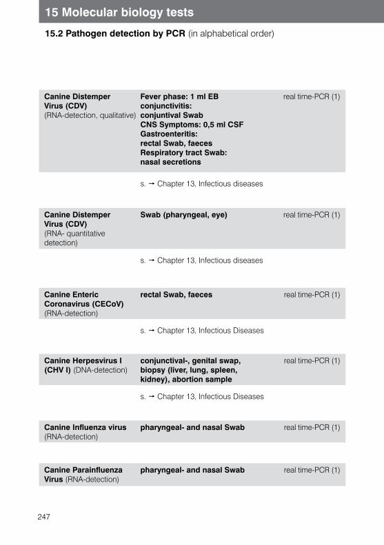
247
15 Molecular biology tests
15.2 Pathogen detection by PCR (in alphabetical order)
Canine Distemper Virus (CDV) (RNA-detection, qualitative)
Fever phase: 1 ml EB conjunctivitis: conjuntival Swab CNS Symptoms: 0,5 ml CSF Gastroenteritis: rectal Swab, faeces Respiratory tract Swab: nasal secretions
real time-PCR (1)
s. Chapter 13, Infectious diseases
Canine Distemper Virus (CDV) (RNA- quantitative detection)
Swab (pharyngeal, eye) real time-PCR (1)
s. Chapter 13, Infectious diseases
Canine EntericCoronavirus (CECoV)(RNA-detection)
rectal Swab, faeces real time-PCR (1)
s. Chapter 13, Infectious Diseases
Canine Herpesvirus I (CHV I) (DNA-detection)
conjunctival-, genital swap, biopsy (liver, lung, spleen, kidney), abortion sample
real time-PCR (1)
s. Chapter 13, Infectious Diseases
Canine Influenza virus (RNA-detection)
pharyngeal- and nasal Swab real time-PCR (1)
Canine Parainfluenza Virus (RNA-detection)
pharyngeal- and nasal Swab real time-PCR (1)
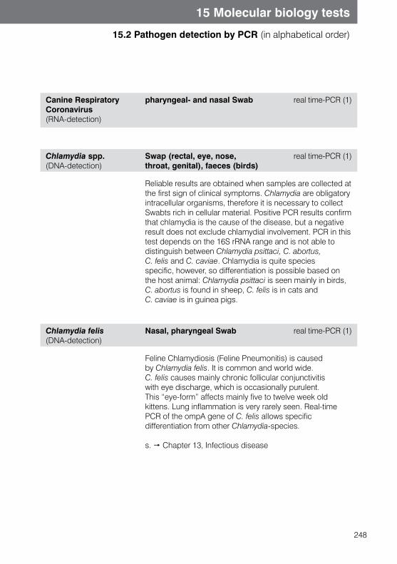
248
15 Molecular biology tests
15.2 Pathogen detection by PCR (in alphabetical order)
Canine Respiratory Coronavirus (RNA-detection)
pharyngeal- and nasal Swab real time-PCR (1)
Chlamydia spp. (DNA-detection)
Swap (rectal, eye, nose, throat, genital), faeces (birds)
real time-PCR (1)
Reliable results are obtained when samples are collected at the first sign of clinical symptoms. Chlamydia are obligatory intracellular organisms, therefore it is necessary to collect Swabts rich in cellular material. Positive PCR results confirm that chlamydia is the cause of the disease, but a negative result does not exclude chlamydial involvement. PCR in this test depends on the 16S rRNA range and is not able to distinguish between Chlamydia psittaci, C. abortus, C. felis and C. caviae. Chlamydia is quite species specific, however, so differentiation is possible based on the host animal: Chlamydia psittaci is seen mainly in birds, C. abortus is found in sheep, C. felis is in cats and C. caviae is in guinea pigs.
Chlamydia felis(DNA-detection)
Nasal, pharyngeal Swab real time-PCR (1)
Feline Chlamydiosis (Feline Pneumonitis) is caused by Chlamydia felis. It is common and world wide. C. felis causes mainly chronic follicular conjunctivitis with eye discharge, which is occasionally purulent. This “eye-form” affects mainly five to twelve week old kittens. Lung inflammation is very rarely seen. Real-time PCR of the ompA gene of C. felis allows specific differentiation from other Chlamydia-species.
s. Chapter 13, Infectious disease
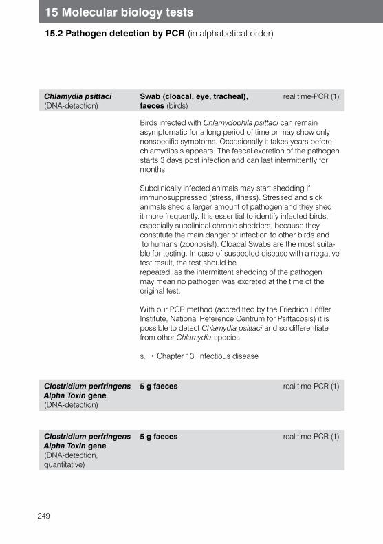
249
15 Molecular biology tests
15.2 Pathogen detection by PCR (in alphabetical order)
Chlamydia psittaci(DNA-detection)
Swab (cloacal, eye, tracheal), faeces (birds)
real time-PCR (1)
Birds infected with Chlamydophila psittaci can remain asymptomatic for a long period of time or may show only nonspecific symptoms. Occasionally it takes years before chlamydiosis appears. The faecal excretion of the pathogen starts 3 days post infection and can last intermittently for months.
Subclinically infected animals may start shedding if immunosuppressed (stress, illness). Stressed and sick animals shed a larger amount of pathogen and they shed it more frequently. It is essential to identify infected birds, especially subclinical chronic shedders, because they constitute the main danger of infection to other birds and to humans (zoonosis!). Cloacal Swabs are the most suita-ble for testing. In case of suspected disease with a negative test result, the test should berepeated, as the intermittent shedding of the pathogen may mean no pathogen was excreted at the time of the original test.
With our PCR method (accreditted by the Friedrich Löffler Institute, National Reference Centrum for Psittacosis) it is possible to detect Chlamydia psittaci and so differentiate from other Chlamydia-species.
s. Chapter 13, Infectious disease
Clostridium perfringens Alpha Toxin gene(DNA-detection)
5 g faeces real time-PCR (1)
Clostridium perfringens Alpha Toxin gene(DNA-detection, quantitative)
5 g faeces real time-PCR (1)
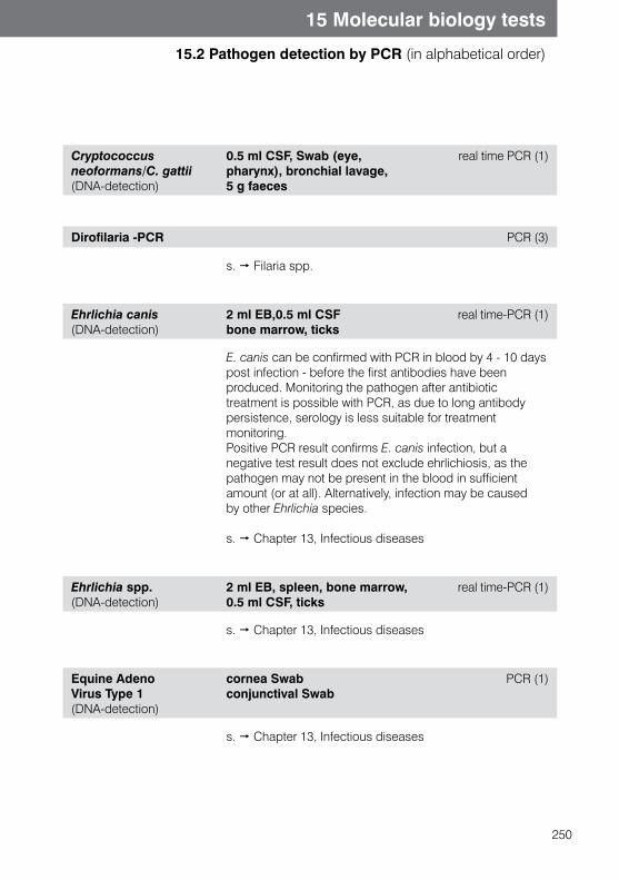
250
15 Molecular biology tests
15.2 Pathogen detection by PCR (in alphabetical order)
Cryptococcus neoformans/C. gattii (DNA-detection)
0.5 ml CSF, Swab (eye, pharynx), bronchial lavage, 5 g faeces
real time PCR (1)
Dirofilaria -PCR PCR (3)
s. Filaria spp.
Ehrlichia canis(DNA-detection)
2 ml EB,0.5 ml CSFbone marrow, ticks
real time-PCR (1)
E. canis can be confirmed with PCR in blood by 4 - 10 days post infection - before the first antibodies have been produced. Monitoring the pathogen after antibiotic treatment is possible with PCR, as due to long antibody persistence, serology is less suitable for treatment monitoring.Positive PCR result confirms E. canis infection, but a negative test result does not exclude ehrlichiosis, as the pathogen may not be present in the blood in sufficient amount (or at all). Alternatively, infection may be caused by other Ehrlichia species.
s. Chapter 13, Infectious diseases
Ehrlichia spp.(DNA-detection)
2 ml EB, spleen, bone marrow, 0.5 ml CSF, ticks
real time-PCR (1)
s. Chapter 13, Infectious diseases
Equine Adeno Virus Type 1 (DNA-detection)
cornea Swabconjunctival Swab
PCR (1)
s. Chapter 13, Infectious diseases
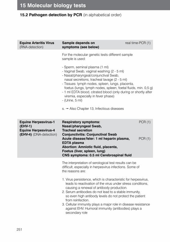
251
15 Molecular biology tests
15.2 Pathogen detection by PCR (in alphabetical order)
Equine Arteritis Virus (RNA-detection)
Sample depends on symptoms (see below)
real time-PCR (1)
For the molecular genetic tests different sample sample is used:
- Sperm, seminal plasma (1 ml) - Vaginal Swab, vaginal washing (2 - 5 ml) - Nasal/pharyngeal/conjunctival Swab, nasal secretions, tracheal lavage (2 - 5 ml) - Tissues: lymph nodes, spleen, lungs, placenta, foetus (lungs, lymph nodes, spleen, foetal fluids, min. 0,5 g) - 1 ml EDTA blood, citrated blood (only during or shortly after
viremia, especially in fever phase) - (Urine, 5 ml)
s. Also Chapter 13, Infectious diseases
Equine Herpesvirus-1(EHV-1)Equine Herpesvirus-4(EHV-4) (DNA-detection)
Respiratory symptoms: PCR (1) Nasal/pharyngeal Swab, Tracheal secretionConjunctivitis: Conjunctival SwabAcute disease/feler: 1 ml heparin plasma, PCR (1) EDTA plasmaAbortion: Amniotic fluid, placenta, Foetus (liver, spleen, lung) CNS symptoms: 0.5 ml Cerebrospinal fluid
The interpretation of serological test results can bedifficult, especially in herpesvirus infections. Some ofthe reasons are:
1. Virus persistence, which is characteristic for herpesvirus, leads to reactivation of the virus under stress conditions, causing a renewal of antibody production.
2. Serum antibodies do not lead to a stable immunity, so even high antibody levels do not protect the patient from reinfection.
3. Cellular immunity plays a major role in disease resistance against EHV. Humoral immunity (antibodies) plays a secondary role
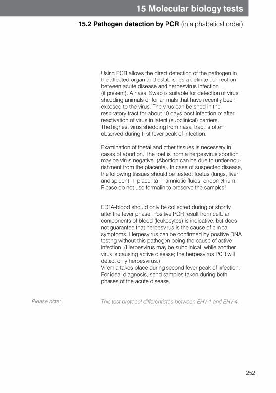
252
15 Molecular biology tests
15.2 Pathogen detection by PCR (in alphabetical order)
Using PCR allows the direct detection of the pathogen in the affected organ and establishes a definite connection between acute disease and herpesvirus infection (if present). A nasal Swab is suitable for detection of virus shedding animals or for animals that have recently been exposed to the virus. The virus can be shed in the respiratory tract for about 10 days post infection or after reactivation of virus in latent (subclinical) carriers. The highest virus shedding from nasal tract is often observed during first fever peak of infection.
Examination of foetal and other tissues is necessary in cases of abortion. The foetus from a herpesvirus abortion may be virus negative. (Abortion can be due to under-nou-rishment from the placenta). In case of suspected disease, the following tissues should be tested: foetus (lungs, liver and spleen) + placenta + amniotic fluids, endometrium. Please do not use formalin to preserve the samples!
Please note:
EDTA-blood should only be collected during or shortly after the fever phase. Positive PCR result from cellular components of blood (leukocytes) is indicative, but does not guarantee that herpesvirus is the cause of clinical symptoms. Herpesvirus can be confirmed by positive DNA testing without this pathogen being the cause of active infection. (Herpesvirus may be subclinical, while another virus is causing active disease; the herpesvirus PCR will detect only herpesvirus.)Viremia takes place during second fever peak of infection. For ideal diagnosis, send samples taken during both phases of the acute disease.
This test protocol differentiates between EHV-1 and EHV-4.
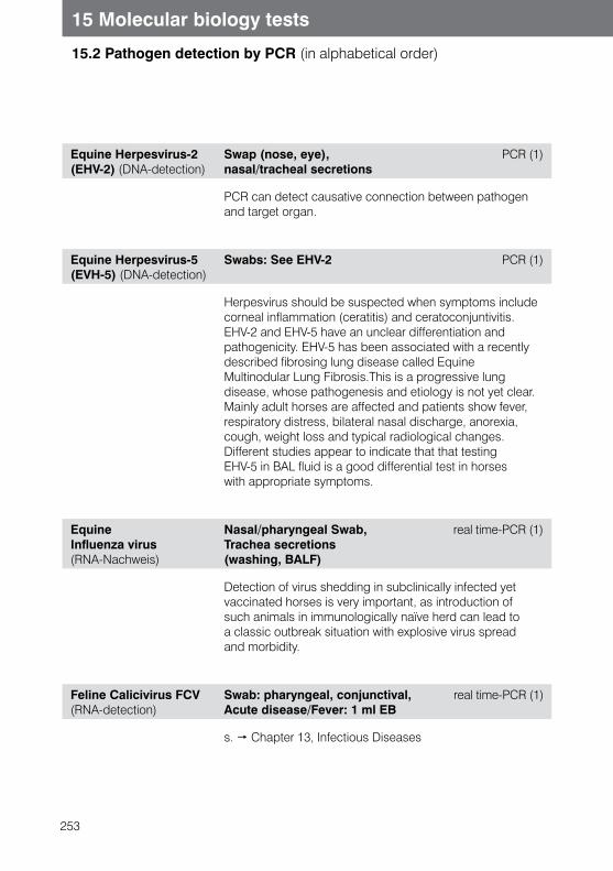
253
15 Molecular biology tests
15.2 Pathogen detection by PCR (in alphabetical order)
Equine Herpesvirus-2(EHV-2) (DNA-detection)
Swap (nose, eye), nasal/tracheal secretions
PCR (1)
PCR can detect causative connection between pathogen and target organ.
Equine Herpesvirus-5(EVH-5) (DNA-detection)
Swabs: See EHV-2 PCR (1)
Herpesvirus should be suspected when symptoms include corneal inflammation (ceratitis) and ceratoconjuntivitis. EHV-2 and EHV-5 have an unclear differentiation and pathogenicity. EHV-5 has been associated with a recently described fibrosing lung disease called Equine Multinodular Lung Fibrosis.This is a progressive lung disease, whose pathogenesis and etiology is not yet clear.Mainly adult horses are affected and patients show fever, respiratory distress, bilateral nasal discharge, anorexia, cough, weight loss and typical radiological changes. Different studies appear to indicate that that testing EHV-5 in BAL fluid is a good differential test in horses with appropriate symptoms.
EquineInfluenza virus (RNA-Nachweis)
Nasal/pharyngeal Swab, Trachea secretions (washing, BALF)
real time-PCR (1)
Detection of virus shedding in subclinically infected yet vaccinated horses is very important, as introduction of such animals in immunologically naïve herd can lead to a classic outbreak situation with explosive virus spread and morbidity.
Feline Calicivirus FCV (RNA-detection)
Swab: pharyngeal, conjunctival, real time-PCR (1) Acute disease/Fever: 1 ml EB
s. Chapter 13, Infectious Diseases
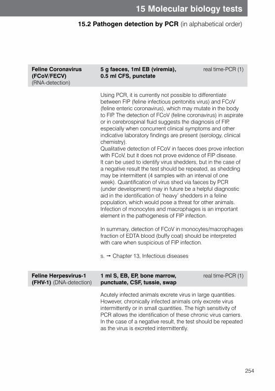
254
15 Molecular biology tests
15.2 Pathogen detection by PCR (in alphabetical order)
Feline Coronavirus (FCoV/FECV)(RNA-detection)
5 g faeces, 1ml EB (viremia), 0.5 ml CFS, punctate
real time-PCR (1)
Using PCR, it is currently not possible to differentiate between FIP (feline infectious peritonitis virus) and FCoV (feline enteric coronavirus), which may mutate in the bodyto FIP. The detection of FCoV (feline coronavirus) in aspirate or in cerebrospinal fluid suggests the diagnosis of FIP, especially when concurrent clinical symptoms and other indicative laboratory findings are present (serology, clinical chemistry). Qualitative detection of FCoV in faeces does prove infection with FCoV, but it does not prove evidence of FIP disease. It can be used to identify virus shedders, but in the case of a negative result the test should be repeated, as shedding may be intermittent (4 samples with an interval of one week). Quantification of virus shed via faeces by PCR (under development) may in future be a helpful diagnostic aid in the identification of ‘heavy’ shedders in a feline population, which would pose a threat for other animals. Infection of monocytes and macrophages is an important element in the pathogenesis of FIP infection.
In summary, detection of FCoV in monocytes/macrophages fraction of EDTA blood (buffy coat) should be interpreted with care when suspicious of FIP infection.
s. Chapter 13, Infectious diseases
Feline Herpesvirus-1(FHV-1) (DNA-detection)
1 ml S, EB, EP, bone marrow, punctuate, CSF, tussie, swap
real time-PCR (1)
Acutely infected animals excrete virus in large quantities. However, chronically infected animals only excrete virus intermittently or in small quantities. The high sensitivity of PCR allows the identification of these chronic virus carriers. In the case of a negative result, the test should be repeated as the virus is excreted intermittently.
255
15 Molecular biology tests
15.2 Pathogen detection by PCR (in alphabetical order)
FIV Feline Immunodeficiency Virus (Progenome-DNA and virus RNA detection)
1 ml EB, bone marrow aspirate, CSF, tissues, Swab
real time-PCR (1)
Our real-time PCR system detects genetic FIV DNA (progenome) and also RNA free replicating FIV. Positive FIV PCR result confirms infection of the tested cat with FIV, based on the high specificity of the test system (99,9%). However a negative PCR result cannot exclude an infection. This is due to FIV having many different subtypes, a high number of genetic mutations with possibly decreased genomic integration rate, but also because of the replication rate of FIV, PCR analysis can lead to false negative results in infected cats. Our tests are being continuously improved in order to minimize these false negative results.
PCR may be used to clarify doubtful positive or negative serological results:
- Infected animals may show false negative serological results in the early stage of disease; antibodies are usually detectable after 2-4 weeks post infection, but in some animals they appear much later, and in end stage disease, the antibody titer drops under detection limits.
- In kittens, interference with maternal antibodies (up to 6 months of age) can lead to positive serological results, but infection cannot be excluded.
s. Chapter 13, Infectious diseases
256
15 Molecular biology tests
15.2 Pathogen detection by PCR (in alphabetical order)
FeLV Feline Leukemia Virus (DNA and RNA detection)
1 ml EB or bone marrow
real time-PCR (1)
Please note:
Using real-time PCR, viral DNA integrated in the host geno-me (called progenome or provirus) can be detected. This detection is used for diagnosis of latent infections. Thanks to its high specificity, PCR can be used to clarify inconclusi-ve results and confirm infection. The sensitivity of PCR is highly dependent on the number of infected cells (provirus load). Therefore negative result does not rule out an infection.
This test provides no information on virus replication ability.
s. Chapter 13, Infectious diseases
Filaria spp. (DNA-detection)
1 ml EB PCR (3)
Using this PCR, Microfilaria that have been detected in filtration test or blood smear can be further differentiated. This way it can be detected if they are pathogenic or non-pathogenic filarias and optimal treatment can be chosen.
This test can differentiate several adult Filaria species, which can originate from subcutaneous lymph nodes (Dirofilaria repens or Acanthocheilonema reconditum), from the heart (Dirofilaria immitis) or peritoneal cavity (Dipetalonema dracunculoides).
For each sample a single PCR for Dirofilaria immitis and Dirofilaria repens, and a 6-species PCR panel will be performed. The panel contains four further (rare) species of Microilaria. (Acanthocheilonema reconditum, D. dracunculoide, Brugia malayi and B. pahangi).
257
15 Molecular biology tests
15.2 Pathogen detection by PCR (in alphabetical order)
Gastroenteritis Virus (TGEV) (RNA-detection)
Swab, intestinal mucosal membrane
s. Chapter 13, Infectious diseases
Haemobartonella felis
s. Feline Haemotropic Mycoplasma Profile: Mycoplasma haemofelis, Candidatus M. haemominutum, Candidatus Mycoplasma turicensis
Helicobacter spp. (DNA-detection, Many species)
Stomach biopsy, faeces PCR (1)
s. Chapter 13, Infectious diseases
Hepatozoon canis (DNA-detection)
1 ml EB, ticks real time-PCR (1)
Hepatozoonosis in dogs is a disease which is caused by Hepatozoon canis. Transmission of Protozoa is by the ingestion of infected brown dog ticks (Rhipizephalus sanguineus), or by vertical transmission of parasites from the mother to the puppies.(Tick bite does not lead to infection.) Hepatozoon canis is widespread in Southeast Europe e.g. Italy, Spain, Southern France, in the Middle and Far East, Asia Africa, South America and the US.In many cases Hepatozoon canis infection is found with Babesiosis, Ehrlichiosis, Leishmaniasis or Dirofilariasis.
258
15 Molecular biology tests
15.2 Pathogen detection by PCR (in alphabetical order)
Herpesvirus (Tortoise) (DNA-detection)
swap, oral cavity (wetted with NaCl)
PCR (3)
s. Chapter 13, Infectious diseases
Herpesvirus-Infektion (Koi fish) (DNA-detection)
EB,HB,gills Swab in isopropanol, gill biopsy, organ probes in isopropanol. Send cooled.
PCR (3)
s. Chapter 10.1
Iridovirus (Reptiles) (DNA-detection)
mouth Swab without medium PCR (3)
Lawsonia intracellulis (DNA-detection)
faeces real-time PCR (1)

259
15 Molecular biology tests
15.2 Pathogen detection by PCR (in alphabetical order)
Leishmania spp.(DNA-detection, quantitative)
Bone marrow, EB, Lymhnodes aspirate, eye Swab, urine, skin biopsy
real time-PCR (1)
This PCR test is highly precise and suited for samples from lymph node aspirate or bone marrow. The advantage of pathogen detection is that you can identify animals withsubclinical disease, as the antibody level can oftenbe below the detection limit. Using real time-PCR it is possible to precisely quantify the number of Leishmania in the test sample.
Knowing the parasite concentration allows exact estimation of the infectious status in cases where:- ELISA-results were not convincing- Dogs show clinical signs, but do not show seroconversion- Dogs do not show clinical signs, but originate from
endemic regions.
Studies have shown that dogs with moderate to high concentration of Leishmania in bone marrow or blood are sick, or are very likely to be develop active clinical infection. Leishmania-quantifying is a highly useful method for treat-ment monitoring (beginning: one month after beginning of treatment).
s. Chapter 13, Infectious diseases.
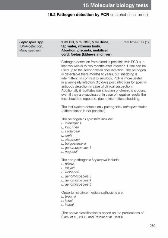
260
15 Molecular biology tests
15.2 Pathogen detection by PCR (in alphabetical order)
Leptospira spp.(DNA-detection, Many species)
2 ml EB, 5 ml CSF, 5 ml Urine, tap water, vitreous body, Abortion: placenta, umbilical cord, foetus (kidneys and liver)
real time-PCR (1)
Pathogen detection from blood is possible with PCR is in first two weeks to two months after infection. Urine can be used up to the second week post infection. The pathogen is detectable there months to years, but shedding is intermittent. In contrast to serology, PCR is more useful in a very early infection (10 days post infection) for specific antibody detection in case of clinical suspection. Additionally it facilitates identification of chronic shedders, even if they are vaccinated. In case of negative results the test should be repeated, due to intermittent shedding.
The test system detects only pathogenic Leptospira strains (differentiation is not possible):
The pathogenic Leptospira include:L. interrogansL. kirschneriL. santarosaiL. weiliiL. alexanderiL. borgpeterseniiL. genomospecies 1L. noguchii
The non-pathogenic Leptospira include:L. biflexaL. meyeriL. wolbachiiL. genomospecies 3L. genomospecies 4L. genomospecies 5
Opportunistic/intermediate pathogens are:L. broomiiL. faineiL. inadai
(The above classification is based on the publications of Slack et al., 2006, and Perolat et al., 1998).
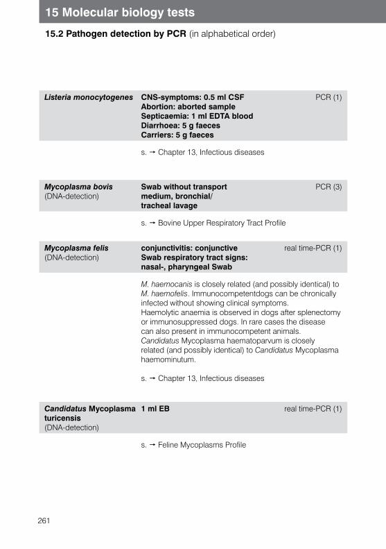
261
15 Molecular biology tests
15.2 Pathogen detection by PCR (in alphabetical order)
Listeria monocytogenes CNS-symptoms: 0.5 ml CSF Abortion: aborted sampleSepticaemia: 1 ml EDTA bloodDiarrhoea: 5 g faecesCarriers: 5 g faeces
PCR (1)
s. Chapter 13, Infectious diseases
Mycoplasma bovis (DNA-detection)
Swab without transport medium, bronchial/ tracheal lavage
PCR (3)
s. Bovine Upper Respiratory Tract Profile
Mycoplasma felis (DNA-detection)
conjunctivitis: conjunctive Swab respiratory tract signs: nasal-, pharyngeal Swab
real time-PCR (1)
M. haemocanis is closely related (and possibly identical) to M. haemofelis. Immunocompetentdogs can be chronically infected without showing clinical symptoms.Haemolytic anaemia is observed in dogs after splenectomy or immunosuppressed dogs. In rare cases the disease can also present in immunocompetent animals. Candidatus Mycoplasma haematoparvum is closely related (and possibly identical) to Candidatus Mycoplasma haemominutum.
s. Chapter 13, Infectious diseases
Candidatus Mycoplasma turicensis (DNA-detection)
1 ml EB real time-PCR (1)
s. Feline Mycoplasms Profile
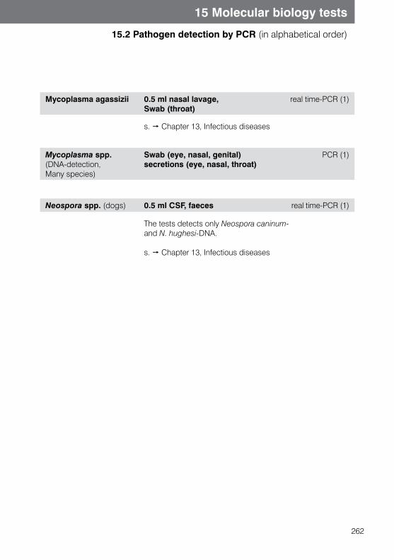
262
15 Molecular biology tests
15.2 Pathogen detection by PCR (in alphabetical order)
Mycoplasma agassizii 0.5 ml nasal lavage, Swab (throat)
real time-PCR (1)
s. Chapter 13, Infectious diseases
Mycoplasma spp. (DNA-detection, Many species)
Swab (eye, nasal, genital) secretions (eye, nasal, throat)
PCR (1)
Neospora spp. (dogs) 0.5 ml CSF, faeces real time-PCR (1)
The tests detects only Neospora caninum- and N. hughesi-DNA.
s. Chapter 13, Infectious diseases
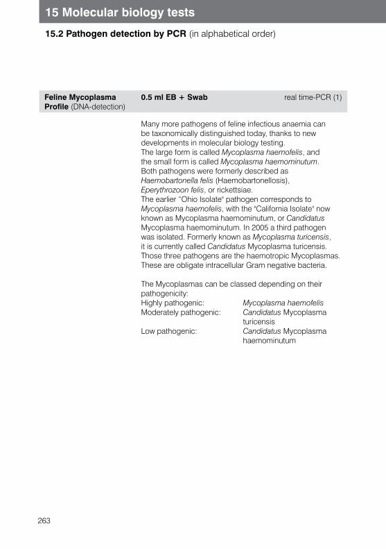
263
15 Molecular biology tests
15.2 Pathogen detection by PCR (in alphabetical order)
Feline Mycoplasma Profile (DNA-detection)
0.5 ml EB + Swab real time-PCR (1)
Many more pathogens of feline infectious anaemia can be taxonomically distinguished today, thanks to new developments in molecular biology testing.The large form is called Mycoplasma haemofelis, and the small form is called Mycoplasma haemominutum. Both pathogens were formerly described as Haemobartonella felis (Haemobartonellosis), Eperythrozoon felis, or rickettsiae.The earlier “Ohio Isolate" pathogen corresponds to Mycoplasma haemofelis, with the "California Isolate" now known as Mycoplasma haemominutum, or Candidatus Mycoplasma haemominutum. In 2005 a third pathogen was isolated. Formerly known as Mycoplasma turicensis, it is currently called Candidatus Mycoplasma turicensis.Those three pathogens are the haemotropic Mycoplasmas. These are obligate intracellular Gram negative bacteria.
The Mycoplasmas can be classed depending on their pathogenicity:Highly pathogenic: Mycoplasma haemofelis Moderately pathogenic: Candidatus Mycoplasma turicensis Low pathogenic: Candidatus Mycoplasma haemominutum
264
15 Molecular biology tests
15.2 Pathogen detection by PCR (in alphabetical order)
Parvovirus 2, canine CPV-2 (DNA) - PCR; includes the types CPV-2, CPV-2a, CPV-2b and CPV-2c
5 g faeces, rectal Swab, tissue, (1 ml EB),
real time-PCR (1)
Direct pathogen detection from faeces or rectal Swab by PCR is possible in dogs and cats. It is important to note the animal species on the laboratory form. In dogs differentiation between vaccine strain CPV2 and wild strain CPV 2a/2b can be performed, if the animal was vaccinated with attenuated CPV type 2b in the 2 to 3 weeks before sampling. (Virbagen Puppy 2b, Quantum DA2pi/CvL and Duramune are common vaccines in Germany.) For Vaccine virus can be shed 2-12 days after vaccination. Shedding of field virus begins 3-4 days post infection and usually lasts 7-10 days. In some cases longer shedding is possible. A negative PCR result does not exclude infection.
Parvovirus 2 differentiation in CPV-2 and CPV-2a/b (DNA detection)
if CPV-2 detection is positive real-time PCR
Parvovirus, feline FPV (DNA-detection)
5 g faeces, rectal Swab PCR (1)
265
15 Molecular biology tests
15.2 Pathogen detection by PCR (in alphabetical order)
PBFD-Virus(DNA-detection)
Diarrhoea: cloacal Swab, faeces PCR (1) Feather deformations: damaged feathers Post mortem: kidneys, spleen, liver Chronic form: 0.1 - 0.5 ml EB, feathers
The pathogen causing PBFD (psittacine beak and feather disease) is a circovirus. Initial virus replication occurs in lymphatic tissue, gastrointestinal tract and liver, but the target organ is the epidermis.The acute form mainly affects hatchlings. Patients show diarrhoea, occasionally hepatitis and but a particular feature of disease is deformed feathers. Many young birds overcome the acute disease and develop a chronic infection. The chronic form is characterised by growth of deformed feathers after moulting and changes to the beak.Latently infected animals and animals in the incubation period pose the biggest threat for introducing the virus to a population. PCR is the method of choice to identify these animals. A positive PCR test is not proof of active infection, as inactive virus DNA is detectable in the blood for up to three months after exposure. Because of this, PCR positive animals that do not show clinical symptoms should be isolated and re-tested after three months. If they are still positive after three months, they must be considered chronically infected, posing a threat to other birds.
Polyomavirus, avian(BFD-Virus) (DNA-detection)
Diarrhoea: cloacal Swab, faeces PCR (1) Feather deformation: affected feathers Post mortem: kidneys, spleen, liverChronic form: 0.1 ml EB, feathers
Vertical spread and virus spread by clinically inapparent animals are the main means of transmission of BFD (budge-rigar fledgling disease). Carriers can be identified by testing several cloacal Swabs using PCR, in three month intervals, to be able to detect even intermittently shedding animals.When feather deformities are present, clinical suspicion can be confirmed using PCR on deformed feathers. The liver, spleen and kidneys can be used from young birds that have died of acute disease.
266
15 Molecular biology tests
15.2 Pathogen detection by PCR (in alphabetical order)
Porcine Circovirus 2 (PCV-2) (DNA-detection)
Lymph node, tissue, PCR (3)Nasal Swab
Porcine Circovirus 2 has only recently been described (1998, Canada). (Porcine Circovirus PCV-1 has been known for a long time and is not pathogenic. PCV-2 causes different symptoms in weaned and fattening pigs (e.g. dyspnoea, lymph node oedema, icterus, diarrhoea) and is also known as PMWS (Postweaning Multisystemic Wasting Syndrome). Clinical disease has only been found in combination with other infections (PRRS, PPV).The other syndrome which may be associated with PCV-2 is PDNS (Porcine Dermatitis and Nephropathy Syndrome). PDNS is an immune complex disease, but is not currently well understood.Not much is known about virus shedding. Experimentally the virus can be detected in eye secretions, saliva and faeces. Transplacenta transmission is possible, but does not play a large role in spread. The virus shows affinity to lymph tissue and causes immunosuppression, which leads to secondary infections. Stomach problems can favour outbreak of the syndrome, and mortality can reach over 80%. Subclinical infections are possible.
Ranavirus (Reptiles) (DNA-detection)
Swab (throat) PCR (3)
Ranaviruses belong to the family Iridoviridae. They are detected in frogs, turtles, tortoises and snakes. Affected animals can display respiratory symptoms (conjunctivitis, stomatitis, pneumonia) and gastrointestinal symptoms (diarrhoea, anorexia). The differential diagnosis should include herpesvirus infection. Transmission appears to be horizontal. Direct detection is by throat Swab.
267
15 Molecular biology tests
15.2 Pathogen detection by PCR (in alphabetical order)
Rhodococcus equi(DNA-detection)
Tracheal secretion (-lavage, BALF), Synovial membrane, tissues (lungs), faeces
real time-PCR (1)
Please note:
R. equi is the most frequent pneumonia pathogen in foals aged between one and six months. It is a Gram positive facultative intracellular pathogen, which can survive in high temperatures and dry environment. Spread is via inhalation of contaminated dust or coprophagy (faecal ingestion). R. equi strains with virus plasmid VapA are the most frequent cause of clinical disease. Symptoms are characterized by acute, subacute or chronic bronchopneumonia. Extrapulmonary forms can occur through internal spread of pathogen, as mesenteric lymphadenopathy, ulcerative colitis (diarrhoea), septic polyarthritis and osteomyelitis. Severe or untreated disease may be fatal.
Pathogen detection from nasal sample (nasal-, pharyngeal Swab) is rarely successful!
268
15 Molecular biology tests
15.2 Pathogen detection by PCR (in alphabetical order)
Toxoplasma gondii (DNA-detection)
CNS symptoms: 0.5 ml CSF real time-PCR (1)Abortion (dog, small ruminants): Vaginal smear, Placenta, Foetal (liver, spleen, lung, heart, gut)Respiratory symptoms: Bronchial washOcular symptoms (esp. cats): Aqueous humour Pyrexia: 0.5 ml EB
The high prevalence of antibodies against toxoplasma in dogs and cats restricts the use of serology in the diagnosis. Only high IgM titers (in combination with low IgG titers) gives a clear indication for acute infection, which may be accompanied by faecal oocyst shedding in cats. Most infected animals remain seropositive (IgG) at high levels for several years or for life, which can even make it difficult to judge paired serum samples.Please note that a positive PCR test does not prove acute infection with Toxoplasma gondii. The pathogen has been isolated from cerebrospinal fluid and aqueous humour inclinically healthy animals. The PCR test cannot currently be performed on faeces, but other sample material, based on clinical symptoms, can be used to detect infection. Normal chemical preparations cannot extract the DNA.In order to absolutely rule out the shedding of oocysts in all circumstances (for example if the owner is pregnant or immune compromised) classical methods such as serology and microscopic faecal examination must be performed. This is because despite the high sensitivity, false negative results can occur.
s. Chapter 13, Infectious diseases
269
15 Molecular biology tests
15.2 Pathogen detection by PCR (in alphabetical order)
Tickborne Encephalitisvirus(RNA-detection)
ticks, 0.5 ml CSF PCR (1)
This test is used to investigate CNS symptoms in animals from regions endemic for tickborne encephalitis pathogen, in association with or as confirmation for serological CSF testing. This test is also capable of detecting the virus directly in ticks.
Tritrichomonas foetus (DNA-detection) )
2 g faeces real time-PCR (1)
Tritrichomonas foetus (syn.: Tritrichomonas suis) is transmitted via mating, and occasionally infected semen. It can be an important factor in fertility disorders and abortions in cattle. T. foetus is not strictly host specific: cattle, swine, and cats may all be infected. The prevalence of T. foetus in cats is very high - a figure of 34% was reported by Gookin et al. 2004. In cats trichomonads stay in the colon and can cause diarrhoea. Also, according to a recent study in dogs, T. foetus will be confirmed in diarrhea cases. (Gookin et al., 2005).
s. Chapter 13, Infectious diseases
270
15 Molecular biology tests
15.3 Hereditary Diseases
General Information on Hereditary Diseases
Hereditary diseases are due to genetic mutations. The mutation can be passed on from parents to offspring.
Fundamental genetics
Sexual reproduction means that the offspring receives a double set of chromosomes. One set of chromosomes originates from the mother and father respectively. Therefore each gene is found in duplicate, meaning as two alleles.- If both alleles carry the same characteristic or the same defect,
the individual is homozygotic for that defect- If only one of the alleles carries the characteristic or the defect, the individual is
heterozygotic. The phenotype of the individual is the result of the gene expression. For hereditary disease this implies:
- Dominant genes will be expressed even if only one of the two alleles is affected by the defect. Thus, the hereditary disease will be expressed in the offspring if the mother or the father passes on the mutant gene.
- Recessive genes are only expressed when both alleles are affected by the defect. This means that both mother and father must both pass the mutant gene to the offspring. If an animal carries only one allele for the recessive hereditary disease, it will not express the disease all its life. But it is a carrier and can pass on the gene to its offspring. If two recessive carriers are matched, the offspring may receive two defective alleles and subsequently express the disease.
- X-chromosomal gene expression: male animals will express the hereditary disease, because the defect is located on the gene that is responsible for the x-chromosome.Female individuals can be carriers, or if they are homozygotic, they may also express the hereditary disease.
- Autosomal gene expression: the responsible gene is not located on the x-chromosome, therefore the hereditary disease can be expressed by both male and female individuals and they can both pass it on to their offspring.
271
15 Molecular biology tests
15.3 Hereditary Diseases
Molecular Genetic Testing for Hereditary Diseases
Molecular genetic testing can be performed at an early age, before the expression of hereditary disease occurs. It is suitable for identifying carriers of genetic defects. These animals can then be excluded from breeding.PCR is used for hereditary disease testing. The DNA which contains the possible mutantgene is amplified and the sequence is compared to a similar sequence from a geneti-cally sound animal.
The following results are possible:1. The animal is genetically sound with respect to the disease tested. Neither of the two
alleles carries the mutation in question. The animal cannot express the hereditary disease, nor can it pass it on.
2. The animal is heterozygous in respect of the mutation in question. It carries a mutant gene from either the mother or the father. Autosomal dominant gene expression will lead to phenotypic expression of the gene: the animal will express the hereditary disease. Autosomal recessive gene expression will not lead to an expression of the disease. In both cases, the animal can pass on the genetic defect to its offspring.
3. The animal is homozygous in respect of the genetic defect in question. Both alleles are mutants. The animal will express the hereditary disease and it will pass on the genetic defect to its offspring.
BLAD (Bovine leukocyte adhesion deficiency)
1 ml EB PCR (3)
Occurrence:
Symptoms:
Laboratory findings:
Inheritance:
BLAD (Bovine leukocyte adhesion deficiency) leads to fatalimmunodeficiency in calves and young cattle.
Holstein-Frisian cattle.
- Recurrent infections of the respiratory and gastrointesti-naltract, as well as the nasopharyngeal region
- Low birth weight- Delayed wound healing, necrosis, gangrene
Leukocytosis
Autosomal recessive
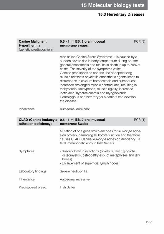
272
15 Molecular biology tests
15.3 Hereditary Diseases
Canine Malignant Hyperthermia (genetic predisposition)
0.5 - 1 ml EB, 2 oral mucosal membrane swaps
PCR (3)
Inheritance:
Also called Canine Stress Syndrome. It is caused by a sudden severe rise in body temperature during or after general anaesthesia and results in death in up to 70% of cases. The severity of the symptoms varies. Genetic predisposition and the use of depolarizing muscle relaxants or volatile anaesthetic agents leads to disturbance in calcium homeostasis and subsequent increased prolonged muscle contractions, resulting in tachycardia, tachypnoea, muscle rigidity, increased lactic acid, hypercalcaemia and myoglobinuria.Homozygous and heterozygous carriers can develop the disease.
Autosomal dominant
CLAD (Canine leukocyte adhesion deficiency)
0.5 - 1 ml EB, 2 oral mucosal membrane Swabs
PCR (1)
Symptoms:
Laboratory findings:
Inheritance:
Predisposed breed:
Mutation of one gene which encodes for leukocyte adhe-sion protein, damaging leukocyte function and therefore causes CLAD (Canine leukocyte adhesion deficiency), a fatal immunodeficiency in Irish Setters.
- Susceptibility to infections (phlebitis, fever, gingivitis, osteomyelitis, osteopathy esp. of metaphyses and jaw bones).
- Enlargement of superficial lymph nodes
Severe neutrophilia
Autosomal recessive
Irish Setter
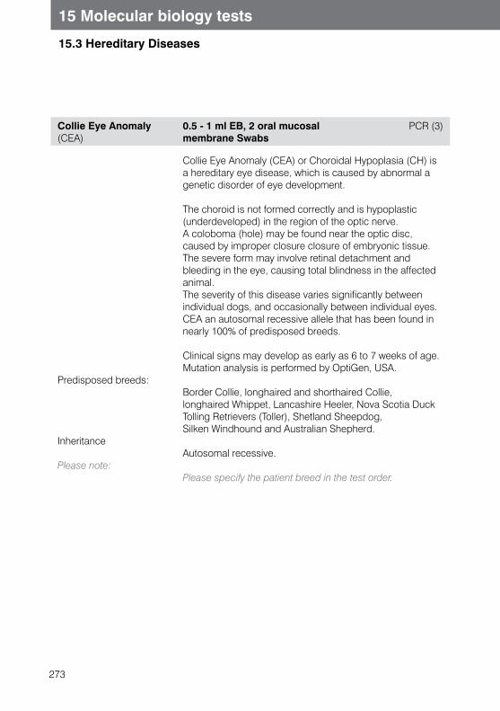
273
15 Molecular biology tests
15.3 Hereditary Diseases
Collie Eye Anomaly (CEA)
0.5 - 1 ml EB, 2 oral mucosal membrane Swabs
PCR (3)
Predisposed breeds:
Inheritance
Please note:
Collie Eye Anomaly (CEA) or Choroidal Hypoplasia (CH) is a hereditary eye disease, which is caused by abnormal a genetic disorder of eye development.
The choroid is not formed correctly and is hypoplastic (underdeveloped) in the region of the optic nerve. A coloboma (hole) may be found near the optic disc, caused by improper closure closure of embryonic tissue. The severe form may involve retinal detachment and bleeding in the eye, causing total blindness in the affected animal.The severity of this disease varies significantly between individual dogs, and occasionally between individual eyes. CEA an autosomal recessive allele that has been found in nearly 100% of predisposed breeds.
Clinical signs may develop as early as 6 to 7 weeks of age.Mutation analysis is performed by OptiGen, USA.
Border Collie, longhaired and shorthaired Collie, longhaired Whippet, Lancashire Heeler, Nova Scotia Duck Tolling Retrievers (Toller), Shetland Sheepdog, Silken Windhound and Australian Shepherd.
Autosomal recessive.
Please specify the patient breed in the test order.
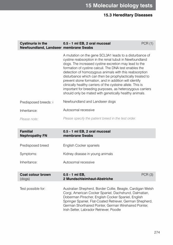
274
15 Molecular biology tests
15.3 Hereditary Diseases
Cystinuria in the Newfoundland, Landseer
0.5 - 1 ml EB, 2 oral mucosal membrane Swabs
PCR (1)
Predisposed breeds: i
Inheritance:
Please note:
A mutation on the gene SCL3A1 leads to a disturbance of cystine reabsorption in the renal tubuli in Newfoundland dogs. The increased cystine excretion may lead to the formation of cystine calculi. The DNA test enables the detection of homozygous animals with this reabsorptiondisturbance which can then be prophylactically treated to prevent stone formation, and in addition will identify clinically healthy carriers of the cysteine allele. This is important for breeding purposes, as heterozygous carriers should only be mated with genetically healthy animals.
Newfoundland and Landseer dogs
Autosomal recessive
Please specify the patient breed in the test order.
Familial Nephropathy FN
0.5 - 1 ml EB, 2 oral mucosal membrane Swabs
Predisposed breed
Symptoms:
Inheritance:
English Cocker spaniels
Kidney disease in young animals
Autosomal recessive
Coat colour brown (dogs)
0.5 - 1 ml EB, 2 Mundschleimhaut-Abstriche
PCR (3)
Test possible for: Australian Shepherd, Border Collie, Beagle, Cardigan Welsh Corgi, American Cocker Spaniel, Dachshund, Dalmatian, Doberman Pinscher, English Cocker Spaniel, English Springer Spaniel, Flat-Coated Retriever, German Shepherd, German Shorthaired Pointer, German Wirehaired Pointer, Irish Setter, Labrador Retriever, Poodle
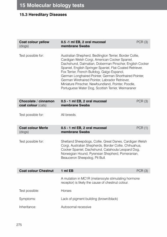
275
15 Molecular biology tests
15.3 Hereditary Diseases
Coat colour yellow(dogs)
0.5 -1 ml EB, 2 oral mucosal membrane Swabs
PCR (3)
Test possible for: Australian Shepherd, Bedlington Terrier, Border Collie, Cardigan Welsh Corgi, American Cocker Spaniel, Dachshund, Dalmatian, Doberman Pinscher, English Cocker Spaniel, English Springer Spaniel, Flat-Coated Retriever, Fox Terrier, French Bulldog, Galgo Espanol, German Longhaired Pointer, German Shorthaired Pointer, German Wirehaired Pointer, Labrador Retriever, Miniature Pinscher, Newfoundland, Pointer, Poodle, Portuguese Water Dog, Scottish Terrier, Weimaraner
Chocolate / cinnamon coat colour (cats)
0.5 - 1 ml EB, 2 oral mucosal membrane Swabs
PCR (3)
Test possible for: All breeds.
Coat colour Merle (dogs)
0.5 - 1 ml EB, 2 oral mucosal membrane Swabs
PCR (1)
Test possible for: Shetland Sheepdogs, Collie, Great Danes, Cardigan Welsh Corgi, Australian Shepherds, Border Collie, Chihuahua, Cocker Spaniel, Dachshund, Catahoula Leopard Dog, Norwegian Hound, Pyrenean Shepherd, Pomeranian, Beauceron Sheepdog, Pit Bull.
Coat colour Chestnut 1 ml EB PCR (3)
Test possible:
Symptoms:
Inheritance:
A mutation in MC1R (melanocyte stimulating hormone receptor) is likely the cause of chestnut colour.
Horses
Lack of pigment building (brown/black)
Autosomal recessive
276
15 Molecular biology tests
15.3 Hereditary Diseases
Fucosidosis 0.5 - 1 ml EB, 2 oral mucosal membrane Swabs
PCR (3)
Genetic test possible:
Inheritance:
Fucosidosis is an autosomal recessively inherited fatal disease of dogs. It occurs mainly in English Springer Spaniels.
Fucosidosis is characterized by the deposit of fucose containing complex molecules in the nervous system, but also in the liver, kidneys, lungs, lymph nodes and bone marrow. Due to a genetic deficiency of the enzyme alpha- L-fucosidase, these substrates can not be broken down.First neurological symptoms are seen at the age of 12-18 months (behavioural changes, locomotor disorders, blindness, deafness, problems swallowing), eventually leading to the death of the animal. Dogs often show a rough, dry haircoat and are unable to breed. Additionally affected dogs loose weight and may vomit.Newborns show no clinical signs, as the earliest signs of illness present between 4-24 months of age. The disease has a progressive course and is invariably fatal. Affected dogs are usually put to sleep because of the severity of symptoms in the end stage of disease.Molecular genetic tests allow reliable diagnosis at birth. Clinically healthy carriers of the gene mutation can be identified. Carriers should not be bred, or at least not with other carriers, to avoid breeding affected animals and to reduce disease occurrence in predisposed breeds.
English Springer Spaniel
Autosomal recessive
Gangliosidosis GM1in dogs
0.5 - 1 ml EB, 2 oral mucosal membrane Swabs
PCR (3)
Test is possible in Husky, Portuguese Water Dog
277
15 Molecular biology tests
15.3 Hereditary Diseases
GM1 und GM2- Gangliosidosis in the Cat
0.5 - 1 ml EB, 2 oral mucosal membrane Swabs
PCR (3)
Symptoms:
Inheritance:
Predisposed breed:
Gangliosidosis is an autosomal recessive hereditary lipid storage disease. Gangliosides accumulate in lysosomes because of the absence of the necessary enzymes for their breakdown. Gangliosidosis is seen in certain dog and cat breeds and in humans. Two main forms are differentiated, depending on the stored ganglioside or the missing enzyme respectively.
GM1-gangliosidosis is characterized by the lack of β-galactosidase, and GM2-gangliosidosis ischaracterized by the absence of β-hexosaminidase. Both forms lead to severe progressive CNS disorders with tremor and paralysis. GM1-gangliosidosis is expressed earlier and the clinical symptoms progress more quickly than in GM2-gangliosidosis. Both forms are expressed inthe first few months of life. The Korat cat expresses both forms of the disease. GM2-gangliosidosis is widespread in the Korat cat population, which poses a serious problem for breeders. All cats should be tested before they are used for breeding, to avoid breeding genetic carriers.
- CNS symptoms- Tremor- Paralysis
Autosomal recessive
Siamese (GM1), Burmese (GM2), and Korat cats (GM1 and GM2)

278
15 Molecular biology tests
15.3 Hereditary Diseases
Globoid cell leukodystrophy
0.5 - 1 ml EB, 2 oral mucosal membrane Swabs
PCR (3)
Globoid cell leukodystrophy (Krabbe’s disease) is found is certain dog and cat breeds and in humans. It is a hereditary lipid storage disease. The lack of the lysosomal enzymegalactocerebrocide-β-galactosidase leads to the deposition of of cerebrosides in the CNS. As a consequence the white matter is demyelinated. In West Highland White and Cairn Terriers the defect is an autosomal recessive disorder.First symptoms are usually seen at the age of 2-6 months.
Predisposed breeds:
Symptoms:
Behaviour disorders:
Inheritance:
West Highland White Terrier, Cairn Terrier.
CNS disorders: Ataxia, Paresis of hind limbs, head tremors
Reduced spinal reflexes, muscle atrophy
Autosomal recessive
GlycogenStorage disease Type IV
0.5 - 1 ml EB, 2 oral mucosal membrane Swabs
PCR (3)
Glycogen storage disease type IV (GSD IV) is one of the different forms of a heterogenic group of glycogen metabolism disorders, which are described also as glycogenosis.In GSD IV, glycogen branching enzyme (amylo-1,4-1,6-transglucosidase) expression is reduced, which results in accumulation of abnormal long-branched glycogen in different tissues. Clinical symptoms are reduced muscle tone and cirrhosis.Two different forms of GSD IV in Norwegian Forest Cats are described: cats affected by the first form die at or shortly after birth.In the second form, development of cats is normal for the first 5-7 months, but then slows down and affected cats show tremors, high fever, muscle contractions and progressive muscle atrophy. Paralysis eventually develops, and affected animals die between 7 and 14 months of age.
Predisposed breed:
Inheritance:
Please note:
Norwegian Forest cats
Autosomal recessive
Please state the patient breed on the test order form!
279
15 Molecular biology tests
15.3 Hereditary Diseases
HCM (hypertrophic cardiomyopathy) Mutations A31P, A74T, R820W
0.5 - 1 ml EB, 2 oral mucosal membrane Swabs
PCR (1, 3)
Hypertrophic cardiomyopathy (HCM) is the most commonly diagnosed heart disease in cats. Caused by concentric hypertrophy of the heart, the following clinical symptoms can occur: - Rhythm disorders - Sudden death, - Cardiac insufficiency, - Tachycardia, - Dyspnoea - Congestive heart failure- Thrombus formation in aortic bifurcation of pelvic and limb arteries.
Persians and Maine Coon cats are some of the most likely breeds to develop heart failure. In Maine Coon cats, mutation A31P may be a genetic trigger of primary HCM in MYBPC3 (cardiac myosin binding protein 3). It is not currently possible to define how widespread the mutation is, due to differences in the published data.The inheritance is autosomally dominant, so animals with a single allele can develop disease. In homozygous cats and purebred cats the disease is more severe.According to the most recent research, the A74T mutation has not yet been proven to cause HCM in Maine Coon Cats. The R820W mutation has been detected only in ragdoll cats and is autosomal recessive inherited. It is not known in cats how many genes are involved in the development of HCM (in humans more than 100 mutations have been described until now).
Gene test A31P and A74T possible in:
Gene test R820W possible in:
Inheritance A31P Inheritance R320W
Please note:
Maine Coon cats and Maine Coon mixes which will test if the mutation has been passed on by the parent.
Ragdoll cats and Ragdoll mixes, which will test if the mutation has been passed on by the parent.
Autosomal dominant (?)Autosomal recessive
Please state the patient breed in the test order form!
280
15 Molecular biology tests
15.3 Hereditary Diseases
HYPP 1 ml EB PCR (1)
Genetic test possible:
Symptoms:
During training:
Inheritance:
HYPP (Hyperkalaemic periodic paralysis) is a muscular disease that is most likely due to disturbed electrolyte transportation in the muscle cell membrane. The responsible mutation is on the gene which codes for the sodium channels in the muscle cells. Potassium-induced attacks of skeletal muscle paralysis occur.
American Quarter horse and their cross breeds
Increased breathing sounds, muscle weakness,muscle tremor, collapse
Laryngospasm, hypoxia, hypercapnia, arrhythmias
Autosomal co-dominant (more severe symptoms inhomozygous animals than in heterozygous animals)
Copper Storage disease 0.5 - 1 ml EB, 2 oral mucosal membrane Swabs
PCR (3)
Predisposed breed:
Symptoms:
Inheritance:
Copper storage hepatopathy is due to a disturbance in copper excretion, which leads to an accumulation of copper in the liver with subsequent damage to the liver cells. For the detection of this disease, a DNA microsatellite marker is used, which is closely linked to the responsible gene mutation.
Bedlington Terrier
Severe liver damage, tremors, occasionally haemolytic anaemia
Autosomal recessive
281
15 Molecular biology tests
15.3 Hereditary Diseases
(L-2-Hydroxyglutaraciduria)
0.5 - 1 ml EB, 2 oral mucosal membrane Swabs
PCR (3)
Predisposed breed:
Inheritance:
Please note:
L-2-HGA (L-2-Hydroxyglutaraciduria) is a progressive neurodegenerative disease with mostly neurological mani-festations, which are characterised by increased levels of 2-hydroxy-glutaric acid in urine, plasma and cerebrospinal fluid.First clinical signs occur usually in between the ages of 6 months to a year. (Occasionally signs develop later.) HGA causes many neurological deficits, such as psychomotoric retardation (especially in the first year of life), seizures and ataxia. Affected animals show weak gait, tremors, musc-le weakness after excercise, excitement and behaviour changes
Staffordshire Bull Terrier
Autosomal recessive
Please state the patient breed in the test order form!
Canine Malignant Hyperthermia
s. Canine Malignant Hyperthermia
Porcine Malignant Hyperthermia Syndrome
s. Porcine Malignant Hyperthermia Syndrome
282
15 Molecular biology tests
15.3 Hereditary Diseases
Mucopolysaccharidosis VII
0.5 - 1 ml EB, 2 oral mucosal membrane Swabs
PCR (3)
Predisposed breeds:
Erbgang
Mucopolysaccharidosis VII occurs in various dog breeds and their crosses and is well known in cats, mice and human. Lack of ß-D-Glucuronidase enzyme, which is important for normal cell functions, causes progressive lysosomal accumulation of glycosaminoglycans in some tissues. Various symptoms occur are similar to those in human. Symptoms include joint deformations, reduced weight loss, mental retardation, eye and heart diseases, hepato and splenomegaly. Animals are affected at the age of approximately six months and often cannot stand or walk. The disease leads to early death. The molecular genetic test allows the most reliable diagnosis of diseased animals and identification of gene carriers. Carriers should not be mated, in order to avoid breeding diseased animals and to reduce the incidence of disease in affected breeds.
German shepards
Autosomal recessive
Myopathy, Labrador Retriever (CNM, HMLR, LRM)
0.5 ml EB, 2 buccal Swabs PCR (3)
283
15 Molecular biology tests
15.3 Hereditary Diseases
Myotonia congenita in Miniature Schnauzers
0.5 - 1 ml EB, 2 oral mucosal membrane Swabs
PCR (3)
Predisposed breed:
Symptoms:
Inheritance:
Myotonia congenita is well described in Miniature Schnauzers, but is also seen in other dog breeds (Chow-Chow, Staffordshire Bull Terrier, Great Danes) and other domestic animals (cats, horses, sheep, goats, and mice). The gene mutation, which encodes for ion channels in skeletal muscle membranes, causes delayed electrical impulses in muscle and abnormal muscle relaxation. Affected animals show clinical symptoms within a few weeks after birth. Disease does not cause abnormal contractions or pain.
Miniature schnaucers
- Muscle hypertrophy, stiff gait, hopping gait (“bunny hopping“)
- Enlarged tongue, swallowing difficulties, increased salivation
- Loud breathing, altered voice (barking) - Only in Miniature Schnauzers: shortened mandibula,
missing teeth
Autosomal recessive
Night BlindnessBriard dog
0.5 - 1 ml EB, 2 oral mucosal membrane Swabs
PCR (3)
Predisposed Breed
Symptoms
Inheritance:
A deletion in the RPE65 gene, which codes for the protein in the stratum pigmentosum of the retina, is responsible for the defect. Young puppies show signs of night blindness. This progresses until the the patient is completely blind.
Berger de Briard dog
Night blindness, eventually total blindness
Autosomal recessive

284
15 Molecular biology tests
15.3 Hereditary Diseases
Overo Lethal White Syndrome (OLWS)
1 ml EB PCR (3)
Predisposed breeds:
Symptoms:
Inheritance:
OLWS (Overo Lethal White Syndrome) concerns of a missense-mutation of gene for endothelin receptor B. This receptor is involved in development of some neural cells, which later become intestinal ganglions. By pairing two heterozygotic carriers for this gene defect, a white homozygotic foal can be born with non-functioning intestinal ganglions. Animals are born apparently healthy, but due to a non-functioning colon, die of colic in a few days. A molecular genetic test is always recommended in suspected cases.
American Paint Horses, Appaloosa, Pinto, Quarter Horses, Thoroughbreds, American Miniature Horses, Mustangs, Arabian crosses.
The foals are born completely white and have an innervation defect of the gastrointestinal tract (intestinal agangionosis).
Autosomal recessive
285
15 Molecular biology tests
15.3 Hereditary Diseases
Phosphofructokinase deficiency
0.5 - 1 ml EB, 2 oral mucosal membrane Swabs
PCR (3)
Predisposed breeds:
Inheritance:
Phosphofructokinase (PFK) is a glycolytic enzyme that is involved in the energy supply to erythrocytes and myelocytes. Hereditary phosphofructokinase deficiency leads to chronic haemolysis (chronic hyperbilirubinaemia, increased reticulocyte count with normal haematocrit). However, stress situations will lead to a haemolytic crisis (brown/red urine due to haemoglobinuria and hyperbilirubinuria, icterus, severe anaemia, lethargy) and to stress myopathies (reluctance to move, convulsions). With adequate good care and rest, animals may have a normal life span. The disease is caused by a point mutation in the muscle-type phosphofructokinase gene.Point mutation causes reduced enzyme production, which results in metabolic myopathy and chronic haemolysis (chronic hiperbilirubinemia, icterus, severe anaemia, lethargy) and stress myopathias (movement ability, spasms). Affected dogs show only 6 - 22 % of normal erythrocytic PFK activity and only 1- 4 % of normal muscular PFK activity. Treatment is not possible. By careful husbandry and optimum living conditions, a normal life expectancy is possible for affected animals. This molecular genetic test allows reliable diagnosis to identify asymptomatic carriers of the gene mutation. The carriers should not be bred, or at least not bred together, in order to avoid breeding sick animals and to reduce prevalence of the gene and the disease in affected breeds.
English Springer Spaniel, American Cocker Spaniel and their crosses
Autosomal recessive
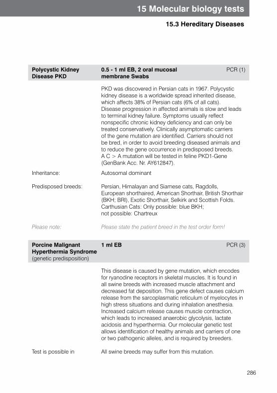
286
15 Molecular biology tests
15.3 Hereditary Diseases
Polycystic Kidney Disease PKD
0.5 - 1 ml EB, 2 oral mucosal membrane Swabs
PCR (1)
PKD was discovered in Persian cats in 1967. Polycystic kidney disease is a worldwide spread inherited disease, which affects 38% of Persian cats (6% of all cats). Disease progression in affected animals is slow and leads to terminal kidney failure. Symptoms usually reflect nonspecific chronic kidney deficiency and can only be treated conservatively. Clinically asymptomatic carriers of the gene mutation are identified. Carriers should not be bred, in order to avoid breeding diseased animals and to reduce the gene occurrence in predisposed breeds. A C > A mutation will be tested in feline PKD1-Gene (GenBank Acc. Nr. AY612847).
Inheritance:
Predisposed breeds:
Please note:
Autosomal dominant
Persian, Himalayan and Siamese cats, Ragdolls, European shorthaired, American Shorthair, British Shorthair (BKH; BRI), Exotic Shorthair, Selkirk and Scottish Folds. Carthusian Cats: Only possible: blue BKH; not possible: Chartreux
Please state the patient breed in the test order form!
Porcine Malignant Hyperthermia Syndrome (genetic predisposition)
1 ml EB PCR (3)
Test is possible in
This disease is caused by gene mutation, which encodes for ryanodine receptors in skeletal muscles. It is found in all swine breeds with increased muscle attachment and decreased fat deposition. This gene defect causes calcium release from the sarcoplasmatic reticulum of myelocytes in high stress situations and during inhalation anesthesia. Increased calcium release causes muscle contraction, which leads to increased anaerobic glycolysis, lactate acidosis and hyperthermia. Our molecular genetic test allows identification of healthy animals and carriers of one or two pathogenic alleles, and is required by breeders.
All swine breeds may suffer from this mutation.
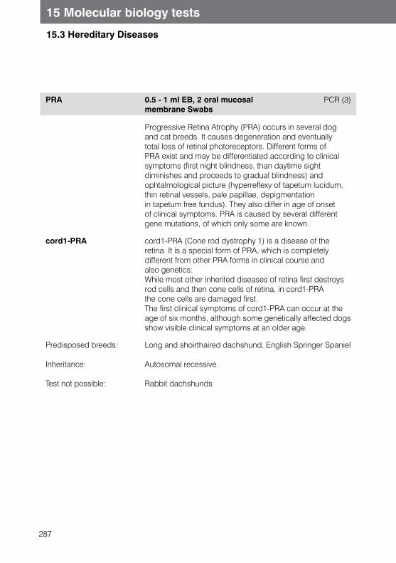
287
15 Molecular biology tests
15.3 Hereditary Diseases
PRA 0.5 - 1 ml EB, 2 oral mucosal membrane Swabs
PCR (3)
Progressive Retina Atrophy (PRA) occurs in several dog and cat breeds. It causes degeneration and eventually total loss of retinal photoreceptors. Different forms of PRA exist and may be differentiated according to clinical symptoms (first night blindness, than daytime sight diminishes and proceeds to gradual blindness) and ophtalmological picture (hyperreflexy of tapetum lucidum, thin retinal vessels, pale papillae, depigmentation in tapetum free fundus). They also differ in age of onset of clinical symptoms. PRA is caused by several different gene mutations, of which only some are known.
cord1-PRA cord1-PRA (Cone rod dystrophy 1) is a disease of the retina. It is a special form of PRA, which is completely different from other PRA forms in clinical course and also genetics:While most other inherited diseases of retina first destroys rod cells and then cone cells of retina, in cord1-PRA the cone cells are damaged first.The first clinical symptoms of cord1-PRA can occur at the age of six months, although some genetically affected dogs show visible clinical symptoms at an older age.
Predisposed breeds:
Inheritance:
Test not possible:
Long and shoirthaired dachshund, English Springer Spaniel
Autosomal recessive.
Rabbit dachshunds
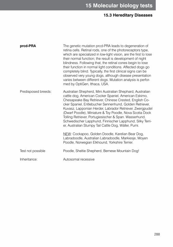
288
15 Molecular biology tests
15.3 Hereditary Diseases
prcd-PRA The genetic mutation prcd-PRA leads to degeneration of retina cells. Retinal rods, one of the photoreceptors type, which are specialized in low-light vision, are the first to lose their normal function; the result is development of night blindness. Following that, the retinal cones begin to lose their function in normal light conditions. Affected dogs go completely blind. Typically, the first clinical signs can be observed very young dogs, although disease presentation varies between different dogs. Mutation analysis is perfor-med by OptiGen, Ithaca, USA.
Predisposed breeds:
Test not possible
Inheritance:
Australian Shepherd, Mini Australian Shephard, Australian cattle dog, American Cocker Spaniel, American Eskimo, Chesapeake Bay Retriever, Chinese Crested, English Co-cker Spaniel, Entlebucher Sennenhund, Golden Retriever, Kuvasz, Lapponian Herder, Labrador Retriever, Zwergpudel (Dwarf Poodle), Miniature & Toy Poodle, Nova Scotia Dock Tolling Retriever, Portugiesischer & Span. Wasserhund, Schwedischer Lapphund, Finnischer Lapphund, Silky Terri-er, Australian Stumpy Tail Cattle Dog, Wäller, Pumi.
NEW: Cockapoo, Golden Doodle, Karelian Bear Dog, Labradoodle, Australian Labradoodle, Markiesje, Moyen Poodle, Norwegian Elkhound, Yorkshire Terrier.
Poodle, Sheltie Shepherd, Bernese Mountain Dog!
Autosomal recessive
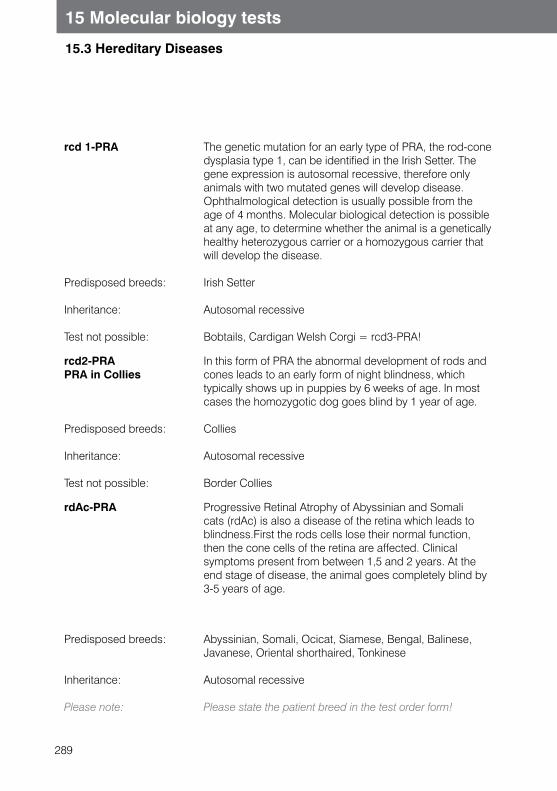
289
15 Molecular biology tests
15.3 Hereditary Diseases
rcd 1-PRA
Predisposed breeds:
Inheritance:
Test not possible:
The genetic mutation for an early type of PRA, the rod-cone dysplasia type 1, can be identified in the Irish Setter. The gene expression is autosomal recessive, therefore only animals with two mutated genes will develop disease.Ophthalmological detection is usually possible from the age of 4 months. Molecular biological detection is possible at any age, to determine whether the animal is a genetically healthy heterozygous carrier or a homozygous carrier that will develop the disease.
Irish Setter
Autosomal recessive
Bobtails, Cardigan Welsh Corgi = rcd3-PRA!
rcd2-PRAPRA in Collies
Predisposed breeds:
Inheritance:
Test not possible:
In this form of PRA the abnormal development of rods and cones leads to an early form of night blindness, which typically shows up in puppies by 6 weeks of age. In most cases the homozygotic dog goes blind by 1 year of age.
Collies
Autosomal recessive
Border Collies
rdAc-PRA Progressive Retinal Atrophy of Abyssinian and Somali cats (rdAc) is also a disease of the retina which leads to blindness.First the rods cells lose their normal function, then the cone cells of the retina are affected. Clinical symptoms present from between 1,5 and 2 years. At the end stage of disease, the animal goes completely blind by 3-5 years of age.
Predisposed breeds:
Inheritance:
Please note:
Abyssinian, Somali, Ocicat, Siamese, Bengal, Balinese, Javanese, Oriental shorthaired, Tonkinese
Autosomal recessive
Please state the patient breed in the test order form!
290
15 Molecular biology tests
15.3 Hereditary Diseases
Pyruvate kinase deficiency
0.5 - 1 ml EDTA plasma, 2 oral mucosal membrane Swabs
PCR (3)
Predisposed breeds:i
Test not possible:
Inheritance:
Please note:
Pyruvate kinase deficiency is well researched in Basenji dogs and West Highland White Terriers, but it also occurs in other dog breeds (Cairn Terrier, Beagle, Miniature Poodle, etc.), cats and humans. It is a specific mutation in the gene, which encodes for functional pyruvate kinase enzyme.The lack of the enzyme, which plays an important role in red blood cell metabolism, leads to premature breakdown and destruction of red blood cells.The affected animals subsequently develop chronic regenerative haemolytic anaemia, progressive myelofibrosis and osteosclerosis and have a much reduced life span. The onset of disease is between the ages of four to twelve months.
Dogs: Basenji, West Highland White TerrierCats: Abyssinian, Somalian, (Ocicat)
Pug dog, Cairn Terrier
Autosomal recessive
Please state the patient breed in the test order form!
291
15 Molecular biology tests
15.3 Hereditary Diseases
SCID in Arabians 1 ml EB PCR (3)
Predisposed breed:
Symptoms:
Inheritance:
SCID (Severe combined immunodeficiency) in the Arabian foal is probably due to a defect in the lymphoid stem cells, which leads to disturbed maturation of the B- and T-cell lymphocytes, resulting in severe lymphopenia. Affected foals develop disease at the age of approximately 1 month and the majority of them die within 5 months of birth, due to opportunistic infections.The disease is caused by a deletion in a gene that codes for DNA-dependent protein kinase. Only foals that carry two copies of the gene will develop disease. The genetic test can detect diseased foals as well as clinically healthy carriers, which is important for breeding purposes.
Arabian horse
Susceptibility to infection
Autosomal recessive
292
15 Molecular biology tests
15.3 Hereditary Diseases
SCID in Jack Russell Terrier
0.5 - 1 ml EB, 2 oral mucosal membrane Swabs
PCR (3)
SCID (severe combined immunodeficiency) is a genetically determined severe immune disease that has a range of clinical symptoms and is described in many different dogs breeds. The disease is also possible in horses, mice and humans.A point mutation results in B- and T-lymphocyte dysfunction, which leads to extreme lymphopenia, agammaglobulinemia, thymus dysplasia and peripheral lymphoid aplasia. Affected dogs die shortly after weaning. Only puppies which carry a double mutation in their genome will develop disease. Affected puppies can be diagnosed by gene test and is very important for breeders. Clinically asymptomatic SCID carriers can be distinguished from non-carriers. SCID carriers are excluded from breeding, but they have to be paired with non-carriers.
Predisposed breed:
Inheritance:
Jack Russell Terrier
Autosomal recessive
293
15 Molecular biology tests
15.3 Hereditary Diseases
Scrapie (genetic predisposition)
1ml EB PCR (3)
Scrapie is a non febrile, chronic progressive degenerative CNS disorder in sheep, rarely seen in goats and cattle. It is caused by the formation of an endogenous glycoprotein on the neuronal surface which folds incorrectly and therefore cannot break down. This results in the formation of amyloid aggregates which deposit in certain tissues, causing CNS symptoms.
Disease is transmitted horizontally and vertically in sheep.In sheep, horizontal and vertical transmission can occur. Whether or not an individual is susceptible to Scrapie is determined by the prion protein gene (PrP). Molecular genetic testing of the gene make it possible to assess the risk for the animal to develop Scrapie.
The following amino acids can occur depending on position: Alanine (A), Histidine (H), Glutamine (Q), Arginine (R) or Valine (V).
By gene testing all variants of three specific gene parts (Codons 136, 154 and 171) the coding for these amino acids will be analysed. The BMVEL and Project Group of German Society of Breeders are breeding for TSE-resis-tance in sheep, and sheep are classified as one of five genotype classes:
Genotype class:
G5
G4
G3
G2
G1
Test method:
Predisposed breeds:
TSE-resistance genotype
VRQ/VRQ, ARQ/VRQ, ARH/VRQ, VRQ/AHQ
ARR/VRQ
AHQ/AHQ, AHQ/ARH, AHQ/ARQ, ARH/ARH, ARH/ARQ
ARQ/ARQ
ARR/AHQ, ARR/ARH, ARR/ARQ
ARR/ARR
Direct DNA-sequencing. This is a new method, with which all currently identified mutations can be diagnosed.
All sheep breeds
294
15 Molecular biology tests
15.3 Hereditary Diseases
Von Willebrand disease (vWF)
0.5 - 1 ml EB, 2 oral mucosal membrane Swabs
PCR (3)
The Von Willebrand factor (vWF) mediates the adhesion of thrombocytes to the subendothelial cells of a damaged blood vessel. It also acts as a carrier protein for Factor VII of the plasma coagulation system and protects it from premature proteolytic decomposition. Reducedconcentration or complete absence of functional vWF leads to coagulation disorders of various degrees of severity. Characteristic symptoms are bleeding from the mucous membranes and vigorous bleeding during teething, oestrus and trauma.There are three types of Von Willebrand Disease, two of which can be genetically determined: type 1 and type 3.
vWF type 1
Predisposed breeds
Often a milder course of the disease. The gene expression is autosomal incompletely dominant, e.g. heterozygous animals possess moderate vWF plasma concentrations and may be clinically inapparent, whereas homozygous animals possess low vWF plasma concentrations and show more severe clinical symptoms.
Dobermann Pinscher, Poodle, Manchester Terrier, Coton de Tulear, Doberman Pinscher, Drentsche Patrijshond, Bernese Mountain Dog, German Pinscher, Kerry Blue Terrier, Poodle, Papillon, Welsh Corgi
vWF type 2
Predisposed breed:
Inheritance:
Shows a mild to severe course with variable vWF concentrations.
German wirehaired pointer, German Pointers
Autosomal recessive
295
15 Molecular biology tests
15.3 Hereditary Diseases
vWF type 3
Predisposed breeds:
Test not possible:
Please note:
The most severe course of the disease. The gene expression is autosomal recessive. Homozygous animals possess no detectable vWF plasma concentrations and suffer from severe coagulation disorders. Heterozygous animals possess reduced vWF plasma concentrations and are carriers of the disease, but do not usually show any clinical symptoms.
Scottish Terrier, Sheltie, Kooiker dog
Rhodesian Ridgeback
Please state the patient breed in the test order form
X-SCID 0.5 - 1 ml EB, 2 oral mucosal membrane Swabs
PCR (3)
Predisposed Breeds:
Symptoms:
Laboratory findings:
Gene expression
X-SCID (X-linked Severe Combined Immuno Deficiency) in the dog is caused by a defect in the γ-chain of the interleucine-2 receptor. The cellular and humoral immune systems are markedly impaired. Only male dogs are affected by the disease; female dogs are only carriers. Recurring and chronic infections (due to opportunistic pathogens) start once maternal antibody protection ends. Most affected puppies die at the age of three to four months.
Welsh Corgi, Basset
- Developmental disorders, thymus dysplasia, - Susceptibility to infections, inadequately formed
peripheral lymph nodes
Lymphopenia, reduced IgG and IgA levels, variable IgM levels
X-chromosome bound
296
15 Molecular biology tests
15.4 Avian Sex Determination
General Information
Avian sex determination is performed using DNA extracted from feather pulp or from EDTA blood. A specific region of the DNA is amplified using PCR. Two different molecular biological methods are employed to identify the gender-specific gene sequences of the sex chromosomes.This test can be used in several hundred avian species. It can not be used in ratites (emu, ostrich, nandu and kiwi) but it can be used in the cassowary.
Sex determination, avian 100 µl EB, feathers real time-PCR (1)
EDTA blood:
Feathers:
Egg shells:
only 2-3 drops are required
Several small feathers or one large feather can be submitted, but it is important that the quill is intact. Growing feathers contain more DNA than mature feathers, which is why they are most suitable for sex identification. It is possible to use mature feathers or even feathers thathave recently been moulted. Clear identification of the source of the feathers must be made and contamination with foreign genetic sample (cage dust or sand) must be avoided. Bloody feathers should be submitted in a sterile tub. The vane (feathered end) of the feather may be shortened. Dry feathers may be sent in a sealed plastic bag.
Isolation of DNA from egg membrane is possible; therefore the egg shell should be mostly intact. A drop of blood taken from the egg shell with a sterile Swab is even more suitable. Please make sure the egg shell is from the desired individual.
Please note: In order to avoid false or inconclusive results, the sample material must be protected from contamination with other samples which may contain DNA.- The sample material (blood or feather pulp) can be
stored at 4-8 °C for several days.- Dry feathers can be stored at room temperature
for several weeks.- Detailed identification must accompany the sample at
submission, including the accurate avian species (scientific name if possible), ring number and the date of collection.
297
15 Molecular biology tests
15.5 Parentage verification/ genetic fingerprint
General Information
Parentage verification is sometimes required to verify that the parents are the trueparents of the offspring. Parentage verification is done using microsatellite analysis.
Principle:A genome contains a large number of DNA “microsatellite” segments, which are multiple repeated copies of short DNA sections. The number of copies and the length of the microsatellites vary from one individual to another. The human genome is estimated to contain up to 100,000 of these microsatellite gene sites. Each individual therefore possesses a unique genome and there are practically no two identical individuals (except monozygotic twins).
In principle, offspring inherit 50% of its genetic sample from its mother and 50% fromits father. This means that any variation of the genome (e.g. in the highly variablemicrosatellite sequence) that is not inherited from the mother must be inherited fromthe father.For example, in the case of paternity testing, if the mother is known, and microsatellites are found in the offspring that do not match either the mother or claimed father, then fatherhood can be ruled out with a high probability.Detection reliability increases with the comparison of several microsatellite gene sites.This is why we examine 17 microsatellites in horses, 10 in cats and 9 in dogs(19 (ISAG 2006) as recommended by ISAG(International Society for Animal Genetics).
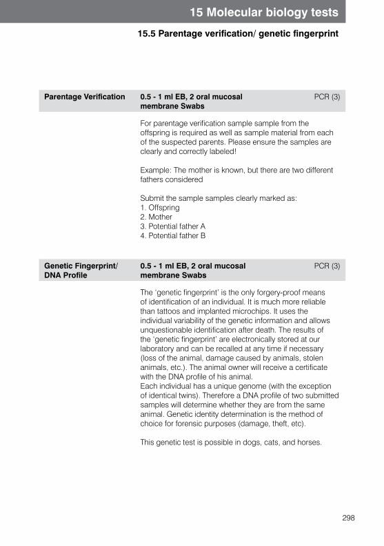
298
15 Molecular biology tests
15.5 Parentage verification/ genetic fingerprint
Parentage Verification 0.5 - 1 ml EB, 2 oral mucosal membrane Swabs
PCR (3)
For parentage verification sample sample from theoffspring is required as well as sample material from eachof the suspected parents. Please ensure the samples are clearly and correctly labeled!
Example: The mother is known, but there are two different fathers considered
Submit the sample samples clearly marked as:1. Offspring2. Mother3. Potential father A4. Potential father B
Genetic Fingerprint/DNA Profile
0.5 - 1 ml EB, 2 oral mucosal membrane Swabs
PCR (3)
The ‘genetic fingerprint’ is the only forgery-proof means of identification of an individual. It is much more reliable than tattoos and implanted microchips. It uses the individual variability of the genetic information and allows unquestionable identification after death. The results of the ‘genetic fingerprint’ are electronically stored at our laboratory and can be recalled at any time if necessary (loss of the animal, damage caused by animals, stolen animals, etc.). The animal owner will receive a certificate with the DNA profile of his animal.Each individual has a unique genome (with the exception of identical twins). Therefore a DNA profile of two submitted samples will determine whether they are from the same animal. Genetic identity determination is the method of choice for forensic purposes (damage, theft, etc).
This genetic test is possible in dogs, cats, and horses.
299
15 Molecular biology tests
15.5 Parentage verification/ genetic fingerprint
Sequence analysis PCR (1)
DNA-sequence analysis in molecular biology and bioinformatics is an automatic computer analysis of characteristic features, especially genes in DNA-strand.Information is gained by DNA-sequencing on the sequence and position of base pairs. When compared for similarities in the international sequence data banks, information can be found about the organism species and about differences (mutations) in the nucleic acid sequence, compared to the standard sequence.This method is used to analyze many inherited diseases, but is also used in single cases to characterise or to differenciate PCR products from test results. Such species differentiation is possible after consultation with the laboratory.
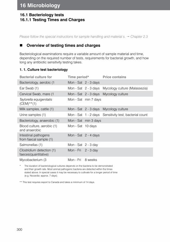
300
16.1 Bacteriology tests16.1.1 Testing Times and Charges
16 Microbiology
Please follow the special instructions for sample handling and material s. Chapter 2.3
Overview of testing times and charges
Bacteriological examinations require a variable amount of sample material and time, depending on the required number of tests, requirements for bacterial growth, and how long any antibiotic sensitivity testing takes.
1. 1. Culture test bacteriology
Bacterial culture for Time period* Price contains
Bacteriology, aerobic (1 Mon - Sat 2 - 3 days
Ear Swab (1) Mon - Sat 2 - 3 days Mycology culture (Malassezia)
Cervical Swab, mare (1 Mon - Sat 2 - 3 days Mycology culture
Taylorella equigenitalis Mon - Sat min 7 days (CEM)**(1)
Milk samples, cattle (1) Mon - Sat 2 - 3 days Mycology culture
Urine samples (1) Mon - Sat 1 - 2 days Sensitivity test, bacterial count
Bacteriology, anaerobic (1) Mon - Sat min 3 days
Blood culture, aerobic (1) Mon - Sat 10 days and anaerobic Intestinal pathogens Mon - Sat 2 - 4 daysfrom faecal sample (1)
Salmonellas (1) Mon - Sat 2 - 3 day
Clostridium detection (1) Mon - Fri 2 - 3 dayfaeces(quantitative)
Mycobacterium (3 Mon - Fri 8 weeks
* The duration of bacteriological cultures depends on the bacteria to be demonstrated and their growth rate. Most animal pathogenic bacteria are detected within the times stated above. In special cases it may be necessary to cultivate for a longer period of time (e.g. Nocardia: approx. 7 days).
** This test requires export to Canada and takes a minimum of 14 days.
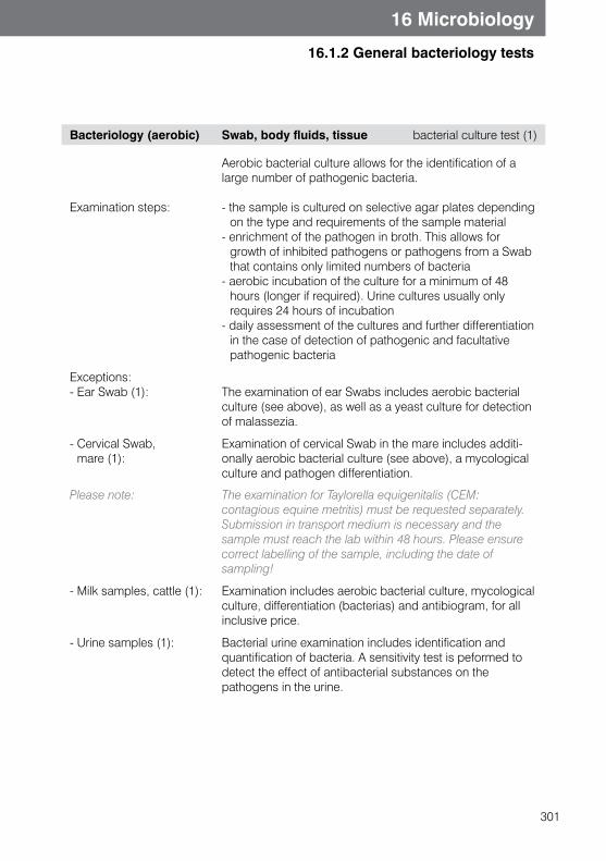
301
16.1.2 General bacteriology tests
16 Microbiology
Bacteriology (aerobic) Swab, body fluids, tissue bacterial culture test (1)
Examination steps:
Aerobic bacterial culture allows for the identification of a large number of pathogenic bacteria.
- the sample is cultured on selective agar plates depending on the type and requirements of the sample material
- enrichment of the pathogen in broth. This allows for growth of inhibited pathogens or pathogens from a Swab that contains only limited numbers of bacteria
- aerobic incubation of the culture for a minimum of 48 hours (longer if required). Urine cultures usually only requires 24 hours of incubation
- daily assessment of the cultures and further differentiation in the case of detection of pathogenic and facultative pathogenic bacteria
Exceptions:- Ear Swab (1): The examination of ear Swabs includes aerobic bacterial
culture (see above), as well as a yeast culture for detection of malassezia.
- Cervical Swab, mare (1):
Examination of cervical Swab in the mare includes additi-onally aerobic bacterial culture (see above), a mycological culture and pathogen differentiation.
Please note: The examination for Taylorella equigenitalis (CEM: contagious equine metritis) must be requested separately. Submission in transport medium is necessary and the sample must reach the lab within 48 hours. Please ensure correct labelling of the sample, including the date ofsampling!
- Milk samples, cattle (1): Examination includes aerobic bacterial culture, mycological culture, differentiation (bacterias) and antibiogram, for all inclusive price.
- Urine samples (1): Bacterial urine examination includes identification and quantification of bacteria. A sensitivity test is peformed to detect the effect of antibacterial substances on the pathogens in the urine.
302
16 Microbiology
16.1.2 General bacteriology tests
Bacteriology (anaerobic) Swabs, body fluids, tissue parts and others
bacteriology culture (1)
Examination steps:
The examination for anaerobic organisms is recommended in addition to the aerobic culture from the following sample materials: abscess material, pus, wound Swabs (especially from bite wounds), body fluids (aspirates, synovial fluid, cerebrospinal fluid, etc.), Swabs from inner organs andserous membranes, and nail bed infection. Please note that Swabs must be sent in transport medium!
- culture on special nutrient medium- enrichment of the bacteria in nutrient broth- anaerobic incubation of the culture for a minimum
of 72 hrs (longer if required)- regular assessment of the cultures and further
differentiation in the case of detection of pathogenic and facultatively pathogenic anaerobic bacteria
Blood Culture blood must be sent in special container
bacteriology culture test (1)
Handling:
In case of bacteraemia or suspected bacteraemia, blood is collected from the patient in a sterile manner and transferred into special blood culture bottles directly in the surgery. These bottles, plus the necessary blood collection set, are provided by the lab free of charge on request.The bottles are incubated for 10 days. A detection system recognises and reports any bacterial growth.
- Always use one aerobic and one anaerobic bottle for culture
- Thorough disinfection of the collection site is necessary to prevent contamination with bacteria from the skin
- Use a syringe or collection set for blood collection- Fill each bottle with 3-10 ml (ideally 8-10 ml) of blood.
First fill the aerobic bottle and then the anaerobic bottle. If you are not using a blood collection set, please inject the blood through the rubber plug into the bottle.
- Collect the blood and submit your sample at the beginning of the week, if possible
- Inoculated bottles should be stored at room temperature (not in the fridge).

303
16 Microbiology
16.1.2 General bacteriology tests
MRS Screening (methicillin resistant staphylococci)
Swab, body fluid Cultures examination (1)
304
16 Microbiology
16.2 Faecal tests
Bacteriology, enteropathogenic germs
faeces, rectal Swab culture test (1)
Faecal samples or rectal Swabs are tested with help of selective medium and enrichment method for the intestinal pathogens under examination.
- Salmonellas - Thermophile Campylobacter species,
Campylobacter jejuni, Campylobacter coli - Yersinia enterocolitica - Different pathogens of different animal species
and facultative pathogens Enterobacteriaceae (e.g. Klebsiella, haemolytic and mucoid E. coli-strains, Proteus spp.)
- Coagulase positive Staphylococci (Staphylococcus aureus, Staphylococcus pseudintermedius)
- Pseudomonas aeruginosa - Yeasts (semiquantative confirmation of
abnormal increase)
In carnivores, if no pathogens were detected, the composi-tion of faecal flora will be semiquantatively estimated.A quantative increase of Gram positive or Gram negative bacteria can prove dysbiosis of large intestinal flora.
Salmonella-detection faeces, rectal Swab culture test (1)
Culture test of faecal samples exclusively to confirm Salmonella. Collective faecal samples can be used for this test
305
16 Microbiology
16.2 Faecal tests
Clostridium perfringens (quantitative without pathogen differentiation)
¼ faecal tube bacterial culture test (1)
In carnivores a quantitative increase of Clostridium perfringens is a sign of intestinal flora imbalance. The faecal sample is precisely diluted, followed by anaerobic culture on a selective medium to determine the number of Clostridium per gram of faeces.
Clostridium perfringensEnterotoxin
¼ faecal tube ELISA (2)
C. perfringens enterotoxin can cause diarrhoea in cats and dogs.
Elastase 3 g faeces ELISA (1)
Species:
Indication:
Please note:
Canine faecal elastase-1 is produced in the pancreas and released along with the pancreatic juices into the intestines during digestion. It is stable in the intestines and can be detected unaltered in a faecal sample for some period of time. To rule out EPI it is usually sufficient to test once for elastase, but borderline results should be re-tested or verified using TLI testing from blood.
s. Chapter 7.3 Exocrine Pancreatic Diseases Dog only
Suspected exocrine pancreatic insufficiency
Enzyme substitution does not have to be discontinued before testing as this does not affect the results. The dilution effect of liquid faeces may lead to false low values.
306
16 Microbiology
16.2 Faecal tests
Faecal digestion test 3 g faeces Microscopy (1)
Species:
Indication:
Results affected by:
Detection of undigested dietary components in the faeces: fatty acid globules, neutral fats, muscle fibres and starch.
Test is possible in carnivores, omnivores and birds
Suspected maldigestion e.g. due to exocrine pancreatic Insufficiency
- Faecal digestion is dependent on the composition of the diet, e.g. fatty acid globules and muscle fibres can be found when feeding raw meat
- Diarrhoea (i.e. reduced faecal passage time) will lead to poor faecal digestion results.
General Virology (Virology faecal testing)
min. ½ faecal tube Electron microscopy (3)
Indications:
Viruses which are excreted in the faeces can be detected and classified using electron microscopy
s. Also Chapter 13, Infectious diseases
Corona-, rota- and parvovirus detection
Occult blood Faeces (1/3 faecal tube) Chromatography (2)
To prevent false positive tests, no meat should be fed for three days prior to sample taking.
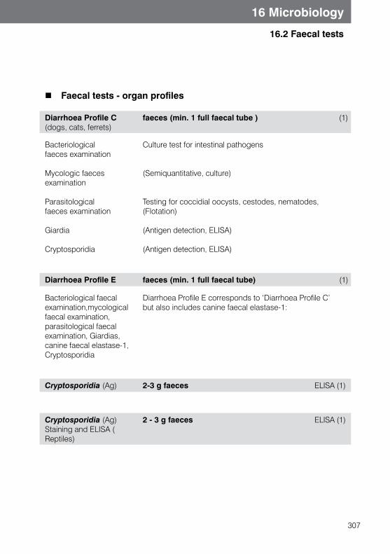
307
16 Microbiology
16.2 Faecal tests
Faecal tests - organ profiles
Diarrhoea Profile C (dogs, cats, ferrets)
faeces (min. 1 full faecal tube ) (1)
Bacteriological faeces examination
Mycologic faeces examination
Parasitological faeces examination
Giardia
Cryptosporidia
Culture test for intestinal pathogens
(Semiquantitative, culture)
Testing for coccidial oocysts, cestodes, nematodes, (Flotation)
(Antigen detection, ELISA)
(Antigen detection, ELISA)
Diarrhoea Profile E faeces (min. 1 full faecal tube) (1)
Bacteriological faecal examination,mycological faecal examination,parasitological faecal examination, Giardias, canine faecal elastase-1,Cryptosporidia
Diarrhoea Profile E corresponds to ‘Diarrhoea Profile C’ but also includes canine faecal elastase-1:
Cryptosporidia (Ag) 2-3 g faeces ELISA (1)
Cryptosporidia (Ag) Staining and ELISA ( Reptiles)
2 - 3 g faeces ELISA (1)

308
16 Microbiology
16.3 Mycology tests 16.3.1 Testing Times and Charges
1. Mycological culture:
Dermatophytes Mon - Fri 4 weeks
Yeasts and moulds Mon - Sat 2 - 3 days
Yeasts in faeces, quantitatively Mon - Fri 2 - 3 days
* The duration of mycological culture depends on the organism to be demonstrated and their growth rate. Most animal pathogenic fungi are detected within the times stated above. In special cases it may be necessary to cultivate for a longer period of time (e.g. Cryptococcus neoformans: 7 days).
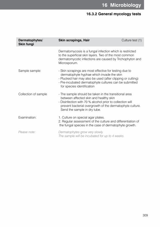
309
16 Microbiology
16.3.2 General mycology tests
Dermatophytes/Skin fungi
Skin scrapings, Hair Culture test (1)
Sample sample:
Collection of sample
Examination:
Dermatomycosis is a fungal infection which is restricted to the superficial skin layers. Two of the most common dermatomycotic infections are caused by Trichophyton and Microsporum.
- Skin scrapings are most effective for testing due to dermatophyte hyphae which invade the skin
- Plucked hair may also be used (after clipping or cutting)- Pre-incubated dermatophyte cultures can be submitted
for species identification
- The sample should be taken in the transitional area between affected skin and healthy skin
- Disinfection with 70 % alcohol prior to collection will prevent bacterial overgrowth of the dermatophyte culture. Send the sample in dry tube.
1. Culture on special agar plates.2. Regular assessment of the culture and differentiation of the fungal species in the case of dermatophyte growth.
Please note: Dermatophytes grow very slowly. The sample will be incubated for up to 4 weeks.
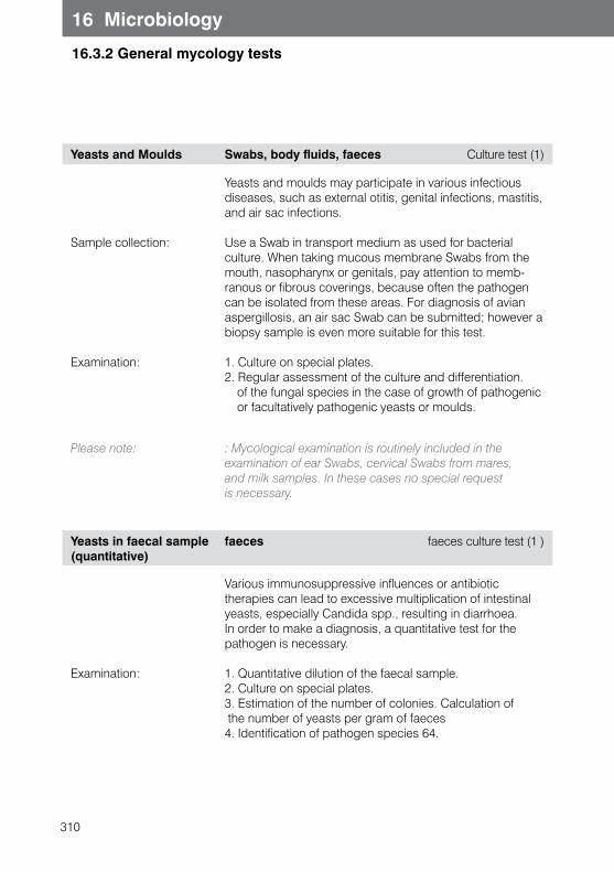
310
16 Microbiology
16.3.2 General mycology tests
Yeasts and Moulds Swabs, body fluids, faeces Culture test (1)
Sample collection:
Examination:
Yeasts and moulds may participate in various infectious diseases, such as external otitis, genital infections, mastitis, and air sac infections.
Use a Swab in transport medium as used for bacterial culture. When taking mucous membrane Swabs from the mouth, nasopharynx or genitals, pay attention to memb-ranous or fibrous coverings, because often the pathogen can be isolated from these areas. For diagnosis of avian aspergillosis, an air sac Swab can be submitted; however a biopsy sample is even more suitable for this test.
1. Culture on special plates.2. Regular assessment of the culture and differentiation.
of the fungal species in the case of growth of pathogenic or facultatively pathogenic yeasts or moulds.
Please note: : Mycological examination is routinely included in the examination of ear Swabs, cervical Swabs from mares, and milk samples. In these cases no special request is necessary.
Yeasts in faecal sample (quantitative)
faeces faeces culture test (1 )
Examination:
Various immunosuppressive influences or antibiotic therapies can lead to excessive multiplication of intestinal yeasts, especially Candida spp., resulting in diarrhoea. In order to make a diagnosis, a quantitative test for the pathogen is necessary.
1. Quantitative dilution of the faecal sample.2. Culture on special plates.3. Estimation of the number of colonies. Calculation of the number of yeasts per gram of faeces4. Identification of pathogen species 64.
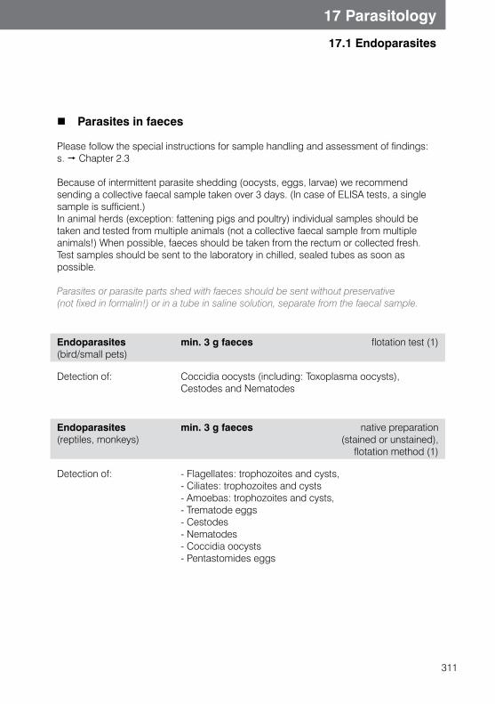
311
17.1 Endoparasites
17 Parasitology
Parasites in faeces
Please follow the special instructions for sample handling and assessment of findings: s. Chapter 2.3
Because of intermittent parasite shedding (oocysts, eggs, larvae) we recommend sending a collective faecal sample taken over 3 days. (In case of ELISA tests, a single sample is sufficient.)In animal herds (exception: fattening pigs and poultry) individual samples should be taken and tested from multiple animals (not a collective faecal sample from multiple animals!) When possible, faeces should be taken from the rectum or collected fresh. Test samples should be sent to the laboratory in chilled, sealed tubes as soon as possible.
Parasites or parasite parts shed with faeces should be sent without preservative (not fixed in formalin!) or in a tube in saline solution, separate from the faecal sample.
Endoparasites(bird/small pets)
min. 3 g faeces flotation test (1)
Detection of: Coccidia oocysts (including: Toxoplasma oocysts),Cestodes and Nematodes
Endoparasites (reptiles, monkeys)
min. 3 g faeces native preparation (stained or unstained),
flotation method (1)
Detection of: - Flagellates: trophozoites and cysts, - Ciliates: trophozoites and cysts- Amoebas: trophozoites and cysts,- Trematode eggs- Cestodes- Nematodes- Coccidia oocysts- Pentastomides eggs
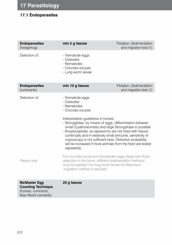
312
17 Parasitology
17.1 Endoparasites
Endoparasites (hedgehog)
min 5 g faeces Flotation, Sedimentation and migration test (1)
Detection of: - Trematode eggs- Cestodes- Nematodes- Coccidia oocysts- Lung worm larvae
Endoparasites (ruminants)
min 10 g faeces Flotation, Sedimentation and migration test (1)
Detection of:
Please note:
- Trematode eggs- Cestodes- Nematodes- Coccidia oocysts
Interpretation guidelines in horses:- Strongylidae: by means of eggs, differentiation between
small (Cyathostomids) and large Strongylidae is possible- Anoplocephala: as tapeworms are not shed with faeces
continually and in relatively small amounts, sensitivity of coproscopy is not sufficient here. Detection probability will be increased if more animals from the herd are tested repeatedly.
For coccidia oocyst and trematodes eggs (large liver fluke) detection in the horse, different sedimentation methods must be applied. For lung worm larvae the Baermann migration method is required.
McMaster Egg Counting Technique (horses, ruminants, New World camelids)
20 g faeces
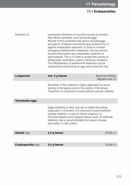
313
17 Parasitology
17.1 Endoparasites
Detection of: quantitative detection of coccidia oocysts (ruminants, New World camelids), and nematode eggs. Results of this quantative test will be oocysts/eggs per gramm of faeces and should give guidance for or against antiparasitic treatment. In order to combat increasing antihelminthic resistance, this test should be peformed before any antiparasitic treatment is administered. This is in order to protect the activity of antiparasitic medication used in veterinary medicine. The effectiveness of antihelminth treatment can be measured by performing an egg count reduction test.
Lungworms min. 5 g faeces Baermann-Wetzel Migration test (1)
Sensitivity of this method is highly dependent on larval density in the faeces and on the activity of the larvae. Therefore it is important to send sufficent sample material.
Trematodes eggs
Eggs shedding is often very low or highly fluctuating, especially in ruminants. It is important to send sufficient sample material. In case of clinical suspicion of Fasciola hepatica and negative faecal result, an antibodydetection test is recommended from serum (horses and cattle) or milk (cattle).
Giardia (Ag) 2-3 g faeces ELISA (1)
Cryptosporidia (Ag) 2-3 g faeces ELISA (1)
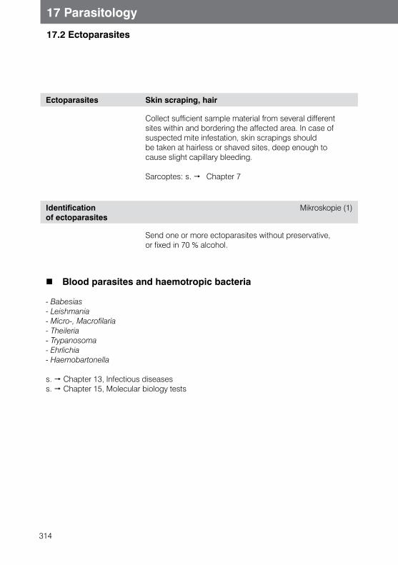
314
17 Parasitology
17.2 Ectoparasites
Ectoparasites Skin scraping, hair
Collect sufficient sample material from several different sites within and bordering the affected area. In case of suspected mite infestation, skin scrapings should be taken at hairless or shaved sites, deep enough to cause slight capillary bleeding.
Sarcoptes: s. Chapter 7
Identification of ectoparasites
Mikroskopie (1)
Send one or more ectoparasites without preservative, or fixed in 70 % alcohol.
Blood parasites and haemotropic bacteria
- Babesias- Leishmania - Micro-, Macrofilaria - Theileria - Trypanosoma - Ehrlichia - Haemobartonella
s. Chapter 13, Infectious diseases s. Chapter 15, Molecular biology tests
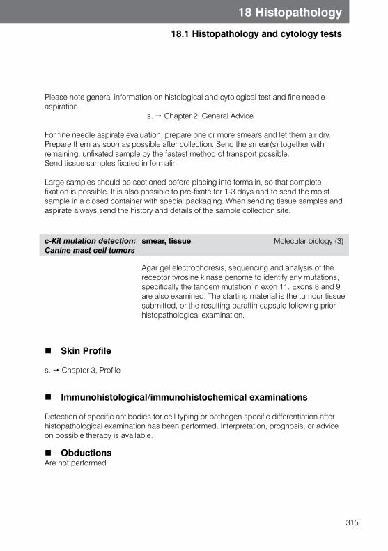
315
18.1 Histopathology and cytology tests
18 Histopathology
Please note general information on histological and cytological test and fine needle aspiration. s. Chapter 2, General Advice
For fine needle aspirate evaluation, prepare one or more smears and let them air dry. Prepare them as soon as possible after collection. Send the smear(s) together with remaining, unfixated sample by the fastest method of transport possible.Send tissue samples fixated in formalin.
Large samples should be sectioned before placing into formalin, so that complete fixation is possible. It is also possible to pre-fixate for 1-3 days and to send the moist sample in a closed container with special packaging. When sending tissue samples and aspirate always send the history and details of the sample collection site.
c-Kit mutation detection: Canine mast cell tumors
smear, tissue Molecular biology (3)
Agar gel electrophoresis, sequencing and analysis of the receptor tyrosine kinase genome to identify any mutations, specifically the tandem mutation in exon 11. Exons 8 and 9 are also examined. The starting material is the tumour tissue submitted, or the resulting paraffin capsule following prior histopathological examination.
Skin Profile
s. Chapter 3, Profile
Immunohistological/immunohistochemical examinations
Detection of specific antibodies for cell typing or pathogen specific differentiation after histopathological examination has been performed. Interpretation, prognosis, or advice on possible therapy is available.
ObductionsAre not performed
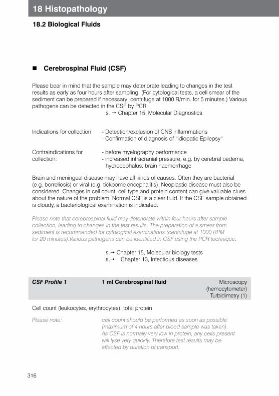
316
18 Histopathology
18.2 Biological Fluids
Cerebrospinal Fluid (CSF)
Please bear in mind that the sample may deteriorate leading to changes in the test results as early as four hours after sampling. (For cytological tests, a cell smear of the sediment can be prepared if necessary; centrifuge at 1000 R/min. for 5 minutes.) Various pathogens can be detected in the CSF by PCR. s. Chapter 15, Molecular Diagnostics
Indications for collection
Contraindications for collection:
- Detection/exclusion of CNS inflammations - Confirmation of diagnosis of “idiopatic Epilepsy“
- before myelography performance - increased intracranial pressure, e.g. by cerebral oedema,
hydrocephalus, brain haemorrhage
Brain and meningeal disease may have all kinds of causes. Often they are bacterial (e.g. borreliosis) or viral (e.g. tickborne encephalitis). Neoplastic disease must also be considered. Changes in cell count, cell type and protein content can give valuable clues about the nature of the problem. Normal CSF is a clear fluid. If the CSF sample obtained is cloudy, a bacteriological examination is indicated.
Please note that cerebrospinal fluid may deteriorate within four hours after sample collection, leading to changes in the test results. The preparation of a smear from sediment is recommended for cytological examinations (centrifuge at 1000 RPM for 20 minutes).Various pathogens can be identified in CSF using the PCR technique.
s. Chapter 15, Molecular biology tests s. Chapter 13, Infectious diseases
CSF Profile 1 1 ml Cerebrospinal fluid Microscopy (hemocytometer)
Turbidimetry (1)
Cell count (leukocytes, erythrocytes), total protein
Please note: cell count should be performed as soon as possible (maximum of 4 hours after blood sample was taken). As CSF is normally very low in protein, any cells present will lyse very quickly. Therefore test results may be affected by duration of transport.
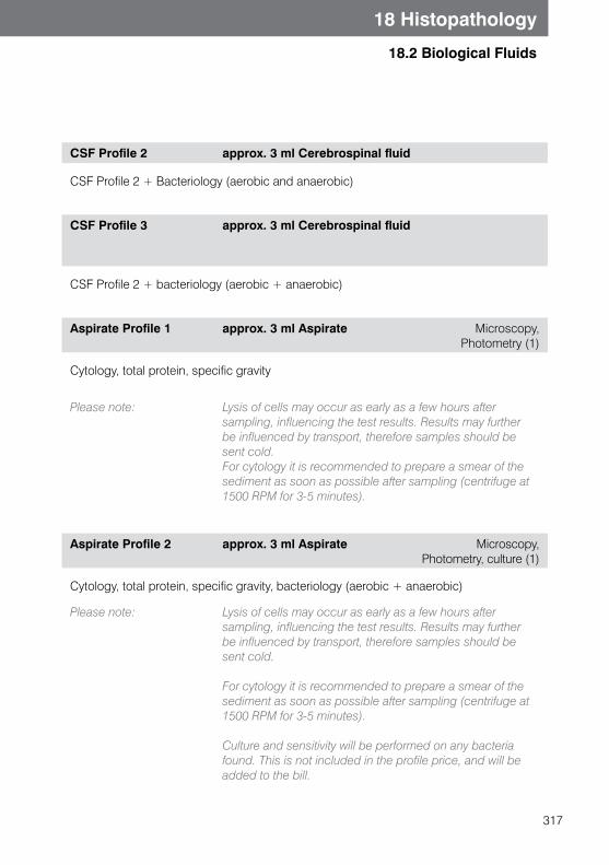
317
18 Histopathology
18.2 Biological Fluids
CSF Profile 2 approx. 3 ml Cerebrospinal fluid
CSF Profile 2 + Bacteriology (aerobic and anaerobic)
CSF Profile 3 approx. 3 ml Cerebrospinal fluid
CSF Profile 2 + bacteriology (aerobic + anaerobic)
Aspirate Profile 1 approx. 3 ml Aspirate Microscopy,Photometry (1)
Cytology, total protein, specific gravity
Please note: Lysis of cells may occur as early as a few hours after sampling, influencing the test results. Results may further be influenced by transport, therefore samples should be sent cold.For cytology it is recommended to prepare a smear of the sediment as soon as possible after sampling (centrifuge at 1500 RPM for 3-5 minutes).
Aspirate Profile 2 approx. 3 ml Aspirate Microscopy, Photometry, culture (1)
Cytology, total protein, specific gravity, bacteriology (aerobic + anaerobic)
Please note: Lysis of cells may occur as early as a few hours after sampling, influencing the test results. Results may further be influenced by transport, therefore samples should be sent cold.
For cytology it is recommended to prepare a smear of the sediment as soon as possible after sampling (centrifuge at 1500 RPM for 3-5 minutes).
Culture and sensitivity will be performed on any bacteria found. This is not included in the profile price, and will be added to the bill.
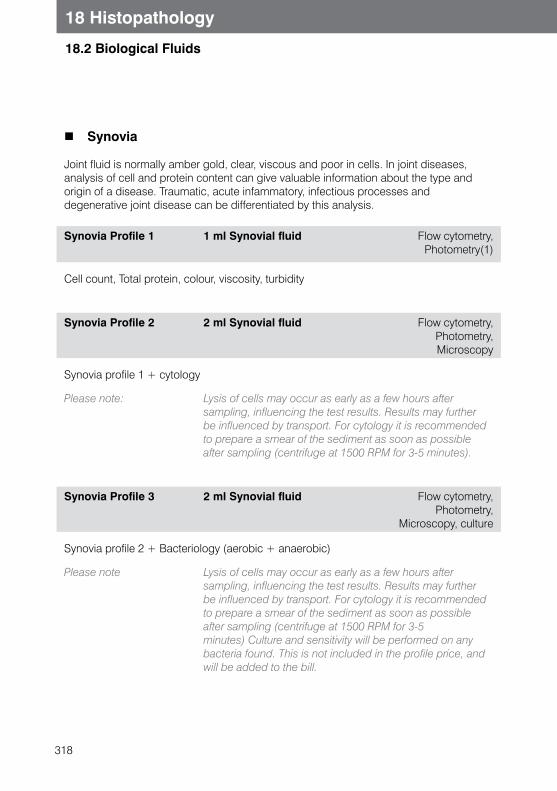
318
18 Histopathology
18.2 Biological Fluids
Synovia
Joint fluid is normally amber gold, clear, viscous and poor in cells. In joint diseases, analysis of cell and protein content can give valuable information about the type and origin of a disease. Traumatic, acute infammatory, infectious processes and degenerative joint disease can be differentiated by this analysis.
Synovia Profile 1 1 ml Synovial fluid Flow cytometry, Photometry(1)
Cell count, Total protein, colour, viscosity, turbidity
Synovia Profile 2 2 ml Synovial fluid Flow cytometry, Photometry, Microscopy
Synovia profile 1 + cytology
Please note: Lysis of cells may occur as early as a few hours after sampling, influencing the test results. Results may further be influenced by transport. For cytology it is recommended to prepare a smear of the sediment as soon as possible after sampling (centrifuge at 1500 RPM for 3-5 minutes).
Synovia Profile 3 2 ml Synovial fluid Flow cytometry, Photometry,
Microscopy, culture
Synovia profile 2 + Bacteriology (aerobic + anaerobic)
Please note Lysis of cells may occur as early as a few hours after sampling, influencing the test results. Results may further be influenced by transport. For cytology it is recommended to prepare a smear of the sediment as soon as possible after sampling (centrifuge at 1500 RPM for 3-5 minutes) Culture and sensitivity will be performed on any bacteria found. This is not included in the profile price, and will be added to the bill.

319
Notes

320
Notes