1
2624-Pos Board B394 Site-Directed Fluorescence Studies of Purified, Functional Cannabinoid Receptor Cb1: Agonist-Induced Conformational Changes in TM6 are Blocked by an Allosteric Modulator and Enhanced by a Novel CB1 Specific Antibody Jonathan F. Fay, David L. Farrens. Oregon Health and Science University, Portland, OR, USA. The human cannabinoid receptor CB1 is one of the most highly expressed GPCRs in the central nervous system, and a promising target for therapeutic applications. However, structural and biophysical information about this re- ceptor have been limited due to difficulties in purifying the receptor in a func- tional form and working with its hydrophobic ligands. Here we report on our ability to purify and study a detergent-solubilized functional form of the CB1 receptor. Our site-directed fluorescence labeling studies show specific confor- mational changes in CB1 in response to different cannabinoid ligands. This work involved attaching a specific fluorescent label to transmembrane helix 6 (TM6) of CB1. We then studied this labeled sample using various fluores- cence approaches. Our data shows specific spectral changes of the attached probe upon addition of various cannabinoid ligands, which we can clearly in- terpret to be due to drug-induced conformational changes in the receptor. We have subsequently used this labeled CB1 mutant to explore the effect of allo- steric ligands and conformationally sensitive antibodies on TM6 movements in CB1. 2625-Pos Board B395 Model for the Oligomer Formation of Serotonin Receptors Based on Quan- titative lux-FRET Measurements Andre Zeug 1 , Andrew Woehler 2 , Erwin Neher 2 , Evgeni Ponimaskin 1 . 1 Hannover Medical School, Hanover, Germany, 2 Max-Planck Institute for Biophysical Chemistry, Go ¨ttingen, Germany. Fo ¨rster Resonant Energy Transfer (FRET) is often used in experimental methods which aim at investigating the interaction of molecules at distances beyond diffraction limited resolution. Here we apply ’linear unmixing FRET’ (lux-FRET) [1]. By using the lux-FRET we can obtain three important quantities, the FRET-efficiency multiplied by the fraction of donors and accep- tors in FRET state as well as measures for the total donor and acceptor concen- trations. In the present study we applied lux-FRET to analyse the oligomerisation behaviour of two serotonin receptors 5-HT 1A and 5-HT 7 , which tend to form a dynamic system of homo- and hetero-dimers. From the modelling of measured lux-FRET data we con- clude that the receptors have significantly dif- ferent affinities to form oligomers with the dissociation constant order: K 5-HT1A-5-HT1A > K 5-HT7-5-HT1A >K 5-HT7-5-HT7 . Quantitative FRET measurements on a custom tailored spinning disk system at single-cell level confirmed these results and also allowed to visualize distribution of mono- and dimers within the cell (Figure 1). Together with observation that the 5-HT 1A receptor-mediated signalling is significantly im- paired in hetero-oligomers, our data suggest that receptor functions can be modulated by the dy- namic interaction within oligomeric complexes. REFERENCE: [1] Wlodarczyk et al. Biophysical J, 94:986-1000. (2008) 2626-Pos Board B396 Understanding the Interaction Between Melanopsin and Arrestin using FRET Devyani T. Ujla, Evan Cameron, Phyllis Robinson, Ph.D. University of Maryland, Baltimore County, Baltimore, MD, USA. A small subset of retinal ganglion cells known as the ipRGCs regulate several non-visual processes including pupillary light reflex, circadian rhythmicity, and sleep. These processes are mediated by the photopigment, melanopsin, ex- pressed in the ipRGCs. Upon illumination, melanopsin initiates a signaling transduction cascade within the cell. This signal induces a depolarization result- ing in the firing of action potentials that carry light information to higher order processing centers in the brain. Like most G-protein coupled receptors (GPCRs), melanopsin signaling is attenuated by G-protein coupled receptor ki- nase (GRK) phosphorylation. This phosphorylation is a cue for arrestin binding which terminates the signal. However, it is unknown if arrestin deactivates mel- anopsin. In mammals, three isoforms of arrestin are expressed: visual arrestin, barrestin 1 and barrestin 2. Studies have shown that barrestin 1 and 2 are co- expressed with melanopsin, while visual arrestin localizes exclusively in the rods and cones. Therefore, we hypothesize that melanopsin is deactivated by either barrestin 1 and/or 2. Using Forester Resonance Energy Transfer (FRET) we will determine the degree with which melanopsin and arrestin in- teract. To date, we have successfully constructed melanopsin-eGFP, barrestin 1-eRFP, and barrestin 2-eRFP expression vectors. We are currently attempting to express these constructs in HEK-293 cells and verify their expression and localization by Western blot assay and confocal microscopy. 2627-Pos Board B397 Mapping Interactions Between the Amino-Terminal Region of Secretin and its Receptor using Disulfide-Trapping Maoqing Dong 1 , Xiequn Xu 1 , Alicja Ball 1 , Joshua A. Makhoul 1 , Polo P.C. Lam 2 , Delia I. Pinon 1 , Patrick M. Sexton 3 , Ruben Abagyan 2 , Laurence J. Miller 1 . 1 Mayo Clinic, Scottsdale, AZ, USA, 2 University of California, San Diego, CA, USA, 3 Monash University, Parkville, Victoria, Australia. While NMR and crystal structures have defined the molecular basis for docking the carboxyl-terminal region of natural peptide ligands with the disulfide- bonded amino-terminal tail of class B GPCRs, the structural basis for docking the biologically critical amino-terminal regions of these ligands is much less clear. We previously utilized photoaffinity labeling to define spatial approxima- tions between distinct positions within this region of secretin and residues within the core domain of its intact receptor. Now, we use a more powerful disulfide-trapping approach to systematically explore spatial proximities be- tween cysteine residues incorporated into the amino terminus of secretin and into each of the extracellular loops (ECLs) of its receptor. This approach is less disruptive, due to the use of cysteines in place of the large photolabile moi- eties necessary for photolabeling, and the spatial information is more useful due to the shorter length of the disulfide bonds formed. Cysteines were incorporated into each of the six amino-terminal positions of secretin, with only positions 2 and 5 tolerated to yield reasonable binding affinities and biological activities. These two peptides were used to probe 61 ECL mutants in which cysteine res- idues were incorporated into the ECLs. The patterns of disulfide formation were quite distinct for the two probes. The position 2 probe predominantly la- beled residues in the carboxyl-terminal region of ECL1 and amino-terminal re- gions of ECL2 and ECL3, while the position 5 probe labeled those within the carboxyl-terminal region of ECL2 and throughout ECL3. These data add sub- stantial new insights for refining our understanding of secretin binding and ac- tivation of its receptor. 2628-Pos Board B398 Cell-Derived Vesicles as a Minimal Cell Prototype Luigino Grasso, Romain Wyss, Joachim Piguet, Michael Werner, Pedro Pascoal, Ghe ´rici Hassaı ¨ne, Ruud Hovius, Horst Vogel. EPFL, Lausanne, Switzerland. Cellular signaling reactions are classically investigated by measuring optical or electrical properties of individual living cells or suspensions of cells. Here we show how the binding of ligands to cell surface receptors and the subsequent activation of signaling reactions can be monitored in single, sub-micrometer sized native vesicles with single molecule sensitivity. The native vesicles are derived from live mammalian cells either by incubation with chemicals (e.g. cytochalasin) known to destabilize the interaction of the cytoskeleton with the plasma membrane, or by micromanipulation using optical tweezers. Such native vesicles comprise parts of the plasma membrane and the cytosol of the mother cell. They represent the smallest autonomous containers capable of performing cellular signaling reactions thus functioning like minimal cells. To demonstrate this, we measured in individual vesicles derived from individ- ual cells with single molecule resolution the different steps of G protein- coupled receptor mediated signalling like ligand binding to receptors, subsequent G protein activation and finally receptor deactivation by interaction with arrestin. The observation of cellular signalling reactions in individual (sub)micrometer sized vesicles opens the door to downscale the analysis of cel- lular functions to the atto- and femtoliter range for multiplexing single cell analysis or investigating receptor mediated signalling in multiarray format. 2629-Pos Board B399 Probing the Receptor Dimer Interfaces of G-Protein Coupled Receptor in Model Membranes Xavier Periole 1 , Adam M. Knepp 2 , Tom P. Sakmar 2 , Siewert-Jan Marrink 1 , Thomas Huber 2 . 1 University of Groningen, Groningen, Netherlands, 2 www.sakmarlab.org, Rockefeller University, New York, NY, USA. How the components of the G protein-coupled receptor (GPCR) ‘‘signalo- some’’ assemble and function in the membrane bilayer is not known. Using Tuesday, February 28, 2012 515a

Mapping Interactions Between the Amino-Terminal Region of Secretin and its Receptor using Disulfide-Trapping

Embed Size (px)

Citation preview

Tuesday, February 28, 2012 515a

2624-Pos Board B394Site-Directed Fluorescence Studies of Purified, Functional CannabinoidReceptor Cb1: Agonist-Induced Conformational Changes in TM6 areBlocked by an AllostericModulator and Enhanced by a Novel CB1 SpecificAntibodyJonathan F. Fay, David L. Farrens.Oregon Health and Science University, Portland, OR, USA.The human cannabinoid receptor CB1 is one of the most highly expressedGPCRs in the central nervous system, and a promising target for therapeuticapplications. However, structural and biophysical information about this re-ceptor have been limited due to difficulties in purifying the receptor in a func-tional form and working with its hydrophobic ligands. Here we report on ourability to purify and study a detergent-solubilized functional form of the CB1receptor. Our site-directed fluorescence labeling studies show specific confor-mational changes in CB1 in response to different cannabinoid ligands. Thiswork involved attaching a specific fluorescent label to transmembrane helix6 (TM6) of CB1. We then studied this labeled sample using various fluores-cence approaches. Our data shows specific spectral changes of the attachedprobe upon addition of various cannabinoid ligands, which we can clearly in-terpret to be due to drug-induced conformational changes in the receptor. Wehave subsequently used this labeled CB1 mutant to explore the effect of allo-steric ligands and conformationally sensitive antibodies on TM6 movementsin CB1.

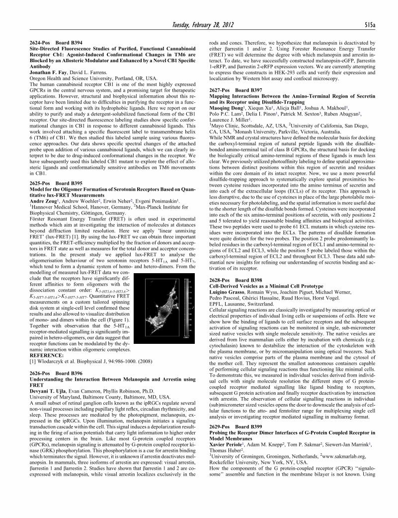
2625-Pos Board B395Model for the Oligomer Formation of Serotonin Receptors Based on Quan-titative lux-FRET MeasurementsAndre Zeug1, Andrew Woehler2, Erwin Neher2, Evgeni Ponimaskin1.1Hannover Medical School, Hanover, Germany, 2Max-Planck Institute forBiophysical Chemistry, Gottingen, Germany.Forster Resonant Energy Transfer (FRET) is often used in experimentalmethods which aim at investigating the interaction of molecules at distancesbeyond diffraction limited resolution. Here we apply ’linear unmixingFRET’ (lux-FRET) [1]. By using the lux-FRET we can obtain three importantquantities, the FRET-efficiency multiplied by the fraction of donors and accep-tors in FRET state as well as measures for the total donor and acceptor concen-trations. In the present study we applied lux-FRET to analyse theoligomerisation behaviour of two serotonin receptors 5-HT1A and 5-HT7,which tend to form a dynamic system of homo- and hetero-dimers. From the

modelling of measured lux-FRET data we con-clude that the receptors have significantly dif-ferent affinities to form oligomers with thedissociation constant order: K5-HT1A-5-HT1A>K5-HT7-5-HT1A>K5-HT7-5-HT7. Quantitative FRETmeasurements on a custom tailored spinningdisk system at single-cell level confirmed theseresults and also allowed to visualize distributionof mono- and dimers within the cell (Figure 1).Together with observation that the 5-HT1A

receptor-mediated signalling is significantly im-paired in hetero-oligomers, our data suggest thatreceptor functions can be modulated by the dy-

namic interaction within oligomeric complexes.REFERENCE:[1] Wlodarczyk et al. Biophysical J, 94:986-1000. (2008)

2626-Pos Board B396Understanding the Interaction Between Melanopsin and Arrestin usingFRETDevyani T. Ujla, Evan Cameron, Phyllis Robinson, Ph.D.University of Maryland, Baltimore County, Baltimore, MD, USA.A small subset of retinal ganglion cells known as the ipRGCs regulate severalnon-visual processes including pupillary light reflex, circadian rhythmicity, andsleep. These processes are mediated by the photopigment, melanopsin, ex-pressed in the ipRGCs. Upon illumination, melanopsin initiates a signalingtransduction cascade within the cell. This signal induces a depolarization result-ing in the firing of action potentials that carry light information to higher orderprocessing centers in the brain. Like most G-protein coupled receptors(GPCRs), melanopsin signaling is attenuated by G-protein coupled receptor ki-nase (GRK) phosphorylation. This phosphorylation is a cue for arrestin bindingwhich terminates the signal. However, it is unknown if arrestin deactivates mel-anopsin. In mammals, three isoforms of arrestin are expressed: visual arrestin,barrestin 1 and barrestin 2. Studies have shown that barrestin 1 and 2 are co-expressed with melanopsin, while visual arrestin localizes exclusively in the

rods and cones. Therefore, we hypothesize that melanopsin is deactivated byeither barrestin 1 and/or 2. Using Forester Resonance Energy Transfer(FRET) we will determine the degree with which melanopsin and arrestin in-teract. To date, we have successfully constructed melanopsin-eGFP, barrestin1-eRFP, and barrestin 2-eRFP expression vectors. We are currently attemptingto express these constructs in HEK-293 cells and verify their expression andlocalization by Western blot assay and confocal microscopy.

2627-Pos Board B397Mapping Interactions Between the Amino-Terminal Region of Secretinand its Receptor using Disulfide-TrappingMaoqing Dong1, Xiequn Xu1, Alicja Ball1, Joshua A. Makhoul1,Polo P.C. Lam2, Delia I. Pinon1, Patrick M. Sexton3, Ruben Abagyan2,Laurence J. Miller1.1Mayo Clinic, Scottsdale, AZ, USA, 2University of California, San Diego,CA, USA, 3Monash University, Parkville, Victoria, Australia.While NMR and crystal structures have defined the molecular basis for dockingthe carboxyl-terminal region of natural peptide ligands with the disulfide-bonded amino-terminal tail of class B GPCRs, the structural basis for dockingthe biologically critical amino-terminal regions of these ligands is much lessclear. We previously utilized photoaffinity labeling to define spatial approxima-tions between distinct positions within this region of secretin and residueswithin the core domain of its intact receptor. Now, we use a more powerfuldisulfide-trapping approach to systematically explore spatial proximities be-tween cysteine residues incorporated into the amino terminus of secretin andinto each of the extracellular loops (ECLs) of its receptor. This approach isless disruptive, due to the use of cysteines in place of the large photolabile moi-eties necessary for photolabeling, and the spatial information is more useful dueto the shorter length of the disulfide bonds formed. Cysteines were incorporatedinto each of the six amino-terminal positions of secretin, with only positions 2and 5 tolerated to yield reasonable binding affinities and biological activities.These two peptides were used to probe 61 ECL mutants in which cysteine res-idues were incorporated into the ECLs. The patterns of disulfide formationwere quite distinct for the two probes. The position 2 probe predominantly la-beled residues in the carboxyl-terminal region of ECL1 and amino-terminal re-gions of ECL2 and ECL3, while the position 5 probe labeled those within thecarboxyl-terminal region of ECL2 and throughout ECL3. These data add sub-stantial new insights for refining our understanding of secretin binding and ac-tivation of its receptor.

2628-Pos Board B398Cell-Derived Vesicles as a Minimal Cell PrototypeLuigino Grasso, Romain Wyss, Joachim Piguet, Michael Werner,Pedro Pascoal, Gherici Hassaıne, Ruud Hovius, Horst Vogel.EPFL, Lausanne, Switzerland.Cellular signaling reactions are classically investigated by measuring optical orelectrical properties of individual living cells or suspensions of cells. Here weshow how the binding of ligands to cell surface receptors and the subsequentactivation of signaling reactions can be monitored in single, sub-micrometersized native vesicles with single molecule sensitivity. The native vesicles arederived from live mammalian cells either by incubation with chemicals (e.g.cytochalasin) known to destabilize the interaction of the cytoskeleton withthe plasma membrane, or by micromanipulation using optical tweezers. Suchnative vesicles comprise parts of the plasma membrane and the cytosol ofthe mother cell. They represent the smallest autonomous containers capableof performing cellular signaling reactions thus functioning like minimal cells.To demonstrate this, we measured in individual vesicles derived from individ-ual cells with single molecule resolution the different steps of G protein-coupled receptor mediated signalling like ligand binding to receptors,subsequent G protein activation and finally receptor deactivation by interactionwith arrestin. The observation of cellular signalling reactions in individual(sub)micrometer sized vesicles opens the door to downscale the analysis of cel-lular functions to the atto- and femtoliter range for multiplexing single cellanalysis or investigating receptor mediated signalling in multiarray format.

2629-Pos Board B399Probing the Receptor Dimer Interfaces of G-Protein Coupled Receptor inModel MembranesXavier Periole1, Adam M. Knepp2, Tom P. Sakmar2, Siewert-Jan Marrink1,Thomas Huber2.1University of Groningen, Groningen, Netherlands, 2www.sakmarlab.org,Rockefeller University, New York, NY, USA.How the components of the G protein-coupled receptor (GPCR) ‘‘signalo-some’’ assemble and function in the membrane bilayer is not known. Using