Embed Size (px)
Citation preview
Mar. Drugs 2015, 13, 4654-4681; doi:10.3390/md13084654
marine drugs ISSN 1660-3397
www.mdpi.com/journal/marinedrugs
Article
The Bright Side of Gelatinous Blooms: Nutraceutical Value and Antioxidant Properties of Three Mediterranean Jellyfish (Scyphozoa)
Antonella Leone 1,2,*, Raffaella Marina Lecci 1,2, Miriana Durante 1, Federica Meli 3
and Stefano Piraino 2,4
1 Institute of Sciences of Food Production, National Research Council, Unit of Lecce (CNR, ISPA),
Via Prov.le Lecce-Monteroni, 73100 Lecce, Italy; E-Mails: [email protected] (R.M.L.);
[email protected] (M.D.) 2 Consorzio Nazionale Interuniversitario per le Scienze del Mare (CoNISMa), Local Unit of Lecce,
Via Prov.le Lecce-Monteroni, 73100 Lecce, Italy; E-Mail: [email protected] 3 Dipartimento di Scienze degli Alimenti, Università di Parma, Parco Area delle Scienze, 59/A,
43124 Parma, Italy; E-Mail: [email protected] 4 Università del Salento, DiSTeBA Via Prov.le Lecce-Monteroni, 73100 Lecce, Italy
* Author to whom correspondence should be addressed; E-Mail: [email protected];
Tel.: +39-0832-422615; Fax: +39-0832-422620.
Academic Editor: Colin Barrow
Received: 1 May 2015 / Accepted: 20 July 2015 / Published: 29 July 2015
Abstract: Jellyfish are recorded with increasing frequency and magnitude in many coastal
areas and several species display biological features comparable to the most popular Asiatic
edible jellyfish. The biochemical and antioxidant properties of wild gelatinous biomasses, in
terms of nutritional and nutraceutical values, are still largely unexplored. In this paper, three of
the most abundant and commonly recorded jellyfish species (Aurelia sp.1, Cotylorhiza
tuberculata and Rhizostoma pulmo) in the Mediterranean Sea were subject to investigation.
A sequential enzymatic hydrolysis of jellyfish proteins was set up by pepsin and
collagenase treatments of jellyfish samples after aqueous or hydroalcoholic protein
extraction. The content and composition of proteins, amino acids, phenolics, and fatty
acids of the three species were recorded and compared. Protein content (mainly represented
by collagen) up to 40% of jellyfish dry weight were found in two of the three jellyfish
species (C. tuberculata and R. pulmo), whereas the presence of ω-3 and ω-6
polyunsaturated fatty acids (PUFAs) was significantly higher in the zooxanthellate jellyfish
OPEN ACCESS
Mar. Drugs 2015, 13 4655
C. tuberculata only. Remarkable antioxidant ability was also recorded from both
proteinaceous and non proteinaceous extracts and the hydrolyzed protein fractions in all
the three species. The abundance of collagen, peptides and other bioactive molecules make
these Mediterranean gelatinous biomasses a largely untapped source of natural compounds
of nutraceutical, cosmeceutical and pharmacological interest.
Keywords: novel foods; marine jellyfish; macrozooplankton; antioxidants; collagen;
nutraceuticals
1. Introduction
In the last decades, the positive association between nutraceutical or functional food and human
health prompted the search for bioactive compounds from living organisms. Although terrestrial plants
and marine taxa represent the main sources of bioactive natural products, the inherent difficulties of
sampling the diversity of aquatic environments have meant that the biochemistry of marine unicellular
and multicellular organisms remains less explored [1–3]. The oceans represent the largest biome on
earth, covering 71% of the planet surface, with a much higher diversity of body plans than on land [4].
Therefore, marine organisms still represent a largely unexplored reservoir of natural products, a vast
potential source of diverse and healthy food, new drugs and bioactive compounds, due to the presence
of secondary metabolites spanning a wide range of structural classes and various biosynthetic origins [5].
For this reason, many bioactive molecules from marine organisms with potential antimicrobial,
anti-inflammatory and anticancer properties have represented the focus of recent researches [1,5–9].
Marine fishery resources have been exploited over a long time, leading to increasing vulnerability,
irreversible change, and collapse of fish stocks [10,11]. Therefore, the search for new potential sources of
bioactive compounds and their direct exploitation from living biomass primarily requires considerations
on resource supply and its sustainable harvesting and must be supported by ecosystem-based studies of
environmental sustainability to prevent overexploitation risks and ecological imbalances.
In this framework, cnidarians have increasingly become an important source of physiologically
active compounds [8,9]. Particularly, jellyfish represent a conspicuous component of marine
ecosystems and their populations are known to experience large seasonal and inter-annual fluctuations,
characterized by sudden outbreaks—also known as “blooms”—followed by rarity periods [12]. In
recent years, evidence is accumulating concerning increasing magnitude and frequency of jellyfish
blooms [13,14], which represent a widely distributed and abundant gelatinous biomass in the world
oceans. Jellyfish blooms are mainly sustained from large Rhizostomoidea and Semaeostomeae
medusae at all latitudes of the Southern and Northern hemispheres, with many coastal locations
sustaining high gelatinous biomasses directly influenced by global warming [15–17]. From an
anthropocentric point of view, jellyfish outbreaks are often negatively perceived because they (i) may
determine severe negative consequences on human health and coastal tourism due to painful stings; (ii)
may impair coastal industries by clogging cooling pipelines [18]; (iii) affect fishery by the reduction of
fish stocks through competition for food or direct predation [19,20]; or (iv) produce mass mortalities in
caged fish aquaculture. Management and adaptation strategies have been developed aiming to prevent
Mar. Drugs 2015, 13 4656
negative impacts [17,21] also in the framework of dedicated research projects, like the MED-JELLYRISK
project within ENPI CBC MED (European Neighbourhood and Partnership Instrument Cross-Border
Cooperation in the Mediterranean) [22]. However, due to their high abundances and high regenerative
and reproductive potentials, jellyfish may be regarded as a new source of pharmacologic, nutraceutical
and food/feed compounds, and their potential use in tissue engineering, food industry and medicine
may provide the opportunity of showcasing jellyfish in a more positive light [12,23–26]. Information
on the biochemical composition and biomass of jellyfish has been available in recent years. The dry
mass (DW) is in the range of about 3%–5% of the fresh weight, and jellyfish carbon (C) is typically
<15% of DW [27] where in non-gelatinous groups it accounts for up to 30%–60% [28]. The organic
content is mainly represented by protein (collagen) while lipids and carbohydrates represent minor
components of jellyfish tissue [29,30].
Despite a low organic content, jellyfish have long been highly considered in Asiatic countries for their
therapeutic value in the treatment of arthritis, hypertension, bone pain, and ulcers, as softening skin and
improving digestion [31,32]. These properties, described mainly in non-scientific publications [33],
are likely attributable to the collagen, a structural protein family widely present throughout the
animal tissues as prevailing component of extracellular matrices in connective tissues [34,35] and
the main structural protein in the jellyfish body mass [32,36]. Collagen has diverse general and
biomedical applications and is also a common constituent of many cosmetic and food products in the
form of gelatin. Interest in collagen as a biomaterial is due to its low immunogenicity and high
biocompatibility [37], and because it can be extracted from a variety of organisms, such as bovine and
porcine skins. However, these sources of collagen are increasingly rejected for disease risks (like bovine
spongiform encephalopathy) or religious reasons, while marine organisms, especially marine
invertebrates are becoming an attractive source for collagen industrial uses. Structure and sequences of
fibrillar collagen are highly conserved, and cnidarian collagen shares several features with their human
counterparts [38]. Jellyfish collagen from Rhopilema esculentum could protect mice skin from the
ultra-violet (UV) radiation damages alleviating the UV-inducing abnormal changes of antioxidative
indicators [39,40]. Collagen from the giant edible jellyfish Nemopilema nomurai showed
immunostimulatory effect in vitro, on hybridoma line HB4C5, human peripheral blood lymphocytes [41],
and in vivo [42]. In addition, the oral administration of type II-like collagen of cannonball jellyfish
Stomolophus meleagris delayed the onset and suppressed collagen-induced arthritis in animal models [43].
More recent studies have shown medical properties of this polymer extracted from Rhopilema
esculentum for cartilage tissue engineering [44]. Accordingly, jellyfish collagen might be also used in
the cosmetics, in creams and lotions for the skin as well as in the biomedical and pharmaceutical industry.
Collagen can also be a source of bioactive peptides. Bioactive peptides are 2–20 amino acid
fragments inactive in the parent protein; when released by enzymatic hydrolysis, these fragments may
exert various physiological functions, depending on their specific amino acid composition [45]. Food
derived peptides may have several functions such as immunomodulatory [46], antimicrobial [47],
antioxidative [48], and antihypertensive [49] properties. Due to their high bioactivity and biocompatibility,
collagen peptides and hydrolysate may be used as functional ingredients in medicine and food industries.
Collagen hydrolysate of the jellyfish Rhopilema esculentum has shown antioxidant activity, ability to
chelate Cu2+ ions and to inhibit tyrosinase activity [39]. Enzymatic hydrolysis, indeed, may improve
the functional properties of proteins, such as solubility and emulsification [50]. Enzymatic hydrolysates
Mar. Drugs 2015, 13 4657
from jellyfish collagen are known to have protective effects on mice skin photoaging induced by UV
irradiation higher than non-hydrolyzed jellyfish collagen [40]. This mechanism is probably related to
the in vivo antioxidative properties showed by collagen and peptides with high contents of glycine,
proline, and hydrophobic amino acids [51].
Besides specific properties of the proteins and derived peptides, the whole mass of jellyfish, as other
marine products, might be considered for food or feed purpose due to their content of essential nutrients
or biochemical characteristics unavailable or poorly present in products from terrestrial plants and
animals. Indeed, several species of the scyphozoan jellyfish with middle stings in South-East Asia,
mainly in China and in Japan, represent a part of the multimillion-dollar seafood business and are
appreciated not only for its texture and taste, but also for its composition which ensures a low calorie
diet being low in fat, cholesterol, and salt [31–33].
Seafood deserves a key role in nutrition and health because it provides omega-3 and omega-6 fatty
acids known for the reduction power of cholesterol levels and the decrease of incidence of coronary
heart diseases. The lipid composition of several cnidarians may vary substantially [52,53], depending on
diet or symbiotic association with unicellular algae. For instance, lipid composition seems determined by
diet in non-symbiotic jellyfish, i.e., the moon jellyfish Aurelia sp. [54]. Conversely, in zooxanthellate
cnidarians lipids are regularly translocated in their tissues from their unicellular symbionts [8,55]. The
absence of storage lipids, such as wax esters, also suggests that proteins govern energy storage [56,57].
Remarkably, jellyfish feed used for chickens and pigs determined an increase of muscle to bone ratio
and of the overall body tissue without toxic effect on blood, liver and muscle [32].
In this study, original data on the biochemical composition and nutraceutical properties of three
jellyfish, namely the scyphozoan Aurelia sp.1 (commonly known as moon jellyfish), Cotylorhiza
tuberculata (known as fried-egg jellyfish) and Rhizostoma pulmo (known as sea lung jellyfish) are
provided. These species bloom yearly along the Mediterranean coastal areas from Spain to the North
Adriatic Sea, forming large populations of considerable and totally unexploited biomass, thus
representing excellent candidates for the isolation and potentially sustainable production of bioactive
compounds in the fields of nutraceuticals, animal feeds, and pharmaceutics. Quali-quantitative
identification and measurement of proteins, together with their antioxidant activity, and analysis of lipid
content were carried out to assess biochemical values of these gelatinous organisms as putative novel
food or for the production of jelly-related, low-cost raw materials for either animal feed or for
applications in cosmetics or biomedical industries.
2. Results and Discussion
2.1. Jellyfish Blooms and Biomass Characterization
The semaeostome jellyfish Aurelia sp.1 can be found in marinas and coastal lagoons of the
Mediterranean Sea. This is a non-indigenous or alien species introduced in the Mediterranean Sea by
shellfish aquaculture, by the transfer of the polyp stage commonly living on bivalve shells. In the
Varano lagoon (Apulia, SE Italy), a dense population of Aurelia sp.1 medusae (up to 80 individuals·m−3)
is generated yearly from February to July. Differently, the rhizostome jellyfish Cotylorhiza tuberculata
and Rhizostoma pulmo [58] are typical species of marine coastal waters and can be encountered across
Mar. Drugs 2015, 13 4658
the Western and Central Mediterranean Sea. These two species are among the top five most frequently
recorded species along the Italian coastlines by the citizen science project METEOMEDUSE carried out
by the MED-JELLYRISK project [22] and represent the largest part of jellyfish biomass off the Apulia
coasts across the years 2010–2014. In September 2013, a high density (>48,000 individuals/km2) of
R. pulmo jellyfish was assessed along the Apulian shores in southwestern part the Gulf of Taranto by
an ultralight aerial survey in the framework of the MED-JELLYRISK project [22], with an estimated
biomass range of 100–300/km2 (MED-JELLYRISK, unpublished data). Biometric and average
biomass data of individuals of the three jellyfish species are shown in Table 1.
Table 1. Biometric measures, fresh and dry weights and organic matter of the three
jellyfish species sampled in the 2010–2014 summers.
Jellyfish Samples
Umbrella
Diameter Range *
Fresh Weight
Range * Ratio
FW/Diameter
Range of DW * Organic Matter
(OM) Mean **
Mean (cm) Mean (g) (% of FW) (% of DW)
Aurelia sp.1 10–23
16.2 ± 4.9
47–604
257 ± 237
6.8–26.3
13.5 ± 9.2 2.2–3.0 23.9 ± 3.3 a
Cotylorhiza tuberculata 6–29
17.7 ± 6.3
19–1770
638 ± 475
3–61
24.3 ± 16.9 3.9–32.4 30.2 ± 2.4 b
Rhizostoma pulmo 8–37
20.8 ± 7.2
42–2440
860 ± 720
5.3–65.9
32.6 ± 14.2 4.1–6.8 29.5 ± 6.6 b
FW, fresh weight; DW, dry weight; * Data are expressed as range and/or means ± standard deviation
(10 < n < 41); ** Organic matter data are the mean of two independent experiments each performed in
quintuplicate, superscript lower case letters indicate significant differences (p < 0.05) within the column.
The umbrella diameter of Aurelia sp.1 specimens ranged from about 10–23 cm, C. tuberculata and
R. pulmo umbrellas were 6–29 cm and 8–37 cm, respectively, with a proportionally increasing biomass
with jellyfish size. The variability of the biometric measures, including the fresh-weight (FW)/diameter
ratio and dry weight (DW) percentage values, is representative of the seasonal growth of jellyfish
collected at different times throughout the spring-summer months and at different growth stages [8].
After lyophilisation, the DW of Aurelia sp.1 from Varano ranged from 2.2% to 3%, C. tuberculata
3.9%–32.4% and R. pulmo 4.1%–6.8% of the FW, displaying a high and quite constant water content
for Aurelia sp.1 and R. pulmo and high variability in tissue consistency in C. tuberculata specimens,
which reached also the highest DW proportion. The DW of Aurelia sp.1 from Varano was slightly
lower than for moon jellyfish collected along the Slovenian coasts (4.0% of FW) whereas similar results
were obtained for R. pulmo [59]. The organic matter content (OM) was highly comparable between
C. tuberculata and R. pulmo (30.2% ± 2.4% and 29.5% ± 6.6% of DW, respectively), while Aurelia sp.1
(23.9% ± 3.3% of DW) showed a statistically significant lower OM than C. tuberculata. The fried-egg
jellyfish also showed higher DW values than R. pulmo or Aurelia sp.1, providing critical information
for the potential exploitation of these jellyfish biomass.
The potential use of jellyfish biomass should be assessed also by taking into consideration the total
energy or gross energy value (GE), which is directly related to DW and OM values. The GE of jellyfish
biomass has long been neglected and poorly documented, compared to other planktonic taxa with
prominent roles in marine food webs, such as crustaceans or fish, and only a few studies dealt with
Mar. Drugs 2015, 13 4659
jellyfish GE to assess their value as a prey for apical gelativorous predators, such as fish or turtles.
This is because the energetic value of jellyfish biomass was long considered poor compared to other
prey items. However, recent evidence shows jellyfish biomass may represent a key component of diet of
several organisms: leatherback sea turtles may consume up to 261 jellyfish·day−1 (330 kg jellyfish
wet mass·day−1), whereas several fish species may rely on jellyfish prey up to 100% of their diet [60–62].
Measured by bomb calorimetric or calculated from carbon content, the GE for Cyanea capillata,
Rhizostoma octopus and Chrysaora hysoscella [63], Atolla wyvillei, Aurelia aurita and Pelagia
noctiluca [64], was in the range of 2.3–5.95 kJ/g of DW, with differences between species and body
parts. These values are lower than the range of energy values of protein (10.2–18.2 kJ/g), fat (35.0–
37.7 kJ/g) and total carbohydrate (11.3–17.4 kJ/g) in ordinary human diet [65], and actually lower than
other marine organisms used as human foods or animal feed. In this framework, jellyfish may
represent a healthy energy-restricted food that may reduce caloric intake and over-nutrition trends in
the typical Western human lifestyle, without necessarily decreasing the amount of consumed food [66].
2.2. Jellyfish Protein
2.2.1. Amino Acid Composition
The amino acid (AA) composition has been determined for the three species of jellyfish (Table 2). None
of the jellyfish protein samples contained the essential amino acid (EAA) tryptophan (Try), as previously
reported for collagen peptides derived from Rhopilema esculentum umbrella [67] and gonads [68],
whole tentacles and nematocyst suspensions of Chrysaora quinquecirrha [69], and total proteins
profiles from Chrysaora hysoscella, Pelagia noctiluca and also R. pulmo [59]. All the remaining EAA,
namely histidine (His), isoleucine (Ile), leucine (Leu), lysine (Lys), methionine (Met), phenylalanine
(Phe), threonine (Thr) and valine (Val), were found in R. pulmo and C. tuberculata specimens.
Differently, the EAAs His and Leu were not detected in Aurelia sp.1, as well as cysteine (Cys) and
arginine (Arg) were not found in C. tuberculata (Table 2).
The proportion of EAAs out of the total AAs in the whole tissues of the Aurelia sp.1, C. tuberculata
and R. pulmo, was 31.4%, 53.6%, and 50.8%, respectively. The last two EAA percentages were higher
than those recorded from the gonads of the edible Asiatic jellyfish Rhopilema esculentum [68], which
may depend on the advanced reproductive status of many jellyfish specimens sampled during this
study. Overall, the EEAs content of the three Mediterranean jellyfish is comparable to those recorded
in other high-value Asiatic and European seafood, at least in terms of percentage composition [68,70].
The most abundant amino acid found in Aurelia sp.1 was Gly, followed by Glu, Ser, Thr and Tyr
(Table 2). Gly is the fixed constituent of collagen-typical repeating triplets with a repeating X-Y-Gly
sequence, where X and Y can be any amino acid, although proline (Pro) and hydroxyproline (Hyp)
residues are the most common triplet in collagen [71,72]. The amounts of aromatic amino acids (AAA)
were 12.7%, 22.9% and 22.5% of the total amino acids in Aurelia sp.1, C. tuberculata and R. pulmo,
respectively. The AA profiles were more similar between the two rhizostome jellyfish C. tuberculata
and R. pulmo, both as EAA and AAA percentages, as well as single AAs, where glutamine/glutamate
were the most representative followed by Phe, Leu, Tyr, Thr, His and Ser (Table 2). The putative
occurrence of a wider diversity of proteins other than collagen in the two Rhizostomeae could justify
Mar. Drugs 2015, 13 4660
their different AA profiles compared to Aurelia sp.1. Overall, the finding of important proteinogenic
and non-proteinogenic AAs (such as Glu, Gly, Phe, Asp, Met, Leu, Tyr, Lys, and Arg) may account
for the long tradition of therapeutic value of jellyfish food in Chinese pharmacopeia.
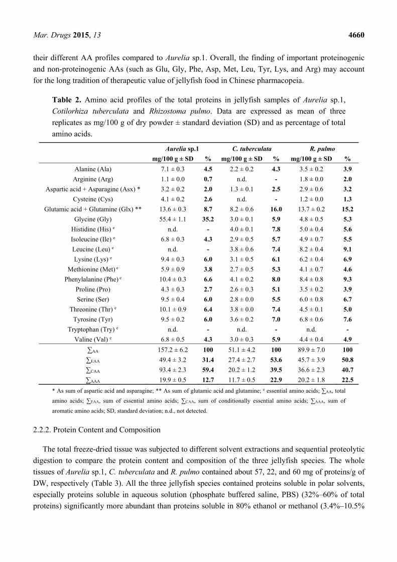
Table 2. Amino acid profiles of the total proteins in jellyfish samples of Aurelia sp.1,
Cotilorhiza tuberculata and Rhizostoma pulmo. Data are expressed as mean of three
replicates as mg/100 g of dry powder ± standard deviation (SD) and as percentage of total
amino acids.
Aurelia sp.1 C. tuberculata R. pulmo
mg/100 g ± SD % mg/100 g ± SD % mg/100 g ± SD %
Alanine (Ala) 7.1 ± 0.3 4.5 2.2 ± 0.2 4.3 3.5 ± 0.2 3.9
Arginine (Arg) 1.1 ± 0.0 0.7 n.d. - 1.8 ± 0.0 2.0
Aspartic acid + Asparagine (Asx) * 3.2 ± 0.2 2.0 1.3 ± 0.1 2.5 2.9 ± 0.6 3.2
Cysteine (Cys) 4.1 ± 0.2 2.6 n.d. - 1.2 ± 0.0 1.3
Glutamic acid + Glutamine (Glx) ** 13.6 ± 0.3 8.7 8.2 ± 0.6 16.0 13.7 ± 0.2 15.2
Glycine (Gly) 55.4 ± 1.1 35.2 3.0 ± 0.1 5.9 4.8 ± 0.5 5.3
Histidine (His) e n.d. - 4.0 ± 0.1 7.8 5.0 ± 0.4 5.6
Isoleucine (Ile) e 6.8 ± 0.3 4.3 2.9 ± 0.5 5.7 4.9 ± 0.7 5.5
Leucine (Leu) e n.d. - 3.8 ± 0.6 7.4 8.2 ± 0.4 9.1
Lysine (Lys) e 9.4 ± 0.3 6.0 3.1 ± 0.5 6.1 6.2 ± 0.4 6.9
Methionine (Met) e 5.9 ± 0.9 3.8 2.7 ± 0.5 5.3 4.1 ± 0.7 4.6
Phenylalanine (Phe) e 10.4 ± 0.3 6.6 4.1 ± 0.2 8.0 8.4 ± 0.8 9.3
Proline (Pro) 4.3 ± 0.3 2.7 2.6 ± 0.3 5.1 3.5 ± 0.2 3.9
Serine (Ser) 9.5 ± 0.4 6.0 2.8 ± 0.0 5.5 6.0 ± 0.8 6.7
Threonine (Thr) e 10.1 ± 0.9 6.4 3.8 ± 0.0 7.4 4.5 ± 0.1 5.0
Tyrosine (Tyr) 9.5 ± 0.2 6.0 3.6 ± 0.2 7.0 6.8 ± 0.6 7.6
Tryptophan (Try) e n.d. - n.d. - n.d. -
Valine (Val) e 6.8 ± 0.5 4.3 3.0 ± 0.3 5.9 4.4 ± 0.4 4.9
∑AA 157.2 ± 6.2 100 51.1 ± 4.2 100 89.9 ± 7.0 100
∑EAA 49.4 ± 3.2 31.4 27.4 ± 2.7 53.6 45.7 ± 3.9 50.8
∑CAA 93.4 ± 2.3 59.4 20.2 ± 1.2 39.5 36.6 ± 2.3 40.7
∑AAA 19.9 ± 0.5 12.7 11.7 ± 0.5 22.9 20.2 ± 1.8 22.5
* As sum of aspartic acid and asparagine; ** As sum of glutamic acid and glutamine; e essential amino acids; ∑AA, total
amino acids; ∑EAA, sum of essential amino acids; ∑CAA, sum of conditionally essential amino acids; ∑AAA, sum of
aromatic amino acids; SD, standard deviation; n.d., not detected.
2.2.2. Protein Content and Composition
The total freeze-dried tissue was subjected to different solvent extractions and sequential proteolytic
digestion to compare the protein content and composition of the three jellyfish species. The whole
tissues of Aurelia sp.1, C. tuberculata and R. pulmo contained about 57, 22, and 60 mg of proteins/g of
DW, respectively (Table 3). All the three jellyfish species contained proteins soluble in polar solvents,
especially proteins soluble in aqueous solution (phosphate buffered saline, PBS) (32%–60% of total
proteins) significantly more abundant than proteins soluble in 80% ethanol or methanol (3.4%–10.5%
Mar. Drugs 2015, 13 4661
of the total proteins). The fried-egg jellyfish C. tuberculata showed the highest percentage of both
PBS- and hydroalcoholic-soluble proteins, as compared to Aurelia sp.1 and R. pulmo.
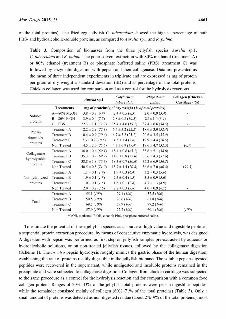
Table 3. Composition of biomasses from the three jellyfish species Aurelia sp.1,
C. tuberculata and R. pulmo. The polar solvent extraction with 80% methanol (treatment A)
or 80% ethanol (treatment B) or phosphate buffered saline (PBS) (treatment C) was
followed by enzymatic digestion with pepsin and then collagenase. Data are presented as
the mean of three independent experiments in triplicate and are expressed as mg of protein
per gram of dry weight ± standard deviation (SD) and as percentage of the total proteins.
Chicken collagen was used for comparison and as a control for the hydrolysis reactions.
Aurelia sp.1
Cotylorhiza
tuberculata
Rhizostoma
pulmo
Collagen (Chicken
Cartilage) (%)
Treatments mg of proteins/g of dry weight (% of total proteins)
Soluble
proteins
A—80% MeOH
B—80% EtOH
C—PBS
3.8 ± 0.8 (6.9)
3.9 ± 0.4 (7.7)
22.3 ± 1.1 (32.2)
2.4 ± 0.5 (8.3)
2.8 ± 0.8 (10.5)
35.4 ± 4.6 (59.1)
2.0 ± 0.9 (3.4)
2.1± 1.0 (3.4)
37.4 ± 6.6 (38.5)
-
-
-
Pepsin
digestible
proteins
Treatment A
Treatment B
Treatment C
Non Treated
12.2 ± 2.9 (22.1)
10.4 ± 0.9 (20.6)
7.3 ± 0.2 (10.6)
14.5 ± 2.0 (25.5)
6.4 ± 3.2 (22.2)
6.7 ± 3.2 (25.1)
4.5 ± 1.4 (7.6)
4.3 ± 0.9 (19.4)
18.6 ± 3.0 (32.4)
20.6 ± 3.5 (33.4)
19.9 ± 4.4 (20.5)
19.6 ± 4.7 (32.5)
(0.7)
Collagenase
hydrolysable
proteins
Treatment A
Treatment B
Treatment C
Non Treated
38.0 ± 0.6 (69.1)
35.3 ± 0.9 (69.9)
38.8 ± 1.8 (55.8)
40.5 ± 0.5 (71.0)
18.4 ± 0.8 (63.1)
14.6 ± 0.8 (55.0)
18.3 ± 0.7 (30.6)
15.7 ± 4.4 (70.8)
33.6 ± 5.1 (58.6)
35.6 ± 4.3 (57.6)
35.2 ± 6.9 (36.2)
36.6 ± 7.0 (60.8)
(99.3)
Not-hydrolyzed
proteins
Treatment A
Treatment B
Treatment C
Non Treated
1.1 ± 0.1 (1.9)
1.9 ± 0.1 (1.8)
1.0 ± 0.1 (1.5)
2.0 ± 0.2 (3.6)
1.9 ± 0.3 (6.4)
2.5 ± 0.4 (9.5)
1.6 ± 0.1 (2.8)
2.2 ± 0.3 (9.8)
3.2 ± 0.3 (5.6)
3.5 ± 0.9 (5.6)
4.7 ± 1.3 (4.9)
4.0 ± 0.9 (6.7)
-
Total
Treatment A
Treatment B
Treatment C
Non Treated
55.1 (100)
50.5 (100)
69.5 (100)
57.0 (100)
29.1 (100)
26.6 (100)
59.9 (100)
22.2 (100)
57.5 (100)
61.8 (100)
97.2 (100)
60.1 (100)
(100)
MeOH, methanol; EtOH, ethanol; PBS, phosphate buffered saline.
To estimate the potential of these jellyfish species as a source of high value and digestible peptides,
a sequential protein extraction procedure, by means of consecutive enzymatic hydrolysis, was designed.
A digestion with pepsin was performed as first step on jellyfish samples pre-extracted by aqueous or
hydroalcoholic solutions, or on non-treated jellyfish tissues, followed by the collagenase digestion
(Scheme 1). The in vitro pepsin hydrolysis roughly mimics the gastric phase of the human digestion,
establishing the rate of proteins readily digestible in the jellyfish biomass. The soluble pepsin-digested
peptides were recovered in the supernatant, while undigested and insoluble proteins remained in the
precipitate and were subjected to collagenase digestion. Collagen from chicken cartilage was subjected
to the same procedure as a control for the hydrolysis reaction and for comparison with a common food
collagen protein. Ranges of 20%–33% of the jellyfish total proteins were pepsin-digestible peptides,
while the remainder consisted mainly of collagen (60%–71% of the total proteins) (Table 3). Only a
small amount of proteins was detected as non-digested residue (about 2%–9% of the total proteins), most
Mar. Drugs 2015, 13 4662
of them occurring in the untreated samples (without pre-extraction), putatively indicating a limited
accessibility to the enzymes, rather than indigestibility. These results suggest that a significant proportion
of both collagen- and pepsin-digestible jellyfish peptides can be dispersed in aqueous solutions by PBS
pre-extraction, while the pre-treatments with methanol and ethanol solutions allow limited extraction of
proteins. Indeed, treatment with ethanol-based solutions can represent a method to remove fats and
pigments [8], leaving the protein fraction, including collagen, quite clean. Indeed, partially fractionated
hydroalcoholic extracts from C. tuberculata jellyfish contained carotenoids and fatty acids derived by
algal symbionts, and yielded compounds with biological activity, representing a high potential protocol
for nutraceuticals and drug discovery [8].
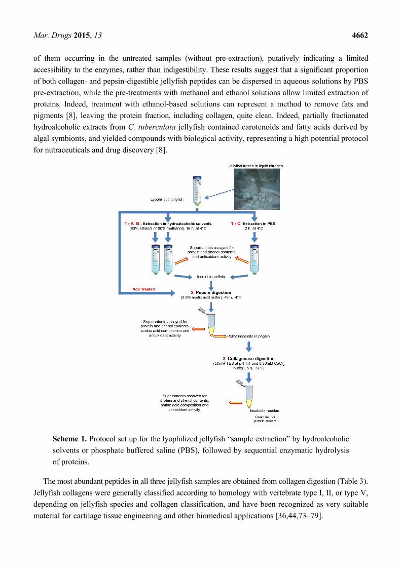
Scheme 1. Protocol set up for the lyophilized jellyfish “sample extraction” by hydroalcoholic
solvents or phosphate buffered saline (PBS), followed by sequential enzymatic hydrolysis
of proteins.
The most abundant peptides in all three jellyfish samples are obtained from collagen digestion (Table 3).
Jellyfish collagens were generally classified according to homology with vertebrate type I, II, or type V,
depending on jellyfish species and collagen classification, and have been recognized as very suitable
material for cartilage tissue engineering and other biomedical applications [36,44,73–79].
Mar. Drugs 2015, 13 4663
The pre-digestion with pepsin and the evaluation of only collagenase-digested polypeptides implies
that just pure collagen was evaluated. It is noteworthy that Aurelia sp.1 and R. pulmo tissues contained up
to about 40% of pure collagen based on the lyophilized dry weight. That percentage is consistent with
previous reported data on different species of edible Asiatic jellyfish, obtained after pepsin
digestion [74,78,80] with a yield of collagen of 46.4% based on the lyophilized dry weight for
Stomolophus meleagris [74] and 35.2% for Rhopilema asamushi [80]. A pepsin-solubilized collagen was
obtained from Chrysaora sp. with a maximum yield of 19% (ash-free lyophilized dry weight) [74].
Also, the pre-digestion by pepsin leads to the elimination of non-helical terminal regions of collagen
(telopeptides), which are also the major antigenic determinants giving a collagen product (atelocollagen)
with high purity and increased solubility. The reduced degree of antigenicity and the solubility features
of the jellyfish-derived collagen have also prompted the use of collagen in the food and cosmetics
sector [1,72,77].
Enzymatic hydrolysis of seafood and other fish biomass has been employed as an alternative
approach to the conversion of underutilized biomass or by-products into edible protein products [81],
and similar processing methodologies might be used to successfully exploit the exceedingly large
amount of jellyfish biomasses.
2.3. Phenolic Compound Content in Jellyfish Hydroalcoholic Soluble Extracts
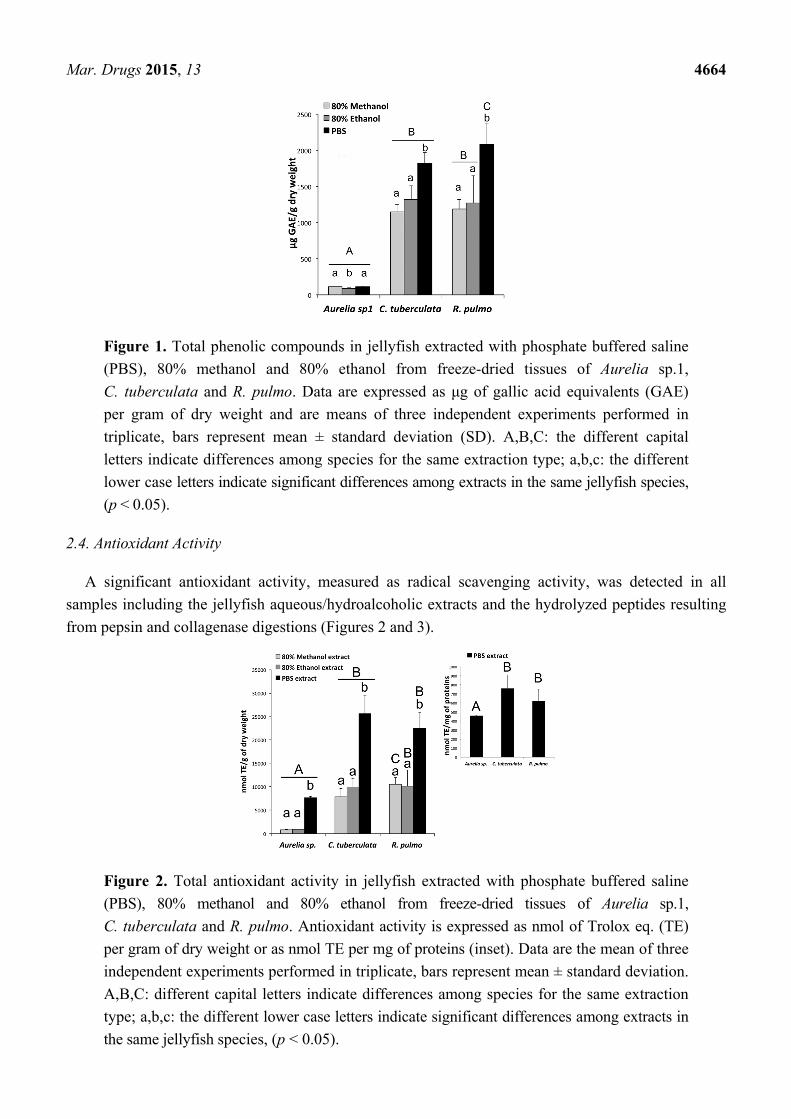
The total phenolic content of the jellyfish extracts was significantly different among the three
jellyfish species (Figure 1). In all extracts from Aurelia sp.1 samples, a low content of total phenols
was detected, as compared to the other two jellyfish species, with 113.2 ± 0.4 μg GAE (gallic acid
equivalent) per gram of DW in the 80% methanol extract, 86.4 ± 9.4 μg GAE/g in the 80% ethanol
extract and 115.5 ± 2.0 μg GAE/g in the PBS extract. The total phenol content detected in all extracts
of both C. tuberculata and R. pulmo was significantly higher than in Aurelia (Figure 1). The highest
concentration was detected for both species in the PBS extracts, reaching 1817.7 ± 153.7 μg GAE/g DW
and 2079.3 ± 301.9 μg GAE/g DW, for C. tuberculata and R. pulmo, respectively.
The presence of phenolic compounds in jellyfish is yet poorly documented. Phenols were detected
in the podocyst cuticle of Chrysaora quinquecirrha [82], and benzene-1,2-dicarboxylic acid or
phthalates were detected in the adult tissues of Cyanea capillata and Chrysaora quinquecirrha [83] as
well as in C. tuberculata extracts [8]. Recent data showed that polyphenols may enhance the
biostability and biomechanical properties of collagen based tissues by modulation of mechanisms of
collagen fibers cross-linking at molecular, inter-molecular and inter-microfibrillar levels [84,85]. There
is a remarkable difference in the stiffness and consistency of jellyfish extracellular matrix between the
highly flexible and soft Aurelia spp. jellyfish against the robust and hardened mesoglea of C. tuberculata
and R. pulmo, and it can be hypothesized that the higher observed concentration of phenols in the large
rhizostomate jellies may be responsible for or contribute to the evolution of different jellyfish functional
and anatomical adaptations.
The high phenolic content in the PBS extracts could also be related to the measurements of phenolic
amino acidic residues of the proteins. However, the observed differences of total phenol contents
between the three jellyfish species is not paralleled by differences in protein contents, suggesting that the
two rhizostome species really contain higher phenol concentrations than Aurelia sp.1.
Mar. Drugs 2015, 13 4664
Figure 1. Total phenolic compounds in jellyfish extracted with phosphate buffered saline
(PBS), 80% methanol and 80% ethanol from freeze-dried tissues of Aurelia sp.1,
C. tuberculata and R. pulmo. Data are expressed as μg of gallic acid equivalents (GAE)
per gram of dry weight and are means of three independent experiments performed in
triplicate, bars represent mean ± standard deviation (SD). A,B,C: the different capital
letters indicate differences among species for the same extraction type; a,b,c: the different
lower case letters indicate significant differences among extracts in the same jellyfish species,
(p < 0.05).
2.4. Antioxidant Activity
A significant antioxidant activity, measured as radical scavenging activity, was detected in all
samples including the jellyfish aqueous/hydroalcoholic extracts and the hydrolyzed peptides resulting
from pepsin and collagenase digestions (Figures 2 and 3).
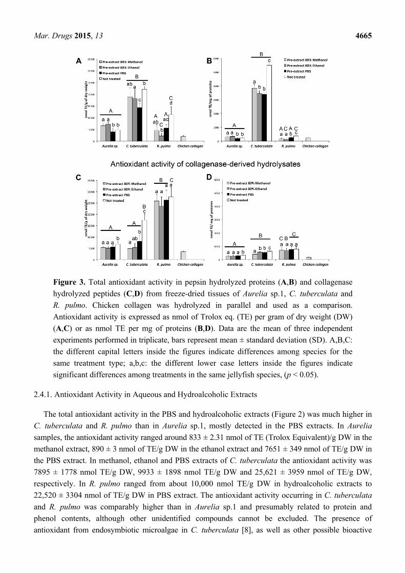
Figure 2. Total antioxidant activity in jellyfish extracted with phosphate buffered saline
(PBS), 80% methanol and 80% ethanol from freeze-dried tissues of Aurelia sp.1,
C. tuberculata and R. pulmo. Antioxidant activity is expressed as nmol of Trolox eq. (TE)
per gram of dry weight or as nmol TE per mg of proteins (inset). Data are the mean of three
independent experiments performed in triplicate, bars represent mean ± standard deviation.
A,B,C: different capital letters indicate differences among species for the same extraction
type; a,b,c: the different lower case letters indicate significant differences among extracts in
the same jellyfish species, (p < 0.05).
Mar. Drugs 2015, 13 4665
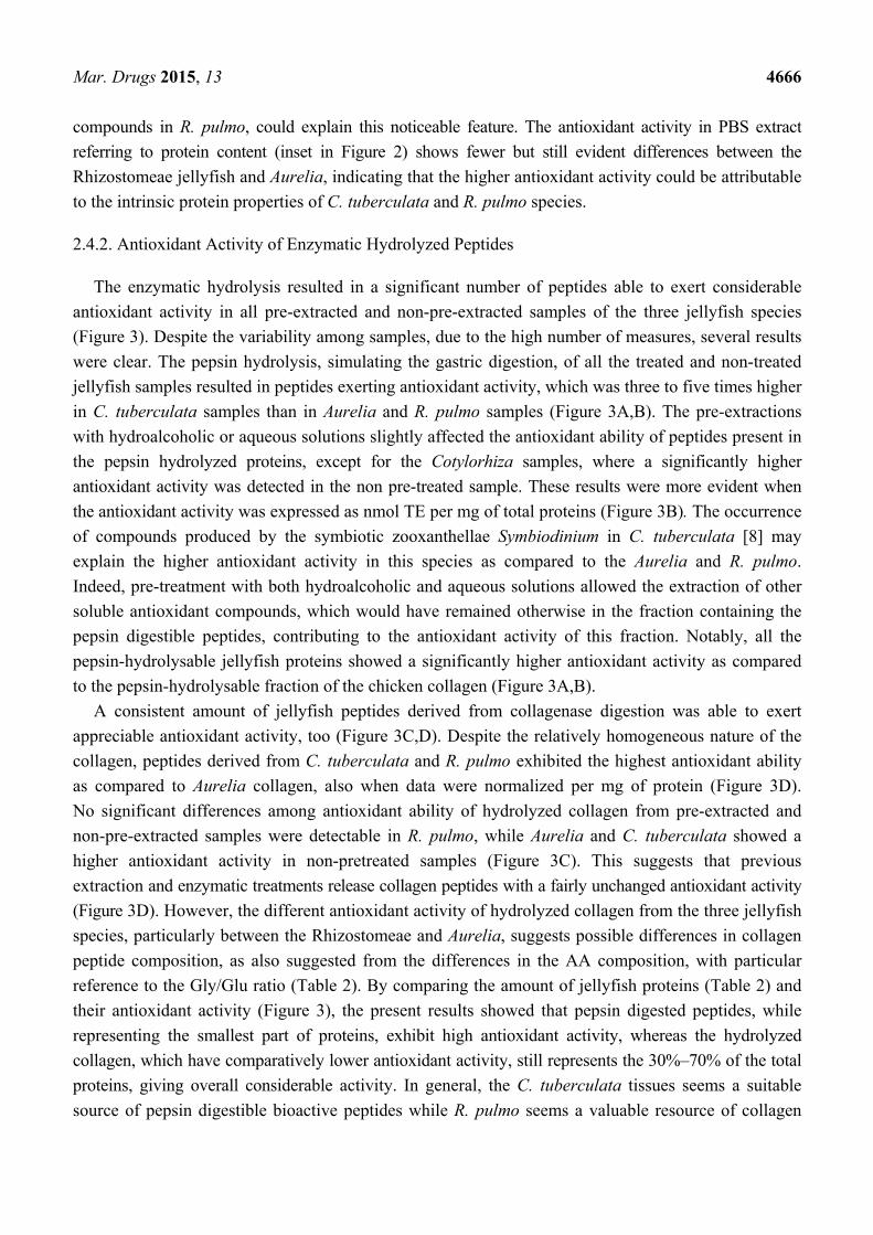
Figure 3. Total antioxidant activity in pepsin hydrolyzed proteins (A,B) and collagenase
hydrolyzed peptides (C,D) from freeze-dried tissues of Aurelia sp.1, C. tuberculata and
R. pulmo. Chicken collagen was hydrolyzed in parallel and used as a comparison.
Antioxidant activity is expressed as nmol of Trolox eq. (TE) per gram of dry weight (DW)
(A,C) or as nmol TE per mg of proteins (B,D). Data are the mean of three independent
experiments performed in triplicate, bars represent mean ± standard deviation (SD). A,B,C:
the different capital letters inside the figures indicate differences among species for the
same treatment type; a,b,c: the different lower case letters inside the figures indicate
significant differences among treatments in the same jellyfish species, (p < 0.05).
2.4.1. Antioxidant Activity in Aqueous and Hydroalcoholic Extracts
The total antioxidant activity in the PBS and hydroalcoholic extracts (Figure 2) was much higher in
C. tuberculata and R. pulmo than in Aurelia sp.1, mostly detected in the PBS extracts. In Aurelia
samples, the antioxidant activity ranged around 833 ± 2.31 nmol of TE (Trolox Equivalent)/g DW in the
methanol extract, 890 ± 3 nmol of TE/g DW in the ethanol extract and 7651 ± 349 nmol of TE/g DW in
the PBS extract. In methanol, ethanol and PBS extracts of C. tuberculata the antioxidant activity was
7895 ± 1778 nmol TE/g DW, 9933 ± 1898 nmol TE/g DW and 25,621 ± 3959 nmol of TE/g DW,
respectively. In R. pulmo ranged from about 10,000 nmol TE/g DW in hydroalcoholic extracts to
22,520 ± 3304 nmol of TE/g DW in PBS extract. The antioxidant activity occurring in C. tuberculata
and R. pulmo was comparably higher than in Aurelia sp.1 and presumably related to protein and
phenol contents, although other unidentified compounds cannot be excluded. The presence of
antioxidant from endosymbiotic microalgae in C. tuberculata [8], as well as other possible bioactive
Mar. Drugs 2015, 13 4666
compounds in R. pulmo, could explain this noticeable feature. The antioxidant activity in PBS extract
referring to protein content (inset in Figure 2) shows fewer but still evident differences between the
Rhizostomeae jellyfish and Aurelia, indicating that the higher antioxidant activity could be attributable
to the intrinsic protein properties of C. tuberculata and R. pulmo species.
2.4.2. Antioxidant Activity of Enzymatic Hydrolyzed Peptides
The enzymatic hydrolysis resulted in a significant number of peptides able to exert considerable
antioxidant activity in all pre-extracted and non-pre-extracted samples of the three jellyfish species
(Figure 3). Despite the variability among samples, due to the high number of measures, several results
were clear. The pepsin hydrolysis, simulating the gastric digestion, of all the treated and non-treated
jellyfish samples resulted in peptides exerting antioxidant activity, which was three to five times higher
in C. tuberculata samples than in Aurelia and R. pulmo samples (Figure 3A,B). The pre-extractions
with hydroalcoholic or aqueous solutions slightly affected the antioxidant ability of peptides present in
the pepsin hydrolyzed proteins, except for the Cotylorhiza samples, where a significantly higher
antioxidant activity was detected in the non pre-treated sample. These results were more evident when
the antioxidant activity was expressed as nmol TE per mg of total proteins (Figure 3B). The occurrence
of compounds produced by the symbiotic zooxanthellae Symbiodinium in C. tuberculata [8] may
explain the higher antioxidant activity in this species as compared to the Aurelia and R. pulmo.
Indeed, pre-treatment with both hydroalcoholic and aqueous solutions allowed the extraction of other
soluble antioxidant compounds, which would have remained otherwise in the fraction containing the
pepsin digestible peptides, contributing to the antioxidant activity of this fraction. Notably, all the
pepsin-hydrolysable jellyfish proteins showed a significantly higher antioxidant activity as compared
to the pepsin-hydrolysable fraction of the chicken collagen (Figure 3A,B).
A consistent amount of jellyfish peptides derived from collagenase digestion was able to exert
appreciable antioxidant activity, too (Figure 3C,D). Despite the relatively homogeneous nature of the
collagen, peptides derived from C. tuberculata and R. pulmo exhibited the highest antioxidant ability
as compared to Aurelia collagen, also when data were normalized per mg of protein (Figure 3D).
No significant differences among antioxidant ability of hydrolyzed collagen from pre-extracted and
non-pre-extracted samples were detectable in R. pulmo, while Aurelia and C. tuberculata showed a
higher antioxidant activity in non-pretreated samples (Figure 3C). This suggests that previous
extraction and enzymatic treatments release collagen peptides with a fairly unchanged antioxidant activity
(Figure 3D). However, the different antioxidant activity of hydrolyzed collagen from the three jellyfish
species, particularly between the Rhizostomeae and Aurelia, suggests possible differences in collagen
peptide composition, as also suggested from the differences in the AA composition, with particular
reference to the Gly/Glu ratio (Table 2). By comparing the amount of jellyfish proteins (Table 2) and
their antioxidant activity (Figure 3), the present results showed that pepsin digested peptides, while
representing the smallest part of proteins, exhibit high antioxidant activity, whereas the hydrolyzed
collagen, which have comparatively lower antioxidant activity, still represents the 30%–70% of the total
proteins, giving overall considerable activity. In general, the C. tuberculata tissues seems a suitable
source of pepsin digestible bioactive peptides while R. pulmo seems a valuable resource of collagen
Mar. Drugs 2015, 13 4667
and collagen derived bioactive peptides, also taking into account the specimen size and yield in dry
weight (Table 1).
In this study, different collagen from diverse sources (vertebrate and cnidarians) was hydrolyzed by
the same enzymatic system, producing a different set of peptides with different antioxidant activity.
Remarkably, the antioxidant activity exerted by hydrolyzed collagen of chicken cartilage was actually
much lower than hydrolyzed collagen from all the three jellyfish (Figure 3C,D). Antioxidant
compounds are known to originate by enzymatic hydrolysis of parent proteins from terrestrial and
marine organisms, showing novel antihypertensive, antioxidant, antimicrobial and antiproliferative or
immunomodulatory properties [46,86–91].
Many marine peptides including collagen exhibit multifunctional activities of interest for food,
cosmetics and pharmaceutical industries [90,92]. In particular, studies on jellyfish proteins showed that
the oral administration of collagen and collagen hydrolysate from the edible jellyfish Rhopilema were able
to alleviate the skin photoaging in mice through antioxidant, anti-melanogenic and immunity-enhancing
biochemical activities [40,67,93]. More recently, collagen hydrolysate from Rhopilema esculentum and
the ribbon jellyfish Chrysaora sp. were shown to exert antioxidant and anti-hypertensive activities [94,95].
The amino acid composition of food protein hydrolysates is also known to have strong influence on
their antioxidant properties. The amounts of histidine, cysteine, proline, methionine, and aromatic
amino acids have been reported to significantly contribute to the antioxidant activity of food peptides.
The amino acid composition of the three Mediterranean jellyfish (Table 2) investigated here shows that
higher amounts of amino acids with potential antioxidant activity, including aromatic amino acids, occur
in the two Rhizostomeae, C. tuberculata and R. pulmo, as compared to Aurelia.
2.5. Sodium Dodecyl Sulfate-Polyacrylamide Gel Electrophoresis (SDS-PAGE) Analysis of
Hydrolyzed Peptides
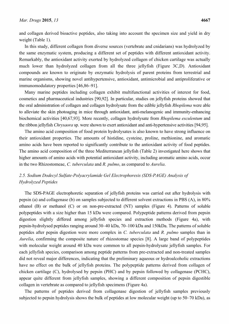
The SDS-PAGE electrophoretic separation of jellyfish proteins was carried out after hydrolysis with
pepsin (a) and collagenase (b) on samples subjected to different solvent extractions in PBS (A), in 80%
ethanol (B) or methanol (C) or on non-pre-extracted (NT) samples (Figure 4). Patterns of soluble
polypeptides with a size higher than 15 kDa were compared. Polypeptide patterns derived from pepsin
digestion slightly differed among jellyfish species and extraction methods (Figure 4a), with
pepsin-hydrolysed peptides ranging around 30–40 kDa, 70–100 kDa and 150kDa. The patterns of soluble
peptides after pepsin digestion were more complex in C. tuberculata and R. pulmo samples than in
Aurelia, confirming the composite nature of rhizostomae species [8]. A large band of polypeptides
with molecular weight around 40 kDa were common to all pepsin-hydrolysate jellyfish samples. For
each jellyfish species, comparison among peptide patterns from pre-extracted and non-treated samples
did not reveal major differences, indicating that the preliminary aqueous or hydroalcoholic extractions
have no effect on the bulk of jellyfish proteins. The polypeptide patterns derived from collagen of
chicken cartilage (C), hydrolysed by pepsin (PHC) and by pepsin followed by collagenase (PCHC),
appear quite different from jellyfish samples, showing a different composition of pepsin digestible
collagen in vertebrate as compared to jellyfish specimens (Figure 4a).
The patterns of peptides derived from collagenase digestion of jellyfish samples previously
subjected to pepsin hydrolysis shows the bulk of peptides at low molecular weight (up to 50–70 kDa), as
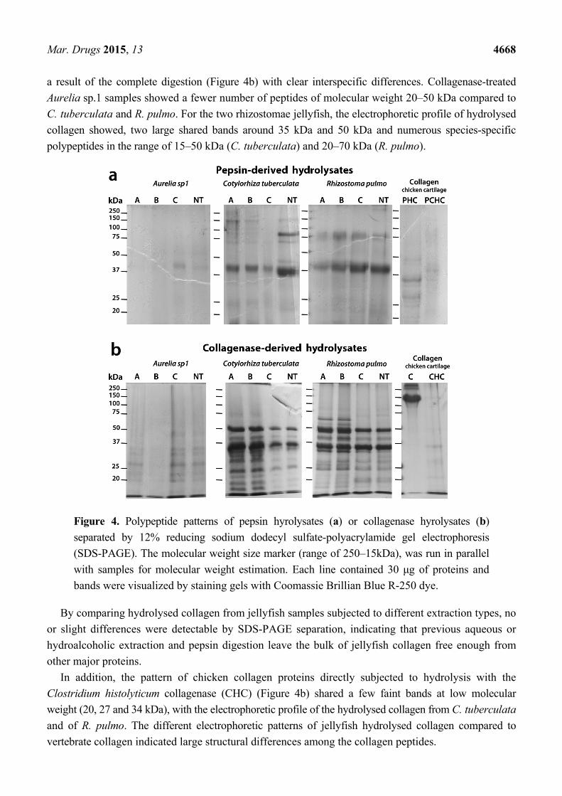
Mar. Drugs 2015, 13 4668
a result of the complete digestion (Figure 4b) with clear interspecific differences. Collagenase-treated
Aurelia sp.1 samples showed a fewer number of peptides of molecular weight 20–50 kDa compared to
C. tuberculata and R. pulmo. For the two rhizostomae jellyfish, the electrophoretic profile of hydrolysed
collagen showed, two large shared bands around 35 kDa and 50 kDa and numerous species-specific
polypeptides in the range of 15–50 kDa (C. tuberculata) and 20–70 kDa (R. pulmo).
Figure 4. Polypeptide patterns of pepsin hyrolysates (a) or collagenase hyrolysates (b)
separated by 12% reducing sodium dodecyl sulfate-polyacrylamide gel electrophoresis
(SDS-PAGE). The molecular weight size marker (range of 250–15kDa), was run in parallel
with samples for molecular weight estimation. Each line contained 30 μg of proteins and
bands were visualized by staining gels with Coomassie Brillian Blue R-250 dye.
By comparing hydrolysed collagen from jellyfish samples subjected to different extraction types, no
or slight differences were detectable by SDS-PAGE separation, indicating that previous aqueous or
hydroalcoholic extraction and pepsin digestion leave the bulk of jellyfish collagen free enough from
other major proteins.
In addition, the pattern of chicken collagen proteins directly subjected to hydrolysis with the
Clostridium histolyticum collagenase (CHC) (Figure 4b) shared a few faint bands at low molecular
weight (20, 27 and 34 kDa), with the electrophoretic profile of the hydrolysed collagen from C. tuberculata
and of R. pulmo. The different electrophoretic patterns of jellyfish hydrolysed collagen compared to
vertebrate collagen indicated large structural differences among the collagen peptides.
Mar. Drugs 2015, 13 4669
Because of the higher antioxidant activity exerted by jellyfish hydrolysed collagen, as compared to
chicken hydrolysed collagen (Figure 3A,B), the exploitation of jellyfish biomasses as functional food
and/or as natural antioxidant peptide source appears to be a particularly promising strategy.
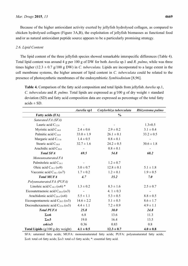
2.6. Lipid Content
The lipid content of the three jellyfish species showed remarkable interspecific differences (Table 4).
Total lipid content was around 4 g per 100 g of DW for both Aurelia sp.1 and R. pulmo, while was three
times higher (12.3 ± 0.7 g/100 g DW) in C. tuberculata. Lipids are incorporated to a large extent in the
cell membrane systems, the higher amount of lipid content in C. tuberculata could be related to the
presence of photosynthetic membranes of the endosymbiotic Symbiodinium [8,96].
Table 4. Comparison of the fatty acid composition and total lipids from jellyfish Aurelia sp.1,
C. tuberculata and R. pulmo. Total lipids are expressed as g/100 g of dry weight ± standard
deviation (SD) and fatty acid composition data are expressed as percentage of the total fatty
acids ± SD.
Aurelia sp1 Cotylorhiza tuberculata Rhizostoma pulmo
Fatty acids (FA) %
Saturated FA (SFA) Lauric acid C12:0 - - 1.3±0.5
Myristic acid C14:0 2.4 ± 0.6 2.9 ± 0.2 3.1 ± 0.4 Palmitic acid C16:0 33.0 ± 1.9 26.1 ± 0.1 33.2 ± 0.5 Margaric acid C17:0 1.4 ± 0.5 0.8 ± 0.1 - Stearic acid C18:0 32.7 ± 1.6 24.2 ± 0.5 30.6 ± 1.8
Arachidic acid C20:0 - 0.8 ± 0.1 - Total SFA 69.5 54.8 68.2
Monounsaturated FA Palmitoleic acid C16:1 - 1.2 ± 0.7 Oleic acid C18:1 (ω9) 3.0 ± 0.7 12.8 ± 0.1 5.1 ± 1.8
Vaccenic acid C18:1 (ω7) 1.7 ± 0.2 1.2 ± 0.1 1.9 ± 0.5 Total MUFA 4.7 15.2 7.0
Polyunsaturated FA (PUFA) Linoleic acid C18:2 (ω6) * 1.3 ± 0.2 8.3 ± 1.6 2.5 ± 0.7
Eicosatetraenoic acid C20:4 (ω3) - 4. 1 ± 0.3 - Arachidonic acid C20:4 (ω6) 5.5 ± 1.1 5.3 ± 0.5 8.8 ± 0.5
Eicosapentaenoic acid C20:5 (ω3) 14.6 ± 2.2 5.1 ± 0.5 8.6 ± 1.7 Docosahexaenoic acid C22:6 (ω3) 4.4 ± 1.1 7.2 ± 0.9 4.9 ± 1.1
Total PUFA 25.8 30.0 24.8 Σω6 6.8 13.6 11.3 Σω3 19.0 16.4 13.5 ω6/ω3 0.36 0.83 0.8
Total Lipids (g/100 g dry weight) 4.1 ± 0.5 12.3 ± 0.7 4.0 ± 0.8
SFA: saturated fatty acids; MUFA: monounsaturated fatty acids; PUFA: polyunsaturated fatty acids;
Σω6: total ω6 fatty acids; Σω3: total ω3 fatty acids; *: essential fatty acid.
Mar. Drugs 2015, 13 4670
The fatty acid (FA) quantitative composition (as percentage values) showed similar FA profiles in the
three jellyfish species (Table 4). Saturated fatty acids (SFA), accounted for two third of total FA (about
55%–70%), followed by polyunsaturated fatty acid (PUFA), representing about one third of the total
FA (about 25%–30%), and a few amount of monounsaturated fatty acids (MUFA), representing about
4%–15% of the total FA. Saturated fatty acids consisted mostly of palmitic (C16:0) and stearic (C18:0)
acids, followed by myristic (C14:0) and margaric (C17:0) acids. Lauric acid was detected only in
R. pulmo, while traces of arachidic acid (C20:0) were detected in C. tuberculata. Among MUFA, oleic
acid (C18:1) was the prevalent FA and palmitoleic acid (C16:1) was detected only in C. tuberculata.
Remarkable differences were observed in the composition of PUFAs among the three jellyfish (Table 4),
being mostly represented by the ω-3 eicosapentaenoic acid (C20:5) in Aurelia sp.1, while the ω-6
arachidonic (C20:4) and the ω-3 eicosapentaenoic acid (C20:5) were prevalent in R. pulmo. A peculiar
PUFA composition was detected in C. tuberculata samples, where the essential ω-6 FA, linoleic acid
(C18:2) was the major component, together with the ω-3 eicosatetraenoic acid (C20:4), docosahexaenoic
acid (C22:6), and the eicosapentaenoic and arachidonic acids. The presence of unsaturated long chain
fatty acids in this species is due to the presence of microalgal symbionts (Symbiodinium spp.), an
important and significant source of essential ω-3 fatty acids.
Overall, ω-3 PUFAs were abundant in the three jellyfish species, with the ratio of ω-6 to ω-3
resulting always in favouring ω-3 fatty acids (Table 4), as generally observed in fish and marine foods.
The value of ω-6/ω-3 ratio was lower in Aurelia sp.1 (0.36) than in the two Rhizostomeae (0.8).
Opposite differences in the ω-6/ω-3 ratio have been noted between fatty acid compositions of marine
and freshwater fish, with marine fish containing higher levels of ω-3 than freshwater fish [97].
Explanations for such opposite patterns for jellyfish and fish are merely speculative. Overall,
differences between marine and freshwater taxa might be related to specific requirements both to
physiological adaptations to different habitats and to deep evolutionary constraints across distant
phylogenetic lineages, such as jellyfish and fish.
The ω-3 types of FA are known to be involved in a number of biological processes including
growth, development, tissue and cell homeostasis [98] and have a variety of health benefits including
hypo-triglyceridemic, anti-inflammatory antihypertensive, anticancer, antioxidant, antidepressive,
antiaging, and antiarthritis effects [99]. In humans, the dietary patterns are important in the
pathogenesis of chronic disease, which appears to be due to proinflammatory effects of the Western
diet, with particular reference to the high ω-6/ω-3 ratio [100].
3. Experimental Section
3.1. Materials and Chemicals
Methanol, ethanol and acetic acid were purchased from Merck (Darmstadt, Germany); potassium
persulfate (dipotassium peroxdisulfate), 6-hydroxy-2,5,7,8-tetramethylchroman-2-carboxylic acid
(Trolox), 2,20-azinobis(3-ethylben-zothiazoline-6-sulfonic acid)diammonium salt (ABTS), gallic acid,
Folin-Ciocalteu’s phenol reagent, Coomassie Brilliant Blue R-250, pepsin from porcine gastric mucosa
(≥2500 U/mg), collagenase from Clostridium histolyticum (0.5–5.0 furylacryloyl-Leu-Gly-Pro-Ala
(FALGPA) units/mg solid, ≥125 collagen digestion unit (CDU)/mg solid), collagen from chicken
Mar. Drugs 2015, 13 4671
sternal cartilage, fatty acid methyl esters (FAME) Mix (C8–C24) and PUFA-3 were all purchased from
Sigma-Aldrich (Milan, Italy). Acrylamide solution was purchased from Euroclone (Milan, Italy). All
other reagents were of analytical grade.
3.2. Sample Collection and Preparation
Specimens of three jellyfish species (Aurelia sp.1, Cotylorhiza tuberculata and Rhizostoma pulmo)
were collected offshore of Apulia coasts (Italy) in the 2011–2014 summers. Aurelia samples were
collected in May 2012 in the Varano Lake (Foggia, Italy, +41°52′45.01″, +15°44′46.00″); C. tuberculata
and R. pulmo samples were collected in the Southern Adriatic (Otranto, Italy) and Ionian (Castellaneta
Marina and Pulsano, Italy) Seas. After the biometric measurements (weight and diameter), each
specimen was frozen in liquid nitrogen and stored at −80 °C until lyophilization. Frozen jellyfish were
freeze-dried for 4 days at −55 °C using a chamber pressure of 0.110 mbar in a freeze dryer (Freezone
4.5L Dry System, Labconco Co. Thermo Scientific, Milan, Italy). Lyophilized samples were weighed
to annotate the dry weight and stored at −20 °C until use.
3.3. Sequential Extraction and Hydrolysis
3.3.1. Polar Solvent Extraction
Lyophilized samples (100 mg) of total jellyfish were subjected to extraction in hydroalcoholic (80%
methanol or 80% ethanol) or aqueous solvents PBS as shown in the Scheme 1. Samples were either
stirred with 16 volumes (w/v) of 80% methanol or 80% ethanol (16 h at 4 °C) or with 16 volumes of
PBS (2 h at 4 °C). Samples were then centrifuged at 9000× g for 30 min at 4 °C and the supernatants
were essayed for protein and phenol contents and antioxidant activity. The insoluble pellets were dried
under a stream of nitrogen and subsequently subjected to sequential enzymatic digestions.
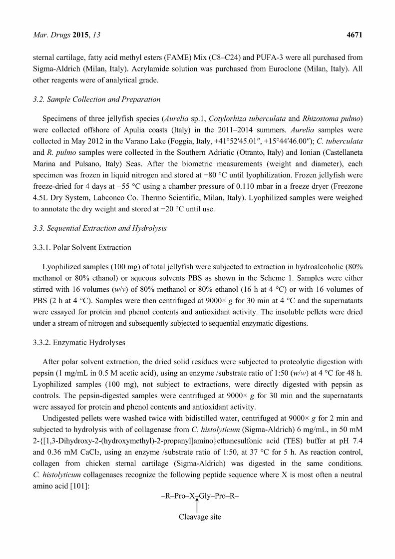
3.3.2. Enzymatic Hydrolyses
After polar solvent extraction, the dried solid residues were subjected to proteolytic digestion with
pepsin (1 mg/mL in 0.5 M acetic acid), using an enzyme /substrate ratio of 1:50 (w/w) at 4 °C for 48 h.
Lyophilized samples (100 mg), not subject to extractions, were directly digested with pepsin as
controls. The pepsin-digested samples were centrifuged at 9000× g for 30 min and the supernatants
were assayed for protein and phenol contents and antioxidant activity.
Undigested pellets were washed twice with bidistilled water, centrifuged at 9000× g for 2 min and
subjected to hydrolysis with of collagenase from C. histolyticum (Sigma-Aldrich) 6 mg/mL, in 50 mM
2-{[1,3-Dihydroxy-2-(hydroxymethyl)-2-propanyl]amino}ethanesulfonic acid (TES) buffer at pH 7.4
and 0.36 mM CaCl2, using an enzyme /substrate ratio of 1:50, at 37 °C for 5 h. As reaction control,
collagen from chicken sternal cartilage (Sigma-Aldrich) was digested in the same conditions.
C. histolyticum collagenases recognize the following peptide sequence where X is most often a neutral
amino acid [101]:
Mar. Drugs 2015, 13 4672
After proteolytic reaction, samples were centrifuged at 9000× g for 30 min, the pellets were
considered as non-hydrolysable protein fractions, and supernatants were essayed for protein and phenol
contents and antioxidant activity.
3.4. Protein Content
Total protein content was estimated by modified Bradford assay [102] using bovine serum albumin
(BSA) as a standard.
3.5. Amino Acidic Composition Analysis
Each sample of total dried jellyfish tissue was analysed in triplicate for the determination of free
amino acids. In particular, 0.2 g of lyophilized jellyfish powder was stirred for 20 min in the following
mixture: 5 mL of bidistilled water, 0.75 mL trifluoroacetic acid and 50 µL internal standard solution (5 mM
DL-Norleucine in water). The extract was then centrifuged at 4 °C, 3500 rpm, for 20 min. After
filtration, the extract was dried under a nitrogen stream and the residue was re-dissolved in 1 mL
bidistilled water. According to the AccQ-Tag protocol (Waters, Milford, MA, USA) and after a
pre-column derivatization step, each sample was analyzed on a C18 AccQ-Tag column (3.9 × 150 mm)
(Waters). A gradient elution was performed according to the AccQ-Tag protocol, using phosphate
buffer solution (eluent A) and acetonitrile:water 60:40 (v/v) (eluent B), the flow rate was 1 mL/min,
temperature was set at 37 °C. The fluorescent detector parameters were set as follows: λex = 250 nm,
λem = 395 nm, gain = 1, EUFS (emission/energy units full scale) = 100.
3.6. Phenol Content
The total content of phenols was determined by a modified Folin-Ciocalteau colorimetric method.
Samples (100 µL) were mixed with 500 µL of Folin-Ciocalteu’s phenol reagent and 500 µL of 7.5%
sodium carbonate (Na2CO3). After 2 h of incubation at room temperature in the dark, the absorbance
was spectrophotometrically measured at 760 nm. Gallic acid, ranging from 25 to 200 µg/mL, was used
as a standard. The results were expressed as gallic acid equivalents (GAE) per gram of dry extract.
3.7. Antioxidant Activity
The total antioxidant activity was determined spectrophotometrically using the Trolox Equivalent
Antioxidant Capacity (TEAC) method, as described by Longo et al. [103]. Ten microliters of the
jellyfish samples (extracts or hydrolyzed fractions) were assayed in 1 mL of the reaction mixture and
the depletion of the radical cation ABTS+ was measured following the decrease of absorbance at 734 nm.
Comparable solutions of 80% methanol, 80% ethanol, PBS and enzymatic reaction mixtures without
substrates were used as controls. A calibration curve was prepared with different concentrations of
Trolox (2.5–20 μM). The antioxidant capacity of the samples was calculated as the absorbance
decrease at 734 nm at 6 min as fixed time, and results were expressed as nmol of Trolox equivalents
(TE) per gram of sample or per mg of contained proteins.
Mar. Drugs 2015, 13 4673
3.8. SDS-PAGE
Hydrolysed polypeptides were separated by electrophoresis through 12% polyacrylamide gels
containing SDS (SDS-PAGE) as described by Leone et al. 2013 [8]. Precision Plus Protein Dual Color
Standard (Bio-Rad, Hertfordshire, UK) was used as the molecular weight marker. Polypeptides on gels
were detected by Coomassie Brilliant Blue staining (0.25% Coomassie Brilliant Blue R-250 in 10%
acetic acid and 50% methanol) for 20 min followed by destaining with 10% acetic acid and 30%
methanol, overnight. Molecular masses of proteins were estimated by comparing the migration of
proteins of interest to the standards of known sizes.
3.9. Total Lipid Extraction
Total lipids were extracted using the modified method of Bligh and Dyer [104] with some
modifications. Lyophilized powder (100 mg) was mixed with a total of 114 mL solvent added in this
sequence: chloroform, methanol, water to achieve a final chloroform/methanol/water ratio of 1:2:0.8
(by volume). Samples were shaken for 15 s after addition of each solvent, and incubated overnight at
4 °C. After centrifugation at 6500× g for 10 min, the supernatant was transferred into a separating
funnel, and phase separation of the biomass-solvent mixtures was achieved by adding chloroform and
water to obtain a final chloroform/methanol/water ratio of 2:2:1.8 (by volume). After settling, the bottom
phase was collected, evaporated in the presence of nitrogen flux and re-suspended in chloroform (1 mL).
Fatty Acid Profiles Determination
Fatty acid methyl esters (FAME) were obtained using boron trifluoride (BF3) according to [105]
with some modifications. Total lipid extract (200 µL) was saponified at 90 °C for 20 min with 0.5 M
KOH in methanol (3 mL). Forty-nine micrograms of the internal standard (methyl tricosanoate) were
added before saponification. The fatty acids were methylated by adding 14% BF3 in MeOH (2 mL) and
heating at 90 °C for 10 min. After cooling, hexane (1 mL) was added and vigorously stirred for 30 s
before the addition of 1 mL of sodium chloride solution (0.6%). The esterified samples were placed at
4 °C for a better phase separation. After collecting the supernatant, another 1.0 mL of hexane was
added and the resulting mixture was vortexed. The samples were evaporated under a stream of
nitrogen, the dried samples were dissolved in 1.0 mL of hexane and analyzed by gas chromatography-mass
spectrometry (GC-MS).
3.10. GC-MS Analysis
The analyses were performed on a GC-MS system consisted of a Shimadzu GC-17A version 3.0
coupled with MS QP5050A according to Talà et al. [106]. Compounds were separated on DB-5
capillary column having 30 m length, 0.25 mm ID (internal diameter) and 0.25 µm thickness. The GC
parameters were as follows: the temperature of the column was 80 °C after injection then programmed at
10 °C/min to 150 °C, at 5 °C/min to 250 °C and maintained at that temperature for 15 min. Split injection
was conducted with a split ratio of 50:1, the flow-rate was 1.0 mL/min, carrier gas used was 99.999%
pure helium, the injector temperature was 250 °C and the column inlet pressure was 74 Kpa. The MS
detection conditions were as follows: interface temperature was set 250 °C; ionization mode, EI+;
Mar. Drugs 2015, 13 4674
electron energy, 70 eV; scanning method of acquisition, ranging from 30 to 450, for mass/charge (m/z)
was optimized. Spectrum data were collected at 0.5 s intervals. Solvent cut time was set at 2 min and
45 min retention time sufficient for separating all the fatty acid. Compounds were identified by using
online the National Institute of Standards and Technology (NIST)-library spectra and published MS data.
Moreover, FAME Mix (C8–C24) and PUFA-3 (from menhaden oil) authentic standards (both from
Sigma-Aldrich) were used to confirm MS data.
3.11. Statistical Analysis
Statistical analysis was based on a one-way ANOVA test. Tukey’s post hoc method was applied to
establish significant differences between means (p < 0.05). Data are mean ± standard deviation (SD).
SigmaPlot Ver 12.0 (Sistat Software, Inc., San Jose, CA, USA) was used.
4. Conclusions
Strategic integrative research on marine biodiversity and biotechnology is key to meeting the
growing demand for healthy food in a sustainable way, calling for urgent diversification of marine
food products [107]. This study demonstrates that the adult stages of three jellyfish species commonly
recorded in the Mediterranean Sea in massive populations contain high amounts of collagen,
anti-oxidant peptides and other bioactive molecules. These findings suggest Mediterranean jellyfish
biomasses can represent a valuable source of natural compounds, not only for biomedical or
pharmaceutical applications, but also as food ingredients, comparable to the most popular Asiatic
jellyfish species. The biochemical and nutraceutical characterization of these gelatinous biomasses,
together with adequate toxicological assays and the development of processing technologies, represent
fundamental information to support future use of the vast but still unexploited resource potential of
Mediterranean jellyfish.
Acknowledgments
This work was funded by the European Community Seventh Framework Programme (FP7/2007–2013)
under Grant Agreement No. 266445 for the project Vectors of Change in Oceans and Seas Marine
Life, Impact on Economic Sectors (VECTORS) and by the ENPI CBCMED programme
MED-JELLYRISK—integrated monitoring of jellyfish outbreaks under anthropogenic and climatic
impacts in the Mediterranean Sea coastal zones: Trophic and socio-economic risks (Project registration
number I-A/1.3/098). Thanks to Gessica Bufano and to Giorgio Aglieri, Rosa Caprioli, Giacomo
Milisenda, Simonetta Scorrano for their support in jellyfish samplings.
Author Contributions
Antonella Leone designed and performed experiments, developed the methodology, analyzed the
data and wrote the manuscript; Raffaella Marina Lecci performed all experiments and wrote the
manuscript; Federica Meli performed amino acid analysis, Miriana Durante performed lipid analysis;
Stefano Piraino analyzed the data and wrote the manuscript.
Mar. Drugs 2015, 13 4675
Conflicts of Interest
The authors declare no conflict of interest.
References
1. Gates, K.W. Marine Products for Healthcare: Functional and Bioactive Nutraceutical Compounds
from the Ocean, Vazhiyil Venugopal. J. Aquat. Food Prod. Technol. 2010, 19, 48–54.
2. Blunt, J.W.; Copp, B.R.; Keyzers, R.A.; Munro, M.H.G.; Prinsep, M.R. Marine natural products.
Nat. Prod. Rep. 2014, 31, 160–258.
3. Hu, Y.; Chen, J.; Hu, G.; Yu, J.; Zhu, X.; Lin, Y.; Chen, S.; Yuan, J. Statistical Research on the
Bioactivity of New Marine Natural Products Discovered during the 28 Years from 1985 to 2012.
Mar. Drugs 2015, 13, 202–221.
4. Jaume, D.; Duarte, C.M. General aspects concerning marine and terrestrial biodiversity. In The
Exploration of Marine Biodiversity Scientific and Technological Challenges; Duarte, C.M., Ed.;
Fundatiòn BBVA: Bilbao, Spain, 2006; pp. 19–32.
5. Mayer, A.M.S.; Rodríguez, A.D.; Taglialatela-Scafati, O.; Fusetani, N. Marine pharmacology in
2009–2011: Marine compounds with antibacterial, antidiabetic, antifungal, anti-inflammatory,
antiprotozoal, antituberculosis, and antiviral activities; affecting the immune and nervous systems,
and other miscellaneous mechanisms of. Mar. Drugs 2013, 11, 2510–2523.
6. Blunden, G. Biologically active compounds from marine organisms. Phyther. Res. 1990, 15, 89–94.
7. Guérard, F.; Decourcelle, N.; Sabourin, C.; Floch-Laizet, C.; le Grel, L.; le Floch, P.; Gourlay, F.;
le Delezir, R.; Jaouen, P.; Bourseau, P. Recent developments of marine ingredients for food and
nutraceutical applications: A review. J. Sci. Halieut. Aquat. 2010, 2, 21–27.
8. Leone, A.; Lecci, R.M.; Durante, M.; Piraino, S. Extract from the zooxanthellate jellyfish
Cotylorhiza tuberculata modulates gap junction intercellular communication in human cell cultures.
Mar. Drugs 2013, 11, 1728–1762.
9. Mariottini, G.L.; Pane, L. Cytotoxic and cytolytic cnidarian venoms. A review on health
implications and possible therapeutic applications. Toxins (Basel) 2013, 6, 108–151.
10. Pauly, D.; Watson, R.; Alder, J. Global trends in world fisheries: Impacts on marine ecosystems
and food security. Philos. Trans. R. Soc. Lond. B Biol. Sci. 2005, 360, 5–12.
11. Bundy, A.; Shannon, L.J.; Rochet, M.-J.; Neira, S.; Shi, Y.-J.; Hill, L.; Aydin, K. The Good (ish),
the Bad and the Ugly: A tripartite classification of ecosystem trends. ICES J. Mar. Sci. 2010, 67,
745–768.
12. Boero, F.; Bouillon, J.; Gravili, C.; Miglietta, M.P.; Parsons, T.; Piraino, S. Gelatinous plankton:
Irregularities rule the world (sometimes). Mar. Ecol. Prog. Ser. 2008, 356, 299–310.
13. Graham, W.M.; Pagès, F.; Hamner, W.M. A physical context for gelatinous zooplankton
aggregations: A review. Hydrobiologia 2001, 451, 199–212.
14. Brotz, L.; Cheung, W.W.L.; Kleisner, K.; Pakhomov, E.; Pauly, D. Increasing jellyfish populations:
Trends in Large Marine Ecosystems. Hydrobiologia 2012, 690, 3–20.
15. Mariottini, G.L.; Pane, L. Mediterranean jellyfish venoms: A review on scyphomedusae. Mar.
Drugs 2010, 8, 1122–1152.
Mar. Drugs 2015, 13 4676
16. Dong, Z.; Liu, D.; Keesing, J.K. Jellyfish blooms in China: Dominant species, causes and
consequences. Mar. Pollut. Bull. 2010, 60, 954–963.
17. Lucas, C.H.; Gelcich, S.; Uye, S.I. Living with jellyfish: Management and adaptation strategies.
In Jellyfish Blooms; Pitt, K.A., Lucas, C.H., Eds; Springer: Dordrecht, The Netherlands, 2014;
pp. 129–150.
18. Purcell, J.E.; Uye, S.I.; Lo, W.T. Anthropogenic causes of jellyfish blooms and their direct
consequences for humans: A review. Mar. Ecol. Prog. Ser. 2007, 350, 153–174.
19. Lynam, C.P.; Gibbons, M.J.; Axelsen, B.E.; Sparks, C.A.J.; Coetzee, J.; Heywood, B.G.;
Brierley, A.S. Jellyfish overtake fish in a heavily fished ecosystem. Curr. Biol. 2006, 16, 1976.
20. Purcell, J.E.; Milisenda, G.; Rizzo, A.; Carrion, S.A.; Zampardi, S.; Airoldi, S.; Zagami, G.;
Guglielmo, L.; Boero, F.; Doyle, T.K.; Piraino, S. Digestion and predation rates of zooplankton
by the pleustonic hydrozoan Velella velella and widespread blooms in 2013 and 2014. Plankt.
Res. 2015, in press.
21. Matsushita, Y.; Honda, N.; Kawamura, S. Design and tow trial of JET (Jellyfish Excluder for
Towed fishing gear). Nippon Suisan Gakk. 2005, 71, 965–967.
22. MED-JELLYRISK, Enhancing management approach and mitigation measures against jellyfish
proliferations impacts. Project funded by the ENPI CBC MED (European Neighbourhood and
Partnership Instrument Cross-Border Cooperation in the Mediterranean). Available online:
http://www.jellyrisk.eu and http://meteomeduse.focus.it/ (accessed on 23 December 2013).
23. Boero, F. Review of jellyfish blooms in the Mediterranean and Black Sea. In General Fisheries
Commission for the Mediterranean. Studies and Reviews. No. 92. Food and Agriculture
Organization of the United Nations (FAO), Ed.; FAO: Rome, Italy, 2013; p. 53.
24. Doyle, T.K.; Hays, G.C.; Harrod, C.; Houghton, J.D.R. Ecological and societal benefits of jellyfish.
In Jellyfish Blooms; Pitt, K.A., Lucas, C.H., Eds.; Springer Science + Business Media: Dordrecht,
The Netherlands, 2014; pp. 105–127.
25. Boero, F.; Bouillon, J.; Piraino, S.; Schmid, V. Asexual reproduction in the Hydrozoa (Cnidaria).
In Reproductive Biology of Invertebrates XI: Progress in Asexual Reproduction; Hughes, R.N.,
Ed.; Oxford & IBH Publishing Co.: New Delhi, India, 2002; pp. 141–158.
26. Piraino, S.; de Vito, D.; Schmich, J.; Bouillon, J.; Boero, F. Reverse development in Cnidaria.
Can. J. Zool. 2004, 82, 1748–1754.
27. Larson, R.J. Water content, organic content, and carbon and nitrogen composition of medusae
from the northeast Pacific. J. Exp. Mar. Biol. Ecol. 1986, 99, 107–120.
28. Lucas, C.H.; Pitt, K.A.; Purcell, J.E.; Lebrato, M.; Condon, R.H. What’s in a jellyfish? Proximate
and elemental composition and biometric relationships for use in biogeochemical studies.
Ecology 2011, 92, 1704.
29. Li, J.; Hsieh, Y.H.P. Traditional Chinese food technology and cuisine. Asia Pac. J. Clin. Nutr.
2004, 13, 147–155.
30. Lucas, C.H. Biochemical composition of the mesopelagic coronate jellyfish Periphylla
periphylla from the Gulf of Mexico. J. Mar. Biol. Assoc. UK 2009, 89, 77–81.
31. Omori, M.; Nakano, E. Jellyfish fisheries in southeast Asia. Hydrobiologia 2001, 451, 19–26.
32. Hsieh, Y.H.P.; Rudloe, J. Potential of utilizing jellyfish as food in Western countries. Trends
Food Sci. Technol. 1994, 5, 225–229.
Mar. Drugs 2015, 13 4677
33. Hsieh, Y.H.P.; Leong, F.M.; Rudloe, J. Jellyfish as food. Hydrobiologia 2001, 451, 11–17.
34. Aouacheria, A.; Geourjon, C.; Aghajari, N.; Navratil, V.; Deléage, G.; Lethias, C.; Exposito, J.Y.
Insights into early extracellular matrix evolution: Spongin short chain collagen-related proteins
are homologous to basement membrane type IV collagens and form a novel family widely
distributed in invertebrates. Mol. Biol. Evol. 2006, 23, 2288–2302.
35. Ricard-Blum, S. The Collagen Family. Cold Spring Harb. Perspect. Biol. 2011, 3, 1–19.
36. Addad, S.; Exposito, J.Y.; Faye, C.; Ricard-Blum, S.; Lethias, C. Isolation, characterization and
biological evaluation of jellyfish collagen for use in biomedical applications. Mar. Drugs 2011, 9,
967–983.
37. Meena, C.; Mengi, S.A.; Deshpande, S.G. Biomedical and industrial applications of collagen.
Proc. Indian Acad. Sci. 1999, 111, 319–329.
38. Exposito, J.Y.; Valcourt, U.; Cluzel, C.; Lethias, C. The fibrillar collagen family. Int. J. Mol. Sci.
2010, 11, 407–426.
39. Zhuang, Y.; Sun, L.; Zhao, X.; Wang, J.; Hou, H.; Li, B. Antioxidant and melanogenesis-inhibitory
activities of collagen peptide from jellyfish (Rhopilema esculentum). J. Sci. Food Agric. 2009, 89,
1722–1727.
40. Zhuang, Y.; Hou, H.; Zhao, X.; Zhang, Z.; Li, B. Effects of collagen and collagen hydrolysate
from jellyfish (Rhopilema esculentum) on mice skin photoaging induced by UV irradiation.
J. Food Sci. 2009, 74, 183–188.
41. Nishimoto, S.; Goto, Y.; Morishige, H.; Shiraishi, R.; Doi, M.; Akiyama, K.; Yamauchi, S.;
Sugahara, T. Mode of action of the immunostimulatory effect of collagen from jellyfish. Biosci.
Biotechnol. Biochem. 2008, 72, 2806–2814.
42. Morishige, H.; Sugahara, T.; Nishimoto, S.; Muranaka, A.; Ohno, F.; Shiraishi, R.; Doi, M.
Immunostimulatory effects of collagen from jellyfish in vivo. Cytotechnology 2011, 63, 481–492.
43. Hsieh, Y.H.P. Use of Jellyfish Collagen (type II) in the Treatment of Rheumatoid Arthritis.
U.S. Patent 6,894,029 B1, 17 May 2005.
44. Hoyer, B.; Bernhardt, A.; Lode, A.; Heinemann, S.; Sewing, J.; Klinger, M.; Notbohm, H.;
Gelinsky, M. Jellyfish collagen scaffolds for cartilage tissue engineering. Acta Biomater. 2014,
10, 883–892.
45. Sarmadi, B.H.; Ismail, A. Antioxidative peptides from food proteins: A review. Peptides 2010,
31, 1949–1956.
46. Yang, R.; Zhang, Z.; Pei, X.; Han, X.; Wang, J.; Wang, L.; Long, Z.; Shen, X.; Li, Y.
Immunomodulatory effects of marine oligopeptide preparation from Chum Salmon
(Oncorhynchus keta) in mice. Food Chem. 2009, 113, 464–470.
47. McCann, K.B.; Shiell, B.J.; Michalski, W.P.; Lee, A.; Wan, J.; Roginski, H.; Coventry, M.J.
Isolation and characterisation of antibacterial peptides derived from the f(164-207) region of
bovine alphaS2-casein. Int. Dairy J. 2005, 15, 133–143.
48. Mendis, E.; Rajapakse, N.; Kim, S.K. Antioxidant properties of a radical-scavenging peptide
purified from enzymatically prepared fish skin gelatin hydrolysate. J. Agric. Food Chem. 2005, 53,
581–587.
Mar. Drugs 2015, 13 4678
49. Hata, Y.; Yamamoto, M.; Ohni, M.; Nakajima, K.; Nakamura, Y.; Takano, T. A placebo-controlled
study of the effect of sour milk on blood pressure in hypertensive subjects. Am. J. Clin. Nutr.
1996, 64, 767–771.
50. Panyam, D. Enhancing the functionality of food proteins by enzymatic modification. Trends
Food Sci. Technol. 1996, 7, 120–125.
51. Li, Y.; Jiang, B.; Zhang, T.; Mu, W.; Liu, J. Antioxidant and free radical-scavenging activities of
chickpea protein hydrolysate (CPH). Food Chem. 2008, 106, 444–450.
52. De Souza, L.M.; Iacomini, M.; Gorin, P.A.J.; Sari, R.S.; Haddad, M.A.; Sassaki, G.L. Glyco- and
sphingophosphonolipids from the medusa Phyllorhiza punctata: NMR and ESI-MS/MS
fingerprints. Chem. Phys. Lipids 2007, 145, 85–96.
53. Dunn, S.R.; Thomas, M.C.; Nette, G.W.; Dove, S.G. A lipidomic approach to understanding free
fatty acid lipogenesis derived from dissolved inorganic carbon within cnidarian-dinoflagellate
symbiosis. PLoS ONE 2012, 7, doi:10.1371/journal.pone.0046801.
54. Fukuda, Y.; Naganuma, T. Potential dietary effects on the fatty acid composition of the common
jellyfish Aurelia aurita. Mar. Biol. 2001, 138, 1029–1035.
55. Papina, M.; Meziane, T.; van Woesik, R. Symbiotic zooxanthellae provide the host-coral
Montipora digitata with polyunsaturated fatty acids. Comp. Biochem. Physiol. B Biochem. Mol.
Biol. 2003, 135, 533–537.
56. Morris, R.J.; McCartney, M.J.; Schulze-Röbbecke, A. Bolinopsis infundibulum (O.F. Müller):
Biochemical composition in relation to diet. J. Exp. Mar. Bio. Ecol. 1983, 67, 149–157.
57. Clarke, A. The lipid content and composition of some Antarctic macrozooplankton. Br. Antarct.
Surv. Bull. 1984, 63, 57–70.
58. Macrì, S. Nuove osservazioni intorno la storia naturale del polmone marino degli antichi;
Biblioteca Regia Monacensis: Munich, Germany, 1778. (In Italian)
59. Kogovšek, T.; Tinta, T.; Klun, K.; Malej, A. Jellyfish biochemical composition: Importance of
standardised sample processing. Mar. Ecol. Prog. Ser. 2014, 510, 275–288.
60. Heaslip, S.G.; Iverson, S.J.; Bowen, W.D.; James, M.C. Jellyfish support high energy intake of
leatherback sea turtles (Dermochelys coriacea): Video evidence from animal-borne cameras.
PLoS ONE 2012, 7, doi:10.1371/journal.pone.0033259.
61. Milisenda, G.; Rosa, S.; Fuentes, V.L.; Boero, F.; Guglielmo, L.; Purcell, J.E.; Piraino, S.
Jellyfish as prey: Frequency of predation and selective foraging of boops boops (vertebrata,
actinopterygii) on the mauve stinger Pelagia noctiluca (Cnidaria, scyphozoa). PLoS ONE 2014, 9,
doi:10.1371/journal.pone.0094600.
62. D’Ambra, I.; Graham, W.M.; Carmichael, R.H.; Hernandez, F.J. Fish rely on scyphozoan hosts
as a primary food source: Evidence from stable isotope analysis. Mar. Biol. 2014, 162, 247–252.
63. Doyle, T.K.; Houghton, J.D.R.; McDevitt, R.; Davenport, J.; Hays, G.C. The energy density of
jellyfish: Estimates from bomb-calorimetry and proximate-composition. J. Exp. Mar. Bio. Ecol.
2007, 343, 239–252.
64. Arai, M.N. A Functional Biology of Scyphozoa; Springer Science & Business Media: Dordrecht,
The Netherlands, 1997.
65. Food energy—Methods of analysis and conversion. In Report of a Technical Workshop; FAO:
Rome, Italy, 2002; p. 93.
Mar. Drugs 2015, 13 4679
66. Rolls, B.J.; Drewnowski, A.; Ledikwe, J.H. Changing the energy density of the diet as a strategy
for weight management. J. Am. Diet. Assoc. 2005, 105, S98–S103.
67. Zhuang, Y.L.; Sun, L.P.; Zhao, X.; Hou, H.; Li, B.F. Investigation of gelatin polypeptides of
jellyfish (Rhopilema esculentum) for their antioxidant activity in vitro. Food Technol. Biotechnol.
2010, 48, 222–228.
68. Yu, H.; Li, R.; Liu, S.; Xing, R.; Chen, X.; Li, P. Amino acid composition and nutritional quality
of gonad from jellyfish Rhopilema esculentum. Biomed. Prev. Nutr. 2014, 4, 399–402.
69. Krishnan, S.; Perumal, P. Preparation and biomedical characterization of jellyfish (Chrysaora
Quinquecirrha) collagen from southeast coast of India. Int. J. Pharm. Pharm. Sci. 2013, 5, 698–701.
70. Usydus, Z.; Szlinder-Richert, J.; Adamczyk, M. Protein quality and amino acid profiles of fish
products available in Poland. Food Chem. 2009, 112, 139–145.
71. Shoulders, M.D.; Raines, R.T. Collagen structure and stability. Annu. Rev. Biochem. 2009, 78,
929–958.
72. Gomez-Guillen, M.C.; Gimenez, B.; Lopez-Caballero, M.E.; Montero, M.P. Functional and
bioactive properties of collagen and gelatin from alternative sources: A review. Food Hydrocoll.
2011, 25, 1813–1827.
73. Miura, S.; Kimura, S. Jellyfish mesogloea collagen. Characterization of molecules as alpha 1
alpha 2 alpha 3 heterotrimers. J. Biol. Chem. 1985, 260, 15352–15356.
74. Nagai, T.; Ogawa, T.; Nakamura, T.; Ito, T.; Nakagawa, H.; Fujiki, K.; Nakao, M.; Yano, T.
Collagen of edible jellyfish exumbrella. J. Sci. Food Agric. 1999, 79, 855–858.
75. Calejo, M.T.; Morais, Z.B.; Fernandes, A.I. Isolation and biochemical characterisation of a novel
collagen from Catostylus tagi. J. Biomater. Sci. Polym. Ed. 2009, 20, 2073–2087.
76. Barzideh, Z.; Latiff, A.A.; Gan, C.Y.; Benjakul, S.; Karim, A.A. Isolation and characterisation of
collagen from the ribbon jellyfish (Chrysaora sp.). Int. J. Food Sci. Technol. 2014, 49, 1490–1499.
77. Silva, T.; Moreira-Silva, J.; Marques, A.; Domingues, A.; Bayon, Y.; Reis, R. Marine origin
collagens and its potential applications. Mar. Drugs 2014, 12, 5881–5901.
78. Zhang, J.; Duan, R.; Huang, L.; Song, Y.; Regenstein, J.M. Characterisation of acid-soluble and
pepsin-solubilised collagen from jellyfish (Cyanea nozakii Kishinouye). Food Chem. 2014, 150,
22–26.
79. Sewing, J.; Klinger, M.; Notbohm, H. Jellyfish collagen matrices conserve the chondrogenic
phenotype in two- and three-dimensional collagen matrices. J. Tissue Eng. Regen. Med. 2015,
doi:10.1002/term.1993.
80. Nagai, T.; Worawattanamateekul, W.; Suzuki, N.; Nakamura, T.; Ito, T.; Fujiki, K.; Nakao, M.;
Yano, T. Isolation and characterization of collagen from rhizostomous jellyfish (Rhopilema
asamushi). Food Chem. 2000, 70, 205–208.
81. Anal, A.K.; Noomhorm, A.; Vongsawasdi, P. Protein hydrolysates and bioactive peptides from
seafood and crustacean waste: Their extraction, bioactive properties and industrial perspectives.
Mar. Proteins Pept. Biol. Act. Appl. 2013, 709–735.
82. Blanquet, R. Structural and chemical aspects of the podocyst cuticle of the scyphozoan medusa,
Chrysaora quinquecirrha. Biol. Bull. 1972, 142, 1–10.
Mar. Drugs 2015, 13 4680
83. Mitchell, K. Organic Compounds in Cyanea capillata and Chrysaora quinquecirrha. Available
online: http://digitalcommons.iwu.edu/cgi/viewcontent.cgi?article=1012&context=bio_honproj
(accessed on 26 March 2014).
84. Madhan, B.; Subramanian, V.; Rao, J.R.; Nair, B.U.; Ramasami, T. Stabilization of collagen
using plant polyphenol: Role of catechin. Int. J. Biol. Macromol. 2005, 37, 47–53.
85. Vidal, C.M.P.; Leme, A.A.; Aguiar, T.R.; Phansalkar, R.; Nam, J.W.; Bisson, J.; McAlpine, J.B.;
Chen, S.N.; Pauli, G.F.; Bedran-Russo, A. Mimicking the hierarchical functions of dentin
collagen cross-links with plant derived phenols and phenolic acids. Langmuir 2014, 30,
14887–14993.
86. Shahidi, F.; Han, X.Q.; Synowiecki, J. Production and characteristics of protein hydrolysates
from capelin (Mallotus villosus). Food Chem. 1995, 53, 285–293.
87. Kitts, D.D.; Weiler, K. Bioactive proteins and peptides from food sources. Applications of
bioprocesses used in isolation and recovery. Curr. Pharm. Des. 2003, 9, 1309–1323.
88. Wu, H.C.; Chen, H.M.; Shiau, C.Y. Free amino acids and peptides as related to antioxidant
properties in protein hydrolysates of mackerel (Scomber austriasicus). Food Res. Int. 2003, 36,
949–957.
89. Korhonen, H.; Pihlanto, A. Bioactive peptides: Production and functionality. Int. Dairy J. 2006,
16, 945–960.
90. Kim, S.K.; Wijesekara, I. Development and biological activities of marine-derived bioactive peptides:
A review. J. Funct. Foods 2010, 2, 1–9.
91. Hernández-Ledesma, B.; de Lumen, B.O.; Hsieh, C.; Clemente, A.; Marín-manzano, C.; Arques, C.;
Domoney, C.; Norris, R.; Fitzgerald, J.; Tavares, T.G.; et al. Bioactive Food Peptides in Health
and Disease; Hernández-Ledesma, B., Hsieh, C.C., Eds; InTech: Rijeka, Croatia, 2013.
92. Kim, S.K. Marine Proteins and Peptides: Biological Activities and Applications; John Wiley &
Sons, Ltd: Chichester, West Sussex, UK, 2013.
93. Fan, J.; Zhuang, Y.; Li, B. Effects of collagen and collagen hydrolysate from jellyfish umbrella
on histological and immunity changes of mice photoaging. Nutrients 2013, 5, 223–233.
94. Zhuang, Y.; Sun, L.; Zhang, Y.; Liu, G. Antihypertensive effect of long-term oral administration
of jellyfish (Rhopilema esculentum) collagen peptides on renovascular hypertension. Mar. Drugs
2012, 10, 417–426.
95. Barzideh, Z.; Latiff, A.A.; Gan, C.; Alias, A.K. ACE inhibitory and antioxidant activities of
collagen hydrolysates from the ribbon jellyfish (Chrysaora sp.). Food Technol. Biotecnol. 2014,
52, 495–504.
96. Leblond, J.D.; Chapman, P.J. Lipid class distribution of highly unsaturated long chain fatty acids
in marine dinoflagellates. J. Phycol. 2000, 36, 1103–1108.
97. Halver, J.E. Chapter 4: Lipids and fatty acids. In ADCP/REP/80/11—Fish Feed Technology,
United Nations Development Programme, Food and Agriculture Organization of the United
Nations; FAO: Rome, Italy, 1980; pp. 41–53.
98. Russo, G.L. Dietary n-6 and n-3 polyunsaturated fatty acids: From biochemistry to clinical
implications in cardiovascular prevention. Biochem. Pharmacol. 2009, 77, 937–946.
Mar. Drugs 2015, 13 4681
99. Siriwardhana, N.; Kalupahana, N.S.; Moustaid-Moussa, N. Health benefits of n-3
polyunsaturated fatty acids: Eicosapentaenoic acid and docosahexaenoic acid. Adv. Food Nutr. Res.
2012, 65, 211–222.
100. Simopoulos, A.P. The importance of the ratio of omega-6/omega-3 essential fatty acids. Biomed.
Pharmacother. 2002, 56, 365–379.
101. Bond, M.D.;Van Wart, H.E. Purification and separation of individual collagenases of
Clostridium histolyticum using red dye ligand chromatography. Biochem. 1984, 23, 3077–3085.
102. Bradford, M.M. A rapid and sensitive method for the quantitation of microgram quantities of
protein utilizing the principle of protein-dye binding. Anal. Biochem. 1976, 72, 248–254.
103. Longo, C.; Leo, L.; Leone, A. Carotenoids, fatty acid composition and heat stability of
supercritical carbon dioxide-extracted-oleoresins. Int. J. Mol. Sci. 2012, 13, 4233–4254.
104. Bligh, E.G.; Dyer, W.J. A rapid method of total lipid extraction and purification. Can. J. Biochem.
Physiol. 1959, 37, 911–917.
105. Szczęsna-Antczak, M.; Antczak, T.; Piotrowicz-Wasiak, M.; Rzyska, M.; Binkowska, N.;
Bielecki, S. Relationships between lipases and lipids in mycelia of two Mucor strains. Enzyme
Microb. Technol. 2006, 39, 1214–1222.
106. Talà, A.; Lenucci, M.S.; Gaballo, A.; Durante, M.; Tredici, S.M.; Debowles, D.A.; Pizzolante, G.;
Marcuccio, C.; Carata, E.; Piro, G.; et al. Sphingomonas cynarae sp. nov., a proteobacterium that
produces an unusual type of sphingan. Int. J. Syst. Evol. Microbiol. 2013, 63, 72–79.
107. Querellou, J.; Børresen, T.; Boyen, C.; Dobson, A.; Höfle, M.; Ianora, A.; Jaspars, M.; Kijjoa, A.;
Olafsen, J.; Rigos, G.; Wijffels, R.H. Marine Biotechnology: A New Vision and Strategy for
Europe; Marine Board, Ed.; European Science Foundation: Beernem, Belgium, 2010.
© 2015 by the authors; licensee MDPI, Basel, Switzerland. This article is an open access article
distributed under the terms and conditions of the Creative Commons Attribution license
(http://creativecommons.org/licenses/by/4.0/).


![Mar. Drugs 2015 OPEN ACCESS marine drugs · 2017. 4. 5. · Mar. Drugs 2015, 13 4232 Union of Biochemistry recommendations 1989 [1] (Figure 1). As shown, phytosterols are characterised](https://img.pdfslide.net/doc/110x75/5fd3a13b4683886d126e8b1a/mar-drugs-2015-open-access-marine-drugs-2017-4-5-mar-drugs-2015-13-4232.jpg)


![Mar. Drugs 2014 OPEN ACCESS marine drugs€¦ · With the exception of Adonis annua, which produces astaxanthin in its flower petals [53], higher plants are unable to synthesize astaxanthin](https://img.pdfslide.net/doc/110x75/60929f5e84cf735ded698e12/mar-drugs-2014-open-access-marine-drugs-with-the-exception-of-adonis-annua-which.jpg)


![Mar. Drugs 2013 OPEN ACCESS marine drugs · 2017. 4. 3. · Mar. Drugs 2013, 11 2697 bacteria [9,10]. This hypothesis is supported by studies in which TTX-producing bacteria have](https://img.pdfslide.net/doc/110x75/609d7fba14e91a4e2a37b950/mar-drugs-2013-open-access-marine-drugs-2017-4-3-mar-drugs-2013-11-2697.jpg)

![Mar. Drugs 2013 OPEN ACCESS Marine Drugs - CNR et al 2013... · 2020-01-01 · Mar. Drugs 2013, 11 1730 bathing areas [34]. In 2002 and 2003, approximately five thousands tons of](https://img.pdfslide.net/doc/110x75/5e4b8780eecf701da9572194/mar-drugs-2013-open-access-marine-drugs-cnr-et-al-2013-2020-01-01-mar.jpg)
![Mar. Drugs 2007 Marine Drugs - MDPI · Mar. Drugs 2007, 5 222 biofilms [17, 82, 98]. This activity is believed to be due mainly to the production of antibiotic compounds as well as](https://img.pdfslide.net/doc/110x75/5ea93f6dcd25c638f931a69e/mar-drugs-2007-marine-drugs-mdpi-mar-drugs-2007-5-222-biofilms-17-82-98.jpg)