Embed Size (px)
Citation preview
Marcia R S IlhaMarcia R S IlhaResident, Anatomic PathologyResident, Anatomic Pathology
College of Veterinary MedicineCollege of Veterinary MedicineUniversity of TennesseeUniversity of Tennessee
Case Number: 08-89Case Number: 08-89
Presented at SEVPAC 2008 – Permission Presented at SEVPAC 2008 – Permission granted for use on SEVPAC website onlygranted for use on SEVPAC website only

SignalmentSignalment
16-month-old male wallaby 16-month-old male wallaby
Ollie Wallaby
Presented at SEVPAC 2008 – Permission Presented at SEVPAC 2008 – Permission granted for use on SEVPAC website onlygranted for use on SEVPAC website only
HistoryHistory
lethargylethargywatery diarrheawatery diarrheadifficulty breathingdifficulty breathingmild uveitismild uveitisincreased ALT, ALP and CKincreased ALT, ALP and CKseizuresseizurescardiorespiratory arrestcardiorespiratory arrest
pet with access to yardpet with access to yardanother wallaby diedanother wallaby died
Presented at SEVPAC 2008 – Permission Presented at SEVPAC 2008 – Permission granted for use on SEVPAC website onlygranted for use on SEVPAC website only
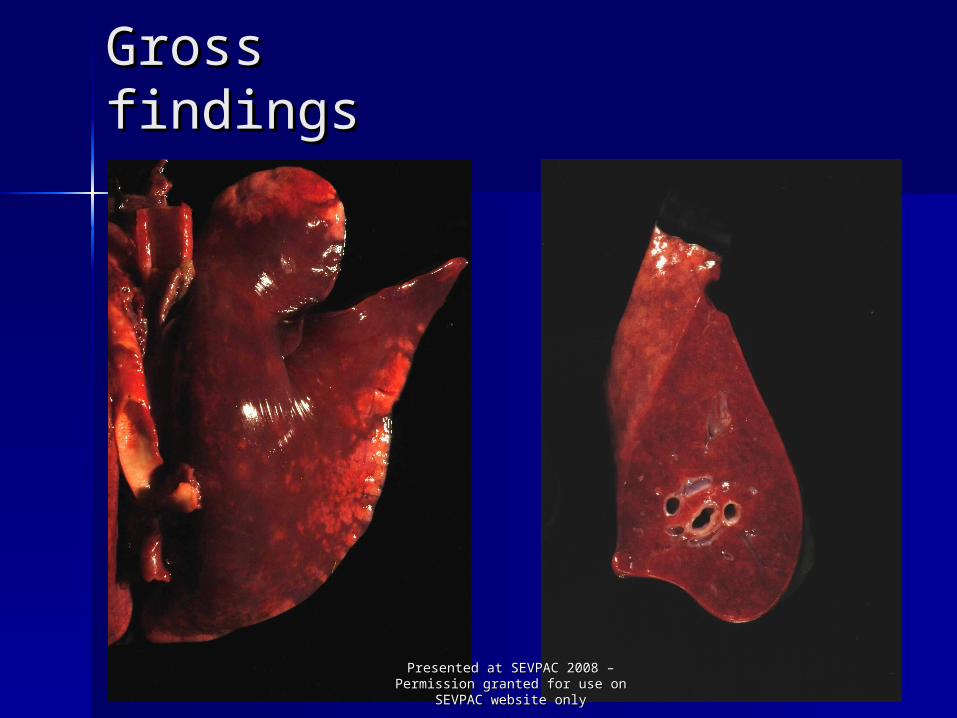
Gross findingsGross findings
Presented at SEVPAC 2008 – Permission Presented at SEVPAC 2008 – Permission granted for use on SEVPAC website onlygranted for use on SEVPAC website only
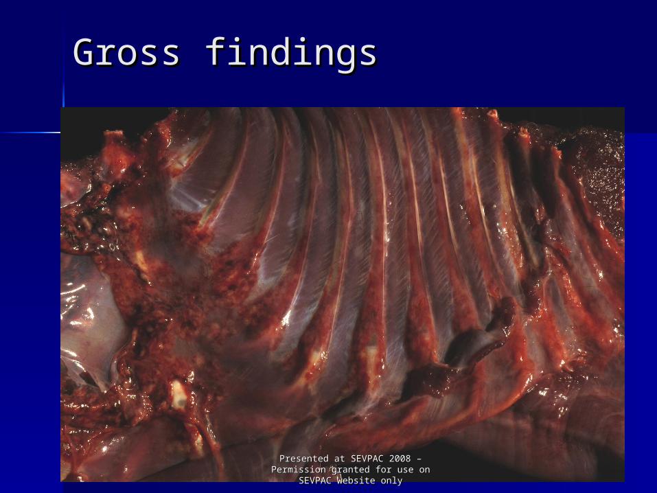
Gross findingsGross findings
Presented at SEVPAC 2008 – Permission Presented at SEVPAC 2008 – Permission granted for use on SEVPAC website onlygranted for use on SEVPAC website only

Gross Gross findingsfindings
Presented at SEVPAC 2008 – Permission Presented at SEVPAC 2008 – Permission granted for use on SEVPAC website onlygranted for use on SEVPAC website only

Gross Gross findingsfindings
Presented at SEVPAC 2008 – Permission Presented at SEVPAC 2008 – Permission granted for use on SEVPAC website onlygranted for use on SEVPAC website only

Gross Gross findingsfindings
Presented at SEVPAC 2008 – Permission Presented at SEVPAC 2008 – Permission granted for use on SEVPAC website onlygranted for use on SEVPAC website only

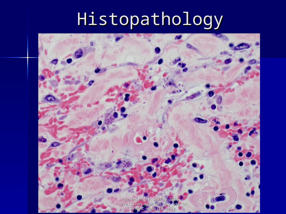
HistopathologyHistopathology
Presented at SEVPAC 2008 – Permission Presented at SEVPAC 2008 – Permission granted for use on SEVPAC website onlygranted for use on SEVPAC website only

HistopathologyHistopathology
Presented at SEVPAC 2008 – Permission Presented at SEVPAC 2008 – Permission granted for use on SEVPAC website onlygranted for use on SEVPAC website only
HistopathologyHistopathology
Presented at SEVPAC 2008 – Permission Presented at SEVPAC 2008 – Permission granted for use on SEVPAC website onlygranted for use on SEVPAC website only

HistopathologyHistopathology
Presented at SEVPAC 2008 – Permission Presented at SEVPAC 2008 – Permission granted for use on SEVPAC website onlygranted for use on SEVPAC website only
Morphologic diagnosisMorphologic diagnosis
Cecum: Cecum: Diffuse, transmural, marked, Diffuse, transmural, marked, subacute, subacute, necrotizing typhlitis and necrotizing typhlitis and gut-associated gut-associated lymphoid tissue lymphoid tissue necrosis with necrosis with intralesional intralesional protozoan organisms protozoan organisms consistent consistent with with Toxoplasma gondiiToxoplasma gondii
Presented at SEVPAC 2008 – Permission Presented at SEVPAC 2008 – Permission granted for use on SEVPAC website onlygranted for use on SEVPAC website only

SerologySerology
Positive for Toxoplasma gondii Positive for Toxoplasma gondii
[IgG > = 8192 UI/ml (>32 UI/ml positive)][IgG > = 8192 UI/ml (>32 UI/ml positive)]
Presented at SEVPAC 2008 – Permission Presented at SEVPAC 2008 – Permission granted for use on SEVPAC website onlygranted for use on SEVPAC website only
HistopathologHistopathologyy
similar lesions stomach, small intestine, and colon
lymphadenitis of mesenteric lymph nodes
myocarditis, adrenalitis and thyroiditis
encephalitis
plasmacytic uveitis and choroiditis
interstitial pneumonia, hepatitis, and splenitis
Presented at SEVPAC 2008 – Permission Presented at SEVPAC 2008 – Permission granted for use on SEVPAC website onlygranted for use on SEVPAC website only

HistopathologyHistopathology
Presented at SEVPAC 2008 – Permission Presented at SEVPAC 2008 – Permission granted for use on SEVPAC website onlygranted for use on SEVPAC website only
HistopathologyHistopathology
Intralesional protozoan organisms consistent with T. gondii
stomach heartsmall intestine adrenal
gland colon thyroid gland cecum brainmesenteric lymph nodes eye mesentery
interstitial pneumonia, hepatitis, and splenitis (not observed)
Presented at SEVPAC 2008 – Permission Presented at SEVPAC 2008 – Permission granted for use on SEVPAC website onlygranted for use on SEVPAC website only

ImmunohistochemistryImmunohistochemistry
Presented at SEVPAC 2008 – Permission Presented at SEVPAC 2008 – Permission granted for use on SEVPAC website onlygranted for use on SEVPAC website only

ImmunohistochemistryImmunohistochemistry
Presented at SEVPAC 2008 – Permission Presented at SEVPAC 2008 – Permission granted for use on SEVPAC website onlygranted for use on SEVPAC website only

DiscussionDiscussion
Wallabies and other Australian marsupials are among the most susceptible species to Toxoplasma gondii
Severe acute disease with widespread dissemination of the organism is frequently recognized
Contamination with T. gondii oocysts from domestic cats and wild felids is suspected as the main source of infection for herbivorous marsupials
In this case, feral cats were occasionally observed in the same area where the wallaby was kept
Most reported cases of fatal toxoplasmosis have been described in marsupials in captivity, especially in zoos
Presented at SEVPAC 2008 – Permission Presented at SEVPAC 2008 – Permission granted for use on SEVPAC website onlygranted for use on SEVPAC website only

ReferencesReferences
Dubey JP et al. Toxoplasmosis in black-faced kangaroos Dubey JP et al. Toxoplasmosis in black-faced kangaroos (Macropus fuliginosus melanops). (Macropus fuliginosus melanops). Veterinary Veterinary ParasitologyParasitology; 30: 97-105. 1988.; 30: 97-105. 1988.
Johnson AM et al. Serodiagnosis of acute toxoplasmosis Johnson AM et al. Serodiagnosis of acute toxoplasmosis in macropods. in macropods. Veterinary ParasitologyVeterinary Parasitology; 34: 25-33. ; 34: 25-33. 1989.1989.
Miller MA et al. Outbreak of toxoplasmosis in wallabies Miller MA et al. Outbreak of toxoplasmosis in wallabies on an exotic animal farm. on an exotic animal farm. J Vet Diagn InvestJ Vet Diagn Invest; 4: 480-; 4: 480-483. 1992.483. 1992.
Basso W et al. Toxoplasmosis in captive Bennett’s Basso W et al. Toxoplasmosis in captive Bennett’s wallabies (wallabies (Macropus rufogriseusMacropus rufogriseus) in Argentina. ) in Argentina. Veterinary ParasitologyVeterinary Parasitology; 144: 157-161. 2007.; 144: 157-161. 2007.
Presented at SEVPAC 2008 – Permission Presented at SEVPAC 2008 – Permission granted for use on SEVPAC website onlygranted for use on SEVPAC website only
AcknowledgementsAcknowledgements
Dr. Robert DonnellDr. Robert Donnell
Dr. Shelley NewmanDr. Shelley Newman
Histopathology Lab Staff, UTHistopathology Lab Staff, UT
UC Davis, IHCUC Davis, IHC
Presented at SEVPAC 2008 – Permission Presented at SEVPAC 2008 – Permission granted for use on SEVPAC website onlygranted for use on SEVPAC website only

Questions?Questions?
Presented at SEVPAC 2008 – Permission Presented at SEVPAC 2008 – Permission granted for use on SEVPAC website onlygranted for use on SEVPAC website only