Embed Size (px)
Citation preview
MARFAN SYNDROME: CT FINDINGS
H. RIAHI, M. BEN MESSAOUD, O. AZAIZ, A. AKROUT, B. SOUISSI, R. ALLANI, I. TURKI, E. MENIF
Radiology service, La Rabta Hospital, Tunis, Tunisie
CR 9
INTRODUCTION
Marfan syndrome is a multisystemic
connective tissue disorder that affects both
sexes equally and is characterized by
skeletal, cardiovascular, and ocular
abnormalities.
Its prevalence has been estimated at two to
three persons per 10,000.
INTRODUCTION It is an autosomal dominant transmitted
disorder (70%–75% of cases) but is also
associated with sporadic mutations.
The diagnostic process to identify patients with
MFS is challenging because it is based on the
Ghent criteria, which requires the assessment
of a number of clinical, genetic, and radiologic
features.
OBJECTIVES
The aim of this work is to illustrate CT findings in
Marfan syndrome revealed by acute chest pain.
MATERIEL AND PATIENTS
We report the case of 2 patients aged 28
and 32 who were seen in the emergency
department with acute chest pain. They
underwent contrast enhanced CT.
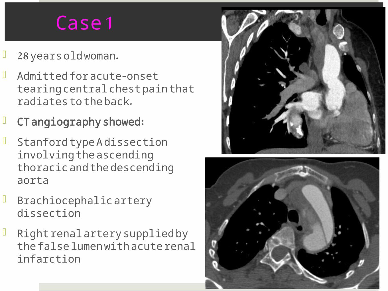
Case 1
28 years old woman.
Admitted for acute-onset tearing central chest pain that radiates to the back.
CT angiography showed:
Stanford type A dissection involving the ascending thoracic and the descending aorta
Brachiocephalic artery dissection
Right renal artery supplied by the false lumen with acute renal infarction
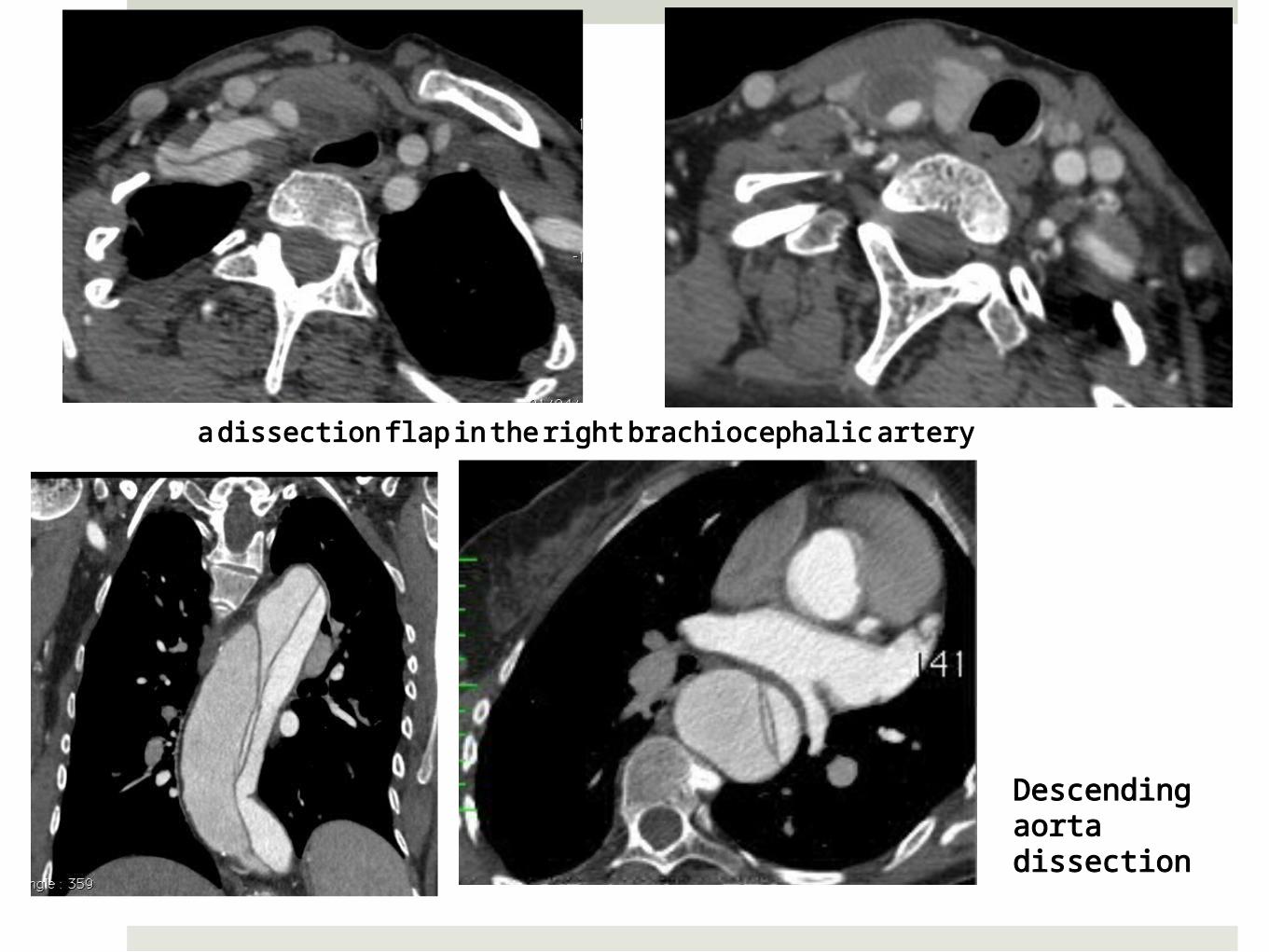
a dissection flap in the right brachiocephalic artery
Descending aorta dissection
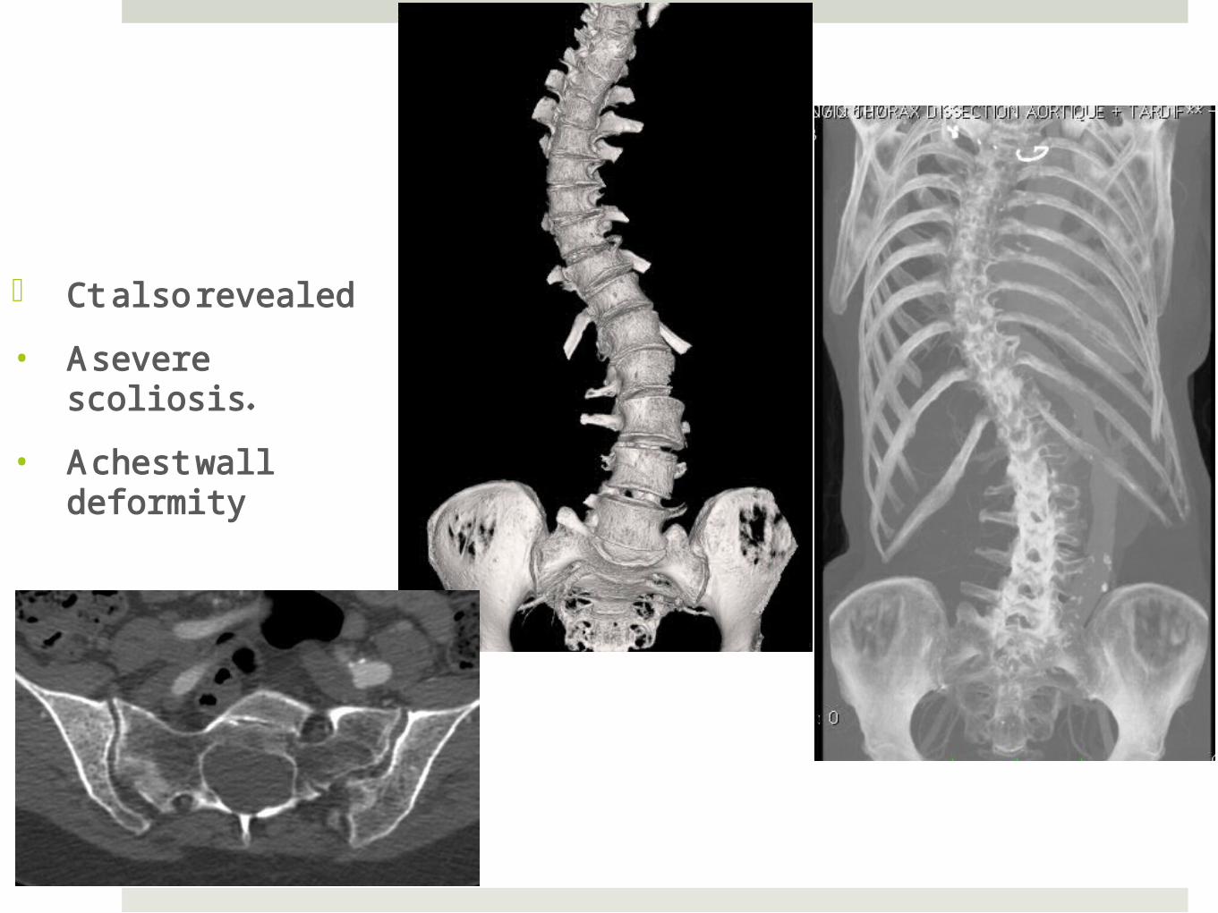
Ct also revealed
• A severe scoliosis.
• A chest wall deformity
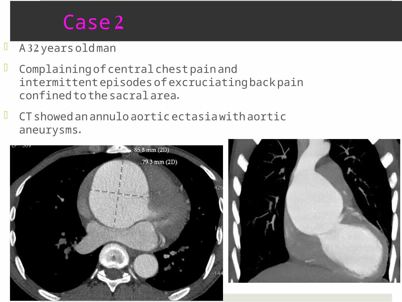
Case 2 A 32 years old man
Complaining of central chest pain and intermittent episodes of excruciating back pain confined to the sacral area.
CT showed an annulo aortic ectasia with aortic aneurysms.
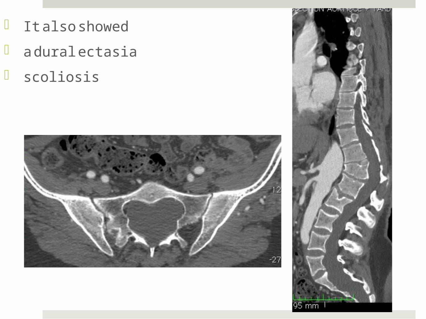
It also showed
a dural ectasia
scoliosis
DISCUSSION
Marfan syndrome may affect various systems,
including the cardiovascular, musculoskeletal, central
nervous, pulmonary, ocular, and integumentary
systems.
Diagnosis is based on the revised 1996 Ghent criteria,
which include cardiovascular, ocular, and pulmonary
abnormalities.
The presence of either two major features and one
minor feature or one major feature and four minor
features supports a diagnosis of Marfan syndrome.
Cardiovascular Manifestations
Cardiovascular major criteria include
• dilatation of the ascending aorta (involving at least the sinuses of Valsalva)
• with or without aortic regurgitation,
• as well as dissection of the descending aorta.
Minor criteria consist of
• dilatation or dissection of the descending or abdominal aorta before the age of 50 years,
• dilatation of the main pulmonary artery before the age of 40 years,
• mitral valve prolapse, and calcification of the mitral annulus before the age of 40 years
Cardiovascular Manifestations
1 . Annuloaortic Ectasia and Aortic Aneurysm:
Annuloaortic ectasia, especially with dilatation of the aortic root, is found in 60%–80% of adults with Marfan syndrome.
In annuloaortic ectasia, severe aortic regurgitation occurs that may progress to aortic root dissection or rupture
Cardiovascular Manifestations
• Aortic aneurysm without annuloaortic ectasia also is common.
• Compared with atherosclerotic aneurysms, it occur in younger patients and enlarge more rapidly.
• The diameters of the dilated ascending aorta, sinotubular junction, and aortic root are clearly demonstrated on multiplanar CT images obtained with three-dimensional reconstruction techniques.
Cardiovascular Manifestations
2 . Aortic Dissection
• Dissection develops more often in young patients with Marfan syndrome than it does in the general population.
• Multidetector CT is the radiologic modality most frequently used for diagnosis of aortic dissection.
• It clearly demonstrates the extent of dissection, the relationship of the true lumen and false lumen, and any involvement of major aortic branch vessels.
Cardiovascular Manifestations
3. Pulmonary Artery Dilatation
• Dilatation of the main pulmonary artery is one of the established criteria for the diagnosis of Marfan syndrome. Like dilatation of the ascending aorta, it occurs predominantly in the root.
• The upper limits of a normal main pulmonary artery diameter at the root and at the level of bifurcation, were 34.8 mm and 28.0 mm, respectively

Musculoskeletal Manifestations
1 . Scoliosis
• Scoliosis is a frequent and potentially severe manifestation of Marfan syndrome. It occurs in approximately 62% of patients.
• Scoliosis in Marfan syndrome is more severe, rigid, and progressive, requiring surgical correction.
• When it occurs in combination with straight back syndrome, kyphosis, or a chest wall deformity, it may contribute to cardiopulmonary compromise and restriction of lung volume

Musculoskeletal Manifestations
• Measurement of the severity of a scoliotic curve
has practical applications and the Lippman-Cobb
method is widely used to measure the degree of
scoliotic curvature.
• CT and MR imaging are helpful to evaluate the
bone structure, associated abnormalities of the
spinal cord, and the nerve roots before treatment
planning.
Musculoskeletal Manifestations
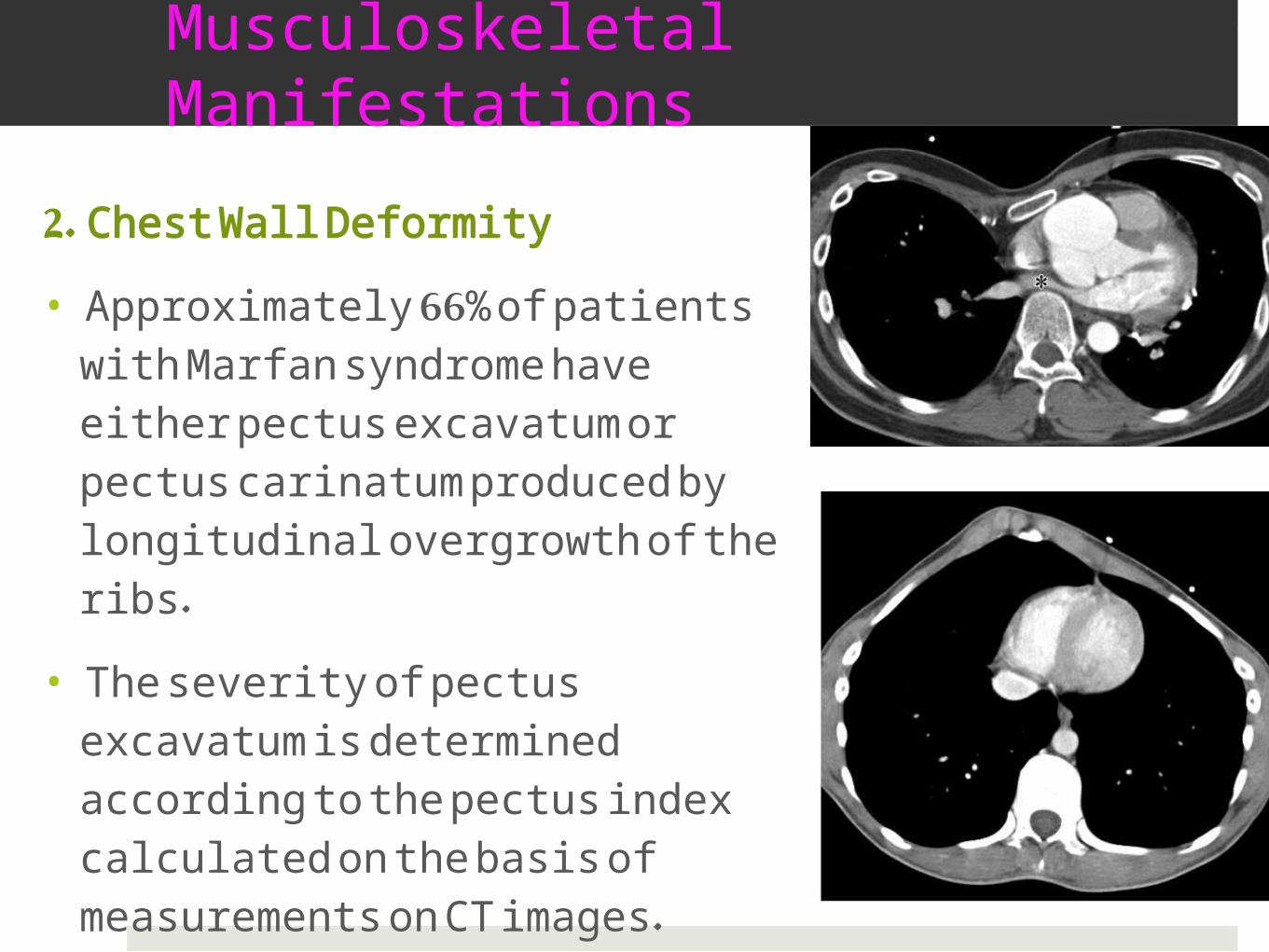
2. Chest Wall Deformity
• Approximately 66% of patients with Marfan syndrome have either pectus excavatum or pectus carinatum produced by longitudinal overgrowth of the ribs.
• The severity of pectus excavatum is determined according to the pectus index calculated on the basis of measurements on CT images.
Musculoskeletal Manifestations
3. Acetabular Protrusion
• Acetabular protrusion is a deformity of the hip
joint and is distinguished by the invasion of the
acetabulum and femoral head into the pelvic
cavity.
• Radiographic findings, including an increased
center-edge angle of Wiberg and an obscured
teardrop sign, allow the diagnosis.
Dural Ectasia and Associated Central Nervous System Manifestations
• Dural ectasia, which has been observed in 56%–
65% of patients with Marfan syndrome, is a
ballooning or significant widening of the dural sac
or neural root sleeves.
• It is sometimes accompanied by bone erosion,
meningoceles, and arachnoid cysts.
• Most occurrences of dural ectasia in Marfan
syndrome affect the lumbosacral spine.
• Dural ectasia is depicted on radiographs as a widening of the interpediculate distance.
• Vertebral body scalloping occurs with a high prevalence in transition vertebrae.
MR imaging and CT are the reference standards for
diagnosis of dural ectasia.
Dural ectasia appears as widening of the dural sac,
dilatation of the nerve root sleeve, and scalloping of
vertebral bodies in the lumbosacral spine on MR and
CT images.
In addition, MR and CT images may demonstrate an
accompanying meningocele or arachnoid cyst

Pulmonary Manifestations
The lungs are rarely involved in Marfan syndrome.
various pulmonary manifestations have been described, including
interstitial parenchymal disease and
honeycombing,
diffuse and apical bullous emphysema
, congenital malformation of the bronchus,
bronchiectasis, and spontaneous pneumothorax.
CONCLUSION
With the increasing availability of whole-body
imaging with multidetector CT or MR imaging, the
role of the radiologist has expanded beyond the
simple achievement of a diagnosis of Marfan
syndrom to include the comprehensive identification
of its various systemic manifestations.
In patient without a family history of Marfan
syndrome, computed tomography can play an
impotant role in diagnosis by identifying one of major
cardiovascular criteria or dural ectasia

![Local maxima of the systole functionrafi/Papers/systole.pdfLocal maxima of the systole function 3 Motivation Akrout [1] proved that sys is a topological4 Morse function on T g.This](https://img.pdfslide.net/doc/110x75/600ce32329f987282c3a39ad/local-maxima-of-the-systole-rafipaperssystolepdf-local-maxima-of-the-systole.jpg)