Embed Size (px)
Citation preview
Hindawi Publishing CorporationJournal of Biomedicine and BiotechnologyVolume 2010, Article ID 740403, 17 pagesdoi:10.1155/2010/740403
Review Article
Biological Role of Dystroglycan in Schwann Cell Function andIts Implications in Peripheral Nervous System Diseases
Toshihiro Masaki1, 2 and Kiichiro Matsumura3
1 Centre for Neuroregeneration, University of Edinburgh, Chancellor’s Building, 49 Little France Crescent,Edinburgh EH16 4SB, UK
2 Faculty of Medical Sciences, Teikyo University of Science, 2-2 Senju-Sakuragi, Adachi-ku, Tokyo 120-0045, Japan3 Department of Neurology and Neuroscience, Teikyo University, 2-11-1 Kaga, Itabashi-ku, Tokyo 173-8605, Japan
Correspondence should be addressed to Toshihiro Masaki, [email protected]
Received 22 October 2009; Accepted 20 April 2010
Academic Editor: Chung-Liang Chien
Copyright © 2010 T. Masaki and K. Matsumura. This is an open access article distributed under the Creative CommonsAttribution License, which permits unrestricted use, distribution, and reproduction in any medium, provided the original work isproperly cited.
Dystroglycan is a central component of the dystrophin-glycoprotein complex (DGC) that links extracellular matrix withcytoskeleton, expressed in a variety of fetal and adult tissues. Dystroglycan plays diverse roles in development and homeostasisincluding basement membrane formation, epithelial morphogenesis, membrane stability, cell polarization, and cell migration.In this paper, we will focus on biological role of dystroglycan in Schwann cell function, especially myelination. First, we reviewthe molecular architecture of DGC in Schwann cell abaxonal membrane. Then, we will review the loss-of-function studies usingtargeted mutagenesis, which have revealed biological functions of each component of DGC in Schwann cells. Based on thesefindings, roles of dystroglycan in Schwann cell function, in myelination in particular, and its implications in diseases will bediscussed in detail. Finally, in view of the fact that understanding the role of dystroglycan in Schwann cells is just beginning, futureperspectives will be discussed.
1. Introduction
Dystroglycan was originally isolated from skeletal muscle asone of dystrophin-associated proteins, and found to be amain component of the dystrophin-glycoprotein complex(DGC), a multimeric transmembrane protein complex [1,2]. In skeletal muscle, α- and β-dystroglycan constitute amembrane-spanning complex and interact with various sub-sarcolemmal and transmembrane proteins and componentsof basement membrane. Thus, DGC provides the physi-cal link between extracellular matrix and subsarcolemmalcytoskeleton, indicating its role in structural stability ofsarcolemma during contraction and extension of skeletalmuscles [3]. In fact, mutations in DGC components lead toprogressive muscle fiber degeneration, causing various typesof muscular dystrophies [4].
Dystroglycan is also expressed in many other cell typesand it plays a variety of roles in nonmuscle tissues. It has
been implicated in basal lamina development in mouseembryo [5], epithelial development [6], somitegenesis [7],cell polarization [8–10], carcinogenesis [11, 12], infectivepathogen targeting [13–15], and development of the centralnervous system (CNS) [16, 17].
In this paper, we review current understanding of dys-troglycan function in Schwann cells such as ensheathment,myelination as well as maintenance of other Schwann cellstructures, and the implications of these findings to periph-eral nervous diseases. In broad sense, “myelination” mayinclude entire developmental process of myelin formationfrom radial sorting of axons to membrane wrapping formingcompact myelin as well as assembly of the nodes of Ranvierand growth of internodes, which are inextricably linked witheach other [18]. However, in this paper, we will use “myelina-tion” mainly in a rather narrow sense, as a membrane wrap-ping process forming compact myelin after radial sorting iscompleted.
2 Journal of Biomedicine and Biotechnology
2. Molecular Architecture ofDystrophin-Glycoprotein Complex (DGC)in Schwann Cells
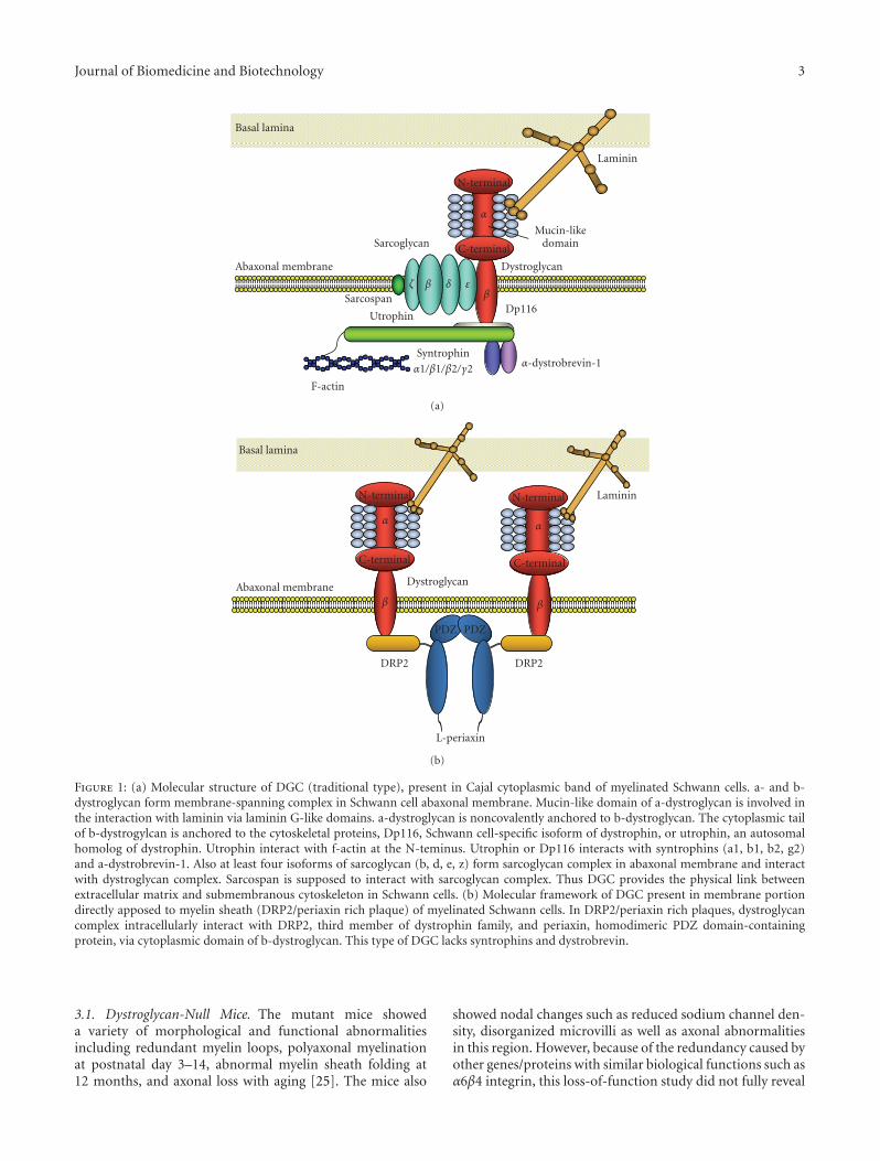
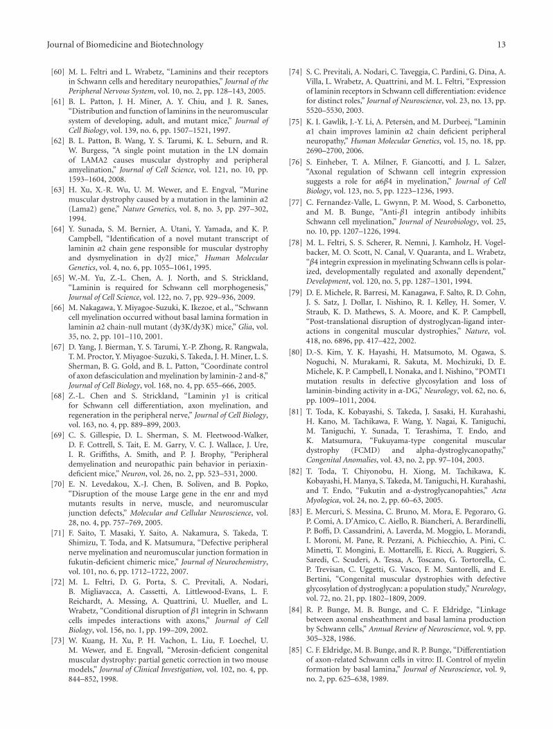
Molecular architecture of DGC in Schwann cells has beenextensively studied by our group and others ([19–29],Figure 1). The biochemical analysis revealed the similaritiesof the fundamental molecular architecture of DGC betweenSchwann cells and muscle cells as well as several criticaldifferences between them. In peripheral nerve, dystroglycanis mainly present in abaxonal membrane of both myelinatingSchwann cells and nonmyelinating Schwann cells [19, 22, 24,30, 31], while it is also expressed in perineurial cells as well assatellite cells of dorsal root ganglia [32]. As in skeletal muscle,dystroglycan in Schwann cells consists of two subunits (α andβ), which are translated from a single mRNA as a propeptidethat is proteolytically cleaved into two noncovalently associ-ated proteins [2, 19, 33]. In Schwann cells, α-dystroglycan isknown to bind to two of the extracellular ligands, laminin-2(α2β1γ1), major laminin isoform in Schwann cell basementmembrane, and agrin [19, 21–23]. Each of these has lamininG (LG)-like domains that mediate their high-affinity Ca2+-dependent binding to α-dystroglycan [34, 35]. While smallamount of laminin-8 (α4β1γ1) is also present in matureSchwann cell basement membrane, LG domains of laminin-8 showed only a low affinity for the α-dystroglycan receptor[36]. Apparent molecular mass of α-dystroglycan in Schwanncells is 120 kDa [19], which is smaller than that of skeletalmuscle (156 kDa), probably due to the difference of tissue-specific O-glycosylation within the mucin domain [4]. Whilethe removal of N-linked glycans alters the molecular weightof α-dystroglycan by only 4 kDa [1], and does not effecton its activity as an extracellular matrix receptor [37], fulldeglycosylation of α-dystroglycan results in the completeloss of ligand-binding activity [37]. Thus, the sugar chainson the mucin-like domain are supposed to mediate theseinteractions. Actually, structural analysis of the sialylatedO-linked oligosaccharides of bovine peripheral nerve α-dystroglycan revealed a high abundance of a novel O-mannosyl-type oligosaccharide, Siaα2-3Galβ1-4GlcNAcβ1-2Man-Ser/Thr (where Sia is sialic acid), and this tetrasac-charide was involved in the interaction of the α-dystroglycanwith laminin [38]. In other cell types, there are evidences thatα-dystroglycan binds to laminin-1 via sugar moieties otherthan the tetrasaccharide [39], while this is not confirmed inSchwann cells. Also, it was reported that nonglyosylated N-terminal fragment of α-dystroglycan bound to laminin-2/-4,laminin-1, fibronectin and fibrinogen [40].
On the other hand, α-dystroglycan is noncovalentlyanchored to β-dystroglycan in the Schwann cell membrane.β-dystroglycan is a type I transmembrane protein with asingle transmembrane domain and a 120 amino acid long C-terminal cytoplasmic tail. The cytoplasmic tail is anchoredto the cytoskeletal proteins, Dp116, Schwann cell-specificisoform of dystrophin, and utrophin, an autosomal homologof dystrophin [19, 20, 24, 26, 41–43]. β-dystroglycan issupposed to interact with cytoskeleton via interaction withthese submembranous proteins [42], while direct interactionof β-dystroglycan with f-actin was also reported [44]. α1-
dystrobrevin and four syntrophin isoforms (α1, β1, β2,and γ2), which interact with dystrophin and utrophin, arealso expressed in Schwann cells [25, 29]. These proteinsare crucial for the formation of DGC-associated signalingscaffolds in membrane of many cell types including Schwanncell abaxonal membrane [45]. As for binding between β-dystroglycan and Dp116, 15 C-terminal amino acids ofthe cytoplasmic domain of β-dystroglycan are involved inthe high affinity binding of Dp116, while 26 N-terminalamino acids of the cytoplasmic domain are also involvedin the low affinity binding of Dp116 [24]. In Schwanncells, MMP2 and MMP9 are suggested to be involved in β-dystroglycan processing, and produce a 30 kDa fragment ofβ-dystroglycan [46–49].
As other membrane protein members of DGC, β-, δ-,ε-, and ζ-sarcoglycans are expressed in Schwann cells. Thefour isoforms of sarcoglycans in Schwann cells seem to formstable tetrameric sarcoglycan complex that associates withdystrogycan and Dp116 to constitute larger DGC complex,as in the case of skeletal muscle [25, 26, 50]. Also, sarcospanis expressed in Schwann cells as a member of DGC [25].
Recently Albrecht et al. [29] reported that moleculararchitecture of DGC is different between the two dis-tinctive Schwann cell abaxonal membrane compartments,membrane covering Cajal cytoplasmic band and membranedirectly apposed to myelin sheath (DRP2/periaxin richplaque). In the former compartment, β-dystroglycan formsa complex with Dp116, utrophin, α1-dystrobrevin, α1-,β1-, β2-, and γ-syntrophins, and ABCA1 (traditional type,Figure 1(a)), and in the latter, β-dystroglycan forms acomplex with L-Periaxin and DRP2 (Figure 1(b)), which wasoriginally characterized by Sherman et al. [27, 51] and Courtet al. [52]. Also, Occhi et al. found expression of DGC innode of Ranvier, and the DGC in node of Ranvier seems tobe compatible with the traditional type, comprising Dp116and utrophin, but not periaxin and DRP2 [28]. They alsofound that laminin-2 (α2β1γ1) and laminin-10 (α5β1γ1) areexpressed in the nodal region [28].
Among these DGC components, only laminin-2 andperiaxin do not depend on dystroglycan for their localizationin Schwann cell abaxonal membrane [25]. Recently, a giantprotein, AHNAK was reported to modify laminin bindingcapacity of Schwann cell abaxonal membrane probably viainteraction with DGC [53], while precise molecular interac-tion between AHNAK and DGC remain to be elucidated.
3. Loss-of-Function Studies forDGC Components
Functions of DGC components including core compo-nents such as dystroglycans in Schwann cells were mainlyrevealed by loss-of-function studies in animals by targetingmutagenesis as well as gene mutation survey for humanand mouse muscular dystrophies (Table 1). In particular,double knockout of two functionally related genes as well asSchwann cell-selective gene ablation system such as Cre/loxPcontributed to these studies (Table 1). Therefore, the findingsobtained from these studies will be described in detail.
Journal of Biomedicine and Biotechnology 3
F-actin
Syntrophin
α1/β1/β2/γ2 α-dystrobrevin-1
Dp116Utrophin
Sarcospan
Abaxonal membrane
Basal lamina
Dystroglycan
Sarcoglycan
N-terminal
C-terminal
α
Mucin-likedomain
Laminin
ζ β δ εβ
(a)
DRP2DRP2
PDZPDZ
L-periaxin
Abaxonal membrane
Basal lamina
Dystroglycan
C-terminal
N-terminal
α
β β
N-terminal
C-terminal
α
Laminin
(b)
Figure 1: (a) Molecular structure of DGC (traditional type), present in Cajal cytoplasmic band of myelinated Schwann cells. a- and b-dystroglycan form membrane-spanning complex in Schwann cell abaxonal membrane. Mucin-like domain of a-dystroglycan is involved inthe interaction with laminin via laminin G-like domains. a-dystroglycan is noncovalently anchored to b-dystroglycan. The cytoplasmic tailof b-dystrogylcan is anchored to the cytoskeletal proteins, Dp116, Schwann cell-specific isoform of dystrophin, or utrophin, an autosomalhomolog of dystrophin. Utrophin interact with f-actin at the N-teminus. Utrophin or Dp116 interacts with syntrophins (a1, b1, b2, g2)and a-dystrobrevin-1. Also at least four isoforms of sarcoglycan (b, d, e, z) form sarcoglycan complex in abaxonal membrane and interactwith dystroglycan complex. Sarcospan is supposed to interact with sarcoglycan complex. Thus DGC provides the physical link betweenextracellular matrix and submembranous cytoskeleton in Schwann cells. (b) Molecular framework of DGC present in membrane portiondirectly apposed to myelin sheath (DRP2/periaxin rich plaque) of myelinated Schwann cells. In DRP2/periaxin rich plaques, dystroglycancomplex intracellularly interact with DRP2, third member of dystrophin family, and periaxin, homodimeric PDZ domain-containingprotein, via cytoplasmic domain of b-dystroglycan. This type of DGC lacks syntrophins and dystrobrevin.
3.1. Dystroglycan-Null Mice. The mutant mice showeda variety of morphological and functional abnormalitiesincluding redundant myelin loops, polyaxonal myelinationat postnatal day 3–14, abnormal myelin sheath folding at12 months, and axonal loss with aging [25]. The mice also
showed nodal changes such as reduced sodium channel den-sity, disorganized microvilli as well as axonal abnormalitiesin this region. However, because of the redundancy caused byother genes/proteins with similar biological functions such asα6β4 integrin, this loss-of-function study did not fully reveal
4 Journal of Biomedicine and Biotechnology
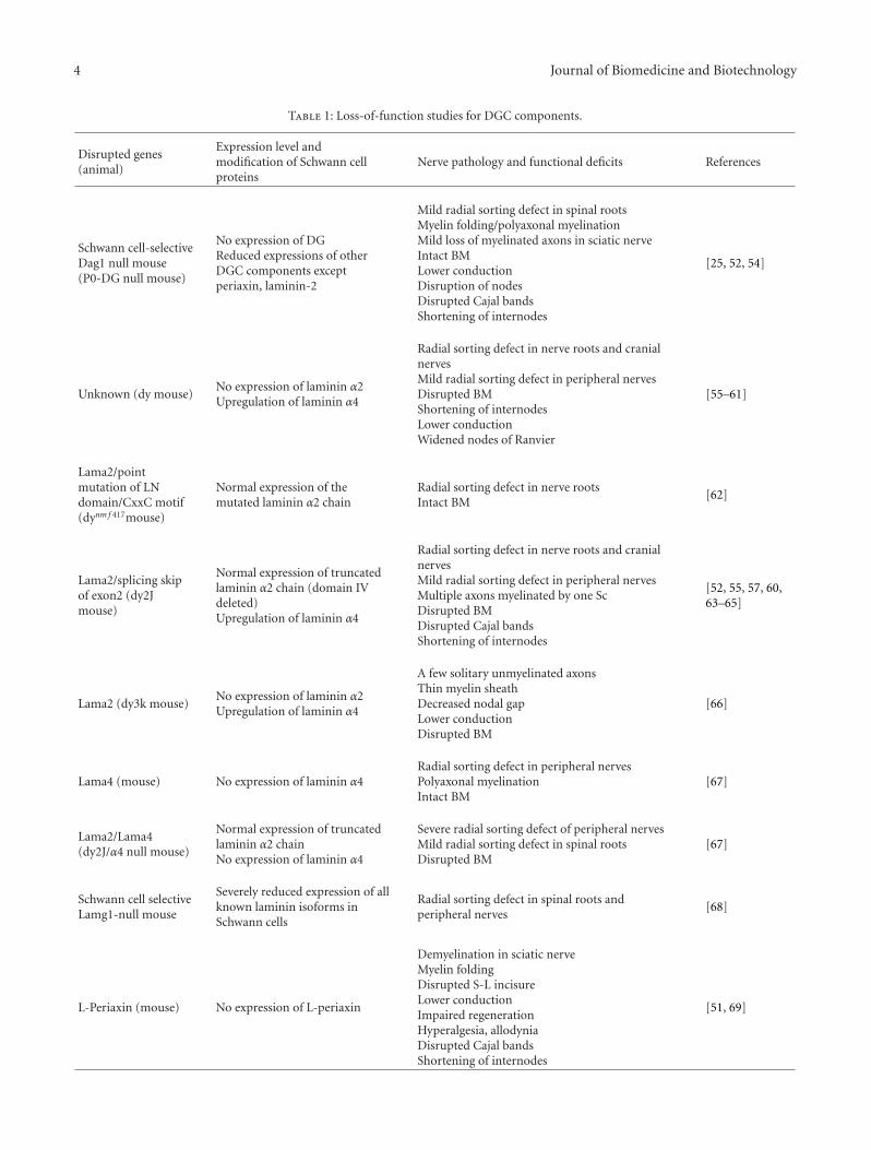
Table 1: Loss-of-function studies for DGC components.
Disrupted genes(animal)
Expression level andmodification of Schwann cellproteins
Nerve pathology and functional deficits References
Schwann cell-selectiveDag1 null mouse(P0-DG null mouse)
No expression of DGReduced expressions of otherDGC components exceptperiaxin, laminin-2
Mild radial sorting defect in spinal rootsMyelin folding/polyaxonal myelinationMild loss of myelinated axons in sciatic nerveIntact BMLower conductionDisruption of nodesDisrupted Cajal bandsShortening of internodes
[25, 52, 54]
Unknown (dy mouse)No expression of laminin α2Upregulation of laminin α4
Radial sorting defect in nerve roots and cranialnervesMild radial sorting defect in peripheral nervesDisrupted BMShortening of internodesLower conductionWidened nodes of Ranvier
[55–61]
Lama2/pointmutation of LNdomain/CxxC motif(dynm f 417mouse)
Normal expression of themutated laminin α2 chain
Radial sorting defect in nerve rootsIntact BM
[62]
Lama2/splicing skipof exon2 (dy2Jmouse)
Normal expression of truncatedlaminin α2 chain (domain IVdeleted)Upregulation of laminin α4
Radial sorting defect in nerve roots and cranialnervesMild radial sorting defect in peripheral nervesMultiple axons myelinated by one ScDisrupted BMDisrupted Cajal bandsShortening of internodes
[52, 55, 57, 60,63–65]
Lama2 (dy3k mouse)No expression of laminin α2Upregulation of laminin α4
A few solitary unmyelinated axonsThin myelin sheathDecreased nodal gapLower conductionDisrupted BM
[66]
Lama4 (mouse) No expression of laminin α4Radial sorting defect in peripheral nervesPolyaxonal myelinationIntact BM
[67]
Lama2/Lama4(dy2J/α4 null mouse)
Normal expression of truncatedlaminin α2 chainNo expression of laminin α4
Severe radial sorting defect of peripheral nervesMild radial sorting defect in spinal rootsDisrupted BM
[67]
Schwann cell selectiveLamg1-null mouse
Severely reduced expression of allknown laminin isoforms inSchwann cells
Radial sorting defect in spinal roots andperipheral nerves
[68]
L-Periaxin (mouse) No expression of L-periaxin
Demyelination in sciatic nerveMyelin foldingDisrupted S-L incisureLower conductionImpaired regenerationHyperalgesia, allodyniaDisrupted Cajal bandsShortening of internodes
[51, 69]
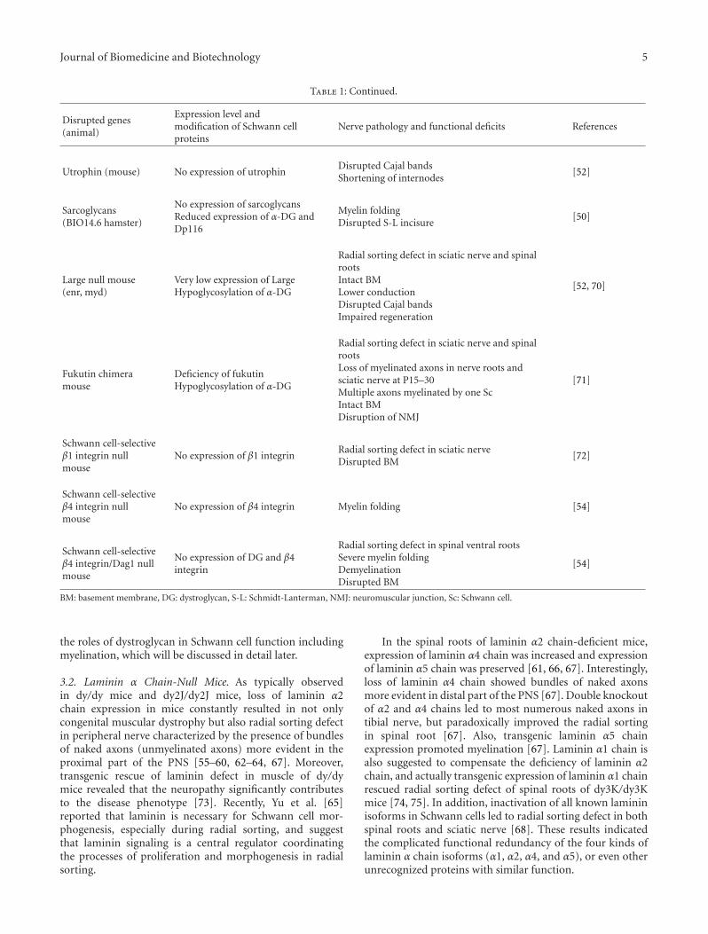
Journal of Biomedicine and Biotechnology 5
Table 1: Continued.
Disrupted genes(animal)
Expression level andmodification of Schwann cellproteins
Nerve pathology and functional deficits References
Utrophin (mouse) No expression of utrophinDisrupted Cajal bandsShortening of internodes
[52]
Sarcoglycans(BIO14.6 hamster)
No expression of sarcoglycansReduced expression of α-DG andDp116
Myelin foldingDisrupted S-L incisure
[50]
Large null mouse(enr, myd)
Very low expression of LargeHypoglycosylation of α-DG
Radial sorting defect in sciatic nerve and spinalrootsIntact BMLower conductionDisrupted Cajal bandsImpaired regeneration
[52, 70]
Fukutin chimeramouse
Deficiency of fukutinHypoglycosylation of α-DG
Radial sorting defect in sciatic nerve and spinalrootsLoss of myelinated axons in nerve roots andsciatic nerve at P15–30Multiple axons myelinated by one ScIntact BMDisruption of NMJ
[71]
Schwann cell-selectiveβ1 integrin nullmouse
No expression of β1 integrinRadial sorting defect in sciatic nerveDisrupted BM
[72]
Schwann cell-selectiveβ4 integrin nullmouse
No expression of β4 integrin Myelin folding [54]
Schwann cell-selectiveβ4 integrin/Dag1 nullmouse
No expression of DG and β4integrin
Radial sorting defect in spinal ventral rootsSevere myelin foldingDemyelinationDisrupted BM
[54]
BM: basement membrane, DG: dystroglycan, S-L: Schmidt-Lanterman, NMJ: neuromuscular junction, Sc: Schwann cell.
the roles of dystroglycan in Schwann cell function includingmyelination, which will be discussed in detail later.
3.2. Laminin α Chain-Null Mice. As typically observedin dy/dy mice and dy2J/dy2J mice, loss of laminin α2chain expression in mice constantly resulted in not onlycongenital muscular dystrophy but also radial sorting defectin peripheral nerve characterized by the presence of bundlesof naked axons (unmyelinated axons) more evident in theproximal part of the PNS [55–60, 62–64, 67]. Moreover,transgenic rescue of laminin defect in muscle of dy/dymice revealed that the neuropathy significantly contributesto the disease phenotype [73]. Recently, Yu et al. [65]reported that laminin is necessary for Schwann cell mor-phogenesis, especially during radial sorting, and suggestthat laminin signaling is a central regulator coordinatingthe processes of proliferation and morphogenesis in radialsorting.
In the spinal roots of laminin α2 chain-deficient mice,expression of laminin α4 chain was increased and expressionof laminin α5 chain was preserved [61, 66, 67]. Interestingly,loss of laminin α4 chain showed bundles of naked axonsmore evident in distal part of the PNS [67]. Double knockoutof α2 and α4 chains led to most numerous naked axons intibial nerve, but paradoxically improved the radial sortingin spinal root [67]. Also, transgenic laminin α5 chainexpression promoted myelination [67]. Laminin α1 chain isalso suggested to compensate the deficiency of laminin α2chain, and actually transgenic expression of laminin α1 chainrescued radial sorting defect of spinal roots of dy3K/dy3Kmice [74, 75]. In addition, inactivation of all known lamininisoforms in Schwann cells led to radial sorting defect in bothspinal roots and sciatic nerve [68]. These results indicatedthe complicated functional redundancy of the four kinds oflaminin α chain isoforms (α1, α2, α4, and α5), or even otherunrecognized proteins with similar function.
6 Journal of Biomedicine and Biotechnology
Another important finding in dy mice is the abnormalsodium channel clusters in nodes of Ranvier [28], whichwas identical in quality but less severe than those observedin dystroglycan-null mice [25]. This nodal abnormality wassupposed to be caused by laminin-2 deficiency becauselaminin-2 and laminin-10 are expressed in normal nodes andparanodes [28]. Notably, laminin-α1 chain was upregulatedin dy mice nodes [28].
3.3. Integrin-Null Mice. While integrin is not supposed tobe a member of DGC, α6β4 and α6β1 integrins are anotherimportant laminin receptors in Schwann cells, and have beensuggested to be involved in Schwann cell myelination [76–78]. Dissecting functions of those integrins is critical fordelineating exact role of dystroglycan, and therefore, thestudies for integrin-null mice will be reviewed in this paper.
Schwann cell specific disruption of β1 integrin showedlarge bundle of naked axons in both spinal roots and sciaticnerves suggesting severe impairment of radial sorting process[72]. Notably, myelinated Schwann cells never expressed β1integrin, suggesting that β1 integrin is not always necessaryfor myelination process once Schwann cells have achieved1 : 1 relationship with large axons [72].
In contrast, Schwann cell specific disruption of β4integrin did not affect peripheral nerve development, myelinformation, maturation, or regeneration except abnormalmyelin folding with ageing [54]. However, disruption of bothβ4 integrin and dystroglycan showed severer hypomyeli-nation in spinal roots than dystroglycan-null mice, withacute sign of demyelination such as myelin degeneration,macrophage infiltration and remyelination [54]. It alsoshowed major folding abnormalities in sciatic nerve myelin,suggesting a role of dystroglycan in myelin stabilization withthe cooperation of β4 integrin.
3.4. Mice with α-Dystroglycan Glycosylation Defects. Recentadvances have highlighted the importance of α-dystroglycanglycosylation in dystroglycan functions. Actually, muta-tion in the genes encoding LARGE, POMGnT1, POMTand fukutin cause defects in α-dystroglycan glycosylation,and altered capacity for binding to laminin, agrin, andneurexin [79, 80]. As a result, the glycosylation defectslead to the human disorders, congenital muscular dys-trophy 1D (MDC1D), muscle-eye-brain disease (MEB),Walker-Warburg disease (WWS), and Fukuyama congen-ital muscular dystrophy (FCMD), which are collectivelycalled α-dystroglycanopathy [81–83]. Because defects inα-dystroglycan glycosylation should reduce the laminin-2binding capacity of α-dystroglycan in not only muscle cellsbut also Schwann cells, Schwann cell functions associatedwith dystroglycan-laminin-2 interaction should be impairedin α-dystroglycanopathy. However, peripheral nerve involve-ment in α-dysroglycanopathy has not been extensively stud-ied. Therefore, we embarked on the investigation whetherperipheral nerve development, especially myelination, isdefective or not in fukutin-deficient chimeric mice, a mousemodel of FCMD [71]. As a result, we demonstrated thatthe sugar chain moiety and laminin-binding activity of α-dystroglycan were severely reduced, while the expression
level of laminin-2 seemed to be unaffected. The fukutinchimeric mice showed cluster of naked axons and loss ofmyelinated fibers in both sciatic nerve and spinal roots atP30. After 20 months of age, some spinal roots showedstriking loss of myelinated axons as well as degeneratedaxons suggesting the progressive neuropathy with aging.Also, occasionally multiple axons were myelinated by singleSchwann cell, which was also found in dy2J/dy2J mice [59].These results suggest that α-dystroglycan glycosylation playsroles in Schwann cell differentiation including ensheathmentand myelination, as well as maintenance of myelin in adultmice [71].
Similar to fukutin chimeric mice, Large-null mice (enrand myd mice) showed cluster of naked axons in sciaticnerve, while the sodium channel clustering in nodes ofRanvier was unaffected [70].
Altogether, Schwann cell myelination is disturbedin α-dystroglycanopathy mice. Therefore, scrutinizationof peripheral nerve involvement in human α-dystrogly-canopathy will reveal more detailed characterization of thosediseases.
3.5. Periaxin-Null Mice. Studies of periaxin-null micerevealed a unique aspect of DGC biological function inSchwann cells. Periaxin-null mice did not show defect inperipheral nerve development, instead they showed pro-gressive demyelination as well as abnormal myelin sheathfoldings in sciatic nerve, suggesting unstable myelin sheath[69]. Also periaxin-null mice showed abnormal Schwann cellcompartmentalization, disruption of protoplasmic bands ofCajal, and impaired Schwann cell internodal growth [51,52]. Recently, Court et al. [52] demonstrated that laminin-2, dystroglycan, utrophin axis is also required for properSchwann cell compartmentalization, and correct internodallength.
3.6. Sarcoglycan-Null Hamster. Sarcoglycan-null hamster(BIO14.6) showed that δ-sarcoglycan induced age-dependent myelin disruption or abnormal foldings andperturbed Schmidt-Lanterman incisures [50]. Togetherwith loss of sarcoglycan complex and reduced expressionof α-dystroglycan and Dp116, these findings suggestedthat sarcoglycans in Schwann cells stabilize DGC-mediatedtransmembrane extracellular matrix-cytoskeleton linkage,and thus play a role in myelin stability.
4. Detailed Role of Dystroglycan inSchwann Cell Function
Based on the findings derived from loss-of-function studiesdescribed above and other previous studies, detailed role ofdystroglycan in Schwann cell function will be discussed indetail in this section.
4.1. Role of Dystroglycan in Myelination and Myelin Main-tenance. It is well known that basement membrane playsan important role in Schwann cell ensheathment andmyelination [84, 85]. Therefore, the discovery in early 1990sthat congenital muscular dystrophy as well as dysmyelination
Journal of Biomedicine and Biotechnology 7
of nerve roots in dy/dy mice are due to lack of laminin-2 prompted us to investigate whether dystroglycan, a highaffinity receptor for laminin-2, plays a role in myelination[86].
In order to address this issue, we analyzed Schwann cellexpression of β-dystroglycan and laminin α2 chain during ratperipheral nerve regeneration and development. As a result,β-dystroglycan protein was present most densely in earlymyelinating Schwann cells, intermediately in promyelinatingSchwann cells, and most faintly in the Schwann cells inthe transient stage between immature Schwann cells andpromyelinating Schwann cells in rats at the age of P3 [87].Another group also reported that β-dystroglycan appear asa protein peinataly, just before myelination, exclusively inouter surface of mouse Schwann cells [74], which was con-sistant with our results derived from rat peripheral nerves.Also, the expression of β-dystroglycan protein dramaticallyincreased during first week after birth of rats, and wasmaintained until adulthood. The expression of laminin α2chain also increased during the first week, and continuedto increase until adulthood [87]. The expression of β-dystroglycan was down-regulated by axonal degenerationin adult rats, and was induced again by axon contactduring axonal regeneration [30]. These results indicate thatdystroglycan is associated with myelination as well as myelinmaintenance rather than radial sorting. Second, Schwanncell-specific dystroglycan-null mice showed abnormally thinmyelinated fibers as well as abnormal myelin folding inwhich Schwann cell established 1 : 1 relationship with axon,suggesting that dystroglycan plays a role in myelination afterradial sorting is completed [25, 54]. Also the dystroglycan-null mice showed mild loss of myelinated axons with aging.Third, double knockout of β4 integrin and dystroglycanrevealed far severer hypomyelination than respective knock-out of each of the two genes, as well as signs of acutedemyelination [54]. Notably the hypomyelination causedby disruption of dystroglycan was most severe in spinalventral roots [54]. Fourth, fukutin chimeric mice and Large-null mice showed hypomyelination as well as signs ofprogressive demyelinating neuropathy [70, 71]. Consideringthese evidences, dystroglycan no doubt plays a role inmyelination as well as in myelin maintenance.
While α6β4 integrin was also suggested to play a role inmyelination and myelin maintenance [76, 78], single disrup-tion of β4 integrin unexpectedly did not show any abnormal-ities of peripheral nerve myelin except age-dependent myelinfolding [54]. However double knockout of dystroglycan andβ4 integrin described above showed cooperative role ofβ4 integrin with dystroglycan in myelination and myelinmaintenance [54].
It is interesting to note that α-dystroglycan serves asa receptor for several pathogens such as Mycobacteriumleprae (M.leprae), lymphocytic choriomeningitis virus andthe Lassa fever virus [13–15, 88–90]. In particular, M.lepraecause acute nonimmune-mediated demyelination in mouseperipheral nerve [91], and the demyelination is at least partlymediated by ErbB2 receptor tyrosine kinase signaling [92].However, it remains to be elucidated whether M.leprae causedemyelination via perturbation of dystroglycan function as
well as whether dystroglycan has some interaction with theErbB2 signaling, while it was reported that Lassa fever virusefficiently competes with laminin α1 and α2 chains for α-dystroglycan binding [90].
Dystroglycan is also expressed in oligodendrocyte as alaminin receptor along with β1 integrins, and may play a rolein myelination by oligodendrocyte [93]. This study offerednew insight into α-dystroglycanopathies that cause braindysmyelination, as well as into the mechanism that underliesCNS myelin abnormalities caused by laminin deficiencies[93]. While structural myelin abnormalities are usually notvery clear in CNS of laminin deficient human and mice[94], meticulous quantitative and morphological analysissuggested the presence of defects of CNS myelin in dy/dymice, in particular in small-sized axons [95].
4.2. Role of Dystroglycan in Radial Sorting. It was unclearwhether dystroglycan played a role in radial sorting processfrom the loss-of-function studies of dystroglycan [25].However the possibility cannot be denied because radialsorting defect in spinal roots was observed in both Large-null mice and fukutin chimeric mice [70, 71]. This suggestsglycosylation of α-dystroglycan may be necessary for radialsorting, although the possibility that unidentified substrateproteins of these glycosylating enzymes play a role in radialsorting cannot be denied.
On the other hand, β1 integrin null mice showed amajor abnormality in radial sorting [72]. β1 integrin bindsto LG domains of laminin-2 different from α-dystroglycanbinding sites. Taken together, there is still a possibility thatdystroglycan may cooperate with β1 integrin in radial sortingprocess.
4.3. Role of Dystroglycan in Development and Maintenanceof Schwann Cell Structures such as Cajal Cytoplasmic Band,Internode and Node of Ranvier. The loss-of-function studiesprovided evidences that dystroglycan plays roles in devel-opment and maintenance of Schwann cell structures suchas Cajal cytoplasmic band, internode and node of Ranvier.First, Schwann cell-specific dystroglycan null mice showednodal changes including reduced sodium channel densityand disorganized microvilli [25, 28]. Second, dystroglycanalong with periaxin, utrophin and laminin-2, is necessaryfor compartmentalization including Cajal cytoplasmic bandformation and elongation of myelin segments [52]. Thesestudies offered new aspects of dystroglycan function inSchwann cells, and will stimulate further studies of DGCconcerning internode growth as well as nodal functions.
5. Molecular Mechanisms of Dystroglycan inSchwann Cell Myelin Formation
Molecular mechanisms of how dystroglycan regulatesSchwann cell myelination remain to be elucidated. Onepossibility is that the link between dystroglycan and laminin-2 provide anchorage between Schwann cell abaxonal mem-brane and basal lamina on which progression of inner lip ofSchwann cells over the axonal surface is based during myelin
8 Journal of Biomedicine and Biotechnology
formation [96]. Evidences that dystroglycan has bindingcapacities between Schwann cell abaxonal membrane andbasement membrane or laminin support this idea [25, 97,98]. Notably, basement membrane is not always necessaryfor myelination [60, 99]. Probably it is just because less orga-nized extracellular matrix components, which are not visibleas electron dense basement membrane, are enough for myeli-nation. In support of this idea, Podratz et al. [99] showedabundant laminin deposition on Schwann cell abaxonalsurface in spite of the lack of visible basement membrane.
Another possibility is that dystroglycan plays a rolein myelination by regulating signaling from extracellularmatrix, especially laminin-2, to intracellular signaling path-ways or cytoskeleton. Actually, laminin signaling seemsto play an essential role in Schwann cell proliferationand survival as well as cytoskeletal regulation-associatedensheathment and myelination [60, 100]. While dystroglycanis a major laminin receptor in Schwann cells along withintegrins such as α6β1, α6β4, and α7β1, the receptorfunction of dystroglycan mediating laminin signaling hasbeen less extensively studied compared with that of integrins.Recently, however, ample evidences are accumulating thatdystroglycan is associated with cell signaling.
First, β-dystroglycan can interact with a varietyof signaling proteins, at least partly via SH2 and SH3binding sites in the C-terminus. While dephosphorylationof Y892 (pY892) in β-dystroglycan enables binding toutrophin/dystrophin[101, 102], phosphorylation of β-dystroglycan enhances recruitment of SH2/SH3 domaincontaining signaling-associated proteins like c-Src, Fyn,Csk, Nck, Shc, and Grb-2 [103–106]. Grb-2 is known tobe involved in ERK-MAP kinase cascade and cytoskeletalorganization [103]. Also β-dystroglycan can interact withFAK [104]. In addition, DAMAGE was reported to be a newmember of DGC as a protein associated with α-dytrobrevin[43]. DAMAGE has a potential nuclear localization signal, 30contiguous 12-aminoacid repeats and two MAGE homologydomains, suggesting it is involved in membrane signaling[43].
Moreover, in nonperipheral nerve tissues, dystroglycanhas been shown to be involved in several signaling path-ways. Association of dystroglycan with MAPK/ERK cascadeand GTPase signaling was reported [107–109]. Filopodiaformation is often governed by Cdc42, a GTPase signaling-associated protein playing diverse roles in cell polarity,cytoskeletal regulation as well as cell cycle [109]. Dystro-glycan plays a role in filopodia formation via forming acomplex with ezrin and Dbl, and activating Cdc42 [107–109]. Fibronectin or biglycan may induce signaling viadystroglycan leading to calcium flux and alteration ofcytoskeletal architecture [110]. Syntrophin contains twopleckstrin homology (PH) domains and one PDZ domain[111]. Binding of laminin to DGC induced heterotrimericG protein binding to α-syntrophin’s PDZ domain, whichleads to activation of PI3K/Act signaling and alteration ofintracellular Ca2+ in muscle [111–114]. Laminin-1 inducedGrb binding to syntrophin, recruited Sos1/Rac1/PAK1/JNK,and eventually led to c-jun phosphorylation [115]. Recently,genetic modifier screens using Drosophila melanogaster
revealed that DGC interacts with genes involved in Notch,TGF-β and EGFR signaling pathways as well as those associ-ated with muscle function and cellular or axonal migration[116]. In Schwann cells also, laminin assembly initiateddystroglycan-dependent Src/Fyn activation and utrophinrecruitment that contributed to their survival [117]. Takentogether, these evidences strongly suggest that dystroglycansignaling plays diverse roles in Schwann cell functionsincluding myelination. The hypothetical role of dystroglycansignaling in Schwann cell myelination will be discussed indetail in the section of future perspectives.
6. Human Peripheral Nervous System (PNS)Diseases Associated with DGC
Among the human diseases caused by mutation of DGCcomponents, there are only two diseases in which periph-eral nerve involvement was clearly demonstrated, MDC1A(LAMA2 mutation) and neuropathy caused by PRX muta-tion, which will be described in detail below. In general,peripheral nerve involvement caused by mutation of DGCcomponents has not been studied as extensively as musculardystrophies. Therefore, there is still a possibility that furtherstudy in the future will reveal new evidences that otherDGC components play roles in the pathogenesis of humanperipheral neuropathies.
6.1. MDC1A (Merosin (Laminin-2)-Deficient CongenitalMuscular Dystrophy). MDC1A is the most frequent congen-ital muscular dystrophy in Europe with autosomal recessiveinheritance caused by LAMA2 mutation [94, 118]. Completelaminin-2 deficiency causes early-onset muscular dystrophy,peripheral neuropathy and white matter lesions in CNS. Par-tial laminin-2 deficiency presents variant phenotypes withlater onset muscular dystrophy, or even predominant PNSor CNS abnormalities. While peripheral nerve involvementis not extensively studied in MDC1A, nerve conductionvelocity is reduced in most of the patients [119–123]. As abasis for the slowed conduction velocity, abnormal sodiumchannel clusters were found in these patients [28]. Theneuropathy is predominantly motor or sensory-motor [120–123]. Sural nerve biopsy showed mild loss of myelinatedfibers, globular thickening of myelin sheath at paranodalregion, myelin foldings, shortened internodes, widenednodes of Ranvier [121, 124], and compartmentalizationdefects [52]. Unfortunately, radial sorting defect in spinalroots in human has not been confirmed because of theabsence of autopsy study.
Patients with MDC1A show striking white matterchanges in T2 weighted brain magnetic resonance imaging[125], which is diffuse, bilateral, and symmetrical. It appearsafter the first 6 months of life, and nonprogressive [126].However, morphological changes of cerebral white matter arenot clearly demonstrated in human patients. Rather, mainpathological findings in CNS are developmental anomaliessuch as abnormal cerebral cortical gyration, hypoplasia ofvermis, hemisphere, or pons. At least, part of these abnor-malities is supposed to be caused by neuronal migration
Journal of Biomedicine and Biotechnology 9
defects associated with laminin-2-α-dystroglycan interac-tion, which are also demonstrated in α-dystroglycanopathies.The full aspects of dystroglycan roles in CNS are beyond thescope of this review, and other comprehensive reviews dealin detail with these issues [81, 127–130].
6.2. Charcot-Marie-Tooth Neuropathy Type 4F and DejerineSottas Neuropathy Caused by PRX (Periaxin) Mutations.Periaxin is the only one gene of members of DGC reportedto be associated with human hereditary demyelinatingneuropathy. Mutation of PRX causing loss of L-periaxinexpression induces sensory-motor demyelinating neuropa-thy consistent with Charcot-Marie-Tooth neuropathy orDejerine Sottas neuropathy [131–133]. Homozygous PRXmutation C715X causing expression of truncated form ofL-periaxin showed relatively milder phenotype of Charcot-Marie-Tooth neuropathy with sensory dominant involve-ment [134]. Nerve biopsy showed loss of myelinated fibers,onion bulbs, focal thickening of myelin sheath, and myelinfolding, which were similar to the nerve pathology of Prx-/-mice [131–134].
7. Future Perspectives
Schwann cell is one of the somatic cell types with robustregenerative capacity. The regenerative capacity of Schwanncell depends on its property of plasticity, which enablesSchwann cell to change its phenotype between differenti-ated Schwann cell (myelinating type and nonmyelinatingtype) and denervated (dedifferentiated) Schwann cell [135,136]. Recent advances have highlighted the importanceof gene/protein network maintaining the identity of cellphenotype, especially from the study of embryonic stemcells [137, 138]. Then, there must be molecular networkspecific to each of the Schwann cell phenotypes. Definitelydystroglycan and other DGC components belong to themolecular network specific to myelinating Schwann cellphenotype, and now it is important to understand the roleof dystroglycan in the context how this protein contributesto the gene/protein network. Cutting edge technologies suchas DNA microarray, ultra-high-throughput sequencing andmass spectroscopy-based proteomics have made it possibleto study epigenome, transcriptome, and proteome as a whole[137, 139]. Analyzing gene/protein network maintainingidentity of myelinating Schwann cell phenotype, and com-paring with that of dedifferentiated Schwann cell phenotypewill reveal a whole framework of molecular mechanisms ofmyelination.
At the same time, it is important to study dystroglycanfunction focusing on specific molecular aspects such as pro-tein interaction, signaling, transcriptional regulation includ-ing chromatin modifications, post-transcriptional modi-fication, post-translational modification, and intracellulartransport and degradation. In addition, anatomical aspectsshould be taken into consideration. Myelinating Schwanncell has four distint domains: internode, juxtaparanode,paranode, and node [140, 141]. Moreover, internode domainis divided into two compartments; Cajal cytoplasmic band,and compartment occupied by myelin sheath [51]. Each
domain is playing a specific role in maintaining myelinatingSchwann cells, and DGC is likely to play different rolein each of the domain. In many of these specific aspects,dystroglycan function is just beginning to be revealed, andso many questions must be answered. A few of the mutuallynonexclusive topics with high priority will be discussedbelow.
(1) Protein Interaction. various proteins have been and arebeginning to be revealed to interact with DGC in non-peripheral nerve tissues. Examples are extracellular matrixproteins such as fibronectin/biglycan [110, 142], perlecan[143–145], pikachurin [146], membrane proteins/receptorssuch as integrins [147, 148], AHNAK [53], aquaporins [149,150], MLC1 [151], caveolins [147], Na and K channels [141,149, 152–154], and submembranous or cytoskeletal proteinssuch as G proteins or other signaling-associated proteins[103, 106, 111, 113, 115], nitric-oxide synthase [147], actins[43], tubulins [155], ERMs (ezrin-radixin-moesin) [108,109], and Par1 [10]. Dissecting their interactions with DGCin Schwann cells one by one will reveal more comprehensivemolecular architecture, and specific functions associatedwith it.
(2) Signaling. Myelinating Schwann cell phenotype issupposed to be maintained by signaling from bothabaxonal membrane contacting extracellular matrix andadaxonal membrane apposing to axon. And the inte-grated signaling from both directions is supposed tomaintain transcriptome or cytoskeletal structures specificto myelinating Schwann cells. Dystroglycan may playa role in signaling from abaxonal membrane via theinteraction with other proteins present in extracellularmatrix /abaxonal membrane/cytoskeleton, especially withlaminin-2 and α6β4 integrin. Then dystroglycan-associatedsignaling is supposed to activate positive regulators of myeli-nation, and inactivate negative regulators of myelination.Evidences accumulated by studies of nonperipheral nervetissues suggest a number of hypotheses in this issue. Asexamples, several hypotheses will be described below.
First is about the association of dystroglycan or syn-trophin with signaling-associated proteins such as Src,Fyn, Csk, Nck, Shc, and Grb2 [103–106, 108, 109, 115].Interaction of DGC with these adaptor proteins implies thatDGC may regulate Rho family GTPase signaling as well asMAPK signaling cascade [103–106, 108, 109, 115, 156]. Oryeast two hybrid screens suggested that dystroglycan candirectly activate MEK or ERK, members of MAPK cascade[107]. Because it was reported that Ras signaling promotesdifferentiation of Schwann cells [157], dystroglycan maypromote myelination through Ras signaling. However, it iscontroversial whether Ras/Raf/ERK signaling is promotingSchwann cell differentiation because there is a report thatRas/Raf/ERK signaling drives Schwann cell dedifferentiation[158]. On the other hand, Cdc42, one of the Rho familyGTPases, was suggested to promote radial sorting andmyelination [65]. So Cdc42 may be another mediator ofdystroglycan signaling.
Second, genetic modifier screens suggested the associa-tion of dystroglycan with Notch signaling [116]. In Schwann
10 Journal of Biomedicine and Biotechnology
cells, Notch acts as a negative regulator of myelination [135,159]. Dystroglycan, therefore, can promote myelination viaregulating Notch signaling. Notably, Notch signaling andWNT signaling seem to prevent oligodendrocyte differen-tiation (myelination). Notch signaling and WNT signalingrespectively can induce HES5 and ID2/4, repressors ofmyelin genes, via transcription factor activity of NICD(Notch1 intracellular domain)/CBF1 and β-catenin/TCF7L2,and then HES5 or ID2/4 represses the transcription of myelingenes [160, 161]. Moreover, HDACs (histone deacetylases)can relieve these repressions by competing with the NICDto bind to CBF1 and competing with β-catenin to bindto TCF7L2. At the same time, the HDACs might increasethe state of chromatin compaction around genes such asHES5, ID2, and ID4 that encode repressors of oligoden-drocyte differentiation, thus preventing their transcriptionand providing permissive conditions for oligodendrocytedifferentiation [160, 161].
Third, Krox20, one of the key myelin-associated tran-scription factors [162], might be another candidate medi-ator of dystroglycan signaling, because laminin signalingincreases Krox-20 expression [163]. Krox 20 can inhibit c-Junactivity [164]. Also Krox 20 can suppress Notch signaling byreducing NICD post-translationally [159].
Fourth, DGC can interact with FAK [104]. FAK is anECM-associated signaling protein and is at the crossroad ofmultiple signaling pathways, interacting with Rho GTPasesignaling as well as MAPK signaling [165]. Effects of FAKon cytoskeletal organization were well demonstrated [165].Moreover, FAK was recently reported to be required forradial sorting [166]. FAK can interact also with Erb2/Erb3receptor [167], and then Erb2/Erb3 receptor can promotemyelination [100]. Notably, laminin deficiency caused dra-matic decrease of Erb2/Erb3 receptor phosphorylation [100].Therefore, DGC may regulate radial sorting or myelinationvia interaction with FAK.
(3) Transcriptional Regulation. little is known about tran-scriptional regulation of dystroglycan or other DGC compo-nents in Schwann cells. Recently, Rettino et al. [168] reportedthat the expression of dystroglycan was regulated by SP1transcription factor in muscle cells, and DNA methylationas well as histone acetylation may be involved in theregulation. In addition, Miura et al. [169] reported thatPPARβ/δ agonist stimulated the transcription of utrophin,which restored the expression of α1 syntrophin and β-dystroglycan at the sarcolemma of the mdx mice. However,so many issues still remain to be answered consideringthe extremely complicated mechanisms of transcriptionalregulation exerted by various transcriptional regulators aswell as chromatin modifications including DNA methylationand all kinds of histone modifications [170].
(4) Node of Ranvier. While dystroglycan-null mice showedvariety of nodal abnormalities including disorganizedmicrovilli and reduced sodium channel density, the molec-ular mechanism remains to be unknown. In order to addressthis issue, it will be important to study the interaction ofdystroglycan with node-associated proteins. For example,ERMs are expressed in the microvilli of many cell types
including those of Schwann cells [171]. In nonperipheralnervous tissues, it is known that dystroglycan plays arole in filopodia formation via interaction with ezrin, andsubsequent activation of Cdc42 [108, 109]. Then Cdc42is supposed to induce cytoskeletal changes necessary forfilopodia formation [108, 109]. Therefore, dystroglycanmay be associated with Schwann cell microvilli formationthrough interaction with ERMs.
(5) Cell Polarity. While Schwann cells do not belong toepithelial cells, Schwann cells have many molecular andstructural properties similar to epithelial cells [140, 172]. Inparticular, cell polarity is a central feature shared by Schwanncells with epithelial cells [140, 172]. Several lines of evidencessuggest that dystroglycan plays a role in maintaining cellpolarity of not only epithelial cells but also Schwann cells [8–10, 30, 172]. First, in Wallerian degeneration, Schwann cellphenotype changes from myelinating type to dedifferentiatedtype [135]. During this process, the cell polarity specificto myelinating Schwann cells is lost along with the disso-ciation of basement membrane from abaxonal membraneas well as downregulation of dystroglycan and laminin-2 [30]. This finding suggests that dystroglycan-laminin-2 interaction is involved in cell polarity maintenance ofmyelinating Schwann cells. Second, Cdc42, which seems tohave several links with dystroglycan signaling, is one of thecental regulators of cell polarity in epithelial cells as well asglial cells [172]. Third, cell polarity protein Par-1 not onlyregulates the basolateral localization of DGC, but also isrequired for the formation of a functional DGC in epithelialcells [10]. Analyzing molecular interactions of DGC withcell polarity-associated proteins will reveal further detailedfunction of dystroglycan.
8. Conclusion
Dystroglycan plays diverse roles in Schwann cells suchas myelination and maintenance of myelin and nodalstructures. However, the molecular mechanisms on whichdystroglycan functions are based are just beginning to berevealed. Analyzing gene/protein network as a whole systemusing cutting edge technologies as well as individual studiesfocusing on a variety of biological aspects specific to Schwanncells hold promise for elucidating molecular mechanismof dystroglycan functions, and eventual development ofeffective treatments for human peripheral neuropathies.
References
[1] J. M. Ervasti and K. P. Campbell, “Membrane organizationof the dystrophin-glycoprotein complex,” Cell, vol. 66, no. 6,pp. 1121–1131, 1991.
[2] O. Ibraghimov-Beskrovnaya, J. M. Ervasti, C. J. Leveille, C.A. Slaughter, S. W. Sernett, and K. P. Campbell, “Primarystructure of dystrophin-associated glycoproteins linking dys-trophin to the extracellular matrix,” Nature, vol. 355, no.6362, pp. 696–702, 1992.
[3] B. J. Petrof, J. B. Shrager, H. H. Stedman, A. M. Kelly, andH. L. Sweeney, “Dystrophin protects the sarcolemma fromstresses developed during muscle contraction,” Proceedings
Journal of Biomedicine and Biotechnology 11
of the National Academy of Sciences of the United States ofAmerica, vol. 90, no. 8, pp. 3710–3714, 1993.
[4] R. Barresi and K. P. Campbell, “Dystroglycan: from biosyn-thesis to pathogenesis of human disease,” Journal of CellScience, vol. 119, no. 2, pp. 199–207, 2006.
[5] R. A. Williamson, M. D. Henry, K. J. Daniels, R. F. Hrstka,J. C. Lee, Y. Sunada, O. Ibraghimov-Beskrovnaya, and K.P. Campbell, “Dystroglycan is essential for early embryonicdevelopment: disruption of Reichert’s membrane in Dag1-null mice,” Human Molecular Genetics, vol. 6, no. 6, pp. 831–841, 1997.
[6] M. Durbeej, E. Larsson, O. Ibraghimov-Beskrovnaya, S. L.Roberds, K. P. Campbell, and P. Ekblom, “Non-muscle α-dystroglycan is involved in epithelial development,” Journalof Cell Biology, vol. 130, no. 1, pp. 79–91, 1995.
[7] M. Hidalgo, C. Sirour, V. Bello, N. Moreau, M. Beaudry,and T. Darribere, “In vivo analyzes of dystroglycan functionduring somitogenesis in Xenopus laevis,” DevelopmentalDynamics, vol. 238, no. 6, pp. 1332–1345, 2009.
[8] S. Li, D. Edgar, R. Fassler, W. Wadsworth, and P. D.Yurchenco, “The role of laminin in embryonic cell polariza-tion and tissue organization,” Developmental Cell, vol. 4, no.5, pp. 613–624, 2003.
[9] W.-M. Deng, M. Schneider, R. Frock, C. Castillejo-Lopez,E. A. Gaman, S. Baumgartner, and H. Ruohola-Baker,“Dystroglycan is required for polarizing the epithelial cellsand the oocyte in Drosophila,” Development, vol. 130, no. 1,pp. 173–184, 2003.
[10] M. Masuda-Hirata, A. Suzuki, Y. Amano, K. Yamashita, M.Ide, T. Yamanaka, M. Sakai, M. Imamura, and S. Ohno,“Intracellular polarity protein PAR-1 regulates extracellularlaminin assembly by regulating the dystroglycan complex,”Genes to Cells, vol. 14, no. 7, pp. 835–850, 2009.
[11] M. D. Henry, M. B. Cohen, and K. P. Campbell, “Reducedexpression of dystroglycan in breast and prostate cancer,”Human Pathology, vol. 32, no. 8, pp. 791–795, 2001.
[12] A. Sgambato and A. Brancaccio, “The dystroglycan complex:from biology to cancer,” Journal of Cellular Physiology, vol.205, no. 2, pp. 163–169, 2005.
[13] W. Cao, M. D. Henry, P. Borrow, H. Yamada, J. H. Elder,E. V. Ravkov, S. T. Nichol, R. W. Compans, K. P. Campbell,and M. B. A. Oldstone, “Identification of α-dystroglycan asa receptor for lymphocytic choriomeningitis virus and Lassafever virus,” Science, vol. 282, no. 5396, pp. 2079–2081, 1998.
[14] A. Rambukkana, H. Yamada, G. Zanazzi, T. Mathus, J. L.Salzer, P. D. Yurchenco, K. P. Campbell, and V. A. Fischetti,“Role of α-dystroglycan as a Schwann cell receptor forMycobacterium leprae,” Science, vol. 282, no. 5396, pp. 2076–2079, 1998.
[15] A. Rambukkana, S. Kunz, J. Min, K. P. Campbell, and M. B.A. Oldstone, “Targeting Schwann cells by nonlytic arenaviralinfection selectively inhibits myelination,” Proceedings of theNational Academy of Sciences of the United States of America,vol. 100, no. 26, pp. 16071–16076, 2003.
[16] S. A. Moore, F. Saito, J. Chen, D. E. Michele, M. D. Henry,A. Messing, R. D. Cohn, S. E. Ross-Barta, S. Westra, R. E.Williamson, T. Hosl, and K. P. Campbell, “Deletion of braindystroglycan recapitulates aspects of congenital musculardystrophy,” Nature, vol. 418, no. 6896, pp. 422–425, 2002.
[17] J. E. Schroder, M. R. Tegeler, U. Großhans, E. Porten, M.Blank, J. Lee, C. Esapa, D. J. Blake, and S. Kroger, “Dystro-glycan regulates structure, proliferation and differentiationof neuroepithelial cells in the developing vertebrate CNS,”Developmental Biology, vol. 307, no. 1, pp. 62–78, 2007.
[18] D. L. Sherman and P. J. Brophy, “Mechanisms of axonensheathment and myelin growth,” Nature Reviews Neuro-science, vol. 6, no. 9, pp. 683–690, 2005.
[19] K. Matsumura, H. Yamada, T. Shimizu, and K. P. Camp-bell, “Differential expression of dystrophin, utrophin anddystrophin-associated proteins in peripheral nerve,” FEBSLetters, vol. 334, no. 3, pp. 281–285, 1993.
[20] T. J. Byers, H. G. W. Lidov, and L. M. Kunkel, “An alternativedystrophin transcript specific to peripheral nerve,” NatureGenetics, vol. 4, no. 1, pp. 77–81, 1993.
[21] H. Yamada, T. Shimizu, T. Tanaka, K. P. Campbell, and K.Matsumura, “Dystroglycan is a binding protein of lamininand merosin in peripheral nerve,” FEBS Letters, vol. 352, no.1, pp. 49–53, 1994.
[22] H. Yamada, A. Chiba, T. Endo, A. Kobata, L. V. B.Anderson, H. Hori, H. Fukuta-Ohi, I. Kanazawa, K. P.Campbell, T. Shimizu, and K. Matsumura, “Characterizationof dystroglycan-laminin interaction in peripheral nerve,”Journal of Neurochemistry, vol. 66, no. 4, pp. 1518–1524,1996.
[23] H. Yamada, A. J. Denzer, H. Hori, T. Tanaka, L. V. B.Anderson, S. Fujita, H. Fukuta-Ohi, T. Shimizu, M. A. Ruegg,and K. Matsumura, “Dystroglycan is a dual receptor foragrin and laminin-2 in Schwann cell membrane,” Journal ofBiological Chemistry, vol. 271, no. 38, pp. 23418–23423, 1996.
[24] F. Saito, T. Masaki, K. Kamakura, L. V. B. Anderson, S. Fujita,H. Fukuta-Ohi, Y. Sunada, T. Shimizu, and K. Matsumura,“Characterization of the transmembrane molecular architec-ture of the dystroglycan complex in Schwann cells,” Journalof Biological Chemistry, vol. 274, no. 12, pp. 8240–8246,1999.
[25] F. Saito, S. A. Moore, R. Barresi, M. D. Henry, A. Messing,S. E. Ross-Barta, R. D. Cohn, R. A. Williamson, K. A.Sluka, D. L. Sherman, P. J. Brophy, J. D. Schmelzer, P. A.Low, L. Wrabetz, M. L. Feltri, and K. P. Campbell, “Uniquerole of dystroglycan in peripheral nerve myelination, nodalstructure, and sodium channel stabilization,” Neuron, vol. 38,no. 5, pp. 747–758, 2003.
[26] M. Imamura, K. Araishi, S. Noguchi, and E. Ozawa,“A sarcoglycan-dystroglycan complex anchors Dp116 andutrophin in the peripheral nervous system,” Human Molec-ular Genetics, vol. 9, no. 20, pp. 3091–3100, 2000.
[27] D. L. Sherman, C. Fabrizi, C. S. Gillespie, and P. J. Brophy,“Specific disruption of a Schwann cell dystrophin-relatedprotein complex in a demyelinating neuropathy,” Neuron,vol. 30, no. 3, pp. 677–687, 2001.
[28] S. Occhi, D. Zambroni, U. Del Carro, S. Amadio, E. E.Sirkowski, S. S. Scherer, K. P. Campbell, S. A. Moore, Z.-L.Chen, S. Strickland, A. Di Muzio, A. Uncini, L. Wrabetz, andM. L. Feltri, “Both laminin and Schwann cell dystroglycan arenecessary for proper clustering of sodium channels at nodesof Ranvier,” Journal of Neuroscience, vol. 25, no. 41, pp. 9418–9427, 2005.
[29] D. E. Albrecht, D. L. Sherman, P. J. Brophy, and S. C.Froehner, “The ABCA1 cholesterol transporter associateswith one of two distinct dystrophin-based scaffolds inSchwann cells,” Glia, vol. 56, no. 6, pp. 611–618, 2008.
[30] T. Masaki, K. Matsumura, F. Saito, Y. Sunada, T. Shimizu,H. Yorifuji, K. Motoyoshi, and K. Kamakura, “Expression ofdystroglycan and laminin-2 in peripheral nerve under axonaldegeneration and regeneration,” Acta Neuropathologica, vol.99, no. 3, pp. 289–295, 2000.
12 Journal of Biomedicine and Biotechnology
[31] T. Masaki, K. Matsumura, F. Saito, H. Yamada, S. Higuchi, K.Kamakura, H. Yorifuji, and T. Shimizu, “Association of dys-troglycan and laminin-2 coexpression with myelinogenesis inperipheral nerves,” Medical Electron Microscopy, vol. 36, no.4, pp. 221–239, 2003.
[32] T. Masaki, K. Matsumura, A. Hirata, H. Yamada, A. Hase,T. Shimizu, H. Yorifuji, K. Motoyoshi, and K. Kamakura,“Expression of dystroglycan complex in satellite cells ofdorsal root ganglia,” Acta Neuropathologica, vol. 101, no. 2,pp. 174–178, 2001.
[33] K. H. Holt, R. H. Crosbie, D. P. Venzke, and K. P. Campbell,“Biosynthesis of dystroglycan: processing of a precursorpropeptide,” FEBS Letters, vol. 468, no. 1, pp. 79–83, 2000.
[34] E. Hohenester, D. Tisi, J. F. Talts, and R. Timpl, “The crystalstructure of a laminin G-like module reveals the molecularbasis of α-dystroglycan binding to laminins, perlecan, andagrin,” Molecular Cell, vol. 4, no. 5, pp. 783–792, 1999.
[35] D. Tisi, J. F. Talts, R. Timpl, and E. Hohenester, “Structure ofthe C-terminal laminin G-like domain pair of the lamininα2 chain harbouring binding sites for α-dystroglycan andheparin,” The EMBO Journal, vol. 19, no. 7, pp. 1432–1440,2000.
[36] J. F. Talts, T. Sasaki, N. Miosge, W. Gohring, K. Mann, R.Mayne, and R. Timpl, “Structural and functional analysisof the recombinant G domain of the laminin α4 chain andits proteolytic processing in tissues,” Journal of BiologicalChemistry, vol. 275, no. 45, pp. 35192–35199, 2000.
[37] J. M. Ervasti and K. P. Campbell, “A role for the dystrophin-glycoprotein complex as a transmembrane linker betweenlaminin and actin,” Journal of Cell Biology, vol. 122, no. 4,pp. 809–823, 1993.
[38] A. Chiba, K. Matsumura, H. Yamada, T. Inazu, T. Shimizu, S.Kusunoki, I. Kanazawa, A. Kobata, and T. Endo, “Structuresof sialylated O-linked oligosaccharides of bovine peripheralnerve α-dystroglycan. The role of a novel O-mannosyl-type oligosaccharide in the binding of α-dystroglycan withlaminin,” Journal of Biological Chemistry, vol. 272, no. 4, pp.2156–2162, 1997.
[39] A. C. Combs and J. M. Ervasti, “Enhanced laminin bindingby α-dystroglycan after enzymatic deglycosylation,” Biochem-ical Journal, vol. 390, no. 1, pp. 303–309, 2005.
[40] H. Hall, D. Bozic, K. Michel, and J. A. Hubbell, “N-terminal α-dystroglycan binds to different extracellularmatrix molecules expressed in regenerating peripheral nervesin a protein-mediated manner and promotes neurite exten-sion of PC12 cells,” Molecular and Cellular Neuroscience, vol.24, no. 4, pp. 1062–1073, 2003.
[41] W. Chung and J. T. Campanelli, “WW and EF handdomains of dystrophin-family proteins mediate dystroglycanbinding,” Molecular Cell Biology Research Communications,vol. 2, no. 3, pp. 162–171, 1999.
[42] D. J. Blake and E. Martin-Rendon, “Intermediate filamentsand the function of the dystrophin-protein complex,” Trendsin Cardiovascular Medicine, vol. 12, no. 5, pp. 224–228, 2002.
[43] D. E. Albrecht and S. C. Froehner, “DAMAGE, a novel α-dystrobrevin-associated MAGE protein in dystrophin com-plexes,” Journal of Biological Chemistry, vol. 279, no. 8, pp.7014–7023, 2004.
[44] Y.-J. Chen, H. J. Spence, J. M. Cameron, T. Jess, J. L. Ilsley, andS. J. Winder, “Direct interaction of β-dystroglycan with F-actin,” Biochemical Journal, vol. 375, no. 2, pp. 329–337, 2003.
[45] D. E. Albrecht and S. C. Froehner, “Syntrophins anddystrobrevins: defining the dystrophin scaffold at synapses,”NeuroSignals, vol. 11, no. 3, pp. 123–129, 2002.
[46] K. Hnia, G. Hugon, A. Masmoudi, J. Mercier, F. Rivier,and D. Mornet, “Effect of β-dystroglycan processing onutrophin/Dp116 anchorage in normal and mdx mouseSchwann cell membrane,” Neuroscience, vol. 141, no. 2, pp.607–620, 2006.
[47] H. Yamada, F. Saito, H. Fukuta-Ohi, D. Zhong, A. Hase, K.Arai, A. Okuyama, R. Maekawa, T. Shimizu, and K. Mat-sumura, “Processing of β-dystroglycan by matrix metallo-proteinase disrupts the link between the extracellular matrixand cell membrane via the dystroglycan complex,” HumanMolecular Genetics, vol. 10, no. 15, pp. 1563–1569, 2001.
[48] K. Matsumura, D. Zhong, F. Saito, K. Arai, K. Adachi, H.Kawai, I. Higuchi, I. Nishino, and T. Shimizu, “Proteolysisof β-dystroglycan in muscular diseases,” NeuromuscularDisorders, vol. 15, no. 5, pp. 336–341, 2005.
[49] D. Zhong, F. Saito, Y. Saito, A. Nakamura, T. Shimizu, andK. Matsumura, “Characterization of the protease activitythat cleaves the extracellular domain of β-dystroglycan,”Biochemical and Biophysical Research Communications, vol.345, no. 2, pp. 867–871, 2006.
[50] H. Cai, R. A. Erdman, L. Zweier, J. Chen, J. H. Shaw IV, K.A. Baylor, M. M. Stecker, D. J. Carey, and Y.-M. M. Chan,“The sarcoglycan complex in Schwann cells and its role inmyelin stability,” Experimental Neurology, vol. 205, no. 1, pp.257–269, 2007.
[51] F. A. Court, D. L. Sherman, T. Pratt, E. M. Garry, R. R.Ribchester, D. F. Cottrell, S. M. Fleetwood-Walker, and P. J.Brophy, “Restricted growth of Schwann cells lacking Cajalbands slows conduction in myelinated nerves,” Nature, vol.431, no. 7005, pp. 191–195, 2004.
[52] F. A. Court, J. E. Hewitt, K. Davies, B. L. Patton, A. Uncini,L. Wrabetz, and M. L. Feltri, “A laminin-2, dystroglycan,utrophin axis is required for compartmentalization andelongation of myelin segments,” Journal of Neuroscience, vol.29, no. 12, pp. 3908–3919, 2009.
[53] C. Salim, Y. V. Boxberg, J. Alterio, S. Fereol, and F. Nothias,“The giant protein AHNAK involved in morphogenesis andlaminin substrate adhesion of myelinating Schwann cells,”Glia, vol. 57, no. 5, pp. 535–549, 2009.
[54] A. Nodari, S. C. Previtali, G. Dati, S. Occhi, F. A. Court,C. Colombelli, D. Zambroni, G. Dina, U. Del Carro, K.P. Campbell, A. Quattrini, L. Wrabetz, and M. L. Feltri,“α6 β4 integrin and dystroglycan cooperate to stabilize themyelin sheath,” Journal of Neuroscience, vol. 28, no. 26, pp.6714–6719, 2008.
[55] A. M. Michelson, E. S. Russell, and P. J. Harman, “Dystrophiamuscularis: a hereditary primary myopathy in the housemouse,” Proceedings of the National Academy of Sciences of theUnited States of America, vol. 41, no. 12, pp. 1079–1084, 1955.
[56] W. G. Bradley and M. Jenkison, “Neural abnormalities in thedystrophic mouse,” Journal of the Neurological Sciences, vol.25, no. 2, pp. 249–255, 1975.
[57] W. G. Bradley, E. Jaros, and M. Jenkison, “The nodes ofRanvier in the nerves of mice with muscular dystrophy,”Journal of Neuropathology and Experimental Neurology, vol.36, no. 5, pp. 797–806, 1977.
[58] C. A. Stirling, “Abnormalities in Schwann cell sheaths inspinal nerve roots of dystrophic mice,” Journal of Anatomy,vol. 119, no. 1, pp. 169–180, 1975.
[59] E. Jaros and W. G. Bradley, “Atypical axon-Schwanncell relationships in the common peroneal nerve of thedystrophic mouse: an ultrastructural study,” Neuropathologyand Applied Neurobiology, vol. 5, no. 2, pp. 133–147, 1979.
Journal of Biomedicine and Biotechnology 13
[60] M. L. Feltri and L. Wrabetz, “Laminins and their receptorsin Schwann cells and hereditary neuropathies,” Journal of thePeripheral Nervous System, vol. 10, no. 2, pp. 128–143, 2005.
[61] B. L. Patton, J. H. Miner, A. Y. Chiu, and J. R. Sanes,“Distribution and function of laminins in the neuromuscularsystem of developing, adult, and mutant mice,” Journal ofCell Biology, vol. 139, no. 6, pp. 1507–1521, 1997.
[62] B. L. Patton, B. Wang, Y. S. Tarumi, K. L. Seburn, and R.W. Burgess, “A single point mutation in the LN domainof LAMA2 causes muscular dystrophy and peripheralamyelination,” Journal of Cell Science, vol. 121, no. 10, pp.1593–1604, 2008.
[63] H. Xu, X.-R. Wu, U. M. Wewer, and E. Engval, “Murinemuscular dystrophy caused by a mutation in the laminin α2(Lama2) gene,” Nature Genetics, vol. 8, no. 3, pp. 297–302,1994.
[64] Y. Sunada, S. M. Bernier, A. Utani, Y. Yamada, and K. P.Campbell, “Identification of a novel mutant transcript oflaminin α2 chain gene responsible for muscular dystrophyand dysmyelination in dy2J mice,” Human MolecularGenetics, vol. 4, no. 6, pp. 1055–1061, 1995.
[65] W.-M. Yu, Z.-L. Chen, A. J. North, and S. Strickland,“Laminin is required for Schwann cell morphogenesis,”Journal of Cell Science, vol. 122, no. 7, pp. 929–936, 2009.
[66] M. Nakagawa, Y. Miyagoe-Suzuki, K. Ikezoe, et al., “Schwanncell myelination occurred without basal lamina formation inlaminin α2 chain-null mutant (dy3K/dy3K) mice,” Glia, vol.35, no. 2, pp. 101–110, 2001.
[67] D. Yang, J. Bierman, Y. S. Tarumi, Y.-P. Zhong, R. Rangwala,T. M. Proctor, Y. Miyagoe-Suzuki, S. Takeda, J. H. Miner, L. S.Sherman, B. G. Gold, and B. L. Patton, “Coordinate controlof axon defasciculation and myelination by laminin-2 and-8,”Journal of Cell Biology, vol. 168, no. 4, pp. 655–666, 2005.
[68] Z.-L. Chen and S. Strickland, “Laminin γ1 is criticalfor Schwann cell differentiation, axon myelination, andregeneration in the peripheral nerve,” Journal of Cell Biology,vol. 163, no. 4, pp. 889–899, 2003.
[69] C. S. Gillespie, D. L. Sherman, S. M. Fleetwood-Walker,D. F. Cottrell, S. Tait, E. M. Garry, V. C. J. Wallace, J. Ure,I. R. Griffiths, A. Smith, and P. J. Brophy, “Peripheraldemyelination and neuropathic pain behavior in periaxin-deficient mice,” Neuron, vol. 26, no. 2, pp. 523–531, 2000.
[70] E. N. Levedakou, X.-J. Chen, B. Soliven, and B. Popko,“Disruption of the mouse Large gene in the enr and mydmutants results in nerve, muscle, and neuromuscularjunction defects,” Molecular and Cellular Neuroscience, vol.28, no. 4, pp. 757–769, 2005.
[71] F. Saito, T. Masaki, Y. Saito, A. Nakamura, S. Takeda, T.Shimizu, T. Toda, and K. Matsumura, “Defective peripheralnerve myelination and neuromuscular junction formation infukutin-deficient chimeric mice,” Journal of Neurochemistry,vol. 101, no. 6, pp. 1712–1722, 2007.
[72] M. L. Feltri, D. G. Porta, S. C. Previtali, A. Nodari,B. Migliavacca, A. Cassetti, A. Littlewood-Evans, L. F.Reichardt, A. Messing, A. Quattrini, U. Mueller, and L.Wrabetz, “Conditional disruption of β1 integrin in Schwanncells impedes interactions with axons,” Journal of CellBiology, vol. 156, no. 1, pp. 199–209, 2002.
[73] W. Kuang, H. Xu, P. H. Vachon, L. Liu, F. Loechel, U.M. Wewer, and E. Engvall, “Merosin-deficient congenitalmuscular dystrophy: partial genetic correction in two mousemodels,” Journal of Clinical Investigation, vol. 102, no. 4, pp.844–852, 1998.
[74] S. C. Previtali, A. Nodari, C. Taveggia, C. Pardini, G. Dina, A.Villa, L. Wrabetz, A. Quattrini, and M. L. Feltri, “Expressionof laminin receptors in Schwann cell differentiation: evidencefor distinct roles,” Journal of Neuroscience, vol. 23, no. 13, pp.5520–5530, 2003.
[75] K. I. Gawlik, J.-Y. Li, A. Petersen, and M. Durbeej, “Lamininα1 chain improves laminin α2 chain deficient peripheralneuropathy,” Human Molecular Genetics, vol. 15, no. 18, pp.2690–2700, 2006.
[76] S. Einheber, T. A. Milner, F. Giancotti, and J. L. Salzer,“Axonal regulation of Schwann cell integrin expressionsuggests a role for α6β4 in myelination,” Journal of CellBiology, vol. 123, no. 5, pp. 1223–1236, 1993.
[77] C. Fernandez-Valle, L. Gwynn, P. M. Wood, S. Carbonetto,and M. B. Bunge, “Anti-β1 integrin antibody inhibitsSchwann cell myelination,” Journal of Neurobiology, vol. 25,no. 10, pp. 1207–1226, 1994.
[78] M. L. Feltri, S. S. Scherer, R. Nemni, J. Kamholz, H. Vogel-backer, M. O. Scott, N. Canal, V. Quaranta, and L. Wrabetz,“β4 integrin expression in myelinating Schwann cells is polar-ized, developmentally regulated and axonally dependent,”Development, vol. 120, no. 5, pp. 1287–1301, 1994.
[79] D. E. Michele, R. Barresi, M. Kanagawa, F. Salto, R. D. Cohn,J. S. Satz, J. Dollar, I. Nishino, R. I. Kelley, H. Somer, V.Straub, K. D. Mathews, S. A. Moore, and K. P. Campbell,“Post-translational disruption of dystroglycan-ligand inter-actions in congenital muscular dystrophies,” Nature, vol.418, no. 6896, pp. 417–422, 2002.
[80] D.-S. Kim, Y. K. Hayashi, H. Matsumoto, M. Ogawa, S.Noguchi, N. Murakami, R. Sakuta, M. Mochizuki, D. E.Michele, K. P. Campbell, I. Nonaka, and I. Nishino, “POMT1mutation results in defective glycosylation and loss oflaminin-binding activity in α-DG,” Neurology, vol. 62, no. 6,pp. 1009–1011, 2004.
[81] T. Toda, K. Kobayashi, S. Takeda, J. Sasaki, H. Kurahashi,H. Kano, M. Tachikawa, F. Wang, Y. Nagai, K. Taniguchi,M. Taniguchi, Y. Sunada, T. Terashima, T. Endo, andK. Matsumura, “Fukuyama-type congenital musculardystrophy (FCMD) and alpha-dystroglycanopathy,”Congenital Anomalies, vol. 43, no. 2, pp. 97–104, 2003.
[82] T. Toda, T. Chiyonobu, H. Xiong, M. Tachikawa, K.Kobayashi, H. Manya, S. Takeda, M. Taniguchi, H. Kurahashi,and T. Endo, “Fukutin and α-dystroglycanopahties,” ActaMyologica, vol. 24, no. 2, pp. 60–63, 2005.
[83] E. Mercuri, S. Messina, C. Bruno, M. Mora, E. Pegoraro, G.P. Comi, A. D’Amico, C. Aiello, R. Biancheri, A. Berardinelli,P. Boffi, D. Cassandrini, A. Laverda, M. Moggio, L. Morandi,I. Moroni, M. Pane, R. Pezzani, A. Pichiecchio, A. Pini, C.Minetti, T. Mongini, E. Mottarelli, E. Ricci, A. Ruggieri, S.Saredi, C. Scuderi, A. Tessa, A. Toscano, G. Tortorella, C.P. Trevisan, C. Uggetti, G. Vasco, F. M. Santorelli, and E.Bertini, “Congenital muscular dystrophies with defectiveglycosylation of dystroglycan: a population study,” Neurology,vol. 72, no. 21, pp. 1802–1809, 2009.
[84] R. P. Bunge, M. B. Bunge, and C. F. Eldridge, “Linkagebetween axonal ensheathment and basal lamina productionby Schwann cells,” Annual Review of Neuroscience, vol. 9, pp.305–328, 1986.
[85] C. F. Eldridge, M. B. Bunge, and R. P. Bunge, “Differentiationof axon-related Schwann cells in vitro: II. Control of myelinformation by basal lamina,” Journal of Neuroscience, vol. 9,no. 2, pp. 625–638, 1989.
14 Journal of Biomedicine and Biotechnology
[86] K. Matsumura, H. Yamada, F. Saito, Y. Sunada, and T.Shimizu, “Peripheral nerve involvement in merosin-de-ficient congenital muscular dystrophy and dy mouse,”Neuromuscular Disorders, vol. 7, no. 1, pp. 7–12, 1997.
[87] T. Masaki, K. Matsumura, A. Hirata, H. Yamada, A. Hase,K. Arai, T. Shimizu, H. Yorifuji, K. Motoyoshi, and K.Kamakura, “Expression of dystroglycan and the laminin-α2chain in the rat peripheral nerve during development,”Experimental Neurology, vol. 174, no. 1, pp. 109–117, 2002.
[88] A. Rambukkana, J. L. Salzer, P. D. Yurchenco, and E. I.Tuomanen, “Neural targeting of Mycobacterium lepraemediated by the G domain of the laminin-α2 chain,” Cell,vol. 88, no. 6, pp. 811–821, 1997.
[89] S. Kunz, N. Sevilla, D. B. McGavern, K. P. Campbell, andM. B. A. Oldstone, “Molecular analysis of the interaction ofLCMV with its cellular receptor α-dystroglycan,” Journal ofCell Biology, vol. 155, no. 2, pp. 301–310, 2001.
[90] S. Kunz, J. M. Rojek, M. Perez, C. F. Spiropoulou, and M.B. A. Oldstone, “Characterization of the interaction of lassafever virus with its cellular receptor α-dystroglycan,” Journalof Virology, vol. 79, no. 10, pp. 5979–5987, 2005.
[91] A. Rambukkana, G. Zanazzi, N. Tapinos, and J. L. Salzer,“Contact-dependent demyelination by Mycobacteriumleprae in the absence of immune cells,” Science, vol. 296, no.5569, pp. 927–931, 2002.
[92] N. Tapinos, M. Ohnishi, and A. Rambukkana, “ErbB2receptor tyrosine kinase signaling mediates earlydemyelination induced by leprosy bacilli,” Nature Medicine,vol. 12, no. 8, pp. 961–966, 2006.
[93] H. Colognato, J. Galvin, Z. Wang, J. Relucio, T. Nguyen, D.Harrison, P. D. Yurchenco, and C. Ffrench-Constant,“Identification of dystroglycan as a second lamininreceptor in oligodendrocytes, with a role in myelination,”Development, vol. 134, no. 9, pp. 1723–1736, 2007.
[94] Y. Miyagoe-Suzuki, M. Nakagawa, and S. Takeda, “Merosinand congenital muscular dystrophy,” Microscopy Researchand Technique, vol. 48, no. 3-4, pp. 181–191, 2000.
[95] S. J. Chun, M. N. Rasband, R. L. Sidman, A. A. Habib,and T. Vartanian, “Integrin-linked kinase is required forlaminin-2-induced oligodendrocyte cell spreading and CNSmyelination,” Journal of Cell Biology, vol. 163, no. 2, pp.397–408, 2003.
[96] R. P. Bunge, M. B. Bunge, and M. Bates, “Movements ofthe Schwann cell nucleus implicate progression of the inner(axon-related) Schwann cell process during myelination,”Journal of Cell Biology, vol. 109, no. 1, pp. 273–284, 1989.
[97] R. Han, M. Kanagawa, T. Yoshida-Moriguchi, E. P. Rader, R.A. Ng, D. E. Michele, D. E. Muirhead, S. Kunz, S. A. Moore,S. T. Iannaccone, K. Miyake, P. L. McNeil, U. Mayer, M. B.A. Oldstoned, J. A. Faulkner, and K. P. Campbell, “Basallamina strengthens cell membrane integrity via the lamininG domain-binding motif of α-dystroglycan,” Proceedingsof the National Academy of Sciences of the United States ofAmerica, vol. 106, no. 31, pp. 12573–12579, 2009.
[98] K. Matsumura, A. Chiba, H. Yamada, H. Fukuta-Ohi, S.Fujita, T. Endo, A. Kobata, L. V. B. Anderson, I. Kanazawa,K. P. Campbell, and T. Shimizu, “A role of dystroglycan inschwannoma cell adhesion to laminin,” Journal of BiologicalChemistry, vol. 272, no. 21, pp. 13904–13910, 1997.
[99] J. L. Podratz, E. Rodriguez, and A. J. Windebank, “Role ofthe extracellular matrix in myelination of peripheral nerve,”Glia, vol. 35, no. 1, pp. 35–40, 2001.
[100] M. A. Chernousov, W. M. Yu, Z. L. Chen, D. J. Carey, andS. Strickland, “Regulation of Schwann cell function by theextracellular matrix,” Glia, vol. 56, no. 14, pp. 1498–1507,2008.
[101] M. James, A. Nuttall, J. L. Ilsley, K. Ottersbach, J. M. Tinsley,M. Sudol, and S. J. Winder, “Adhesion-dependent tyrosinephosphorylation of β-dystroglycan regulates its interactionwith utrophin,” Journal of Cell Science, vol. 113, no. 10, pp.1717–1726, 2000.
[102] J. L. Ilsley, M. Sudol, and S. J. Winder, “The interactionof dystrophin with β-dystroglycan is regulated by tyrosinephosphorylation,” Cellular Signalling, vol. 13, no. 9, pp.625–632, 2001.
[103] B. Yang, D. Jung, D. Motto, J. Meyer, G. Koretzky, andK. P. Campbell, “SH3 domain-mediated interaction ofdystroglycan and Grb2,” Journal of Biological Chemistry, vol.270, no. 20, pp. 11711–11714, 1995.
[104] M. Cavaldesi, G. Macchia, S. Barca, P. Defilippi, G. Tarone,and T. C. Petrucci, “Association of the dystroglycan complexisolated from bovine brain synaptosomes with proteinsinvolved in signal transduction,” Journal of Neurochemistry,vol. 72, no. 4, pp. 1648–1655, 1999.
[105] K. Russo, E. Di Stasio, G. Macchia, G. Rosa, A. Brancaccio,and T. C. Petrucci, “Characterization of the β-dystroglycan-growth factor receptor 2 (Grb2) interaction,” Biochemicaland Biophysical Research Communications, vol. 274, no. 1,pp. 93–98, 2000.
[106] F. Sotgia, H. Lee, M. T. Bedford, T. Petrucci, M. Sudol, andM. P. Lisanti, “Tyrosine phosphorylation of β-dystroglycanat its WW domain binding motif, PPxY, recruits SH2domain containing proteins,” Biochemistry, vol. 40, no. 48,pp. 14585–14592, 2001.
[107] H. J. Spence, A. S. Dhillon, M. James, and S. Winder,“Dystroglycan, a scaffold for the ERK-MAP kinase cascade,”EMBO Reports, vol. 5, no. 5, pp. 484–489, 2004.
[108] C. L. Batchelor and S. J. Winder, “Sparks, signals andshock absorbers: how dystrophin loss causes musculardystrophy,” Trends in Cell Biology, vol. 16, no. 4, pp. 198–205,2006.
[109] C. L. Batchelor, J. R. Higginson, Y.-J. Chen, C. Vanni, A. Eva,and S. J. Winder, “Recruitment of Dbl by ezrin and dystrogly-can drives membrane proximal Cdc42 activation and filopo-dia formation,” Cell Cycle, vol. 6, no. 3, pp. 353–363, 2007.
[110] N. P. J. Vogtlander, H. J. Visch, M. A. H. Bakker, J. H. M.Berden, and J. van der Vlag, “Ligation of α-dystroglycan onpodocytes induces intracellular signaling: a new mechanismfor podocyte effacement?” PLoS ONE, vol. 4, no. 6, articlee5979, 2009.
[111] Y. W. Zhou, S. A. Oak, S. E. Senogles, and H. W. Jarrett,“Laminin-alpha1 globular domains 3 and 4 induceheterotrimeric G protein binding to alpha-syntrophin’s PDZdomain and alter intracellular Ca2+ in muscle,” AmericanJournal of Physiology, vol. 288, no. 2, pp. C377–C388, 2005.
[112] K. J. Langenbach and T. A. Rando, “Inhibition ofdystroglycan binding to laminin disrupts the PI3K/AKTpathway and survival signaling in muscle cells,” Muscle andNerve, vol. 26, no. 5, pp. 644–653, 2002.
[113] Y. Zhou, D. Jiang, D. B. Thomason, and H. W. Jarrett,“Laminin-induced activation of Rac1 and JNKp46 isinitiated by Src family kinases and mimics the effects ofskeletal muscle contraction,” Biochemistry, vol. 46, no. 51,pp. 14907–14916, 2007.
Journal of Biomedicine and Biotechnology 15
[114] Y. Xiong, Y. Zhou, and H. W. Jarrett, “Dystrophin gly-coprotein complex-associated Gβγ subunits activatephosphatidylinositol-3-kinase/akt signaling in skeletalmuscle in a laminin-dependent manner,” Journal of CellularPhysiology, vol. 219, no. 2, pp. 402–414, 2009.
[115] S. A. Oak, Y. W. Zhou, and H. W. Jarrett, “Skeletal musclesignaling pathway through the dystrophin glycoproteincomplex and Rac1,” Journal of Biological Chemistry, vol. 278,no. 41, pp. 39287–39295, 2003.
[116] M. M. Kucherenko, M. Pantoja, A. S. Yatsenko, H. R.Shcherbata, K. A. Fischer, D. V. Maksym IV, Y. I. Chernyk,and H. Ruohola-Baker, “Genetic modifier screens reveal newcomponents that interact with the Drosophila dystroglycan-dystrophin complex,” PLoS ONE, vol. 3, no. 6, article e2418,2008.
[117] S. Li, P. Liquari, K. K. McKee, D. Harrison, R. Patel, S.Lee, and P. D. Yurchenco, “Laminin-sulfatide bindinginitiates basement membrane assembly and enables receptorsignaling in Schwann cells and fibroblasts,” Journal of CellBiology, vol. 169, no. 1, pp. 179–189, 2005.
[118] P. Guicheney, N. Vignier, A. Helbling-Leclerc, M. Nissinen,X. Zhang, C. Cruaud, J.-C. Lambert, C. Richelme, H.Topaloglu, L. Merlini, A. Barois, K. Schwartz, F. M. S. Tome,K. Tryggvason, and M. Fardeau, “Genetics of laminin α2chain (or merosin) deficient congenital muscular dystrophy:from identification of mutations to prenatal diagnosis,”Neuromuscular Disorders, vol. 7, no. 3, pp. 180–186,1997.
[119] Z. Shorer, J. Philpot, F. Muntoni, C. Sewry, and V. Dubowitz,“Demyelinating peripheral neuropathy in merosin-deficientcongenital muscular dystrophy,” Journal of Child Neurology,vol. 10, no. 6, pp. 472–475, 1995.
[120] M. Mora, I. Moroni, G. Uziel, C. Di Blasi, R. Barresi, L.Farina, and L. Morandi, “Mild clinical phenotype in a 12-year-old boy with partial merosin deficiency and central andperipheral nervous system abnormalities,” NeuromuscularDisorders, vol. 6, no. 5, pp. 377–381, 1996.
[121] A. Di Muzio, M. V. De Angelis, P. Di Fulvio, A. Ratti,A. Pizzuti, L. Stuppia, D. Gambi, and A. Uncini,“Dysmyelinating sensory-motor neuropathy in merosin-deficient congenital muscular dystrophy,” Muscle and Nerve,vol. 27, no. 4, pp. 500–506, 2003.
[122] S. Quijano-Roy, F. Renault, N. Romero, P. Guicheney, M.Fardeau, and B. Estournet, “EMG and nerve conductionstudies in children with congenital muscular dystrophy,”Muscle and Nerve, vol. 29, no. 2, pp. 292–299, 2004.
[123] Y. K. R. Habeeb, M. Al-Bloushi, E. S. Al-Jumah, T. M. DeSouza, and A. Moosa, “Congenital muscular dystrophy inArab children,” Journal of Child Neurology, vol. 21, no. 5, pp.400–405, 2006.
[124] F. Deodato, M. Sabatelli, E. Ricci, E. Mercuri, F.Muntoni, C. Sewry, I. Naom, P. Tonali, and F. Guzzetta,“Hypermyelinating neuropathy, mental retardation andepilepsy in a case of merosin deficiency,” NeuromuscularDisorders, vol. 12, no. 4, pp. 392–398, 2002.
[125] M. Vainzof, S. K. N. Marie, U. C. Reed, J. S. Schwartzmann,R. C. M. Pavanello, M. R. Passos-Bueno, and M. Zatz, “Defi-ciency of merosin (laminin M or α2) in congenital musculardystrophy associated with cerebral white matter alterations,”Neuropediatrics, vol. 26, no. 6, pp. 293–297, 1995.
[126] C. P. Trevisan, F. Martinello, E. Ferruzza, M. Fanin, M.Chevallay, and F. M. S. Tome, “Brain alterations in theclassical form of congenital muscular dystrophy. Clinical andneuroimaging follow-up of 12 cases and correlation with the
expression of merosin in muscle,” Child’s Nervous System,vol. 12, no. 10, pp. 604–610, 1996.
[127] T. Haenggi and J.-M. Fritschy, “Role of dystrophin andutrophin for assembly and function of the dystrophinglycoprotein complex in non-muscle tissue,” Cellular andMolecular Life Sciences, vol. 63, no. 14, pp. 1614–1631, 2006.
[128] F. Muntoni, S. Torelli, and M. Brockington, “Musculardystrophies due to glycosylation defects,” Neurotherapeutics,vol. 5, no. 4, pp. 627–632, 2008.
[129] T. Yamamoto, Y. Kato, M. Kawaguchi-Niida, N. Shibata,M. Osawa, K. Saito, S. Kroger, and M. Kobayashi,“Characteristics of neurons and glia in the brain ofFukuyama type congenital muscular dystrophy,” ActaMyologica, vol. 27, pp. 9–13, 2008.
[130] A. Waite, C. L. Tinsley, M. Locke, and D. J. Blake, “The neuro-biology of the dystrophin-associated glycoprotein complex,”Annals of Medicine, vol. 41, no. 5, pp. 344–359, 2009.
[131] V. Delague, C. Bareil, S. Tuffery, P. Bouvagnet, E. Chouery,S. Koussa, T. Maisonobe, J. Loiselet, A. Megarbane, and M.Claustres, “Mapping of a new locus for autosomal recessivedemyelinating charcot- marie-tooth disease to 19q13.1-13.3in a large consanguineous Lebanese family: exclusion ofMAG as a candidate gene,” American Journal of HumanGenetics, vol. 67, no. 1, pp. 236–243, 2000.
[132] C. F. Boerkoel, H. Takashima, P. Stankiewicz, C. A. Garcia, S.M. Leber, L. Rhee-Morris, and J. R. Lupski, “Periaxin muta-tions cause recessive dejerine-sottas neuropathy,” AmericanJournal of Human Genetics, vol. 68, no. 2, pp. 325–333, 2001.
[133] A. Guilbot, A. Williams, N. Ravise, C. Verny, A. Brice, D.L. Sherman, P. J. Brophy, E. LeGuern, V. Delague, C. Bareil,A. Megarbane, and M. Claustres, “A mutation in periaxinis responsible for CMT4F, an autosomal recessive form ofCharcot-Marie-Tooth disease,” Human Molecular Genetics,vol. 10, no. 4, pp. 415–421, 2001.
[134] H. Takashima, C. F. Boerkoel, P. De Jonghe, C. Ceuterick,J.-J. Martin, T. Voit, J. M. Schroder, A. Williams, P. J. Brophy,V. Timmerman, and J. R. Lupski, “Periaxin mutations causea broad spectrum of demyelinating neuropathies,” Annals ofNeurology, vol. 51, no. 6, pp. 709–715, 2002.
[135] K. R. Jessen and R. Mirsky, “Negative regulation of myelina-tion: relevance for development, injury, and demyelinatingdisease,” Glia, vol. 56, no. 14, pp. 1552–1565, 2008.
[136] R. Mirsky, A. Woodhoo, D. B. Parkinson, P. Arthur-Farraj,A. Bhaskaran, and K. R. Jessen, “Novel signals controllingembryonic Schwann cell development, myelination anddedifferentiation,” Journal of the Peripheral Nervous System,vol. 13, no. 2, pp. 122–135, 2008.
[137] X. Chen, H. Xu, P. Yuan, F. Fang, M. Huss, V. B. Vega, E.Wong, Y. L. Orlov, W. Zhang, J. Jiang, Y.-H. Loh, H. C. Yeo, Z.X. Yeo, V. Narang, K. R. Govindarajan, B. Leong, A. Shahab,Y. Ruan, G. Bourque, W.-K. Sung, N. D. Clarke, C.-L. Wei,and H.-H. Ng, “Integration of external signaling pathwayswith the core transcriptional network in embryonic stemcells,” Cell, vol. 133, no. 6, pp. 1106–1117, 2008.
[138] F.-J. Muller, L. C. Laurent, D. Kostka, I. Ulitsky, R. Williams,C. Lu, I.-H. Park, M. S. Rao, R. Shamir, P. H. Schwartz, N.O. Schmidt, and J. F. Loring, “Regulatory networks definephenotypic classes of human stem cell lines,” Nature, vol.455, no. 7211, pp. 401–405, 2008.
[139] M. Gstaiger and R. Aebersold, “Applying mass spectrometry-based proteomics to genetics, genomics and networkbiology,” Nature Reviews Genetics, vol. 10, no. 9, pp.617–627, 2009.
16 Journal of Biomedicine and Biotechnology
[140] J. L. Salzer, “Polarized domains of myelinated axons,”Neuron, vol. 40, no. 2, pp. 297–318, 2003.
[141] J. L. Salzer, P. J. Brophy, and E. Peles, “Molecular domainsof myelinated axons in the peripheral nervous system,” Glia,vol. 56, no. 14, pp. 1532–1540, 2008.
[142] M. A. Bowe, D. B. Mendis, and J. R. Fallon, “The smallleucine-rich repeat proteoglycan biglycan binds to α-dystroglycan and is upregulated in dystrophic muscle,”Journal of Cell Biology, vol. 148, no. 4, pp. 801–810, 2000.
[143] J. F. Talts, Z. Andac, W. Gohring, A. Brancaccio, and R.Timpl, “Binding of the G domains of laminin α1 and α2chains and perlecan to heparin, sulfatides, α-dystroglycanand several extracellular matrix proteins,” The EMBOJournal, vol. 18, no. 4, pp. 863–870, 1999.
[144] M. D. Henry, J. S. Satz, C. Brakebusch, M. Costell, E.Gustafsson, R. Fassler, and K. P. Campbell, “Distinct rolesfor dystroglycan, β1 integrin and perlecan in cell surfacelaminin organization,” Journal of Cell Science, vol. 114, no. 6,pp. 1137–1144, 2001.
[145] V. Mirouse, C. P. Christoforou, C. Fritsch, D. St. Johnston,and R. P. Ray, “Dystroglycan and perlecan provide a basalcue required for epithelial polarity during energetic stress,”Developmental Cell, vol. 16, no. 1, pp. 83–92, 2009.
[146] S. Sato, Y. Omori, K. Katoh, M. Kondo, M. Kanagawa, K.Miyata, K. Funabiki, T. Koyasu, N. Kajimura, T. Miyoshi, H.Sawai, K. Kobayashi, A. Tani, T. Toda, J. Usukura, Y. Tano,T. Fujikado, and T. Furukawa, “Pikachurin, a dystroglycanligand, is essential for photoreceptor ribbon synapseformation,” Nature Neuroscience, vol. 11, no. 8, pp. 923–931,2008.
[147] P. D. Cote, H. Moukhles, and S. Carbonetto, “Dystroglycanis not required for localization of dystrophin, syntrophin,and neuronal nitric-oxide synthase at the sarcolemmabut regulates integrin α7B expression and caveolin-3distribution,” Journal of Biological Chemistry, vol. 277, no. 7,pp. 4672–4679, 2002.
[148] A. Driss, L. Charrier, Y. Yan, V. Nduati, S. Sitaraman, andD. Merlin, “Dystroglycan receptor is involved in integrinactivation in intestinal epithelia,” American Journal ofPhysiology, vol. 290, no. 6, pp. G1228–G1242, 2006.
[149] E. Guadagno and H. Moukhles, “Laminin-inducedaggregation of the inwardly rectifying potassium channel,Kir4.1, and the water-permeable channel, AQP4, via adystroglycan-containing complex in astrocytes,” Glia, vol.47, no. 2, pp. 138–149, 2004.
[150] G. P. Nicchia, L. Cogotzi, A. Rossi, et al., “Expressionof multiple AQP4 pools in the plasma membrane andtheir association with the dystrophin complex,” Journal ofNeurochemistry, vol. 105, no. 6, pp. 2156–2165, 2008.
[151] E. Ambrosini, B. Serafini, A. Lanciotti, F. Tosini, F. Scialpi, R.Psaila, C. Raggi, F. Di Girolamo, T. C. Petrucci, and F. Aloisi,“Biochemical characterization of MLC1 protein in astrocytesand its association with the dystrophin-glycoproteincomplex,” Molecular and Cellular Neuroscience, vol. 37, no. 3,pp. 480–493, 2008.
[152] D. J. Blake, A. Weir, S. E. Newey, and K. E. Davies, “Functionand genetics of dystrophin and dystrophin-related proteinsin muscle,” Physiological Reviews, vol. 82, no. 2, pp. 291–329,2002.
[153] Y. Eshed, K. Feinberg, S. Poliak, H. Sabanay, O. Sarig-Nadir,I. Spiegel, J. R. Bermingham Jr., and E. Peles, “Gliomedinmediates Schwann cell-axon interaction and the molecularassembly of the nodes of Ranvier,” Neuron, vol. 47, no. 2, pp.215–229, 2005.
[154] G. Noel, M. Belda, E. Guadagno, J. Micoud, N. Klocker,and H. Moukhles, “Dystroglycan and Kir4.1 coclustering inretinal Muller glia is regulated by laminin-1 and requires thePDZ-ligand domain of Kir4.1,” Journal of Neurochemistry,vol. 94, no. 3, pp. 691–702, 2005.
[155] K. W. Prins, J. L. Humston, A. Mehta, V. Tate, E. Ralston,and J. M. Ervasti, “Dystrophin is a microtubule-associatedprotein,” Journal of Cell Biology, vol. 186, no. 3, pp. 363–369,2009.
[156] A. Hall, “Rho GTPases and the control of cell behaviour,”Biochemical Society Transactions, vol. 33, no. 5, pp. 891–895,2005.
[157] T. Rosenbaum, H. A. Kim, Y. L. Boissy, B. Ling, and N.Ratner, “Neurofibromin, the neurofibromatosis type 1RaS-GAP, is required for appropriate P0 expression andmyelination,” Annals of the New York Academy of Sciences,vol. 883, pp. 203–214, 1999.
[158] M. C. Harrisingh, E. Perez-Nadales, D. B. Parkinson, D. S.Malcolm, A. W. Mudge, and A. C. Lloyd, “The Ras/Raf/ERKsignalling pathway drives Schwann cell dedifferentiation,”The EMBO Journal, vol. 23, no. 15, pp. 3061–3071, 2004.
[159] A. Woodhoo, M. B. D. Alonso, A. Droggiti, M. Turmaine,M. D’Antonio, D. B. Parkinson, D. K. Wilton, R. Al-Shawi,P. Simons, J. Shen, F. Guillemot, F. Radtke, D. Meijer, M.L. Feltri, L. Wrabetz, R. Mirsky, and K. R. Jessen, “Notchcontrols embryonic Schwann cell differentiation, postnatalmyelination and adult plasticity,” Nature Neuroscience, vol.12, no. 7, pp. 839–847, 2009.
[160] F. Ye, Y. Chen, T. Hoang, R. L. Montgomery, X.-H. Zhao,H. Bu, T. Hu, M. M. Taketo, J. H. Van Es, H. Clevers, J.Hsieh, R. Bassel-Duby, E. N. Olson, and Q. R. Lu, “HDAC1and HDAC2 regulate oligodendrocyte differentiationby disrupting the β-catenin-TCF interaction,” NatureNeuroscience, vol. 12, no. 7, pp. 829–838, 2009.
[161] H. Li and W. D. Richardson, “Genetics meets epigenetics:HDACs and Wnt signaling in myelin development andregeneration,” Nature Neuroscience, vol. 12, no. 7, pp.815–817, 2009.
[162] P. Topilko, S. Schneider-Maunoury, G. Levi, A. Baron-VanEvercooren, A. B. Younes Chennoufi, T. Seitanidou, C.Babinet, and P. Charnay, “Krox-20 controls myelination inthe peripheral nervous system,” Nature, vol. 371, no. 6500,pp. 796–799, 1994.
[163] W.-M. Yu, M. L. Feltri, L. Wrabetz, S. Strickland, andZ.-L. Chen, “Schwann cell-specific ablation of laminin γ1causes apoptosis and prevents proliferation,” Journal ofNeuroscience, vol. 25, no. 18, pp. 4463–4472, 2005.
[164] D. B. Parkinson, A. Bhaskaran, A. Droggiti, S. Dickinson, M.D’Antonio, R. Mirsky, and K. R. Jessen, “Krox-20 inhibitsJun-NH2-terminal kinase/c-Jun to control Schwann cellproliferation and death,” Journal of Cell Biology, vol. 164, no.3, pp. 385–394, 2004.
[165] D. Ilic, C. H. Damsky, and T. Yamamoto, “Focal adhesionkinase: at the crossroads of signal transduction,” Journal ofCell Science, vol. 110, no. 4, pp. 401–407, 1997.
[166] M. Grove, N. H. Komiyama, K.-A. Nave, S. G. Grant, D.L. Sherman, and P. J. Brophy, “FAK is required for axonalsorting by Schwann cells,” Journal of Cell Biology, vol. 176,no. 3, pp. 277–282, 2007.
[167] T. Vartanian, A. Goodearl, S. Lefebvre, S.-K. Park, and G.Fischbach, “Neuregulin induces the rapid association of focaladhesion kinase with the erbB2-erbB3 receptor complexin Schwann cells,” Biochemical and Biophysical ResearchCommunications, vol. 271, no. 2, pp. 414–417, 2000.
Journal of Biomedicine and Biotechnology 17
[168] A. Rettino, F. Rafaneli, G. Genovese, et al., “Identificationof SP1 and GC-boxes as transcriptional regulators of mouseDag-1 gene promoter,” American Journal of Physiology CellPhysiology, vol. 297, no. 5, pp. C1113–1123, 2009.
[169] P. Miura, J. V. Chakkalakal, L. Boudreault, et al.,“Pharmacological activation of PPARβ/δ stimulatesutrophin A expression in skeletal muscle fibers andrestores sarcolemmal integrity in mature mdx mice,” HumanMolecular Genetics, vol. 18, no. 23, pp. 4640–4649, 2009.
[170] B. E. Bernstein, A. Meissner, and E. S. Lander, “The mam-malian epigenome,” Cell, vol. 128, no. 4, pp. 669–681, 2007.
[171] C. V. Melendez-Vasquez, J. C. Rios, G. Zanazzi, S. Lambert,A. Bretscher, and J. L. Salzer, “Nodes of Ranvier formin association with ezrin-radixin-moesin (ERM)-positiveSchwann cell processes,” Proceedings of the National Academyof Sciences of the United States of America, vol. 98, no. 3, pp.1235–1240, 2001.
[172] S. Etienne-Manneville, “Polarity proteins in glial cellfunctions,” Current Opinion in Neurobiology, vol. 18, no. 5,pp. 488–494, 2008.


