Embed Size (px)
Citation preview

DMD#61416
1
MASS SPECTROMETRIC EVALUATION OF MEPHEDRONE IN VIVO
HUMAN METABOLISM: IDENTIFICATION OF PHASE I AND PHASE II
METABOLITES INCLUDING A NOVEL SUCCINYL CONJUGATE.
Óscar J. Pozo, María Ibáñez, Juan V. Sancho, Julio Lahoz-Beneytez, Magí Farré, Esther
Papaseit, Rafael de la Torre, Félix Hernández
Affiliations:
Bioanalysis Research Group, IMIM, Hospital del Mar Medical Research Institute,
Doctor Aiguader 88, 08003 Barcelona, Spain: OJP
Research Institute for Pesticides and Water, University Jaume I, Avda. Sos Baynat, E-
12071 Castellón, Spain: MI, JVS and FH
Human Pharmacology and Clinical Neurosciences Research Group, IMIM-Hospital del
Mar Medical Research Institute, Universitat Autònoma de Barcelona and Universitat
Pompeu Fabra, Barcelona, Spain: JLB, MF, EP and RT
Warwick Systems Biology Centre, University of Warwick, Coventry, United Kingdom:
JLB
This article has not been copyedited and formatted. The final version may differ from this version.DMD Fast Forward. Published on December 2, 2014 as DOI: 10.1124/dmd.114.061416
at ASPE
T Journals on June 7, 2018
dmd.aspetjournals.org
Dow
nloaded from

DMD#61416
2
RUNNING TITLE:
Mephedrone metabolism in humans
Corresponding author:
María Ibáñez
Avda. Sos Baynat,
E-12071 Castellón,
Spain.
Tel +34 964 387339,
Fax +34 964 387368
Number of text pages: 19
Number of Tables: 1
Number of Figures: 4
Number of references: 25
Number of words in the Abstract: 247
Number of words in the introduction: 504
Number of words in the discussion: 664
This article has not been copyedited and formatted. The final version may differ from this version.DMD Fast Forward. Published on December 2, 2014 as DOI: 10.1124/dmd.114.061416
at ASPE
T Journals on June 7, 2018
dmd.aspetjournals.org
Dow
nloaded from

DMD#61416
3
List of abbreviations:
MDMA: 3,4-methylenedioxy-methamphetamine
LC-MS/MS: Liquid chromatography coupled to tandem mass spectrometry
UHPLC: Ultra-high performance liquid chromatography
QTOF MS: Hybrid quadrupole-time of flight mass spectrometry
FWHM: Full width half maximum
ACN: Acetonitrile
CID: Collision-induced dissociation
MeOH: methanol
HCOOH: formic acid
NaOH: sodium hydroxide
This article has not been copyedited and formatted. The final version may differ from this version.DMD Fast Forward. Published on December 2, 2014 as DOI: 10.1124/dmd.114.061416
at ASPE
T Journals on June 7, 2018
dmd.aspetjournals.org
Dow
nloaded from

DMD#61416
4
ABSTRACT
In recent years, many new designer drugs have emerged, including the group of
cathinone derivatives. One frequently occurring is mephedrone, that although was
originally considered as “legal high” product, is currently banned in most Western
countries. Despite the banning, abuse of the drug and seizures are continuously reported.
Although the metabolism of mephedrone has been studied in rats or in vitro using
human liver microsomes, to the best of our knowledge, no dedicated study with human
volunteers has been performed for studying the in vivo metabolism of mephedrone in
humans. So, the aim of this study was to establish the actual human metabolism of
mephedrone and to compare it with other models. For this purpose, urine samples of
two healthy volunteers, who ingested 200 mg of mephedrone by orally, were taken
before administration and 4 hours after substance intake. The discovery and
identification of the Phase I and Phase II metabolites of mephedrone was based on ultra-
performance liquid chromatography coupled to hybrid quadrupole-time of flight mass
spectrometry (UHPLC-QTOF MS), operating in the so-called MSE mode. Six phase I
metabolites and four phase II metabolites were identified, four of them not previously
reported in the literature. The structure of four of the detected metabolites has been
confirmed by synthesis of the suggested compounds. Remarkably, a mephedrone
metabolite conjugated with succinic acid has been identified and confirmed by synthesis.
According to the reviewed literature, this is the first time that this type of conjugate is
reported for human metabolism.
This article has not been copyedited and formatted. The final version may differ from this version.DMD Fast Forward. Published on December 2, 2014 as DOI: 10.1124/dmd.114.061416
at ASPE
T Journals on June 7, 2018
dmd.aspetjournals.org
Dow
nloaded from

DMD#61416
5
INTRODUCTION
The stimulant designer drug mephedrone (M-CAT, Meow Meow) is a derivative of
cathinone, a monoamine alkaloid found in khat, the effects of which resembles that of
3,4-methylenedioxy methamphetamine (MDMA, ecstasy). There are several reported
fatalities in Europe in which mephedrone intoxication appears to be the sole cause of
death (Torrance at al. 2010, Maskell et al. 2011, Lusthof et al. 2011, Adamowicz et al.
2013, Cosbey et al. 2013), and it has also been detected in numerous post-mortem
biological samples of fatal cases in which the cause of death was not specifically
mephedrone (Torrance at al. 2010, Maskell et al. 2011, Schifano et al. 2013, Cosbey et
al. 2013). Abuse of mephedrone has been documented since 2007; it was originally a
“legal high” drug, but has now been banned in most Western countries. Therefore, an
intake of this drug must be monitored in clinical and forensic toxicology and doping
control. The detection of urinary metabolites is the most suitable strategy for this
purpose (Maurer et al., 2011).
The metabolism of mephedrone has been studied mainly in rats (Meyer et al., 2010,
Martinez-Clemente et al., 2013) or in vitro using either rat liver hepatocytes (Khreit et
al., 2013) or human liver microsomes (Pedersen et al., 2013). Some of the metabolites
found in these studies have also been found in human urine samples submitted for drug
testing, but no dedicated study with human subjects has been performed for studying the
in vivo metabolism of mephedrone in humans.
Liquid chromatography coupled to tandem mass spectrometry (LC-MS/MS) and/or to
high resolution (HR) MS is one of the most useful analytical tools for metabolic studies.
It allows for the direct and simultaneous detection of both phase I and phase II
metabolites. The versatility of liquid chromatography combined with different analyzers
This article has not been copyedited and formatted. The final version may differ from this version.DMD Fast Forward. Published on December 2, 2014 as DOI: 10.1124/dmd.114.061416
at ASPE
T Journals on June 7, 2018
dmd.aspetjournals.org
Dow
nloaded from

DMD#61416
6
favored the development of a large number of analytical approaches for the detection of
metabolites (Prakash et al., 2007, Gomez et al., 2014). Among them, ultra-high
performance liquid chromatography coupled to HR MS, specifically using hybrid
quadrupole-time of flight mass spectrometry (UHPLC-QTOF MS) is one of the most
attractive for the open detection of unknown metabolites. This configuration allows for
operating in the so-called MSE mode i.e. two accurate-mass full spectra are acquired
sequentially. The first one is acquired without applying collision energy, providing
information about the intact molecules (commonly the (de)protonated molecule) present
in the urine sample. The second, applying a collision energy ramp, promotes
fragmentation obtaining accurate-mass fragment ions useful for metabolite
identification. This approach has been successfully applied for the detection of
metabolites/transformation products of several xenobiotics such as omeprazole (Boix et
al., 2013a), cocaine and benzoylecgonine (Bijlsma et al., 2013) or cannabis (Boix et al.,
2014).
In this study, the human metabolism of mephedrone was evaluated using UHPLC-
QTOF MS. For this purpose, urine samples collected at pre-dose and 4 hours after drug
administration were analyzed. Potential structures for the detected metabolites were
established based on their mass spectrometric behavior. Metabolic pathways found in
human were compared with those obtained with other models like rats or microsomes.
This article has not been copyedited and formatted. The final version may differ from this version.DMD Fast Forward. Published on December 2, 2014 as DOI: 10.1124/dmd.114.061416
at ASPE
T Journals on June 7, 2018
dmd.aspetjournals.org
Dow
nloaded from

DMD#61416
7
MATERIALS AND METHODS
Reagents and chemicals
Mephedrone, mephedrone-d3 and nor-mephedrone reference standards were purchased
from LGC-GmbH (Lukenwalde, Germany) and TRC (North York, Canada). NaBH4 and
succinic anhydride were obtained from Sigma Aldrich (St. Louis, MO, USA). HPLC-
grade water was obtained from deionized water passed through a Milli-Q water
purification system (Millipore, Bedford, MA, USA). HPLC-grade methanol (MeOH),
HPLC-grade acetonitrile (ACN), sodium hydroxide (NaOH) and formic acid (HCOOH)
were purchased from ScharLab (Barcelona, Spain). Leucine enkephalin was purchased
from Sigma Aldrich (St. Louis, MO, USA).
Instrumentation
An Acquity ultra-performance liquid chromatography (UPLC) system (Waters, Milford,
MA, USA) was interfaced to a QTOF mass spectrometer (QTOF Xevo G2, Waters
Micromass, Manchester, UK) using an orthogonal Z-spray electrospray interface. The
LC separation was performed using Acquity UPLC BEH C18 1.7 µm particle size
analytical column of 100 x 2.1 mm (from Waters) at a flow rate of 0.3 mL/min. The
mobile phases used were A H2O and B MeOH, both with 0.01% (v/v) HCOOH. The
proportion of MeOH was linearly increased as follows: 0 min, 10%; 14 min, 90%; 16
min, 90%; 16.01 min, 10% and 18 min, 10%. The injection volume was 20 μL.
Nitrogen (Praxair, Valencia, Spain) was used as both drying and nebulizing gas. The
gas flow rate was set at 1000 L/h. The resolution of the TOF mass spectrometer was
approximately 20,000 at full width half maximum (FWHM) at m/z 556. MS data were
acquired over a m/z range of 50–1000 in a scan time of 0.3 s. The MCP detector
This article has not been copyedited and formatted. The final version may differ from this version.DMD Fast Forward. Published on December 2, 2014 as DOI: 10.1124/dmd.114.061416
at ASPE
T Journals on June 7, 2018
dmd.aspetjournals.org
Dow
nloaded from

DMD#61416
8
potential was set to 3700 V. Capillary voltages of 0.7 kV and -2.0 kV were used in
positive and negative ionization modes, respectively. A cone voltage of 15 V was
applied. The collision gas was argon (99.995%, Praxair). The interface temperature was
set to 600 ºC and the source temperature to 130 ºC. The column and autosampler
temperatures were set to 40 ºC and 5 ºC, respectively.
For MSE experiments, two acquisition functions with different collision energies were
created: the low-energy function with a collision energy of 4 eV, and the high energy
function with a collision energy ramp ranging from 15 to 30 eV.
MS/MS experiments were also performed, using a cone voltage of 15 V and collision
energies of 10, 15 and 20 eV. In those cases in which a single abundant product ion was
obtained, a MS3 experiment was performed by selecting the product ion generated in-
source at high cone energy (30 V) and acquiring the product ion scan of this in-source
fragment.
Calibration of the mass-axis from m/z 50 to 1000 was conducted daily with a 1:1
mixture of 0.05M NaOH/5% (v/v) HCOOH diluted (1:25) with water/ACN (20:80 v/v).
For automated accurate mass measurement the lock-spray probe was used, pumping
leucine enkephalin (2 mg/L) in ACN/water (0.1% HCOOH, 50/50) at 20 µL/min
through the lock-spray needle. The leucine enkephalin [M+H]+ ion (m/z 556.2771) for
positive ionization mode, and [M-H]- ion (m/z 554.2615) for negative ionization, were
used for recalibrating the mass axis and to ensure a robust accurate mass measurement
over time.
MS data were acquired in centroid mode and processed by the MetaboLynx XS
application manager (within MassLynx v 4.1; Waters Corporation).
This article has not been copyedited and formatted. The final version may differ from this version.DMD Fast Forward. Published on December 2, 2014 as DOI: 10.1124/dmd.114.061416
at ASPE
T Journals on June 7, 2018
dmd.aspetjournals.org
Dow
nloaded from

DMD#61416
9
Data processing
MetaboLynx XS software was used to process QTOF MS data obtained from
metabolic studies. This software compares eXtracted Ion Chromatograms of a positive
sample with a control sample for detecting, identifying and reporting differential
ions/chromatographic peaks which would correspond, in principle, to metabolites. In
previous studies (Boix et al., 2013a; Boix et al., 2013b;Boix et al., 2014), MetaboLynx
XS proved to be highly useful for the investigation of both expected and unexpected
metabolites/transformation products.
Acquisitions were performed in centroid, in both positive and negative ion mode. For all
compounds detected by MetaboLynx, the accurate mass of protonated/deprotonated
molecules was determined on the basis of averaged spectra obtained in the survey scan.
Then, possible elemental compositions were calculated using the MassLynx elemental
composition calculator with a maximum deviation of 2 mDa from the measured
accurate mass. The maximum and minimum parameters were restricted considering the
elemental composition and structure of mephedrone (C11H15NO) and the possibility of
phase II metabolism, such as glucuronidation, as follows: C 0–25, H 0–50, N 0–2, O 0–
12 and S 0–1. The applied double-bond equivalent filter was set between –0.5 and 50.
To calculate the elemental composition of fragment ions, parameters settings were
restricted as a function of the calculated elemental composition of the (de)protonated
molecule, while for neutral losses no restrictions were applied. Additionally, the option
“even-electrons ions only” was selected for the precursor ion, and “odd- and even-
electrons ions” for the fragment ions.
This article has not been copyedited and formatted. The final version may differ from this version.DMD Fast Forward. Published on December 2, 2014 as DOI: 10.1124/dmd.114.061416
at ASPE
T Journals on June 7, 2018
dmd.aspetjournals.org
Dow
nloaded from

DMD#61416
10
Reduction of reference material: Synthesis of reference standards for M3 and M5
Qualitative reduction of reference material was performed based on a procedure
described elsewhere (Pozo et al., 2012), where the carbonyl group is converted in an
alcohol moiety. Briefly, 100 µL of reference material (both mephedrone and nor-
mephedrone) at 100 µg/mL were evaporated to dryness. After addition of 2 mL of
methanol and 100 mg of NaBH4, the mixture was maintained under agitation for 3 hours.
After evaporation of the solvent, the residue was dissolved in 1 mL of mobile phase.
The presence of the reduced metabolites was confirmed by UHPLC-QTOFMS analysis
using the experimental conditions described above (see section 2.2).
Synthesis of nor-mephedrone succinate (M4)
Nor-mephedrone conjugated with succinic acid was qualitatively synthesized by
mixing 100 µL of nor-mephedrone at 100 µg/mL with 100 mg of succinic anhydride in
acetone. The mixture was heated at 60 ºC for 3 hours. After evaporation of the solvent,
the residue was dissolved in mobile phase. Residues of insoluble salts were removed by
centrifugation. The presence of the succinate metabolite was confirmed by UHPLC-
QTOFMS analysis using the experimental conditions described above (see section 2.2).
Urine samples
Urine samples were obtained at pre-dose and after 4 hours from two volunteers
that were administered with a single oral dose of 200 mg of mephedrone in the IMIM
Clinical Research Unit. Subjects were two Caucasian healthy male recreational users of
psychostimulants and hallucinogens (ages 29 and 36 years old). Both were extensive-
intermediate CYP2D6 metabolizers.. They were free of any medicine or drugs of abuse.
A rapid urine test for main drugs of abuse was negative before administration. Subjects
This article has not been copyedited and formatted. The final version may differ from this version.DMD Fast Forward. Published on December 2, 2014 as DOI: 10.1124/dmd.114.061416
at ASPE
T Journals on June 7, 2018
dmd.aspetjournals.org
Dow
nloaded from

DMD#61416
11
signed an informed consent and the protocol was authorized by the local ethical
committee (CEIC-PSMAR, ref. IMIMFTCL/MEF/1, 2013/5045).
Sample treatment
1 mL of urine sample was diluted two-fold with Milli-Q water and centrifuged at
10,000 r.p.m. for 7 minutes. After that, 20 µL were directly injected in the LC-QTOF
MS system.
This article has not been copyedited and formatted. The final version may differ from this version.DMD Fast Forward. Published on December 2, 2014 as DOI: 10.1124/dmd.114.061416
at ASPE
T Journals on June 7, 2018
dmd.aspetjournals.org
Dow
nloaded from
DMD#61416
12
RESULTS
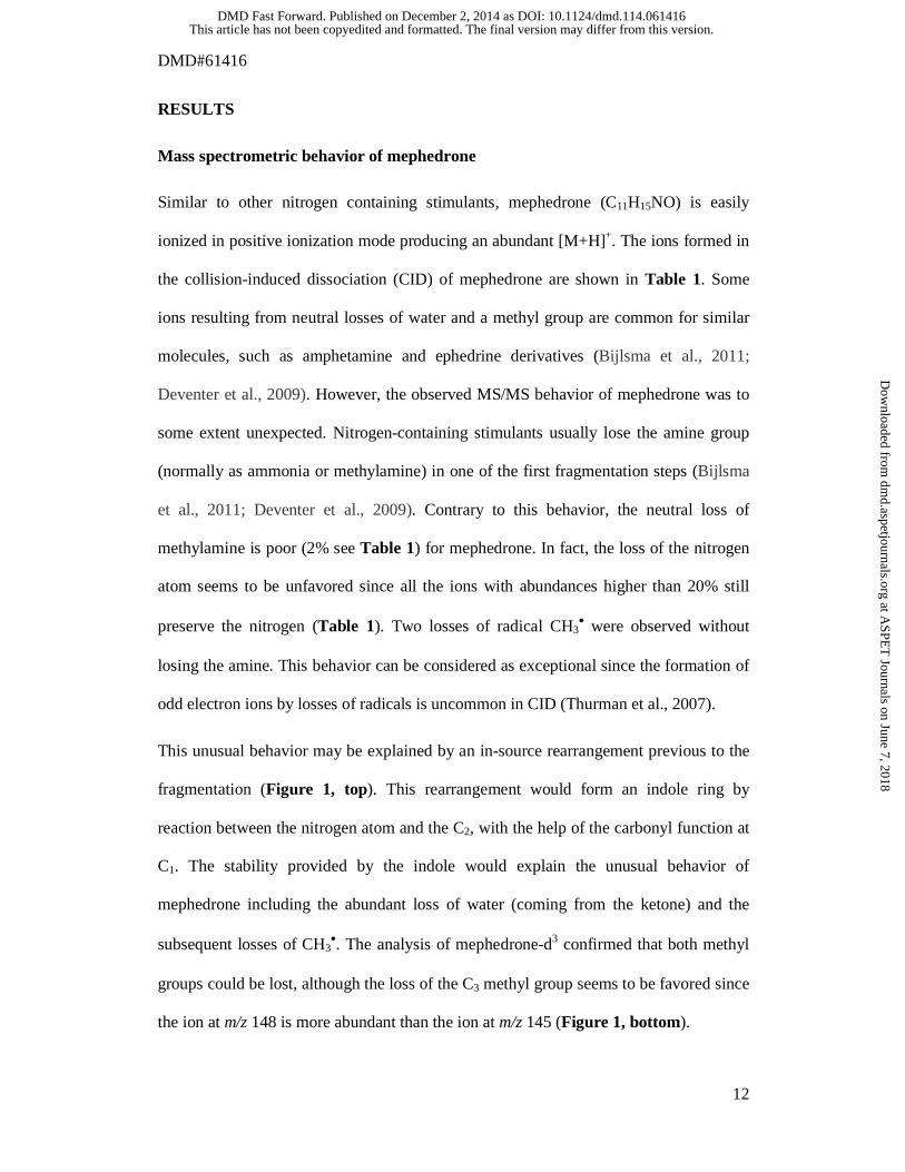
Mass spectrometric behavior of mephedrone
Similar to other nitrogen containing stimulants, mephedrone (C11H15NO) is easily
ionized in positive ionization mode producing an abundant [M+H]+. The ions formed in
the collision-induced dissociation (CID) of mephedrone are shown in Table 1. Some
ions resulting from neutral losses of water and a methyl group are common for similar
molecules, such as amphetamine and ephedrine derivatives (Bijlsma et al., 2011;
Deventer et al., 2009). However, the observed MS/MS behavior of mephedrone was to
some extent unexpected. Nitrogen-containing stimulants usually lose the amine group
(normally as ammonia or methylamine) in one of the first fragmentation steps (Bijlsma
et al., 2011; Deventer et al., 2009). Contrary to this behavior, the neutral loss of
methylamine is poor (2% see Table 1) for mephedrone. In fact, the loss of the nitrogen
atom seems to be unfavored since all the ions with abundances higher than 20% still
preserve the nitrogen (Table 1). Two losses of radical CH3• were observed without
losing the amine. This behavior can be considered as exceptional since the formation of
odd electron ions by losses of radicals is uncommon in CID (Thurman et al., 2007).
This unusual behavior may be explained by an in-source rearrangement previous to the
fragmentation (Figure 1, top). This rearrangement would form an indole ring by
reaction between the nitrogen atom and the C2, with the help of the carbonyl function at
C1. The stability provided by the indole would explain the unusual behavior of
mephedrone including the abundant loss of water (coming from the ketone) and the
subsequent losses of CH3•. The analysis of mephedrone-d3 confirmed that both methyl
groups could be lost, although the loss of the C3 methyl group seems to be favored since
the ion at m/z 148 is more abundant than the ion at m/z 145 (Figure 1, bottom).
This article has not been copyedited and formatted. The final version may differ from this version.DMD Fast Forward. Published on December 2, 2014 as DOI: 10.1124/dmd.114.061416
at ASPE
T Journals on June 7, 2018
dmd.aspetjournals.org
Dow
nloaded from

DMD#61416
13
Detection of mephedrone metabolites
The strategy applied in this study (dilution of the sample, acquisition in UHPLC-QTOF
MS, and processing by MetaboLynx software) to urine samples collected before and
after administration of 200 mg of mephedrone allowed for the detection of the parent
compound and 10 metabolites (Table 1).
The mass accuracy obtained provided only 1 feasible formula for each metabolite at the
selected narrow-mass window (2 mDa). Among the 10 metabolites found, four were
detected only in the positive ionization mode, similar to the behavior observed for
mephedrone. Six of them were also detected in negative ionization mode (Table 1)
suggesting the presence of an acidic moiety.
The product ion scan of each detected metabolite was acquired and the results are
shown in Table 1. Based on the comparison between these results and the CID behavior
of mephedrone (see section 3.1), a putative structure for each metabolite was established
(see section 3.3).
In those cases in which a phase II metabolite was predicted (M4, M8, M9 and M10) or a
single abundant product ion was obtained (M2 and M7), a MS3 experiment was
performed by selecting the product ion generated in-source at high cone voltage and
acquiring the product ion scan of this in-source fragment. This allowed more knowledge
to be garnered or to obtain additional relevant information about the unconjugated
metabolite. The results of these experiments are shown in Table 1.
Mass spectrometric results for mephedrone metabolites
Metabolite M1
This article has not been copyedited and formatted. The final version may differ from this version.DMD Fast Forward. Published on December 2, 2014 as DOI: 10.1124/dmd.114.061416
at ASPE
T Journals on June 7, 2018
dmd.aspetjournals.org
Dow
nloaded from

DMD#61416
14
The molecular formula obtained for metabolite M1 (C10H13NO) suggested the loss of a
methyl group from mephedrone. Since N-demethylation is a common metabolic
pathway of N-methyl stimulants, N-demethyl mephedrone was postulated as feasible
structure for M1.
As expected, the product ion spectrum followed behavior similar to the observed for
mephedrone. Thus, the expected neutral loss of ammonia was substantially less
abundant than the losses of water and CH3• (Table 1). This fact can be explained by the
same rearrangement postulated for mephedrone (Supplemental Figure 1).
The structure of M1 was confirmed by comparison with the reference material (Figure
2, bottom).
Metabolite M2
The molecular formula of M2 (C10H11NO3) indicated a double oxidation from M1.
Additionally, M2 was also detected in negative ionization mode (Table 1) suggesting
the presence of an acidic moiety. Therefore, the oxidation of one of the methyl groups
of M1 to carboxylic acid was the most feasible mechanism to produce M2.
The product ion spectrum provided information about the location of the carboxylic
function. The product ion spectrum of M2 was dominated by the ion at m/z 119.0497
(Table 1). This ion can be obtained after the neutral loss of glycine (Supplemental
Figure 2) suggesting a proximity between the amine and the acidic groups. Additionally,
the formula of this ion (C8H7O) implied that the aromatic zone of the mephedrone
remains intact.
This fact was confirmed by the fragmentation of the ion C8H7O generated in-source.
This fragmentation followed the expected behavior for a CH3PhCO+ ion. It was
This article has not been copyedited and formatted. The final version may differ from this version.DMD Fast Forward. Published on December 2, 2014 as DOI: 10.1124/dmd.114.061416
at ASPE
T Journals on June 7, 2018
dmd.aspetjournals.org
Dow
nloaded from

DMD#61416
15
dominated by the tropylium ion (m/z 91.0552) and its subsequent fragmentation
(Supplemental Figure 2). Additionally, an apparent neutral loss of 10 Da was observed.
This fact can be explained by the addition of water in the collision cell after the CO loss
as it was reported for similar ions (Beuck et al., 2009).
Based on this information N-demethylmephedrone-3-carboxylic acid was selected as
potential structure for M2.
Metabolite M3
The molecular formula obtained for M3 (C11H17NO) indicated a reduction step from
mephedrone.
The product ion spectrum exhibited an abundant neutral loss of water and subsequent
losses of CH3• and methylamine (Table 1). This behavior is similar to the one observed
for ephedrine derivatives which have a hydroxyl group in C1. Therefore, the reduction
of the ketone moiety would hamper the rearrangement postulated for mephedrone
decreasing the number of N-containing product ions (Supplemental Figure 3).
The structure of this metabolite as 1-dihydro mephedrone was confirmed by
comparison with synthesized material (Figure 2, middle).
Metabolite M4
Metabolite M4 exhibited a molecular formula containing three carbon atoms more than
mephedrone (C14H17NO4). This fact suggested that M4 was conjugated by phase II
metabolism. A diacetyl metabolite with the same formula was reported in the in vitro
metabolism of mephedrone (Khreit et al., 2013). However, M4 showed ionization in
This article has not been copyedited and formatted. The final version may differ from this version.DMD Fast Forward. Published on December 2, 2014 as DOI: 10.1124/dmd.114.061416
at ASPE
T Journals on June 7, 2018
dmd.aspetjournals.org
Dow
nloaded from

DMD#61416
16
both positive and negative mode (Table 1) indicating the presence of an acidic moiety
in M4. This result is in disagreement with the diacetyl metabolite.
The product ion spectrum of M4 was one of the most complex among the detected
metabolites (Figure 3a). It showed that the molecule could be divided in two parts: one
fragment at m/z 164 (C10H14NO) and the other at m/z 101 (C4H5O3). The fragment
C10H14NO has the same molecular formula than M1 i.e. nor-mephedrone. Therefore, the
fragment at m/z 101 provided information about the phase II metabolite. The molecular
formula was in agreement with a succinyl conjugate. The product ion spectra could be
explained by this conjugation as shown in Figure 3c.
The structure of M4 as N-succinyl nor-mephedrone was confirmed by comparison
with synthetic material as shown in Figure 3b.
Metabolite M5
Metabolite M5 showed only positive ionization with a molecular formula of C10H15NO.
This formula suggested a demethylation followed by a reduction of the keto function.
Therefore, 1-dihydro nor-mephedrone was selected as most feasible candidate for M5.
This hypothesis was supported by the product ion spectrum. Similar to M3, the absence
of the ketone functionality seems to hamper the rearrangement observed in mephedrone.
In agreement with that, the product ion spectrum of M5 was dominated by subsequent
neutral losses of water and ammonia (Supplemental Figure 4).
The proposed structure was confirmed by the reduction of nor-mephedrone as can be
seen in Figure 2 (top).
This article has not been copyedited and formatted. The final version may differ from this version.DMD Fast Forward. Published on December 2, 2014 as DOI: 10.1124/dmd.114.061416
at ASPE
T Journals on June 7, 2018
dmd.aspetjournals.org
Dow
nloaded from

DMD#61416
17
Metabolite M6
The molecular formula obtained for M6 (C11H15NO2) indicated a hydroxylation from
mephedrone. Since mephedrone contains several potential hydroxylation sites, the
product ion spectrum was used to propose the most feasible one.
The product ion spectrum showed the two expected losses of water: the one coming
from the ketone observed in most of mephedrone metabolites and the other coming
from the incorporated hydroxyl group. Additionally, an abundant neutral loss of
formaldehyde was also observed (see Table 1 and Supplemental Figure 5). This
neutral loss of 30 Da has been reported for hydroxymethyl metabolites in compounds
with stable rings like steroids (Pozo et al., 2008). Therefore, 4’-hydroxymethyl-
mephedrone was proposed for M6 in agreement with previously published studies
(Meyer et al., 2010; Khreit et al., 2013; Pedersen et al., 2013; Martinez-Clemente et al.,
2013).
Metabolite M7
The molecular formula of M7 (C11H13NO3) suggested a double oxidation from
mephedrone. Similar to M2, the oxidation of a methyl group to carboxylic acid seems to
be the most feasible mechanism to obtain M7. This fact was also supported by the
negative ionization observed for M7 (Table 1).
In contrast to M2, the product ion spectrum of M7 did not show the ion C8H7O (m/z
119.0500) indicative of an intact benzylic area. The spectrum of M7 was dominated by
losses of H2O, CO2 and CH3•. These losses could be explained after the rearrangement
described for mephedrone if the structure of M7 is 4’-carboxy-mephedrone
This article has not been copyedited and formatted. The final version may differ from this version.DMD Fast Forward. Published on December 2, 2014 as DOI: 10.1124/dmd.114.061416
at ASPE
T Journals on June 7, 2018
dmd.aspetjournals.org
Dow
nloaded from

DMD#61416
18
(Supplemental Figure 6) as previously reported (Khreit et al., 2013; Pedersen et al.,
2013; Martinez-Clemente et al., 2013).
Metabolite M8
M8 showed a molecular formula of C17H23NO8 which implied an increase of C6H8O6
from hydroxyl-mephedrone. This increase is associated with a glucuronide moiety.
Additionally, M8 was detected in both positive and negative ionization modes (Table 1).
Therefore hydroxyl-mephedrone-3-O-glucuronide was proposed as feasible structure
for M8.
The product ion spectrum confirmed the presence of the glucuronide moiety by the
typical loss of 176 Da in positive ionization mode and the occurrence of the ion at m/z
113 in negative ionization mode associated with the glucuronide functionality (Fabregat
et al., 2013). Additionally, the product ion spectrum helped in the assignation of the
hydroxylation site (Supplemental Figure 7). The product ion spectrum of the in-source
fragment at m/z 194 (produced after the neutral loss of the glucuronide and
corresponding to the unconjugated metabolite) showed an abundant ion at m/z 119.0497
(Table 1) corresponding to CH3-Ph-CO+. As commented for other metabolites, this ion
is indicative of unaltered benzylic zone in M8. Therefore, C3 seems to be the most
suitable hydroxylation site. Similarly to M2 in which a carboxylic acid was proposed in
C3, M8 also showed the ions corresponding to the loss of CO and the subsequent gain of
a water molecule (Supplemental Figure 7) supporting the assignation. Therefore, a C3
hydroxylation previous to glucuronidation is proposed as the most feasible metabolic
pathway to obtain M8.
This article has not been copyedited and formatted. The final version may differ from this version.DMD Fast Forward. Published on December 2, 2014 as DOI: 10.1124/dmd.114.061416
at ASPE
T Journals on June 7, 2018
dmd.aspetjournals.org
Dow
nloaded from

DMD#61416
19
Metabolite M9
The molecular formula for M9 (C17H21NO9) also suggested a glucuronide conjugate. In
that case, the phase I metabolite seemed to be a double oxidized metabolite.
The product ion spectrum of M9 after the loss of glucuronide moiety was identical to
the one observed for M7 (see Table 1 and Supplemental Figure 8). Therefore, the
glucuronide conjugate of 4’-carboxy-mephedrone was proposed as structure of M9.
Based on the structure of M7, N-glucuronidation seems to be the most feasible
metabolic pathway to obtain M9 (4’-carboxy-mephedrone-N-glucuronide).
Metabolite M10
Similarly to M8 and M9, the molecular formula of M10 (C16H20NO8) indicated a
glucuronidation. In that case, the molecular formula of the phase I metabolite produced
after the loss of the glucuronide moiety (C10H13NO2) corresponded to a hydroxylated
metabolite of nor-mephedrone.
The product ion spectrum of M10 exhibited an abundant ion at m/z 119.0478 (C8H7O).
As described for other metabolites like M2 and M8, this ion is indicative of an intact
aromatic area (Supplemental Figure 9). Following the considerations described for
these metabolites, hydroxylation in C3 of nor-mephedrone and subsequent
glucuronidation was proposed as potential metabolic pathway to obtain M10 (hydroxyl
nor-mephedrone-3-O-glucuronide). The C3-hydroxylated metabolite precursor of
M10 might be also proposed as intermediate metabolite in the generation of M2 (Figure
4).
This article has not been copyedited and formatted. The final version may differ from this version.DMD Fast Forward. Published on December 2, 2014 as DOI: 10.1124/dmd.114.061416
at ASPE
T Journals on June 7, 2018
dmd.aspetjournals.org
Dow
nloaded from
DMD#61416
20
DISCUSSION
The study of mephedrone metabolism by combination of UHPLC-QTOF MS and
MetaboLynx after dilution of the sample allowed for the detection of mephedrone and
10 metabolites in human urine. The metabolic pathway proposed based on our results is
summarized in Figure 4.
The accurate mass measurements played a critical role to properly interpret the
fragmentation of mephedrone and to propose suitable structures for detected metabolites.
As an example, mephedrone and most of its metabolites show a product ion with
nominal m/z 119. In a previous report, this ion was assigned to CH3PhCO+ suggesting a
cleavage of the C1-C2 bond of the molecule (Martinez-Clemente et al., 2013). However,
accurate mass measurements revealed that it corresponds to a formula of C9H11
indicating that the expected cleavage is not produced. In contrast, several metabolites
produced the ion CH3PhCO+, which was confirmed by the subsequent loss of CO and
the ion at m/z 109 corresponding to [CH3PhCO – CO + H2O]+. The presence of this ion
would imply a structural change in the surroundings of the nitrogen atom. Therefore,
discerning between both C9H11 and C8H7O formulae for the ion at m/z 119 was found to
be compulsory for structural elucidation.
In summary, six phase I and four phase II mephedrone metabolites were detected and
elucidated in human urine. Phase I reactions included N-demethylation, reduction of the
keto function, hydroxylations in C3 and in the benzylic carbon and oxidation of C3 and
the benzylic methyl to carboxylic acid (Figure 4). Our data confirmed the presence of
some compounds that have been previously described as mephedrone metabolites in
humans or other species (e.g. M1, M3, M5, M6, M7 and M9) (Meyer et al., 2010;
Khreit et al., 2013; Pedersen et al., 2013; Martinez-Clemente et al., 2013). However, it
This article has not been copyedited and formatted. The final version may differ from this version.DMD Fast Forward. Published on December 2, 2014 as DOI: 10.1124/dmd.114.061416
at ASPE
T Journals on June 7, 2018
dmd.aspetjournals.org
Dow
nloaded from

DMD#61416
21
is remarkable that structures for M2, M4, M8 and M10 have been reported for the first
time in this study.
Regarding phase II metabolism, three metabolites conjugated with glucuronic acid (M8,
M9 and M10) were found. More surprising was the detection of a conjugate with
succinic acid (M4). To the best of our knowledge, this is the first reporting of such
conjugate in human urine. Succinylation in drug development falls into the prodrug
category. Succinic acid derivatives are attached to target drugs to produce more
desirable physical and biological properties, with the goal that the resulting conjugates
are degraded by cellular enzymes to release the target drugs in vivo. In the context of
physiologic compounds, biogenic amines like octopamine, dopamine, and serotonin are
typically deactivated by acetylation in insects, nematodes, and other invertebrates.
Recently, succinylation, in addition to acetylation, has been reported for octopamine and
tyramine ascarosides in nematodes opening the question as to which extend this
metabolic reaction may represent a general pathway of biogenic amines metabolism
(Artyukhin et al., 2013). In the context of the present study succinic acid was
conjugated to nor-mephedrone. The hydrolysis of the conjugate would free nor-
mephedrone, a metabolite with unknown pharmacological activity but presumably
lower than the corresponding to mephedrone.
The structure of four of the detected metabolites (M1, M3, M4 and M5) has been
confirmed by comparison with synthesized material. All four were correctly assigned
based on their mass spectrometric behavior. The synthesis of the rest of metabolites
would be required in order to ultimately confirm their structure.
In vitro studies suggest that mephedrone metabolic disposition is regulated by the
highly polymorphic isoenzyme of cytochrome P450, CYP2D6 (Pedersen et al., 2013).
The quantification of metabolites identified in this report is of importance since it is
This article has not been copyedited and formatted. The final version may differ from this version.DMD Fast Forward. Published on December 2, 2014 as DOI: 10.1124/dmd.114.061416
at ASPE
T Journals on June 7, 2018
dmd.aspetjournals.org
Dow
nloaded from

DMD#61416
22
unknown the in vivo relevance of CYP2D6 polymorphism in mephedrone disposition
and which metabolic pathways are regulated by this isoenzyme in order to interpret
metabolic profiles in the context of forensic toxicology.
In the near future, it would be highly recommended to evaluate the behavior of
unchanged mephedrone and the 10 reported metabolites in urine samples collected for a
larger population ideally after administration of different doses, in order to determine
which should be the most appropriate biomarkers when investigating drug use.
This article has not been copyedited and formatted. The final version may differ from this version.DMD Fast Forward. Published on December 2, 2014 as DOI: 10.1124/dmd.114.061416
at ASPE
T Journals on June 7, 2018
dmd.aspetjournals.org
Dow
nloaded from

DMD#61416
23
AUTORSHIP CONTRIBUTIONS:
Participated in research design: de la Torre, Hernandez, Farré, Sancho, Pozo
Conducted experiments: Ibañez, Lahoz- Beneytez, Pozo, Farré, Papaseit, de la Torre
Performed data analysis: Pozo, Ibañez, Sancho
Wrote or contributed to the writing of the manuscript: Pozo, Ibañez, de la Torre,
Hernandez
This article has not been copyedited and formatted. The final version may differ from this version.DMD Fast Forward. Published on December 2, 2014 as DOI: 10.1124/dmd.114.061416
at ASPE
T Journals on June 7, 2018
dmd.aspetjournals.org
Dow
nloaded from

DMD#61416
24
REFERENCES
Adamowicz P, Tokarczyk B, Stanaszek R, Slopianka M. (2013) Fatal mephedrone
intoxication--a case report. J Anal Toxicol. 37:37-42.
Artyukhin AB, Yim JJ, Srinivasan J, Izrayelit Y, Bose N, von Reuss SH, Jo Y, Jordan
JM, Baugh LR, Cheong M, Sternberg PW, Avery L, Schroeder FC (2013) Succinylated
octopamine ascarosides and a new pathway of biogenic amine metabolism in
Caenorhabditis elegans. J Biol Chem. 288:18778-18783.
Beuck S, Schwabe T, Grimme S, Schlorer N, Kamber M, Schanzer W, Thevis M (2009)
Unusual mass spectrometric dissociation pathway of protonated isoquinoline-3-
carboxamides due to multiple reversible water adduct formation in the gas phase. J.
Mass Spectrom. 20: 2034-2048
Bijlsma L, Sancho JV, Hernandez F, Niessen WMA (2011) Fragmentation pathways of
drugs of abuse and their metabolites based on QTOF MS/MS and MSE accurate-mass
spectra. J. Mass Spectrom. 46: 865-875.
Bijlsma L, Boix C, Niessen WMA, Ibáñez M, Sancho JV, Hernández F (2013)
Investigation of degradation products of cocaine and benzoylecgonine in the aquatic
environment. Sci. Tot. Environ. 443: 200-208
Boix C, Ibáñez M, Zamora T, Sancho JV, Niessen WMA, Hernández F (2013a)
Identification of new omeprazole metabolites in wastewaters and surface waters; Sci.
Tot. Environ. 468-469: 706-714
Boix C, Ibáñez M, Sancho JV, Niessen WMA, Hernández F (2013b) Investigating the
presence of omeprazole in waters by liquid chromatography coupled to low and high
This article has not been copyedited and formatted. The final version may differ from this version.DMD Fast Forward. Published on December 2, 2014 as DOI: 10.1124/dmd.114.061416
at ASPE
T Journals on June 7, 2018
dmd.aspetjournals.org
Dow
nloaded from

DMD#61416
25
resolution mass spectrometry: degradation experiments. J. Mass Spectrom. 48: 1091-
1100
Boix C, Ibáñez M, Bijlsma L, Sancho JV, Hernández F (2014) Investigation of cannabis
biomarkers and transformation products in waters by liquid chromatography coupled to
time of flight and triple quadrupole mass spectrometry, Chemosphere, 99: 64-71
Cosbey SH, Peters KL, Quinn A, Bentley A. (2013) Mephedrone (methylmethcathinone)
in toxicology casework: a Northern Ireland perspective. J Anal Toxicol. 37:74-82
Gomez C, Fabregat A, Pozo OJ, Marcos J, Segura J, Ventura R. (2014) Analytical
strategies based on mass spectrometric techniques for the study of steroid metabolism
TrAC-Trends Anal. Chem. 53: 106–116
Deventer K, Pozo OJ; Van Eenoo P, Delbeke FT (2009) Development and validation of
an LC–MS/MS method for the quantification of ephedrines in urine J. Chromatogr. B
877: 369-374
Fabregat A, Pozo OJ, Marcos J, Segura J, Ventura R (2013) Use of LC-MS/MS for the
Open Detection of Steroid Metabolites Conjugated with Glucuronic Acid. Anal. Chem.
85: 5005-5014
Khreit O, Helen Grant M, Zhang T, Henderson C, Watson DG, Sutcliffe OB (2013)
Elucidation of the Phase I and Phase II metabolic pathways of (±)-4 -
methylmethcathinone (4-MMC) and (±)-4 -(trifluoromethyl)methcathinone (4-TFMMC)
in rat liver hepatocytes using LC–MS and LC–MS2. J. Pharm Biomed Anal 72: 177-185
This article has not been copyedited and formatted. The final version may differ from this version.DMD Fast Forward. Published on December 2, 2014 as DOI: 10.1124/dmd.114.061416
at ASPE
T Journals on June 7, 2018
dmd.aspetjournals.org
Dow
nloaded from

DMD#61416
26
Lusthof KJ, Oosting R, Maes A, Verschraagen M, Dijkhuizen A, Sprong AG. (2011) A
case of extreme agitation and death after the use of mephedrone in The Netherlands.
Forensic Sci Int. 206: e93-e95.
J. Martinez-Clemente, R. López-Arnau, M. Carbó, D. Pubill, J. Camarasa, E. Escubedo
(2013) Mepedrone pharmacokinetics after intravenous and oral administration in rats:
relation to pharmacodynamics, Psychofarmacology, 229: 295-306
Maurer HH, Pfleger K, Weber AA (2011) Mass spectral and GC data of drugs, poisons,
pesticides, pollutants and their metabolites. John Wiley & Sons ISBN 978-3-527-
31538-3
Maskell PD, De Paoli G, Seneviratne C, Pounde DJ (2011) Mephedrone (4-
methylmethcathinone)-related deaths. J Anal Toxicol 35: 188-191
Meyer MR, Wilhelm J, Peters FT, Maurer HH (2010) Beta-keto amphetamines: studies
on the metabolism of the designer drug mephedrone and toxicological detection of
mephedrone, butylone, and methylone in urine using gas chromatography–mass
spectrometry Anal Bioanal Chem 397: 1225-1233
Pedersen AJ, Reitzel LA, Johansen SS, Linnet K (2013) In vitro metabolism studies on
mephedrone and analysis of forensic cases. Drug Test Anal 5: 430-438
Pozo OJ, Van Eenoo P, Deventer K; Grimalt S, Sancho JV, Hernandez F, Delbeke FT
(2008) Collision-induced dissociation of 3-keto anabolic steroids and related
compounds after electrospray ionization. Considerations for structural elucidation Rapid
Commun. Mass Spectrom. 22: 4009-4024.
This article has not been copyedited and formatted. The final version may differ from this version.DMD Fast Forward. Published on December 2, 2014 as DOI: 10.1124/dmd.114.061416
at ASPE
T Journals on June 7, 2018
dmd.aspetjournals.org
Dow
nloaded from

DMD#61416
27
Pozo OJ, Marcos J, Matabosch X, Ventura R, Segura J. (2012) Using complementary
mass spectrometric approaches for the determination of methylprednisolone metabolites
in human urine. Rapid Commun. Mass Spectrom. 26: 541-553.
Prakash C, Shaffer CL, Nedderman A (2007) Analytical strategies for identifying drug
metabolites Mass Spectrom. Rev. 26: 340-369.
Schifano F, Corkery J, Ghodse AH. (2012) Suspected and confirmed fatalities
associated with mephedrone (4-methylmethcathinone, "meow meow") in the United
Kingdom. J Clin Psychopharmacol. 32:710-714.
Thurman EM, Ferrer I, Pozo OJ, Sancho JV, Hernandez F (2007) The even-electron
rule in electrospray mass spectra of pesticides Rapid Commun. Mass Spectrom. 21:
3855-3868.
Torrance H, Cooper G. (2010) The detection of mephedrone (4-methylmethcathinone)
in 4 fatalities in Scotland. Forensic Sci Int. 202:e62-e63.
This article has not been copyedited and formatted. The final version may differ from this version.DMD Fast Forward. Published on December 2, 2014 as DOI: 10.1124/dmd.114.061416
at ASPE
T Journals on June 7, 2018
dmd.aspetjournals.org
Dow
nloaded from

DMD#61416
28
FOOTNOTES
Financial support: Spanish Ministry of Economy and Competitiveness [Ref CTQ2012-
36189], Ministry of Health [ISCIII PI11/01961], DIUE de la Generalitat de Catalunya
[2014 SGR680]. and Generalitat Valenciana [research group of excellence PROMETEO
II/2014/023; ISIC 2012/016]. Supported in part by grants from Instituto de Salud Carlos
III-FEDER [FIS PI11/0196, RTA RD12/0028/0009 and Rio Hortega fellowship ISC-
III-CM13/00016].
Reprint requests: [email protected]
This article has not been copyedited and formatted. The final version may differ from this version.DMD Fast Forward. Published on December 2, 2014 as DOI: 10.1124/dmd.114.061416
at ASPE
T Journals on June 7, 2018
dmd.aspetjournals.org
Dow
nloaded from
DMD#61416
29
FIGURE LEGENDS
Figure 1. Proposed fragmentation pathway for mephedrone reference standard (top),
product ion spectrum at 20 eV for mephedrone (middle) and product ion spectrum at 20
eV for mephedrone-d3 (bottom).
Figure 2. MS/MS spectra (at 20 eV) of (a) metabolites found in urine sample, and (b)
synthesized material/reference standards.
Figure 3. MS/MS spectra at 20eV in positive ionization mode (top) and negative
ionization mode (bottom) for metabolite 4 (N-succinyl nor-mephedrone) in (a) urine and
(b) synthesized reference standard. (c) proposed fragmentation pathway for this
metabolite.
Figure 4. Metabolic pathway proposed for mephedrone in human.
This article has not been copyedited and formatted. The final version may differ from this version.DMD Fast Forward. Published on December 2, 2014 as DOI: 10.1124/dmd.114.061416
at ASPE
T Journals on June 7, 2018
dmd.aspetjournals.org
Dow
nloaded from
DMD#61416
30
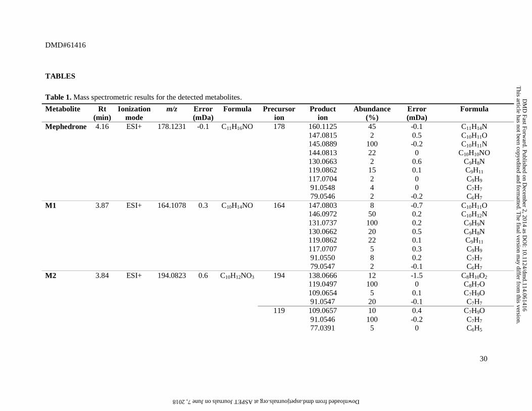
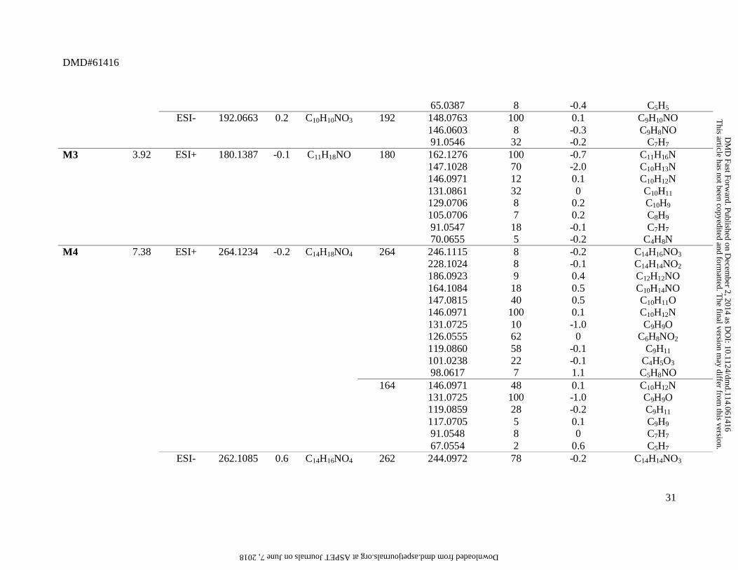
TABLES
Table 1. Mass spectrometric results for the detected metabolites.
Metabolite Rt (min)
Ionization mode
m/z Error (mDa)
Formula Precursor ion
Product ion
Abundance (%)
Error (mDa)
Formula
Mephedrone 4.16 ESI+ 178.1231 -0.1 C11H16NO 178 160.1125 45 -0.1 C11H14N 147.0815 2 0.5 C10H11O 145.0889 100 -0.2 C10H11N 144.0813 22 0 C10H10NO 130.0663 2 0.6 C9H8N 119.0862 15 0.1 C9H11 117.0704 2 0 C9H9 91.0548 4 0 C7H7 79.0546 2 -0.2 C6H7 M1 3.87 ESI+ 164.1078 0.3 C10H14NO 164 147.0803 8 -0.7 C10H11O 146.0972 50 0.2 C10H12N 131.0737 100 0.2 C9H9N 130.0662 20 0.5 C9H8N 119.0862 22 0.1 C9H11 117.0707 5 0.3 C9H9 91.0550 8 0.2 C7H7 79.0547 2 -0.1 C6H7 M2 3.84 ESI+ 194.0823 0.6 C10H12NO3 194 138.0666 12 -1.5 C8H10O2 119.0497 100 0 C8H7O 109.0654 5 0.1 C7H9O 91.0547 20 -0.1 C7H7 119 109.0657 10 0.4 C7H9O 91.0546 100 -0.2 C7H7 77.0391 5 0 C6H5
This article has not been copyedited and form
atted. The final version m
ay differ from this version.
DM
D Fast Forw
ard. Published on Decem
ber 2, 2014 as DO
I: 10.1124/dmd.114.061416
at ASPET Journals on June 7, 2018 dmd.aspetjournals.org Downloaded from
DMD#61416
31
65.0387 8 -0.4 C5H5 ESI- 192.0663 0.2 C10H10NO3 192 148.0763 100 0.1 C9H10NO 146.0603 8 -0.3 C9H8NO 91.0546 32 -0.2 C7H7 M3 3.92 ESI+ 180.1387 -0.1 C11H18NO 180 162.1276 100 -0.7 C11H16N 147.1028 70 -2.0 C10H13N 146.0971 12 0.1 C10H12N 131.0861 32 0 C10H11 129.0706 8 0.2 C10H9 105.0706 7 0.2 C8H9 91.0547 18 -0.1 C7H7 70.0655 5 -0.2 C4H8N M4 7.38 ESI+ 264.1234 -0.2 C14H18NO4 264 246.1115 8 -0.2 C14H16NO3 228.1024 8 -0.1 C14H14NO2 186.0923 9 0.4 C12H12NO 164.1084 18 0.5 C10H14NO 147.0815 40 0.5 C10H11O 146.0971 100 0.1 C10H12N 131.0725 10 -1.0 C9H9O 126.0555 62 0 C6H8NO2 119.0860 58 -0.1 C9H11 101.0238 22 -0.1 C4H5O3 98.0617 7 1.1 C5H8NO 164 146.0971 48 0.1 C10H12N 131.0725 100 -1.0 C9H9O 119.0859 28 -0.2 C9H11 117.0705 5 0.1 C9H9 91.0548 8 0 C7H7 67.0554 2 0.6 C5H7 ESI- 262.1085 0.6 C14H16NO4 262 244.0972 78 -0.2 C14H14NO3
This article has not been copyedited and form
atted. The final version m
ay differ from this version.
DM
D Fast Forw
ard. Published on Decem
ber 2, 2014 as DO
I: 10.1124/dmd.114.061416
at ASPET Journals on June 7, 2018 dmd.aspetjournals.org Downloaded from

DMD#61416
32
226.0847 42 -2.1 C14H12NO2 200.1071 100 -0.4 C13H14NO 162.0932 18 1.3 C10H12NO 133.0639 18 -1.4 C9H9O 99.0079 38 -0.3 C4H3O3 98.0271 25 2.9 C4H4NO2 M5 3.84 ESI+ 166.1231 -0.1 C10H16NO 166 148.1122 72 -0.4 C10H14N 131.0861 100 0 C10H11 116.0627 28 0.1 C9H8 115.0545 12 -0.3 C9H7 105.0703 5 -0.1 C8H9 91.0548 60 0 C7H7 56.0488 2 -1.2 C3H6N M6 2.61 ESI+ 194.1178 0.3 C11H16NO2 194 176.1057 2 -1.8 C11H14NO 158.0961 100 -0.9 C11H12N 146.0962 100 -0.6 C10H12N 133.0651 8 -0.2 C9H9O 131.0728 43 -0.7 C9H9N 105.0696 12 -0.8 C8H9 M7 1.19 ESI+ 208.0973 -0.1 C11H14NO3 208 190.0879 5 1.1 C11H12NO2 172.0762 35 0 C11H10NO 146.0968 81 -0.2 C10H12N 144.0813 18 0 C10H10N 131.0735 100 0 C9H9N 130.0656 8 -0.1 C9H8N 105.0702 20 -0.2 C8H9 ESI- 206.0812 -0.5 C11H12NO3 206 162.0914 100 -0.5 C10H12NO 162 160.0766 62 0.4 C10H10NO 147.0696 20 1.2 C9H9NO 84.0438 8 -0.1 C4H6NO
This article has not been copyedited and form
atted. The final version m
ay differ from this version.
DM
D Fast Forw
ard. Published on Decem
ber 2, 2014 as DO
I: 10.1124/dmd.114.061416
at ASPET Journals on June 7, 2018 dmd.aspetjournals.org Downloaded from

DMD#61416
33
M8 6.62 ESI+ 370.1509 0.7 C17H24NO8 370 209.1169 12 -0.9 C12H17O3 194.1176 100 -0.5 C11H16NO2 191.1075 22 0.3 C12H15O2 178.1234 8 0.2 C11H16NO 160.1121 32 -0.5 C11H14N 119.0497 68 0 C8H7O 194 161.0818 10 0.7 C10H11NO 148.1096 8 -0.3 C7H16O3 119.0502 100 0.5 C8H7O 109.0656 8 0.3 C7H9O 91.0554 35 0.6 C7H7 79.0545 5 -0.3 C6H7 ESI- 368.1358 1.1 C17H22NO8 368 193.0335 62 -1.3 C6H9O7 157.0137 18 0 C6H5O5 131.0350 38 0.7 C5H7O4 113.0239 100 0.4 C5H5O3 103.0022 40 -0.9 C3H3O4 72.9917 32 -0.9 C2HO3 M9 1.27 ESI+ 384.1302 0.7 C17H22NO9 384 208.0982 100 0.8 C11H14NO3 190.0862 22 -0.6 C11H12NO2 172.0763 58 0.1 C11H10NO 146.0968 32 -0.2 C10H12N 208 190.0853 5 -1.5 C11H12NO2 172.0763 25 0.1 C11H10NO 146.0972 82 0.2 C10H12N 131.0737 100 0.2 C9H9N 105.0707 22 0.3 C8H9 ESI- 382.1128 -1.0 C17H20NO9 382 206.0812 100 -0.5 C11H12NO3 162.0915 58 -0.4 C10H12NO 113.0234 72 -0.5 C5H5O3
This article has not been copyedited and form
atted. The final version m
ay differ from this version.
DM
D Fast Forw
ard. Published on Decem
ber 2, 2014 as DO
I: 10.1124/dmd.114.061416
at ASPET Journals on June 7, 2018 dmd.aspetjournals.org Downloaded from

DMD#61416
34
85.0280 22 -1.0 C4H5O2 206 162.0923 100 0.4 C10H12NO 160.0762 8 -0.9 C10H10NO 77.0386 5 -0.5 C6H5 M10 5.71 ESI+ 356.1346 0.1 C16H22NO8 356 180.1028 100 0.3 C10H14NO2 162.0919 72 0 C10H12NO 119.0478 45 -1.9 C8H7O 180 149.0974 22 0.8 C10H13O 131.0864 100 0.3 C10H11 107.0867 18 0.6 C8H11 91.0543 50 -0.5 C7H7 ESI- 354.1185 -0.4 C16H20NO8 354 193.0364 38 1.1 C9H7NO4 113.0237 42 -0.2 C5H5O3 85.0303 18 1.3 C4H5O2
This article has not been copyedited and form
atted. The final version m
ay differ from this version.
DM
D Fast Forw
ard. Published on Decem
ber 2, 2014 as DO
I: 10.1124/dmd.114.061416
at ASPET Journals on June 7, 2018 dmd.aspetjournals.org Downloaded from

This article has not been copyedited and formatted. The final version may differ from this version.DMD Fast Forward. Published on December 2, 2014 as DOI: 10.1124/dmd.114.061416
at ASPE
T Journals on June 7, 2018
dmd.aspetjournals.org
Dow
nloaded from

This article has not been copyedited and formatted. The final version may differ from this version.DMD Fast Forward. Published on December 2, 2014 as DOI: 10.1124/dmd.114.061416
at ASPE
T Journals on June 7, 2018
dmd.aspetjournals.org
Dow
nloaded from

This article has not been copyedited and formatted. The final version may differ from this version.DMD Fast Forward. Published on December 2, 2014 as DOI: 10.1124/dmd.114.061416
at ASPE
T Journals on June 7, 2018
dmd.aspetjournals.org
Dow
nloaded from

This article has not been copyedited and formatted. The final version may differ from this version.DMD Fast Forward. Published on December 2, 2014 as DOI: 10.1124/dmd.114.061416
at ASPE
T Journals on June 7, 2018
dmd.aspetjournals.org
Dow
nloaded from