Embed Size (px)
Citation preview
PG TUTORIAL
MAXILLOFACIAL TRAUMA
DR. AHMED AL-ARFAJ
Asst. Professor / Consultant
ORL Department, KAUH
Epidemiology
Incidence 50/100000
M:F
Causes
Paediatrics
General Consideration H&N
ABCDs
Soft tissues

History & Physical
examination
Face & Facial Skeleton
Inspection
Palpation
Assess function
HEAD & NECK RADIOLOGICAL EXAMS
Facial Plain Films
Largely been replaced by computer tomography (except for the mandible)
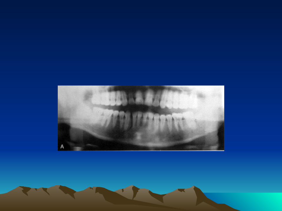
Plain Film Mandible Series and Panorex
Computed Tomography (CT) Most informative radiographic exam fro head and neck Trauma Axial and coronal facial CT with bone and soft tissue window, 2-3 mm sections
Special Radiologic Exams
Angiography
Magnetic Resonance Imaging
Modified Barium Swallow and Esophagram
MANDIBULAR FRACTURES
Introduction
Most common in young males (ages 18-30)
Causes: assault , motor vehicle accidents, sports and gunshots wounds
Most common Fractures Sites
Risks: impacted teeth, osteoporosis, edentulous areas, pathologic, lytic lesions
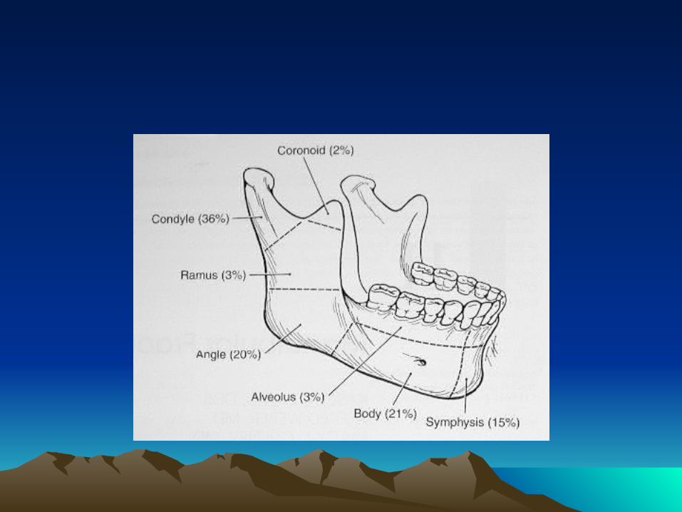
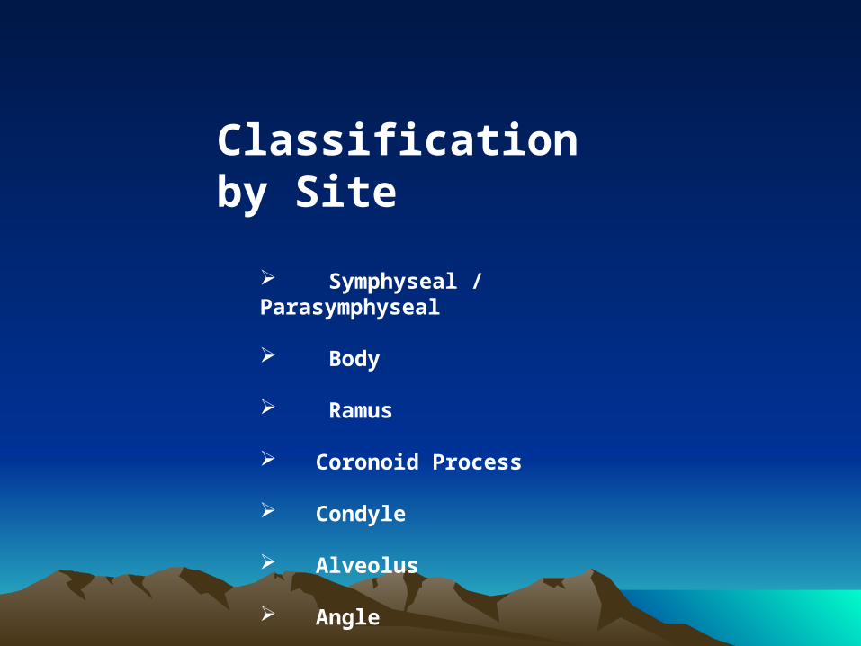
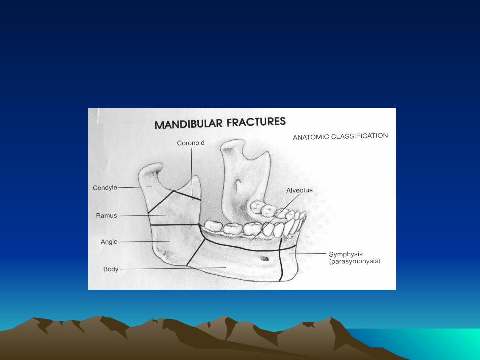
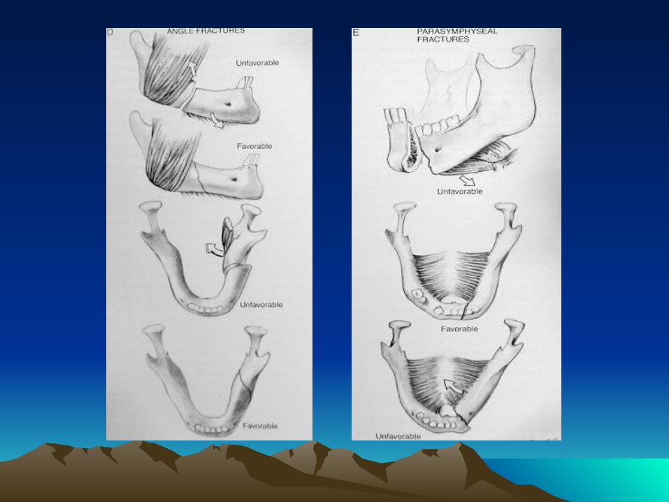
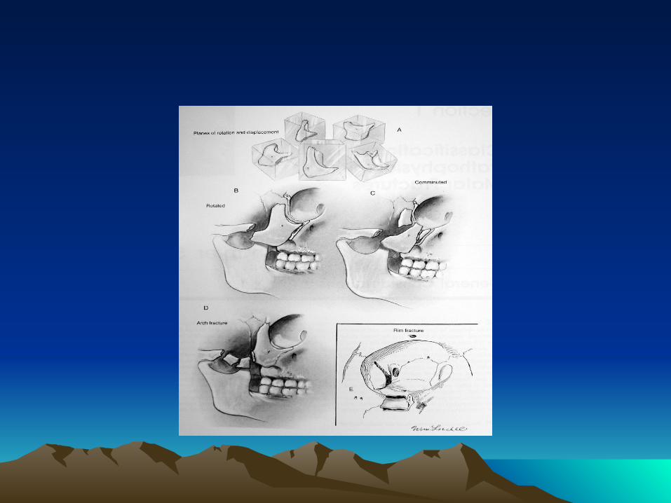
Classification by Site
Symphyseal / Parasymphyseal
Body
Ramus
Coronoid Process Condyle
Alveolus
Angle

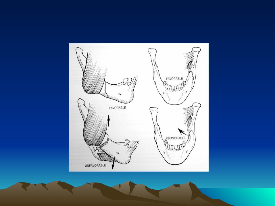
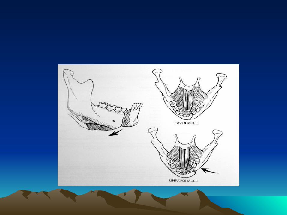
Classification by Favorability
Favorable
Unfavorable
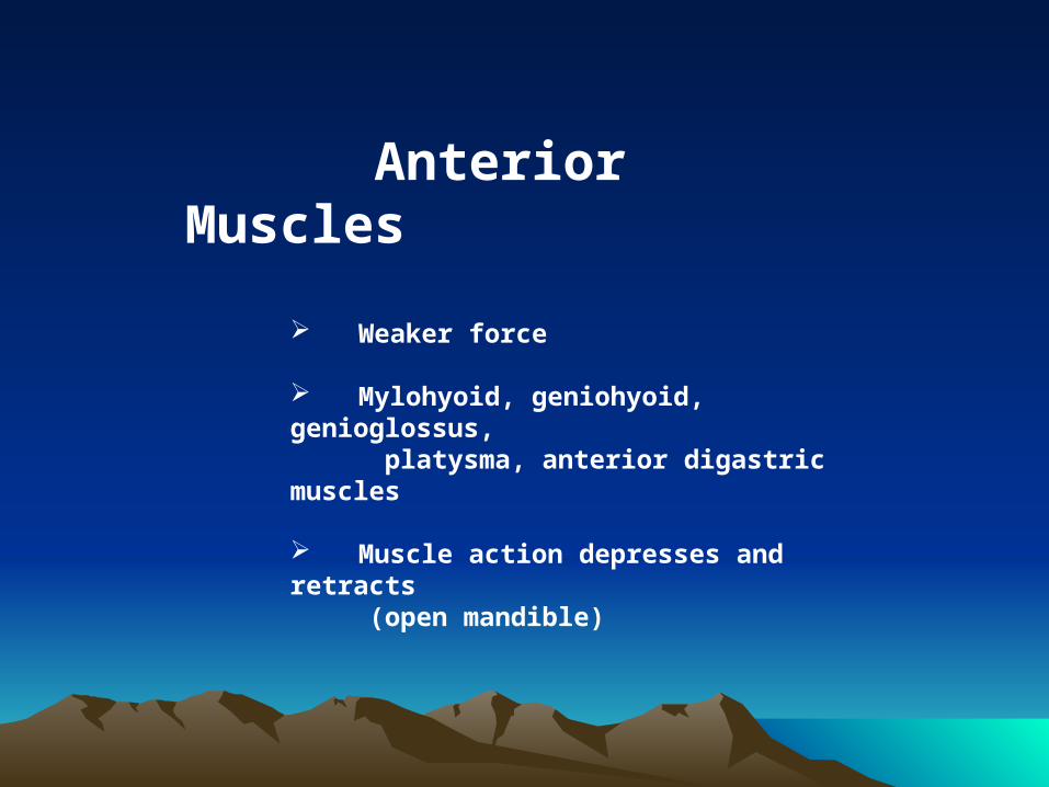
Anterior Muscles
Weaker force
Mylohyoid, geniohyoid, genioglossus, platysma, anterior digastric muscles
Muscle action depresses and retracts (open mandible)
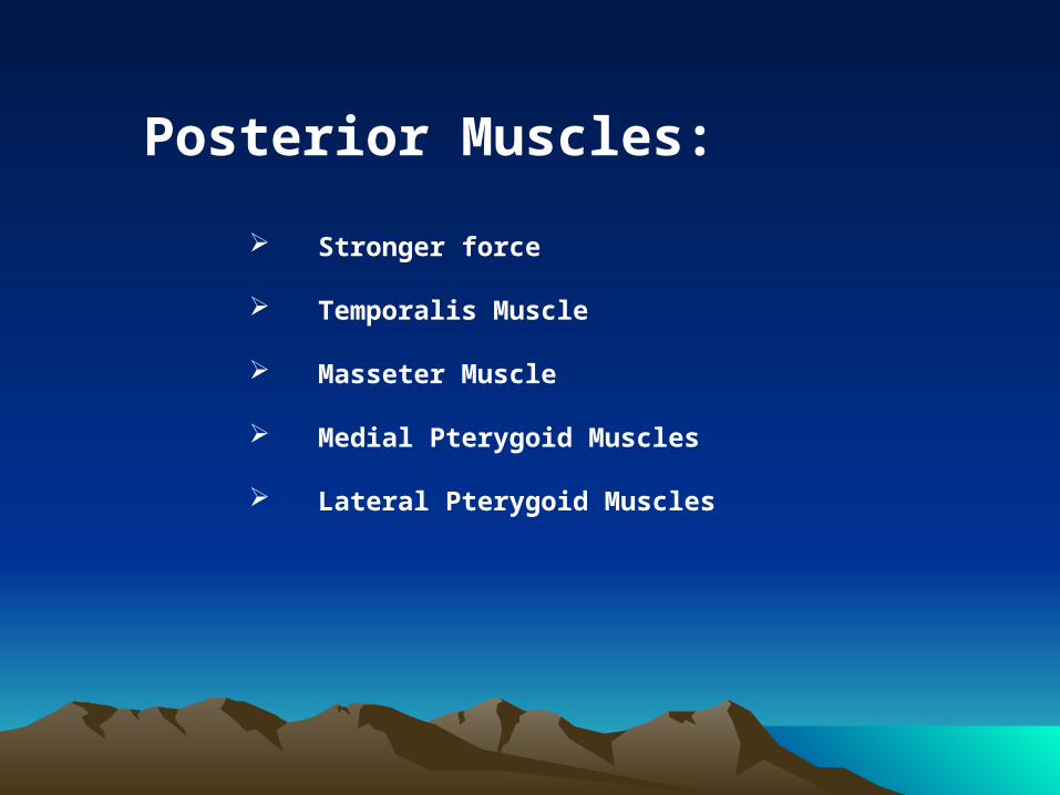
Posterior Muscles:
Stronger force
Temporalis Muscle Masseter Muscle
Medial Pterygoid Muscles
Lateral Pterygoid Muscles
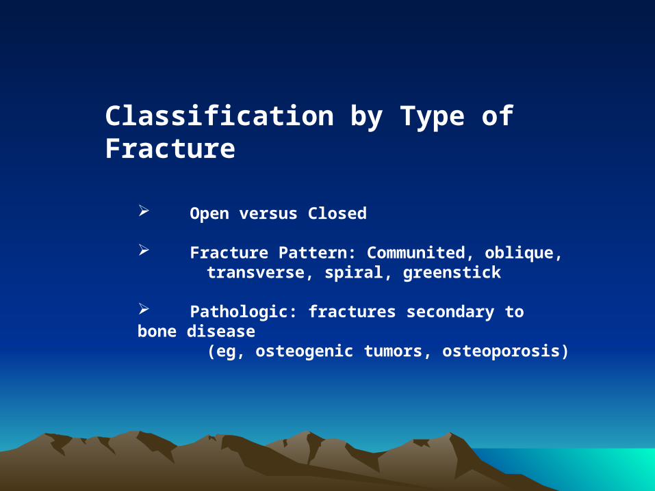
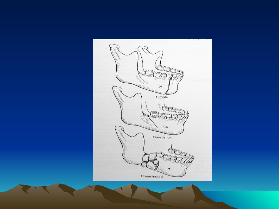
Classification by Type of Fracture
Open versus Closed
Fracture Pattern: Communited, oblique, transverse, spiral, greenstick
Pathologic: fractures secondary to bone disease (eg, osteogenic tumors, osteoporosis)
Dental Classification
Class I
Class II
Class III
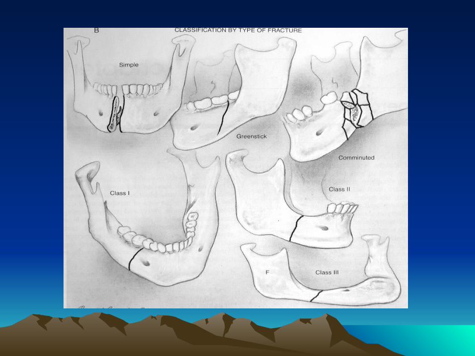
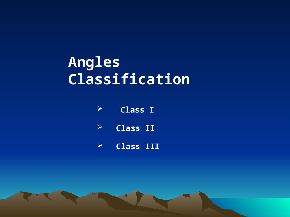
Angles Classification
Class I
Class II
Class III
MANAGEMENT
Management Concepts
Goals: restore occlusion, establish bony union& avoid TMJ pathology
Repair within first week
In general favorable fractures may only need closed reduction
Postoperative Care
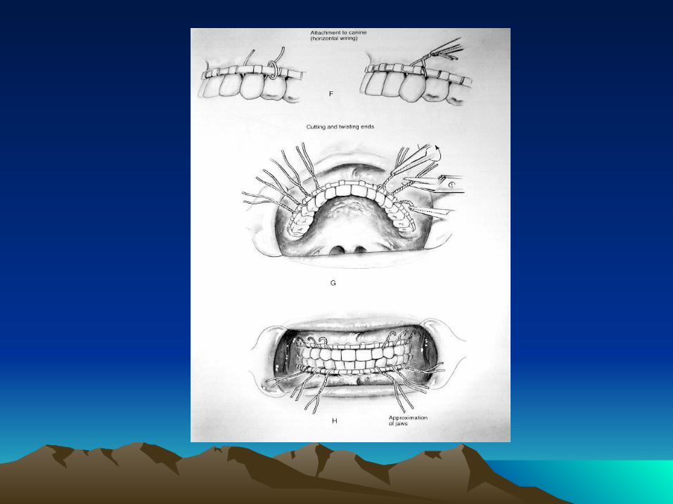
Maxillo-Mandibular Fixation (MMF) - Closed Reduction
Indications
Methods
Requires an intact maxilla
Typically MMF may be removed after 2-8 weeks
Complications
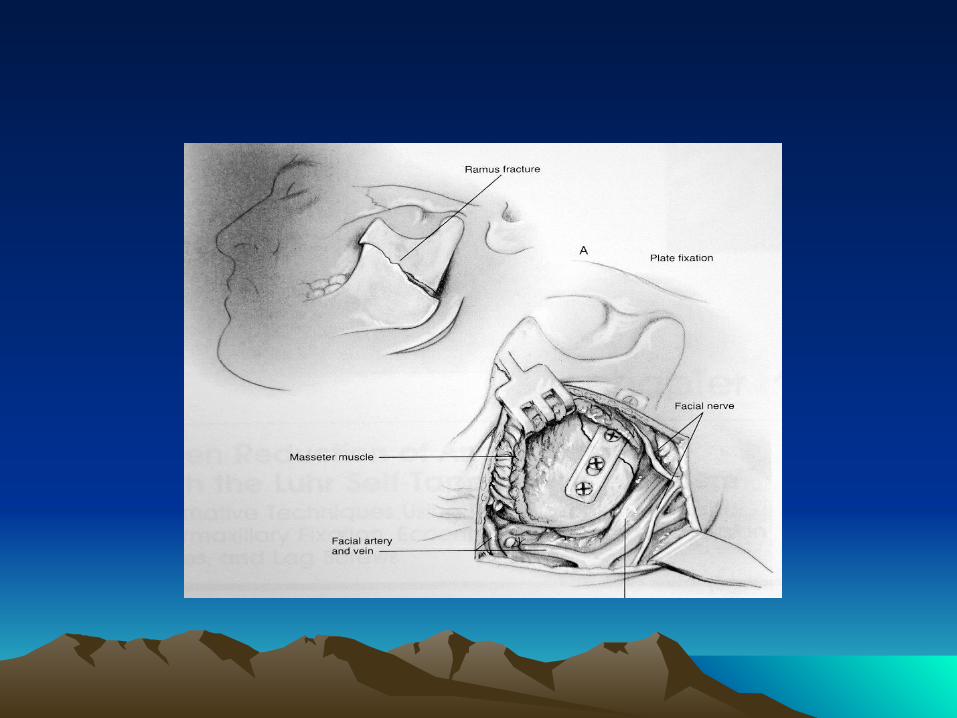
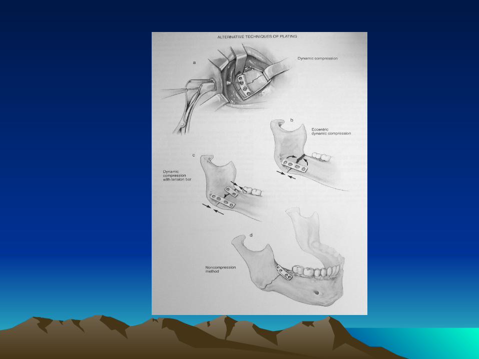
Open Reduction &Internal Fixation (ORIF)
Indications
Approaches:
1. Transoral
2. External
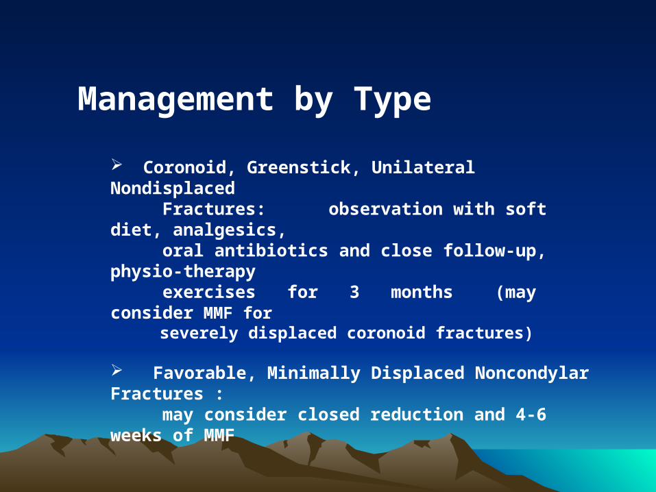
Management by Type
Coronoid, Greenstick, Unilateral Nondisplaced Fractures: observation with soft diet, analgesics, oral antibiotics and close follow-up, physio-therapy exercises for 3 months (may consider MMF for severely displaced coronoid fractures)
Favorable, Minimally Displaced Noncondylar Fractures : may consider closed reduction and 4-6 weeks of MMF
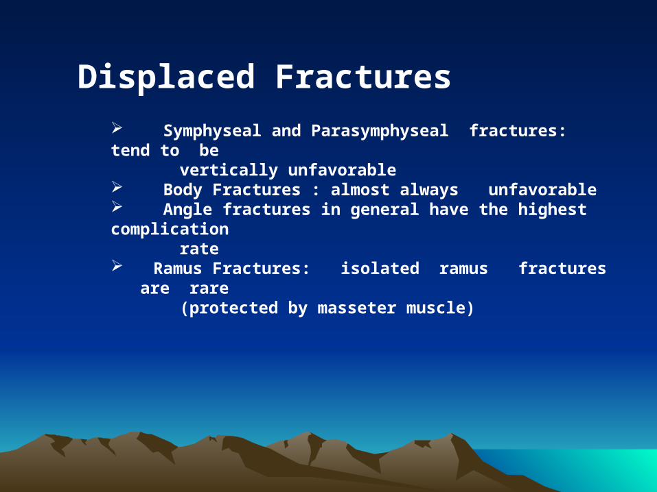
Displaced Fractures
Symphyseal and Parasymphyseal fractures: tend to be vertically unfavorable Body Fractures : almost always unfavorable Angle fractures in general have the highest complication rate Ramus Fractures: isolated ramus fractures are rare (protected by masseter muscle)
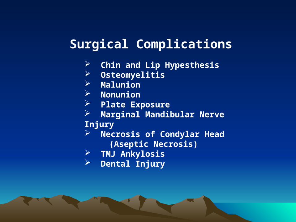
Surgical Complications
Chin and Lip Hypesthesis Osteomyelitis Malunion Nonunion Plate Exposure Marginal Mandibular Nerve Injury Necrosis of Condylar Head (Aseptic Necrosis) TMJ Ankylosis Dental Injury
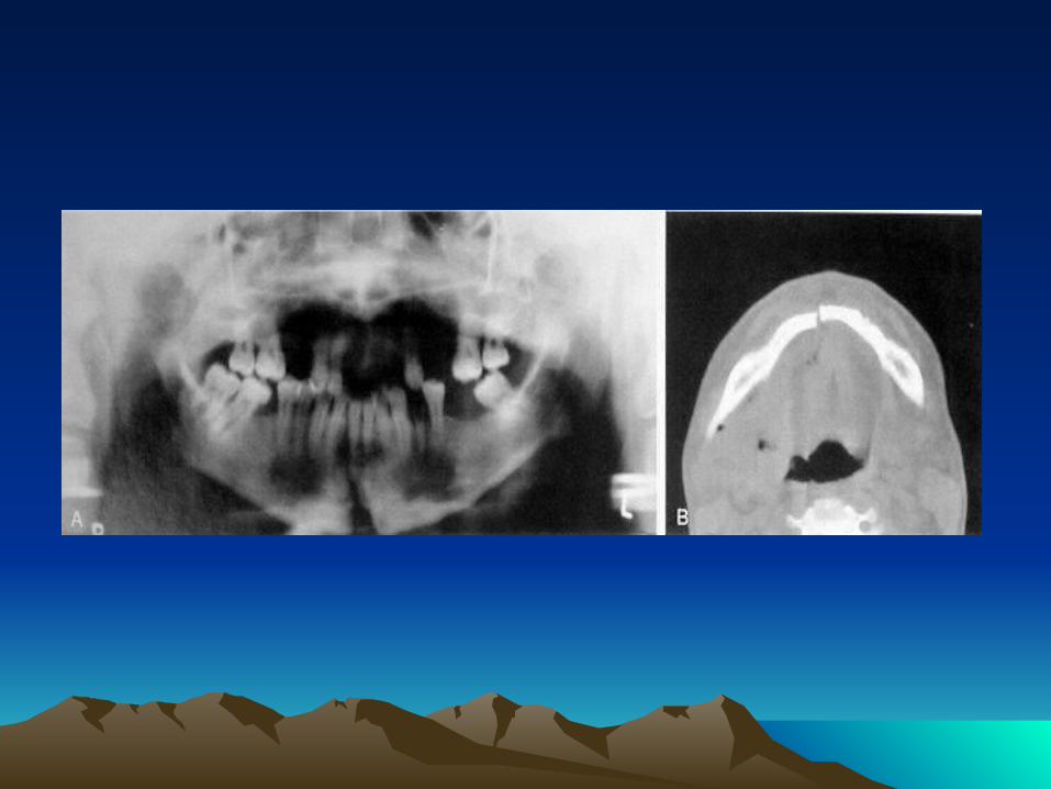
MAXILLARY FRACTURES
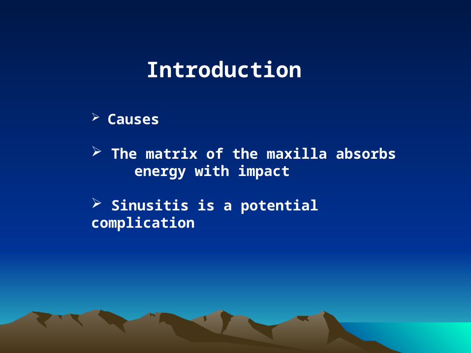
Introduction
Causes
The matrix of the maxilla absorbs energy with impact
Sinusitis is a potential complication
Classification
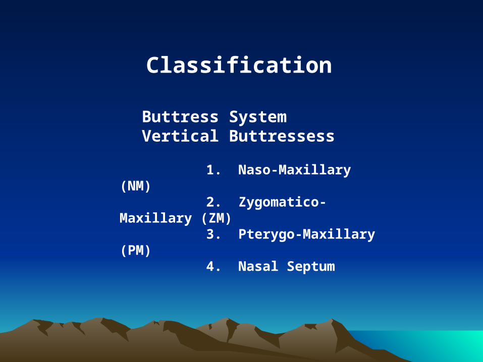
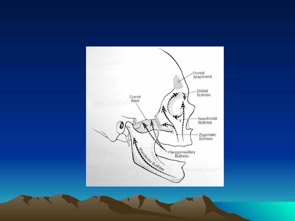
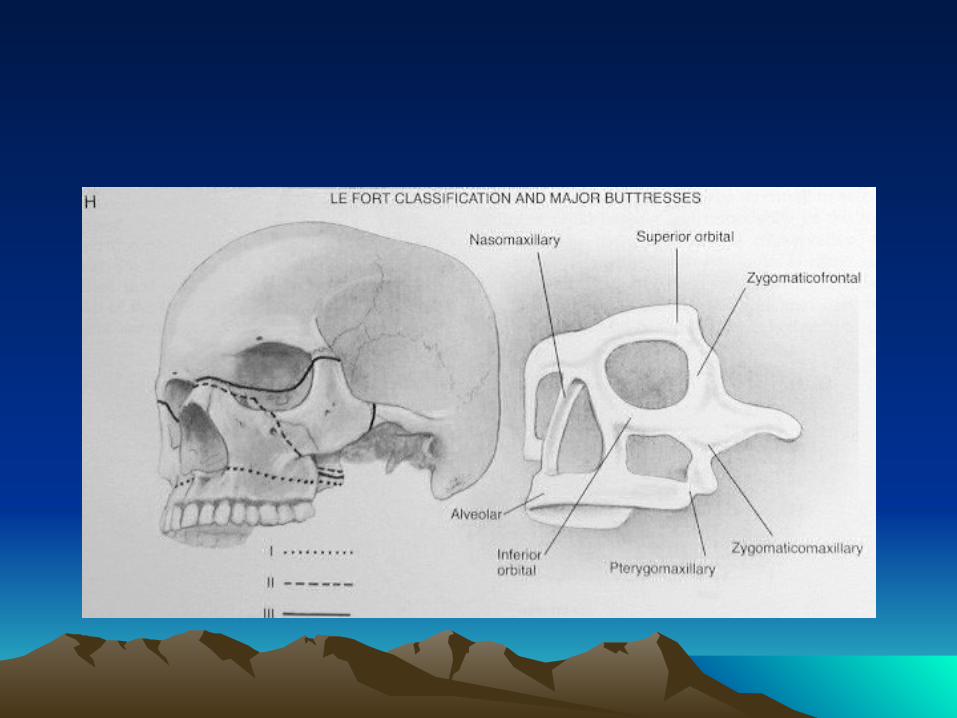
Buttress System
Vertical Buttressess
1. Naso-Maxillary (NM) 2. Zygomatico-Maxillary (ZM) 3. Pterygo-Maxillary (PM) 4. Nasal Septum
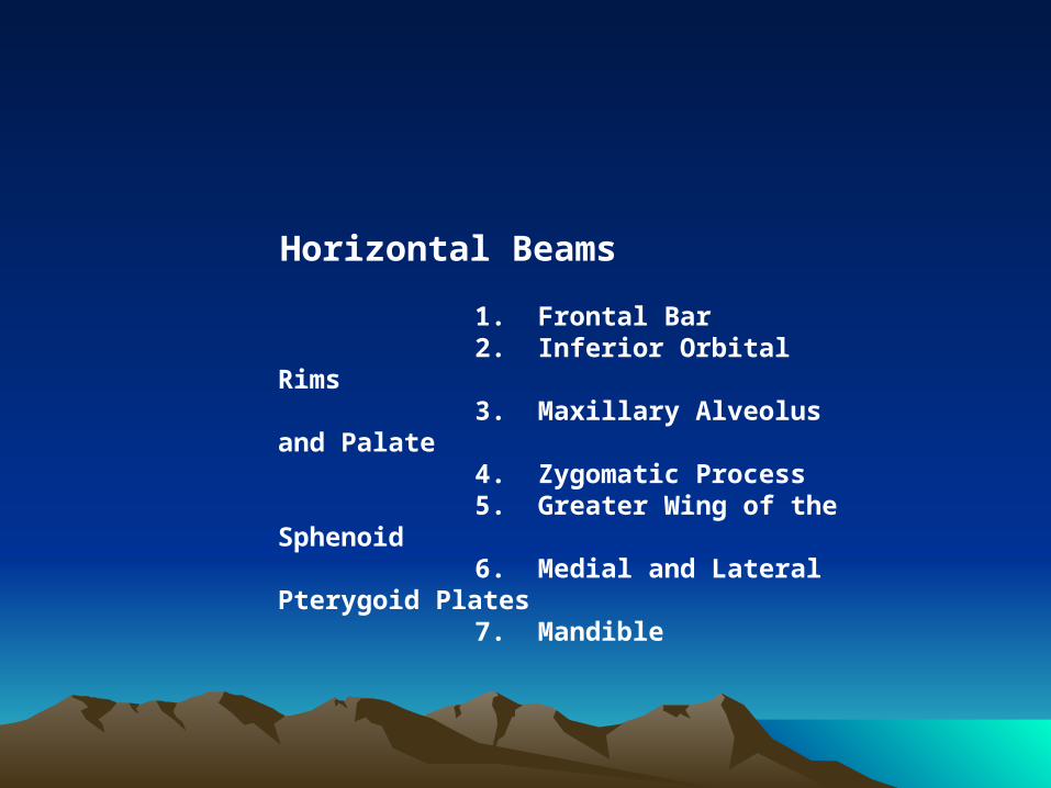
Horizontal Beams
1. Frontal Bar 2. Inferior Orbital Rims 3. Maxillary Alveolus and Palate 4. Zygomatic Process 5. Greater Wing of the Sphenoid 6. Medial and Lateral Pterygoid Plates 7. Mandible
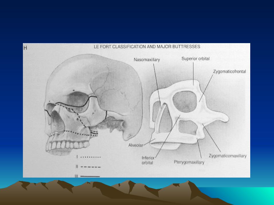
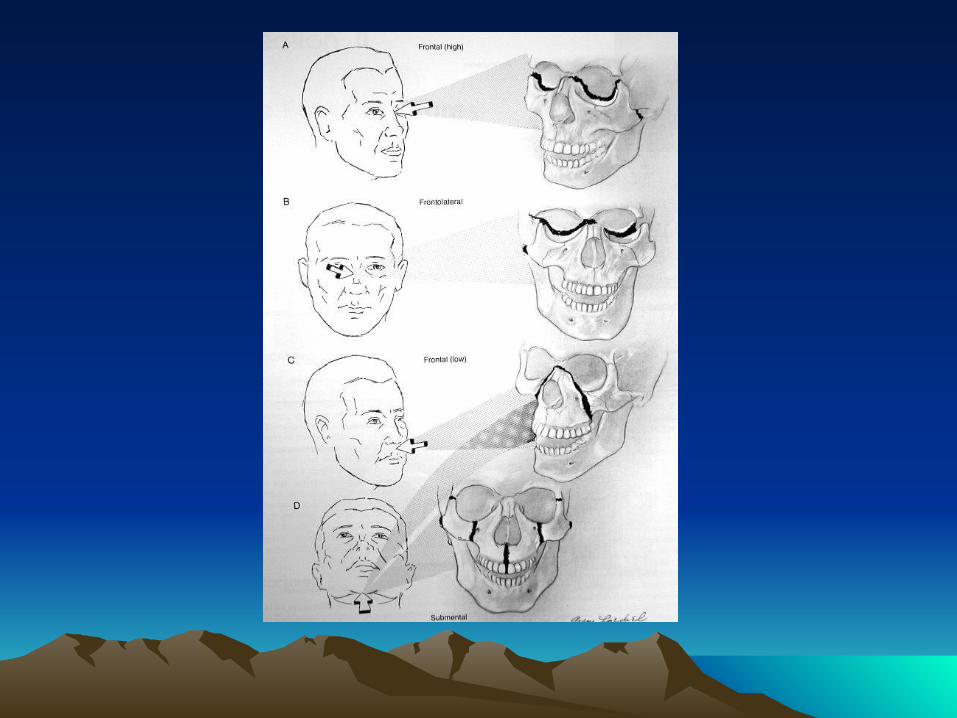
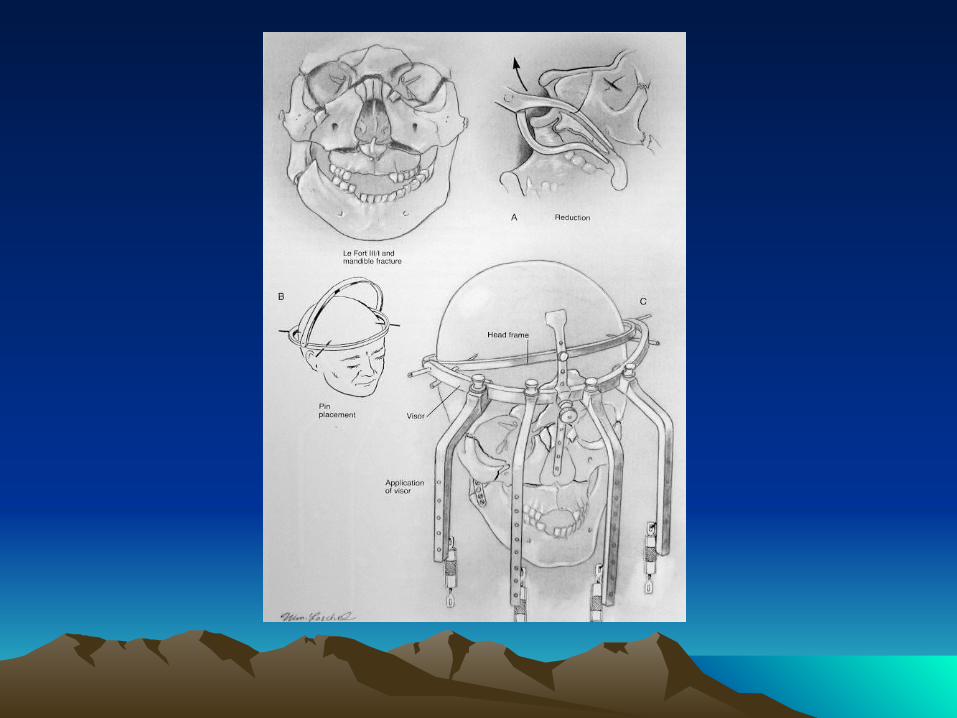
Le Fort Classification
Based on patterns of fractures (lines of minimal resistance) classified according to the highest level of Injury In many cases Le Fort classification is incomplete for maxillary fractures Le Fort fractures may present in many combinations or
on one side (hemi-Le Fort)
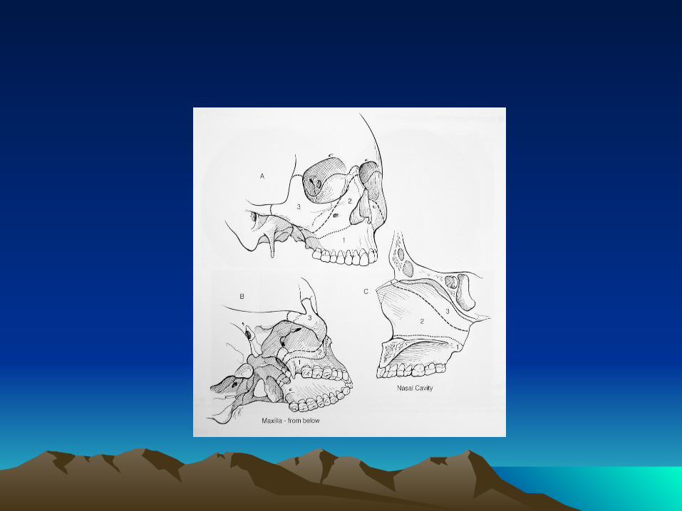
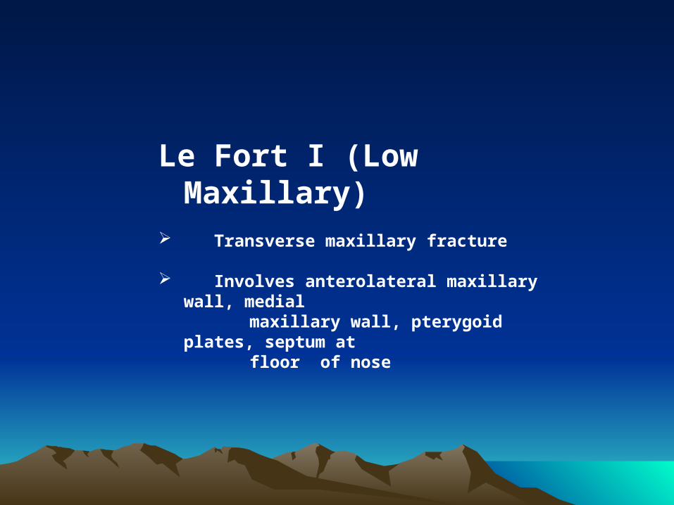
Le Fort I (Low Maxillary) Transverse maxillary fracture
Involves anterolateral maxillary wall, medial maxillary wall, pterygoid plates, septum at floor of nose

Le Fort II (Pyramidal ) Caused typically from a superiorly directed force against the maxilla. Involves nasofrontal suture, orbital foramen, rim, and floor frontal process of lacrimal bone, zygomaxillary suture, lamina papyracea of ethmoid; pterygoid plate and high septum

Le Fort III (Craniofacial Dysjunction)
Separates facial skeleton from base of skull, typically caused by high velocity impacts. Involves nasofrontal suture, zygoma and zygomatic arch; pterygoid plates and nasal septum

Management
Principles
Goals of Reconstruction Exposure/Approaches Timing Postoperative Care
Cont: Management
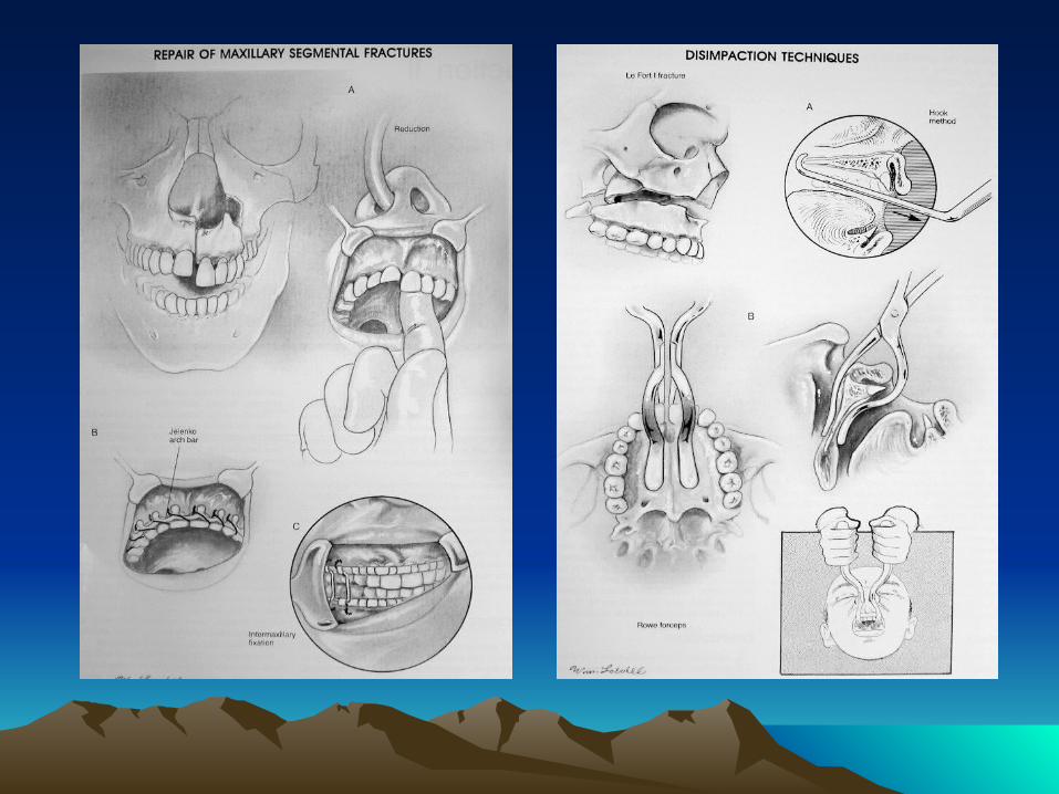
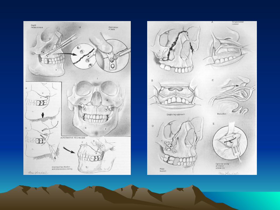
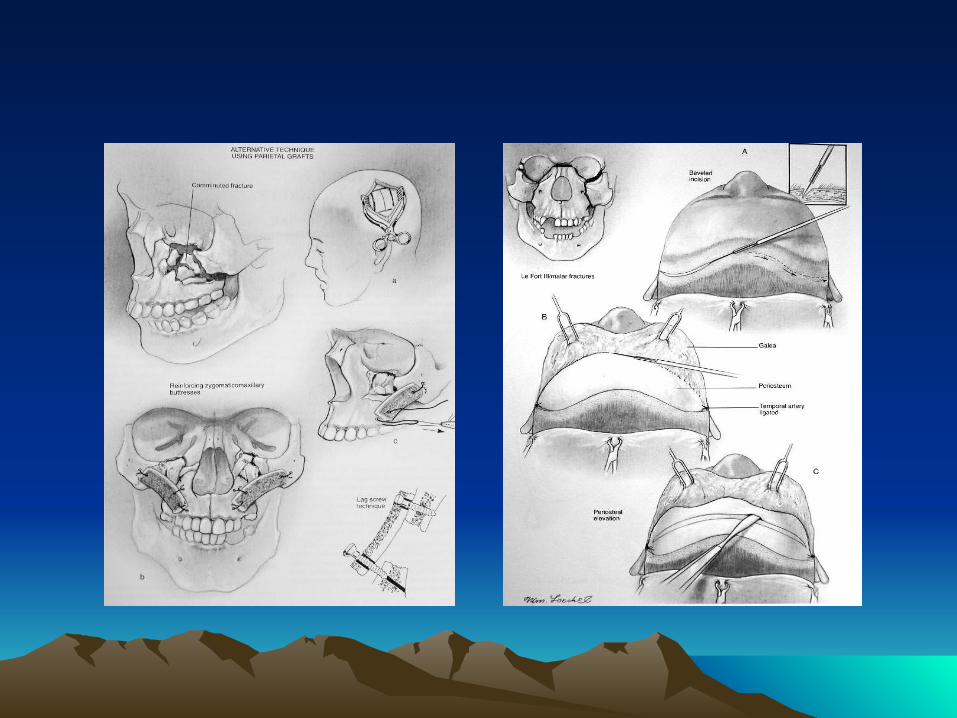
Techniques
Plate Fixation (Miniplates) Interosseous Wire Fixation Bone Grafts
Management by Le Fort Classification
Le Fort I: reduced digitally, MMF, fixation of ZM Le Fort II: stabilization of the ZM buttress, MMF , nasofrontal
process and inferior orbital rim.
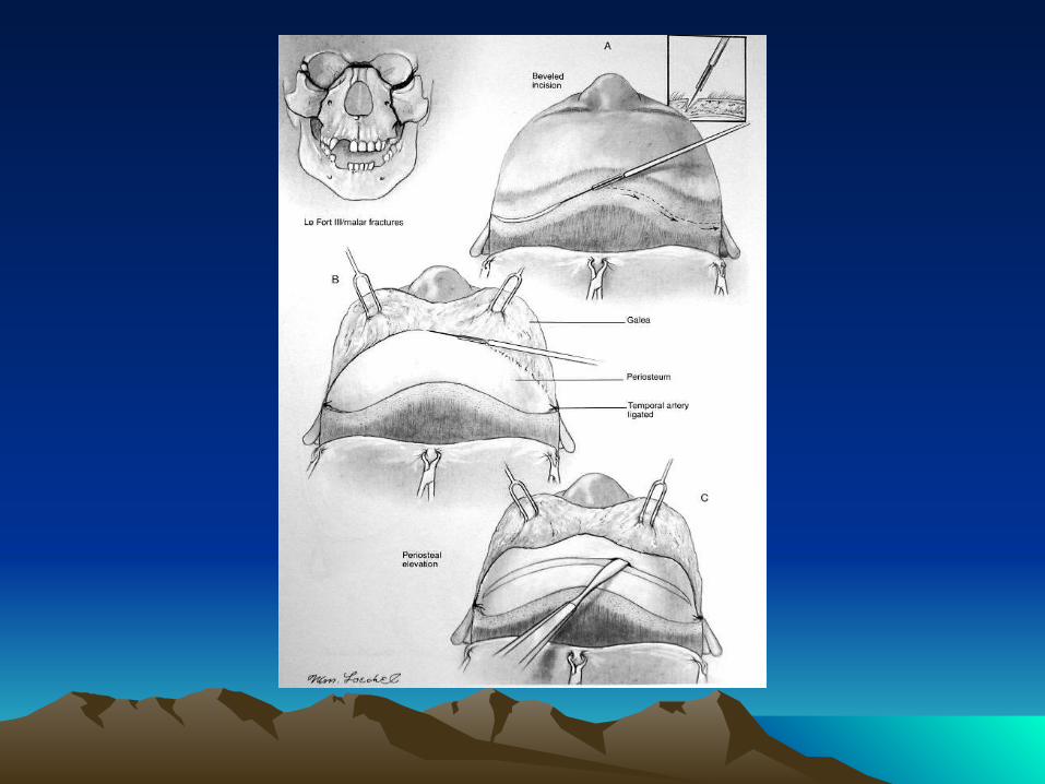
Le Fort III: usually requires coronal flap for adequate exposure for exploration and miniplate fixation
Surgical complications
Malunion, Nonunion, Plate Exposure
Palpable or Observable Plates
Forehead or Cheek Hypesthesi
Osteomyelitis
Dental Injury
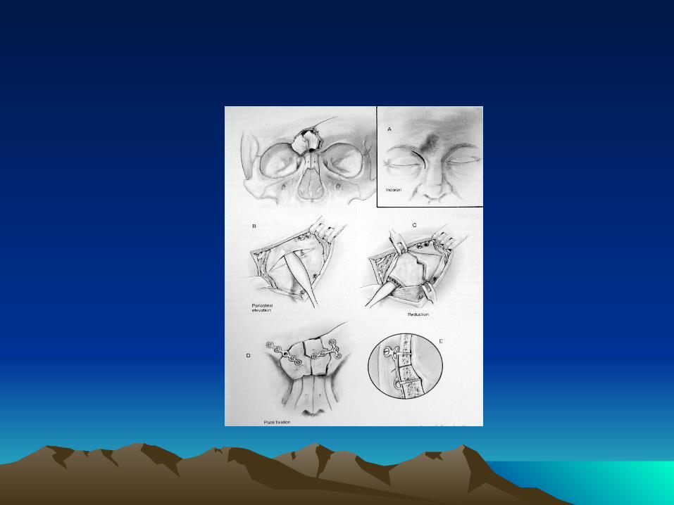
ZYGOMATICOMAXILLARY & ORBITAL FRACTURES
Zygomaticomaxillary Complex(Trimalar) Fractures
Introduction
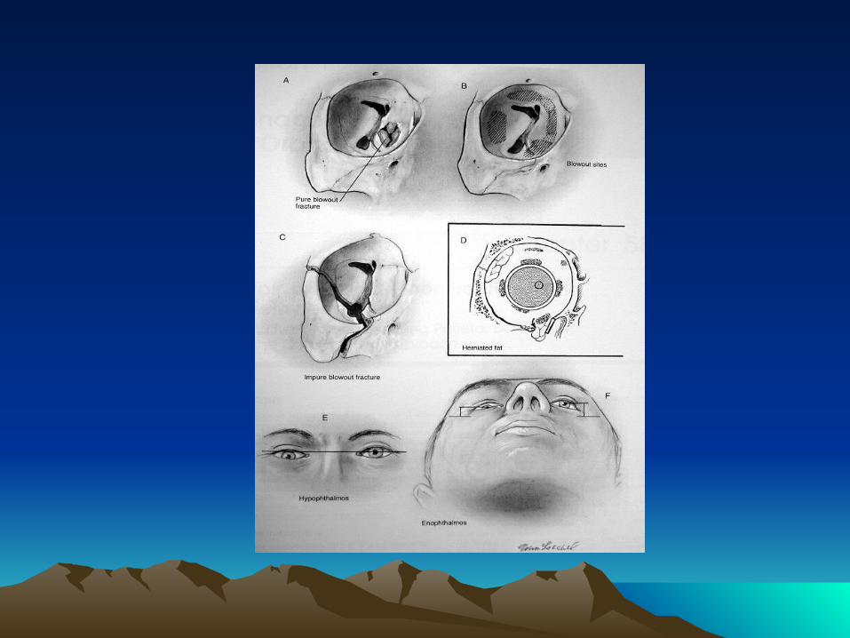
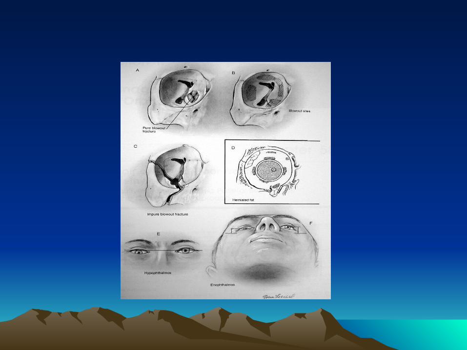
Symptom: • Subconjunctival & periorbital ecchymosis • Eyelid edema• Epistaxis • Cheek hypesthesia• Diplopia • Hypophthalmos • Enophthalmos• Trismus
Zygomaticomaxillary Complex (Trimalar) Fractures
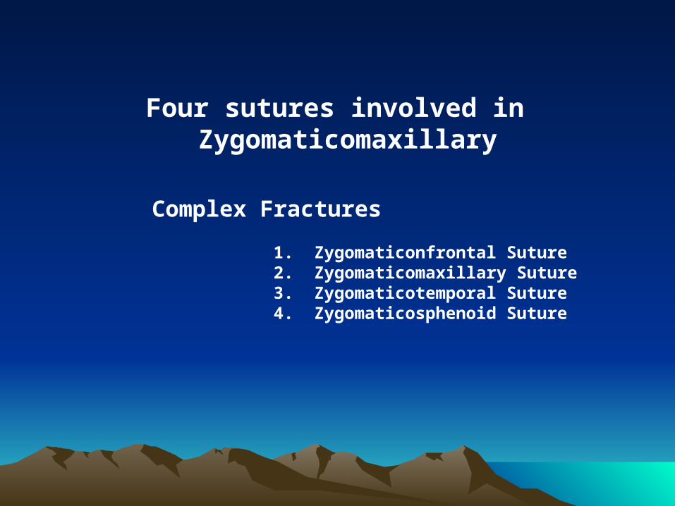
Four sutures involved in Zygomaticomaxillary
Complex Fractures 1. Zygomaticonfrontal Suture 2. Zygomaticomaxillary Suture 3. Zygomaticotemporal Suture 4. Zygomaticosphenoid Suture
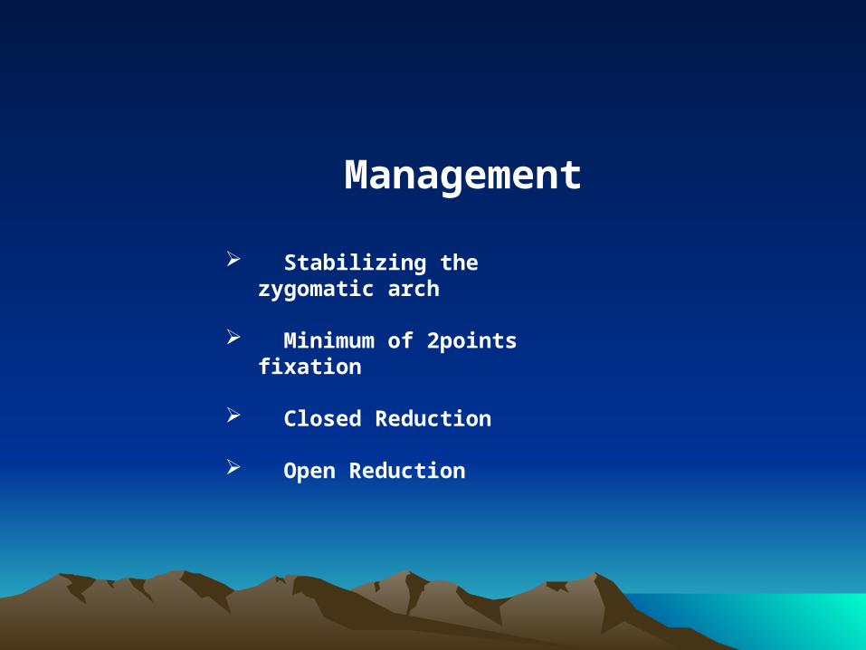
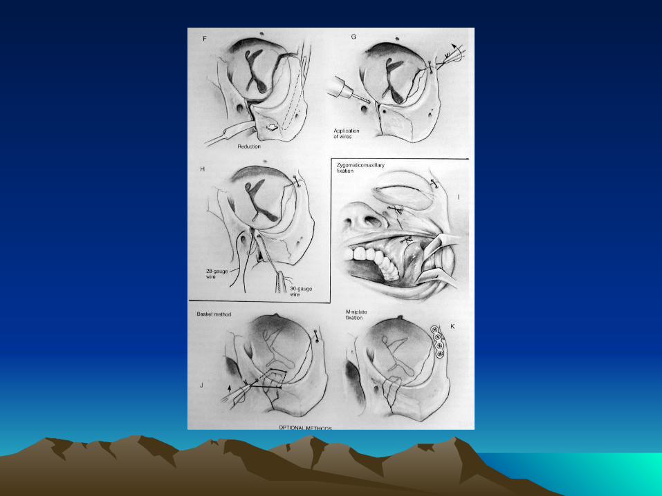
Management
Stabilizing the zygomatic arch
Minimum of 2points fixation
Closed Reduction
Open Reduction
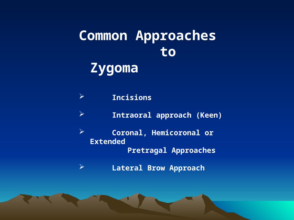
Common Approaches to Zygoma
Incisions
Intraoral approach (Keen)
Coronal, Hemicoronal or Extended Pretragal Approaches
Lateral Brow Approach
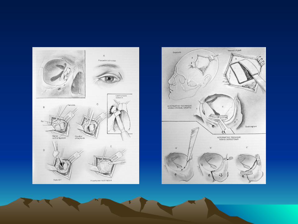
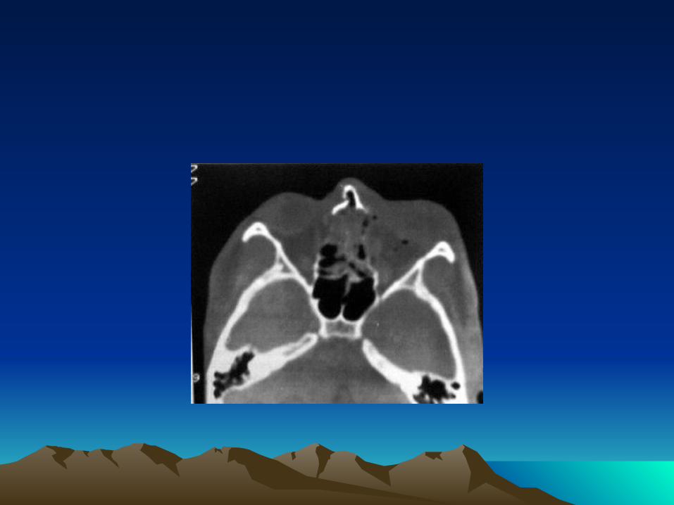
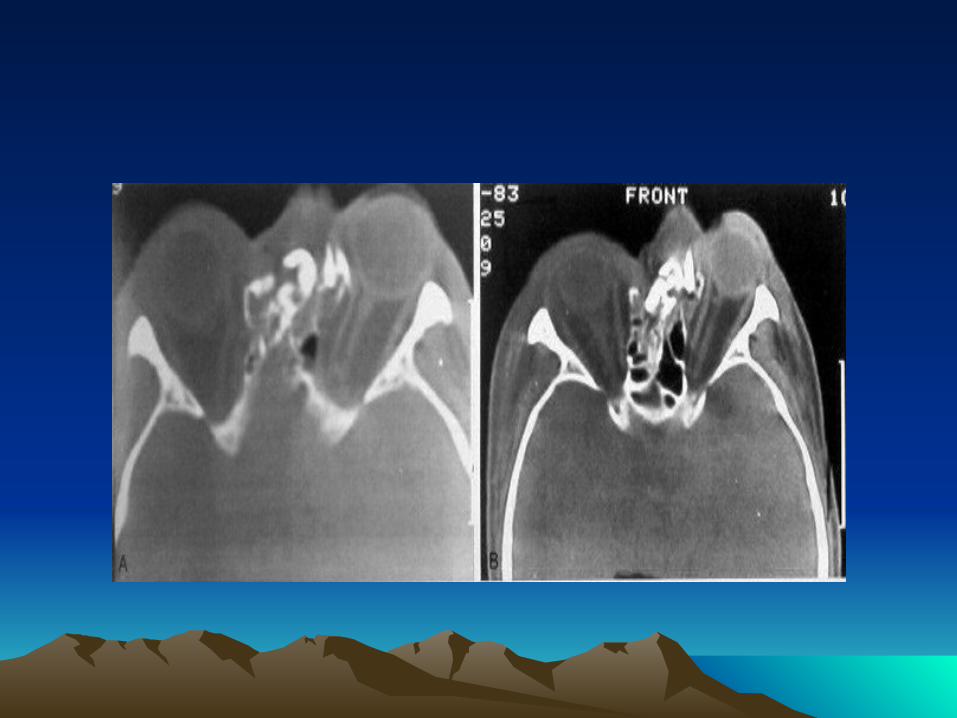
ORBITAL FRACTURES
INTRODUCTION
Orbital Bones Optic Canal& Orbital Fissures Contents Sign& Symptoms
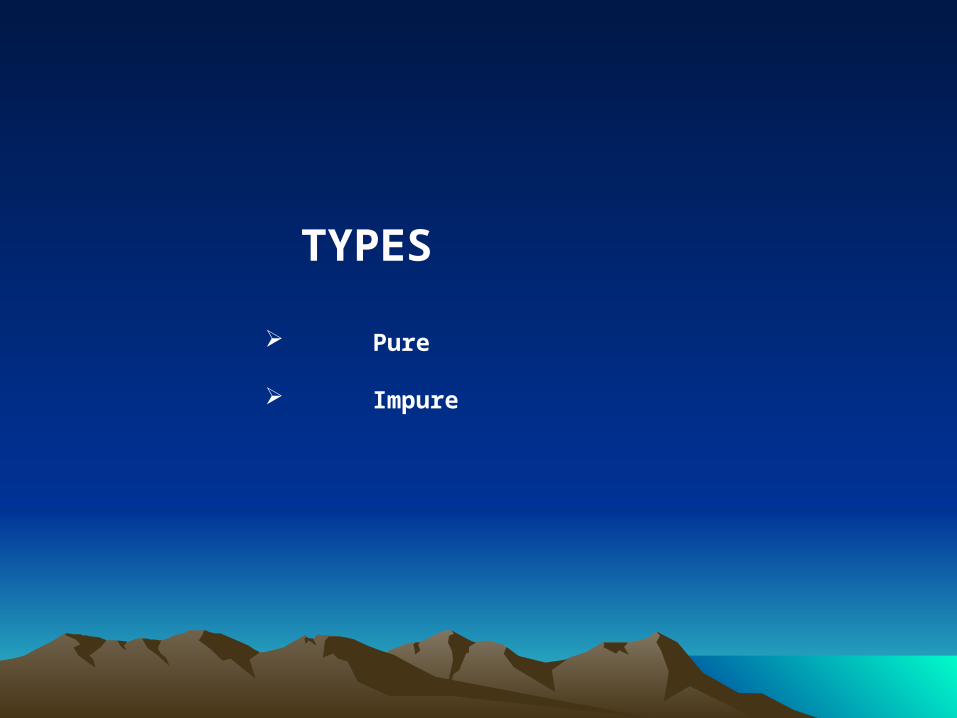
TYPES
Pure
Impure
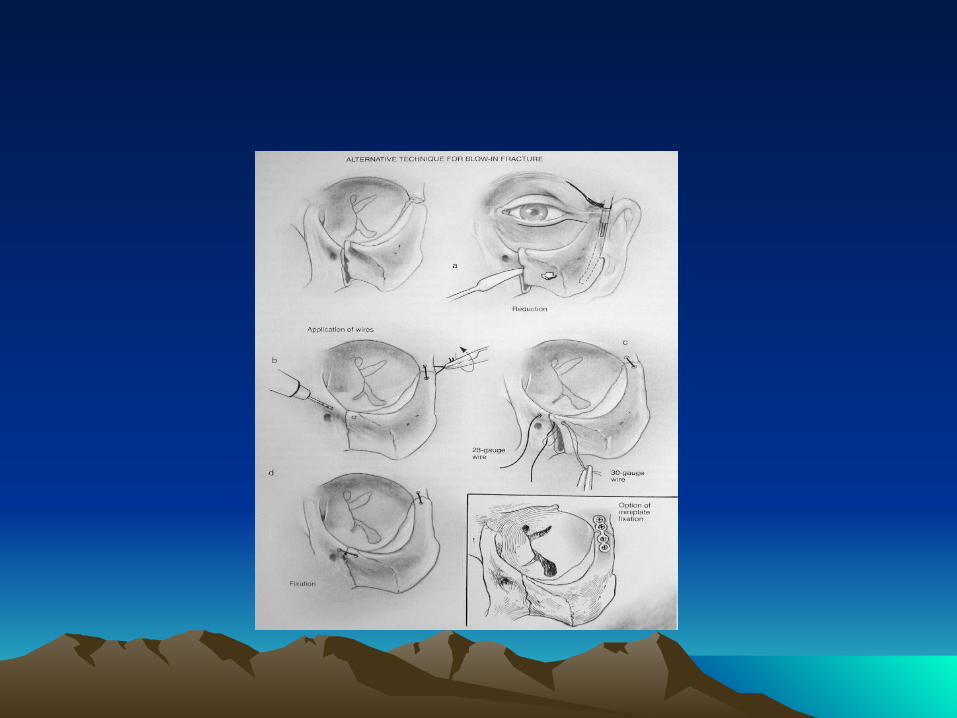
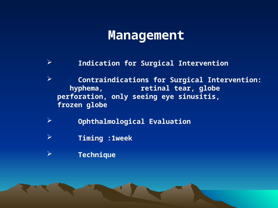
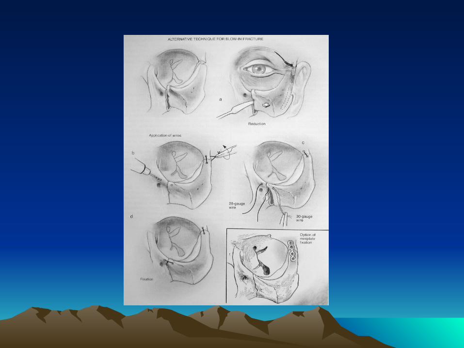
Management
Indication for Surgical Intervention
Contraindications for Surgical Intervention: hyphema, retinal tear, globe perforation, only seeing eye
sinusitis, frozen globe
Ophthalmological Evaluation
Timing :1week
Technique
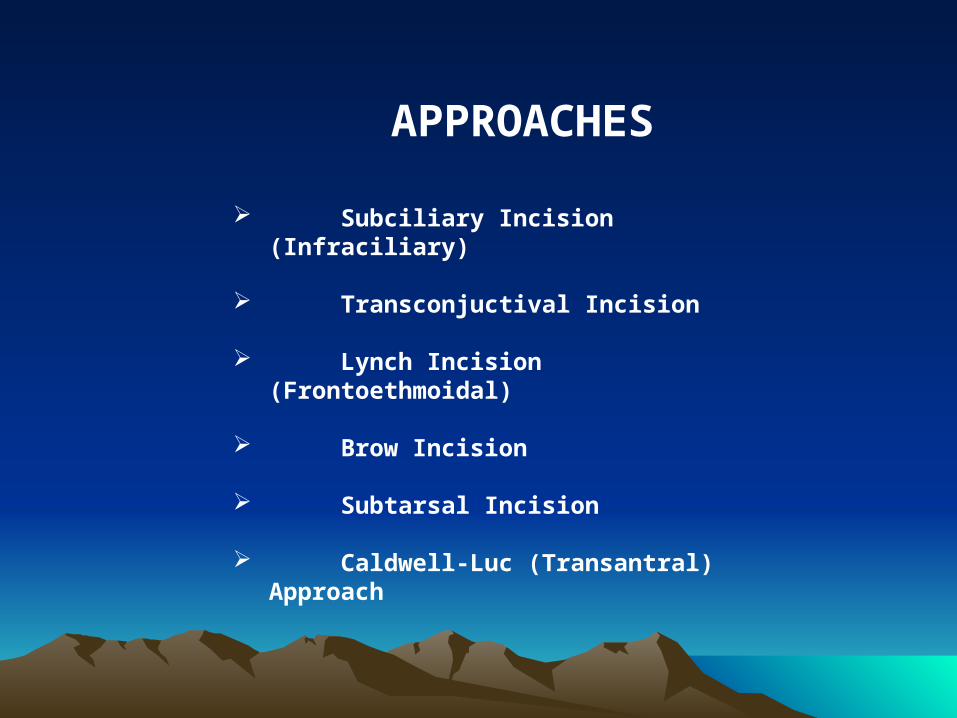
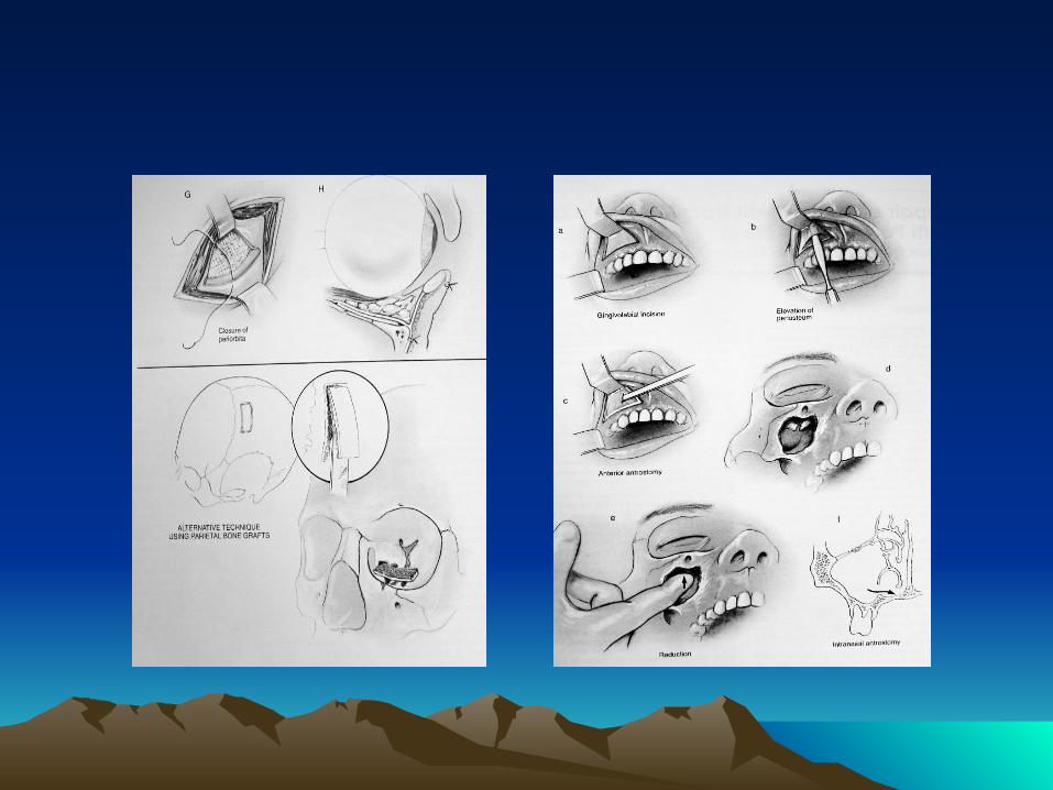
APPROACHES
Subciliary Incision (Infraciliary)
Transconjuctival Incision
Lynch Incision (Frontoethmoidal) Brow Incision
Subtarsal Incision
Caldwell-Luc (Transantral) Approach
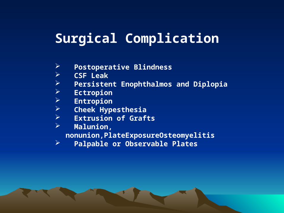
Surgical Complication
Postoperative Blindness CSF Leak Persistent Enophthalmos and Diplopia Ectropion Entropion Cheek Hypesthesia Extrusion of Grafts Malunion, nonunion,PlateExposureOsteomyelitis Palpable or Observable Plates
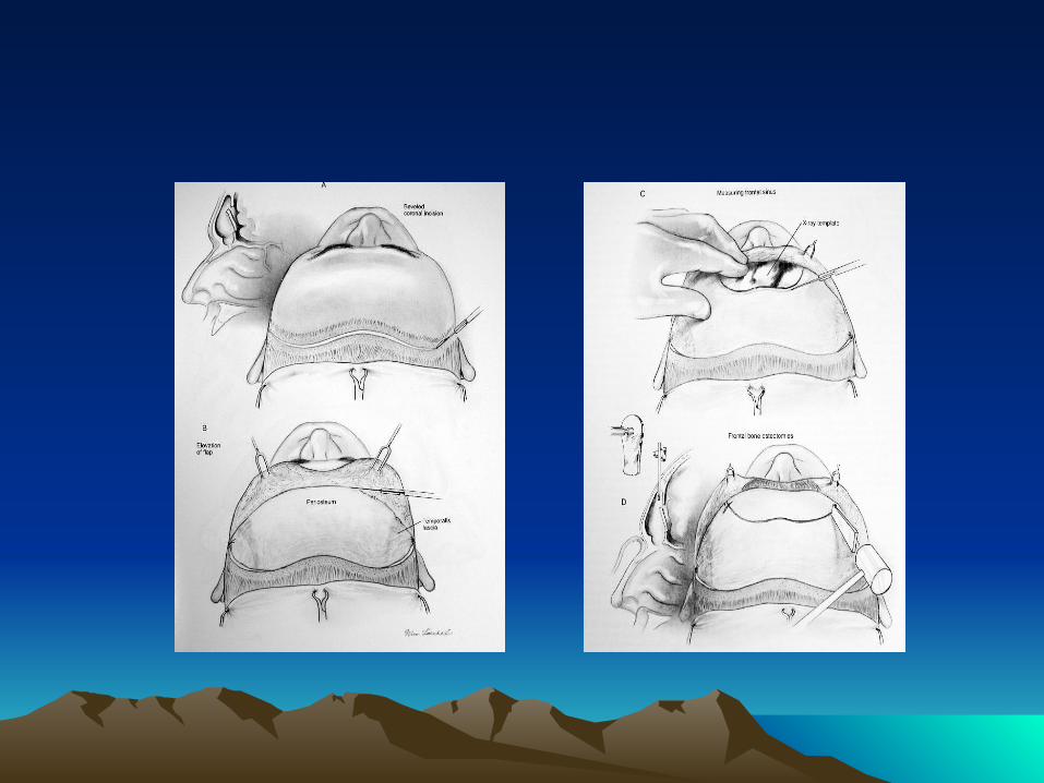
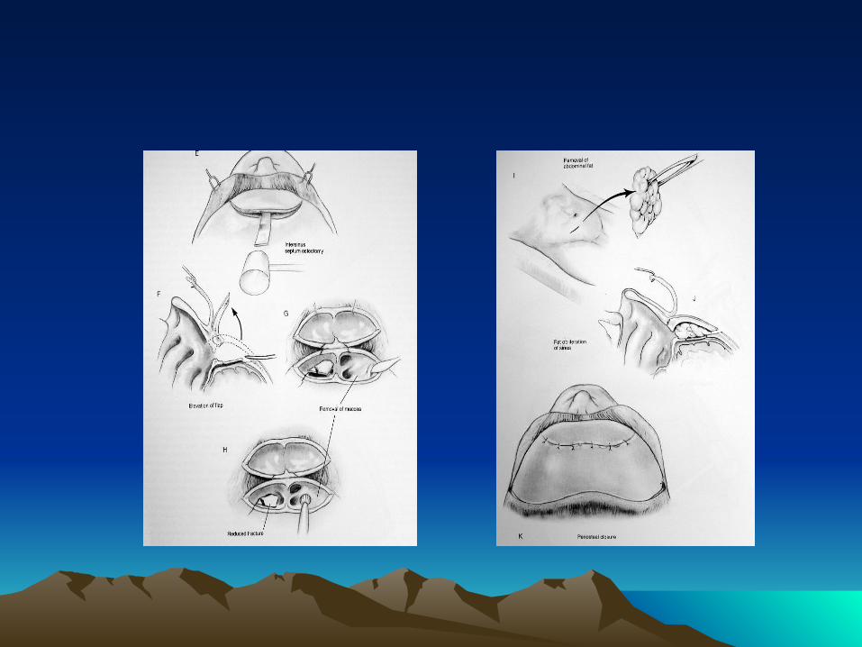
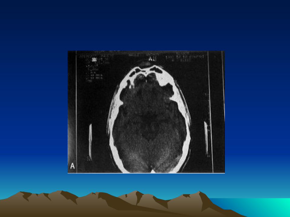
FRONTAL SINUS FRACTURE
FRONTAL SINUS FRACTURE
• Sign& Symptoms• Risk
MANAGEMENT
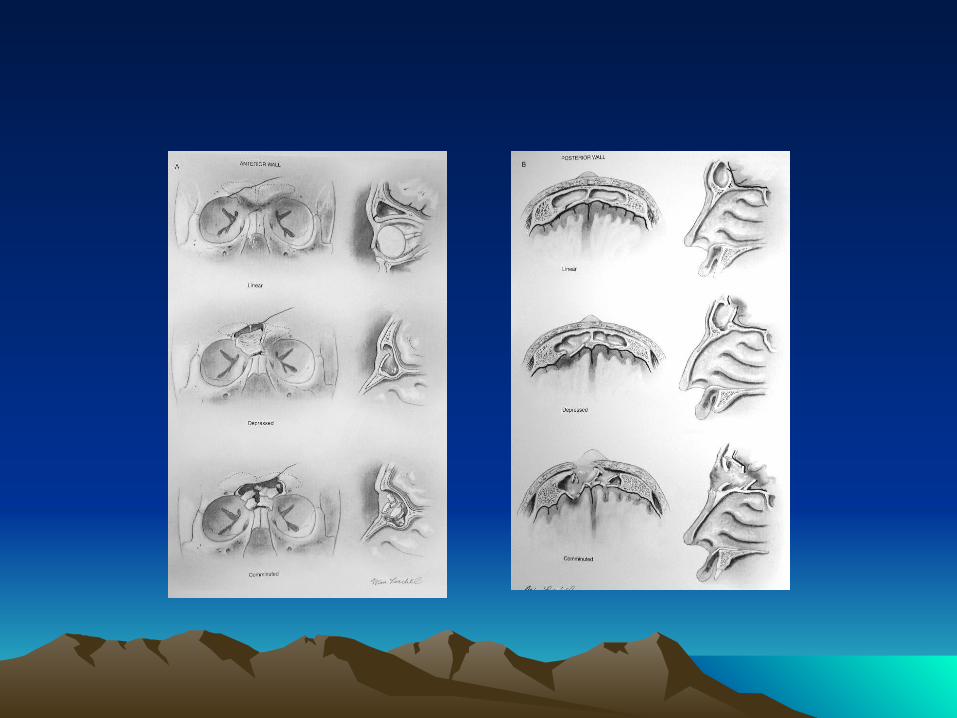
Anterior Table Fractures
Linear, Minimally Displaced
Depressed Fractures
Comminuted or Unstable Fractures
Posterior Table Fractures
Isolated Nondisplaced Psoterior Table Fracture
Displaced Posterior Table Fracture
Comminuted, Contaminated or through and Through Fractures--Cranialization
Surgical Complications
Mucocele, Mucopyoceles Sinusitis Forehead Contour Deformity Intracranial Infections Osteomyelitis CSF leak Forehead Hypesthesia Forehead Paralysis
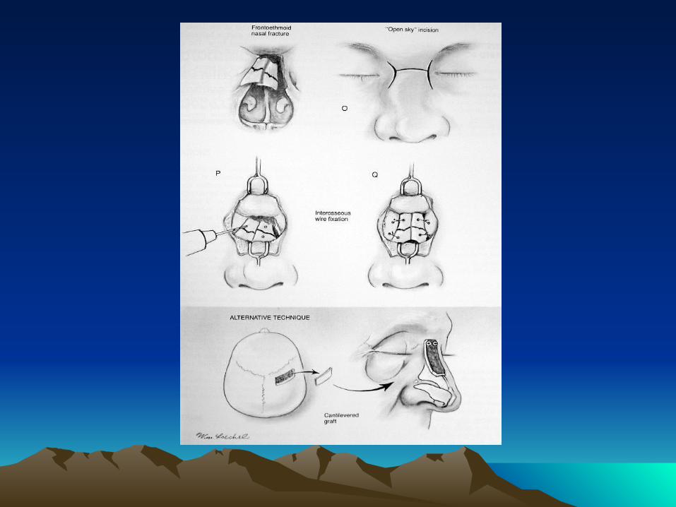
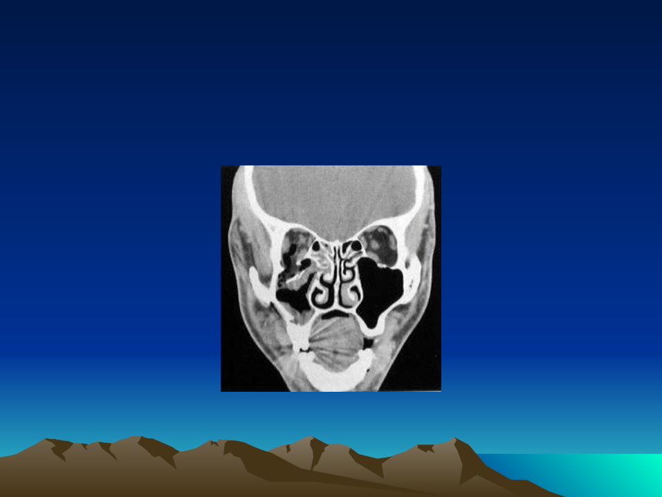
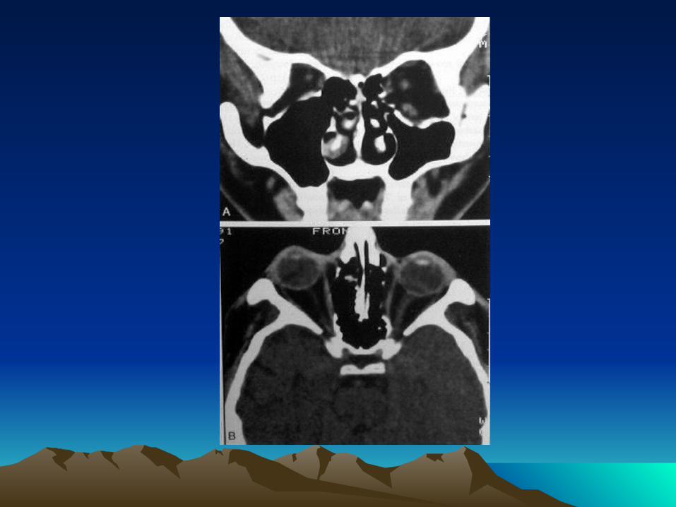
NASO-ORBITOETHMOID (NOE)FRACTURES
Introduction
NOE: frontal process of maxilla, nasal bones, and orbital space Sign& Symptoms Pseudohypertelorism (Traumatic Telecanthus)
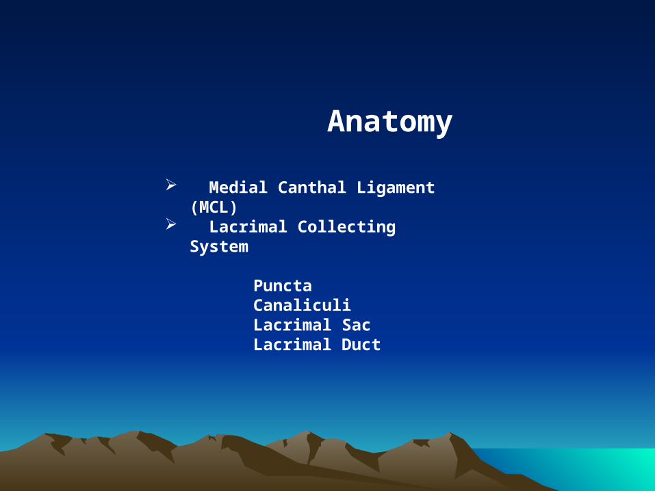
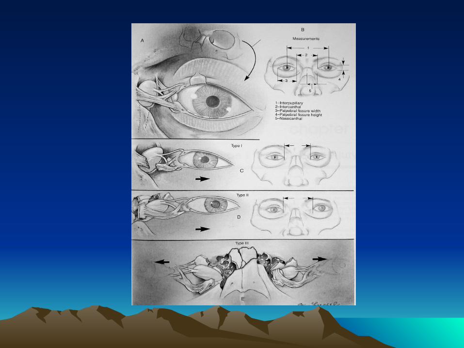
Anatomy
Medial Canthal Ligament (MCL) Lacrimal Collecting System Puncta Canaliculi Lacrimal Sac Lacrimal Duct
Management
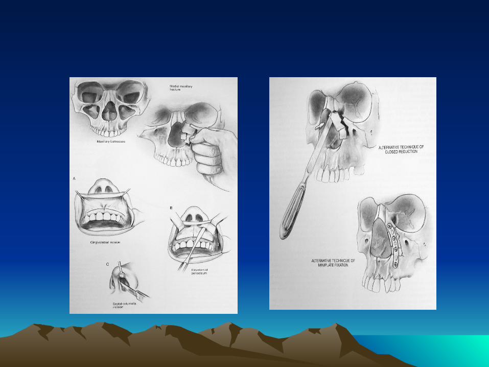
First reconstruct medial orbital wall prior to repair of the MCL Must consider associated injuries
May attempt closed reduction if MCL and lacrimal system is intact
Telescoping Nasal Bones and Frontal Process of the Maxilla
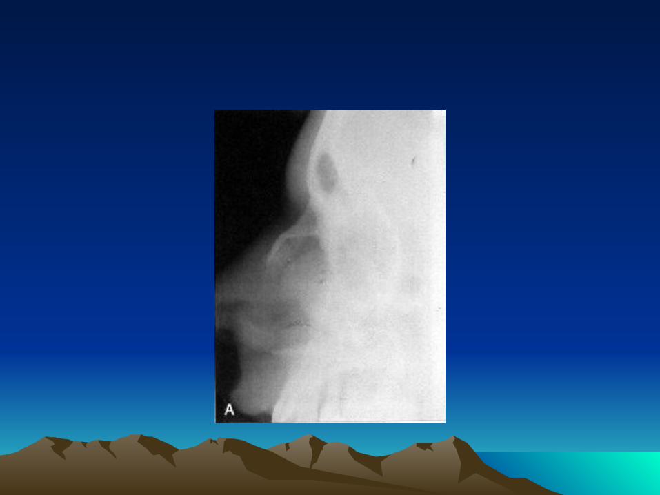
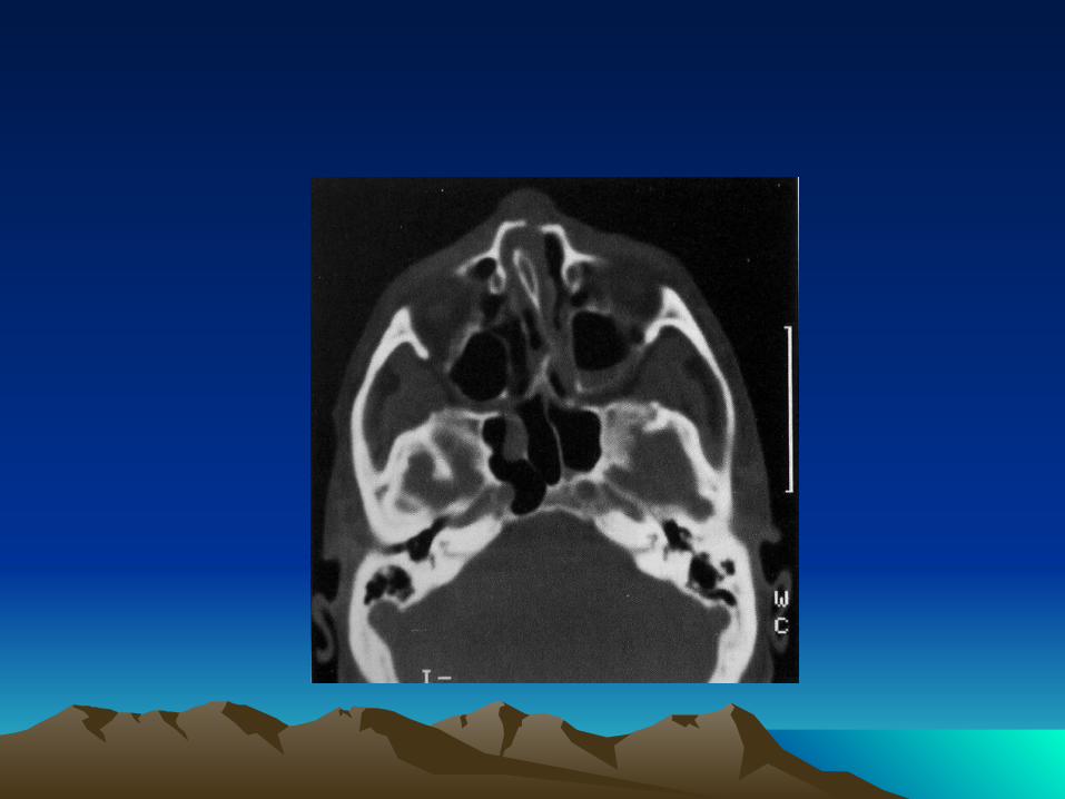
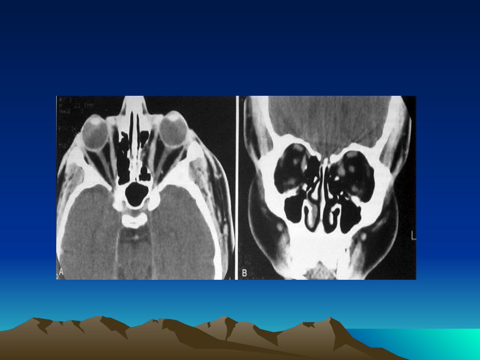
Nasal Fractures
Introduction
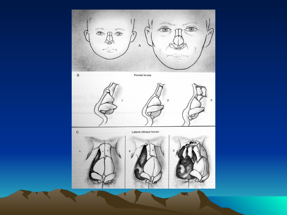
Most common Anterior impacts Lateral impacts Dislocated quadrangular cartilage inferiorly or “C-shaped” Children usually have dislocated or green stick fractures and have a higher risk of septal hematomas) Comminutions are more common in adults Sign& Symptoms Diagnosis
Management
Initial Management
Preoperative photographs/x-ray may be considered for medicolegal
documentation
Septal hematomas
Open fractures must be cleaned then given antibiotics
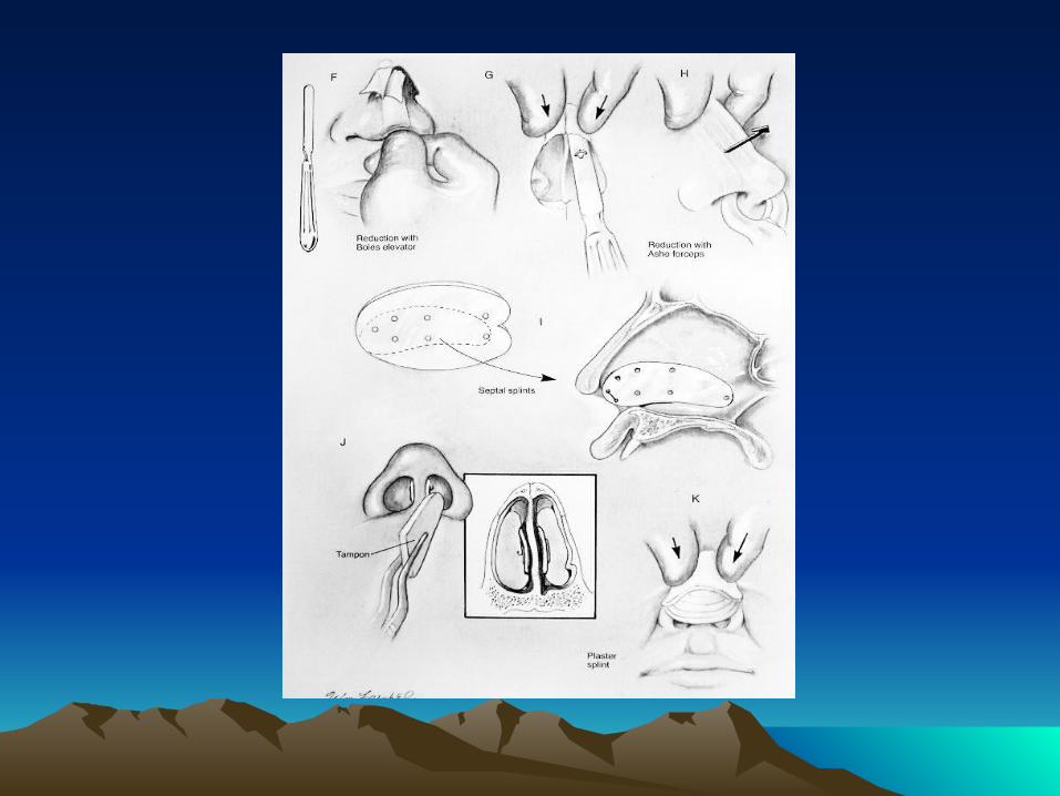
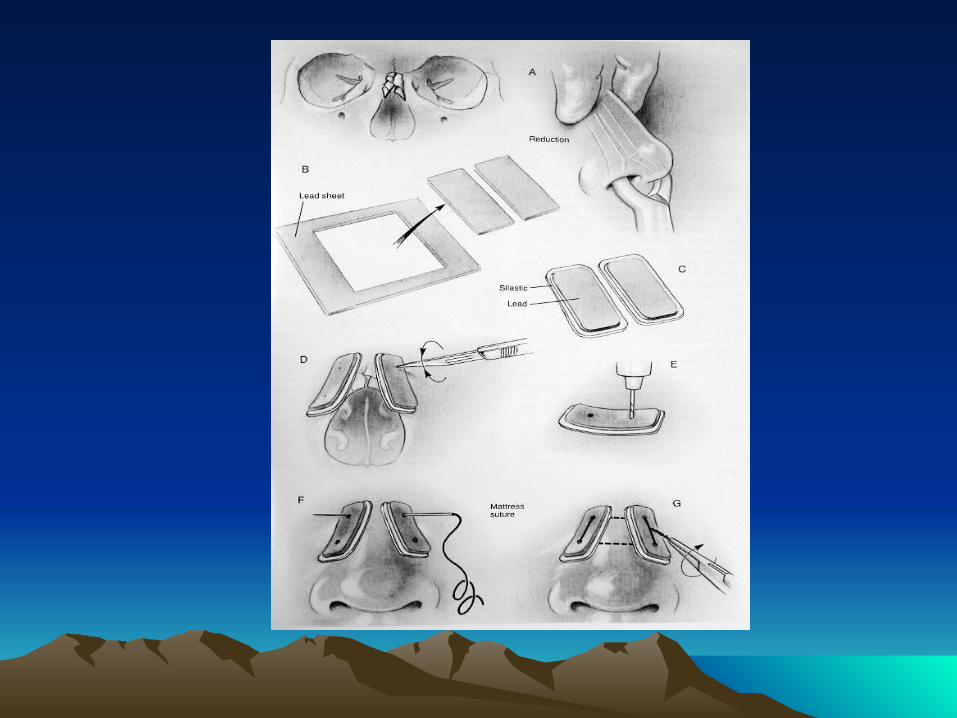
Cont : Management
Surgical Management
Generally nasal bone depressed or deviation may undergo closed reduction Open Reduction with Internal Fixation (Septorhinoplasty)
Pediatric Nasal Fractures: generally should be treated conservatively
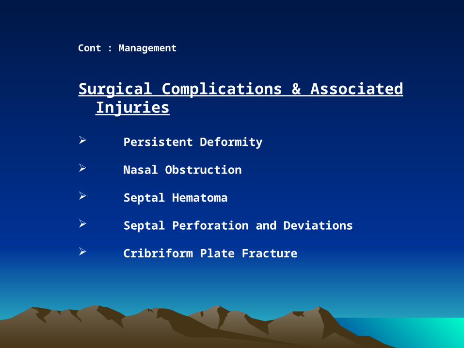
Cont : Management
Surgical Complications & Associated Injuries
Persistent Deformity
Nasal Obstruction
Septal Hematoma
Septal Perforation and Deviations
Cribriform Plate Fracture

Thank you&
Good luck to your examination