Embed Size (px)
Citation preview

Measurement of CT
radiation profile width
using CR imaging plates
Med Phys 2005; 32(9):2881-2887
Speaker:Shin-Lei Peng
2007, April 18

Outline
Introduction
Materials and methods
Results
Discussion
Conclusions
Introduction
Collimation of computed tomography(CT) radiation beams is crucial to both image quality and patient dose
How do we measure the width of the beam?
--Full Width at Half Maximum, FWHM
tradition: film processing
today: imaging plate, IP
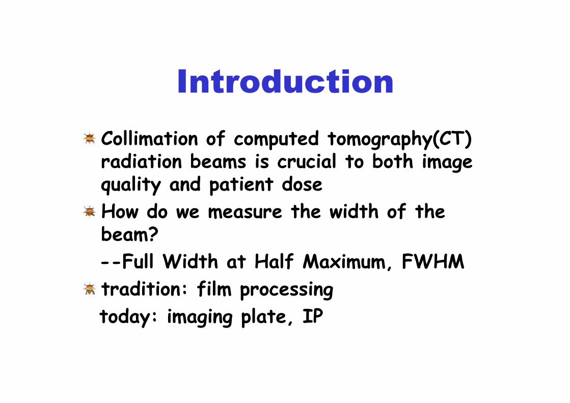
Materials
CT:Light-Speed 16 multislice scanner
(General Electric)
IP system:Fuji Model 5000
Film system:X-Omat V(XV) ready-pack
film,X-Omat processor
(Kodak)
Array 2905 Laser Film Digitizer
MethodsMethods
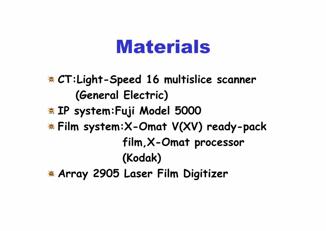
Interpreting Fuji IP pixel--the pixel value Q(E) resulting from exposure(E, in mR)
L:latitude, 0.5~4
S:sensitivity, 2~20000k:calibration constant, approximately 0.005
1 0
1 0 2 4( ) lo g ( ) 5 1 1Q E k S E
L= × × +

MethodsMethods
Interpreting Fuji IP pixel--a S value of 200:1 mR exposure, 80 kVp, 1mm Cuand 1 mm Al filtration
--three Exposure Data Recognizer,EDR--automatic--semiautomatic--fixed
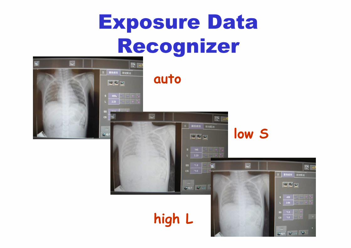
Exposure Data
Recognizer
auto
low S
high L

Methods
Characterization of IP saturation
--prior to each IP exposure, the
free-in-air exposure was measured
--80kVp,EDR mode with L=4,S=5,
80kVp,EDR mode with L=4,S=50,
mAs settings were varied between 2~40

Methods
CT radiation profile experiment
--using several collimation settings and
detector configurations
--the image receptor was placed on a
Styrofoam spacer on top of the table
--positioned at the isocenter of the CT
gantry during exposure
Methods
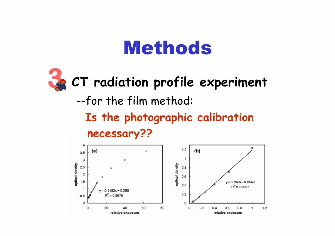
CT radiation profile experiment
--for the film method:
Is the photographic calibration
necessary??
Methods
CT radiation profile experiment
--for the CR method:
--the IP was selected at random
--to evaluate the scattering and
attenuation effects from the
cassette:w/ cassette & w/o cassette
Results
Four experiments were set up--the effects of attenuation and scattingby the the cassette
--pixel value versus logarithm of CTexposure
--CT radiation profile measured by film &IP
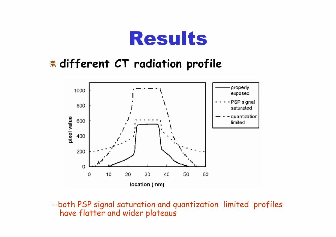
--different CT radiation profile

Results
the effects of attenuation and scatting by the the cassette
suggestion:using a IP exposed inside a cassette

Results
pixel value versus logarithm of CTexposure
--PSP signal saturation for CT techniques greater
than 20mAs at 80 kVp--pixel value=869 for S=50
pixel value=613 for S=5

Results
CT radiation profile measured by film & IP

Results
CT radiation profile measured by film & IP

Results CT radiation profile measured by film & IP
--discrepancies between the film and IP: < 3%
Results
different CT radiation profile
--both PSP signal saturation and quantization limited profiles have flatter and wider plateaus

Discussion
Under the properly exposed and processed, IP is an adequate detector for measurements of CT radiation profiles
Why we need low S and high L number?
--S=5~50, L=4
Q(E)=(1024/L)log(kxSxE)+511

Discussion
Different CT scanner, different exposure conditions
Different manufacturers, different relationship between the olgarithm of the exposure and pixel value
--for Agfa==logE(E)=1249.5*log(S*E/200)+2774.5
--for Kodak==EI(E)=1000*log(E)+Co

Conclusion
The FWHM of CT radiation profiles can be accurately measured using CR imaging plates.
Because of slight attenuation of the x-ray beam by the CR cassette, we recommend exposing the IP inside its cassette

References
D.M Tucker and P.S Rezentes, “The relationship between pixel value and beam quality in photostimulable phosphor imaging,” Med. Phys. 24.887-893(1997)Roberto Molteni, ”Effect of visible light on photostimulated phosphor imaging plates”, International Congress Series. 1256.1100-1205(2003)C.E. Floyd, Jr., H.C. Chotas, J.T. Dobbins, and C.E. Ravin, “Quantitative radiographic imaging using a photostimulable phosphor system”, Med. Phys. 17. 454-459(1990)


![[width=0.2]LogoMines [width=0.3]LogoINRIA [width=0.15](https://img.pdfslide.net/doc/110x75/6201e72d8bfe977ad8268cb6/width02logomines-width03logoinria-width015-.jpg)