Embed Size (px)
Citation preview
Mecanismes moleculars implicats en la secreció de pèptids en cèl·lules glials del sistema nerviós central
Sonia Paco Mercader
ADVERTIMENT. La consulta d’aquesta tesi queda condicionada a l’acceptació de les següents condicions d'ús: La difusió d’aquesta tesi per mitjà del servei TDX (www.tdx.cat) ha estat autoritzada pels titulars dels drets de propietat intel·lectual únicament per a usos privats emmarcats en activitats d’investigació i docència. No s’autoritza la seva reproducció amb finalitats de lucre ni la seva difusió i posada a disposició des d’un lloc aliè al servei TDX. No s’autoritza la presentació delseu contingut en una finestra o marc aliè a TDX (framing). Aquesta reserva de drets afecta tant al resum de presentació de la tesi com als seus continguts. En la utilització o cita de parts de la tesi és obligat indicar el nom de la persona autora.
ADVERTENCIA. La consulta de esta tesis queda condicionada a la aceptación de las siguientes condiciones de uso: La difusión de esta tesis por medio del servicio TDR (www.tdx.cat) ha sido autorizada por los titulares de los derechos de propiedad intelectual únicamente para usos privados enmarcados en actividades de investigación y docencia. No se autoriza su reproducción con finalidades de lucro ni su difusión y puesta a disposición desde un sitio ajeno al servicio TDR. No se autoriza la presentación de su contenido en una ventana o marco ajeno a TDR (framing). Esta reserva de derechos afecta tanto al resumen de presentación de la tesis como a sus contenidos. En la utilización o cita de partes de la tesis es obligado indicar el nombre de la persona autora.
WARNING. On having consulted this thesis you’re accepting the following use conditions: Spreading this thesis by the TDX (www.tdx.cat) service has been authorized by the titular of the intellectual property rights only for private uses placed in investigation and teaching activities. Reproduction with lucrative aims is not authorized neither its spreading and availability from a site foreign to the TDX service. Introducing its content in a window or frame foreign to the TDX service isnot authorized (framing). This rights affect to the presentation summary of the thesis as well as to its contents. In the usingor citation of parts of the thesis it’s obliged to indicate the name of the author.
UNIVERSITAT DE BARCELONA FACULTAT DE BIOLOGIA
Departament de Biologia Cel·lular
Tesi presentada per Sonia Paco Mercader
Per optar al grau de
DOCTORA
Director: Dr. Fernando Aguado Tomás
Programa de doctorat de Biologia Cel·lular i Molecular Bienni: 2005-2007
Vist-i-plau del director, La interessada, Dr. Fernando Aguado Tomás Sonia Paco Mercader
Barcelona, Novembre 2011


iii
Índex de figures ................................................................................................................................ vi
Índex de taules ................................................................................................................................ vii
Abreviatures .................................................................................................................................... viii
INTRODUCCIÓ ...................................................................................................................................... 1
1. Composició cel·lular del SNC ............................................................................................. 3
1.1. Neurones ....................................................................................................................... 3
1.2. Cèl·lules glials ................................................................................................................ 3
1.2.1. Oligodendròcits ....................................................................................................... 3
1.2.2. Astròcits ..................................................................................................................... 5
1.2.3. Micròglia .................................................................................................................... 5
1.2.4. Cèl·lules ependimàries ............................................................................................ 6
1.2.5. Cèl·lules NG2 ............................................................................................................ 6
2. Desenvolupament dels astròcits ........................................................................................ 7
2.1. De la neurogènesi a la gliogènesi ............................................................................ 9
2.2. Diversitat astrogènica ................................................................................................ 10
2.2.1. Heterogeneïtat regional ........................................................................................... 11
2.2.2. Heterogeneïtat fenotípica ........................................................................................ 12
3. Funcions dels astròcits ....................................................................................................... 14
3.1. Desenvolupament ...................................................................................................... 15
3.1.1. Els astròcits com a elements stem ..................................................................... 15
3.1.2. Guia axonal ............................................................................................................. 15
3.1.3. Regulació de la sinaptogènesi ............................................................................ 15
3.2. Funció estructural ....................................................................................................... 16
3.2.1. Formació de la barrera hematoencefàlica. ...................................................... 17
3.3. Funció metabòlica ...................................................................................................... 17
iv
3.3.1. Regulació de la micro circulació cerebral ........................................................ 17
3.3.2. Suport metabòlic a les neurones ....................................................................... 18
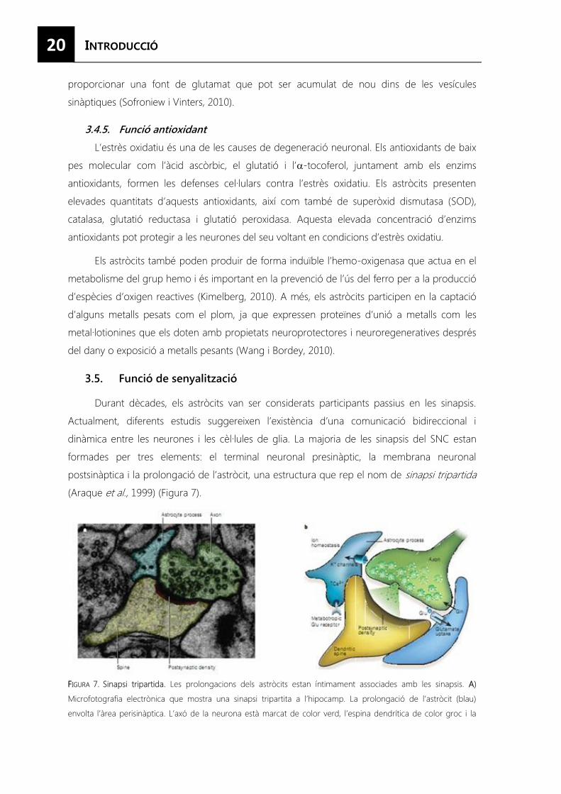
3.4. Funció homeostàtica ................................................................................................. 19
3.4.1. Homeòstasi d’ions ................................................................................................. 19
3.4.2. Homeòstasi de l’aigua .......................................................................................... 19
3.4.3. Homeòstasi del pH ................................................................................................ 19
3.4.4. Homeòstasi de neurotransmissors .................................................................... 19
3.4.5. Funció antioxidant ................................................................................................. 20
3.5. Funció de senyalització ............................................................................................. 20
3.5.1. Senyalització dins del sincici astrocitari: fisiologia astrocitària .................... 21
3.5.2. Alliberació de neurotransmissors ....................................................................... 23
3.5.3. Modulació de la transmissió i la plasticitat sinàptica ..................................... 23
3.6. Resposta glial al dany ............................................................................................... 23
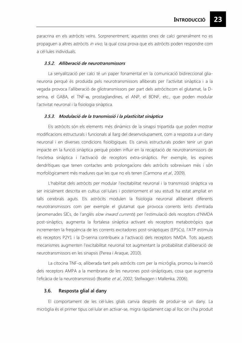
3.6.1. Canvis morfològics i d’expressió de l’astrocitosi ............................................ 24
3.6.2. Factors d’activació i vies implicades .................................................................. 25
4. Secreció ................................................................................................................................. 27
4.1. Tipus de vesícules de secreció ................................................................................ 28
4.1.1. Vesícules constitutives .......................................................................................... 28
4.1.2. Grànuls de secreció ............................................................................................... 29
4.1.3. Vesícules sinàptiques o SLMVs ........................................................................... 29
4.1.4. Lisosomes secretors o LROs ............................................................................... 29
4.2. Principis del transport vesicular .............................................................................. 30
4.2.1. Molècules de recobriment .................................................................................. 30
4.2.2. Proteïnes adaptadores ......................................................................................... 30
4.2.3. Senyals de classificació ......................................................................................... 31
4.3. Transport a la membrana plasmàtica ................................................................... 36
4.3.1. Proteïnes de transport .......................................................................................... 36
4.3.2. Rab GTPases ........................................................................................................... 36
4.3.3. Proteïnes SNAREs .................................................................................................. 37
v
5. Secreció en els astròcits .................................................................................................... 40
5.1. Mecanismes de secreció en astròcits .................................................................... 40
5.1.1. Secreció no exocítica en astròcits ...................................................................... 41
5.1.2. Exocitosi en astròcits ............................................................................................. 43
OBJECTIUS .......................................................................................................................................... 49
RESULTATS ......................................................................................................................................... 53
CAPÍTOL I: La senyalització per AMP cíclic reprimeix l’activació i promou la maduració
en els astròcits..................................................................................................................................55
CAPÍTOL II: L’AMP cíclic regula l’expressió de proteïnes exocítiques i potencia la secreció
regulada de pèptids en astròcits en cultiu ............................................................................... 81
CAPÍTOL III: Secretogranina III és una granina astrocitària que es sobreexpressa en glia
reactiva ............................................................................................................................................. 97
DISCUSSIÓ ........................................................................................................................................ 111
CONCLUSIONS ................................................................................................................................. 127
BIBLIOGRAFIA ................................................................................................................................... 131
ANNEX.............................................................................................................................................. 149
Informe del director .................................................................................................................... 151
Agraïments…………………………………………………………………………………………………………………….153
vi
FIGURA 1. Les prolongacions dels oligodendròcits envolten els axons per formar la beina de mielina. ............................................................................................................................................ 4
FIGURA 2. Relació de la micròglia amb les cèl·lules hematopoètiques i les cèl·lules del sistema nerviós central. .............................................................................................................................. 7
FIGURA 3. Patró de gliogènesi a l’embrió i a les zones progenitores en l’adult.. .......................... 8
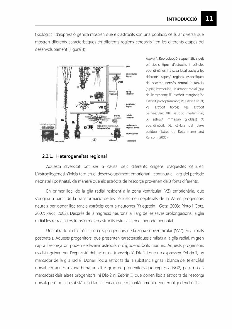
FIGURA 4. Reproducció esquemàtica dels principals tipus d’astròcits i cèl·lules ependimals i la seva localització a les diferents capes/ regions específiques del sistema nerviós central.. .......................................................................................................................................... 11
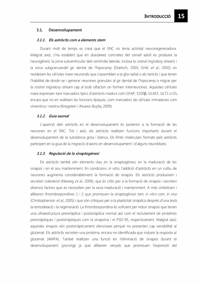
FIGURA 5. Microdominis anatòmics formats per les cèl·lules glials. ................................................ 16
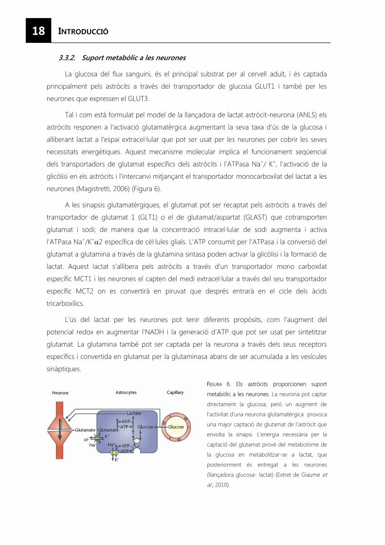
FIGURA 6. Els astròcits proporcionen suport metabòlic a les neurones. ....................................... 18
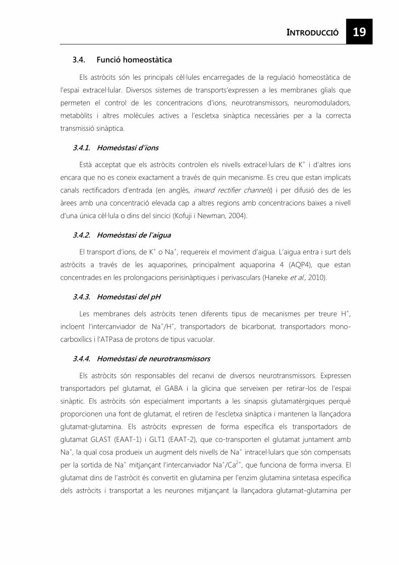
FIGURA7. Sinapsi tripartita......................................................................................................................... 20
FIGURA 8. Fonts de calci dins l’astròcit. .................................................................................................. 22
FIGURA 9. Esquema dels canvis morfològics i d’expressió de l’astrocitosi al llarg del temps. .. 24
FIGURA 10. Esquema on es mostren els factors que indueixen l’astrocitosi i les principals vies moleculars activades. ................................................................................................................. 26
FIGURA 11. Vies de secreció en les cèl·lules eucariotes. .................................................................... 28
FIGURA 12. Mecanismes de classificació als grànuls densos. ............................................................ 34
FIGURA 13. Vies de secreció i biogènesi de grànuls densos en cèl·lules (neuro)endocrines. ... 35
FIGURA 14. Exemple funcional de la fusió de grànuls mitjançada per proteïnes SNARE. ......... 38
FIGURA 15. Mecanismes d’exocitosi de neurotransmissors no exocítica i híbrida. calci ............. 43
FIGURA 16. Les SLMVs es situen en els terminals dels astròcits pròxims a les membranes neuronals que contenen receptors NMDA. ......................................................................... 45
FIGURA 17. Microscopia electrònica on es mostra la localització de CGB i SgII en vesícules similars als grànuls de secreció (grànuls densos) en astròcits en teixit. ......................... 47
FIGURA 18. Expressió de proteïnes exocítiques en cèl·lules en cultiu…………..………………..……….114 FIGURA 19. Proteïnes implicades en l’exocitosi de neurones, astròcits i micròglia .................... 115
FIGURA 20. La micròglia sobreexpressa algunes proteïnes SNARE en el tractament amb citocines pro inflamatòries. ..................................................................................................... 116
FIGURA 21. L’alliberació de TNF-� es produeix de forma ràpida i depenent de transport vesícular i proteïnes SNARE... .................................................................................................. 117
FIGURA 22. Dependència de les proteïnes SNARE en l’alliberació de SgII regulada per calci en astròcits.. ..................................................................................................................................... 120
FIGURA 23. El marcatge de SgIII en plaques senils en humans està associat amb neurites distròfiques i amb astròcits activats. ..................................................................................... 125

vii
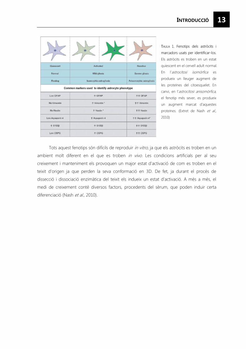
TAULA 1. Fenotips dels astròcits i marcadors usats per identificar-los. ......................................... 13
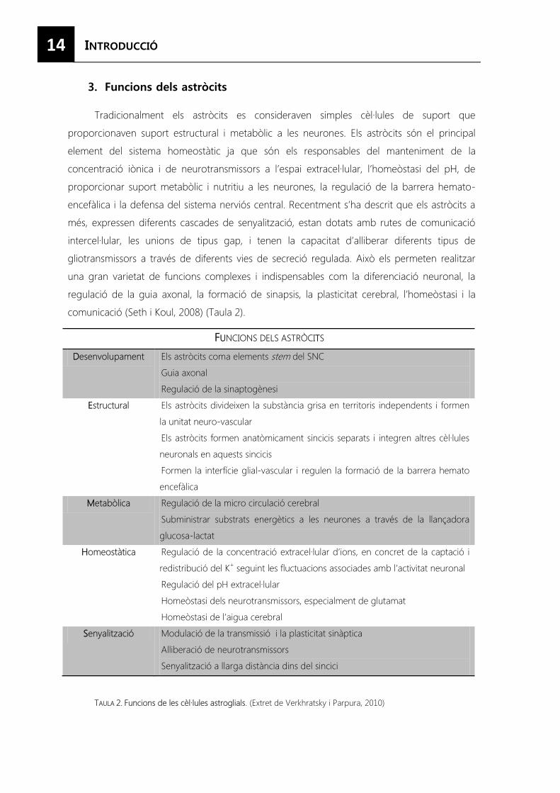
TAULA 2. Funcions de les cèl·lules astroglials. ...................................................................................... 14
TAULA 3. Classificació dels membres de la familia SNARE. .............................................................. 37
viii
Aldh1L1 Aldehid deshidrogenasa família 1, membre L1 (Aldehyde Dehydrogenase 1 family,
member L1)
AMPA (α-amino-3-hydroxy-5-methyl-4-isoxazolepropionic acid)
AMPc Adenosina 3’- 5’- Monofosfat cíclica
ANP Pèptid natriurètic atrial (Atrial Natriuretic Peptide)
AP Proteïnes adaptadores (Adaptor Protein)
ATP Adenosina trifosfat
BDNF Factor neurotròfic derivat del cervell (Brain Derived Neurotrophic Factor)
CD (Cluster of Differentiation)
CgA Cromogranina A (Chromogranin A)
CgB Cromogranina B (Chromogranin B)
CNTF Factor neurotròfic ciliar (Ciliary Neurotrophic Factor)
CPE Carboxipeptidasa E (Carboxipeptidase E)
CRE Element de resposta al cAMP (cAMP response element)
CREB Proteïna d’unió al element de resposta al AMPc (cAMP response element-binding
protein)
CT-1 Cardiotrofina-1 (Cardiotrophin-1)
EAA Aminoàcid excitador (Excitatory Amino Acid)
EGF Factor de creixement epidèrmic (Epidermal Growth Factor)
ER Reticle endoplasmàtic (Endoplasmic Reticulum)
FGF Factor de creixement de fibroblasts ( FibroblastGrowth Factor)
GABA Àcid gamma aminobutíric (gamma-aminobutyric acid)
GDNF Factor neurotròfic derivat de cèl·lules glials (Glial cell-line Derived Neurotrophic
Factor)
GFAP Proteïna fibril·lar acídica glial (Glial Fibrillary Acidic Protein)
GLAST Transportador de glutamat aspartat (Glutamate Aspartate Transporter)
GLT-1 Transportador de glutamat 1 (Glutamate Transporter)
GLUT Transportador de glucosa (Glucose Transporter)
GS Glutamina sintetasa (Glutamine Synthase)
IFN Interferó
IGF-1 Factor de creixement similar a la insulina 1 (Insulin-like Growth Factor 1)
IL Interleucina (Interleukin)
IP3 Inositol trifosfat (Inositol trisphosphate)
IP3R Receptor d’IP3
ix
LIF Factor inhibidor de leucèmia (Leukemia Inhibitory Factor)
LPS Lipopolisacàrid (Lipopolysaccharide)
LRO Orgànuls relacionats amb els lisosomes (Lysosome- related organelles)
mGluRs Receptors metabotròpics de glutamate (Metabotropic Glutamate Receptor)
MHC Complex major d’histocompatibilitat (Major Histocompatibility Complex)
MMPs (Matrix metalloproteinases)
NADH Nicotinamida adenina dinucleótid reduït
NG2 Antigen neuro-glial 2
NGF Factor de creixement nerviós (Nerve Growth Factor)
NMDA (N-Methyl-D-aspartate)
NPCs Cèl·lula precursora de neurones ( Neural Precursor Cells)
NSF N-ethylmaleimide- sensitive factor
OPCs Cèl·lula precursora d’oligodendròcits (OligodendrocytePrecursor Cells)
PC (Proprotein convertase)
PGE Prostaglandina E
PKA Proteïna quinasa dependent d’AMPc
PKC Proteïna quinasa C
RyR Receptor de rianodina (Ryanodine Receptors)
SG Grànul de secreció (Secretory granule)
SgII Secretogranina II (Secretogranin II)
SgIII Secretogranina III (Secretogranin III)
SLMVs Microvesícules similar a les sinàptiques (Synaptic-like micro-vesicles)
SNAP Soluble NSF Attachment Protein
SNARE Soluble NSF Attachment Protein Receptor
SNC Sistema nerviós central
SNP Sistema nerviós perifèric
SOD Superòxid dismutasa (Superoxide dismutase)
Stxbp Sintaxin binding protein
SV Vesícula sinàptica (Synaptic vesicle)
SVZ Zona subventricular (Subventricular zone)
TGN Xarxa de trans-Golgi (Trans-Golgi Network)
TNF Factor de necrosi tumoral (Tumor Necrosis Factor)
TPA 12-O-tetradecanoylphorbol-13-acetate
VAMP Proteïna de membrane associada a la vesícula (vesicle-associated membrane protein)
VEGF Factor de creixement endotelial vascular (Vascular Endothelial Growth Factor)


3
El sistema nerviós és un sistema de processament de la informació amb dues funcions
fonamentals: la relació amb el medi extern i el control de tots els òrgans de l’individu. Permet
rebre la informació, ja sigui externa o interna, analitzar la situació i elaborar una resposta.
També és el responsable de coordinar totes les funcions, conscients o inconscients, de
l’individu (Kandel et al., 2000).
A part de les neurones, que representen el 10% de les cèl·lules al SNC, el SNC està
format per la glia que representa el 90% restant.
1.1. Neurones
La neurona és la unitat bàsica del cervell i la seva característica diferencial és la capacitat
de generar potencials d’acció. Les neurones s’organitzen en xarxes (o circuits) i es
comuniquen entre elles mitjançant les sinapsis. La senyalització neuronal implica la propagació
del potencial d’acció al llarg de l’axó fins al terminal presinàptic i l’alliberació dels
neurotransmissors que s’uneixen als receptors localitzats a la membrana de la neurona
postsinàptica, generant la despolarització de la neurona que continua propagant el senyal
(Allen i Barres, 2009).
1.2. Cèl·lules glials
El terme de cèl·lula glial clàssicament es refereix a tres tipus de cèl·lules. La primera
categoria inclou les cèl·lules de Schawnn i els oligodendròcits, que s’encarreguen de produir la
mielina en el sistema nerviós perifèric i central, respectivament. Aquestes cèl·lules envolten els
axons de les neurones amb una beina de mielina per aïllar i facilitar la transmissió ràpida i
eficient del’impuls nerviós. La segona categoria està formada pels astròcits, unes cèl·lules de
morfologia estrellada que proporcionen suport a les neurones. El tercer tipus de cèl·lula glial
és la micròglia, que no té un origen neural sinó que deriva del mesoderma, és originada pels
macròfags que envaeixen el cervell en estats molt primerencs del desenvolupament i
s’estableixen en el SNC. Les cèl·lules ependimàries també estan incloses dins de la paraula glia,
ja que deriven de la glia radial i comparteixen algunes propietats amb els astròcits.
Recentment, s’ha descrit un nou tipus de cèl·lula glial, caracteritzada per l’expressió del
proteoglicà condroïtin sulfat NG2, les cèl·lules NG2 o polidendròcits.
1.2.1. Oligodendròcits
Els oligodendròcits envolten llargs segments dels axons amb una beina multi laminada
formada per l’extensió de la membrana cel·lular que s’ajunta amb la membrana de l’axó
4
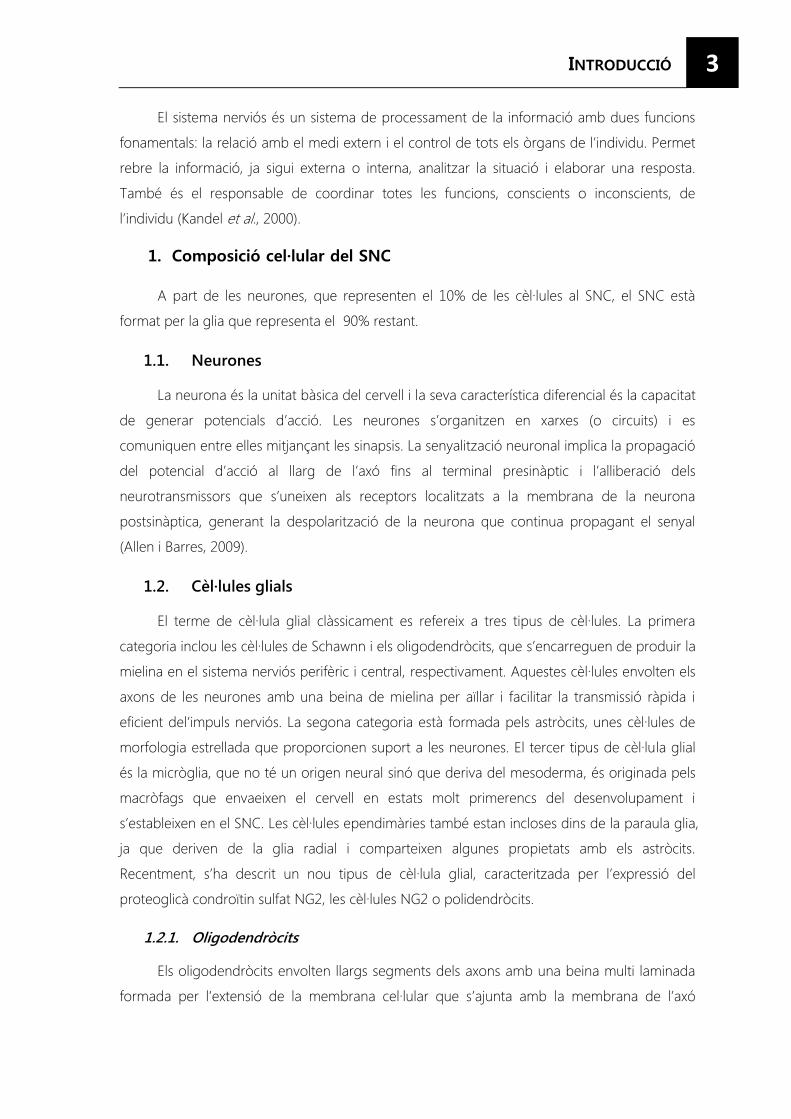
mitjançant unes juntes especials que defineixen els nodes de Ranvier entre els llargs segments
de mielina (Figura 1).
FFIGURA 1. Les prolongacions dels oligodendròcits envolten els axons per formar la beina de mielina. Les regions
mielinitzades dels axons (internodes) estan interrompudes per regions no mielinitzades (nodes de Ranvier). Les
prolongacions dels astròcits contacten amb els nodes (Extret de Jackman et al., 2009).
Els oligodendròcits només són competents per iniciar el procés de mielinització en un
període de temps molt reduït (generalment 12-18h) durant el desenvolupament, ja que un
cop madurs són relativament incapaços de mielinitzar. El procés d’embolcallament de
múltiples axons per part d’un únic oligodendròcit és altament regulat. Es seleccionen axons
amb un diàmetre superior als 0,2 �m i encara que no es coneixen els mecanismes moleculars
de reconeixement, són diferents en el SNC i en el SNP. Per exemple, un senyal crític per la
mielinització en el sistema nerviós perifèric és la interacció de la neuregulin-1 (NRG1) neuronal
tipus III amb els receptors glials ErbB, mentre que no és essencial per la mielinització del SNC,
per això es consideren altres factors axonals alternatius. El factor de creixement semblant a la
insulina 1 (IGF-1) i el factor inhibidor de leucèmia astroglial (LIF) estimulen la mielinització. En
canvi, AKT, mTOR i la sobreexpressió d’NRG1 en les neurones produeixen la hipermielinització
dels axons (Nave, 2010).
Es considerava que un senyal indispensable per l’inici de la mielinització era l’activitat
elèctrica de les neurones, però els oligodendròcits en cultiu poden envoltar axons que han
estat fixats prèviament (Rosenberg et al., 2008) de manera que sembla ser que els
oligodendròcits mitjançant diferents senyals poden mielinitzar els axons per defecte quedant
restringits localment per altres canvis en els axons, com la disminució de l’expressió de PSA-
NCAM i de LINGO-1, una proteïna transmembrana amb repeticions de residus de leucina i un
domini d’immunoglobulina (Charles et al., 2000).
Els oligodendròcits no només envolten els axons per aïllar-los elèctricament sinó que a
més indueixen l’agrupament dels canals de sodi al llarg de l’axó, en els nòduls de Ranvier. La
presència de la beina de mielina també provoca un augment del diàmetre de l’axó
possiblement per l’acumulació i fosforilació local del neurofilament. A més, proporcionen
suport tròfic a les neurones ja que sintetitzen el factor neurotròfic derivat de cèl·lules glials
5
(GDNF), el factor neurotròfic derivat del cervell (BDNF) i el factor de creixement derivat de la
insulina-1 (IGF-1) (Bradl i Lassmann, 2010).
1.2.2. Astròcits
Els astròcits són les cèl·lules més nombroses i heterogènies del SNC. Els subtipus
d’astròcits es poden definir seguint paràmetres que es sobreposen com la localització del cos
cel·lular, característiques morfològiques, propietats electro fisiològiques i el perfil d’expressió
gènic. Bioquímicament es defineixen per l’expressió d’unes proteïnes específiques com són el
filament intermedi GFAP, l’enzim glutamina sintetasa (GS), els transportadors EAA específics
d’astròcits GLAST i GLT-1, la proteïna d’unió al calci S100��� o Aldh1L1 (aldehid
deshidrogenasa família 1, membre L1) (Kimelberg et al., 2009)
Els astròcits madurs tenen una morfologia molt característica. Des del seu soma
s’originen unes ramificacions primàries que gradualment es van dividint en ramificacions cada
cop més fines fins a donar lloc a una xarxa densa de terminals que estan íntimament units
amb les sinapsis. Un únic astròcit de ratolí, depenent de la regió cerebral, pot arribar a
ocupar una àrea d’entre 20.000 i 80.000 �m3, envoltar nombrosos somes neuronals, associar-
se amb 300-600 dendrites i establir contacte amb unes 100.000 sinapsis individuals
aproximadament. En humans, aquestes xifres són molt més elevades, ja que un únic astròcit
ocupa un volum 30 vegades superior al que ocupa en els rosegadors i pot arribar a contactar
amb 2.000.000 de sinapsis (Oberheim et al., 2006)
1.2.3. Micròglia
La micròglia representa el 5- 20% de les cèl·lules en el SNC, de manera que és tan
nombrosa com les neurones.
Manté un fenotip quiescent en el SNC normal, contínuament monitoritzant el seu
microambient a través de la pinocitosi i la interacció amb les neurones, expressant nivells
baixos de MHC classe I i II així com molècules co-estimuladores com CD86 i CD40. Quan es
produeix un dany en el SNC, la micròglia es converteix en un fenotip activat amb una elevada
proliferació, motilitat, activitat fagocítica i alliberació de citocines (IL-1, IL-6, TNF-�, MCP-1,
RANTES) i espècies d’oxigen reactives (Yang et al., 2010) que recluten la micròglia, activen els
astròcits i augmenten l’excitabilitat neuronal (Milligan i Walkins, 2009).
Com a cèl·lules presentadores d’antígens tenen un paper similar als macròfags perifèrics,
i són components essencials tant de la immunitat innata com l’adaptativa; ja que durant
l’activació augmenta l’expressió d’MHC i de molècules co-estimuladoresque contribueixen a la
resposta de les cèl·lules T CD4 i CD8 (Olson i Miller, 2004).
6
La micròglia també presenta una fagocitosi transitòria durant l’ontogènia del SNC, ja
que a través del CR3 està implicada en la fagocitosi de neurones apoptòtiques i les seves
connexions que expressen les molècules del complement C1q i C3 (Stevens et al., 2007).
Recentment també s’ha relacionat la micròglia amb l’aparició del dolor neuropàtic
(Smith, 2010) així com que algunes proteïnes secretades per la micròglia tenen funcions
específiques en el cervell durant el desenvolupament i la plasticitat sinàptica (Bessis et al.,
2007).
1.2.4. Cèl·lules ependimàries
Les cèl·lules ependimàries deriven de la glia radial i comparteixen algunes
característiques amb els astròcits com l’expressió de GFAP. Formen una capa epitelial que
revesteix la superfície dels ventricles cerebrals i presenten característiques morfològiques
diferents als astròcits com la presència de cilis. Actuen com a barrera entre el teixit cerebral i el
líquid cefaloraquidi (LCR), tenen un paper en la secreció i el manteniment de l’equilibri del LCR
i en el metabolisme de les toxines (Wang i Bordey, 2008).
1.2.5. Cèl·lules NG2
Recentment, s’ha identificat un nou tipus de cèl·lula glial: les cèl·lules NG2, també
conegudes com sinantòcits o polidendròcits. Són identificables per l’expressió del proteoglicà
condroïtin sulfat NG2 i es troben tant el cervell en desenvolupament com en l’adult (Peters,
2004). Primer es van identificar com una cèl·lula progenitora d’oligodendròcits (OPCs) perquè
expressen diversos marcadors comuns, però tenen unes característiques diferencials que els
permeten classificar com a unes cèl·lules de glia diferents. Presenten una morfologia i
distribució similar als astròcits tant en la substància grisa com en la blanca del SNC adult.
Tenen una morfologia estrellada amb nombroses prolongacions primàries que es van
bifurcant finsa formar una arborització d’un diàmetre de 100�m, estenent les seves
prolongacions fins els nodes de Ranvier i les sinapsis on interactuen amb les neurones.
Fisiològicament moltes NG2 expressen canals de voltatge de Na+, receptors AMPA
permeables al Ca2+, receptors de GABA i purinoreceptors. A l’hipocamp reben projeccions de
les neurones piramidals de CA3 i d’interneurones gabaèrgiques (Sakry et al., 2011) el que
suggereix que podrien tenir un paper important d’integració al cervell. A més, són cèl·lules
progenitores altament plàstiques que poden donar lloc a astròcits i fins i tot a neurones
(Nishiyama et al., 2009; Heneka et al., 2010) i com a resposta al dany augmenten la seva
proliferació de manera que contribueixen a l’astrogliosi reactiva (Di Bello et al., 1999; Levine et
al., 1994; McTigue et al., 2001)
7
El sistema nerviós és un dels òrgans que es diferencia abans a partir de l’estat
embrionari de blàstula. En els humans, el tub neural primitiu es forma a partir de la quarta
setmana de gestació i la neurogènesi comença a partir de la cinquena setmana. El seu
desenvolupament és bastant llarg, es produeix al llarg de tota l’embriogènesi i no finalitza la
mielinització fins etapes post-natals. A mesura que avança el desenvolupament neural, el tub
neural experimenta una expansió diferencial i una regionalització per formar regions rostro-
caudals identificables que formaran les futures subdivisions del cervell. La part anterior del tub
neural pateix una expansió dramàtica on es poden distingir tres vesícules primàries: el cervell
anterior (prosencèfal), el cervell mig (mesencèfal) i el cervell posterior (romboencèfal). El
creixement diferencial i la posterior segregació produeix una subdivisió addicional del
prosencèfal en telencèfal i diencèfal i el romboencèfal en metencèfal i mielencèfal. La part
caudal del tub neural no pateix aquesta expansió, però si que augmenta de mida de forma
paral·lela a l’embrió i es diferencia per donar lloc a la medul·la espinal. (Liu i Rao, 2004).
Totes les neurones i la macròglia del sistema nerviós deriven del neuroepiteli pseudo-
estratificat d’origen ectodèrmicque folra els ventricles cerebrals i el canal espinal en el
desenvolupament embrionari. En canvi, la micròglia no té un origen neural, sinó que deriva
del mesoderma, i és originada pels macròfags que envaeixen el cervell en estats molt
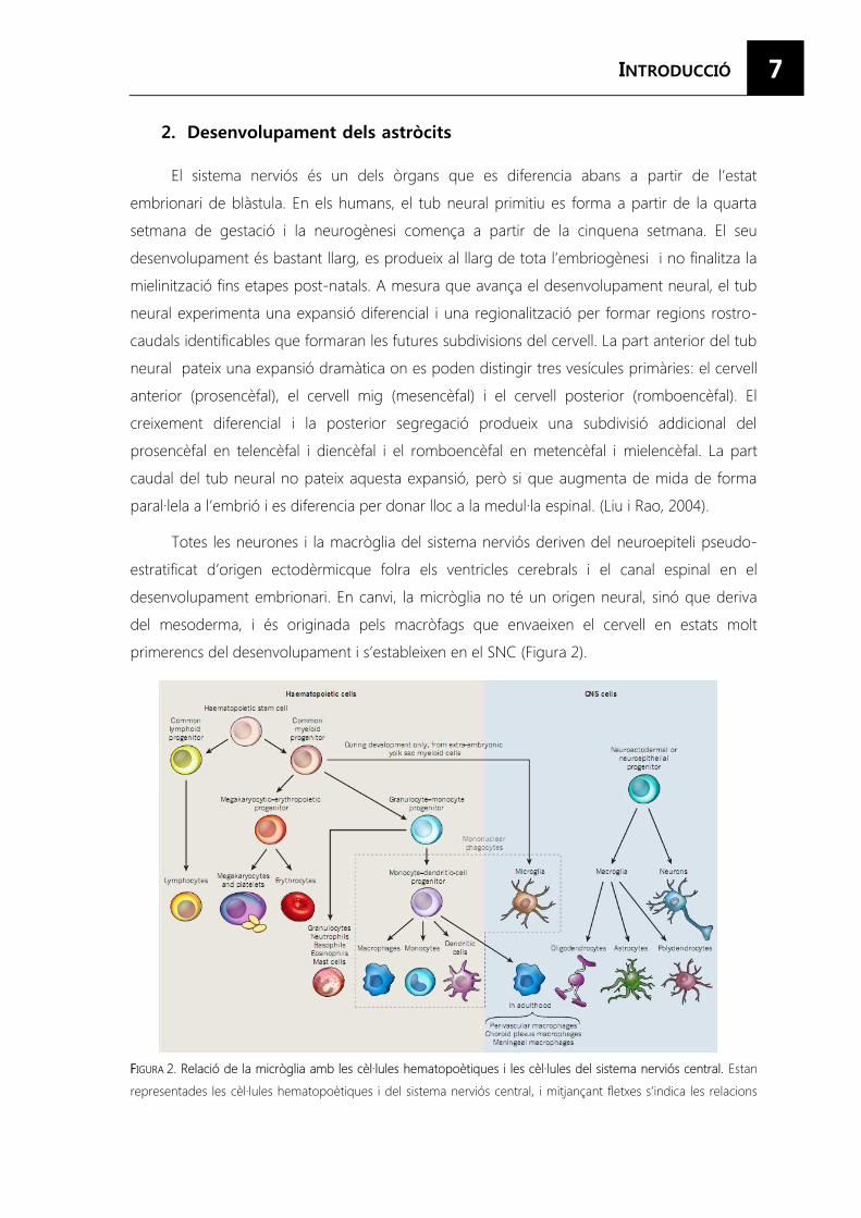
primerencs del desenvolupament i s’estableixen en el SNC (Figura 2).
FFIGURA 2. Relació de la micròglia amb les cèl·lules hematopoètiques i les cèl·lules del sistema nerviós central. Estan
representades les cèl·lules hematopoètiques i del sistema nerviós central, i mitjançant fletxes s’indica les relacions
8
del seu llinatge. La micròglia són les úniques cèl·lules hematopoètiques que es troben al parènquima del SNC.
L’origen dels macròfags perivasculars, els macròfags del plexes coroïdals i els macròfags de les meninges és
desconegut, encara que aquí s’indica que deriven de la cèl·lula progenitora de monòcits i cèl·lules dendrítiques de
forma especulativa (Extret de Ransohoff i Cardona, 2010)
Una característica fonamental del desenvolupament neural en els vertebrats és que els
diferents tipus cel·lulars són generats en un seqüència específica, primer les neurones,
seguides dels astròcits i els oligodendròcits.
Les cèl·lules neuroepitelials inicialment es divideixen d’una manera simètrica, produint
una ràpida expansió dels progenitors. El començament de la neurogènesi està marcat per
l’inici d’una manera asimètrica de divisió, on les cèl·lules neuroepitelials produeixen una altra
cèl·lula neuroepitelial i una neurona o un progenitor intermedi destinat a la neurogènesi. Les
cèl·lules de la glia radial són les cèl·lules progenitores primàries en les etapes embrionàries de
la neurogènesi, i al’igual que les cèl·lules de les que deriven, folren els ventricles i el canal
espinal, mantenint la polaritat apical-basal. Els precursors d’oligodendròcits i les cèl·lules
ependimàries també deriven de la glia radial però els progenitors intermedis involucrats no
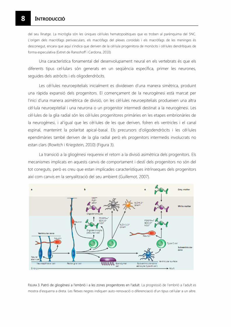
estan clars (Rowitch i Kriegstein, 2010) (Figura 3).
La transició a la gliogènesi requereix el retorn a la divisió asimètrica dels progenitors. Els
mecanismes implicats en aquests canvis de comportament i destí dels progenitors no són del
tot coneguts, però es creu que estan implicades característiques intrínseques dels progenitors
així com canvis en la senyalització del seu ambient (Guillemot, 2007).
FIGURA 3. Patró de gliogènesi a l’embrió i a les zones progenitores en l’adult. La progressió de l’embrió a l’adult es
mostra d’esquerra a dreta. Les fletxes negres indiquen auto-renovació o diferenciació d’un tipus cel·lular a un altre.
9
Estan llistats els marcadors de la macròglia i dels seus precursors. AA) Auto-renovació de la línea de cèl·lules
neuroepitelials del ventricles al llarg dels neuroeixos a les etapes del tancament del tub neural. Aquestes cèl·lules
poden generar alguna neurona. Les cèl·lules neuroepitelials es transformen en cèl·lules de glia radial i comença la
neurogènesi. BB) La glia radialprodueix cèl·lules progenitores intermèdies i cèl·lules precursores d’oligodendròcits
(OPCs) que donen lloc a neurones i oligodendròcits, respectivament. La glia radial també pot donar lloc a astròcits
produint progenitors intermedis que s’expandeixen en nombre abans de produir astròcits. Els astròcits
protoplàsmics i els astròcits fibrosos poden sorgir de progenitors comuns o independents. La glia radial també
produeix cèl·lules ependimàries. CC) En adults, els oligodendròcits provenen de dues vies independents: les cèl·lules
tipus B de la zona subventricular donen lloc a una cèl·lula amplificadora de trànsit, anomenada cèl·lula tipus C, que
pot produir tant OPCs com neurones. Les OPCs generades i les que ja residien a la substància grisa produeixen
oligodendròcits. ALDH1L1, família aldehid deshidrogenasa 1 membre L1; APC, adenomatous polyposis coli; GFAP,
proteïna acídica fibril·lar glial; MBP, proteïna bàsica de mielina; PDGFR-�, receptor alfa del factor de creixement
derivat de plaquetes; PLP, proteïna proteolipídica 1. Totes les cèl·lules verdes són progenitors intermedis, sent les
cèl·lules tipus C un subconjunt d’aquestes, i totes les cèl·lules blaves són cèl·lules progenitores neurals (Extret de
Rowitch i Kriegstein, 2010).
2.1. De la neurogènesi a la gliogènesi
El potencial in vivo de les cèl·lules neuroepitelials i la glia radial està restringit de forma
regional per l’acció de senyals organitzadores com sonic hegdehog (SHH), factors de
creixement de fibroblasts (FGF), WNTs i proteïnes morfogèniques d’ós (BMPs), que
proporcionen informació posicional mitjançant gradients al llarg dels eixos dorsal-ventral,
anterior-posterior i medial-lateral.
La gliogènesi generalment segueix a la neurogènesi en el desenvolupament del SNC i
els mateixos progenitors donen lloc a diferents tipus cel·lulars, de manera que s’ha de produir
un canvi perquè aquests progenitors generin tipus cel·lulars distints.
Els mecanismes que regulen la transició entre la formació de neurones i astròcits són
complexes i no estan molt ben entesos. Treballs recents suggereixen que aquest canvi és
produït tant per factors extrínsecs que promouen l’astrogènesi com intrínsecs que
disminueixen la neurogènesi a la vegada que promouen l’astrogènesi (Freeman, 2010).
• Wnt: És necessari per a l’activació de gens neurogènics (neurogenina 1 i neurogenina
2) que indueixen la diferenciació neuronal i bloquegen l’activació de gens glials.
Malgrat això, els lligands de Wnt es continuen expressant després de la transició
neurona-glia, encara que ja no indueixen l’expressió de neurogenines a les NPCs a
causa de canvis a l’acetilació i la metilació dels seu promotor.
• Canvis a l’acetilació i la metilació dels promotors neurogenina 1 i neurogenina 2. En
etapes inicials, es produeix una elevada acetilació i una baixa metilació que correspon
a una conformació de la cromatina oberta que permet l’associació amb la RNA
polimerasa II, per tant, les neurogenines s’expressen i promouen la neurogènesi
10
inhibint l’astrogènesi. En canvi, en etapes posteriors, es produeix una baixa acetilació i
una elevada metilació que correspon a una conformació de la cromatina tancada, de
manera que no s’expressen neurogenina 1 ni neurogenina 2 i no s’inhibeix
l’astrogènesi (Hirabayashi et al., 2009)
• CCitocines: Com el factor neurotròfic ciliar (CNTF), el factor inhibidor de leucèmia (LIF) i
la cardiotrofina-1 (CT-1) (Miller i Gauthier, 2007) activen la via JAK/STAT que uneix
CBP/p300 i es trasllada al nucli on promou la diferenciació astroglial. Malgrat això,
aquesta activació no és suficient si no està activada la via de senyalització de Notch.
• Notch: Aquesta via s’activa per Jag1 i Dll2 produït per les neurones neurogenina+.
Aquests lligands activen NFIA (factor nuclear augmentat IA), que inhibeix Dnmt1 (DNA
(citosina-5-)metiltransferasa 1) provocant la desmetilació dels llocs d’unió de STAT en
els promotors i l’activació de l’expressió de GFAP i S100�.
• BMPs: Tenen un paper dual ja que tant promouenla neurogènesi com la gliogènesi. La
presència de BMP2 juntament amb citocines gliogèniquespromou la formació d’un
complex SMAD-p300 proteïna d’unió a CRE- STAT que activa l’expressió de gens
astroglials (Nakashima et al., 2001).
Tots aquests factors regulen la capacitat de les NPCs per donar lloc als astròcits, però
no la funcionalitat, el creixement i la diversificació dels astròcits.
2.2. Diversitat astrogènica
Els astròcits són una població morfològicament heterogènia. Els astròcits madurs en els
rosegadors es poden dividir en dos grups segons la seva morfologia i localització: astròcits
fibrosos i protoplàsmics.
Els astròcits protoplasmàtics, que presenten una morfologia estrellada, es troben a la
substància grisa i les seves prolongacions envolten les sinapsis i els vasos sanguinis; en canvi,
els astròcits fibrosos es troben a la substància blanca, on contacten amb els nodes de Ranvier
i els vasos sanguinis. Alguns tipus d’astròcits morfològicament diferents van ser descrits amb
anterioritat i tenen noms especials com la glia de Müller a la retina i la glia de Bergmann al
cerebel. També hi ha petites poblacions d’astròcits especialitzats localitzats en regions
específiques del SNC com són els astròcits velats del cerebel i el bulb olfactori, els astròcits
interlaminarsal còrtex dels grans primats, elstanicits presents en els òrgans periventriculars i la
hipòfisi, els pituícits a la neurohipòfisi i els astròcits perivasculars i marginals que es localitzen
molt a prop de la pia formant la glia limitant que ajuda a l’aïllament del parènquima cerebral
del compartiment vascular i subaracnoïdeu (Verkhartsky i Butt, 2007). Estudis recents
11
fisiològics i d’expressió gènica mostren que els astròcits són una població cel·lular diversa que
mostren diferents característiques en diferents regions cerebrals i en les diferents etapes del
desenvolupament (Figura 4).
FFIGURA 4. Reproducció esquemàtica dels
principals tipus d’astròcits i cèl·lules
ependimàries i la seva localització a les
diferents capes/ regions específiques
del sistema nerviós central. I: tanicits
(a:pial; b:vascular); II: astròcit radial (glia
de Bergmann); III: astròcit marginal; IV:
astròcit protoplasmàtic; V: astròcit velat;
VI: astròcit fibrós; VII: astròcit
perivascular; VIII: astròcit interlaminar;
IX: astròcit immadur/ glioblast; X:
ependimòcit; XI: cèl·lula del plexe
corideu (Extret de Kettenmann and
Ransom, 2005).
2.2.1. Heterogeneïtat regional
Aquesta diversitat pot ser a causa dels diferents orígens d’aquestes cèl·lules.
L’astrogliogènesi s’inicia tard en el desenvolupament embrionari i continua al llarg del període
neonatal i postnatal, de manera que els astròcits de l’escorça provenen de 3 fonts diferents.
En primer lloc, de la glia radial resident a la zona ventricular (VZ) embrionària, que
s’origina a partir de la transformació de les cèl·lules neuroepitelials de la VZ en progenitors
neurals per donar lloc tant a astròcits com a neurones (Kriegstein i Gotz, 2003; Pinto i Gotz,
2007; Rakic, 2003). Després de la migració neuronal al llarg de les seves prolongacions, la glia
radial les retracta i es transforma en astròcits estrellats en el període perinatal.
Una altra font d’astròcits són els progenitors de la zona subventricular (SVZ) en animals
postnatals. Aquests progenitors, que presenten característiques similars a la glia radial, migren
cap a l’escorça on poden esdevenir astròcits o oligodendròcits madurs. Aquests progenitors
es distingeixen per l’expressió del factor de transcripció Dlx-2 i que no expressen Zebrin II, un
marcador de la glia radial. Donen lloc a astròcits de la substància grisa i blanca del telencèfal
dorsal. En aquesta zona hi ha un altre grup de progenitors que expressa NG2, però no els
marcadors dels altres progenitors, ni Dlx-2 ni Zebrin II, que donen lloc a astròcits de l’escorça
dorsal, però no a la substància blanca, encara que majoritàriament generen oligodendròcits.
12 I finalment, un petit grup de progenitors resideix a la zona marginalde l’escorça en
desenvolupament i són diferents dels precursors de la zona ventricular i subventricular
(Hewett, 2009).
Aquests 3 llinatges astrocitaris diferenciats suggereixen que no tots els astròcits s’han
format de la mateixa manera, cosa que permet explicar la diversitat d’astròcits en una mateixa
àrea del cervell (Wang i Bordey, 2008).
2.2.2. Heterogeneïtat fenotípica
Peròaquesta diversitat fenotípica també pot ser causada per factors ambientals.Els
astròcits són cèl·lules molt plàstiques i algunes de les seves característiques fenotípiques les
adquireixen al llarg del desenvolupament normal. Els astròcits estan íntimament relacionats
amb les neurones de manera que l’entorn neuronal defineix el fenotip astrocític per facilitar i
optimitzar el processament de la informació a nivell local. Això afecta als nivells d’expressió de
canals de K+ i Ca2+, receptors de neurotransmissors, transportadors, connexines,… la qual cosa
afecta la comunicació i l’excitabilitat (Matyash i Kettenmann, 2010).
Un dels exemples més clarsés el que es produeix com a resposta a la inflamació. En el
SNC normal, els astròcits són descrits com a cèl·lules en un estat quiescent o en repòs que
participen en la funció cerebral normal; però com a resposta a un dany es tornen reactius i
canvien la seva morfologia, la mida i el secretoma en un procés anomenat astrocitosi anisomòrfica. Alguns autors suggereixen que existeix un fenotip intermedi en el qual els
astròcits estan activats i que s’anomena astrocitosi isomòrfica. Aquestes categories no són
excloents, sinó que existeix una progressió gradual entre elles, la qual cosa dóna lloc a
fenotips intermedis (Sofroniew i Vinters, 2010) (Taula 1).
En l’astrocitosi isomòrfica, també anomenada astrocitosi lleu o activació astrocitària, es
produeix un lleuger augment de l’expressió de GFAP, hi ha una certa proliferació i la secreció
de citocines antiinflamatòries (John et al., 2005; Meeuwsen et al., 2003). De fet, el tractament
amb citocines pro-inflamatòries es creu que provoca l’activació dels astròcits (Herx i Yong,
2001; John et al., 2003).
En canvi, en l’astrocitosi anisomòrfica, també anomenada astrocitosi severa o reactivitat astrocitària, es produeix un gran augment de l’expressió de GFAP, augmenten les taxes de
proliferació i la secreció de citocines pro-inflamatòries com TNF-� i CXCL10 (Daginakatte et al., 2008). Aquest fenotip generalment està associat amb la formació de la cicatriu glial, on els
astròcits delimiten l’àrea danyada ( Sofroniew i Vinters, 2010).
13
TTAULA 1. Fenotips dels astròcits i
marcadors usats per identificar-los.
Els astròcits es troben en un estat
quiescent en el cervell adult normal.
En l’astrocitosi isomòrfica es
produeix un lleuger augment de
les proteïnes del citoesquelet. En
canvi, en l’astrocitosi anisomòrfica,
el fenotip més sever, es produeix
un augment marcat d’aquestes
proteïnes. (Extret de Nash et al., 2010)
Tots aquest fenotips són difícils de reproduir in vitro, ja que els astròcits es troben en un
ambient molt diferent en el que es troben in vivo. Les condicions artificials per al seu
creixement i manteniment els provoquen un major estat d’activació de com es troben en el
teixit d’origen ja que perden la seva conformació en 3D. De fet, ja durant el procés de
dissecció i dissociació enzimàtica del teixit els indueix un estat d’activació. A més a més, el
medi de creixement conté diversos factors, procedents del sèrum, que poden induir certa
diferenciació (Nash et al., 2010).
14
Tradicionalment els astròcits es consideraven simples cèl·lules de suport que
proporcionaven suport estructural i metabòlic a les neurones. Els astròcits són el principal
element del sistema homeostàtic ja que són els responsables del manteniment de la
concentració iònica i de neurotransmissors a l’espai extracel·lular, l’homeòstasi del pH, de
proporcionar suport metabòlic i nutritiu a les neurones, la regulació de la barrera hemato-
encefàlica i la defensa del sistema nerviós central. Recentment s’ha descrit que els astròcits a
més, expressen diferents cascades de senyalització, estan dotats amb rutes de comunicació
intercel·lular, les unions de tipus gap, i tenen la capacitat d’alliberar diferents tipus de
gliotransmissors a través de diferents vies de secreció regulada. Això els permeten realitzar
una gran varietat de funcions complexes i indispensables com la diferenciació neuronal, la
regulació de la guia axonal, la formació de sinapsis, la plasticitat cerebral, l’homeòstasi i la
comunicació (Seth i Koul, 2008) (Taula 2).
FFUUNCIONS DELS ASTRÒCITTS DDesenvolupament Els astròcits coma elements stem del SNC
Guia axonal
Regulació de la sinaptogènesi
EEstructural Els astròcits divideixen la substància grisa en territoris independents i formen
la unitat neuro-vascular
Els astròcits formen anatòmicament sincicis separats i integren altres cèl·lules
neuronals en aquests sincicis
Formen la interfície glial-vascular i regulen la formació de la barrera hemato
encefàlica
MMetabòlica Regulació de la micro circulació cerebral
Subministrar substrats energètics a les neurones a través de la llançadora
glucosa-lactat
HHomeostàtica Regulació de la concentració extracel·lular d’ions, en concret de la captació i
redistribució del K+ seguint les fluctuacions associades amb l’activitat neuronal
Regulació del pH extracel·lular
Homeòstasi dels neurotransmissors, especialment de glutamat
Homeòstasi de l’aigua cerebral
SSenyalització Modulació de la transmissió i la plasticitat sinàptica
Alliberació de neurotransmissors
Senyalització a llarga distància dins del sincici
TAULA 2. Funcions de les cèl·lules astroglials. (Extret de Verkhratsky i Parpura, 2010)
15
3.1. Desenvolupament
3.1.1. Els astròcits com a elements stem
Durant molt de temps es creia que el SNC no tenia activitat neuroregeneradora.
Malgrat això, s’ha establert que en duesàrees concretes del cervell adult es produeix la
neurogènesi: la zona subventricular dels ventricles laterals, inclosa la rostral migratory stream, i
la zona subgranulardel gir dentat de l’hipocamp (Doetsch, 2003; Gritti et al., 2002) on
resideixen les cèl·lules mare neuronals que s’assemblen a la glia radial o als tanicits i que tenen
l’habilitat de dividir-se i generar neurones granulars al gir dentat de l’hipocamp o migrar per
la rostral migratory stream cap al bulb olfactori on formen interneurones. Aquestes cèl·lules
mare expressen tant marcadors típics d’astròcits madurs com GFAP, S100�, GLAST, GLT1 o GS,
encara que no en realitzen les funcions típiques, com marcadors de cèl·lules immadures com
vimentina i nestina (Kriegstein i Alvarez-Buylla, 2009).
3.1.2. Guia axonal
L’aparició dels astròcits en el desenvolupament és posterior a la formació de les
neurones en el SNC. Tot i això, els astròcits realitzen funcions importants durant el
desenvolupament de la substància grisa i blanca. Els límits moleculars formats pels astròcits
participen en la guia de la migració d’axons en desenvolupament i d’alguns neuroblasts.
3.1.3. Regulació de la sinaptogènesi
Els astròcits també són elements clau en la sinaptogènesi, en la maduració de les
sinapsis i en el seu manteniment. En condicions in vitro, l’addició d’astròcits en un cultiu de
neurones augmenta considerablement la formació de sinapsis. Els astròcits produeixen i
secreten colesterol (Nieweg et al., 2009), que és crític per a la formació de sinapsis i secreten
diversos factors que es necessiten per la seva maduració i manteniment. A més sintetitzen i
alliberen thrombospondines 1 i 2 que promouen la sinaptogènesi tant in vitro com in vivo (Christopherson et al., 2005) i que són crítiques per a la plasticitat sinàptica després d’una lesió,
la remodelació i la regeneració. La thrombospondina és suficient per induir sinapsis que tenen
una ultraestructura presinàptica i postsinàptica normal així com el reclutament de proteïnes
presinàptiques i postsinàptiques com la sinapsina i el PSD-95, respectivament. Malgrat això,
aquestes sinapsis són postsinàpticament silencioses perquè no presenten cap sensibilitat al
glutamat. Els astròcits secreten una proteïna, encara no identificada que indueix la resposta al
glutamat (AMPA). També realitzen una funció en l’eliminació de sinapsis durant el
desenvolupament (pruning) ja que alliberen senyals que promouen l’expressió del
16
complement C1q a les sinapsis per marcar-les perquè siguin eliminades per la micròglia
(Barres, 2008).
3.2. Funció estructural
Les cèl·lules glials divideixen el parènquima cerebral en dominis morfològics
independents. A la substància grisa els astròcits parcel·len els teixit segons els seus territoris
anatòmics, les cèl·lules de la micròglia estan entreteixides en el circuit cerebral des dels seus
dominis defensius no cavalcats i els oligodendròcits formen els nodes de Ranvier en els axons
que envolten.
Les cèl·lules glials són elements centrals en la microarquitectura cerebral. Els astròcits
divideixen el teixit en dominis estructurals independents i no cavalcats mitjançant un procés
anomenat tiling (Bushong et al., 2004) (Figura 5). En aquests dominis les membranes dels
astròcits cobreixen les sinapsis i estableixen contactes les membranes neuronals i els vasos
sanguinis.
FFIGURA 5. Microdominis anatòmics formats per les cèl·lules glials. (Extret de Verkhratsky, 2010)
Els dominis astroglials s’integren formant una xarxa cel·lular anomenada sincici
astrocitari mitjançant les unions tipus gap localitzades a les prolongacions perifèriques on els
dominis estan en contacte. Aquestes unions formades per connexines (principalment Cx43 i
Cx30), els proporcionen un sistema de transferència d’informació entre els astròcits ja que
permeten la difusió intercel·lular de molècules com ions, IP3 i substrats metabòlits. El sincici
està format dins de les estructures anatòmiques del cervell i contribueix a la seva organització
jeràrquica (Houades et al., 2008).
17
3.2.1. Formació de la barrera hematoencefàlica.
La barrera hematoencefàlica (BHE) és una barrera de difusió que impedeix l’entrada al
parènquima cerebral de molècules segons la seva polaritat i la seva mida. Els principals
components cel·lulars de la BHE són les cèl·lules endotelials dels capil·lars que formen unions
hermètiques i estan envoltades per la làmina basal, els pericits perivasculars i els peus
astrocitaris. El paper dels pericits en la BHE no està del tot estudiat i el paper dels astròcits es
considerava perquè els astròcits reactius tenen un paper clau en el tancament de la BHE
després d’una lesió (Bush et al., 1999) i perquè es creia que la BHE es formava en edats post
natals, coincidint amb la generació dels astròcits. Estudis recents demostren que la BHE està
totalment formada des que els vasos penetren el parènquima cerebral, aproximadament en
els dies de desenvolupament embrionari 11-12 en ratolí (Saunders et al., 2008). Diferents vies
de senyalització controlen diferents aspectes de la formació de la barrera incloent la
senyalització Wnt, que es troba en les cèl·lules mare neurals i dirigeix l’angiogènesi específica
del SNC, la migració de les cèl·lules endotelials al cervell i l’expressió d’alguns transportadors.
En el desenvolupament post natal, quan les cèl·lules mare ja han desaparegut, aquestes
funcions les adquireixen els astròcits, que s’encarreguen més de manteniment que de la
inducció de la formació de la BHE (Abbot et al., 2006).
3.3. Funció metabòlica
Els microdominis anatòmics formats pels astròcits, els permet format la unitat
neurovascular que integra els circuits neuronals amb la circulació sanguínia cerebral i el suport
metabòlic.
3.3.1. Regulació de la micro circulació cerebral
Els astròcits envolten les sinapsis i per tant, poden ser estimulats per l’activitat neuronal,
a la vegada que els seus peus astrocitaris embolcallen els vasos sanguinis i poden enviar
senyals a les cèl·lules de la musculatura llisa que determinen el diàmetre dels vasos.
L’alliberació de glutamat per part de les neurones activa els receptors metabotròpics de
glutamat (mGluRs) dels astròcits, la qual cosa provoca un increment dels nivells de calci
intracel·lular que activen la fosfolipasa A2. S’inicia així la producció d’àcid araquidònic a partir
dels fosfolípids de la membrana, que a la vegada dóna lloc als vasodilatadors prostaglandina
E (PGE) i àcid epoxieicosatrienoic o vasoconstrictor àcid 20-hidroxi eicosatetraenoic (20-HETE)
(Attwell et al., 2010). Treballs recents mostren que la competició entre vasodilatadors i
vasoconstrictors depèn de la pressió parcial d’oxigen (pO2), i predomina la vasodilatació a pO2
baixes (Kleinfeld et al., 2011).
18
3.3.2. Suport metabòlic a les neurones
La glucosa del flux sanguini, és el principal substrat per al cervell adult, i és captada
principalment pels astròcits a través del transportador de glucosa GLUT1 i també per les
neurones que expressen el GLUT3.
Tal i com està formulat pel model de la llançadora de lactat astròcit-neurona (ANLS) els
astròcits responen a l’activació glutamatèrgica augmentant la seva taxa d’ús de la glucosa i
alliberant lactat a l’espai extracel·lular que pot ser usat per les neurones per cobrir les seves
necessitats energètiques. Aquest mecanisme molecular implica el funcionament seqüencial
dels transportadors de glutamat específics dels astròcits i l’ATPasa Na+/ K+, l’activació de la
glicòlisi en els astròcits i l’intercanvi mitjançant el transportador monocarboxilat del lactat a les
neurones (Magistretti, 2006) (Figura 6).
A les sinapsis glutamatèrgiques, el glutamat pot ser recaptat pels astròcits a través del
transportador de glutamat 1 (GLT1) o el de glutamat/aspartat (GLAST) que cotransporten
glutamat i sodi; de manera que la concentració intracel·lular de sodi augmenta i activa
l’ATPasa Na+/K+�2 específica de cèl·lules glials. L’ATP consumit per l’ATPasa i la conversió del
glutamat a glutamina a través de la glutamina sintasa poden activar la glicòlisi i la formació de
lactat. Aquest lactat s’allibera pels astròcits a través d’un transportador mono carboxilat
específic MCT1 i les neurones el capten del medi extracel·lular a través del seu transportador
específic MCT2 on es convertirà en piruvat que després entrarà en el cicle dels àcids
tricarboxílics.
L’ús del lactat per les neurones pot tenir diferents propòsits, com l’augment del
potencial redox en augmentar l’NADH i la generació d’ATP que pot ser usat per sintetitzar
glutamat. La glutamina també pot ser captada per la neurona a través dels seus receptors
específics i convertida en glutamat per la glutaminasa abans de ser acumulada a les vesícules
sinàptiques.
FFIGURA 6. Els astròcits proporcionen suport
metabòlic a les neurones. La neurona pot captar
directament la glucosa, però un augment de
l’activitat d’una neurona glutamatèrgica provoca
una major captació de glutamat de l’astròcit que
envolta la sinapsi. L’energia necessària per la
captació del glutamat prové del metabolisme de
la glucosa en metabolitzar-se a lactat, que
posteriorment és entregat a les neurones
(llançadora glucosa- lactat) (Extret de Giaume et
al., 2010).
19
3.4. Funció homeostàtica
Els astròcits són les principals cèl·lules encarregades de la regulació homeostàtica de
l’espai extracel·lular. Diversos sistemes de transports’expressen a les membranes glials que
permeten el control de les concentracions d’ions, neurotransmissors, neuromoduladors,
metabòlits i altres molècules actives a l’escletxa sinàptica necessàries per a la correcta
transmissió sinàptica.
3.4.1. Homeòstasi d’ions
Està acceptat que els astròcits controlen els nivells extracel·lulars de K+ i d’altres ions
encara que no es coneix exactament a través de quin mecanisme. Es creu que estan implicats
canals rectificadors d’entrada (en anglès, inward rectifier channels) i per difusió des de les
àrees amb una concentració elevada cap a altres regions amb concentracions baixes a nivell
d’una única cèl·lula o dins del sincici (Kofuji i Newman, 2004).
3.4.2. Homeòstasi de l’aigua
El transport d’ions, de K+ o Na+, requereix el moviment d’aigua. L’aigua entra i surt dels
astròcits a través de les aquaporines, principalment aquaporina 4 (AQP4), que estan
concentrades en les prolongacions perisinàptiques i perivasculars (Haneke et al., 2010).
3.4.3. Homeòstasi del pH
Les membranes dels astròcits tenen diferents tipus de mecanismes per treure H+,
incloent l’intercanviador de Na+/H+, transportadors de bicarbonat, transportadors mono-
carboxílics i l’ATPasa de protons de tipus vacuolar.
3.4.4. Homeòstasi de neurotransmissors
Els astròcits són responsables del recanvi de diversos neurotransmissors. Expressen
transportadors pel glutamat, el GABA i la glicina que serveixen per retirar-los de l’espai
sinàptic. Els astròcits són especialment importants a les sinapsis glutamatèrgiques perquè
proporcionen una font de glutamat, el retiren de l’escletxa sinàptica i mantenen la llançadora
glutamat-glutamina. Els astròcits expressen de forma específica els transportadors de
glutamat GLAST (EAAT-1) i GLT1 (EAAT-2), que co-transporten el glutamat juntament amb
Na+, la qual cosa produeix un augment dels nivells de Na+ intracel·lulars que són compensats
per la sortida de Na+ mitjançant l’intercanviador Na+/Ca2+, que funciona de forma inversa. El
glutamat dins de l’astròcit és convertit en glutamina per l’enzim glutamina sintetasa específica
dels astròcits i transportat a les neurones mitjançant la llançadora glutamat-glutamina per
20
proporcionar una font de glutamat que pot ser acumulat de nou dins de les vesícules
sinàptiques (Sofroniew i Vinters, 2010).
3.4.5. Funció antioxidant
L’estrès oxidatiu és una de les causes de degeneració neuronal. Els antioxidants de baix
pes molecular com l’àcid ascòrbic, el glutatió i l’�-tocoferol, juntament amb els enzims
antioxidants, formen les defenses cel·lulars contra l’estrès oxidatiu. Els astròcits presenten
elevades quantitats d’aquests antioxidants, així com també de superòxid dismutasa (SOD),
catalasa, glutatió reductasa i glutatió peroxidasa. Aquesta elevada concentració d’enzims
antioxidants pot protegir a les neurones del seu voltant en condicions d’estrès oxidatiu.
Els astròcits també poden produir de forma induïble l’hemo-oxigenasa que actua en el
metabolisme del grup hemo i és important en la prevenció de l’ús del ferro per a la producció
d’espècies d’oxigen reactives (Kimelberg, 2010). A més, els astròcits participen en la captació
d’alguns metalls pesats com el plom, ja que expressen proteïnes d’unió a metalls com les
metal·lotionines que els doten amb propietats neuroprotectores i neuroregeneratives després
del dany o exposició a metalls pesants (Wang i Bordey, 2010).
3.5. Funció de senyalització
Durant dècades, els astròcits van ser considerats participants passius en les sinapsis.
Actualment, diferents estudis suggereixen l’existència d’una comunicació bidireccional i
dinàmica entre les neurones i les cèl·lules de glia. La majoria de les sinapsis del SNC estan
formades per tres elements: el terminal neuronal presinàptic, la membrana neuronal
postsinàptica i la prolongació de l’astròcit, una estructura que rep el nom de sinapsi tripartida
(Araque et al., 1999) (Figura 7).
FFIGURA 7. Sinapsi tripartida. Les prolongacions dels astròcits estan íntimament associades amb les sinapsis. AA)
Microfotografia electrònica que mostra una sinapsi tripartita a l’hipocamp. La prolongació de l’astròcit (blau)
envolta l’àrea perisinàptica. L’axó de la neurona està marcat de color verd, l’espina dendrítica de color groc i la
21
densitat post-sinàptica de color vermell i negre. BB) Representació esquemàtica de la sinapsi tripartita (Extret de
Eroglu i Barres, 2010).
L’astròcit té un paper dual en aquesta sinapsi tripartida. Primer, perquè els astròcits
tenen l’avantatge de que expressen receptors per neurotransmissors a la seva membrana
(potencialment poden expressar els receptors per tots el neurotransmissors, malgrat això
aquesta expressió està altament controlada in vivo i astròcits de diferents àrees del cervell
estan dotats amb diferents tipus de receptors) i poden sentir l’alliberació de neurotransmissors
en el terminal neuronal (Fellin et al., 2004; Latour et al., 2001; Pasti et al., 1997); i en segon lloc,
modular la transmissió sinàptica (Fellin et al., 2004; Henneberg et al., 2010; Panatier et al.,
2006; Perea i Araque, 2005) alliberant gliotransmissors (Halassa i Haydon, 2010; Volterra i
Meldonesi, 2005).
3.5.1. Senyalització dins del sincici astrocitari: fisiologia astrocitària
Els astròcits són cèl·lules excitables, encara que la seva excitabilitat no està basada en
canvis de voltatge de la seva membrana sinó en oscil·lacions en la concentració intracel·lular
de calci.
Hi ha dos patrons diferencials d’oscil·lacions de calci: en situacions basals, quan no hi ha
cap estímul, es produeixen oscil·lacions de calci espontànies que generalment estan
restringides a un únic astròcit i passen independentment de l’activitat neuronal (Aguado et al.,
2002). En canvi, com a resposta a un estímul, els astròcits presenten un oscil·lació de calci
evocada que implica vàries cèl·lules glials i és generada pel glutamat alliberat a la sinapsi per
les neurones que activa els receptors de glutamat metabotròpics (mGluR1 i mGluR5) i
transportadors presents a la membrana cel·lular dels astròcits (Fellin, 2009).
Per generar aquestes oscil·lacions les cèl·lules de glia usen les reserves intracel·lulars de
calci, el reticle endoplasmàtic (RE) i la mitocòndria (Figura 8). Aquests orgànuls generen i
mantenen les concentracions intracel·lulars i permeten la propagació de les ones de calci
intercel·lulars.
El RE és la principal font de calci intracel·lular ja que pot acumular grans quantitats (400-
800 �mol/L) per acció de les ATPases de calci del reticle sarco(endo)plàsmic, SERCAs. El calci
pot sortir del reticle a través dels receptors d’IP3 (IP3R) i per l’activació dels receptors de
rianodina/cafeïna (RyR). Després de l’activació dels receptors metabotròpics, la fosfolipasa C
hidrolitza el lípid de membrana fosfatidilinositol 4,5-difosfat i genera diacilglicerol (DAG) i IP3,
que activa el receptor d’IP3 i produeix la sortida de calci del reticle. L’obertura dels receptors
de rianodina està regulada pels ions de calci citoplasmàtics, de manera que variacions dels
nivells de calci indueixen la sortida de calci del reticle, procés que es coneix com alliberació de
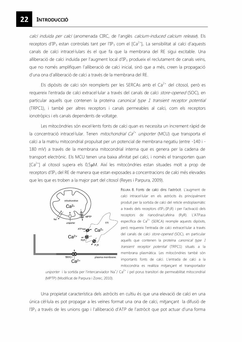
22
calci induïda per calci (anomenada CIRC, de l’anglès calcium-induced calcium release). Els
receptors d’IP3 estan controlats tant per l’IP3 com el [Ca2+]i. La sensibilitat al calci d’aquests
canals de calci intracel·lulars és el que fa que la membrana del RE sigui excitable. Una
alliberació de calci induïda per l’augment local d’IP3 produeix el reclutament de canals veïns,
que no només amplifiquen l’alliberació de calci inicial, sinó que a més, creen la propagació
d’una ona d’alliberació de calci a través de la membrana del RE.
Els dipòsits de calci són reomplerts per les SERCAs amb el Ca2+ del citosol, però es
requereix l’entrada de calci extracel·lular a través del canals de calci store-opened (SOC), en
particular aquells que contenen la proteïna canonical type 1 transient receptor potential (TRPC1), i també per altres receptors i canals permeables al calci, com els receptors
ionotròpics i els canals dependents de voltatge.
Les mitocòndries són excel·lents fonts de calci quan es necessita un increment ràpid de
la concentració intracel·lular. Tenen mitochondrial Ca2+ uniporter (MCU) que transporta el
calci a la matriu mitocondrial propulsat per un potencial de membrana negatiu (entre -140 i -
180 mV) a través de la membrana mitocondrial interna que es genera per la cadena de
transport electrònic. Els MCU tenen una baixa afinitat pel calci, i només el transporten quan
[Ca2+] al citosol supera els 0,5�M. Així les mitocòndries estan situades molt a prop de
receptors d’IP3 del RE de manera que estan exposades a concentracions de calci més elevades
que les que es troben a la major part del citosol (Reyes i Parpura, 2009).
FFIGURA 8. Fonts de calci dins l’astròcit. L’augment de
calci intracel·lular en els astròcits és principalment
produït per la sortida de calci del reticle endoplasmàtic
a través dels receptors d’IP3 (IP3R) i per l’activació dels
receptors de rianodina/cafeïna (RyR). L’ATPasa
específica de Ca2+ (SERCA) reomple aquests dipòsits,
però requereix l’entrada de calci extracel·lular a través
del canals de calci store-opened (SOC), en particular
aquells que contenen la proteïna canonical type 1 transient receptor potential (TRPC1) situats a la
membrana plasmàtica. Les mitocòndries també són
importants fonts de calci. L’entrada de calci a la
mitocondria es realitza mitjançant el transportador
uniporter i la sortida per l’intercanviador Na+/ Ca2+ i pel porus transitori de permeabilitat mitocondrial
(MPTP) (Modificat de Parpura i Zorec, 2010).
Una propietat característica dels astròcits en cultiu és que una elevació de calci en una
única cèl·lula es pot propagar a les veïnes format una ona de calci, mitjançant la difusió de
l’IP3 a través de les unions gap i l’alliberació d’ATP de l’astròcit que pot actuar d’una forma
23
paracrina en els astròcits veïns. Sorprenentment, aquestes ones de calci generalment no es
propaguen a altres astròcits in vivo, la qual cosa prova que els astròcits poden respondre com
a cèl·lules individuals.
3.5.2. Alliberació de neurotransmissors
La senyalització per calci té un paper fonamental en la comunicació bidireccional glia-
neurona perquè és produïda pels neurotransmissors alliberats per l’activitat sinàptica i a la
vegada provoca l’alliberació de gliotransmissors per part dels astròcitscom el glutamat, la D-
serina, el GABA, el TNF-�, prostaglandines, el ANP, el BDNF, etc., que poden modular
l’activitat neuronal i la fisiologia sinàptica.
3.5.3. Modulació de la transmissió i la plasticitat sinàptica
Els astròcits són els elements més dinàmics de la sinapsi tripartida que poden mostrar
modificacions estructurals i funcionals al llarg del desenvolupament, com a resposta a un dany
neuronal i en diverses condicions fisiològiques. Els canvis estructurals poden tenir un gran
impacte en la funció sinàptica perquè poden influir en la recaptació de neurotransmissors de
l’escletxa sinàptica i l’activació de receptors extra-sinàptics. Per exemple, les espines
dendrítiques que tenen contactes amb prolongacions dels astròcits sobreviuen més i són
morfològicament més madures que les que no els tenen (Carmona et al., 2009).
L’habilitat dels astròcits per modular l’excitabilitat neuronal i la transmissió sinàptica va
ser inicialment descrita en cultius cel·lulars i posteriorment el seu estudi ha estat ampliat en
talls cerebrals aguts. Els astròcits modulen la fisiologia neuronal alliberant diferents
neurotransmissors com per exemple el glutamat que provoca corrents lents d’entrada
(anomenades SICs, de l’anglès slow inward currents) per l’estimulació dels receptors d’NMDA
post-sinàptics, augmenta la fortalesa sinàptica activant els receptors metabotròpics que
incrementen la freqüència de les corrents excitadores post-sinàptiques (EPSCs), l’ATP estimula
els receptors P2Y1 i la D-serina contribueix a l’activació dels receptors NMDA. Tots aquests
mecanismes augmenten l’excitabilitat neuronal tot augmentant la probabilitat d’alliberació de
neurotransmissors en les sinapsis (Perea i Araque, 2010).
La citocina TNF-α, alliberada tant pels astròcits com per la micròglia, promou la inserció
dels receptors AMPA a la membrana de les neurones post-sinàptiques, cosa que augmenta
l’eficàcia de la neurotransmissió (Beattie et al., 2002; Stellwagen i Mallenka, 2006).
3.6. Resposta glial al dany
El comportament de les cèl·lules glials canvia després de produir-se un dany. La
micròglia és el primer tipus cel·lular en activar-se, migra ràpidament cap al lloc on s’ha produït
24
la lesió, on inicia encara més la reacció glial i es comunica amb el sistema immune. Les
cèl·lules NG2+ també reaccionen en etapes inicials augmentant la seva proliferació, mentre
que els astròcits són les últimes cèl·lules en activar-se i pateixen canvis morfològics,
d’expressió gènica i de funcionalitat, en un procés que rep el nom d’astrocitosi.
L’astrocitosi es produeix com a resposta a qualsevol dany neuronal, sigui
neurodegeneratiu, inflamatori, traumàtic o per un infart cerebral. En aquests casos, els
astròcits esdevenen hipertròfics i augmenten l’expressió dels filaments intermedis GFAP i
vimentina, però també expressen nestina i la proteïna d’unió a lípids del cervell (BLBP) (Figura
9). A més, en danys més greus com un trauma sever, l’hipòxia o l’infart cerebral, una part dels
astròcits reactius també proliferen i augmenta el nombre d’astròcits que envolten la lesió.
3.6.1. Canvis morfològics i d’expressió de l’astrocitosi
Un dels models més usats per estudiar l’astrocitosi és la lesió amb una fulla de bisturí
(stab wound injury) a l’escorça dels ratolins realitzada de forma paral·lela a la línia mitja ja que
és un mètode repetitiu i proporciona suficient material pel posterior estudi (Hampton et al.,
2004).
FFIGURA 9. Esquema dels canvis
morfològics i d’expressió de l’astrocitosi
al llarg del temps. Els astròcits
comencen a ser hipertròfics a partir del
tercer dia després de la lesió i
augmenten l’expressió de GFAP i
nestina, així com del proteoglicà DSD1 i
de tenascina C, reflectint la
desdiferenciació parcial dels astròcits
reactius. Una setmana després, els
astròcits que envolten la lesió són
clarament hipertròfics, allargats i
expressen de forma molt abundant
BLBP. Això es correspon amb el pic de
proliferació dels astròcits caracteritzat per l’expressió de Ki67 i la incorporació de bromodesoxiuridina (BrdU).
D’acord amb l’anterior, el nombre total d’astròcits (marcat pels nivells d’S100�) augmenta amb el temps. Quan la
proliferació es para, les proteïnes específiques de la glia immadura (DSD1, TNC, nestina i vimentina) disminueixen
però es manté incrementat el nombre total de cèl·lules GFAP+ (Extret de Robel et al., 2011).
La funció dels astròcits reactius és limitar el dany tissular mitjançant diferents
mecanismes: recaptant el glutamat potencialment excitotòxic, protegint de l’estrès oxidatiu
per la via del glutatió, alliberant adenosina, protegint de la toxicitat per NH4+, degradant
pèptids ��amiloides, encapsulant infeccions i ajudant a segellar la BHE lesionada, reduint
25
l’edema provocat per traumes, infarts o malalties obstructives i estabilitzant la concentració
extracel·lular d’ions. Tot i així, pot ser perjudicial en altres casos. De fet, alguns tipus de lesions
poden provocar que els sistemes homeostàtics funcionin d’una manera que exacerben el
dany. Un estrés sever en els astròcits pot provocar una pèrdua de l’homeòstasi iònica que
condueix a una alliberació massiva de glutamat, una pèrdua d’ions K+, alliberació de NO i
espècies d’oxigen reactives, i d’altres agents que promouen la neurotoxicitat (Heneka et al.,
2010).
La cicatriu glial que inicialment és considerada beneficiosa perquè limita la zona
lesionada amb una barrera física, després és considerada com un aspecte negatiu de
l’astrocitosi perquè inhibeix la regeneració dels axons per la secreció de proteoglicans
condroïtin sulfat (CSPGs) (Mingorance et al., 2006) i altres molècules que n’inhibeixen el
creixement (Silver i Miller, 2004).
3.6.2. Factors d’activació i vies implicades
L’astròglia és activada per diferents canvis que es produeixen en el parènquima cerebral
després de la lesió. Aquests canvis inclouen la producció d’una gran varietat de molècules de
senyalització, algunes derivades de l’extravasació de plasma, capaços d’iniciar la reacció
inflamatòria o modular-la al llarg del temps (Figura 10).
Es consideren factors que inicien l’activació glial les purines i pirimidines alliberades per
la mort cel·lular i la transmissió excitotòxica, que activen tant els receptors ionotròpics P2X com
els metabotròpics P2Y, i les citocines pro inflamatòries alliberades inicialment per les cèl·lules
de la micròglia i posteriorment també pels astròcits, com el TNF-�, la IL-1� i l’IFN-�. Aquests a
la vegada indueixen la formació de mediadors secundaris que contribueixen a mantenir la
resposta astrocítica al llarg del temps i es detecten en patologies cròniques,com metabòlits de
l’àcid araquidònic, òxid nítric i enzims incloent metal·loproteïnes de la matriu (MMPs). Tot i així,
altres citocines secretades tant per la micròglia com l’astròglia, atenuen l’astrocitosi com la IL-
10, l’IFN-� i l’Epo.
Després de la lesió els astròcits també sobre expressen els factors de creixement com el
factor de creixement nerviós (NGF), el factor neurotròfic derivat dels cervell (BDNF), la
neurotrofina 3 (NT3), el factor neurotròfic ciliar (CNTF), el factor de creixement endotelial
vascular (VEGF), el factor de creixement epidèrmic (EGF), el factor de creixement de fibroblasts
bàsic (bFGF), el factor de creixement insulínic tipus 1 (IGF-1), el factor de creixement derivat
d’una línia de cèl·lules glials (GDNF), per mantenir el suport tròfic a les neurones que encara
estan intactes.
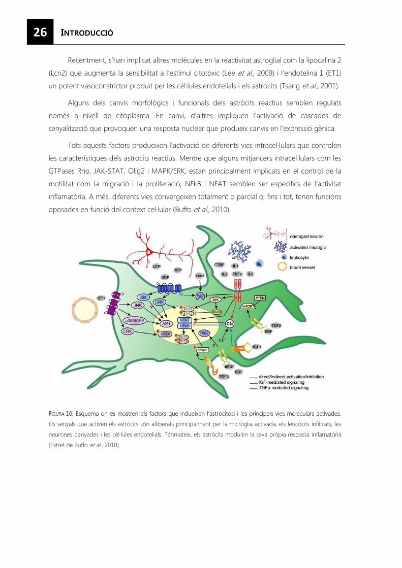
26
Recentment, s’han implicat altres molècules en la reactivitat astroglial com la lipocalina 2
(Lcn2) que augmenta la sensibilitat a l’estímul citotòxic (Lee et al., 2009) i l’endotelina 1 (ET1)
un potent vasoconstrictor produït per les cèl·lules endotelials i els astròcits (Tsang et al., 2001).
Alguns dels canvis morfològics i funcionals dels astròcits reactius semblen regulats
només a nivell de citoplasma. En canvi, d’altres impliquen l’activació de cascades de
senyalització que provoquen una resposta nuclear que produeix canvis en l’expressió gènica.
Tots aquests factors produeixen l’activació de diferents vies intracel·lulars que controlen
les característiques dels astròcits reactius. Mentre que alguns mitjancers intracel·lulars com les
GTPases Rho, JAK-STAT, Olig2 i MAPK/ERK, estan principalment implicats en el control de la
motilitat com la migració i la proliferació, NFkB i NFAT semblen ser específics de l’activitat
inflamatòria. A més, diferents vies convergeixen totalment o parcial o, fins i tot, tenen funcions
oposades en funció del context cel·lular (Buffo et al., 2010).
FFIGURA 10. Esquema on es mostren els factors que indueixen l’astrocitosi i les principals vies moleculars activades.
Els senyals que activen els astròcits són alliberats principalment per la micròglia activada, els leucòcits infiltrats, les
neurones danyades i les cèl·lules endotelials. Tanmateix, els astròcits modulen la seva pròpia resposta inflamatòria
(Extret de Buffo et al., 2010).
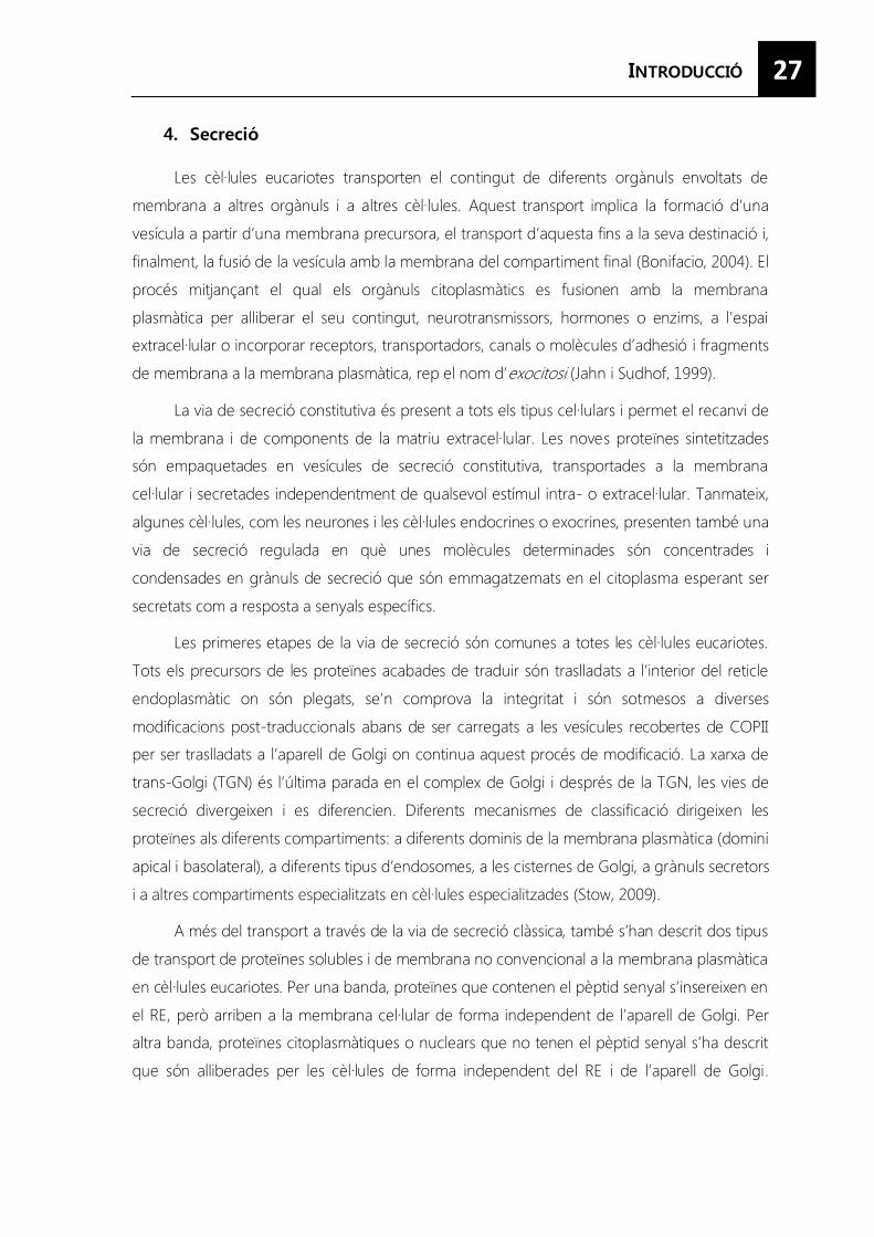
27
Les cèl·lules eucariotes transporten el contingut de diferents orgànuls envoltats de
membrana a altres orgànuls i a altres cèl·lules. Aquest transport implica la formació d’una
vesícula a partir d’una membrana precursora, el transport d’aquesta fins a la seva destinació i,
finalment, la fusió de la vesícula amb la membrana del compartiment final (Bonifacio, 2004). El
procés mitjançant el qual els orgànuls citoplasmàtics es fusionen amb la membrana
plasmàtica per alliberar el seu contingut, neurotransmissors, hormones o enzims, a l’espai
extracel·lular o incorporar receptors, transportadors, canals o molècules d’adhesió i fragments
de membrana a la membrana plasmàtica, rep el nom d’exocitosi (Jahn i Sudhof, 1999).
La via de secreció constitutiva és present a tots els tipus cel·lulars i permet el recanvi de
la membrana i de components de la matriu extracel·lular. Les noves proteïnes sintetitzades
són empaquetades en vesícules de secreció constitutiva, transportades a la membrana
cel·lular i secretades independentment de qualsevol estímul intra- o extracel·lular. Tanmateix,
algunes cèl·lules, com les neurones i les cèl·lules endocrines o exocrines, presenten també una
via de secreció regulada en què unes molècules determinades són concentrades i
condensades en grànuls de secreció que són emmagatzemats en el citoplasma esperant ser
secretats com a resposta a senyals específics.
Les primeres etapes de la via de secreció són comunes a totes les cèl·lules eucariotes.
Tots els precursors de les proteïnes acabades de traduir són traslladats a l’interior del reticle
endoplasmàtic on són plegats, se’n comprova la integritat i són sotmesos a diverses
modificacions post-traduccionals abans de ser carregats a les vesícules recobertes de COPII
per ser traslladats a l’aparell de Golgi on continua aquest procés de modificació. La xarxa de
trans-Golgi (TGN) és l’última parada en el complex de Golgi i després de la TGN, les vies de
secreció divergeixen i es diferencien. Diferents mecanismes de classificació dirigeixen les
proteïnes als diferents compartiments: a diferents dominis de la membrana plasmàtica (domini
apical i basolateral), a diferents tipus d’endosomes, a les cisternes de Golgi, a grànuls secretors
i a altres compartiments especialitzats en cèl·lules especialitzades (Stow, 2009).
A més del transport a través de la via de secreció clàssica, també s’han descrit dos tipus
de transport de proteïnes solubles i de membrana no convencional a la membrana plasmàtica
en cèl·lules eucariotes. Per una banda, proteïnes que contenen el pèptid senyal s’insereixen en
el RE, però arriben a la membrana cel·lular de forma independent de l’aparell de Golgi. Per
altra banda, proteïnes citoplasmàtiques o nuclears que no tenen el pèptid senyal s’ha descrit
que són alliberades per les cèl·lules de forma independent del RE i de l’aparell de Golgi.
28
Malgrat això, aquests dos processos estan restringits a estadis específics del desenvolupament
i es desconeix la via de senyalització que els activa (Nickel i Rabouille, 2010).
4.1. Tipus de vesícules de secreció
Per tant, generalment, la secreció en cèl·lules animals es realitza mitjançant tres tipus de
vesícules de secreció: la vesícula de secreció constitutiva, que és comuna a tots els tipus
cel·lulars; els grànuls de secreció, que només es troben en cèl·lules secretores especialitzades i
alliberen el contingut emmagatzemat depenent d’un estímul extern; i les vesícules sinàptiques
de les neurones i les microvesícules similars a les sinàptiques (SLMVs) en les cèl·lules
neuroendocrines. També trobem un subtipus especial de grànuls de secreció, els orgànuls
relacionats amb els lisosomes (LRO) també anomenats lisosomes secretors en alguns tipus
cel·lulars (Figura 11).
FFIGURA 11. Vies de secreció en les cèl·lules
eucariotes. Aquest esquema mostra les vies de
secreció cel·lular incloent la via de secreció
constitutiva (AA) i la via de secreció regulada (BB-D). BB)
Biogènesi de grànuls de secreció en les cèl·lules
endocrines en la que es forma un grànul immadur
(IG) a partir del TGN, que madura i dóna lloc al
grànul de secreció (SG). CC) Biogènesi de les
vesícules sinàptiques (SV) que implica la secreció
constitutiva seguida pel transport des de la
membrana plasmàtica a les SV o als endosomes
primerencs (EE) com a intermediaris. DD) Biogènesi
dels orgànuls relacionats amb els lisosomes (LRO) que implica la secreció constitutiva seguida de la internalització
als EE o als cossos multivesiculars (MVBs) i l’entrega als orgànuls relacionats amb els lisosomes (LROs). La fusió de
SG, SV i LRO amb la membrana plasmàtica (fletxes vermelles) és dependent d’estímul (Extret de Benado et al., 2009).
4.1.1. Vesícules constitutives
Són petites vesícules secretores formades a la TGN que són contínuament
transportades i fusionades amb la membrana plasmàtica. Aquestes vesícules poden ser
transportades directament des de la TGN o passar a través de compartiments endosomals,
com els endosomes primerencs o tardans i compartiments de reciclatge, abans d’arribar a la
membrana plasmàtica. Com que les proteïnes que contenen aquestes vesícules són
secretades contínuament, la secreció constitutiva és principalment regulada per la taxa
biosintètica de les proteïnes sintetitzades al reticle endoplasmàtic (Ponnambalam i Baldwin,
2003)
29
4.1.2. Grànuls de secreció
La formació dels grànuls de secreció comença a la TGN amb la formació d’un grànul
immadur (IG) que madura i genera el grànul de secreció. Aquest procés de maduració
generalment consisteix en la fusió amb altres IG i l’eliminació de les proteïnes mal classificades.
S’han proposat dos models que no són mútuament exclusius i, que expliquen la
classificació de les proteïnes dins dels grànuls de secreció. En el model de “classificació per
entrada”, la classificació de proteïnes es produeix a la TGN i només les proteïnes
seleccionades entraran dins dels IG. En canvi, en el model de “classificació per retenció”, totes
les proteïnes que es sintetitzen a la TGN entraran als IG, però només aquelles proteïnes
destinades a la secreció regulada quedaran retingudes al llarg de la maduració del grànul, i la
resta seran eliminades per gemmació (Park i Loh, 2008). Aquests dos models s’explicaran amb
més detall en apartats posteriors.
4.1.3. Vesícules sinàptiques o SLMVs
La formació de les vesícules sinàptiques està altament relacionada amb la via endocítica.
El transport de les proteïnes de membrana de les SVs i les SLMVs es pot produir a través de la
via de secreció constitutiva des de la TGN a la membrana plasmàticai d’aquí directament a les
SVs/SLMVs, o mitjançant un intermediari endosomal. La maduració de les SVs és un procés
dinàmic que implica diferents etapes d’exo/endocitosi per remodelar la membrana de la
vesícula. Durant l’endocitosi i la migració cap als llocs d’alliberació les vesícules són
reomplertes amb neurotransmissors.
En un terminal pre-sinàptic madur hi ha tres grups de vesícules: el grup preparat per ser
ràpidament alliberat (RRP), un grup de reciclatge i un grup de reserva. Les vesícules RRP són
alliberades ràpidament quan es produeix l’estimulació, mentre que les vesícules en reciclatge
són alliberades amb una estimulació moderada i el grup de reserva actua com a magatzem i
només és alliberat si es produeix una estimulació molt intensa (Bonanomi et al., 2006).
4.1.4. Lisosomes secretors o LROs
Algunes cèl·lules han modificat el compartiment lisosomal perquè també funcioni com
un compartiment secretor, de manera que també s’usa per emmagatzemar proteïnes
secretores recent sintetitzades. Aquests orgànuls reben el nom de lisosomes secretors o
orgànuls relacionats amb els lisosomes. Els lisosomes secretors comparteixen característiques
amb els lisosomes convencionals, com per exemple, el seu contingut àcid i la presència de
proteïnes de degradació, però es distingeixen per la seva capacitat de ser secretats de forma
regulada.
30
Només unes poques cèl·lules contenen aquests lisosomes secretors i la majoria d’elles
deriven del llinatge hematopoètic com els mastòcits, les cèl·lules T citotòxiques, els neutròfils,
els osteoclasts, les cèl·lules presentadores d’antígens, les plaquetes i també els melanòcits. Els
lisosomes secretors madurs, de la mateixa manera que els lisosomes convencionals reben
transport biosintètic i degradatiu. Això inclou proteïnes que són necessàries per al correcte
funcionament del’orgànul (Blott i Griffiths, 2002).
4.2. Principis del transport vesicular
El transport vesicular és una activitat cel·lular fonamental ja que és responsabledel tràfic
molecular entre els diferents compartiments envoltats de membrana. Per tant, l’especificitat
d’aquest tràfic és clau per al correcte funcionament de la cèl·lula. Aquesta especificitat es basa
en l’empaquetament selectiu de la càrrega seleccionada en vesícules que reconeixen i es
fusionen només amb la membrana diana adequada.
Els mecanismes moleculars implicats en la regulació de la secreció són similars en els
diferents tipus cel·lulars i hi estan implicades diferents famílies de proteïnes.
4.2.1. Molècules de recobriment
Les proteïnes de recobriment citosòlic s’agrupen per induir la formació de les vesícules
de transport i concentrar les proteïnes de càrrega en aquestes vesícules. Aquesta concentració
es produeix per la interacció de les proteïnes de recobriment amb senyals específiques
presents a la cua citoplasmàtica de les proteïnes de càrrega.
Les molècules de recobriment poden classificar-se en tres famílies principals: el complex
COPI, responsable de la formació de les vesícules per el transport retrògrad; el complex COPII,
que mitjança el transport anterògrad; i el recobriment de clatrina, que té un paper principal
en la via endocítica (Dancourt i Barlowe, 2010).
4.2.2. Proteïnes adaptadores
Diferents complexes de proteïnes adaptadores (AP), que es diferencien segons el
reconeixement de la seva càrrega i en la seva localització subcel·lular, s’usen per al transport
vesicular i aporten especificitat en el seu contingut i destí.
La formació de les vesícules recobertes està mitjançada per proteïnes petites d’unió a
GTP. L’agrupament de les proteïnes de recobriment COPI i COPII és produït per les petites
GTPases ARF1 i Sar1 respectivament. L’assemblatge de les cobertes de clatrina és mitjançat
per una família de proteïnes classificadores associades a la clatrina (CLASPs).
31
A més, hi ha tres famílies addicionals de proteïnes adaptadores que comparteixen una
homologia limitada amb els complexes AP: les GGAs (Golgi-localized, γ-ear-containing, ARF-binding proteins), les stonins i una família de proteïnes adaptadores que comprèn Dab 1/ 2,
ARH i numb. A més, hi ha dues classes addicionals de proteïnes adaptadores: les arrestines i
les epsines (Robinson, 2004).
4.2.3. Senyals de classificació
Els adaptadors, les molècules relacionades amb els adaptadors i els complexes, mostren
diferents distribucions dins de la cèl·lula i diferents preferències de càrrega. Aquestes
propietats els proporcionen el potencial de classificar diferents tipus de càrrega en la via
endocítica o exocítica.
El principi més ben entès i caracteritzat és el reconeixement de seqüències específiques
de classificació a la cua citosòlica de càrrega transmembrana per complexes adaptadors (AP) i
proteïnes de recobriment. A la TGN, les senyals de classificació s’han identificat en proteïnes
dirigides a l’endosoma i en proteïnes dirigides a la membrana basolateral, basades en tirosina
amb la seqüència consens NPXY o YXXØ (on N és asparagina; P, prolina; X, qualsevol
aminoàcid; Y, tirosina; i Ø, un aminoàcid amb un residu hidrofòbic voluminós a la cadena
lateral). La seqüència YXXØ també té un paper important en el transport retrògrad de la
membrana plasmàtica a Golgi.
També les seqüències basades en dos residus de leucina, amb les seqüències consens
[DE]XXXL[LI] o DXXLL, on en alguns casos una leucina és reemplaçada per un residu
hidrofòbic gran com una isoleucina o una metionina, realitzen un gran nombre de funcions
que controlen el tràfic de proteïnes tant en la via endocítica com en l’exocítica.
Malgrat que les seqüències específiques de classificació tenen un paper fonamental en
el direccionament i tràfic de proteïnes cap a la seva localització final, modificacions post-
traduccionals de la proteïna poden afectar la seva classificació.
La ffosforilació de proteïnes de membrana pot influenciar el tràfic en la TGN, tant
modulant l’activitat d’un senyal de classificació com generant-ne de nous. La fosforilació
regula la interacció proteïna-proteïna, i en aquest cas modula directament la formació d’una
vesícula recoberta de clatrina.
La NN-glicosilació és important per al plegament i funcionalitat d’algunes proteïnes, però
a més contribueix a la classificació al llarg del sistema endocític. S’ha demostrat que juntament
32
amb el domini citoplasmàtic de Sinaptotagmina I, dirigeix les proteïnes a les vesícules
sinàptiques i el TNF-� als grànuls de secreció en els mastòcits.
La OO-glicosilació és important per a l’estabilitat de les glicoproteïnes i és vital per la
interacció glicoproteïna-proteïna; a més, modula l’activitat específica d’aquestes proteïnes.
Proteïnes citoplasmàtiques i nuclears poden ser O-glicosilades de forma reversible. La O-
glicosilació també és un senyal de classificació determinant per al direccionament cap a la
membrana plasmàtica, però també per a la internalització de proteïnes des de la superfície
cel·lular.
La ppoliubiqüitinació de les proteïnes és un senyal de classificació cap a la degradació als
proteasomes, però també és un senyal d’internalització, un senyal de classificació que dirigeix
les proteïnes directament des de la TGN als endosomes i un senyal per a la invaginació dins
de les vesícules luminals dels cossos multivesiculars (De Matteis i Luini, 2008).
El radical mmanosa-6-fosfat identifica les proteïnes solubles lisosomals que no tenen
pèptids senyals en la seva seqüència, però són modificades durant la seva biosíntesi. Aquest
radical és reconegut pels receptors de manosa-6-fosfat (MPR), uns receptors trans-membrana
que fan un cicle entre la TGN i els endosomes tardans transportant proteïnes solubles als
lisosomes.
El pprocessament proteolític de precursors d’hormones peptídiques és necessari per les
hormones i neuropèptids que són sintetitzats com a precursorspolipeptídics inactius que han
de ser endoproteolitzats per ser biològicament actius. En cèl·lules neuroendocrines aquest
processament és realitzat per dos tipus de proteases: les convertases PC1/3 i PC2, que
processen una gran varietat de prohormones i proneuropèptids, i la catepsina Lque talla la
proencefalina i altres neuropèptids (Hook et al., 2008). La furina, una convertasa expressada
de forma ubiqüita, és la principal responsable del processament de les proteïnes secretades
de forma constitutiva (Seidah, 2011).
Existeix un mecanisme que separa les proteïnes que formaran els grànuls densos
d’aquelles que són secretades de forma constitutiva. La classificació de les proteïnes en els
grànuls densos és un requisit previ per a certs passos de processament post-transcripcional en
l’activació d’hormones i proteases; per tant, existeix un mecanisme que assegura el correcte
direccionament dels precursors de la proteïna i dels seus enzims processadors en el mateix
orgànul. S’han proposat dos models que expliquen la classificació de les proteïnes dins dels
grànuls de secreció: “classificació per entrada” (Blazquez i Shennan, 2000) i “classificació per
retenció” (Dikeakos i Reudelhuber, 2007).
33
Els mecanismes de classificació per entrada es divideixen en diferents subcategories. Un
mecanisme implica inicialment l’agregació de les proteïnes de càrrega com els precursors de
les hormones peptídiques i les granines de forma depenent de pH (6.0- 6.5) i de cations
divalents. Aquest procés d’agregació exclou les proteïnes secretades de forma constitutiva.
Després, els agregats s’uneixen a la membrana de la TGN, en alguns casos a través d’un
receptor, com per exemple, carboxipeptidasa E (CPE) que està descrit que dirigeix la pro-
opiomelanocortina i el pro-BDNF de la TGN als grànuls densos de cèl·lules de la hipòfisi i
neurones hipocampals. La secretogranina III (SgIII), una proteïna que s’associa als
microdominis de membrana rics en colesterol i esfingolípids, ala TGN dirigeix la cromogranina
A (CgA) als grànuls de secreció (Hosaka et al., 2005) (Figura 12).
Les ggranines són una família de proteïnes acídiques que inclou chromogranina A (CgA),
chromogranina B (CgB o SgI), secretogranina II (SgII o CgC), SgIII (o 1B1075), SgIV (o HISL-19),
SgV (o 7B2), SgVI (o NESP55) i SgVII (o VGF). Se’ls han atribuït diferents funcions tant en la
formació dels grànuls densos com en els mecanismes de classificació que succeeixen en
aquests grànuls. Aquestes proteïnes formen agregats en l’entorn lleugerament àcid dels
grànuls electrodensos (pH 5,5) i en presència de calci, la qual cosa suggereix que la seva
agregació serveix per evitar la sortida de les granines i d’altres proteïnes dels grànuls madurs
(Borges et al., 2010). Les granines també poden unir calci amb alta capacitat i amb baixa
afinitat, per això alguns autors consideren que els grànuls de secreció poden actuar com a
reservoris de calci, ja que contenen la major concentració de calci de l’interior de la cèl·lula,
aproximadament 40 mM. A més, els grànuls de secreció també contenen els receptors de IP3
a la seva membrana (Yoo et al., 2010). Les granines, a més, es consideren pro-hormones ja
que donen lloc a pèptids bio-actius i s’usen com a marcadors d’alguns tipus de tumors i com
un signe de malignitat. Recentment, s’ha proposat que les granines estan directament
implicades en el desenvolupament d’algunes malalties neurològiques com l’esquizofrènia i
l’epilèpsia, i en malalties neurodegeneratives com el Parkinson i l’Alzheimer (Borges et al.,
2010).
CgA és un regulador principal en la formació de grànuls de secreció (Taupenot et al.,
2005), però també altres proteïnes són importants com secretogranina II i CgB, ja que la seva
expressió és suficient per induir a la formació de vesícules fins i tot en cèl·lules que no
presenten una via de secreció regulada (Beuret et al., 2004).
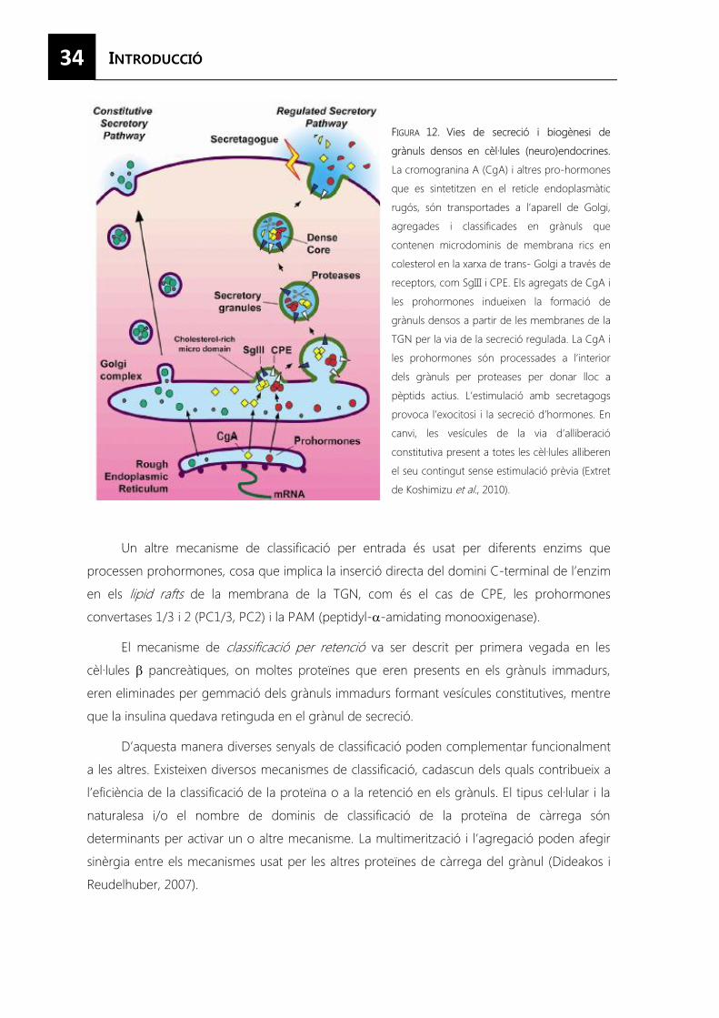
34
FIGURA 12. Vies de secreció i biogènesi de
grànuls densos en cèl·lules (neuro)endocrines.
La cromogranina A (CgA) i altres pro-hormones
que es sintetitzen en el reticle endoplasmàtic
rugós, són transportades a l’aparell de Golgi,
agregades i classificades en grànuls que
contenen microdominis de membrana rics en
colesterol en la xarxa de trans- Golgi a través de
receptors, com SgIII i CPE. Els agregats de CgA i
les prohormones indueixen la formació de
grànuls densos a partir de les membranes de la
TGN per la via de la secreció regulada. La CgA i
les prohormones són processades a l’interior
dels grànuls per proteases per donar lloc a
pèptids actius. L’estimulació amb secretagogs
provoca l’exocitosi i la secreció d’hormones. En
canvi, les vesícules de la via d’alliberació
constitutiva present a totes les cèl·lules alliberen
el seu contingut sense estimulació prèvia (Extret
de Koshimizu et al., 2010).
Un altre mecanisme de classificació per entrada és usat per diferents enzims que
processen prohormones, cosa que implica la inserció directa del domini C-terminal de l’enzim
en els lipid rafts de la membrana de la TGN, com és el cas de CPE, les prohormones
convertases 1/3 i 2 (PC1/3, PC2) i la PAM (peptidyl-�-amidating monooxigenase).
El mecanisme de classificació per retenció va ser descrit per primera vegada en les
cèl·lules � pancreàtiques, on moltes proteïnes que eren presents en els grànuls immadurs,
eren eliminades per gemmació dels grànuls immadurs formant vesícules constitutives, mentre
que la insulina quedava retinguda en el grànul de secreció.
D’aquesta manera diverses senyals de classificació poden complementar funcionalment
a les altres. Existeixen diversos mecanismes de classificació, cadascun dels quals contribueix a
l’eficiència de la classificació de la proteïna o a la retenció en els grànuls. El tipus cel·lular i la
naturalesa i/o el nombre de dominis de classificació de la proteïna de càrrega són
determinants per activar un o altre mecanisme. La multimerització i l’agregació poden afegir
sinèrgia entre els mecanismes usat per les altres proteïnes de càrrega del grànul (Dideakos i
Reudelhuber, 2007).
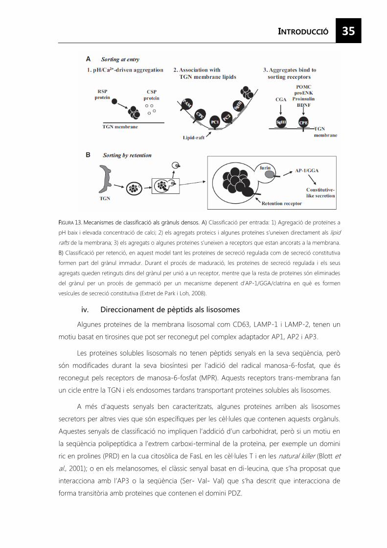
35
FFIGURA 13. Mecanismes de classificació als grànuls densos. A) Classificació per entrada: 1) Agregació de proteïnes a
pH baix i elevada concentració de calci; 2) els agregats proteics i algunes proteïnes s’uneixen directament als lipid
rafts de la membrana; 3) els agregats o algunes proteïnes s’uneixen a receptors que estan ancorats a la membrana.
B) Classificació per retenció, en aquest model tant les proteïnes de secreció regulada com de secreció constitutiva
formen part del grànul immadur. Durant el procés de maduració, les proteïnes de secreció regulada i els seus
agregats queden retinguts dins del grànul per unió a un receptor, mentre que la resta de proteïnes són eliminades
del grànul per un procés de gemmació per un mecanisme depenent d’AP-1/GGA/clatrina en què es formen
vesícules de secreció constitutiva (Extret de Park i Loh, 2008).
Algunes proteïnes de la membrana lisosomal com CD63, LAMP-1 i LAMP-2, tenen un
motiu basat en tirosines que pot ser reconegut pel complex adaptador AP1, AP2 i AP3.
Les proteïnes solubles lisosomals no tenen pèptids senyals en la seva seqüència, però
són modificades durant la seva biosíntesi per l’adició del radical manosa-6-fosfat, que és
reconegut pels receptors de manosa-6-fosfat (MPR). Aquests receptors trans-membrana fan
un cicle entre la TGN i els endosomes tardans transportant proteïnes solubles als lisosomes.
A més d’aquests senyals ben caracteritzats, algunes proteïnes arriben als lisosomes
secretors per altres vies que són específiques per les cèl·lules que contenen aquests orgànuls.
Aquestes senyals de classificació no impliquen l’addició d’un carbohidrat, però si un motiu en
la seqüència polipeptídica a l’extrem carboxi-terminal de la proteïna, per exemple un domini
ric en prolines (PRD) en la cua citosòlica de FasL en les cèl·lules T i en les natural killer (Blott et
al., 2001); o en els melanosomes, el clàssic senyal basat en di-leucina, que s’ha proposat que
interacciona amb l’AP3 o la seqüència (Ser- Val- Val) que s’ha descrit que interacciona de
forma transitòria amb proteïnes que contenen el domini PDZ.
36
4.3. Transport a la membrana plasmàtica
Després del procés de senyalització que dóna lloc a l’exocitosi, es produeixen dos
processos. El primer és el transport depenent de microtúbuls de grànul de secreció cap a la
membrana plasmàtica; i el segon, és el docking i la fusió amb la membrana plasmàtica
alliberant el seu contingut a l’exterior. Mentre que les proteïnes SNAREs s’han relacionat amb
el docking i la fusió, les Rab GTPases s’han descrit com a reguladors importants del procés
d’exocitosi.
4.3.1. Proteïnes de transport
Les vesícules, un cop formades a la TGN, són transportades als llocs de secreció de la
membrana plasmàtica mitjançant un sistema de transport basat en microtúbuls que requereix
la presència d’adaptadors, com la dinactina o la proteïna-1 associada a hungtintina, per
facilitar la subjecció de la vesícula als microtúbuls. Generalment, tots els tipus de vesícula usen
les mateixes proteïnes motores, la cinesina per al transport anterògrad i la dineïna per al
transport retrògrad. Estudis recents indiquen que la cua citosòlica de CPE, present en els
grànuls de secreció, pot interaccionar directament amb la dinactina (Park et al., 2008) que
recluta les proteïnes motores cinesina-2 i -3 (KIF3 i KIF1A).
Al final del transport basat en microtúbuls, les vesícules són transferides a l’esquelet
d’actina pròxim a la membrana plasmàtica. S’ha proposat que miosina V és la responsable de
transferir les vesícules dels microtúbuls a l’actina interaccionant directament amb les proteïnes
motores (Park i Loh, 2008).
4.3.2. Rab GTPases
Formen la família més gran de GTPases, en humans se n’han identificat més de 60, i
tenen com a funció actuar com a interruptors moleculars ja que alternen dos estats
conformacionals: el GTP actiu i el GDP inactiu. Estan associades de forma reversible amb les
membranes a través de grups hidrofòbics geranilgeranil que estan ancorats a un o dos
residus de cisteïna carboxiterminals i això els permet realitzar la seva funció de regular el tràfic
de membranes.
Les Rab GTPases controlen la dinàmica intracel·lular de les vesícules, des de la formació
de la vesícula ja que capten receptors per a la nova vesícula i en modifiquen la composició de
la coberta, fins aparticipar en el tethering de les vesícules interaccionant amb els factors de
lligació que a la vegada interaccionen amb les SNAREs i els seus reguladors per activar la
formació del complex SNARE, que produeix la fusió de membranes. Respecte al seu paper
com a moduladors del transport, les proteïnes Rab s’uneixen a les proteïnes motores com les
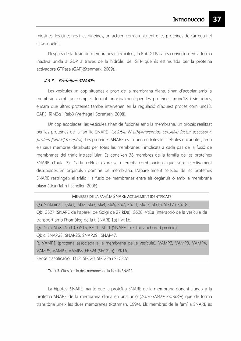
37
miosines, les cinesines i les dineïnes, on actuen com a unió entre les proteïnes de càrrega i el
citoesquelet.
Després de la fusió de membranes i l’exocitosi, la Rab GTPasa es converteix en la forma
inactiva unida a GDP a través de la hidròlisi del GTP que és estimulada per la proteïna
activadora GTPasa (GAP)(Stenmark, 2009).
4.3.3. Proteïnes SNAREs
Les vesícules un cop situades a prop de la membrana diana, s’han d’acoblar amb la
membrana amb un complex format principalment per les proteïnes munc18 i sintaxines,
encara que altres proteïnes també intervenen en la regulació d’aquest procés com unc13,
CAPS, RIM2� i Rab3 (Verhage i Sorensen, 2008).
Un cop acoblades, les vesícules s’han de fusionar amb la membrana, un procés realitzat
per les proteïnes de la família SNARE (soluble-N-ethylmaleimide-sensitive-factor accessory-
protein [SNAP] receptor). Les proteïnes SNARE es troben en totes les cèl·lules eucariotes, amb
els seus membres distribuïts per totes les membranes i implicats a cada pas de la fusió de
membranes del tràfic intracel·lular. Es coneixen 38 membres de la família de les proteïnes
SNARE (Taula 3). Cada cèl·lula expressa diferents combinacions que són selectivament
distribuïdes en orgànuls i dominis de membrana. L’aparellament selectiu de les proteïnes
SNARE restringeix el tràfic i la fusió de membranes entre els orgànuls o amb la membrana
plasmàtica (Jahn i Scheller, 2006).
MMEEMBRES DE LA FAMÍLIA SSNARE AACTUALMENT IDENTIFICAATS
Qa. Sintaxina 1 (Stx1), Stx2, Stx3, Stx4, Stx5, Stx7, Stx11, Stx13, Stx16, Stx17 i Stx18.
Qb. GS27 (SNARE de l’aparell de Golgi de 27 kDa), GS28, Vti1a (interacció de la vesícula de
transport amb l’homòleg de la t-SNARE 1a) i Vti1b.
Qc. Stx6, Stx8 i Stx10, GS15, BET1 i SLT1 (SNARE-like tail-anchored protein)
Qb,c. SNAP23, SNAP25, SNAP29 i SNAP47.
R. VAMP1 (proteïna associada a la membrana de la vesícula), VAMP2, VAMP3, VAMP4,
VAMP5, VAMP7, VAMP8, ERS24 (SEC22b) i YKT6.
Sense classificació. D12, SEC20, SEC22a i SEC22c.
TTAULA 3. Classificació dels membres de la familia SNARE.
La hipòtesi SNARE manté que la proteïna SNARE de la membrana donant s’uneix a la
proteïna SNARE de la membrana diana en una unió (trans-SNARE complex) que de forma
transitòria uneix les dues membranes (Rothman, 1994). Els membres de la família SNARE es
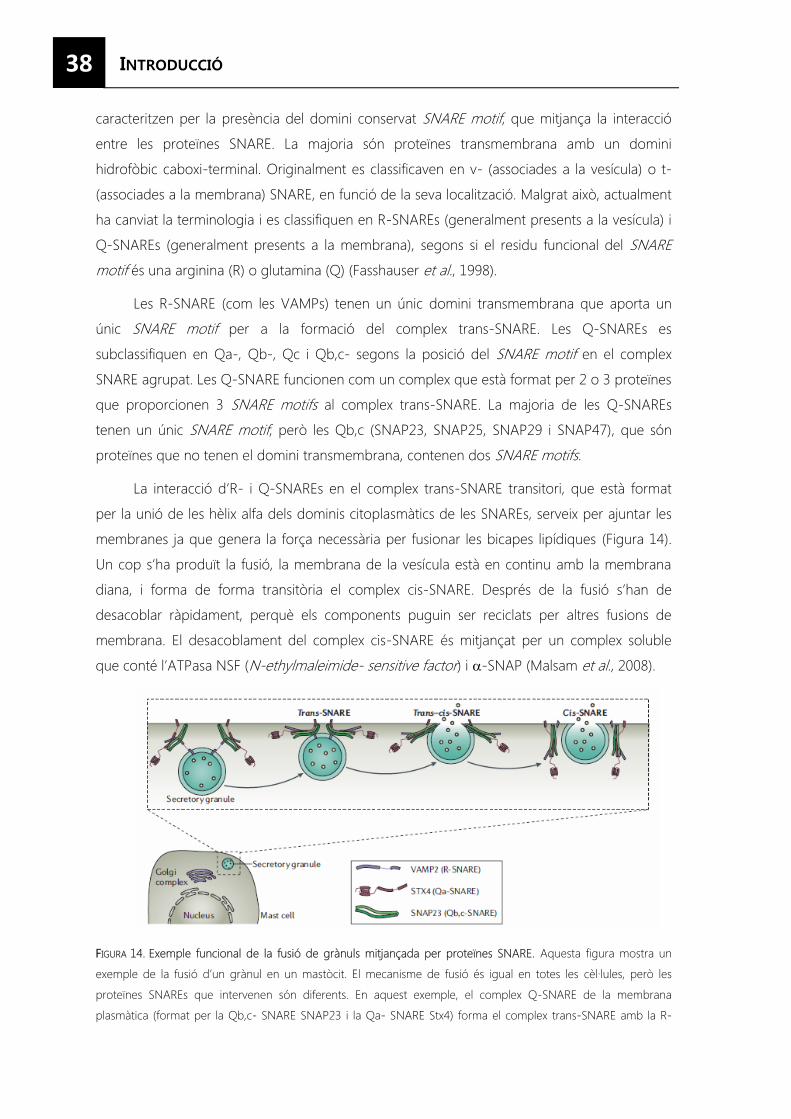
38
caracteritzen per la presència del domini conservat SNARE motif, que mitjança la interacció
entre les proteïnes SNARE. La majoria són proteïnes transmembrana amb un domini
hidrofòbic caboxi-terminal. Originalment es classificaven en v- (associades a la vesícula) o t-
(associades a la membrana) SNARE, en funció de la seva localització. Malgrat això, actualment
ha canviat la terminologia i es classifiquen en R-SNAREs (generalment presents a la vesícula) i
Q-SNAREs (generalment presents a la membrana), segons si el residu funcional del SNARE
motif és una arginina (R) o glutamina (Q) (Fasshauser et al., 1998).
Les R-SNARE (com les VAMPs) tenen un únic domini transmembrana que aporta un
únic SNARE motif per a la formació del complex trans-SNARE. Les Q-SNAREs es
subclassifiquen en Qa-, Qb-, Qc i Qb,c- segons la posició del SNARE motif en el complex
SNARE agrupat. Les Q-SNARE funcionen com un complex que està format per 2 o 3 proteïnes
que proporcionen 3 SNARE motifs al complex trans-SNARE. La majoria de les Q-SNAREs
tenen un únic SNARE motif, però les Qb,c (SNAP23, SNAP25, SNAP29 i SNAP47), que són
proteïnes que no tenen el domini transmembrana, contenen dos SNARE motifs.
La interacció d’R- i Q-SNAREs en el complex trans-SNARE transitori, que està format
per la unió de les hèlix alfa dels dominis citoplasmàtics de les SNAREs, serveix per ajuntar les
membranes ja que genera la força necessària per fusionar les bicapes lipídiques (Figura 14).
Un cop s’ha produït la fusió, la membrana de la vesícula està en continu amb la membrana
diana, i forma de forma transitòria el complex cis-SNARE. Després de la fusió s’han de
desacoblar ràpidament, perquè els components puguin ser reciclats per altres fusions de
membrana. El desacoblament del complex cis-SNARE és mitjançat per un complex soluble
que conté l’ATPasa NSF (N-ethylmaleimide- sensitive factor) i �-SNAP (Malsam et al., 2008).
FFIGURA 14. Exemple funcional de la fusió de grànuls mitjançada per proteïnes SNARE. Aquesta figura mostra un
exemple de la fusió d’un grànul en un mastòcit. El mecanisme de fusió és igual en totes les cèl·lules, però les
proteïnes SNAREs que intervenen són diferents. En aquest exemple, el complex Q-SNARE de la membrana
plasmàtica (format per la Qb,c- SNARE SNAP23 i la Qa- SNARE Stx4) forma el complex trans-SNARE amb la R-
39
SNARE VAMP2 del grànul. El complex trans-SNARE dirigeix la fusió entre les dues membranes i la secreció del
contingut del grànul. El complex post-fusió, abans del desacoblament, es coneix com a complex cis-SNARE (Extret
de Stow et al., 2006).
Els membres de la família Sec1/Munc18 s’uneixen directament als dominis amino-
terminals de les sintaxines i estan posicionats de manera que poden regular (de forma positiva
o negativa) la capacitat d’aquestes Q-SNARE per formar el complex trans-SNARE. Aquestes
proteïnes també poden tenir altres funcions com regular l’estabilitat i el recanvi de les
proteïnes SNARE. A la majoria de cèl·lules de mamífers, aquestes proteïnes estan associades a
la membrana plasmàtica, a l’aparell de Golgi i als endosomes per regular l’activitat de les
SNAREs en aquestes localitzacions. A més, també l’organització espacial de les SNAREs i de
les Sec/Munc-like controla la seva funció. Els dominis de membrana rics en colesterol regulen
la interacció de les proteïnes Q-SNAREs, separant-les de les proteïnes Sec/Munc-like. Altres
proteïnes que s’uneixen a les proteïnes SNARE poden actuar com a moduladors de la seva
funció, com Munc13, el seu homòleg en cèl·lules endocrines CAPS (calcium-activated protein
for secretion) i la proteïna RIM que afavoreixen la formació del complex SNARE. Les proteïnes
que regulen la secreció varien segons el tipus cel·lular, com per exemple la sinaptotagmina 1
(un sensor de calci) i la tomosina (també coneguda com a Stxbp5) en cèl·lules neuronals.
40
L’existència d’un sistema de comunicació basat en l’alliberació de mitjancers químics
entre els astròcits i altres cèl·lules neuronals va ser hipotetitzat a finals dels anys 80 per
l’observació que les cèl·lules glials contenien, sintetitzaven i alliberaven gran varietat de
substàncies.
Actualment està acceptat que els astròcits són cèl·lules secretores que alliberen diferents
transmissors químics com: aminoàcids excitadors i inhibidors (glutamat, aspartat, D-serina,
àcid homocisteic, àcid quinolínic, GABA, glicina, taurina, etc); altres neurotransmissors clàssics
(acetilcolina, noradrenalina, dopamina, serotonina, histamina, etc); ATP i nucleòtids i
nucleòsids relacionats (nucleòtids de purina, ATP, GTP, i els seus derivats di- i mono-fosfat,
adenosina, guanosina, UTP); eicosanoids i altres mitjancers lipídics (productes de la
ciclooxigenasa (COX), prostaglandines (PGs), productes de la lipooxigenasa (LOX), productes
de la epooxigenasa (EPOX)); neuropèptids (proencefalina, angiotensinògen, endotelines);
neurotrofines (factor de creixement neural (NGF), neurotrofina- 3, factor neurotròfic derivat
del cervell (BDNF)); citocines (interleucines (IL), interferons (IFN), factors de necrosi tumoral
(TNF)) i factors de creixement i quimiocines.
Diversos estudis han intentat descriure el “secretoma” d’aquestes cèl·lules mitjançant
diferents aproximacions experimentals, tant en condicions basals (Dowell et al., 2009; Greco et
al., 2010) com en resposta al tractament amb citocines inflamatòries (Keene et al., 2009) o a
l’estimulació colinèrgica (Moore et al., 2008). En l’estudi més recent, es van identificar més de
mig miler de proteïnes, de les quals un 20% tenien el pèptid senyal a l’extrem N-terminal i
estaven enriquides més de 1,5 vegades en el medi dels astròcits.
5.1. Mecanismes de secreció en astròcits
Els mecanismes moleculars responsables de l’alliberació dels gliotransmissors encara no
estan del tot clars. La majoria d’evidències indiquen que l’alliberació de gliotransmissors es
produeix per un mecanisme depenent de calci i de proteïnes SNARE a través de l’exocitosi de
vesícules i lisosomes (Perea i Araque, 2010).
Durant els últims anys, els estudis s’han centrat en descriure els mecanismes
d’alliberació d’aminoàcids per part dels astròcits, sobretot pel glutamat, pel qual s’han descrit
fins a 6 mecanismes diferents d’alliberació (Malarkey i Parpura, 2008). Tots aquests no són
mútuament exclusius i poden coexistir (Perea i Araque, 2010).
41
5.1.1. Secreció no exocítica en astròcits
Malgrat que s’han descrit diferents mecanismes per l’alliberació de glutamat tant a
través de canals, com l’obertura dels canals iònics induïts per volum, la secreció per
connexons i hemicanals o mitjançant receptors purinèrgics ionotròpics; com a través de
transportadors com el funcionament invers dels transportadors de glutamat o l’intercanviador
de cistina-glutamat. Encara falta determinar si aquests mecanismes succeeixen en condicions
fisiològiques o només en determinades condicions patològiques com en l’infart o la isquèmia
o requereixen condicions especials com l’inflament de les cèl·lules o baixes concentracions
extracel·lulars de calci (Figura 15).
El treball pioner del grup de Kimelberg va establir que quan els astròcits en cultiu
s’inflen, simulant un edema cerebral, alliberen glutamat, aspartat i taurina a través dels canals
aniònics regulats per volum (VRAC) (Kimelberg et al., 1990), però no s’ha demostrat
l’alliberació de glutamat a través d’aquests canals en condicions no patològiques. Tanmateix,
algunes substàncies com l’ATP o l’òxid nítric poden actuar sobre els VRACs amplificant
l’alliberació d’aminoàcids en una lleugera hipo-osmolaritat que pot ser que succeeixi in vivo.
L’ATP mitjançant la unió a receptors pot augmentar els nivells de calci intracel·lular provocant
un lleuger inflament transitori de la cèl·lula seguida de l’alliberació de glutamat pels VRACs
(Takano et al., 2005).
Els transportadors de glutamat són una família de proteïnes situades a la membrana
plasmàtica dels astròcits i de les neurones que tenen la funció específica de retirar el glutamat
de l’espai extracel·lular. Per cada cicle de transport, juntament amb una molècula de glutamat,
co-transporta 3 Na+ i 1 H+ i treu 1 K+. Encara que no sembla que succeeixi en la funció
cerebral normal, en condicions patològiques com en la isquèmia on hi ha uns nivells
extracel·lulars de K+ elevats, aquests transportadors poden funcionar de forma inversa i
alliberar glutamat (Szatkowsky et al., 1990). Aquest transport invers del glutamat acaba
provocant la mort dels astròcits, no per l’augment dels nivells de glutamat a l’espai
extracel·lular, sinó per la depleció del glutatió citoplasmàtic causat per la disminució dels
glutamat a l’interior de la cèl·lula que provoca un augment de l’estrès oxidatiu.
42
Són uns transportadors d’aminoàcids independents de Na+ i dependents de Cl- que
intercanvien la cistina extracel·lular amb el glutamat intracel·lular amb una estequiometria 1:1
(Banai, 1986; Reichelt et al., 1997). Aquest transportador és el responsable de l’acumulació de
la cistina necessària per la síntesi de glutatió, un dels principals antioxidants endògens, i a més,
proporciona una via d’alliberació de glutamat (War et al., 1999). Aquesta alliberació de
glutamat està relacionada amb la modulació de la transmissió sinàptica i l’expressió del
comportament animal relacionat amb l’addicció (Parpura i Zorec, 2010).
Les unions tipus gap són porus formats per la unió de dos connexons (hemicanals)
situats de forma juxtaposada en les membranes de dues cèl·lules adjacents que connecten el
seu citoplasma i permeten el pas de molècules de fins a 1 kDa. Els connexons desaparellats
poden actuar com a hemicanals funcionals permetent la difusió de molècules des del
citoplasma cap a l’espai extracel·lular. Els hemicanals formats per conexines (Cx43 en astròcits)
poden alliberar glutamat a l’espai extracel·lular quan hi ha una baixa concentració de cations
divalents a l’espai extracel·lular, però falta determinar si això també succeeix en condicions
fisiològiques. En canvi, les panexines són insensibles a les concentracions extracel·lulars de
calci i poden alliberar glutamat si es produeix un augment de calci intracel·lular a nivells que
es solen observar en els astròcits (Ye et al., 2003).
Els receptors P2X són uns canals de membrana plasmàtica que s’activen per la unió
amb l’ATP i són permeables a cations mono i divalents. Es coneixen 7 tipus de subunitats del
receptor P2X que es poden agrupar per formar canals homomèris o heteromèrics. En moltes
cèl·lules, si hi ha una estimulació mantinguda, el porus aquós es dilata fins a permetre el pas
de molècules grans independentment de la seva càrrega. Quan l’ATP activa el receptor P2X7,
s’obre un porus que és permeable a l’ATP i al glutamat. L’activació d’aquest canal en astròcits
en cultiu i in vivo proporciona una ruta no exocítica per l’alliberació d’ATP, glutamat i
probablement D-serina (Duan et al., 2003). En condicions patològiques on es produeix una
activació prolongada dels canals P2X7 pot conduir a l’alliberació de proteïnes citosòliques i a la
mort cel·lular.
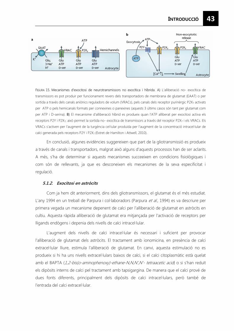
43
FIGURA 15. Mecanismes d’exocitosi de neurotransmissors no exocítica i híbrida. A) L’alliberació no- exocítica de
transmissors es pot produir per funcionament revers dels transportadors de membrana de glutamat (EAAT) o per
sortida a través dels canals aniònics reguladors de volum (VRACs), pels canals dels receptor purinèrgic P2X7 activats
per ATP o pels hemicanals formats per connexines o panexines (aquests 3 últims casos són tant per glutamat com
per ATP i D-serina). BB) El mecanisme d’alliberació híbrid es produeix quan l’ATP alliberat per exocitosi activa els
receptors P2Y i P2X7; això permet la sortida no- exocítica de transmissors a través del receptor P2X7 i els VRACs. Els
VRACs s’activen per l’augment de la turgència cel·lular produïda per l’augment de la concentració intracel·lular de
calci generada pels receptors P2Y i P2X7 (Extret de Hamilton i Attwell, 2010).
En conclusió, algunes evidències suggereixen que part de la gliotransmissió es produeix
a través de canals i transportadors, malgrat això alguns d’aquests processos han de ser aclarits.
A més, s’ha de determinar si aquests mecanismes succeeixen en condicions fisiològiques i
com són de rellevants, ja que es desconeixen els mecanismes de la seva especificitat i
regulació.
5.1.2. Exocitosi en astròcits
Com ja hem dit anteriorment, dins dels gliotransmissors, el glutamat és el més estudiat.
L’any 1994 en un treball de Parpura i col·laboradors (Parpura et al., 1994) es va descriure per
primera vegada un mecanisme depenent de calci per l’alliberació de glutamat en astròcits en
cultiu. Aquesta ràpida alliberació de glutamat era mitjançada per l’activació de receptors per
lligands endògens i depenia dels nivells de calci intracel·lular.
L’augment dels nivells de calci intracel·lular és necessari i suficient per provocar
l’alliberació de glutamat dels astròcits. El tractament amb ionomicina, en presència de calci
extracel·lular lliure, estimula l’alliberació de glutamat. En canvi, aquesta estimulació no es
produeix si hi ha uns nivells extracel·lulars baixos de calci, si el calci citoplasmàtic està quelat
amb el BAPTA (1,2-bis(o-aminophenoxy)-ethane-N,N,N’,N’- tetraacetic acid) o si s’han reduït
els dipòsits interns de calci pel tractament amb tapsigargina. De manera que el calci prové de
dues fonts diferents, principalment dels dipòsits de calci intracel·lulars, però també de
l’entrada del calci extracel·lular.

44
En estudis posteriors s’ha descrit que aquesta alliberació és sensible a inhibidors de
l’exocitosi neuronal com les toxines clostridials (la neurotoxina tetànica (TeNT) i els set tipus
de neurotoxines botulíniques (BoNT A-G)) (Jourdain et al., 2007; Perea i Araque, 2007), per
tant, els astròcits expressen les proteïnes SNAREs que són la diana d’aquestes toxines
(Montana et al., 2006). Aquesta alliberació també és sensible a la bafilomicina A1 (Baf A1) un
bloquejant de la H+-ATPasa vesicular que produeix l’alcalinització del lumen vesicular evitant
el gradient de protons necessari perquè els transportadors de glutamat (VGLUTs)
l’introdueixin dins de les vesícules.
Aquestes vesícules són transportades al llarg del citoesquelet. Aquest transport és
modulat per la dinàmica dels diferents filaments, les diferents proteïnes motores i és controlat
per diferents molècules de senyalització. A més dels microtúbuls i els filaments d’actina, els
astròcits també expressen filaments intermedis: GFAP, vimentina, nestina i sinemina, aquests
dos últims només s’expressen en astròcits reactius. Pel transport ràpid a llarga distància s’usen
preferentment els microtúbuls, encara que la resta de filaments també tenen un paper
fonamental (Potokar et al., 2011).
Tots aquests estudis suggereixen l’existència d’una alliberació vesicular regulada per
calci en astròcits, no només pel glutamat (Bezzi et al., 2004) sinó també per altres
neurotransmissors com la D-serina (Mothet, 2005; Martineau et al., 2008), l’ATP (Coco et al.,
2003; Pangrsic et al., 2007), i pèptids com el ANP (Krzan et al., 2003), el BDNF (Bergami et al.,
2008; Jean et al., 2008) i el neuropèptid Y (NPY) (Ramamoorthy i Whim, 2008), i que poden
ser alliberats mitjançant una exocitosi regulada depenent de calci.
Malgrat totes aquestes evidències, les dades disponibles no proporcionen una
descripció completa dels processos d’exocitosi en astròcits. Per això, l’existència d’una via
d’exocitosi regulada en aquestes cèl·lules encara s’està debatint.
A més, s’ha descrit certa heterogeneïtat en les vies de secreció dels astròcits ja que
s’han proposat diferents orgànuls com els responsables de l’exocitosi dels gliotransmissors. En
astròcits es troben dos tipus de vesícules secretores: les vesícules petites similars a les
sinàptiques (synaptic- like microvesicle : SLMVs) i grànuls densos (large dense-core vesicles:
LDCV). Sempre s’ha pensat que les SLMVs emmagatzemaven molècules petites i s’alliberaven
de forma regulada, participant en la comunicació glia-neurona. Malgrat això, els LDCV
contenen una gran varietat tant de molècules petites com grans que tenen importància en la
comunicació cèl·lula- cèl·lula (Hur et al., 2010). A més, en els astròcits també trobem lisosomes
secretors. La majoria dels estudis no distingien entre les diferents poblacions d’orgànuls
secretors, per tant, la descripció de la secreció en aquestes cèl·lules potser que no sigui molt
acurada hi hagi una barreja de la contribució dels diferents orgànuls.
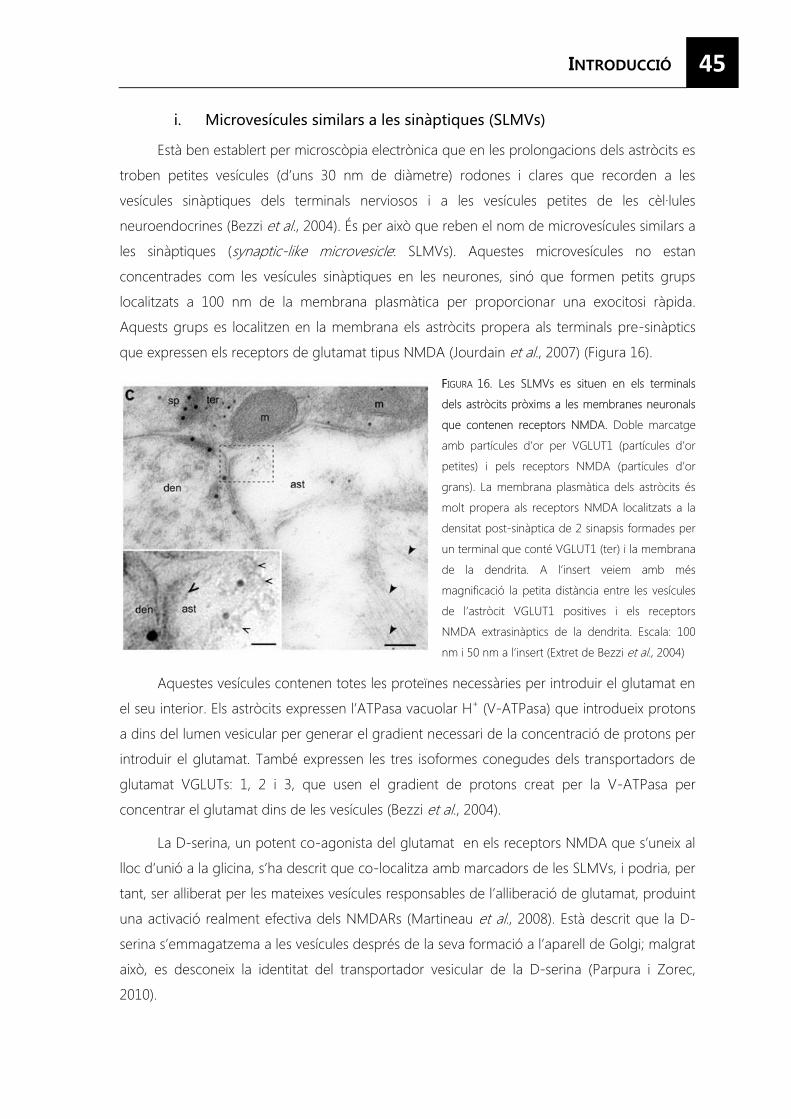
45
Està ben establert per microscòpia electrònica que en les prolongacions dels astròcits es
troben petites vesícules (d’uns 30 nm de diàmetre) rodones i clares que recorden a les
vesícules sinàptiques dels terminals nerviosos i a les vesícules petites de les cèl·lules
neuroendocrines (Bezzi et al., 2004). És per això que reben el nom de microvesícules similars a
les sinàptiques (synaptic-like microvesicle: SLMVs). Aquestes microvesícules no estan
concentrades com les vesícules sinàptiques en les neurones, sinó que formen petits grups
localitzats a 100 nm de la membrana plasmàtica per proporcionar una exocitosi ràpida.
Aquests grups es localitzen en la membrana els astròcits propera als terminals pre-sinàptics
que expressen els receptors de glutamat tipus NMDA (Jourdain et al., 2007) (Figura 16).
FFIGURA 16. Les SLMVs es situen en els terminals
dels astròcits pròxims a les membranes neuronals
que contenen receptors NMDA. Doble marcatge
amb partícules d’or per VGLUT1 (partícules d’or
petites) i pels receptors NMDA (partícules d’or
grans). La membrana plasmàtica dels astròcits és
molt propera als receptors NMDA localitzats a la
densitat post-sinàptica de 2 sinapsis formades per
un terminal que conté VGLUT1 (ter) i la membrana
de la dendrita. A l’insert veiem amb més
magnificació la petita distància entre les vesícules
de l’astròcit VGLUT1 positives i els receptors
NMDA extrasinàptics de la dendrita. Escala: 100
nm i 50 nm a l’insert (Extret de Bezzi et al., 2004)
Aquestes vesícules contenen totes les proteïnes necessàries per introduir el glutamat en
el seu interior. Els astròcits expressen l’ATPasa vacuolar H+ (V-ATPasa) que introdueix protons
a dins del lumen vesicular per generar el gradient necessari de la concentració de protons per
introduir el glutamat. També expressen les tres isoformes conegudes dels transportadors de
glutamat VGLUTs: 1, 2 i 3, que usen el gradient de protons creat per la V-ATPasa per
concentrar el glutamat dins de les vesícules (Bezzi et al., 2004).
La D-serina, un potent co-agonista del glutamat en els receptors NMDA que s’uneix al
lloc d’unió a la glicina, s’ha descrit que co-localitza amb marcadors de les SLMVs, i podria, per
tant, ser alliberat per les mateixes vesícules responsables de l’alliberació de glutamat, produint
una activació realment efectiva dels NMDARs (Martineau et al., 2008). Està descrit que la D-
serina s’emmagatzema a les vesícules després de la seva formació a l’aparell de Golgi; malgrat
això, es desconeix la identitat del transportador vesicular de la D-serina (Parpura i Zorec,
2010).
46 En l’exocitosi d’aquestes vesícules coexisteix la fusió completa de la vesícula, en que
s’allibera tot el contingut a l’espai extracel·lular i la fusió tipus kiss and run. Aquest últim tipus
de fusió, els astròcits la presenten exclusivament en resposta a l’estimulació fisiològica; a cada
exocitosi només s’allibera una fracció del contingut total de la vesícula (<10%), permetent
diversos cicles d’exocitosi sense recarregar la vesícula; cada cicle d’exocitosi només és una
alliberació, no múltiples com en les cèl·lules cromafins; i la latència de l’endocitosi induïda per
l’estímul és 4 vegades més lenta en els astròcits que en les cèl·lules cromafins (Chen et al., 2006).
Contràriament als aminoàcids i a l’ATP, que són introduïts a les vesícules per
transportadors de membrana, els gliotransmissors entren a les vesícules a través de la via de
la secreció regulada. Els pro-pèptids es sintetitzen al reticle endoplasmàtic (ER), passen a
l’aparell de golgi des d’on es formen les vesícules on són concentrats i classificats en orgànuls,
on són processats abans de ser alliberats (Parpura i Zorec, 2010). Aquestes vesícules que
transporten transmissors peptídics tenen una aparença morfològica diferent al microscopi
electrònic: grànuls densos (large dense-core vesicles: LDCV), que són menys abundants que
les SLMVs ( hi ha generalment 0-4 grànuls densos cada 6 �m2 d’astròcits) i que es troben
majoritàriament al voltant de l’aparell de Golgi i també en les prolongacions. Aquests grànuls
tenen un diàmetre de 300- 400 nm, encara que no és estrany trobar-ne de diàmetre superior
a 400 nm. A més, és freqüent trobar grànuls que no tenen l’interior tan electrodens o que hi
ha unes parts de l’interior del grànul que són menys electrodenses que altres, això és degut a
que recentment han endocitat una vesícula o que està en procés de maduració (Hur et al., 2010).
Les cromoganines i les secretogranines són marcadors proteics que identifiquen els grànuls
de secreció. En els astròcits, la cromogranina A i B i la secretogranina II es localitzen
exclusivament en els LDCV (Figura 17). A més, aquestes proteïnes tenen una alta capacitat i
una baixa afinitat per emmagatzemar el calci, unint 30- 93 mols de calci/ mols de proteïna
amb una constant de dissociació (Kd) de 1.5-4.0 mM, permetent als grànuls emmagatzemar
fins a 40 mM de calci, la concentració de calci més gran en qualsevol orgànul subcel·lular.
Aquestes proteïnes també són alliberades juntament amb el contingut dels grànuls en
resposta a un estímul que augmenti les concentracions intracel·lulars de calci (Hur et al., 2010).
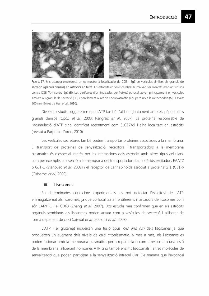
47
FFIGURA 17. Microscopia electrònica on es mostra la localització de CGB i SgII en vesícules similars als grànuls de
secreció (grànuls densos) en astròcits en teixit. Els astròcits en teixit cerebral humà van ser marcats amb anticossos
contra CGB ((A) i contra SgII ((B). Les partícules d’or (indicades per fletxes) es localitzaven principalment en vesícules
similars als grànuls de secreció (SG) i parcilament al reticle endoplasmàtic (er), però no a la mitocondria (M). Escala:
200 nm (Extret de Hur et al., 2010).
Diversos estudis suggereixen que l’ATP també s’allibera juntament amb els pèptids dels
grànuls densos (Coco et al., 2003; Pangrsic et al., 2007). La proteïna responsable de
l’acumulació d’ATP s’ha identificat recentment com SLC17A9 i s’ha localitzat en astròcits
(revisat a Parpura i Zorec, 2010)
Les vesícules secretores també poden transportar proteïnes associades a la membrana.
El transport de proteïnes de senyalització, receptors i transportadors a la membrana
plasmàtica és d’especial interès per les interaccions dels astròcits amb altres tipus cel·lulars,
com per exemple, la inserció a la membrana del transportador d’aminoàcids excitadors EAAT2
o GLT-1 (Stenovec et al., 2008) i el receptor de cannabinoids associat a proteïna G 1 (CB1R)
(Osborne et al., 2009)
En determinades condicions experimentals, es pot detectar l’exocitosi de l’ATP
emmagatzemat als lisosomes, ja que col·localitza amb diferents marcadors de lisosomes com
són LAMP-1 i el CD63 (Zhang et al., 2007). Dos estudis més confirmen que en els astròcits
orgànuls semblants als lisosomes poden actuar com a vesícules de secreció i alliberar de
forma depenent de calci (Jaiswal et al., 2007; Li et al., 2008).
L’ATP i el glutamat indueixen una fusió tipus Kiss and run dels lisosomes ja que
produeixen un augment dels nivells de calci citoplasmàtic. A més a més, els lisosomes es
poden fusionar amb la membrana plasmàtica per a reparar-la o com a resposta a una lesió
de la membrana, alliberant no només ATP sinó també enzims lisosomals i altres molècules de
senyalització que poden participar a la senyalització intracel·lular. De manera que l’exocitosi
48
regulada dels lisosomes pot tenir una funció important en la comunicació cèl·lula- cèl·lula tant
en condicions fisiològiques com patològiques.
El mecanisme mitjançant el qual es produeix l’acumulació de l’ATP dins del lisosoma no
és del tot clar. Com l’ATP està carregat negativament, l’activitat de la bomba de protons
manté el potencial de membrana necessari perquè el transportador de nucleòtids introdueix
l’ATP. També els transportadors ABC (ATP-binding cassette) i les proteïnes multiresistents a
drogues (multidrug resistant proteins) que poden introduir ATP, estan presents a la
membrana dels lisosomes.
A més, s’ha descrit que compartiments membranosos dels astròcits marcats amb
marcadors de endosomes tardans i lisosomes presenten el transportador d’aspartat sialina,
suggerint que aquestes orgànuls poden exocitar aspartat (Li et al., 2008).

51
En els últims anys, diversos treballs han demostrat que els astròcits participen
activament en el desenvolupament i plasticitat del SNC, així com en la modulació de la
neurotransmissió (Araque et al., 2001; Auld i Robitaille, 2003; Slezak i Pfrieger, 2003;
Christopherson et al., 2005).
Característicament, la majoria de les accions descrites dels astròcits sobre la fisiologia i
la patologia neuronal són mitjançades per secreció vesicular (Araque et al., 2001). Recentment,
s’ha donat una especial rellevància a la secreció glial regulada pel calci.
Estudiar els components moleculars i els mecanismes implicats en les vies de secreció
de cèl·lules astroglials.
Objectius concrets:
1. Obtenció d’un fenotip astroglial diferenciat en cultiu competent per a
realitzar estudis de secreció regulada de pèptids.
2. Analitzar proteïnes exocítiques astroglials, la regulació de la seva
expressió i la seva implicació en la secreció regulada.
3. Identificació i anàlisi de nous components moleculars de la via de
secreció regulada de pèptids en astròcits.


55
LLa senyalització per AMP cíclic reprimeix l’activació i promou la maduració en els
astròcits
Sonia Paco1, Manuela Hummel2, Virginia Pla1, Lauro Sumoy3, Fernando Aguado1 1 Department of Cell Biology, University of Barcelona, Barcelona E-08028, Spain. 2Centre for Genomic Regulation,
Barcelona E-08003, Spain, 3Institute for Predictive and Personalized Medicine of Cancer, Badalona E-08916, Spain
En preparació
Resum
La senyalització per AMPc produeix canvis dramàtics a la morfologia i a la fisiologia dels
astròcits. Malgrat això, la seva implicació en l’adquisició del fenotip i els mecanismes
transcripcionals implicats no es coneixen.
En aquest treball analitzem el transcriptoma global d’astròcits en cultiu tractats amb
anàlegs del AMPc. Un gran volum de transcrits, que corresponen a 6296 gens anotats, van ser
regulats de forma diferencial per la via del AMPc. Els anàlegs del AMPc augmenten l’expressió
dels gens involucrats tradicionalment en les funcions dels astròcits madurs, com són el control
homeostàtic, el suport metabòlic i estructural a les neurones, l’activitat antioxidant i la
comunicació; mentre que disminuïa considerablement l’espressió de transcrits relacionats amb
la proliferació i la immaduresa. A més a més, els gens típicament activats en els astròcits
reactius com els mediadors de la resposta immunològica i els components de la cicatriu, estan
reprimits pel AMPc. Els anàlisis d’enriquiment de grups gènics (GSEA) i l’avaluació in situ de
l’expressió gènica dels astròcits en diferents estats mostra que la senyalització per AMPc
confereix un perfil transcripcional madur i semblant a in vivo als astròcits en cultiu.
Aquests resultats mostren que la senyalització per AMPc és una via clau per restringir
les característiques d’immaduresa i activació astroglial promovent la seva maduració.

57
CCYCLIC AMP SIGNALING REPRESSES ACTIVATION AND PROMOTES
MATURATION IN ASTROCYTES
Sonia Paco1, Manuela Hummel2, Virginia Pla1, Lauro Sumoy3, and Fernando Aguado1
From Department of Cell Biology, University of Barcelona, Barcelona E-08028, Spain1, Centre for Genomic
Regulation, Barcelona E-08003, Spain2, Institute for Predictive and Personalized Medicine of Cancer, Badalona E-
08916, Spain3
ABSTRACT
cAMP signaling produces dramatic changes in astrocyte morphology and physiology.
However, its involvement in phenotype acquisition and the transcriptionally mediated
mechanisms of action are largely unknown. Here we analyzed the global transcriptome of
cultured astroglial cells incubated with activators of cAMP pathways. A bulk of astroglial
transcripts, 6,296 annotated genes, were differentially regulated by cAMP signaling. cAMP
analogs strongly upregulated genes involved in typical functions of mature astrocytes, such as
homeostatic control, metabolic and structural support to neurons, antioxidant defense and
communication, whereas they downregulated a considerable number of proliferating and
immaturity-related transcripts. Moreover, genes typically activated in reactive cells, such as
immunological mediators and scar components, were repressed by cAMP. Gene Set
Enrichment Analysis and evaluation in situ of gene expression in astrocytes in different states
showed that cAMP signaling conferred a mature and in vivo–like transcriptional profile to
cultured astrocytes. These results indicate that cAMP signaling is a key pathway restricting
developmental and activation features of astrocytes and promoting their maturation. A
positive modulation of cAMP signaling is suggested to suppress the mechanisms of activation
driven by pathological situations and to promote the physiologically normal state of
differentiated astrocytes.
INTRODUCTION
Mature astrocytes ensheath synapses and fine blood vessels, within the neuro-glio-
vascular units, to shape the functional micro-architecture of the central nervous system (CNS).
Through an array of transporters, ion channels and released and adhesion molecules,
astrocytes play key roles in the regulation of extracellular fluid homeostasis, integrity of the
blood-brain barrier and assurance of metabolic demand and antioxidant defense of neurons
[1,2]. Astrocytes also make a crucial contribution to communication, operating within glial
networks through gap junctions and hemichannels and bidirectionally with neurons and
58
endothelial cells via diffusible and surface molecules [3-5]. Characteristically, in response to
CNS insults, such as trauma, epilepsy or degenerative diseases, astrocytes become reactive
and play a critical role in neuroinflammation and scar formation [6,7].
In contrast to neurons, little is known about the intracellular signaling of astrocytes, and
the pathways controlling their differentiation and activation have not been elucidated. By
means of STAT transcription factors, cytokines of the interleukin-6 family are key cues in the
specification and differentiation of astroglial cells. Moreover, Smad transcription factors
activated by bone morphogenetic proteins also promote astrogliogenesis, through their
interaction with STAT3 [8]. Strikingly, analysis of glial activation in pro-inflammatory cytokine-
administered and genetically-modified mice has shown that JAK-STAT3 signaling is also a key
pathway through which astrocytes become reactive [9-11]. In addition to activating kinase-
dependent receptors, G-protein coupled receptors can trigger astroglial genesis and
maturation via cAMP [12-15]. Changes in cell morphology and protein expression induced by
cAMP have long been considered a feature of astrocyte activation [16-19]. However, the
precise significance of the cAMP pathway in the maturation and activation of these cells
remains elusive. Here we studied the net impact of the cAMP signaling pathway on astrocytes
by analyzing the global transcriptome of cultured cells incubated with permeable cAMP
analogs. Moreover, we perform Gene Set Enrichment Analysis (GSEA) of cAMP-regulated
genes and contrasted our findings with recent microarray reports and the in vivo expression
patterns of astrocytes. Our findings indicate that cAMP signaling restricts developmental and
activation features of astrocytes and promotes their maturation.
EEXPERIMENTAL PROCEDURES
Animals- OF1 mice were provided by Charles River Laboratories, Inc. (Lyon, France),
kept under controlled temperature (22 ± 2 °C), humidity (40–60%), and light (12-h cycles) and
treated in accordance with the European Community Council Directive (86/609/EEC) on
animal welfare. Under ketamine/xylazine anesthesia, stab wound lesions were made in the
parietal cortex with a scalpel blade as described previously [20].
Astrocyte cultures- Cultures were prepared from the cerebral cortex of two-day-old
mice [21]. Most cell culture reagents were obtained from GIBCO (Invitrogen, Paisley, UK).
Briefly, the cerebral cortex was isolated and the meninges were carefully dissected out.
Cortical tissues were then minced and incubated in 0.25% trypsin and 0.01% DNase.
Dissociated cells were seeded in flasks and grown in high-glucose Dulbecco’s Modified Eagle’s
Medium and F-12 (1:1) containing 10% FBS, 10 mM HEPES and penicillin/streptomycin at 37°
59
C in a 5% CO2 incubator. At confluence (days 10–12), the flasks were shaken overnight and
the cells were rinsed, detached and subcultured onto poly-L-lysine-coated plastic culture
dishes and coverslips. Treatment with 1mM 8Br-cAMP (Biolog, Life Science Institute, Bremen,
Germany) was administered two day after subculturing and prolonged for 8 days to induce
described morphological and physiological changes [12,13].
RNA Extraction and Reverse Transcription- RNA was isolated from triplicate cultures
from the four individual experiments. Extractions were performed using using Trizol® Reagent
(Invitrogen™) following the manufacturer’s instructions. The quantity and quality of isolated
RNAs were determined with a NanoDrop ND-1000 (NanoDrop Technologies, Wilmington, DE,
USA) and a Bioanalyzer 2100 (Agilent, Waldbronn, Germany). The RNA integrity numbers (RIN)
in all cases ranged from 8.7 to 10, thereby indicating minimal RNA degradation [22]. First-
strand DNA was synthesized using the Superscript III Reverse Transcriptase kit (Invitrogen). 1
μg of total RNA was added to kit components and nuclease-free water to 20 μl. Reactions
containing nuclease-free water instead of enzyme served as a negative control. Reactions
were incubated at 25ºC for 10 min, 50ºC for 30 min, 85ºC for 5 min. They were then chilled on
ice. E.coli RNAse H was added and incubated at 37ºC for 20 min. The samples were then
cooled to 4ºC and finally stored at -20ºC.
Microarray Analysis- RNA (500 ng) was labeled using Agilent's Low Input RNA Labeling
Kit, which involves reverse transcribing the mRNA in the presence of T7-oligo-dT primer to
produce cDNA and then in vitro transcribing with T7 RNA polymerase in the presence of Cy3-
CTP or Cy5-CTP to produce labeled cRNA. The labeled cRNA was hybridized to the Agilent
Mouse 4x44K 60-mer oligo microarray according to the manufacturer's protocol. The arrays
were washed, dried by centrifugation and scanned on an Agilent G2565BA microarray scanner
at 100% PMT and 5 μm resolution. Data were extracted using Genepix 6.0 (Molecular Devices)
software using the irregular spot finding feature.
Microarray Statistical Processing- Log2ratio values were computed for all pairs of
control and cAMP-stimulated samples. Per-probe log2ratios were aggregated to per-gene
values by taking median values. Analysis for differential expression on a gene-by-gene basis
was performed by limma [23] while distinguishing biological from technical replicates (dye-
swap hybridizations). Correction for multiple testing was done using the False Discovery Rate
(FDR) method. The lists of regulated genes were brought into biological context by Gene
Ontology scoring using GSEA [24]. Also, for cross-study comparisons GSEA, was applied. This
approach was used to test gene sets of interest (defined as lists of differentially expressed
gene symbols derived from other studies) for significant enrichment among differentially
60
expressed genes within our study. Only studies performed on mouse from which astrocyte
cultures were obtained and maintained under similar conditions to those in the present study
were considered. To compare the gene sets by Venn diagrams, we fixed the same fold change
cut-off at 1.5.
Quantitative Real-Time PCR Analysis- RT-qPCR was carried out using Power SYBR®
Green PCR Master Mix (Applied Biosystems, Foster City, CA) on an Applied 7700 machine,
using the primers indicated in Supplementary Table 1, and following the manufacturer’s
protocol. A dilution series (100–10−3) of total cDNA was prepared to determine the standard
curve (relative quantification). The samples (in triplicates) were amplified according to the
following protocol: 10 min at 95 °C, 42 cycles: 15 s at 95 °C, 15 s annealing temperature
(60ºC), 30 s at 72 °C. To control the specificity of the reaction, melting-curve analysis was
performed after amplification. In addition, PCR products were analyzed by agarose gel
electrophoresis to confirm the size of the amplified targets. Levels of Tbp were used as
normalization controls and relative mRNA levels were calculated as indicated by Pfaffl et al..
[25].
Western blotting- Cultured cells were lysed in ice-cold 50mM Tris-HCl, pH 7.4, 150 mM
NaCl, 5mM MgCl2, 1mM EGTA, 1% Triton-X 100 and protease inhibitor cocktail (Roche
Diagnostics). Samples were electrophoresed in 10% SDS-PAGE (BioRad) and then transferred
to nitrocellulose membranes (Whatman® Schleicher & Schuell, Keene, NH). The membranes
were blocked in a solution containing 5% non-fat milk powder in TBST (140 mM NaCl, 10 mM
Tris/HCl, pH 7.4 and 0.1% Tween 20) for 1 h at room temperature and then incubated with
primary antibodies (Supplementary Table 2) in blocking buffer for 2 h at room temperature.
After several washes in blocking solution, the membranes were incubated for 1 h with HRP-
conjugated secondary antibodies (DAKO, Glostrup, Denmark). Bound antibodies were
visualized with enhanced chemiluminescence reagents PIERCE® ECL Western Blotting
Substrate (Thermo Scientific, Rockford, IL, USA).
Immunocytochemistry-Cells grown on glass coverslips were fixed with 4%
paraformaldehyde in PBS for 15 min. Adult and postnatal (P5) animals were perfused
transcardially under deep anesthesia with the same fixative. Brains were removed from the
skulls, postfixed for 4 h and cryoprotected overnight at 4 °C by immersion in a 30% sucrose
solution. Forty-micrometer-thick frozen sections were obtained with a cryostat and collected
in PBS. To suppress nonspecific binding, cell cultures and brain sections were incubated in PBS
containing 10% FBS, 0.1% Triton X-100, 0.2% glycine, and 0.2% gelatin for 1 h at room
temperature. Incubations with the primary antibodies (Supplementary Table 8) were carried
61
out overnight at 4 °C in PBS containing 1% FBS, 0.1% Triton X-100, and 0.2% gelatin. Cell
cultures and brain sections were processed for immunofluorescence using secondary
fluorochrome-conjugated antibodies (Alexa Fluor 488 and Alexa Fluor 568, Molecular Probes,
Eugene, OR). Cell nuclei were stained with 4 ,6-diamidino-2-phenylindole (DAPI, Molecular
Probes). Cell-containing coverslips and histological sections were mounted with Mowiol.
Double immunofluorescences were performed by using primary antibodies raised in different
species. Images were obtained with a Leica TCS SPE scanning confocal microscope. The
specificity of the immunostaining was tested by omitting the primary antibodies or by
replacing them with an equivalent concentration of nonspecific IgG. No immunostaining was
observed in these conditions. Student’s t-test was used for statistical evaluation and data were
expressed as mean ± SEM.
RRESULTS AND DISCUSSION
Genome-wide transcriptional regulation in astrocytes by cAMP signaling.
To activate cAMP signaling we augmented intracellular levels by exposing cultured
astrocytes to the permeable cAMP analog 8Br-cAMP. Before and after treatment cell cultures
were essentially formed by astrocytes (>95% GFAP+ cells). Sustained elevated intracellular
cAMP levels altered the number and morphology of astrocytes. Quantitative analysis showed
almost 25% less GFAP+ astrocytes after treatment compared with controls (54.21±0.78
cell/mm2 and 72.08±1.24 cell/mm2, respectively, p=0.006). In agreement with previous
studies [12,21], control flat polygonal-shaped astroglial cells were converted into process-
bearing stellate astrocytes (Fig 1). Microarray analysis showed that 6,296 of 16,594 annotated
genes with assigned gene symbol were differentially expressed by a factor of 1.2 or greater
(and FDR q-value <5%) in 8Br-cAMP-treated astrocytes. The full list of differentially expressed
genes is shown in Supplementary Table 3. Approximately, one-third of the regulated elements
on the array corresponded to ESTs and genes of unknown function. Of the 6,296 genes with
significant regulation 42.2% were upregulated by cAMP while the rest were downregulated.
We validated the present array results by analyzing astrocyte expression of the mRNA
and protein of 10 representative genes after cAMP augmentation (Figs 1 and 2). qPCR analysis
was performed on the cell cycle regulator cyclin D1 (Ccnd1), the peptidase inhibitor cystatin C
(Cst3), the calcium-dependent exocytotic SNARE Vamp2, the NMDA receptor subunit NR2C
(Grin2c), the adhesion component claudin 10 (Cldn10) and the thyroid hormone transporter
Slco1c1. A correlation was found between RT-qPCR and array results for each gene (Fig 2A).
cAMP-induced changes of cyclin D1, cystatin C, Vamp2 and the apoptotic regulator Bid, as

62
well as their time dependence, were further corroborate at protein level by immunoblotting
(Fig 2B). No changes on the array were found for non-regulated genes, as shown for the
ubiquitous exocytotic protein Snap23 (Fig 2B). Immunofluorescence for the proliferation
marker Mki67, the polyamine enzyme Ssat (Sat1) and the NR2C receptor demonstrated that
cAMP-dependent gene regulation occurred in GFAP-identified astrocytes (Fig 1). Furthermore,
previous studies showing that cAMP regulates the expression of intermediate filaments (GFAP
and Nes), glutamate transporters (Slc1a2 and Slc1a3), deiodinases (Dio2), and secretogranins
(Scg2 and Scg3) in astrocyte further validate the present transcriptomic results [13,14,20,26,
Supplementary Table 3].
Elevated cAMP restricts the activation of astrocytes and promotes their differentiation
The functional roles of up- and downregulated cAMP-responsive genes were
categorized by Gene Ontology enrichment analysis using GSEA. GO categories with GSEA q-
value <2% were considered relevant. We calculated the relative proportion of genes in the
core enrichment of each GO category and grouped them in general categories (Fig 3). All the
GO categories enriched with differentially regulated genes are shown in the bar plot of
Supplementary Figure 1.
More than half (55.2%) the upregulated genes in enriched GO categories corresponded
to cell metabolism and transporters. Thus, genes involved in the uptake and degradation of
neurotransmitters such as glutamate, glycine and catecholamines were overexpressed after
treatment (Slc1a2, Glul, Gldc and MaoB). Ion channels, pumps and transporters with key
homeostatic and metabolic functions were also preferentially upregulated by cAMP. For
instance, genes participating in the membrane transport of K+ (Kcnn2), Cl- (Clcn2), Ca2+
(Atp2a2 and Atp2b2), water (Aqp4), glucose (Slc2a1), thyroid hormones (Slco1c1 and Slc16a2),
and oligopeptides (Slc15a2) were activated by cAMP signaling. Moreover, an increase of
cAMP levels induced expression of genes coding for a wide variety of astrocytic metabolic
enzymes, such as most aldehyde dehydrogenases, tRNA ribonucleases and GPI transamidases.
Interestingly, the third functional category of genes upregulated by cAMP was associated with
antioxidant activities (Table 1). Thus, genes encoding for antioxidant enzymes and molecules,
such as catalase, superoxide dismutases, metallothioneins, heme oxygenases and NADPH-
quinone oxidoreductases were found to be through microarray analysis. Enhanced cAMP also
upregulated numerous genes related to the glutathione system and transport of antioxidant
vitamins. The observation that cAMP agonists upregulated genes typically implicated in
biological processes of mature astrocytes indicates that cAMP signaling promotes cell
differentiation.
63
GO categories enriched with downregulated genes were associated mainly with
proliferating and immaturity-related features of astrocytes (Table 2). Thus, 71.5% of the genes
were involved in the general categories cell cycle and apoptosis, cell signaling, cytoskeleton,
cell shape and motility, and cell development. Expression of genes typically involved in mitotic
processes was dramatically decreased, such as those encoding for the DNA polymerases and
the proliferation antigen Mki67. Transcripts of proliferation and cell death regulators, such as
mitogens and morphogens (IGF-1, PDGF and BMP4), cyclins, caspases and proapoptotic Bcl2
family members (e.i., Casp8, Bid and Bax) were reduced after exposure to cAMP. Moreover,
numerous genes involved in cytoskeletal organization, such as Rho GTPases, myosins, actins,
tropomyosins, kinesins and septins, were notably repressed. Among the GO categories
enriched with downregulated genes, those typically of reactive astrocytes such as components
of the extracellular matrix and mediators of the immune response were predominantly
enriched. Thus, mRNA expression of tenascin C, nidogens and many collagens and
chondroitin and heparan sulfate proteoglycans was reduced. Interestingly, the enzymes
responsible for the major postranscriptional modifications of proteoglycans and collagens
were widely downregulated (sulfotransferases and lysyl oxidases). Finally, prolonged cAMP
signaling stimulation in astrocytes induced a downregulation of numerous mediators of the
immune response, such as cytokines, chemokines and components of the major
histocompatibility complex and phagocytototic and adhesion processes (e.g., Tnf, Ccl2, Tgfb1,
Cxcl10, Csf1r, H2-D1, B2m, Gulp1 and Icam1). We conclude that cAMP contributes to the
restriction of developmental and activation features of astrocytes.
cAMP signaling confers a mature in vivo-like transcriptional profile to cultured astrocytes.
To substantiate the net phenotype induction of cAMP signaling on astroglial cells, we
applied GSEA analysis to contrast the present gene profiles with previous microarray studies
describing the transcriptome signatures of astrocytes in vivo in a range of states. First, we
compared our genome profiling results with gene sets of developing and mature brain
astrocytes reported by Cahoy et al.. [27]. We found that cAMP-induced upregulated genes
were significantly enriched for the mature profile of astrocytes (Fig 4A, C). The 166 common
transcripts included metabolic (Agxt2l1), antioxidant (Sod3) and signaling (Gjb6) genes.
Conversely, 258 genes downregulated by cAMP, such as Pold1, Ccna2 and Npy, were
enriched for the developmental gene set of astrocytes (Fig 4 A,C). The full lists are shown in
Supplementary Table 4 and 5. We conclude that cAMP signaling causes the global
transcriptome of astrocytes to become more differenciated. Previous electrophysiological and
biochemical studies demonstrating that cAMP elevation induces the acquisition of mature
astrocyte signatures, such as proliferation arrest, membrane conductances, neurotransmitter
64
uptake and cytoskeletal construction further substantiate a relevant contribution of this
pathway to astrocyte maturation [12-15]. In addition to supporting roles to neurons,
astrocytes are dynamic regulatory elements implicated in the physiology of neural circuits [4,5].
The observed regulation of connexins and pannexins provides evidence for the crucial
contribution of intracellular cAMP levels to glial crosstalk. Interestingly, cAMP augmentation
also influences astroglial components of the so-called "tripartite synapse". Here we show that
the NMDA receptor subunit, NR2C, and the SNARE proposed to mediate astrocyte-to-neuron
communication, VAMP-2 [28], were found to be under control of cAMP concentrations. These
results together previous observations on regulated exocytosis implicate cAMP as a second
messenger involved in glial communication [5,29].
Next, we compare our profiles of cAMP-regulated genes with genes overexpressed in
activated astrocytes. Due to the lack of microarray reports of reactive astrocytes in vivo, we
compared cAMP-regulated gene sets with previous studies performed on cultured astrocytes
stimulated with pro-inflammatory cytokines [30,31]. It has been established that in vitro
administration of TNF-α, IL-1β and IFN-γ recapitulate many aspects of reactive astrogliosis,
mainly those related to immunity processes [32]. Genes downregulated by cAMP were
significantly enriched in upregulated gene sets of astrocytes treated with IFN-γ alone (n = 313
genes) or with a mix of TNF-α, IL-1β and IFN-γ (n = 183 genes) (Figure 4A,C). Remarkably,
more than a quarter of the genes upregulated by each inflammatory treatment were
downregulated by cAMP. Gene sets downregulated by cAMP and upregulated by IFN-γ alone
and by the cytokine mix shared 17 genes in common, 11 related to immune response (i.e.
Ccl2, Icam1) and 4 to proteasomal and lysosomal components (i.e, Ctsc). The full gene list is in
Supplementary Table 4. These data suggest that cAMP analogs restrict the inflammatory
response to activation of astrocytes.
Our findings demonstrate a wide cAMP-induced downregulation of genes related to the
main hallmarks of astroglial activation, such as hyperplasia, cytoskeletal rearrangement,
triggering of immunological reactions and production of scar components. Many genes
associated with proliferation and cytoskeletal components upregulated during activation were
downregulated by cAMP (as shown for Mki67 and Nes). This is expected because developing
and reactive astrocytes share some cellular features. Moreover, cAMP elevation also represses
a number of genes codifying for immunological mediators and scar components which are
overexpressed in reactive astroglia (cytokines, chemokines, collagens, sulfated proteoglycans
and adhesion molecules). Although some transcripts implicated in glial activation were
upregulated by cAMP (i.e. the complement protein C3), our results suggest that cAMP
signaling essentially represses astroglial activation. This conclusion may diverge from the
65
assumption by some authors that cAMP analogs in cultured astrocytes promote activation
[16-19]. However, this consideration was taken on the basis of changes in shape stellation and
GFAP expression, which are also typical attributes of maturing astrocytes [13,14,33].
Noteworthy, our results showing an opposite transcriptome regulation by cAMP and glial-
activating cytokines are consistent with previous studies demonstrating that elevated cAMP in
astrocytes antagonizes the cytokine-induced expression of adhesion molecules involved in
neuroinflammation [30]. Moreover, a repressive role for cAMP in glial activation is consistent
with the observations that cAMP pathways are involved in the reduction and promotion of the
glial activation triggered by anti-inflammatory and pro-inflamatory extracellular agents [35-37].
Finally, the observation of cAMP-induced upregulation of repressors of the glial inflammatory
response, such as Socs1 (2.3 fold) and Nr4a2/Nurr1 (10.5 fold) [38,39], further support the
notion that cAMP signaling repress glial activation.
In contrast to other neural cell types, such as neurons and oligodendrocytes, astroglial
cells grown in culture do not accurately reflect their attributes in vivo. For instance, cultured
astrocytes display a high proliferative rate and deficient glutamate clearance, and they lack cell
processes and membrane K+ and Cl− conductances [12,13]. Recently, it has been revealed
that in vitro astrocytes show an immature, somewhat reactive, transcriptional profile [27,40]
(see also overlap between developing and in vitro sets in Fig. 4C). Therefore by comparing our
results with gene sets of Cahoy et al.. [27], we also examined whether cAMP-induced changes
make the features of cultured astrocytes more like those observed in vivo. Surprisingly, 334
out of 1147 genes upregulated by cAMP were significantly enriched in the mRNA signature of
brain-purified astrocytes (in vivo set), whereas a 30% of cAMP-downregulated genes were
common with the upregulated set of cultured astrocytes (in vitro set) (Fig 4A-C). For instance,
the enzyme Dio2, the immunosuppressor Nr4a2, the antioxidant Cat and the Notch pathway
effector Hes5 were overexpressed by cAMP, whereas a strong downregulation was found for
many annexins, caveolins and IGFBPs. The full gene lists are shown in Supplementary Table 7
and 8. Moreover, around two-thirds of the overlapped genes in the cAMP-
upregulated/mature and cAMP-downregulated/developing groups were shared with in vivo
(i.e. Adra2a, Grin2c, and Kcnn2) and in vitro (i.e collagens, nidogens and kinesins) enriched
gene sets, respectively. Our observation that the transcriptome of cultured astrocytes acquires
a greater resemblance to that of in vivo cells when these cells are exposed to 8Br-cAMP may
indicate that cAMP signaling confers astrocytes an in vivo-like phenotype by repressing
activation and promoting differentiation.
We next evaluated the regulation of representative astroglial genes controlled by cAMP
in the cerebral cortex in situ in a range of states. Mki67 and Nes genes were downregulated
66
by cAMP in our cultures (-5 and -4.2 fold), but upregulated in acute-purified developing
astrocytes (10.9 and 3.6 fold). Conversely, mRNA expression of the NMDA receptor subunit
NR2C was upregulated by cAMP (3.9 fold) as well as in isolated matured astrocytes (5.9 fold)
[this study, 27]. In agreement with the proliferative state of cells, many nuclei of GFAP- and
Nestin-positive astrocytes were immunolabeled for Mki67 in histological sections of postnatal
and injured adult cortex, but not in the intact adult brain (Fig. 5). Although recent studies have
shown that NR2C mRNA increases with age in cortical astrocytes in situ [41,42], the location of
NMDA receptor protein in glial cells in vivo is unknown. Here we show the protein expression
of NR2C subunit in many astrocytes in the adult cerebral cortex (Fig 5), without changes after
a stab wound lesion (data not shown). In conclusion, the present GSEA results provide a
comprehensive database of transcripts involved in the acquisition of a mature astrocyte
phenotype regulated by the cAMP pathway.
The induction of target genes and cellular programs regulated by the cAMP pathway is
highly dependent on cellular contexts [43]. For instance, elevated cAMP results in cell-cycle
arrest in astrocytes but induces proliferation of neurons [14,44]. We postulate that the potent
gene regulation by cAMP in astrocytes shown here is conveyed through PKA and the
subsequent activation of the CREB transcription factors. Moreover, the recruitment of specific
CREB regulatory partners, the activation of PKA-independent pathways and a secondary gene
regulation may also collaborate to shape the individual astrocyte transcriptome elicited by
cAMP signaling [44,45].
In conclusion, we here show that the net effect of cAMP signaling is to restrict
developmental and activation features of astrocytes and promote their maturation. Therefore,
we propose that controlled positive modulation of cAMP signaling could be used to suppress
the mechanisms of activation driven by pathological situations and to promote the
physiologically normal state of differentiated astrocytes.
RREFERENCES
1. Wang, D.D., and Bordey, A. (2008). The astrocyte odyssey.Prog Neurobiol.86, 342-367.
2. Kimelberg, H.K. (2010). Functions of mature mammalian astrocytes: a current view.
Neuroscientist.116, 79-106.
3. Abbott, N.J., Rönnbäck, L., and Hansson, E. (2006). Astrocyte-endothelial interactions at the
blood-brain barrier.Nat Rev Neurosci.77, 41-53.
67
4. Scemes, E., Suadicani, S.O., Dahl, G., and Spray, D.C. (2007). Connexin and pannexin
mediated cell-cell communication. Neuron Glia Biol. 33, 199-208.
5. Perea, G., Navarrete, M., and Araque, A. (2009). Tripartite synapses: astrocytes process and
control synaptic information. Trends Neurosci.332, 421-431.
6. Farina, C., Aloisi, F., and Meinl, E. (2007). Astrocytes are active players in cerebral innate
immunity. Trends Immunol.228, 138-145.
7. Sofroniew, M.V. (2009). Molecular dissection of reactive astrogliosis and glial scar
formation.Trends Neurosci.332:638-647.
8. Miller, F.D., and Gauthier, A.S. (2007). Timing is everything: making neurons versus glia in
the developing cortex. Neuron.554, 357-369.
9. Giulian, D., Woodward, J., Young, D.G., Krebs, J.F., and Lachman, L.B. (1988). Interleukin-1
injected into mammalian brain stimulates astrogliosis and neovascularization. J
Neurosci8, 2485–2490.
10. Yong, V.W., Moumdjian, R., Yong, F.P., Ruijs, T.C., Freedman, M.S., Cashman, N., and Antel,
J.P. (1991). Gamma-interferon promotes proliferation of adult human astrocytes in vitro
and reactive gliosis in the adult mouse brain in vivo. Proc Natl Acad Sci USA88, 7016–
7020.
11. Herrmann, J.E., Imura, T., Song, B., Qi, J., Ao, Y., Nguyen, T.K., Korsak, R.A., Takeda, K.,
Akira, S., and Sofroniew, M.V. (2008). STAT3 is a critical regulator of astrogliosis and scar
formation after spinal cord injury. J Neurosci.228, 7231-7243.
12. Ferroni, S., Marchini, C., Schubert, P., and Rapisarda, C. (1995). Two distinct inwardly
rectifying conductances are expressed in long term dibutyryl-cyclic-AMP treated rat
cultured cortical astrocytes. FEBS Lett.3367,319-325.
13. Schlag, B.D., Vondrasek J.R., Munir, M., Kalandadze, A., Zelenaia, O.A., Rothstein, J.D.,
Robinson, M.B. (1998). Regulation of the glial Na+-dependent glutamate transporters
by cyclic AMP analogs and neurons.Mol Pharmacol.553, 355-369.
14. McManus, M.F., Chen, L.C., Vallejo, I., and Vallejo, M. (1999). Astroglial differentiation of
cortical precursor cells triggered by activation of the cAMP-dependent signaling
pathway.J Neurosci.119, 9004-9015.
15. Cebolla, B., Fernández-Pérez, A., Perea, G., Araque, A., and Vallejo, M. (2008). DREAM
mediates cAMP-dependent, Ca2+-induced stimulation of GFAP gene expression and
regulates cortical astrogliogenesis. J Neurosci.228, 6703-6713.
68
16. Fedoroff, S., McAuley, W.A., Houle, J.D., and Devon, R.M. (1984). Astrocyte cell lineage. V.
Similarity of astrocytes that form in the presence of dBcAMP in cultures to reactive
astrocytes in vivo. J Neurosci Res.112, 14-27.
17. Miller, C., Tsatas, O., and David, S. (1994). Dibutyryl cAMP, interleukin-1 beta, and
macrophage conditioned medium enhance the ability of astrocytes to promote neurite
growth. J Neurosci Res.38, 56-63.
18. Bochelen, D., Langley, K., Adamczyk, M., Kupferberg, A., Hor, F., Vincendon, G., and Mersel,
M. (2000). 7beta-hydroxysterol is cytotoxic to neonatal rat astrocytes in primary culture
when cAMP levels are increased. J Neurosci Res. 662, 99-111.
19. Daginakatte, G.C., Gadzinski, A., Emnett, R.J., Stark, J.L., Gonzales, E.R., Yan, P., Lee, J.M.,
Cross, A.H, and Gutmann, D.H. (2008). Expression profiling identifies a molecular
signature of reactive astrocytes stimulated by cyclic AMP or proinflammatory cytokines.
Exp Neurol. 2210, 261-267.
20. Paco, S., Pozas, E., and Aguado, F. (2010). Secretogranin III is an astrocyte granin that is
overexpressed in reactive glia. Cereb Cortex. 220, 1386-1397.
21. Paco, S., Margelí, M.A., Olkkonen, V.M., Imai, A., Blasi, J., Fischer-Colbrie, R., and Aguado, F.
(2009). Regulation of exocytotic protein expression and Ca2+-dependent peptide
secretion in astrocytes. J Neurochem. 1110, 143-156.
22. Schroeder, A., Mueller, O., Stocker, S., Salowsky, R., Leiber, M., Gassmann, M., Lightfoot, S.,
Menzel, W., Granzow, M., and Ragg, T. (2006). The RIN: an RNA integrity number for
assigning integrity values to RNA measurements. BMC Mol Biol. 77:3.
23. Smyth, G.K. (2004). Linear models and empirical Bayes methods for assessing differential
expression in microarray experiments.Statistical Applications in Genetics and Molecular
Biology3, Article 3.
24. Subramanian, A., Tamayo, P., Mootha, V.K., Mukherjee, S., Ebert, B.L., Gillette, M.A.,
Paulovich, A., Pomeroy, S.L., Golub, T.R., Lander, E.S., and Mesirov, J.P. (2005). Gene set
enrichment analysis: a knowledge-based approach for interpreting genome-wide
expression profiles. Proc Natl Acad Sci U S A. 1102, 15545-15550.
25. Pfaffl, M.W. (2001) A new mathematical model for relative quantification in real-time RT-
PCR. Nucleic Acids Res. 229, e45.
26. Lamirand, A., Pallud-Mothré, S., Ramaugé, M., Pierre, M., and Courtin, F. (2008). Oxidative
stress regulates type 3 deiodinase and type 2 deiodinase in cultured rat astrocytes.
Endocrinology149, 3713-3721.
69
27. Cahoy, J.D., Emery, B., Kaushal, A., Foo, L.C., Zamanian, J.L., Christopherson, K.S., Xing, Y.,
Lubischer, J.L., Krieg, P.A., Krupenko, S.A., Thompson, W.J., and Barres B.A.. (2008). A
transcriptome database for astrocytes, neurons, and oligodendrocytes: a new resource
for understanding brain development and function. J Neurosci. 228, 264-278.
28. Perea, G., and Araque, A. (2007). Astrocytes potentiate transmitter release at single
hippocampal synapses. Science.3317, 1083-1086.
29. Shiga, H., Murakami, J., Nagao, T., Tanaka, M., Kawahara, K., Matsuoka, I., and Ito, E. (2006).
Glutamate release from astrocytes is stimulated via the appearance of exocytosis during
cyclic AMP-induced morphologic changes. J Neurosci Res. 884, 338-347.
30. Falsig, J., Pörzgen, P., Lund, S., Schrattenholz, A., and Leist, M. (2006). The inflammatory
transcriptome of reactive murine astrocytes and implications for their innate immune
function.J Neurochem.996, 893-907.
31. Halonen, S.K., Woods, T., McInnerney. K., and Weiss L.M..(2006). Microarray analysis of
IFN-gamma response genes in astrocytes.J Neuroimmunol.1175, 19-30.
32. John, G.R., Lee, S.C., and Brosnan, C.F. (2003). Cytokines: powerful regulators of glial cell
activation. Neuroscientist9, 10-22.
33. Wandosell, F., Bovolenta, P., and Nieto-Sampedro, M. (1993). Differences between reactive
astrocytes and cultured astrocytes treated with di-butyryl-cyclic AMP. J Neuropathol Exp
Neurol.52, 205-215.
34. Ballestas, M.E., and Benveniste, E.N. (1997) Elevation of cyclic AMP levels in astrocytes
antagonizes cytokine-induced adhesion molecule expression. J Neurochem. 669, 1438-
1448.
35. Zhang, Y., Luo, Y., Zhai, Q., Ma, L., and Dorf, M.E. (2003). Negative role of cAMP-
dependent protein kinase A in RANTES-mediated transcription of proinflammatory
mediators through Raf. FASEB J.17, 734-736.
36. Delgado, M. (2003). Inhibition of interferon (IFN) gamma-induced Jak-STAT1 activation in
microglia by vasoactive intestinal peptide: inhibitory effect on CD40, IFN-induced
protein-10, and inducible nitric-oxide synthase expression. J Biol Chem. 2278:27620-
27629.
37. Lee, H., Suk, K. (2004). Selective modulation of microglial signal transduction by PACAP.
Neuroreport.115, 1469-1474.
70
38. Qin, H., Niyongere, S.A., Lee, S.J., Baker, B.J., and Benveniste, E.N. (2008). Expression and
functional significance of SOCS-1 and SOCS-3 in astrocytes.J Immunol.1181, 3167-3176.
39. Saijo, K., Winner, B., Carson, C.T., Collier, J.G., Boyer, L., and Rosenfeld, M.G., Gage FH,
Glass CK. 2009. A Nurr1/CoREST pathway in microglia and astrocytes protects
dopaminergic neurons from inflammation-induced death. Cell.1137, 47-59.
40 Ståhlberg, A., Andersson, D., Aurelius, J., Faiz, M., Pekna, M., Kubista, M. and Pekny, M.
(2011).Defining cell populations with single-cell gene expression profiling: correlations
and identification of astrocyte subpopulations.Nucleic Acids Res39,e24.
41. Karavanova, I., Vasudevan, K., Cheng, J., and Buonanno, A. (2007). Novel regional and
developmental NMDA receptor expression patterns uncovered in NR2C subunit-beta-
galactosidase knock-in mice.Mol Cell Neurosci.334, 468-480.
42. Lein, E.S., Hawrylycz, M.J., Ao, N., Ayres, M., Bensinger, A., Bernard, A., Boe, A.F., Boguski,
M.S., Brockway, K.S., Byrnes, E.J., Chen, L., Chen, L., Chen, T.M., Chin M.C., Chong, J.,
Crook, B.E., Czaplinska, A., Dang, C.N., Datta, S., Dee, N.R., Desaki, A.L., Desta, T., Diep,
E., Dolbeare, T.A., Donelan, M.J., Dong, H.W., Dougherty, J.G., Duncan, B.J., Ebbert, A.J.,
Eichele, G., Estin, L.K., Faber, C., Facer, B.A., Fields, R., Fischer, S.R., Fliss, T.P., Frensley, C.,
Gates, S.N., Glattfelder, K.J., Halverson, K.R., Hart, M.R., Hohmann, J.G., Howell, M.P.,
Jeung, D.P., Johnson, R.A., Karr, P.T., Kawal, R., Kidney, J.M., Knapik, R.H., Kuan, C.L.,
Lake, J.H., Laramee, A.R., Larsen, K.D., Lau, C., Lemon, T.A., Liang, A.J., Liu, Y., Luong,
L.T., Michaels, J., Morgan, J., Morgan, R.J., Mortrud, M.T., Mosqueda, N.F., Ng, L.L., Ng,
R., Orta, G.J., Overly, C.C., Pak, T.H., Parry, S.E., Pathak, S.D., Pearson, O.C., Puchalski,
R.B., Riley, Z.L., Rockett, H.R., Rowland, S.A., Royall, J.J., Ruiz, M.J., Sarno, N.R., Schaffnit,
K., Shapovalova, N.V., Sivisay, T., Slaughterbeck, C.R., Smith, S.C., Smith, K.A., Smith, B.I.,
Sodt, A.J., Stewart, N.N., Stumpf, K.R., Sunkin, S.M., Sutram, M., Tam, A., Teemer, C.D.,
Thaller, C., Thompson, C.L., Varnam, L.R., Visel, A., Whitlock, R.M., Wohnoutka, P.E.,
Wolkey, C.K., Wong, V.Y., Wood, M., Yaylaoglu, M.B., Young, R.C., Youngstrom, B.L.,
Yuan, X.F., Zhang, B., Zwingman, T.A., and Jones, A.R. (2007). Genome-wide atlas of
gene expression in the adult mouse brain.Nature.4445, 168-176.
43. Zhang, X., Odom, D.T., Koo, S.H., Conkright, M.D., Canettieri, G., Best, J., Chen, H., Jenner,
R., Herbolsheimer, E., Jacobsen, E., Kadam, S., Ecker, J.R., Emerson, B., Hogenesch, J.B.,
Unterman, T., Young, R.A., and Montminy, M. (2005). Genome-wide analysis of cAMP-
response element binding protein occupancy, phosphorylation, and target gene
activation in human tissues.Proc Natl Acad Sci U S A. 1102, 4459-4464.
71
44. Dugan, L.L., Kim, J.S., Zhang, Y., Bart, R.D., Sun, Y., Holtzman, D.M., and Gutmann, D.H.
(1999). Differential effects of cAMP in neurons and astrocytes.Role of B-raf.J Biol Chem.
2274, 25842-25848.
45. Lastres-Becker, I., Fernández-Pérez, A., Cebolla, B., and Vallejo, M. (2008). Pituitary
adenylate cyclase-activating polypeptide stimulates glial fibrillary acidic protein gene
expression in cortical precursor cells by activating Ras and Rap1. Mol Cell Neurosci.339,
291-301.
FOOTNOTE
This work was supported by Spanish Ministry of Science and Innovation Grants
BFU2007-67889 and BFU2010-22132 to F.A. We are grateful to Robin Rycroft for editorial
assistance.
ABBREVIATIONS
FDR, false discovery rate; GFAP, glial fibrillary acidic protein; GSEA, gene set enrichment
analysis; NES, normalized enrichment score, NR2C, NMDA receptor subunit 2C.
FIGURE LEGENDS
Figure 1. Regulation of microarray expression in identified single astrocytes in culture.
Double immunofluorescence confocal images of the astrocyte marker GFAP (red) and three
proteins differentially regulated by cAMP (green) in control and treated cells. Mki67 and Ssat
were analyzed in permeabilized astrocytes and NR2C in non-permeabilized cells. Nuclear
Mki67 immunfluorescence was abundant in control astrocytes, but almost absent in cAMP-
treated cells. Diffuse cytosolic Ssat and extracellular clusters of NR2C were intensely labeled in
treated cells. Yellow signal in the Ssat labeling indicates colocalization with GFAP cytoskeleton.
Note the star-shaped morphology acquired by astrocytes exposed to permeable cAMP
agonists. Scale bar: 10 �m.
Figure 2. Validation of microarray expression data for selected differentially expressed
genes in cultured astrocytes at the mRNA and protein levels. (A) Comparison of cDNA
microarray and qRT-PCR data. (B) Western blotting analysis of cAMP-regulated genes in
control and treated astrocytes.
Figure 3. Global GO classification of gene expression profile of cAMP-treated astrocytes.
Pie charts depicting the relative proportions of significant up- and downregulated genes in
general categories containing distinctive GO terms. The GO analysis was performed using
GSEA (http://www.broadinstitute.org/gsea/index.jsp). Detailed GO terms are shown in
Supplementary Figure 1.

72 FFigure 4. GSEA analysis comparing the gene profile of cAMP-treated astrocytes with
the transcriptome signatures of astroglial cells in different states. (A) The upregulated gene set
identified in cAMP-treated astrocytes is significantly enriched in mature and in vivo astroglial
cells, while the cAMP-induced downregulated gene set is significantly enriched in developing,
in vitro and proinflammatory cytokine-treated astrocytes. FDR q-value, False discovery rate;
NES, Normalized Enrichment Score. (B) Representative GSEA analysis showing the plots of
cAMP-regulated gene profile with in vivo (n = 2821 genes) and in vitro (n = 2105 genes) gene
sets obtained from Cahoy et al.., 2008. Signal-to-Noise ratio statistic was used to rank the
genes on the basis of their correlation with either the upregulation (red) or downregulation
(blue). On each panel, the vertical black lines indicate the position of each gene of the set in
the ordered, non-redundant data set. The green curve corresponds to the enrichment profile
curve, which is the running sum of the weighted enrichment score obtained whith GSEA
software. (C) Proportional Venn diagrams representing the overlap of astroglial genes
differentially expressed by cAMP compared to the gene sets in different astrocyte states (fold
change cut-off 1.5 in each case).
Figure 5. In situ expression of astroglial genes controlled by cAMP in the cerebral cortex
in different states. Confocal images of double immunofluorescences of Mki67, Nestin, NR2C
and GFAP in the hippocampal dentate gyrus (Mki67/GFAP) and the stratum radiatum of the
CA1 region (NR2C/GFAP), and upper layers of neocortex (Mki67/Nestin) of postnatal and
adult brains. Cortical lesions were analyzed 5 days after a stab wound injury in the
contralateral (control) and ipsilateral (lesion) hemispheres. Arrows point to identified astrocytes
expressing only GFAP. Arrowheads identify double labeled cells. Crosses and the asterisk
indicate the lateral ventricle and the brain surface, respectively. Scale bar: 10 μm.
*Figures suplementàries incloses al CD.
Supplementary Figure 1. Barplot of enriched Gene Ontology categories. The barplot
shows negative log10 q-values of Gene Ontology categories significantly enriched (q-value
<0.02) with regulated genes according to GSEA analysis. The greater the bar height, the more
significant the enrichment of the respective GO category. The vertical line indicates a q-value
of 0.02. Enrichment analysis was done separately for downregulated genes (green bars) and
upregulated genes (red bars). Categories are grouped into the three ontologies, as indicated
within the bar labels (MF: Molecular Function, CC: Cellular Component, BP: Biological Process).
The bar widths are proportional to the numbers of genes associated with the respective GO
categories. The solid-colored part of the bars indicates the fraction of genes within the "core
enrichment" of the respective category, as defined by GSEA analysis.
73
SSupplementary Table 1. Full list of significantly regulated genes in cAMP-treated cells.
Differential expression analysis on a gene-by-gene basis was done using linear models (limma).
The table contains all genes with FDR q-values <5% and absolute fold changes > 1.2 and is
sorted on the basis of fold changes in decreasing order. The first eight columns show a color
representation of the log2ratios for each two-color hybridization of a cAMP-treated together
with a control sample. The sign of dye-swap hybridization log2ratios (indicated by ‘DS’ in the
column name) was flipped, such that values have the same “direction” as in the direct
hybridizations, i.e. red (green) indicates up- (down-) regulation in cAMP-treated as compared
to Control. The 9th column is the average log2ratio between cAMP-treated and Control
samples. In addition to gene symbol and gene description, the remaining three columns show
(symmetric) fold changes, limma p-values and FDR q-values.
Supplementary Tables 2-6. Lists of genes regulated in cAMP-treated cells and present in
published astrocyte signatures. Genes that are regulated in cAMP-treated cells (genes within
the "core enrichment" as defined by GSEA analysis), and that are reported to be regulated in
previously published astrocyte signatures: mature (Supplementary Table 2), developing
(Supplementary Table 3), cytokine-treated (Supplementary Table 4), in vivo (Supplementary
Table 5), and in vitro (Supplementary Table 8) astrocytes. The lists represent the respective
intersections in the Venn diagrams of Figure 4C. In GSEA analysis, genes were ranked on the
basis of signal-to-noise ratio within the microarray data, starting with strongly upregulated
and ending with strongly downregulated genes. The third column of the tables indicate the
rank of the respective gene, i.e. low rank values indicate strong upregulation and high ones
close to the maximal value of 21,492 indicate strong downregulation.
Supplementary Table 7. Primers used in this study.
Supplementary Table 8. Antibodies used in this study.
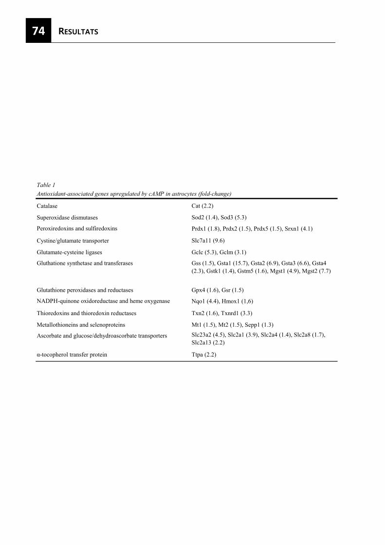
74
Table 1Antioxidant-associated genes upregulated by cAMP in astrocytes (fold-change)
Catalase Cat (2.2)
Superoxidase dismutases Sod2 (1.4), Sod3 (5.3)
Peroxiredoxins and sulfiredoxins Prdx1 (1.8), Prdx2 (1.5), Prdx5 (1.5), Srxn1 (4.1)
Cystine/glutamate transporter Slc7a11 (9.6)
Glutamate-cysteine ligases Gclc (5.3), Gclm (3.1)
Gluthatione synthetase and transferases Gss (1.5), Gsta1 (15.7), Gsta2 (6.9), Gsta3 (6.6), Gsta4 (2.3), Gstk1 (1.4), Gstm5 (1.6), Mgst1 (4.9), Mgst2 (7.7)
Glutathione peroxidases and reductases Gpx4 (1.6), Gsr (1.5)
NADPH-quinone oxidoreductase and heme oxygenase Nqo1 (4.4), Hmox1 (1,6)
Thioredoxins and thioredoxin reductases Txn2 (1.6), Txnrd1 (3.3)
Metallothioneins and selenoproteins Mt1 (1.5), Mt2 (1.5), Sepp1 (1.3)
Ascorbate and glucose/dehydroascorbate transporters Slc23a2 (4.5), Slc2a1 (3.9), Slc2a4 (1.4), Slc2a8 (1.7), Slc2a13 (2.2)
α-tocopherol transfer protein Ttpa (2.2)
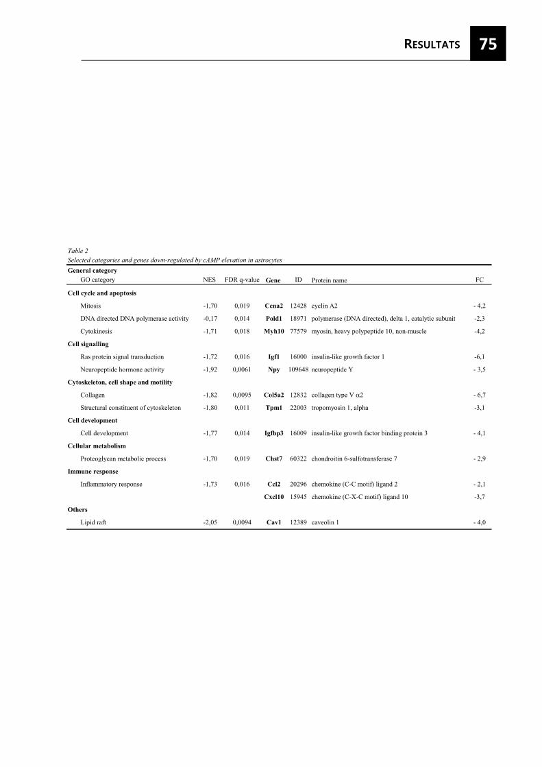
75
Table 2Selected categories and genes down-regulated by cAMP elevation in astrocytesGeneral category
GO category NES FDR q-value Gene ID Protein name FC
Cell cycle and apoptosis
Mitosis -1,70 0,019 Ccna2 12428 cyclin A2 - 4,2
DNA directed DNA polymerase activity -0,17 0,014 Pold1 18971 polymerase (DNA directed), delta 1, catalytic subunit -2,3
Cytokinesis -1,71 0,018 Myh10 77579 myosin, heavy polypeptide 10, non-muscle -4,2
Cell signalling
Ras protein signal transduction -1,72 0,016 Igf1 16000 insulin-like growth factor 1 -6,1
Neuropeptide hormone activity -1,92 0,0061 Npy 109648 neuropeptide Y - 3,5
Cytoskeleton, cell shape and motility
Collagen -1,82 0,0095 Col5a2 12832 collagen type V �2 - 6,7
Structural constituent of cytoskeleton -1,80 0,011 Tpm1 22003 tropomyosin 1, alpha -3,1
Cell development
Cell development -1,77 0,014 Igfbp3 16009 insulin-like growth factor binding protein 3 - 4,1
Cellular metabolism
Proteoglycan metabolic process -1,70 0,019 Chst7 60322 chondroitin 6-sulfotransferase 7 - 2,9
Immune response
Inflammatory response -1,73 0,016 Ccl2 20296 chemokine (C-C motif) ligand 2 - 2,1
Cxcl10 15945 chemokine (C-X-C motif) ligand 10 -3,7
Others
Lipid raft -2,05 0,0094 Cav1 12389 caveolin 1 - 4,0
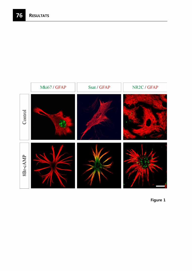
76
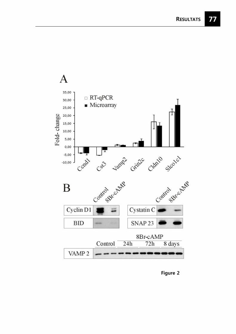
77
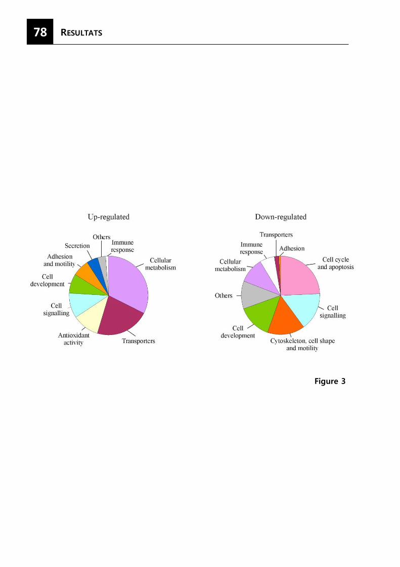
78
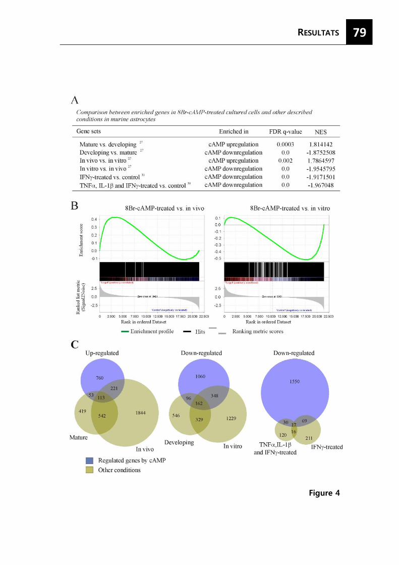
79
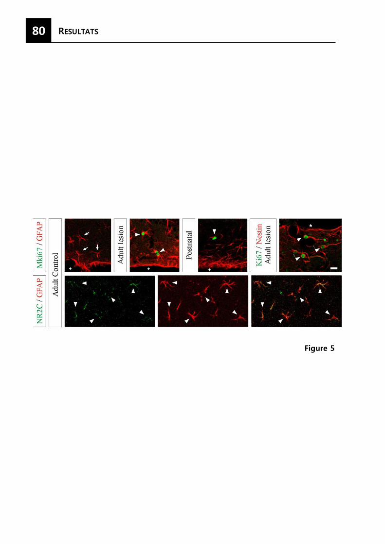
80
81
LL’AMP cíclic regula l’expressió de proteïnes exocítiques i potencia la secreció regulada
de pèptids en astròcits en cultiu
Sonia Paco1 , Mª Ángeles Margelí1 , Vesa M. Olkkonen2, Akane Imai3, Joan Blasi4,
Reiner Fischer-Colbrie5, Fernando Aguado1 1Departament de Biologia Cel·lular, Universitat de Barcelona, Barcelona E-08028, Spain.2Department of Molecular
Medicine, National Public Health Institute, Helsinki, Finland.3Department of Biochemistry, The Nippon Dental
University, Niigata, Japan. 4Department de Patologia i Terapèutica Experimental, IDIBELL-Universitat de Barcelona,
L'Hospitalet de Llobregat, Spain.5Department of Pharmacology, Medical University of Innsbruck, Austria.
Publicat a la revista J Neurochem. 2009 Jul;110(1):143-56.
Resum
L’alliberació vesicular de neurotransmissors, pèptids i factors neurotròfics per part dels
astròcits influencia el desenvolupament, la funció y la plasticitat neuronal. Malgrat això, els
mecanismes moleculars de la secreció en els astròcits són poc coneguts. En aquest estudi
demostrem que els astròcits en cultiu expressen diverses proteïnes SNARE i isoformes de
Munc 18, sent sintaxina-4, SNAP23, VAMP3 i Munc18c les majoritàries. L’expressió de les
proteïnes típicament implicades en la secreció regulada per calci de neurotransmissors i
hormones s’incrementa específicament en cultius d’astròcits per la presència de 8Br-cAMP,
usat com a agent diferenciador. Contràriament, l’administració de citocines pro-inflamatòries
que provoquen la formació de glia reactiva indueix l’augment de l’expressió de proteïnes
exocítiques de distribució més ubiqua. A més, els tractaments a llarg termini amb 8Br-cAMP
incrementen marcadament el contingut cel·lular del marcador de vesícules peptídic
secretogranina-2. Els assajos d’alliberació realitzats en astròcits tractats amb 8Br-cAMP
mostren que tant l’alliberació basal com l’estimulada de secretogranina-2 són dependents de
la concentració intracel·lular de calci. Tal i com es mostra amb l’alliberació de la hormona
quimèrica ANP.emd en cèl·lules transfectades, el tractament amb 8Br-cAMP en astròcits
potencia l’alliberació de pèptids regulada per calci. Per tant, concloem que els astròcits
expressen, respecte a les neurones, un patró distintiu de components moleculars necessari per
l’exocitosi i que aquest patró varia en funció de l’estat en que es troben. L’augment de la
secreció regulada de pèptids i la sobreexpressió de determinades proteïnes exocítiques en
astròcits tractats amb 8Br-cAMP suggereix un important paper de la secreció dependent de
calci en cèl·lules glials diferenciades.

*Department of Cell Biology, University of Barcelona, Barcelona, Spain
�National Institute for Health and Welfare, and FIMM, Institute for Molecular Medicine Finland, University of Helsinki, Biomedicum,
Helsinki, Finland
�Department of Biochemistry, The Nippon Dental University, Niigata, Japan
§Department of Pathology and Experimental Therapeutics, IDIBELL-University of Barcelona, L’Hospitalet de Llobregat, Barcelona,
Spain
¶Department of Pharmacology, Medical University of Innsbruck, Innsbruck, Austria
Astrocyte-secreted peptidergic and non-peptidergic transmit-ters regulate development, physiology and pathology ofneuronal circuits (Araque et al. 2001; Volterra and Meldolesi2005). For instance, astroglial-derived thrombospondins andApoE-containing lipoproteins control synaptogenesis, whileastrocytic glutamate, ATP and D-serine modulate synaptictransmission and network activity (Mauch et al. 2001; Fellinet al. 2004; Christopherson et al. 2005; Pascual et al. 2005;Panatier et al. 2006; Perea and Araque 2007). Moreover,proinflammatory cytokines, growth factors and amino acidsreleased from glia have been associated with neurologicaldisorders and neurodegenerative diseases (Allan andRothwell 2001; Halassa et al. 2007). Although certainchemical messengers can be released from astrocytes bymechanism other than exocytosis, secretory vesicular fusion
is considered as the main astroglial pathway in the glia-neuron signaling (Evanko et al. 2004; Volterra and Meldolesi2005; Montana et al. 2006). As astrocytes can communicate
Received January 8, 2009; revised manuscript received April 9, 2009;accepted April 11, 2009.Address correspondence and reprint requests to Dr Fernando Aguado,
Department of Cell Biology, University of Barcelona, Av. Diagonal 645,Barcelona E-08028, Spain. E-mail: [email protected] used: BAPTA, 1,2-bis(2-amino-phenoxy)ethane-N, N,
N¢, N¢-tetraacetic acid; GFAP, glial fibrillary acidic protein; GFP, greenflourescent protein; IFN, interferon; IL, interleukin; PBS, phosphate-buffered saline; SCG-2, secretogranin-2; SDS–PAGE, sodium dodecylsulfate–polyacrylamide gel electrophoresis; SNAP, synaptosomal-asso-ciated protein; SNAREs, N-ethylmaleimide-sensitive factor attachmentprotein receptors; TNF, tumor necrosis factor; TPA, phorbol-12-myri-state-13-acetate; VAMP, vesicle-associated membrane protein.
Abstract
Vesicular transmitter release from astrocytes influences neu-
ronal development, function and plasticity. However, secretory
pathways and the involved molecular mechanisms in astrog-
lial cells are poorly known. In this study, we show that a variety
of SNARE and Munc18 isoforms are expressed by cultured
astrocytes, with syntaxin-4, Munc18c, SNAP-23 and VAMP-3
being the most abundant variants. Exocytotic protein expres-
sion was differentially regulated by activating and differenti-
ating agents. Specifically, proteins controlling Ca2+-dependent
secretion in neuroendocrine cells were up-regulated after
long-term 8Br-cAMP administration in astrocytes, but not by
proinflammatory cytokines. Moreover, 8Br-cAMP treatment
greatly increased the cellular content of the peptidic vesicle
marker secretogranin-2. Release assays performed on
cAMP-treated astrocytes showed that basal and stimulated
secretogranin-2 secretion are dependent on [Ca2+]i. As shown
release of the chimeric hormone ANP.emd from transfected
cells, cAMP-induced differentiation in astrocytes enhances
Ca2+-regulated peptide secretion. We conclude that astroglial
cells display distinctive molecular components for exocytosis.
Moreover, the regulation of both exocytotic protein expression
and Ca2+-dependent peptide secretion in astrocytes by
differentiating and activating agents suggest that glial secre-
tory pathways are adjusted in different physiological states.
Keywords: astrocyte, calcium, microglia, Munc18, regulated
secretion, SNARE.
J. Neurochem. (2009) 110, 143–156.
JOURNAL OF NEUROCHEMISTRY | 2009 | 110 | 143–156 doi: 10.1111/j.1471-4159.2009.06116.x
� 2009 The AuthorsJournal Compilation � 2009 International Society for Neurochemistry, J. Neurochem. (2009) 110, 143–156 143
with neighboring neurons, synapses and capillaries by raisingthe intracellular free Ca2+ concentration [Ca2+]i, a transmitterrelease regulated by Ca2+ in astrocytes was originallyproposed (Haydon and Carmignoto 2006; Iadecola andNedergaard 2007). It has now been shown that astrocytesrelease gliotransmitters such as glutamate and ATP by aCa2+-dependent exocytosis (for review, see Montana et al.2006). However, the intracellular transport pathways in thesecells are as yet poorly characterized (Bezzi et al. 2004; Chenet al. 2005; Mothet et al. 2005; Bowser and Khakh 2007;Zhang et al. 2007; Li et al. 2008). Even less is known aboutthe Ca2+-dependence of the peptide secretion from astrocytes(Calegari et al. 1999; Krzan et al. 2003).
The core secretory machinery for vesicle exocytosisconsists of N-ethylmaleimide-sensitive factor attachmentprotein receptors (SNAREs) and sec1/Munc18-like proteins(Jahn et al. 2003). During vesicular secretion the plasmamembrane Q-SNAREs syntaxin and synaptosomal-associatedprotein (SNAP)-25 interact with the vesicular R-SNAREsvesicle-associated membrane protein (VAMP) (also known assynaptobrevin) to form a fusion complex which is tightlyregulated by Munc18 (Jahn and Scheller 2006; Toonen andVerhage 2007). Each exocytotic protein is represented bymultiple, closely related isoforms: SNAP-25 and -23; syn-taxin-1, -2, -3 and -4; VAMP-1, -2 and -3; Munc18a, b and c(Jahn and Scheller 2006; Toonen and Verhage 2007).Distinctive sets of exocytotic protein isoforms have beenassociated with different exocytosis events. For instance, theset of syntaxin-1A/SNAP-25/VAMP-2/Munc18a is classicallyinvolved in Ca2+ regulated secretion in neurons and endocrinecells; the cell surface translocation of the glucose transporter 4in insulin-responding cells and the tumor necrosis factor(TNF)a release from macrophages depends on syntaxin-4/SNAP-23/VAMP-2-3/Munc18c; and apical membrane trafficin epithelial cells are controlled by syntaxin-3/SNAP-23/VAMP-3/Munc18b (Riento et al. 1998; Pagan et al. 2003;Toonen and Verhage 2007; Smithers et al. 2008). In recentyears, considerable attention has been focused on the astrocyteexpression and function of the originally described neuronalspecific SNAREs (Montana et al. 2006). Thus, several studieshave demonstrated that the neuronal-enriched VAMP-2 isexpressed in astrocytes and involved in glial Ca2+-dependentrelease of non-peptidergic transmitters (Parpura et al. 1995;Araque et al. 2000; Zhang et al. 2004a; Pascual et al. 2005;Perea and Araque 2007). However, other SNARE isoforms,including SNAP-23 and VAMP-3, which are barely expressedin neurons but importantly involved in different non-neuronalsecretory processes, were robustly detected in astrocytes(Parpura et al. 1995; Hepp et al. 1999; Maienschein et al.1999). As astrocytes are not specialized in secretion but ratherdisplay a broader range of functions, it is expected that themolecular machinery for astroglial exocytosis differs fromthat operating in neurons. To the best of our knowledge,molecular exocytotic machinery in astroglial cells and its
regulation under different cellular states has not yet beencomprehensively identified. In this study, we show thedetailed expression pattern of exocytotic protein isoforms incultured astrocytes. Moreover, we report regulation of theexpression of these proteins in glial cells exposed toproinflammatory and differentiating agents. Finally, we founda correlation between over-expression of certain exocytoticproteins and enhancement of the Ca2+-regulated peptidesecretion in cAMP-differentiated astrocytes.
Material and methods
Antibodies and reagentsActin, syntaxin-1 and b-tubulin isotype III (Tuj-1) monoclonal
antibodies were obtained from Sigma-Aldrich (Diesenhofen,
Germany). Antibodies to syntaxin-2, VAMPs, SNAP-23, and csp
were purchased from Synaptic Systems (SYSY, Goettingen,
Germany). Antibodies against Munc18a, synaptogyrin and syn-
taxin-4 were obtained from BD Transduction Laboratories (San
Jose, CA, USA), glial fibrillary acidic protein (GFAP), GluR5, and
NG2 were obtained from Chemicon (Temecula, CA, USA)
and CD11b from Serotec (Oxford, UK). Synaptophysin, p-STAT-1
and SNAP-25 monoclonal antibodies were obtained from Dako-
Cytomation (Glostrup, Denmark), Zymed Laboratories (South San
Francisco, CA, USA) and Sternberg Monoclonals Inc. (Baltimore,
MD, USA), respectively. Monoclonal antibodies to Rip, SV-2,
Na+/K+ ATPase alpha1 subunit and b-tubulin were purchased from
Developmental Studies Hybridoma Bank (Department of Biological
Sciences, The University of Iowa, Iowa city, IA, USA). Polyclonal
antibodies against syntaxin-3, syntaxin-4, Munc18b, Munc18c, and
secretogranin-2 (SCG-2) were described elsewhere (Fischer-Colbrie
et al. 1993; Riento et al. 1998; Torrejon-Escribano et al. 2002; Imai
et al. 2004). Antibodies against green fluorescent protein (GFP)
were raised in female New Zealand rabbits and were generated and
purificated as described previously (Majo et al. 1998).Recombinant mouse TNFa, interleukin (IL)-b1 and interferon
(IFN) c were purchased from Preprotech, Inc. (London, UK) and
8Br-cAMP from Biolog, Life Science Institute (Bremen, Germany).
1,2-bis(2-amino-phenoxy)ethane-N, N, N¢, N¢-tetraacetic acid (BAP-
TA)-AM was from Invitrogen (Paisley, UK). Most chemicals and
cell culture reagents were obtained from Sigma-Aldrich and Gibco
(Invitrogen), respectively.
Cell cultureThe epithelial MDCK, glioma C6 (ATCC, Rockville, MD, USA)
and U373 (ECACC, Salisbury, UK) cell lines were grown in
Dulbecco’s Modified Eagle’s Medium supplemented with 10% fetal
bovine serum with penicillin/streptomycin at 37�C in a 5% CO2
atmosphere.
Astrocyte and neuron cultures were obtained from OF1 mice
(Charles River Laboratories, Inc., Lyon, France). Animals were kept
under controlled temperature (22 ± 2�C), humidity (40–60%) and
light (12 h cycles) and treated in accordance with the European
Community Council Directive (86/609/EEC) on animal welfare.
Astrocyte cultures were prepared from the cerebral cortex of 2-
day-old mice as described previously (McCarthy and de Vellis 1980).
Briefly, the cerebral cortex was isolated and the meninges were
Journal Compilation � 2009 International Society for Neurochemistry, J. Neurochem. (2009) 110, 143–156� 2009 The Authors
144 | S. Paco et al.
carefully dissected away. Cortical tissues were then minced and
incubated in 0.25% trypsin and 0.01% Dnase. Dissociated cells were
seeded in flasks and grown in high-glucose Dulbecco’s Modified
Eagle’s Medium and F-12 (1 : 1) containing 10% fetal bovine serum,
10 mM HEPES and penicillin/streptomycin at 37�C in a 5% CO2
incubator. At confluence (days 10–12), the flasks were shaken
overnight and the cells were rinsed, detached and subcultured onto
poly-L-lysine-coated plastic culture dishes and coverslips. Under
these conditions, cell cultures were essentially formed by astro-
cytes and virtually devoid of neurons (92.2 ± 1.3% and 0.2 ± 0.2%
GFAP+ and Tuj-1+ cells, respectively) (Fig. S1). Moreover, a small
percentage of microglia (5.2 ± 1% CD11b+ cells) and immature
oligodendrocytes (2.6 ± 1.2% NG2+ cells) was also detected
(Fig. S1). Treatments with 0.01–1 mM 8Br-cAMP, 20 ng/mL
TNFa, 20 ng/mL IL-b1, and 20 ng/mL IFNc were administrated
1 day after subculturing. Astrocytes cultures were transfected with
ANP.emd (Burke et al. 1997) usingLipofectamin� 2000 (Invitrogen)
and peptide release was assayed 48 h later. Because transfection
efficacy was low in replated astrocytes, ANP.emd release was assayed
in primary astrocyte cultures (> 95% of transfected cells were
GFAP+).
Neuronal cultures were prepared from the cerebral cortex of E16
mouse embryos. After trypsin and Dnase treatment, dissociated cells
were seeded onto poly-L-lysine-coated culture plates. Neurons were
grown in Neurobasal medium containing B27 and N2 supplements
(Invitrogen), glutamine and penicillin/streptomycin at 37�C in a 5%
CO2 atmosphere for 10 days. During the first 4 days in culture were
also supplemented with 20 lg/mL 5-Fluoro-2¢-deoxyuridine and
50 lg/mL uridine (Sigma-Aldrich) to inhibit mitotic activity of glial
cells. Tuj-1 immunostaining showed that more than 99% of the cells
were neurons.
Western blottingCultured cells and tissues were homogenized in ice-cold lysis buffer
containing 50 mM Tris–HCl, pH 7.4, 150 mM NaCl, 5 mM MgCl2,
1 mM EGTA, 1% Triton X-100, and protease inhibitor cocktail
(Roche Diagnostics, Indianapolis, IN, USA). Samples of post-
nuclear lysates were electrophoresed in 12% and 8% sodium dodecyl
sulfate–polyacrylamide gel electrophoresis (SDS–PAGE) (Bio-Rad,
Hercules, CA, USA) and then transferred to nitrocellulose mem-
branes (Whatman� Schleicher & Schuell, Keene, NH, USA). Urea–
SDS gels were used to improve resolution of proteins of low
molecular weight (Ruiz-Montasell et al. 1996). The membranes
were blocked in a solution containing 5% non-fat milk powder in
Tris-buffered saline with Tween (140 mM NaCl, 10 mM Tris/HCl,
pH 7.4 and 0.1% Tween 20) for 1 h at 22–24�C and then incubated
with primary antibodies in blocking buffer for 2 h or overnight at
22–24�C or 4�C, respectively. After several washes with blocking
solution, the membranes were incubated for 1 h with horseradish
peroxidase-conjugated secondary antibodies (Vector, Burlingame,
CA, USA). Bound antibodies were visualized with enhanced
chemiluminescence reagents ECL� (GE Healthcare, Buckingham-
shire, UK). Blot images were captured with a scanner and
densitometric values were obtained using ImageJ software (NIH,
Bethesda, MD, USA). To obtain crude membranes, cultured cells
were homogenized in 5 mM Tris–HCl, pH 7.4 containing 0.25 mM
sucrose, 1 mM MgCl2, 1 mM EGTA and protease inhibitors. The
suspension was centrifuged at 600 g for 10 min at 4�C and the
resulting post-nuclear supernatant was centrifuged at 120 000 g for
30 min at 4�C. The pellet was solubilized in 10 mM Tris–HCl, pH
7.4, 2 mM EGTA and 1% Triton X-100. Density gradient subcellular
fractions from astrocytes cultures were obtained as previously
reported (Majo et al. 1998). Cell cultures were homogenized in
0.3 M sucrose, 4 mM HEPES, pH 7.0 and protease inhibitors and
centrifuged for 5 min at 600 g. The post-nuclear supernatants were
collected, and subfraccionated by centrifugation on a 0.3–2.0 M
discontinuous sucrose gradients at 100 000 g overnight in a SW41
rotor (Beckman Instruments, Inc., Palo Alto, CA, USA). Aliquots
from gradient fractions were precipitated with 5% trichloroacetic
acid, resuspended in SDS-sample buffer and analyzed by SDS–
PAGE and immunoblotting.
ImmunocytochemistryCells grown on glass coverslips or plastic tissue dishes were
washed with ice-cold phosphate-buffered saline (PBS) solution and
fixed with 4% paraformaldehyde in PBS for 15 min. To suppress
non-specific binding, cells were incubated in 10% serum-PBS
containing 0.02% Triton X-100, 0.2% glycine and 0.2% gelatin for
1 h at 22–24�C. Then, cells were incubated with the different
primary antibodies diluted in 1% serum-PBS containing 0.2%
gelatin and 0.2% gelatin overnight at 4�C. After washing, the
cells were incubated with secondary fluorochrome-conjugated
antibodies (Alexa Fluor 488 and Alexa Fluor 568; Molecular
Probes, Eugene, OR, USA) and the nuclei were stained with 4¢,6-diamidino-2-phenylindole (DAPI; Molecular Probes). The speci-
ficity of the immunoreaction was tested by omitting the primary
antibody or by replacing it with an equivalent concentration of
non-specific IgG. No immunostaining was observed in these
conditions. Double immunofluorescences were performed by using
primary antibodies raised in different species. Cell-type percent-
ages in control and treated cultures were evaluated in cells grown
on similar plastic dishes used for western blotting analysis.
Fluorescent images were obtained with the Olympus fluorescent
BX-61 (Olympus Corp., Tokyo, Japan) and Leica TCS SPE
scanning confocal microscopes. (Leica microsystems GmbH,
Wetzlar, Germany).
Peptide releaseAstrocytes were seeded in poly-L-lysine-coated tissue culture dishes,
to minimize cell loss; 100- and 35-mm culture plates were used to
analyze SCG-2 and ANP.emd release, respectively. Before the
release experiment, astrocytes were rinsed twice with serum-free
medium and deprivated for 2 h. Cells were rinsed again and
incubated in serum free-medium containing 1 lM ionomycin and
0.1 lM phorbol-12-myristate-13-acetate (TPA) or dimethylsulfox-
ide as control at 37�C in 5% CO2. At the indicated times after
incubation, conditioned medium was collected and lysis buffer was
added to the cells (see above). Cell media were centrifuged at 600 gfor 5 min to remove dislodged cells and proteins were precipitated
with 5% trichloroacetic acid using bovine serum albumin as carrier.
In some cases, proteins from the media were concentrated by
Amicon� Ultra-15 Centrifugal Filter Devices (Millipore Corpora-
tion; Beverly, MA, USA) and then precipitated with 5% trichloro-
acetic acid. SCG-2 and ANP.emd were analyzed both in media and
cells by SDS–PAGE and immunoblotting with antibodies against
SCG-2 and GFP, respectively.
� 2009 The AuthorsJournal Compilation � 2009 International Society for Neurochemistry, J. Neurochem. (2009) 110, 143–156
Exocytotic proteins and peptide release in astrocytes | 145
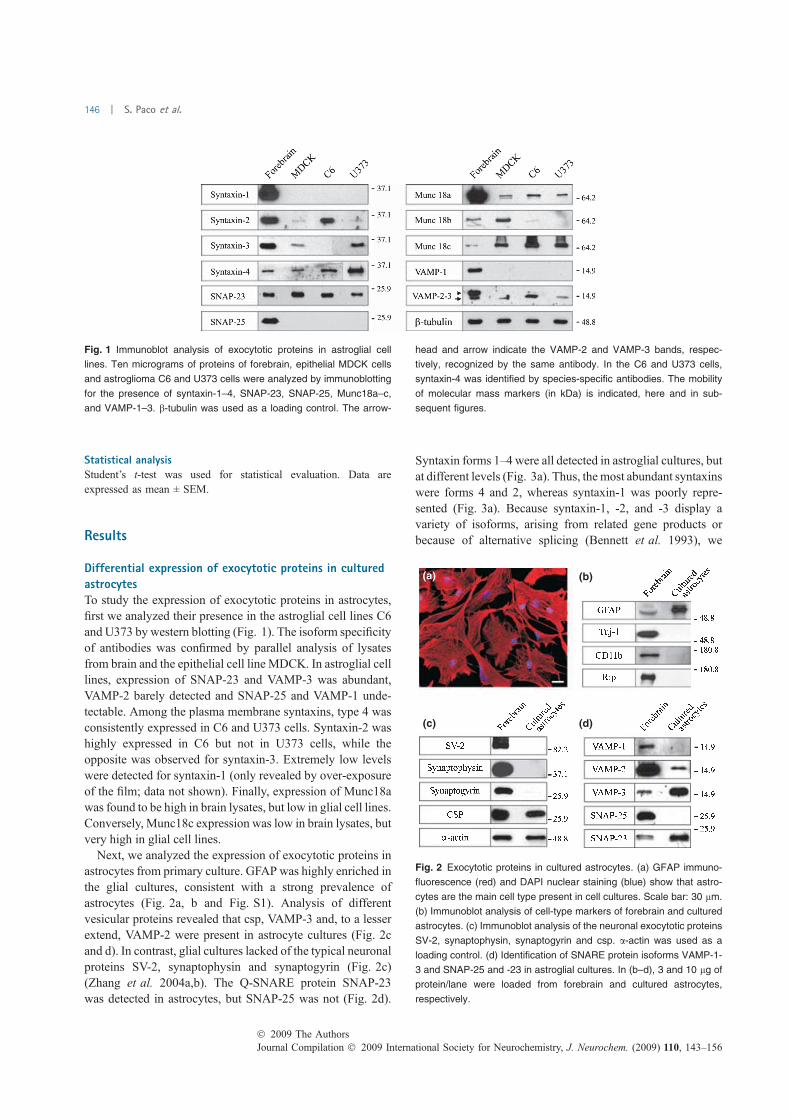
Statistical analysisStudent’s t-test was used for statistical evaluation. Data are
expressed as mean ± SEM.
Results
Differential expression of exocytotic proteins in culturedastrocytesTo study the expression of exocytotic proteins in astrocytes,first we analyzed their presence in the astroglial cell lines C6and U373 by western blotting (Fig. 1). The isoform specificityof antibodies was confirmed by parallel analysis of lysatesfrom brain and the epithelial cell line MDCK. In astroglial celllines, expression of SNAP-23 and VAMP-3 was abundant,VAMP-2 barely detected and SNAP-25 and VAMP-1 unde-tectable. Among the plasma membrane syntaxins, type 4 wasconsistently expressed in C6 and U373 cells. Syntaxin-2 washighly expressed in C6 but not in U373 cells, while theopposite was observed for syntaxin-3. Extremely low levelswere detected for syntaxin-1 (only revealed by over-exposureof the film; data not shown). Finally, expression of Munc18awas found to be high in brain lysates, but low in glial cell lines.Conversely, Munc18c expression was low in brain lysates, butvery high in glial cell lines.
Next, we analyzed the expression of exocytotic proteins inastrocytes from primary culture. GFAP was highly enriched inthe glial cultures, consistent with a strong prevalence ofastrocytes (Fig. 2a, b and Fig. S1). Analysis of differentvesicular proteins revealed that csp, VAMP-3 and, to a lesserextend, VAMP-2 were present in astrocyte cultures (Fig. 2cand d). In contrast, glial cultures lacked of the typical neuronalproteins SV-2, synaptophysin and synaptogyrin (Fig. 2c)(Zhang et al. 2004a,b). The Q-SNARE protein SNAP-23was detected in astrocytes, but SNAP-25 was not (Fig. 2d).
Syntaxin forms 1–4 were all detected in astroglial cultures, butat different levels (Fig. 3a). Thus, the most abundant syntaxinswere forms 4 and 2, whereas syntaxin-1 was poorly repre-sented (Fig. 3a). Because syntaxin-1, -2, and -3 display avariety of isoforms, arising from related gene products orbecause of alternative splicing (Bennett et al. 1993), we
Fig. 1 Immunoblot analysis of exocytotic proteins in astroglial cell
lines. Ten micrograms of proteins of forebrain, epithelial MDCK cells
and astroglioma C6 and U373 cells were analyzed by immunoblotting
for the presence of syntaxin-1–4, SNAP-23, SNAP-25, Munc18a–c,
and VAMP-1–3. b-tubulin was used as a loading control. The arrow-
head and arrow indicate the VAMP-2 and VAMP-3 bands, respec-
tively, recognized by the same antibody. In the C6 and U373 cells,
syntaxin-4 was identified by species-specific antibodies. The mobility
of molecular mass markers (in kDa) is indicated, here and in sub-
sequent figures.
(a)
(c) (d)
(b)
Fig. 2 Exocytotic proteins in cultured astrocytes. (a) GFAP immuno-
fluorescence (red) and DAPI nuclear staining (blue) show that astro-
cytes are the main cell type present in cell cultures. Scale bar: 30 lm.
(b) Immunoblot analysis of cell-type markers of forebrain and cultured
astrocytes. (c) Immunoblot analysis of the neuronal exocytotic proteins
SV-2, synaptophysin, synaptogyrin and csp. a-actin was used as a
loading control. (d) Identification of SNARE protein isoforms VAMP-1-
3 and SNAP-25 and -23 in astroglial cultures. In (b–d), 3 and 10 lg of
protein/lane were loaded from forebrain and cultured astrocytes,
respectively.
Journal Compilation � 2009 International Society for Neurochemistry, J. Neurochem. (2009) 110, 143–156� 2009 The Authors
146 | S. Paco et al.
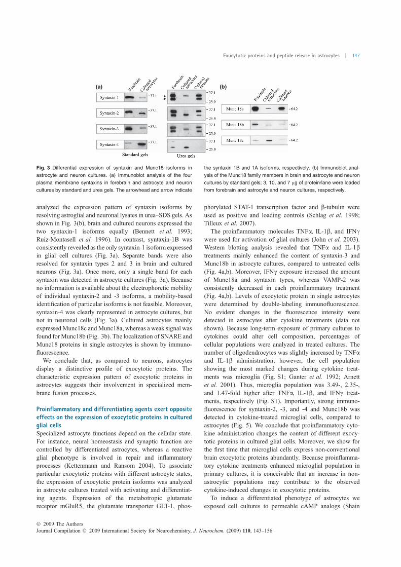
analyzed the expression pattern of syntaxin isoforms byresolving astroglial and neuronal lysates in urea–SDS gels. Asshown in Fig. 3(b), brain and cultured neurons expressed thetwo syntaxin-1 isoforms equally (Bennett et al. 1993;Ruiz-Montasell et al. 1996). In contrast, syntaxin-1B wasconsistently revealed as the only syntaxin-1 isoform expressedin glial cell cultures (Fig. 3a). Separate bands were alsoresolved for syntaxin types 2 and 3 in brain and culturedneurons (Fig. 3a). Once more, only a single band for eachsyntaxin was detected in astrocyte cultures (Fig. 3a). Becauseno information is available about the electrophoretic mobilityof individual syntaxin-2 and -3 isoforms, a mobility-basedidentification of particular isoforms is not feasible. Moreover,syntaxin-4 was clearly represented in astrocyte cultures, butnot in neuronal cells (Fig. 3a). Cultured astrocytes mainlyexpressedMunc18c andMunc18a, whereas a weak signal wasfound for Munc18b (Fig. 3b). The localization of SNARE andMunc18 proteins in single astrocytes is shown by immuno-fluorescence.
We conclude that, as compared to neurons, astrocytesdisplay a distinctive profile of exocytotic proteins. Thecharacteristic expression pattern of exocytotic proteins inastrocytes suggests their involvement in specialized mem-brane fusion processes.
Proinflammatory and differentiating agents exert oppositeeffects on the expression of exocytotic proteins in culturedglial cellsSpecialized astrocyte functions depend on the cellular state.For instance, neural homeostasis and synaptic function arecontrolled by differentiated astrocytes, whereas a reactiveglial phenotype is involved in repair and inflammatoryprocesses (Kettenmann and Ransom 2004). To associateparticular exocytotic proteins with different astrocyte states,the expression of exocytotic protein isoforms was analyzedin astrocyte cultures treated with activating and differentiat-ing agents. Expression of the metabotropic glutamatereceptor mGluR5, the glutamate transporter GLT-1, phos-
phorylated STAT-1 transcription factor and b-tubulin wereused as positive and loading controls (Schlag et al. 1998;Tilleux et al. 2007).
The proinflammatory molecules TNFa, IL-1b, and IFNcwere used for activation of glial cultures (John et al. 2003).Western blotting analysis revealed that TNFa and IL-1btreatments mainly enhanced the content of syntaxin-3 andMunc18b in astrocyte cultures, compared to untreated cells(Fig. 4a,b). Moreover, IFNc exposure increased the amountof Munc18a and syntaxin types, whereas VAMP-2 wasconsistently decreased in each proinflammatory treatment(Fig. 4a,b). Levels of exocytotic protein in single astrocyteswere determined by double-labeling immunofluorescence.No evident changes in the fluorescence intensity weredetected in astrocytes after cytokine treatments (data notshown). Because long-term exposure of primary cultures tocytokines could alter cell composition, percentages ofcellular populations were analyzed in treated cultures. Thenumber of oligodendrocytes was slightly increased by TNFaand IL-1b administration; however, the cell populationshowing the most marked changes during cytokine treat-ments was microglia (Fig. S1; Ganter et al. 1992; Arnettet al. 2001). Thus, microglia population was 3.49-, 2.35-,and 1.47-fold higher after TNFa, IL-1b, and IFNc treat-ments, respectively (Fig. S1). Importantly, strong immuno-fluorescence for syntaxin-2, -3, and -4 and Munc18b wasdetected in cytokine-treated microglial cells, compared toastrocytes (Fig. 5). We conclude that proinflammatory cyto-kine administration changes the content of different exocy-totic proteins in cultured glial cells. Moreover, we show forthe first time that microglial cells express non-conventionalbrain exocytotic proteins abundantly. Because proinflamma-tory cytokine treatments enhanced microglial population inprimary cultures, it is conceivable that an increase in non-astrocytic populations may contribute to the observedcytokine-induced changes in exocytotic proteins.
To induce a differentiated phenotype of astrocytes weexposed cell cultures to permeable cAMP analogs (Shain
(a) (b)
Fig. 3 Differential expression of syntaxin and Munc18 isoforms in
astrocyte and neuron cultures. (a) Immunoblot analysis of the four
plasma membrane syntaxins in forebrain and astrocyte and neuron
cultures by standard and urea gels. The arrowhead and arrow indicate
the syntaxin 1B and 1A isoforms, respectively. (b) Immunoblot anal-
ysis of the Munc18 family members in brain and astrocyte and neuron
cultures by standard gels; 3, 10, and 7 lg of protein/lane were loaded
from forebrain and astrocyte and neuron cultures, respectively.
� 2009 The AuthorsJournal Compilation � 2009 International Society for Neurochemistry, J. Neurochem. (2009) 110, 143–156
Exocytotic proteins and peptide release in astrocytes | 147
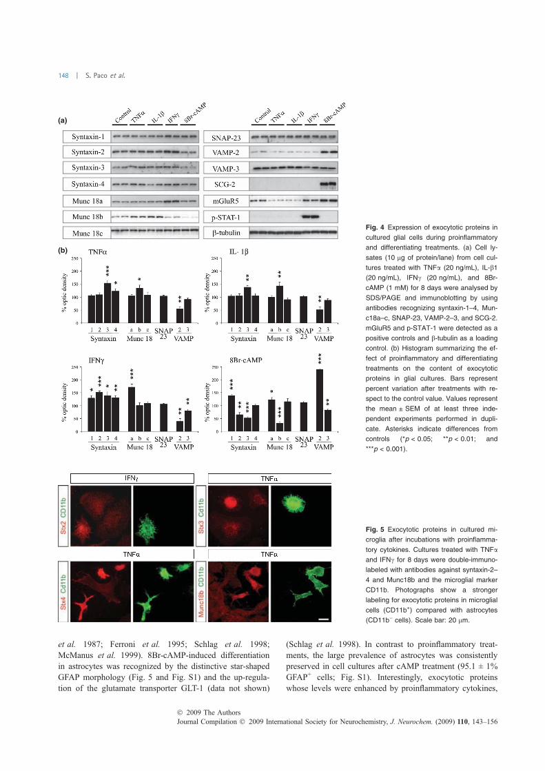
et al. 1987; Ferroni et al. 1995; Schlag et al. 1998;McManus et al. 1999). 8Br-cAMP-induced differentiationin astrocytes was recognized by the distinctive star-shapedGFAP morphology (Fig. 5 and Fig. S1) and the up-regula-tion of the glutamate transporter GLT-1 (data not shown)
(Schlag et al. 1998). In contrast to proinflammatory treat-ments, the large prevalence of astrocytes was consistentlypreserved in cell cultures after cAMP treatment (95.1 ± 1%GFAP+ cells; Fig. S1). Interestingly, exocytotic proteinswhose levels were enhanced by proinflammatory cytokines,
(a)
(b)
Fig. 4 Expression of exocytotic proteins in
cultured glial cells during proinflammatory
and differentiating treatments. (a) Cell ly-
sates (10 lg of protein/lane) from cell cul-
tures treated with TNFa (20 ng/mL), IL-b1
(20 ng/mL), IFNc (20 ng/mL), and 8Br-
cAMP (1 mM) for 8 days were analysed by
SDS/PAGE and immunoblotting by using
antibodies recognizing syntaxin-1–4, Mun-
c18a–c, SNAP-23, VAMP-2–3, and SCG-2.
mGluR5 and p-STAT-1 were detected as a
positive controls and b-tubulin as a loading
control. (b) Histogram summarizing the ef-
fect of proinflammatory and differentiating
treatments on the content of exocytotic
proteins in glial cultures. Bars represent
percent variation after treatments with re-
spect to the control value. Values represent
the mean ± SEM of at least three inde-
pendent experiments performed in dupli-
cate. Asterisks indicate differences from
controls (*p < 0.05; **p < 0.01; and
***p < 0.001).
Fig. 5 Exocytotic proteins in cultured mi-
croglia after incubations with proinflamma-
tory cytokines. Cultures treated with TNFa
and IFNc for 8 days were double-immuno-
labeled with antibodies against syntaxin-2–
4 and Munc18b and the microglial marker
CD11b. Photographs show a stronger
labeling for exocytotic proteins in microglial
cells (CD11b+) compared with astrocytes
(CD11b) cells). Scale bar: 20 lm.
Journal Compilation � 2009 International Society for Neurochemistry, J. Neurochem. (2009) 110, 143–156� 2009 The Authors
148 | S. Paco et al.
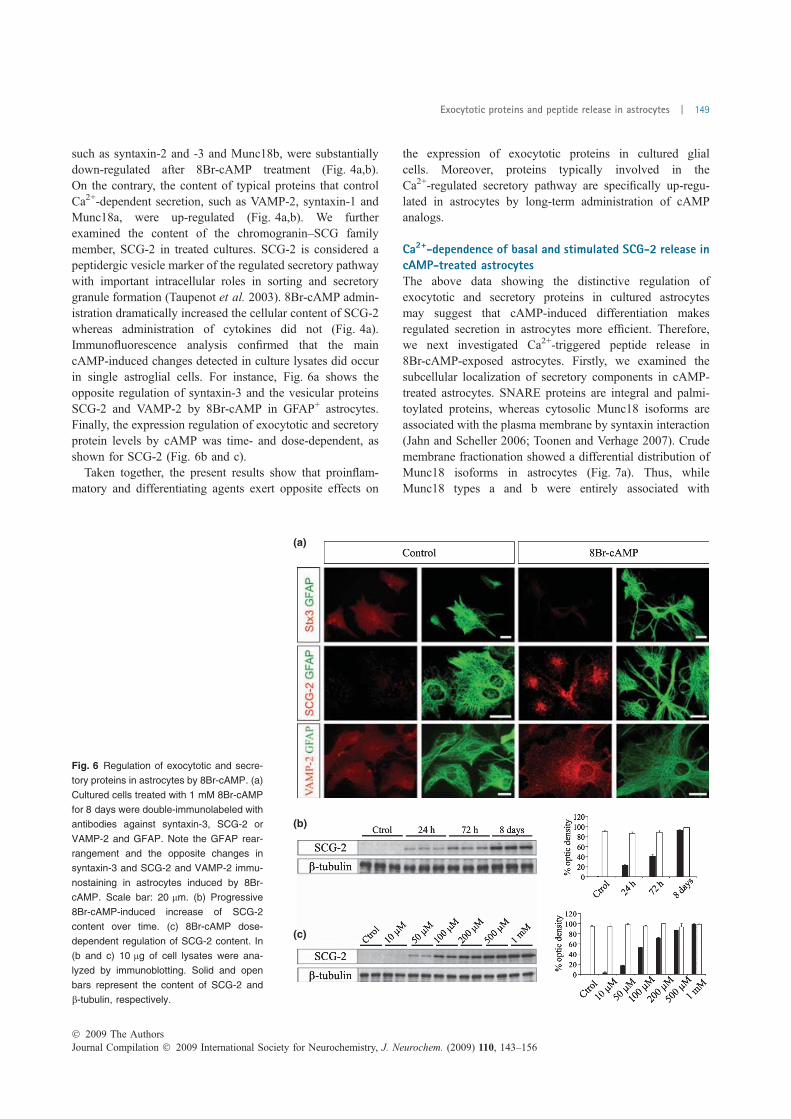
such as syntaxin-2 and -3 and Munc18b, were substantiallydown-regulated after 8Br-cAMP treatment (Fig. 4a,b).On the contrary, the content of typical proteins that controlCa2+-dependent secretion, such as VAMP-2, syntaxin-1 andMunc18a, were up-regulated (Fig. 4a,b). We furtherexamined the content of the chromogranin–SCG familymember, SCG-2 in treated cultures. SCG-2 is considered apeptidergic vesicle marker of the regulated secretory pathwaywith important intracellular roles in sorting and secretorygranule formation (Taupenot et al. 2003). 8Br-cAMP admin-istration dramatically increased the cellular content of SCG-2whereas administration of cytokines did not (Fig. 4a).Immunofluorescence analysis confirmed that the maincAMP-induced changes detected in culture lysates did occurin single astroglial cells. For instance, Fig. 6a shows theopposite regulation of syntaxin-3 and the vesicular proteinsSCG-2 and VAMP-2 by 8Br-cAMP in GFAP+ astrocytes.Finally, the expression regulation of exocytotic and secretoryprotein levels by cAMP was time- and dose-dependent, asshown for SCG-2 (Fig. 6b and c).
Taken together, the present results show that proinflam-matory and differentiating agents exert opposite effects on
the expression of exocytotic proteins in cultured glialcells. Moreover, proteins typically involved in theCa2+-regulated secretory pathway are specifically up-regu-lated in astrocytes by long-term administration of cAMPanalogs.
Ca2+-dependence of basal and stimulated SCG-2 release incAMP-treated astrocytesThe above data showing the distinctive regulation ofexocytotic and secretory proteins in cultured astrocytesmay suggest that cAMP-induced differentiation makesregulated secretion in astrocytes more efficient. Therefore,we next investigated Ca2+-triggered peptide release in8Br-cAMP-exposed astrocytes. Firstly, we examined thesubcellular localization of secretory components in cAMP-treated astrocytes. SNARE proteins are integral and palmi-toylated proteins, whereas cytosolic Munc18 isoforms areassociated with the plasma membrane by syntaxin interaction(Jahn and Scheller 2006; Toonen and Verhage 2007). Crudemembrane fractionation showed a differential distribution ofMunc18 isoforms in astrocytes (Fig. 7a). Thus, whileMunc18 types a and b were entirely associated with
(a)
(b)
(c)
Fig. 6 Regulation of exocytotic and secre-
tory proteins in astrocytes by 8Br-cAMP. (a)
Cultured cells treated with 1 mM 8Br-cAMP
for 8 days were double-immunolabeled with
antibodies against syntaxin-3, SCG-2 or
VAMP-2 and GFAP. Note the GFAP rear-
rangement and the opposite changes in
syntaxin-3 and SCG-2 and VAMP-2 immu-
nostaining in astrocytes induced by 8Br-
cAMP. Scale bar: 20 lm. (b) Progressive
8Br-cAMP-induced increase of SCG-2
content over time. (c) 8Br-cAMP dose-
dependent regulation of SCG-2 content. In
(b and c) 10 lg of cell lysates were ana-
lyzed by immunoblotting. Solid and open
bars represent the content of SCG-2 and
b-tubulin, respectively.
� 2009 The AuthorsJournal Compilation � 2009 International Society for Neurochemistry, J. Neurochem. (2009) 110, 143–156
Exocytotic proteins and peptide release in astrocytes | 149
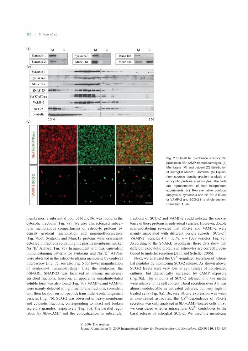
membranes, a substantial pool of Munc18c was found in thecytosolic fractions (Fig. 7a). We also characterized subcel-lular membranous compartments of astrocyte proteins bydensity gradient fractionation and immunofluorescence(Fig. 7b,c). Syntaxin and Munc18 proteins were essentiallydetected in fractions containing the plasma membrane markerNa+/K+ ATPase (Fig. 7b). In agreement with this, equivalentimmunostaining patterns for syntaxins and Na+/K+ ATPasewere observed in the astrocyte plasma membrane by confocalmicroscopy (Fig. 7c, see also Fig. 5 for lower magnificationof syntaxin-4 immunolabeling). Like the syntaxins, thet-SNARE SNAP-23 was localized in plasma membrane-enriched fractions, however, an apparently unpalmitoylatedsoluble form was also found (Fig. 7b). VAMP-2 andVAMP-3were mainly detected in light membrane fractions, consistentwith their location on non-peptide transmitter-containing smallvesicles (Fig. 7b). SCG-2 was observed in heavy membraneand cytosolic fractions, corresponding to intact and brokensecretory granules, respectively (Fig. 7b). The parallel regu-lation by 8Br-cAMP and the colocalization in subcellular
fractions of SCG-2 and VAMP-2 could indicate the coexis-tence of these proteins in individual vesicles. However, doubleimmunolabeling revealed that SCG-2 and VAMP-2 weremainly associated with different vesicle subsets (SCG-2+/VAMP-2+ vesicles 4.7 ± 1.1%, n = 1039 vesicles, Fig. 7c).According to the SNARE hypothesis, these data show thatdifferent exocytotic proteins in astrocytes are correctly posi-tioned to underlie secretion (Jahn and Scheller 2006).
Next, we analyzed the Ca2+-regulated secretion of astrog-lial peptides by monitoring SCG-2 release. As shown above,SCG-2 levels were very low in cell lysates of non-treatedcultures, but dramatically increased by cAMP exposure(Fig. 8a). The amounts of SCG-2 released into the mediawere relative to the cell content. Basal secretion over 3 h wasalmost undetectable in untreated cultures, but very high intreated cells (Fig. 8a). Because SCG-2 expression was weakin non-treated astrocytes, the Ca2+-dependence of SCG-2secretion was only analyzed in 8Br-cAMP-treated cells. First,we considered whether intracellular Ca2+ contributes to thebasal release of astroglial SCG-2. We used the membrane
(a)
(b)
(c)
Fig. 7 Subcellular distribution of exocytotic
proteins in 8Br-cAMP-treated astrocyes. (a)
Membrane (M) and cytosol (C) distribution
of astroglial Munc18 isoforms. (b) Equilib-
rium sucrose density gradient analysis of
exocytotic proteins in astrocytes. The blots
are representative of four independent
experiments. (c) Representative confocal
analysis of syntaxin-4 and Na+/K+ ATPase
or VAMP-2 and SCG-2 in a single section.
Scale bar: 1 lm.
Journal Compilation � 2009 International Society for Neurochemistry, J. Neurochem. (2009) 110, 143–156� 2009 The Authors
150 | S. Paco et al.
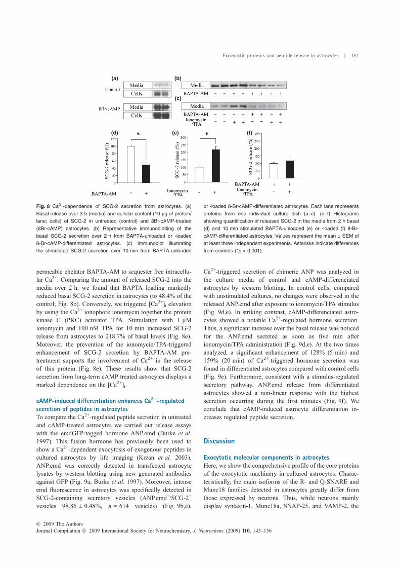
permeable chelator BAPTA-AM to sequester free intracellu-lar Ca2+. Comparing the amount of released SCG-2 into themedia over 2 h, we found that BAPTA loading markedlyreduced basal SCG-2 secretion in astrocytes (to 48.4% of thecontrol; Fig. 8b). Conversely, we triggered [Ca2+]i elevationby using the Ca2+ ionophore ionomycin together the proteinkinase C (PKC) activator TPA. Stimulation with 1 lMionomycin and 100 nM TPA for 10 min increased SCG-2release from astrocytes to 218.7% of basal levels (Fig. 8e).Moreover, the prevention of the ionomycin/TPA-triggeredenhancement of SCG-2 secretion by BAPTA-AM pre-treatment supports the involvement of Ca2+ in the releaseof this protein (Fig. 8e). These results show that SCG-2secretion from long-term cAMP treated astrocytes displays amarked dependence on the [Ca2+]i.
cAMP-induced differentiation enhances Ca2+-regulatedsecretion of peptides in astrocytesTo compare the Ca2+-regulated peptide secretion in untreatedand cAMP-treated astrocytes we carried out release assayswith the emdGFP-tagged hormone ANP.emd (Burke et al.1997). This fusion hormone has previously been used toshow a Ca2+-dependent exocytosis of exogenous peptides incultured astrocytes by life imaging (Krzan et al. 2003).ANP.emd was correctly detected in transfected astrocytelysates by western blotting using new generated antibodiesagainst GFP (Fig. 9a; Burke et al. 1997). Moreover, intenseemd fluorescence in astrocytes was specifically detected inSCG-2-containing secretory vesicles (ANP.emd+/SCG-2+
vesicles 98.86 ± 0.48%, n = 614 vesicles) (Fig. 9b,c).
Ca2+-triggered secretion of chimeric ANP was analyzed inthe culture media of control and cAMP-differenciatedastrocytes by western blotting. In control cells, comparedwith unstimulated cultures, no changes were observed in thereleased ANP.emd after exposure to ionomycin/TPA stimulus(Fig. 9d,e). In striking contrast, cAMP-differenciated astro-cytes showed a notable Ca2+-regulated hormone secretion.Thus, a significant increase over the basal release was noticedfor the ANP.emd secreted as soon as five min afterionomycin/TPA administration (Fig. 9d,e). At the two timesanalyzed, a significant enhancement of 128% (5 min) and159% (20 min) of Ca2+-triggered hormone secretion wasfound in differentiated astrocytes compared with control cells(Fig. 9e). Furthermore, consistent with a stimulus-regulatedsecretory pathway, ANP.emd release from differentiatedastrocytes showed a non-linear response with the highestsecretion occurring during the first minutes (Fig. 9f). Weconclude that cAMP-induced astrocyte differentiation in-creases regulated peptide secretion.
Discussion
Exocytotic molecular components in astrocytesHere, we show the comprehensive profile of the core proteinsof the exocytotic machinery in cultured astrocytes. Charac-teristically, the main isoforms of the R- and Q-SNARE andMunc18 families detected in astrocytes greatly differ fromthose expressed by neurons. Thus, while neurons mainlydisplay syntaxin-1, Munc18a, SNAP-25, and VAMP-2, the
(a) (b)
(c)
(d) (e) (f)
Fig. 8 Ca2+-dependence of SCG-2 secretion from astrocytes. (a)
Basal release over 3 h (media) and cellular content (10 lg of protein/
lane; cells) of SCG-2 in untreated (control) and 8Br-cAMP-treated
(8Br-cAMP) astrocytes. (b) Representative immunoblotting of the
basal SCG-2 secretion over 2 h from BAPTA-unloaded or -loaded
8-Br-cAMP-differentiated astrocytes. (c) Immunoblot illustrating
the stimulated SCG-2 secretion over 10 min from BAPTA-unloaded
or -loaded 8-Br-cAMP-differentiated astrocytes. Each lane represents
proteins from one individual culture dish (a–c). (d–f) Histograms
showing quantification of released SCG-2 in the media from 2 h basal
(d) and 10 min stimulated BAPTA-unloaded (e) or -loaded (f) 8-Br-
cAMP-differentiated astrocytes. Values represent the mean ± SEM of
at least three independent experiments. Asterisks indicate differences
from controls (*p < 0.001).
� 2009 The AuthorsJournal Compilation � 2009 International Society for Neurochemistry, J. Neurochem. (2009) 110, 143–156
Exocytotic proteins and peptide release in astrocytes | 151
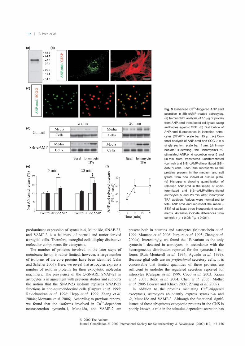
predominant expression of syntaxin-4, Munc18c, SNAP-23,and VAMP-3 is a hallmark of normal and tumor-derivedastroglial cells. Therefore, astroglial cells display distinctivemolecular components for exocytosis.
The number of proteins involved in the later steps ofmembrane fusion is rather limited; however, a large numberof isoforms of the core proteins have been identified (Jahnand Scheller 2006). Here, we reveal that astrocytes express anumber of isoform proteins for their exocytotic molecularmachinery. The prevalence of the Q-SNARE SNAP-23 inastrocytes is in agreement with previous studies and supportsthe notion that the SNAP-23 isoform replaces SNAP-25functions in non-neuroendocrine cells (Parpura et al. 1995;Ravichandran et al. 1996; Hepp et al. 1999; Zhang et al.2004a; Montana et al. 2006). According to previous reports,we found that the isoforms involved in Ca2+-dependentneurosecretion syntaxin-1, Munc18a, and VAMP-2 are
present both in neurons and astrocytes (Maienschein et al.1999; Montana et al. 2006; Parpura et al. 1995; Zhang et al.2004a). Interestingly, we found the 1B variant as the onlysyntaxin-1 detected in astrocytes, in accordance with theheterogeneous distribution reported for the syntaxin-1 iso-forms (Ruiz-Montasell et al. 1996; Aguado et al. 1999).Because glial cells are no professional secretory cells, it isconceivable that limited quantities of these proteins aresufficient to underlie the regulated secretion reported forastrocytes (Calegari et al. 1999; Coco et al. 2003; Krzanet al. 2003; Bezzi et al. 2004; Chen et al. 2005; Mothetet al. 2005 Bowser and Khakh 2007; Zhang et al. 2007).
In addition to the proteins mediating Ca2+-triggeredexocytosis, astrocytes abundantly express syntaxin-4 and-2, Munc18c and VAMP-3. Although the functional signif-icance of these ubiquitous exocytotic proteins in the CNS ispoorly known, a role in the stimulus-dependent secretion has
(a) (b)
(c)
(d)
(e) (f)
Fig. 9 Enhanced Ca2+-triggered ANP.emd
secretion in 8Br-cAMP-treated astrocytes.
(a) Immunoblot analysis of 10 lg of protein
from ANP.emd-transfected cell lysate using
antibodies against GFP. (b) Distribution of
ANP.emd fluorescence in identified astro-
cytes (GFAP+), scale bar: 15 lm. (c) Con-
focal analysis of ANP.emd and SCG-2 in a
single section, scale bar: 1 lm. (d) Immu-
noblots illustrating the ionomycin/TPA-
stimulated ANP.emd secretion over 5 and
20 min from transfected undifferentiated
(control) and 8-Br-cAMP-diferentiated (8Br-
cAMP) cells. Each lane represents all the
proteins present in the medium and cell
lysate from one individual culture plate.
(e) Histograms showing quantification of
released ANP.emd in the media of undif-
ferentiated and 8-Br-cAMP-differentiated
astrocytes 5 and 20 min after ionomycin/
TPA addition. Values were normalized to
total ANP.emd and represent the mean ±
SEM of at least three independent experi-
ments. Asterisks indicate differences from
controls (*p < 0.05; **p < 0.001).
Journal Compilation � 2009 International Society for Neurochemistry, J. Neurochem. (2009) 110, 143–156� 2009 The Authors
152 | S. Paco et al.
been proposed in non-neural cell types (Flaumenhaft 2003;Imai et al. 2004; Borisovska et al. 2005). Therefore, astro-cyte-enriched exocytotic isoforms may participate in regu-lated glial secretion. One basic question about SNAREisoforms is whether co-expression of different members ofthe same family represents a redundant mechanism or confersspecificity (Jahn and Scheller 2006). Recent studies usingknockout mice have suggested that although differentQ-SNARE isoforms can underlie the same exocytoticprocess, particular variants confer distinctive properties tosupport specialized secretory functions (Sørensen et al.2003). Moreover, specific VAMP isoforms selectively drivethe secretion of granule subsets in mast cells (Puri and Roche2008). Thus, it is possible that the presence of a variety ofSNARE isoforms in astrocytes reflects heterogeneity in theglial secretory exocytosis. Particularly, the recent evidencethat astrocytes display a prominent Ca2+-dependent lyso-somal exocytosis suggests that syntaxin-4 and SNAP-23contribute to regulated lysosomal fusion (Rao et al. 2004;Zhang et al. 2007; Li et al. 2008). Finally, as astrocyte-enriched exocytotic proteins are involved in membranefusion processes with non-secretory purposes (i.e. receptor,channel and transporter translocation), specific interactionamong different syntaxin, VAMP, and Munc18 isoformscould underlie astroglial non-secretory exocytosis (Chiereg-atti and Meldolesi 2005; Stenovec et al. 2008). Future loss-of-function experimental approaches are required to elucidatethe contribution of distinctive SNARE isoforms to special-ized glial exocytosis.
Ca2+-regulated exocytotic apparatus and peptide secretionin 8Br-cAMP-differenciated astrocytesMost studies analyzing the regulated secretory pathway inastrocytes have been performed on primary cultures (Calegariet al. 1999; Araque et al. 2000; Coco et al. 2003; Krzan et al.2003; Bezzi et al. 2004; Zhang et al. 2004a,b; Mothet et al.2005; Bowser and Khakh 2007; Zhang et al. 2007; Chenet al. 2005). Although the precise state of cultured astrocytes iscontroversial, it is thought that astroglial cells in culture displaya non-fully differentiated phenotype and are probably acti-vated (Kettenmann and Ransom 2004). Long-term treatmentof astrocytes cultures with permeable cAMP analogs inducesmorphological (stellate appearance), biochemical [productionof GFAP and inositol (1,4,5)-trisphosphate] and physiological(membraneK+ andCl) conductances and glutamate clearance)changes which are believed as indicators for astrocytedifferentiation (Shain et al. 1987; Ferroni et al. 1995; Schlaget al. 1998; McManus et al. 1999). We show here that a set ofproteins typically involved in Ca2+-dependent transmitterrelease are differentially up-regulated in astrocyte culturesduring cAMP-induced differentiation. This finding may sug-gest that cAMP exposure increases regulated secretion inastrocytes. A study showing that long-term addition ofdibutyryl cAMP increased syntaxin-1 and VAMP-2 content
and the evoked-release of glutamate in cultured astrocytessupports these observations (Shiga et al. 2006).
In agreement with previous reports (Fischer-Colbrie et al.1993; Calegari et al. 1999; Zhang et al. 2007), we observedthat SCG-2 content and secretion in cultured astrocytes isextremely low. In contrast, and consistent with the presenceof a functional cAMP-response element in the SCG-2promoter (Taupenot et al. 2003), we found that content ofSCG-2 is dramatically enhanced by 8Br-cAMP in astrocytes.As originally detected in non-treated astrocytes by thesensitive techniques radioimmunoassay and metaboliclabeling (Fischer-Colbrie et al. 1993; Calegari et al. 1999),we show that basal SCG-2 secretion from cAMP-treatedastrocytes is very high. Moreover, we demonstrate here thatbasal SCG-2 secretion in astroglial cells is dependent onresting [Ca2+]i. These findings agree with the proposal thatthe astrocyte-expressed Q-SNARE SNAP-23 leads to highlevels of basal secretion of peptidergic hormones at low[Ca2+]i (Chieregatti et al. 2004). Taking advantage of theincreased levels of intracellular SCG-2 because of 8Br-cAMP, we show here that a Ca2+-triggered release ofendogenous glial peptides is detected in the culture mediumas soon as 5 min after stimulus (data not shown). The Ca2+-dependence on SCG-2 release was further confirmed by thevirtual abolition of ionomycin/TPA-triggered secretion bypre-incubation of astrocytes with the fast Ca2+ chelatorBAPTA-AM. These data support the idea that astrocytesdisplay a Ca2+-regulated secretory pathway (Montana et al.2006). Furthermore, we propose the use of permeable cAMPanalogs as a priming treatment to study regulated secretion ofendogenous peptides in cultured astrocytes.
A Ca2+-dependent release of peptides has been shown incultured astrocytes after ionomycin application by imagingfluorescence of transfected ANP.emd (Krzan et al. 2003).However, under similar conditions, we were not able todetect a increase of released ANP.emd in the medium. Thelimited number of Ca2+-stimulated exocytosis rapidlydetected by live imaging probably cannot be distinguishedfrom the basal secretion in the media by immunoblot analysisover longer times. In contrast to untreated cells, elevation of[Ca2+]i in cAMP-differentiated astrocytes causes an increasein released ANP.emd. This finding indicates that cAMPsignaling improves the regulated secretory pathway inastrocytes. Moreover, the differential changes of exocytoticproteins detected in astrocytes during cAMP-induced differ-entiation suggest the involvement of distinctive exocytoticproteins in regulated peptide release. Because astrocytessecrete proteins that control development and activity ofneuronal circuits, a functional role for Ca2+-regulated releaseof peptides from astrocytes in vivo is conceivable.
Exocytotic proteins in activated gliaTNFa, IL-1b, and IFNc are potent proinflammatory cyto-kines activating astrocytes and microglial cells in vivo and
� 2009 The AuthorsJournal Compilation � 2009 International Society for Neurochemistry, J. Neurochem. (2009) 110, 143–156
Exocytotic proteins and peptide release in astrocytes | 153
in vitro (John et al. 2003). In the present study, we show thatexocytotic protein content in astrocyte cultures is changed byproinflammatory cytokines. Although certain proteins, suchas VAMP-2, were similarly regulated by the three cytokines,other, such as Munc18a and Munc18b, were equally changedby TNFa and IL-1b but differently affected by IFNc. Thesedifferent responses are probably related to divergences in thesignaling initiated by IFNc and those triggered by TNFa andIL-1b. Even though very limited information is availableabout the secretory machinery in activated astrocytes, theidentification of Munc18a (Stxbp1) as a gene induced inresponse to IFNc in cultured astrocytes by microarrayanalysis supports the notion that exocytotic proteins areadjusted during astroglial activation (Halonen et al. 2006).Interestingly, long-term administration of proinflammatoryand differentiating agents exerts opposite effects on theexpression levels of distinctive exocytotic proteins incultured astrocytes. Because astrocytes are very versatile, itis conceivable that expression of exocytotic protein inastrocytes could be regulated during different cellular statesto achieve specific exocytotic processes.
Importantly, the present results show for the first time that aset of exocytotic proteins (i.e. syntaxin-2, -3, and -4) arerobustly expressed bymicroglia. Recently, the over-expressionof these proteins during macrophage activation and theirinvolvement in cytokine release was reported (Pagan et al.2003). Therefore, exocytotic proteins controlling the secretionof adverse mediators in inflammatory diseases have beenproposed as novel targets for therapy (Stow et al. 2006).Because it is believed that cultured microglia display apermanent state of activation, the exocytotic proteins detectedin microglia might reflect the composition of the exocytoticmachinery in activated cells (Kettenmann and Ransom 2004).As microglia and astroglial cells are resident mediators ofimmune and inflammatory responses in the CNS, a role forglial exocytotic proteins in neuroinflammation is suggested.
Acknowledgements
We thank Drs E. Levitan and I. Cristea for ANP.emd and GST-GFP
constructs, respectively; Drs E. Soriano, B. Castellano, F. Perez-
Brangulı, and E. Pozas for helpful discussions; and C. Evans for
editorial assistance. Antibodies to Rip, SV-2, Na+/K+ ATPase a1subunit and b-tubulin were obtained from the Developmental Studies
Hybridoma Bank at the University of Iowa. This study was supported
by Spanish Ministry of Education and Science Grants BFU-2004-
01154 and BFU2007-67889 to FA and BFU2005-02202 to JB.
Supporting information
Additional Supporting Information may be found in the online
version of this article:
Figure S1. Cellular composition of astroglial cultures after
cytokine and cAMP treatments.
Please note: Wiley-Blackwell are not responsible for the content
or functionality of any supporting materials supplied by the authors.
Any queries (other than missing material) should be directed to the
corresponding author for the article.
References
Aguado F., Majo G., Ruiz-Montasell B., Llorens J., Marsal J. and BlasiJ. (1999) Syntaxin 1A and 1B display distinct distribution patternsin the rat peripheral nervous system. Neuroscience 88, 437–446.
Allan S. M. and Rothwell N. J. (2001) Cytokines and acute neurode-generation. Nat. Rev. Neurosci. 2, 734–744.
Araque A., Li N., Doyle R. T. and Haydon P. G. (2000) SNARE protein-dependent glutamate release from astrocytes. J. Neurosci. 20, 666–673.
Araque A., Carmignoto G. and Haydon P. G. (2001) Dynamic signalingbetween astrocytes and neurons. Annu. Rev. Physiol. 63, 795–813.
Arnett H. A., Mason J., Marino M., Suzuki K., Matsushima G. K. andTing J. P. (2001) TNFa promotes proliferation of oligodendrocyteprogenitors and remyelination. Nat. Neurosci. 4, 1116–1122.
Bennett M. K., Garcia-Arraras J. E., Elferink L. A., Peterson K., FlemingA. M., Hazuka C. D. and Scheller R. H. (1993) The syntaxinfamily of vesicular transport receptors. Cell 74, 663–673.
Bezzi P., Gundersen V., Galbete J. L., Seifert G., Steinhauser C., Pilati E.and Volterra A. (2004) Astrocytes contain a vesicular compartmentthat is competent for regulated exocytosis of glutamate. Nat.Neurosci. 7, 613–620.
Borisovska M., Zhao Y., Tsytsyura Y., Glyvuk N., Takamori S., Matti U.,Rettig J., Sudhof T. and Bruns D. (2005) v-SNAREs control exo-cytosis of vesicles from priming to fusion. EMBO J. 24, 2114–2126.
Bowser D. N. and Khakh B. S. (2007) Two forms of single-vesicleastrocyte exocytosis imaged with total internal reflection fluores-cence microscopy. Proc. Natl Acad. Sci. USA 104, 4212–4217.
Burke N. V., Han W., Li D., Takimoto K., Watkins S. C. and LevitanE. S. (1997) Neuronal peptide release is limited by secretorygranule mobility. Neuron 19, 1095–1102.
Calegari F., Coco S., Taverna E., Bassetti M., Verderio C., Corradi N.,Matteoli M. and Rosa P. (1999) A regulated secretory pathway incultured hippocampal astrocytes. J. Biol. Chem. 274, 22539–22547.
Chen X., Wang L., Zhou Y., Zheng L. H. and Zhou Z. (2005) ‘Kiss-and-run’ glutamate secretion in cultured and freshly isolated rathippocampal astrocytes. J. Neurosci. 25, 9236–9243.
Chieregatti E. and Meldolesi J. (2005) Regulated exocytosis: neworganelles for non-secretory purposes. Nat. Rev. Mol. Cell Biol. 6,181–187.
Chieregatti E., Chicka M. C., Chapman E. R. and Baldini G. (2004)SNAP-23 functions in docking/fusion of granules at low Ca2+.Mol. Biol. Cell 15, 1918–1930.
Christopherson K. S., Ullian E. M., Stokes C. C., Mullowney C. E., HellJ. W., Agah A., Lawler J., Mosher D. F., Bornstein P. and Barres B.A. (2005) Thrombospondins are astrocyte-secreted proteins thatpromote CNS synaptogenesis. Cell 120, 421–433.
Coco S., Calegari F., Pravettoni E., Pozzi D., Taverna E., Rosa P.,Matteoli M. and Verderio C. (2003) Storage and release of ATPfrom astrocytes in culture. J. Biol. Chem. 278, 1354–1362.
Evanko D. S., Zhang Q., Zorec R. and Haydon P. G. (2004) Definingpathways of loss and secretion of chemical messengers fromastrocytes. Glia 47, 233–240.
Fellin T., Pascual O., Gobbo S., Pozzan T., Haydon P. G. and Carmig-noto G. (2004) Neuronal synchrony mediated by astrocytic gluta-mate through activation of extrasynaptic NMDA receptors. Neuron43, 729–743.
Journal Compilation � 2009 International Society for Neurochemistry, J. Neurochem. (2009) 110, 143–156� 2009 The Authors
154 | S. Paco et al.
Ferroni S., Marchini C., Schubert P. and Rapisarda C. (1995) Two dis-tinct inwardly rectifying conductances are expressed in long termdibutyryl-cyclic-AMP treated rat cultured cortical astrocytes. FEBSLett. 367, 319–325.
Fischer-Colbrie R., Kirchmair R., Schobert A., Olenik C., Meyer D. K.and Winkler H. (1993) Secretogranin II is synthesized and secretedin astrocyte cultures. J. Neurochem. 60, 2312–2314.
Flaumenhaft R. (2003) Molecular basis of platelet granule secretion.Arterioscler. Thromb. Vasc. Biol. 23, 1152–1160.
Ganter S., Northoff H.,Mannel D. andGebicke-Harter P. J. (1992) Growthcontrol of cultured microglia. J. Neurosci. Res. 33, 218–230.
Halassa M. M., Fellin T. and Haydon P. G. (2007) The tripartite synapse:roles for gliotransmission in health and disease. Trends Mol. Med.13, 54–63.
Halonen S. K., Woods T., McInnerney K. and Weiss L. M. (2006)Microarray analysis of IFNc response genes in astrocytes.J. Neuroimmunol. 175, 19–30.
Haydon P. G. and Carmignoto G. (2006) Astrocyte control of synaptictransmission and neurovascular coupling. Physiol. Rev. 86, 1009–1031.
Hepp R., Perraut M., Chasserot-Golaz S., Galli T., Aunis D., Langley K.and Grant N. J. (1999) Cultured glial cells express the SNAP-25analogue SNAP-23. Glia 27, 181–187.
Iadecola C. and Nedergaard M. (2007) Glial regulation of the cerebralmicrovasculature. Nat. Neurosci. 10, 1369–1376.
Imai A., Nashida T. and Shimomura H. (2004) Roles of Munc18-3 inamylase release from rat parotid acinar cells. Arch. Biochem.Biophys. 422, 175–182.
Jahn R. and Scheller R. H. (2006) SNAREs – engines for membranefusion. Nat. Rev. Mol. Cell Biol. 7, 631–643.
Jahn R., Lang T. and Sudhof T. C. (2003) Membrane fusion. Cell 112,519–533.
John G. R., Lee S. C. and Brosnan C. F. (2003) Cytokines: powerfulregulators of glial cell activation. Neuroscientist 9, 10–22.
Kettenmann H. and Ransom B. R. (2004) Neuroglia, 2nd edn. OxfordUniversity Press, Oxford.
Krzan M., Stenovec M., Kreft M., Pangrsic T., Grilc S., Haydon P. G.and Zorec R. (2003) Calcium-dependent exocytosis of atrialnatriuretic peptide from astrocytes. J. Neurosci. 23, 1580–1583.
Li D., Ropert N., Koulakoff A., Giaume C. and Oheim M. (2008)Lysosomes are the major vesicular compartment undergoing Ca2+-regulated exocytosis from cortical astrocytes. J. Neurosci. 28,7648–7658.
Maienschein V., Marxen M., Volknandt W. and Zimmermann H. (1999)A plethora of presynaptic proteins associated with ATP-storingorganelles in cultured astrocytes. Glia 26, 233–244.
Majo G., Aguado F., Blasi J. and Marsal J. (1998) Synaptobrevin iso-forms in secretory granules and synaptic-like microvesicles inanterior pituitary cells. Life Sci. 62, 607–616.
Mauch D. H., Nagler K., Schumacher S., Goritz C., Muller E.-C., OttoA. and Pfrieger F. W. (2001) CNS synaptogenesis promoted byglia-derived cholesterol. Science 294, 1354–1357.
McCarthy K. D. and de Vellis J. (1980) Preparation of separate astroglialand oligodendroglial cell cultures from rat cerebral tissue. J. CellBiol. 85, 890–902.
McManus M. F., Chen L. C., Vallejo I. and Vallejo M. (1999) Astroglialdifferentiation of cortical precursor cells triggered by activation ofthe cAMP-dependent signaling pathway. J. Neurosci. 19, 9004–9015.
Montana V., Malarkey E. B., Verderio C., Matteoli M. and Parpura V.(2006) Vesicular transmitter release from astrocytes. Glia 54, 700–715.
Mothet J. P., Pollegioni L., Ouanounou G., Martineau M., Fossier P.and Baux G. (2005) Glutamate receptor activation triggers a
calcium-dependent and SNARE protein-dependent release of thegliotransmitter D-serine. Proc. Natl Acad. Sci. USA 102, 5606–5611.
Pagan J. K., Wylie F. G., Joseph S., Widberg C., Bryant N. J., JamesD. E. and Stow J. L. (2003) The t-SNARE syntaxin 4 is regulatedduring macrophage activation to function in membrane traffic andcytokine secretion. Curr. Biol. 13, 56–60.
Panatier A., Theodosis D. T., Mothet J. P., Touquet B., Pollegioni L.,Poulain D. A. and Oliet S. H. (2006) Glia-derived D-serinecontrols NMDA receptor activity and synaptic memory. Cell 125,775–784.
Parpura V., Fang Y., Basarsky T., Jahn R. and Haydon P. G. (1995)Expression of synaptobrevin II, cellubrevin and syntaxin but notSNAP-25 in cultured astrocytes. FEBS Lett. 377, 489–492.
Pascual O., Casper K. B., Kubera C., Zhang J., Revilla-Sanchez R., SulJ. Y., Takano H., Moss S. J., McCarthy K. and Haydon P. G. (2005)Astrocytic purinergic signaling coordinates synaptic networks.Science 310, 113–116.
Perea G. and Araque A. (2007) Astrocytes potentiate transmitterrelease at single hippocampal synapses. Science 317, 1083–1086.
Puri N. and Roche P. A. (2008) Mast cells possess distinct secretorygranule subsets whose exocytosis is regulated by different SNAREisoforms. Proc. Natl Acad. Sci. USA 105, 2580–2585.
Rao S. K., Huynh C., Proux-Gillardeaux V., Galli T. and Andrews N. W.(2004) Identification of SNAREs involved in synaptotagminVII-regulated lysosomal exocytosis. J. Biol. Chem. 279, 20471–20479.
Ravichandran V., Chawla A. and Roche P. A. (1996) Identification ofa novel syntaxin- and synaptobrevin/VAMP-binding protein,SNAP-23, expressed in non-neuronal tissues. J. Biol. Chem. 271,13300–13303.
Riento K., Galli T., Jansson S., Ehnholm C., Lehtonen E. and OlkkonenV. M. (1998) Interaction of Munc-18-2 with syntaxin 3 controls theassociation of apical SNAREs in epithelial cells. J. Cell Sci. 111,2681–2688.
Ruiz-Montasell B., Aguado F., Majo G., Chapman E. R., Canals J. M.,Marsal J. and Blasi J. (1996) Differential distribution of syntaxinisoforms 1A and 1B in the rat central nervous system. Eur. J.Neurosci. 8, 2544–2552.
Schlag B. D., Vondrasek J. R., Munir M., Kalandadze A., Zelenaia O.A., Rothstein J. D. and Robinson M. B. (1998) Regulation of theglial Na+-dependent glutamate transporters by cyclic AMP analogsand neurons. Mol. Pharmacol. 53, 355–369.
Shain W., Forman D. S., Madelian V. and Turner J. N. (1987) Mor-phology of astroglial cells is controlled by b-adrenergic receptors.J. Cell Biol. 105, 2307–2314.
Shiga H., Murakami J., Nagao T., Tanaka M., Kawahara K., Matsuoka I.and Ito E. (2006) Glutamate release from astrocytes is stimulatedvia the appearance of exocytosis during cyclic AMP-inducedmorphologic changes. J. Neurosci. Res. 84, 338–347.
Smithers N. P., Hodgkinson C. P., Cuttle M. and Sale G. J. (2008)Insulin-triggered repositioning of munc18c on syntaxin-4 inGLUT4 signalling. Biochem. J. 410, 255–260.
Sørensen J. B., Nagy G., Varoqueaux F., Nehring R. B., Brose N.,Wilson M. C. and Neher E. (2003) Differential control of thereleasable vesicle pools by SNAP-25 splice variants and SNAP-23.Cell 114, 75–86.
Stenovec M., Kreft M., Grilc S., Pangrsic T. and Zorec R. (2008) EAAT2density at the astrocyte plasma membrane and Ca2+-regulatedexocytosis. Mol. Membr. Biol. 25, 203–215.
Stow J. L., Manderson A. P. and Murray R. Z. (2006) SNAREingimmunity: the role of SNAREs in the immune system. Nat. Rev.Immunol. 6, 919–929.
� 2009 The AuthorsJournal Compilation � 2009 International Society for Neurochemistry, J. Neurochem. (2009) 110, 143–156
Exocytotic proteins and peptide release in astrocytes | 155
Taupenot L., Harper K. L. and O’Connor D. T. (2003) The chro-mogranin-secretogranin family. N. Engl. J. Med. 348, 1134–1149.
Tilleux S., Berger J. and Hermans E. (2007) Induction of astrogliosisby activated microglia is associated with a down-regulation ofmetabotropic glutamate receptor 5. J. Neuroimmunol. 189, 23–30.
Toonen R. F. and Verhage M. (2007) Munc18-1 in secretion: lonelyMunc joins SNARE team and takes control. Trends Neurosci. 30,564–572.
Torrejon-Escribano B., Gomez de Aranda I. and Blasi J. (2002) SNAREexpression and distribution during 3T3-L1 adipocyte differentia-tion. FEBS Lett. 512, 275–281.
Volterra A. and Meldolesi J. (2005) Astrocytes, from brain glue tocommunication elements: the revolution continues. Nat. Rev.Neurosci., 6, 626–640.
Zhang Q., Fukuda M., Van Bockstaele E., Pascual O. and Haydon P. G.(2004a) Synaptotagmin IV regulates glial glutamate release. Proc.Natl Acad. Sci. USA 101, 9441–9446.
Zhang Q., Pangrsic T., Kreft M., Krzan M., Li N., Sul J. Y., Halassa M., VanBockstaele E., Zorec R. and Haydon P. G. (2004b) Fusion-relatedrelease of glutamate from astrocytes. J. Biol. Chem. 279, 12724–12733.
Zhang Z., Chen G., Zhou W., Song A., Xu T., Luo Q., Wang W., Gu X.S. and Duan S. (2007) Regulated ATP release from astrocytesthrough lysosome exocytosis. Nat. Cell Biol. 9, 945–953.
Journal Compilation � 2009 International Society for Neurochemistry, J. Neurochem. (2009) 110, 143–156� 2009 The Authors
156 | S. Paco et al.
97
SSecretogranina III és una granina astrocitària que es sobreexpressa en glia reactiva
Sonia Paco1, Esther Pozas2, Fernando Aguado1 1Department of Cell Biology, University of Barcelona, Barcelona E-08028, Spain.2Department of Brain Ischemia and
Neurodegeneration, Institute for Biomedical Research of Barcelona (IIBB), CSIC-IDIBAPS, Spain
Publicat a la revista Cereb Cortex. 2010 Jun;20(6):1386-97.
Resum
Els astròcits alliberen de manera constitutiva i regulada importants mediadors de la
comunicació glia-neurona. Entre ells trobem transmissors peptídics i no peptídics que
influencien el desenvolupament, la funcionalitat i la plasticitat neuronal. Malgrat això, són
pràcticament inexistents les evidències experimentals de la presència in vivo de components
moleculars de la via de secreció regulada en astròcits. Les granines són marcadors
neuroendocrins de la via de secreció regulada de pèptids. És una família molt àmplia que
inclou la secretogranina III (SgIII). Intracel·lularment, les granines tenen un paper important en
la classificació i agregació de pèptids de la via regulada i conseqüentment en la biogènesi de
vesícules de secreció. A més, la seva proteòlisi dóna lloc a pèptids bioactius amb funcions de
senyalització intercel·lular.
En aquest treball analitzem la producció, la regulació i l’alliberació de la SgIII en astròcits
in vitro i in vivo. Tant les neurones com els astròcits en cultiu sintetitzen i secreten SgIII. A
diferència de les neurones, els astròcits alliberen una forma de SgIII no processada. In vivo, la
SgIII és sintetitzada per ambdues poblacions, encara que els nivells més alts d’expressió es
troben en els astròcits. Característicament, tant a nivell de mRNA com de proteïna, els nivells
d’expressió de SgIII augmenten notablement en astròcits reactius en lesions de l’escorça
cerebral.
Aquests resultats impliquen la SgIII a la via de secreció astroglial in vivo. A més, la
sobreexpressió de SgIII en astròcits reactius suggereix un paper d’aquesta granina glial en els
processos activats en el dany cerebral.

Cerebral Cortex June 2010;20:1386--1397
doi:10.1093/cercor/bhp202
Advance Access publication November 5, 2009
Secretogranin III Is an Astrocyte GraninThat Is Overexpressed in Reactive Glia
Sonia Paco1, Esther Pozas2 and Fernando Aguado1
1Department of Cell Biology, University of Barcelona, Barcelona
E-08028, Spain and 2Department of Brain Ischemia and
Neurodegeneration, Institute for Biomedical Research of
Barcelona, CSIC-IDIBAPS, Barcelona E-08036, Spain
Astrocytes release peptide and nonpeptide transmitters thatinfluence neuronal development, function, and plasticity. However,the molecular components of the astroglial secretory pathways invivo are largely unknown. Here, we analyze in astrocytes theproduction, expression regulation, trafficking, and release ofsecretogranin III (SgIII), a member of the multifunctional graninfamily. We show that astroglial cells in culture synthesize andrelease a nonprocessed form of SgIII. In vivo studies show thatmany neuronal populations produce and transport SgIII. Inparticular, the highest SgIII expression in the cerebral cortex invivo is present in astroglial cells. Both SgIII protein and mRNA areabundantly detected in cortical astrocytes and in Bergmann glialcells. Moreover, the levels of SgIII mRNA and protein in reactiveastrocytes, induced by perforating injury increase dramatically.These results implicate SgIII in the astrocyte secretory pathway invivo and show that its expression is finely regulated during glialactivation. The robust expression of SgIII in astrocytes and itsregulation in the injured brain suggest both intracellular andextracellular roles for this glial granin in the physiology and repair/damage of neuronal circuits.
Keywords: astrocyte, brain injury, glial activation, secretogranin, secretorypathway
Introduction
Exocytotic vesicular secretion of signaling molecules contrib-
utes to glia--neuron communication (Araque et al. 2001;
Volterra and Meldolesi 2005). In particular, astrocyte-secreted
glutamate, adenosine triphosphate (ATP) and D-serine regulate
neuronal physiology by modulating synaptic transmission and
network activity (Pascual et al. 2005; Panatier et al. 2006; Perea
and Araque 2007). In addition to nonpeptide transmitters,
peptides and proteins released from astrocytes, such as growth
factors, cytokines, and extracellular components, control the
development, plasticity, and pathology of neuronal circuits
(Allan and Rothwell 2001; Christopherson et al. 2005).
Although astrocyte vesicular secretion is crucial for neuronal
function, secretory pathways remain poorly characterized. It
has now been shown that astroglial cells contain competent
compartments and molecular machinery for a regulated
secretory pathway (for review, see Montana et al. 2006). Thus,
astrocytes can release gliotransmitters, such as glutamate, ATP,
and neuropeptide Y, from synaptic-like microvesicles, lyso-
somes, and secretory granules in a Ca2+-dependent manner
(Bezzi et al. 2004; Zhang et al. 2007; Ramamoorthy and Whim
2008). However, the molecular components of the transmitter
secretory pathway in glia in vivo are largely unknown (Fiacco
et al. 2009).
A biochemical hallmark of neuroendocrine secretory gran-
ules is the storage and release of uniquely acidic proteins
known as granins, which are considered markers of the
regulated secretory pathway (Taupenot et al. 2003; Helle
2004). The granin family comprises the ‘‘classical’’ members:
chromogranin A (CgA), chromogranin B (CgB), and secretog-
ranin II (SgII); and the 5 less known secretogranin III (SgIII,
1B1075 gene product), secretogranin IV (HISL-19 antigen),
secretogranin V (neuroendocrine secretory protein 7B2),
secretogranin VI (NESP55), and secretogranin VII (the nerve
growth factor inducible protein VGF; Taupenot et al. 2003;
Helle 2004; Montero-Hadjadje et al. 2008). Intracellularly,
granins have a role in the sorting and aggregation of secretory
products in the trans-Golgi network and in the subsequent
biogenesis of secretory granules (Taupenot et al. 2003; Helle
2004). Moreover, proteolytic processing of these proteins, as
for prohormones, gives rise to bioactive peptides (e.g.,
catestatin and secretoneurin) with autocrine, paracrine, and
endocrine functions (Simon et al. 1988; Shyu et al. 2008).
In endocrine cells, SgIII is a key sorting receptor for peptide
hormones (for review, see Takeuchi and Hosaka 2008).
Moreover, SgIII has recently been shown to be expressed and
released by nonneuroendocrine cell types that display a regu-
lated secretory pathway, such as platelets and mast cells
(Coppinger et al. 2004; Prasad et al. 2008). Here, we studied
the little known SgIII in rodent astrocytes. We show that
a nonprocessed form of SgIII is produced and released by
cortical astrocytes in primary cultures. In brain sections, SgIII
transcripts and protein were found in neuronal populations and
abundantly in cortical astrocytes. Moreover, SgIII expression was
specifically upregulated in reactive astrocytes after perforating
brain injury. These results show that SgIII is a reliable
component of the astrocyte secretory pathway and suggest
important roles for glial SgIII in the glia--neuron communication.
Materials and Methods
Antibodies and ReagentsPolyclonal antibodies against SgIII were purchased from Sigma--Aldrich
(Deisenhofen, Germany) and Proteintech Group Inc. (Chicago, IL). SgII
antibodies were kindly provided by Dr R. Fischer-Colbrie. Monoclonal
antibodies against synaptosome-associated protein of 25 kDa (SNAP-
25), glial fibrillary acidic protein (GFAP), neuronal nuclei (NeuN),
carboxypeptidase E (CPE), FLAG, b-tubulin, and vimentin were
obtained from Sternberg Monoclonals Inc. (Baltimore, MD), Chemicon
(Temecula, CA), BD Transduction Laboratories (San Jose, CA), Sigma--
Aldrich, and Developmental Studies Hybridoma Bank (Department of
Biological Sciences, The University of Iowa, Iowa city, IA). Polyclonal
antibody to Olig2 was from Chemicon. FLAG-tagged rat SgIII (1--471)
cDNA was a gift of Dr M. Hosaka. Brefeldin A was obtained by Sigma--
Aldrich and 8Br-cyclic adenosine 3#,5#-monophosphate (cAMP) was
� The Author 2009. Published by Oxford University Press. All rights reserved.
For permissions, please e-mail: [email protected]
at Universitat D
e Barcelona on June 19, 2011
cercor.oxfordjournals.orgD
ownloaded from
from Biolog Life Science Institute (Bremen, Germany). Most chemicals
and cell culture reagents were from Sigma--Aldrich and GIBCO
(Invitrogen, Paisley, UK), respectively.
AnimalsOF1micewere provided byCharles River Laboratories, Inc. (Lyon, France),
andSprague--Dawley ratswereobtained fromtheLaboratoryAnimalService
of the University of Barcelona. Animals were anesthetized with ketamine
(Ketolar, Parke-Davis, Barcelona, Spain) and xylazine (Rompun, Bayer
Healthcare, Kiel, Germany). Stab wound lesions were made as described
previously (Aguado et al. 2002). Under anesthesia, adult animals were fixed
to a stereotactical frame, and the cerebral cortex was then stabbed with
a #11 scalpel blade. Thewoundswere 3-mm long and 2-mmdeepat 1.5mm
from andparallel tomidline and ran 0.5mmaway from the Bregma over the
right parietal cortex. To provoke secretory protein accumulation in neural
bodies, colchicine was administrated intracerebroventricular (i.c.v.)
(Aguado et al. 1999). Seventy-five microgram of colchicine in 10 lL of
phosphate-buffered saline (PBS) was stereotaxically injected in the lateral
ventricle of adult rats. Sham-operated controls only received 10 lL of PBS.
Animals were kept under controlled temperature (22 ± 2 �C), humidity
(40--60%), and light (12-h cycles) and treated in accordance with the
European Community Council Directive (86/609/EEC) on animal welfare.
Every effort was made to minimize animal suffering. In all in vivo
experimental conditions, at least 4 animals were used per time-point.
Cell CulturesThe COS kidney epithelial cell line was grown in Dulbecco’s Modified
Eagle’s Medium supplemented with 10% fetal bovine serum with
penicillin/streptomycin at 37 �C in a 5% CO2 atmosphere. COS cell line
was transfected with FLAG-SgIII using Lipofectamin 2000, and peptide
expression was assayed 48 h later.
Astrocyte cultures were prepared from the cerebral cortex of 2-day-old
mice as describedpreviously (Paco et al. 2009). Briefly, the cerebral cortex
was isolated, and the meninges were carefully dissected away. Cortical
tissueswere thenminced and incubated in 0.25% trypsin and 0.01%DNase
(Roche Diagnostics, Mannheim, Germany). Dissociated cells were seeded
in flasks and grown in high-glucose Dulbecco’s Modified Eagle’s Medium,
and F-12 (1:1) containing 10% fetal bovine serum, 10 mM 4-(2-
hydroxyethyl)-1-piperazineethanesulfonic acid and penicillin/streptomy-
cin at 37 �C in a 5% CO2 incubator. At confluence (days 10--12), the flasks
were shaken over night, and the cells were rinsed, detached, and
subcultured at a density of 50 000 cells/cm2 on poly-L-lysine-coated
plastic culture dishes and coverslips. Under these conditions, cell cultures
were essentially formed by astrocytes (>92% GFAP+cells) and virtually
devoidofneurons ( <0.5%Tuj-1+cells). A small percentageofmicroglia and
immature oligodendrocytes (<8% CD11b+and NG2
+cells) was also
detected (Paco et al. 2009). Long-term treatment with 8Br-cAMP did not
change thecellular composition (Pacoet al. 2009).Neuronalcultureswere
prepared from the cerebral cortex of E16 mouse embryos. After trypsin
and DNase treatment, dissociated cells were seeded on poly-L-lysine-
coated culture plates. Neurons were grown at a density of 50 000 cells/
cm2 in Neurobasal medium containing B27 and N2 supplements,
glutamine, and penicillin/streptomycin at 37 �C in a 5% CO2 atmosphere
for 10 days. During the first 4 days in culture, the media were also
supplemented with 20 lg/mL 5-fluoro-2#-deoxyuridine and 50 lg/mL
uridine to inhibit the mitotic activity of glial cells. More than 99% of the
cells were Tuj-1+neurons and less than 1% of the cells were GFAP
+
astrocytes (Paco et al. 2009). In all in vitro conditions, at least 3
independent experiments in duplicate were performed.
Reverse Transcriptase-Polymerase Chain ReactionTo perform polymerase chain reaction (PCR) experiments, total RNA was
purified from the forebrain and from cultured astrocytes of mice using
Trizol Reagent (Invitrogen) following the manufacturer’s instructions. All
RNA obtained was reverse transcribed using pdN6 (random hexamers)
and AMV reverse transcriptase (Amersham Biosciences, GE Healthcare,
Buckinghamshire, UK). Then, cDNAs were then amplified with specific
primer pairs (5#--3#) for SgIII, forward CAATTCAAGCTTTCCCCAAA and
reverse TTTCACTCGGCTTGCTTTCT (NM_009130); GFAP, forward
TGCTAGCTACATCGAGAAGG and reverse TCCTCTGTCTCTTGCATGTT
(NM_001131020.1); SNAP-25, forward CCTCCACTCTTGCTACCTGC and
reverse CTCCTCTGCATCTCCTCCAG (NM_011428.3); and actin, for-
ward ATATCGCTGCGCTGGTCGTC and reverse AGGATGGCGTGAGG-
GAGAGC (NM_007393.3). PCR products were analyzed on 1--2% agarose
gels, and images were captured with a scanner.
Western BlottingCultured cells and tissues were homogenized in ice-cold lysis buffer
containing 50 mM Tris--HCl pH 7.4, 150 mM NaCl, 5 mM MgCl2, 1 mM
ethyleneglycol-bis(2-aminoethylether)-N,N,N’,N’-tetra acetic acid, 1%
Triton X-100, and protease inhibitor cocktail (Roche Diagnostics).
Samples of postnuclear lysates were electrophoresed in 12% sodium
dodecyl sulfate--polyacrylamide gel electrophoresis (SDS--PAGE; BioRad
Laboratories, Hercules, CA) and then transferred to nitrocellulose
membranes (Whatman Schleicher & Schuell, Keene, NH). The
membranes were blocked in a solution containing 5% nonfat milk
powder in tris-buffered saline tween-20 (140 mM NaCl, 10 mM Tris--
HCl, pH 7.4, and 0.1% Tween 20) for 1 h at room temperature and then
incubated with primary antibodies in blocking buffer for 2 h at room
temperature. After several washes in blocking solution, the membranes
were incubated for 1 h with horseradish peroxidase-conjugated
secondary antibodies (VECTOR, Burlingame, CA). Bound antibodies
were visualized with enhanced chemiluminescence reagents (GE
Healthcare). Blot images were captured with a scanner.
Peptide ReleaseAstrocytes and neurons were seeded on poly-L-lysine-coated tissue
culture dishes, to minimize cell loss. Before the release experiment,
cells were serum starved. Brefeldin A 5 lg/mL was incubated with
astrocytes for 12 h and with neurons for 4 h. After release, conditioned
medium was collected, and lysis buffer was added to the cells (see
above). Cell media were centrifuged at 600 g for 5 min to remove
dislodged cells, and proteins were precipitated with 5% trichloroacetic
acid. Secretory proteins were analyzed in both media and cells by SDS--
PAGE and immunoblotting.
ImmunocytochemistryCells grown on glass coverslips were washed in ice-cold PBS and fixed
with 4% paraformaldehyde in PBS for 15 min. Animals were perfused
transcardially under deep anesthesia with 4% paraformaldehyde in
0.1 M phosphate buffer (PB), pH 7.4. The brains were removed from the
skulls, postfixed for 4 h in the same fixative solution, and cryoprotected
overnight at 4 �C by immersion in a 30% sucrose solution in 0.1 M PB.
Forty-micrometer thick frozen sections were obtained with a cryostat
and collected in PBS. Sections processed for the peroxidase method
were soaked for 30 min in PBS containing 10% methanol and 3% H2O2
and subsequently washed in PBS. To suppress nonspecific binding, cell
cultures and brain sections were incubated in 10% serum--PBS
containing 0.1% Triton X-100, 0.2% glycine, and 0.2% gelatin for 1 h
at room temperature. Incubations with the primary antibodies were
carried out overnight at 4 �C in PBS containing 1% fetal calf serum, 0.1%
Triton X-100, and 0.2% gelatin. Some histological sections were processed
using the avidin--biotin--peroxidase method (Vectastain ABC kit, VEC-
TOR). The peroxidase complex was visualized by incubating the sections
with 0.05% diaminobenzidine and 0.01% H2O2 in PBS. Sections were
mounted, dehydrated, and coverslipped in Eukitt. Cell cultures and some
brain sections were processed for immunofluorescence using secondary
fluorochrome-conjugated antibodies (Alexa Fluor 488 and Alexa Fluor
568, Molecular Probes, Eugene, OR), and cell nuclei were stained with
4#,6-diamidino-2-phenylindole (Molecular Probes). Cell-containing cover-
slips and histological sections were mounted with Mowiol. The specificity
of the immunostaining was tested by omitting the primary antibodies or
by replacing them with an equivalent concentration of nonspecific IgG.
No immunostaining was observed in these conditions. Bright field and
fluorescent images were obtained with the Olympus fluorescent BX-61
and Leica TCS SPE scanning confocal microscopes.
Double immunolabeling using primary antibodies raised in the
same species (SgIII and Olig-2) was performed by a sequential
avidin--biotin--peroxidase method (Levey et al. 1986). The first immunos-
tainingwas performed as above. The second immunolabeling reactionwas
Cerebral Cortex June 2010, V 20 N 6 1387
at Universitat D
e Barcelona on June 19, 2011
cercor.oxfordjournals.orgD
ownloaded from
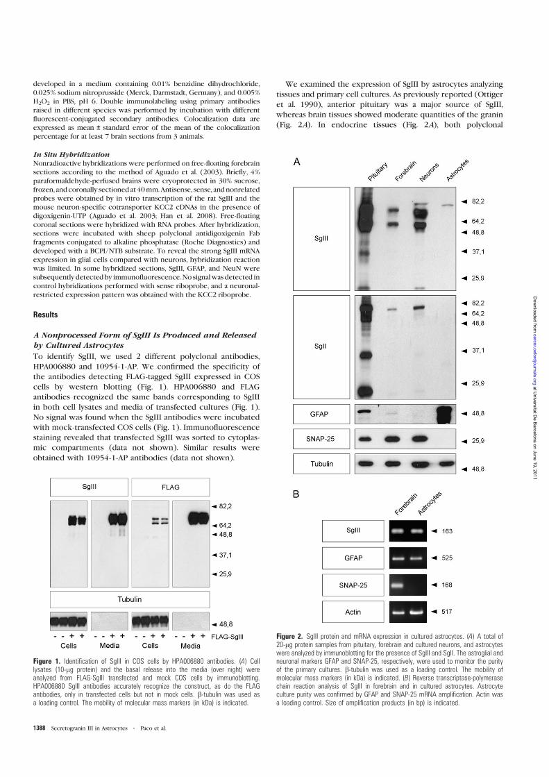
developed in a medium containing 0.01% benzidine dihydrochloride,
0.025% sodium nitroprusside (Merck, Darmstadt, Germany), and 0.005%
H2O2 in PBS, pH 6. Double immunolabeling using primary antibodies
raised in different species was performed by incubation with different
fluorescent-conjugated secondary antibodies. Colocalization data are
expressed as mean ± standard error of the mean of the colocalization
percentage for at least 7 brain sections from 3 animals.
In Situ HybridizationNonradioactive hybridizations were performed on free-floating forebrain
sections according to the method of Aguado et al. (2003). Briefly, 4%
paraformaldehyde-perfused brains were cryoprotected in 30% sucrose,
frozen, andcoronally sectionedat40mm.Antisense, sense, andnonrelated
probes were obtained by in vitro transcription of the rat SgIII and the
mouse neuron-specific cotransporter KCC2 cDNAs in the presence of
digoxigenin-UTP (Aguado et al. 2003; Han et al. 2008). Free-floating
coronal sections were hybridized with RNA probes. After hybridization,
sections were incubated with sheep polyclonal antidigoxigenin Fab
fragments conjugated to alkaline phosphatase (Roche Diagnostics) and
developed with a BCPI/NTB substrate. To reveal the strong SgIII mRNA
expression in glial cells compared with neurons, hybridization reaction
was limited. In some hybridized sections, SgIII, GFAP, and NeuN were
subsequentlydetectedby immunofluorescence.No signalwas detected in
control hybridizations performed with sense riboprobe, and a neuronal-
restricted expression pattern was obtained with the KCC2 riboprobe.
Results
A Nonprocessed Form of SgIII Is Produced and Releasedby Cultured Astrocytes
To identify SgIII, we used 2 different polyclonal antibodies,
HPA006880 and 10954-1-AP. We confirmed the specificity of
the antibodies detecting FLAG-tagged SgIII expressed in COS
cells by western blotting (Fig. 1). HPA006880 and FLAG
antibodies recognized the same bands corresponding to SgIII
in both cell lysates and media of transfected cultures (Fig. 1).
No signal was found when the SgIII antibodies were incubated
with mock-transfected COS cells (Fig. 1). Immunofluorescence
staining revealed that transfected SgIII was sorted to cytoplas-
mic compartments (data not shown). Similar results were
obtained with 10954-1-AP antibodies (data not shown).
We examined the expression of SgIII by astrocytes analyzing
tissues and primary cell cultures. As previously reported (Ottiger
et al. 1990), anterior pituitary was a major source of SgIII,
whereas brain tissues showed moderate quantities of the granin
(Fig. 2A). In endocrine tissues (Fig. 2A), both polyclonal
Figure 1. Identification of SgIII in COS cells by HPA006880 antibodies. (A) Celllysates (10-lg protein) and the basal release into the media (over night) wereanalyzed from FLAG-SgIII transfected and mock COS cells by immunoblotting.HPA006880 SgIII antibodies accurately recognize the construct, as do the FLAGantibodies, only in transfected cells but not in mock cells. b-tubulin was used asa loading control. The mobility of molecular mass markers (in kDa) is indicated.
Figure 2. SgIII protein and mRNA expression in cultured astrocytes. (A) A total of20-lg protein samples from pituitary, forebrain and cultured neurons, and astrocyteswere analyzed by immunoblotting for the presence of SgIII and SgII. The astroglial andneuronal markers GFAP and SNAP-25, respectively, were used to monitor the purityof the primary cultures. b-tubulin was used as a loading control. The mobility ofmolecular mass markers (in kDa) is indicated. (B) Reverse transcriptase-polymerasechain reaction analysis of SgIII in forebrain and in cultured astrocytes. Astrocyteculture purity was confirmed by GFAP and SNAP-25 mRNA amplification. Actin wasa loading control. Size of amplification products (in bp) is indicated.
1388 Secretogranin III in Astrocytes d Paco et al.
at Universitat D
e Barcelona on June 19, 2011
cercor.oxfordjournals.orgD
ownloaded from
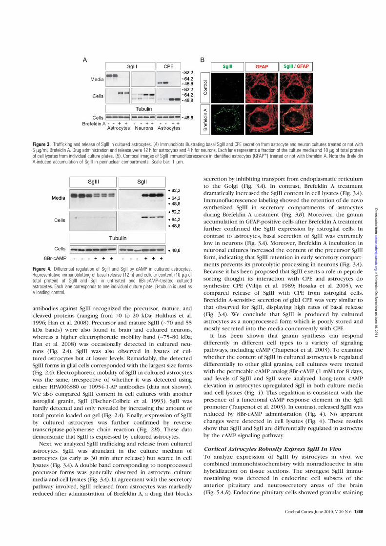
antibodies against SgIII recognized the precursor, mature, and
cleaved proteins (ranging from 70 to 20 kDa; Holthuis et al.
1996; Han et al. 2008). Precursor and mature SgIII (~70 and 55
kDa bands) were also found in brain and cultured neurons,
whereas a higher electrophoretic mobility band (~75--80 kDa;
Han et al. 2008) was occasionally detected in cultured neu-
rons (Fig. 2A). SgIII was also observed in lysates of cul-
tured astrocytes but at lower levels. Remarkably, the detected
SgIII forms in glial cells corresponded with the largest size forms
(Fig. 2A). Electrophoretic mobility of SgIII in cultured astrocytes
was the same, irrespective of whether it was detected using
either HPA006880 or 10954-1-AP antibodies (data not shown).
We also compared SgIII content in cell cultures with another
astroglial granin, SgII (Fischer-Colbrie et al. 1993). SgII was
hardly detected and only revealed by increasing the amount of
total protein loaded on gel (Fig. 2A). Finally, expression of SgIII
by cultured astrocytes was further confirmed by reverse
transcriptase-polymerase chain reaction (Fig. 2B). These data
demonstrate that SgIII is expressed by cultured astrocytes.
Next, we analyzed SgIII trafficking and release from cultured
astrocytes. SgIII was abundant in the culture medium of
astrocytes (as early as 30 min after release) but scarce in cell
lysates (Fig. 3A). A double band corresponding to nonprocessed
precursor forms was generally observed in astrocyte culture
media and cell lysates (Fig. 3A). In agreement with the secretory
pathway involved, SgIII released from astrocytes was markedly
reduced after administration of Brefeldin A, a drug that blocks
secretion by inhibiting transport from endoplasmatic reticulum
to the Golgi (Fig. 3A). In contrast, Brefeldin A treatment
dramatically increased the SgIII content in cell lysates (Fig. 3A).Immunofluorescence labeling showed the retention of de novo
synthetized SgIII in secretory compartments of astrocytes
during Brefeldin A treatment (Fig. 3B). Moreover, the granin
accumulation in GFAP-positive cells after Brefeldin A treatment
further confirmed the SgIII expression by astroglial cells. In
contrast to astrocytes, basal secretion of SgIII was extremely
low in neurons (Fig. 3A). Moreover, Brefeldin A incubation in
neuronal cultures increased the content of the precursor SgIII
form, indicating that SgIII retention in early secretory compart-
ments prevents its proteolytic processing in neurons (Fig. 3A).Because it has been proposed that SgIII exerts a role in peptide
sorting thought its interaction with CPE and astrocytes do
synthesize CPE (Vilijn et al. 1989; Hosaka et al. 2005), we
compared release of SgIII with CPE from astroglial cells.
Brefeldin A-sensitive secretion of glial CPE was very similar to
that observed for SgIII, displaying high rates of basal release
(Fig. 3A). We conclude that SgIII is produced by cultured
astrocytes as a nonprocessed form which is poorly stored and
mostly secreted into the media concurrently with CPE.
It has been shown that granin synthesis can respond
differently in different cell types to a variety of signaling
pathways, including cAMP (Taupenot et al. 2003). To examine
whether the content of SgIII in cultured astrocytes is regulated
differentially to other glial granins, cell cultures were treated
with the permeable cAMP analog 8Br-cAMP (1 mM) for 8 days,
and levels of SgIII and SgII were analyzed. Long-term cAMP
elevation in astrocytes upregulated SgII in both culture media
and cell lysates (Fig. 4). This regulation is consistent with the
presence of a functional cAMP response element in the SgII
promoter (Taupenot et al. 2003). In contrast, released SgIII was
reduced by 8Br-cAMP administration (Fig. 4). No apparent
changes were detected in cell lysates (Fig. 4). These results
show that SgIII and SgII are differentially regulated in astrocyte
by the cAMP signaling pathway.
Cortical Astrocytes Robustly Express SgIII In Vivo
To analyze expression of SgIII by astrocytes in vivo, we
combined immunohistochemistry with nonradioactive in situ
hybridization on tissue sections. The strongest SgIII immu-
nostaining was detected in endocrine cell subsets of the
anterior pituitary and neurosecretory areas of the brain
(Fig. 5A,B). Endocrine pituitary cells showed granular staining
Figure 4. Differential regulation of SgIII and SgII by cAMP in cultured astrocytes.Representative immunoblotting of basal release (12 h) and cellular content (10 lg oftotal protein) of SgIII and SgII in untreated and 8Br-cAMP--treated culturedastrocytes. Each lane corresponds to one individual culture plate. b-tubulin is used asa loading control.
Figure 3. Trafficking and release of SgIII in cultured astrocytes. (A) Immunoblots illustrating basal SgIII and CPE secretion from astrocyte and neuron cultures treated or not with5 lg/mL Brefeldin A. Drug administration and release were 12 h for astrocytes and 4 h for neurons. Each lane represents a fraction of the culture media and 10 lg of total proteinof cell lysates from individual culture plates. (B). Confocal images of SgIII immunofluorescence in identified astrocytes (GFAPþ) treated or not with Brefeldin A. Note the BrefeldinA-induced accumulation of SgIII in perinuclear compartments. Scale bar: 1 lm.
Cerebral Cortex June 2010, V 20 N 6 1389
at Universitat D
e Barcelona on June 19, 2011
cercor.oxfordjournals.orgD
ownloaded from
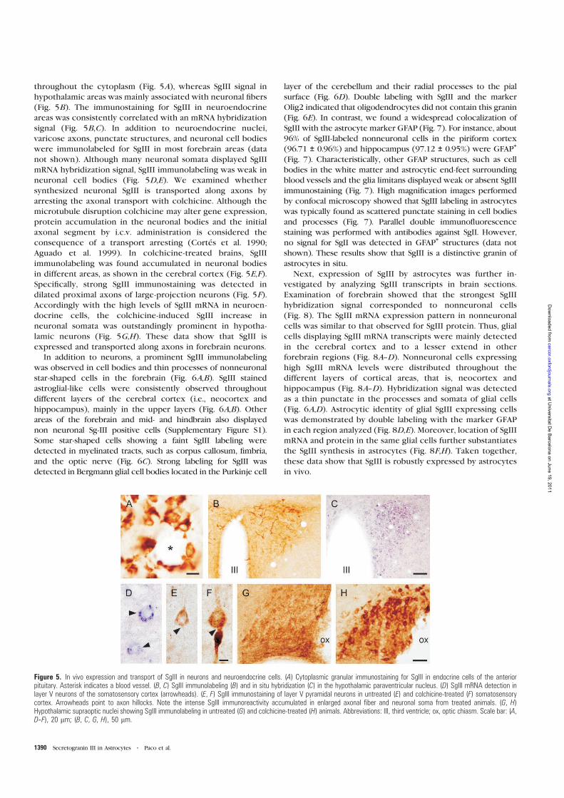
throughout the cytoplasm (Fig. 5A), whereas SgIII signal in
hypothalamic areas was mainly associated with neuronal fibers
(Fig. 5B). The immunostaining for SgIII in neuroendocrine
areas was consistently correlated with an mRNA hybridization
signal (Fig. 5B,C). In addition to neuroendocrine nuclei,
varicose axons, punctate structures, and neuronal cell bodies
were immunolabeled for SgIII in most forebrain areas (data
not shown). Although many neuronal somata displayed SgIII
mRNA hybridization signal, SgIII immunolabeling was weak in
neuronal cell bodies (Fig. 5D,E). We examined whether
synthesized neuronal SgIII is transported along axons by
arresting the axonal transport with colchicine. Although the
microtubule disruption colchicine may alter gene expression,
protein accumulation in the neuronal bodies and the initial
axonal segment by i.c.v. administration is considered the
consequence of a transport arresting (Cortes et al. 1990;
Aguado et al. 1999). In colchicine-treated brains, SgIII
immunolabeling was found accumulated in neuronal bodies
in different areas, as shown in the cerebral cortex (Fig. 5E,F).Specifically, strong SgIII immunostaining was detected in
dilated proximal axons of large-projection neurons (Fig. 5F).Accordingly with the high levels of SgIII mRNA in neuroen-
docrine cells, the colchicine-induced SgIII increase in
neuronal somata was outstandingly prominent in hypotha-
lamic neurons (Fig. 5G,H). These data show that SgIII is
expressed and transported along axons in forebrain neurons.
In addition to neurons, a prominent SgIII immunolabeling
was observed in cell bodies and thin processes of nonneuronal
star-shaped cells in the forebrain (Fig. 6A,B). SgIII stained
astroglial-like cells were consistently observed throughout
different layers of the cerebral cortex (i.e., neocortex and
hippocampus), mainly in the upper layers (Fig. 6A,B). Other
areas of the forebrain and mid- and hindbrain also displayed
non neuronal Sg-III positive cells (Supplementary Figure S1).
Some star-shaped cells showing a faint SgIII labeling were
detected in myelinated tracts, such as corpus callosum, fimbria,
and the optic nerve (Fig. 6C). Strong labeling for SgIII was
detected in Bergmann glial cell bodies located in the Purkinje cell
layer of the cerebellum and their radial processes to the pial
surface (Fig. 6D). Double labeling with SgIII and the marker
Olig2 indicated that oligodendrocytes did not contain this granin
(Fig. 6E). In contrast, we found a widespread colocalization of
SgIII with the astrocyte marker GFAP (Fig. 7). For instance, about
96% of SgIII-labeled nonneuronal cells in the piriform cortex
(96.71 ± 0.96%) and hippocampus (97.12 ± 0.95%) were GFAP+
(Fig. 7). Characteristically, other GFAP structures, such as cell
bodies in the white matter and astrocytic end-feet surrounding
blood vessels and the glia limitans displayed weak or absent SgIII
immunostaining (Fig. 7). High magnification images performed
by confocal microscopy showed that SgIII labeling in astrocytes
was typically found as scattered punctate staining in cell bodies
and processes (Fig. 7). Parallel double immunofluorescence
staining was performed with antibodies against SgII. However,
no signal for SgII was detected in GFAP+structures (data not
shown). These results show that SgIII is a distinctive granin of
astrocytes in situ.
Next, expression of SgIII by astrocytes was further in-
vestigated by analyzing SgIII transcripts in brain sections.
Examination of forebrain showed that the strongest SgIII
hybridization signal corresponded to nonneuronal cells
(Fig. 8). The SgIII mRNA expression pattern in nonneuronal
cells was similar to that observed for SgIII protein. Thus, glial
cells displaying SgIII mRNA transcripts were mainly detected
in the cerebral cortex and to a lesser extend in other
forebrain regions (Fig. 8A--D). Nonneuronal cells expressing
high SgIII mRNA levels were distributed throughout the
different layers of cortical areas, that is, neocortex and
hippocampus (Fig. 8A--D). Hybridization signal was detected
as a thin punctate in the processes and somata of glial cells
(Fig. 6A,D). Astrocytic identity of glial SgIII expressing cells
was demonstrated by double labeling with the marker GFAP
in each region analyzed (Fig. 8D,E). Moreover, location of SgIII
mRNA and protein in the same glial cells further substantiates
the SgIII synthesis in astrocytes (Fig. 8F,H). Taken together,
these data show that SgIII is robustly expressed by astrocytes
in vivo.
Figure 5. In vivo expression and transport of SgIII in neurons and neuroendocrine cells. (A) Cytoplasmic granular immunostaining for SgIII in endocrine cells of the anteriorpituitary. Asterisk indicates a blood vessel. (B, C) SgIII immunolabeling (B) and in situ hybridization (C) in the hypothalamic paraventricular nucleus. (D) SgIII mRNA detection inlayer V neurons of the somatosensory cortex (arrowheads). (E, F) SgIII immunostaining of layer V pyramidal neurons in untreated (E) and colchicine-treated (F) somatosensorycortex. Arrowheads point to axon hillocks. Note the intense SgIII immunoreactivity accumulated in enlarged axonal fiber and neuronal soma from treated animals. (G, H)Hypothalamic supraoptic nuclei showing SgIII immunolabeling in untreated (G) and colchicine-treated (H) animals. Abbreviations: III, third ventricle; ox, optic chiasm. Scale bar: (A,D--F), 20 lm; (B, C, G, H), 50 lm.
1390 Secretogranin III in Astrocytes d Paco et al.
at Universitat D
e Barcelona on June 19, 2011
cercor.oxfordjournals.orgD
ownloaded from
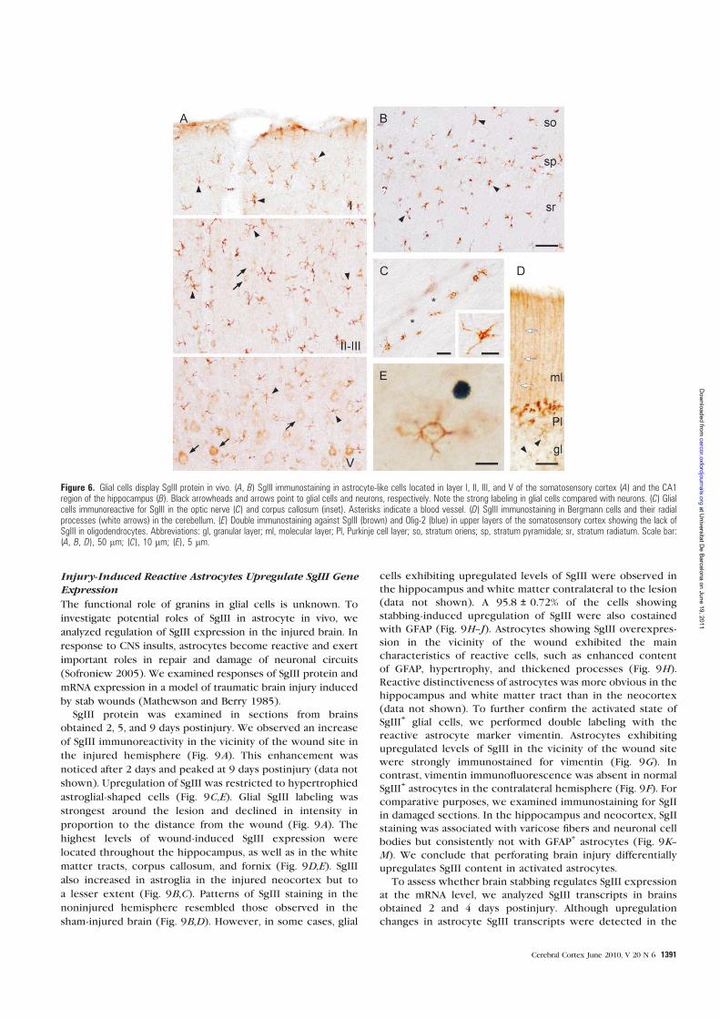
Injury-Induced Reactive Astrocytes Upregulate SgIII GeneExpression
The functional role of granins in glial cells is unknown. To
investigate potential roles of SgIII in astrocyte in vivo, we
analyzed regulation of SgIII expression in the injured brain. In
response to CNS insults, astrocytes become reactive and exert
important roles in repair and damage of neuronal circuits
(Sofroniew 2005). We examined responses of SgIII protein and
mRNA expression in a model of traumatic brain injury induced
by stab wounds (Mathewson and Berry 1985).
SgIII protein was examined in sections from brains
obtained 2, 5, and 9 days postinjury. We observed an increase
of SgIII immunoreactivity in the vicinity of the wound site in
the injured hemisphere (Fig. 9A). This enhancement was
noticed after 2 days and peaked at 9 days postinjury (data not
shown). Upregulation of SgIII was restricted to hypertrophied
astroglial-shaped cells (Fig. 9C,E). Glial SgIII labeling was
strongest around the lesion and declined in intensity in
proportion to the distance from the wound (Fig. 9A). The
highest levels of wound-induced SgIII expression were
located throughout the hippocampus, as well as in the white
matter tracts, corpus callosum, and fornix (Fig. 9D,E). SgIII
also increased in astroglia in the injured neocortex but to
a lesser extent (Fig. 9B,C). Patterns of SgIII staining in the
noninjured hemisphere resembled those observed in the
sham-injured brain (Fig. 9B,D). However, in some cases, glial
cells exhibiting upregulated levels of SgIII were observed in
the hippocampus and white matter contralateral to the lesion
(data not shown). A 95.8 ± 0.72% of the cells showing
stabbing-induced upregulation of SgIII were also costained
with GFAP (Fig. 9H--J). Astrocytes showing SgIII overexpres-
sion in the vicinity of the wound exhibited the main
characteristics of reactive cells, such as enhanced content
of GFAP, hypertrophy, and thickened processes (Fig. 9H).Reactive distinctiveness of astrocytes was more obvious in the
hippocampus and white matter tract than in the neocortex
(data not shown). To further confirm the activated state of
SgIII+
glial cells, we performed double labeling with the
reactive astrocyte marker vimentin. Astrocytes exhibiting
upregulated levels of SgIII in the vicinity of the wound site
were strongly immunostained for vimentin (Fig. 9G). In
contrast, vimentin immunofluorescence was absent in normal
SgIII+astrocytes in the contralateral hemisphere (Fig. 9F). For
comparative purposes, we examined immunostaining for SgII
in damaged sections. In the hippocampus and neocortex, SgII
staining was associated with varicose fibers and neuronal cell
bodies but consistently not with GFAP+astrocytes (Fig. 9K--
M). We conclude that perforating brain injury differentially
upregulates SgIII content in activated astrocytes.
To assess whether brain stabbing regulates SgIII expression
at the mRNA level, we analyzed SgIII transcripts in brains
obtained 2 and 4 days postinjury. Although upregulation
changes in astrocyte SgIII transcripts were detected in the
Figure 6. Glial cells display SgIII protein in vivo. (A, B) SgIII immunostaining in astrocyte-like cells located in layer I, II, III, and V of the somatosensory cortex (A) and the CA1region of the hippocampus (B). Black arrowheads and arrows point to glial cells and neurons, respectively. Note the strong labeling in glial cells compared with neurons. (C) Glialcells immunoreactive for SgIII in the optic nerve (C) and corpus callosum (inset). Asterisks indicate a blood vessel. (D) SgIII immunostaining in Bergmann cells and their radialprocesses (white arrows) in the cerebellum. (E) Double immunostaining against SgIII (brown) and Olig-2 (blue) in upper layers of the somatosensory cortex showing the lack ofSgIII in oligodendrocytes. Abbreviations: gl, granular layer; ml, molecular layer; Pl, Purkinje cell layer; so, stratum oriens; sp, stratum pyramidale; sr, stratum radiatum. Scale bar:(A, B, D), 50 lm; (C), 10 lm; (E), 5 lm.
Cerebral Cortex June 2010, V 20 N 6 1391
at Universitat D
e Barcelona on June 19, 2011
cercor.oxfordjournals.orgD
ownloaded from
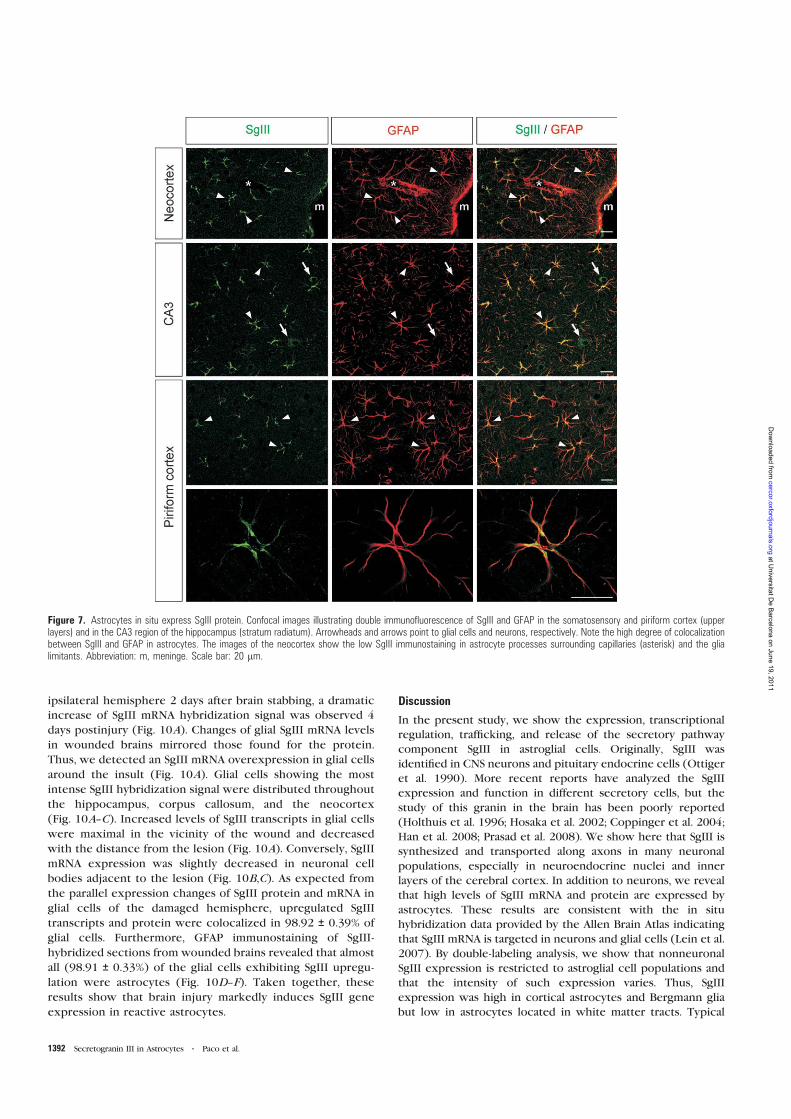
ipsilateral hemisphere 2 days after brain stabbing, a dramatic
increase of SgIII mRNA hybridization signal was observed 4
days postinjury (Fig. 10A). Changes of glial SgIII mRNA levels
in wounded brains mirrored those found for the protein.
Thus, we detected an SgIII mRNA overexpression in glial cells
around the insult (Fig. 10A). Glial cells showing the most
intense SgIII hybridization signal were distributed throughout
the hippocampus, corpus callosum, and the neocortex
(Fig. 10A--C). Increased levels of SgIII transcripts in glial cells
were maximal in the vicinity of the wound and decreased
with the distance from the lesion (Fig. 10A). Conversely, SgIIImRNA expression was slightly decreased in neuronal cell
bodies adjacent to the lesion (Fig. 10B,C). As expected from
the parallel expression changes of SgIII protein and mRNA in
glial cells of the damaged hemisphere, upregulated SgIII
transcripts and protein were colocalized in 98.92 ± 0.39% of
glial cells. Furthermore, GFAP immunostaining of SgIII-
hybridized sections from wounded brains revealed that almost
all (98.91 ± 0.33%) of the glial cells exhibiting SgIII upregu-
lation were astrocytes (Fig. 10D--F). Taken together, these
results show that brain injury markedly induces SgIII gene
expression in reactive astrocytes.
Discussion
In the present study, we show the expression, transcriptional
regulation, trafficking, and release of the secretory pathway
component SgIII in astroglial cells. Originally, SgIII was
identified in CNS neurons and pituitary endocrine cells (Ottiger
et al. 1990). More recent reports have analyzed the SgIII
expression and function in different secretory cells, but the
study of this granin in the brain has been poorly reported
(Holthuis et al. 1996; Hosaka et al. 2002; Coppinger et al. 2004;
Han et al. 2008; Prasad et al. 2008). We show here that SgIII is
synthesized and transported along axons in many neuronal
populations, especially in neuroendocrine nuclei and inner
layers of the cerebral cortex. In addition to neurons, we reveal
that high levels of SgIII mRNA and protein are expressed by
astrocytes. These results are consistent with the in situ
hybridization data provided by the Allen Brain Atlas indicating
that SgIII mRNA is targeted in neurons and glial cells (Lein et al.
2007). By double-labeling analysis, we show that nonneuronal
SgIII expression is restricted to astroglial cell populations and
that the intensity of such expression varies. Thus, SgIII
expression was high in cortical astrocytes and Bergmann glia
but low in astrocytes located in white matter tracts. Typical
Figure 7. Astrocytes in situ express SgIII protein. Confocal images illustrating double immunofluorescence of SgIII and GFAP in the somatosensory and piriform cortex (upperlayers) and in the CA3 region of the hippocampus (stratum radiatum). Arrowheads and arrows point to glial cells and neurons, respectively. Note the high degree of colocalizationbetween SgIII and GFAP in astrocytes. The images of the neocortex show the low SgIII immunostaining in astrocyte processes surrounding capillaries (asterisk) and the glialimitants. Abbreviation: m, meninge. Scale bar: 20 lm.
1392 Secretogranin III in Astrocytes d Paco et al.
at Universitat D
e Barcelona on June 19, 2011
cercor.oxfordjournals.orgD
ownloaded from
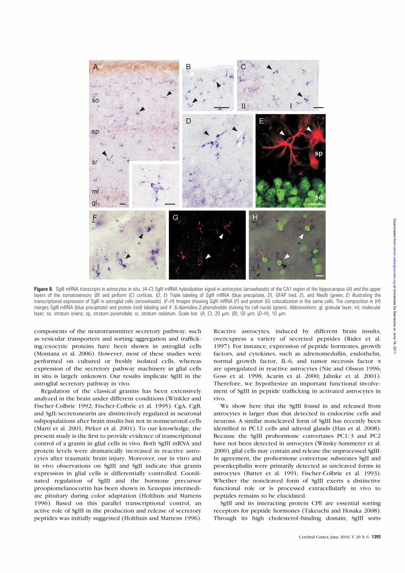
components of the neurotransmitter secretory pathway, such
as vesicular transporters and sorting/aggregation and traffick-
ing/exocytic proteins have been shown in astroglial cells
(Montana et al. 2006). However, most of these studies were
performed on cultured or freshly isolated cells, whereas
expression of the secretory pathway machinery in glial cells
in situ is largely unknown. Our results implicate SgIII in the
astroglial secretory pathway in vivo.
Regulation of the classical granins has been extensively
analyzed in the brain under different conditions (Winkler and
Fischer-Colbrie 1992; Fischer-Colbrie et al. 1995). CgA, CgB,
and SgII/secretoneurin are distinctively regulated in neuronal
subpopulations after brain insults but not in nonneuronal cells
(Martı et al. 2001; Pirker et al. 2001). To our knowledge, the
present study is the first to provide evidence of transcriptional
control of a granin in glial cells in vivo. Both SgIII mRNA and
protein levels were dramatically increased in reactive astro-
cytes after traumatic brain injury. Moreover, our in vitro and
in vivo observations on SgIII and SgII indicate that granin
expression in glial cells is differentially controlled. Coordi-
nated regulation of SgIII and the hormone precursor
proopiomelanocortin has been shown in Xenopus intermedi-
ate pituitary during color adaptation (Holthuis and Martens
1996). Based on this parallel transcriptional control, an
active role of SgIII in the production and release of secretory
peptides was initially suggested (Holthuis and Martens 1996).
Reactive astrocytes, induced by different brain insults,
overexpress a variety of secreted peptides (Ridet et al.
1997). For instance, expression of peptide hormones, growth
factors, and cytokines, such as adrenomedullin, endothelin,
normal growth factor, IL-6, and tumor necrosis factor aare upregulated in reactive astrocytes (Nie and Olsson 1996;
Goss et al. 1998; Acarin et al. 2000; Jahnke et al. 2001).
Therefore, we hypothesize an important functional involve-
ment of SgIII in peptide trafficking in activated astrocytes in
vivo.
We show here that the SgIII found in and released from
astrocytes is larger than that detected in endocrine cells and
neurons. A similar noncleaved form of SgIII has recently been
identified in PC12 cells and adrenal glands (Han et al. 2008).
Because the SgIII prohormone convertases PC1/3 and PC2
have not been detected in astrocytes (Winsky-Sommerer et al.
2000), glial cells may contain and release the unprocessed SgIII.
In agreement, the prohormone convertase substrates SgII and
proenkephalin were primarily detected as uncleaved forms in
astrocytes (Batter et al. 1991; Fischer-Colbrie et al. 1993).
Whether the noncleaved form of SgIII exerts a distinctive
functional role or is processed extracellularly in vivo to
peptides remains to be elucidated.
SgIII and its interacting protein CPE are essential sorting
receptors for peptide hormones (Takeuchi and Hosaka 2008).
Through its high cholesterol--binding domain, SgIII sorts
Figure 8. SgIII mRNA transcripts in astrocytes in situ. (A--C) SgIII mRNA hybridization signal in astrocytes (arrowheads) of the CA1 region of the hippocampus (A) and the upperlayers of the somatosensory (B) and piriform (C) cortices. (D, E) Triple labeling of SgIII mRNA (blue precipitate, D), GFAP (red, E), and NeuN (green, E) illustrating thetranscriptional expression of SgIII in astroglial cells (arrowheads). (F--H) Images showing SgIII mRNA (F) and protein (G) colocalization in the same cells. The composition in (H)merges SgIII mRNA (blue precipitate) and protein (red) labeling and 4#,6-diamidino-2-phenylindole staining for cell nuclei (green). Abbreviations: gl, granular layer; ml, molecularlayer; so, stratum oriens; sp, stratum pyramidale; sr, stratum radiatum. Scale bar: (A, C), 20 lm; (B), 50 lm; (D--H), 10 lm.
Cerebral Cortex June 2010, V 20 N 6 1393
at Universitat D
e Barcelona on June 19, 2011
cercor.oxfordjournals.orgD
ownloaded from
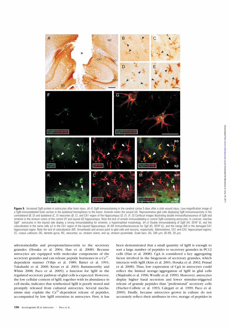
adrenomedullin and proopiomelanocortin to the secretory
granules (Hosaka et al. 2004; Han et al. 2008). Because
astrocytes are equipped with molecular components of the
secretory granules and can release peptide hormones in a Ca2+-
dependent manner (Vilijn et al. 1989; Batter et al. 1991;
Takahashi et al. 2000; Krzan et al. 2003; Ramamoorthy and
Whim 2008; Paco et al. 2009), a function for SgIII in the
regulated secretory pathway of glial cells is expected. However,
the low cellular content of SgIII, together with its abundance in
cell media, indicates that synthesized SgIII is poorly stored and
promptly released from cultured astrocytes. Several mecha-
nisms may explain the Ca2+-dependent release of peptides,
accompanied by low SgIII retention in astrocytes. First, it has
been demonstrated that a small quantity of SgIII is enough to
sort a large number of peptides to secretory granules in PC12
cells (Han et al. 2008). CgA is considered a key aggregating
factor involved in the biogenesis of secretory granules, which
interacts with SgIII (Kim et al. 2001; Hosaka et al. 2002; Prasad
et al. 2008). Thus, low expression of CgA in astrocytes could
reflect the limited storage/aggregation of SgIII in glial cells
(Majdoubi et al. 1996; Woulfe et al. 1999). Moreover, astrocytes
display higher basal secretion and lower stimulus-triggered
release of granule peptides than ‘‘professional’’ secretory cells
(Fischer-Colbrie et al. 1993; Calegari et al. 1999; Paco et al.
2009). Finally, because astrocytes grown in culture do not
accurately reflect their attributes in vivo, storage of peptides in
Figure 9. Increased SgIII protein in astrocytes after brain injury. (A--E) SgIII immunostaining in the cerebral cortex 5 days after a stab wound injury. Low-magnification image ofa SgIII-immunolabeled brain section in the ipsilateral hemispheres to the lesion. Asterisk marks the wound (A). Representative glial cells displaying SgIII immunoreactivity in thecontralateral (B, D) and ipsilateral (C, E) neocortex (B, C), and CA1 region of the hippocampus (D, E). (F, G) Confocal images illustrating double immunofluorescence of SgIII andvimentin in the stratum oriens of the control (F) and injured (G) hippocampus. Note the lack of vimetin immunolabeling in control SgIII-containing astrocytes. In contrast, reactiveSgIIIþ astrocytes in the injured side display a strong immunolabeling for vimentin, a hypertrophied morphology. (H--J) Double immunolabeling of SgIII (H), GFAP (I), and thecolocalization in the same cells (J) in the CA1 region of the injured hippocampus. (K--M) Immunofluorescences for SgII (K), GFAP (L), and the merge (M) in the damaged CA1hippocampal region. Note the lack of colocalization (M). Arrowheads and arrows point to glial cells and neurons, respectively. Abbreviations: CA1 and CA3, hippocampal regions;CC, corpus callosum; DG, dentate gyrus; NC, neocortex; so, stratum oriens; and sp, stratum pyramidale. Scale bars: (A), 200 lm; (B--M), 20 lm.
1394 Secretogranin III in Astrocytes d Paco et al.
at Universitat D
e Barcelona on June 19, 2011
cercor.oxfordjournals.orgD
ownloaded from
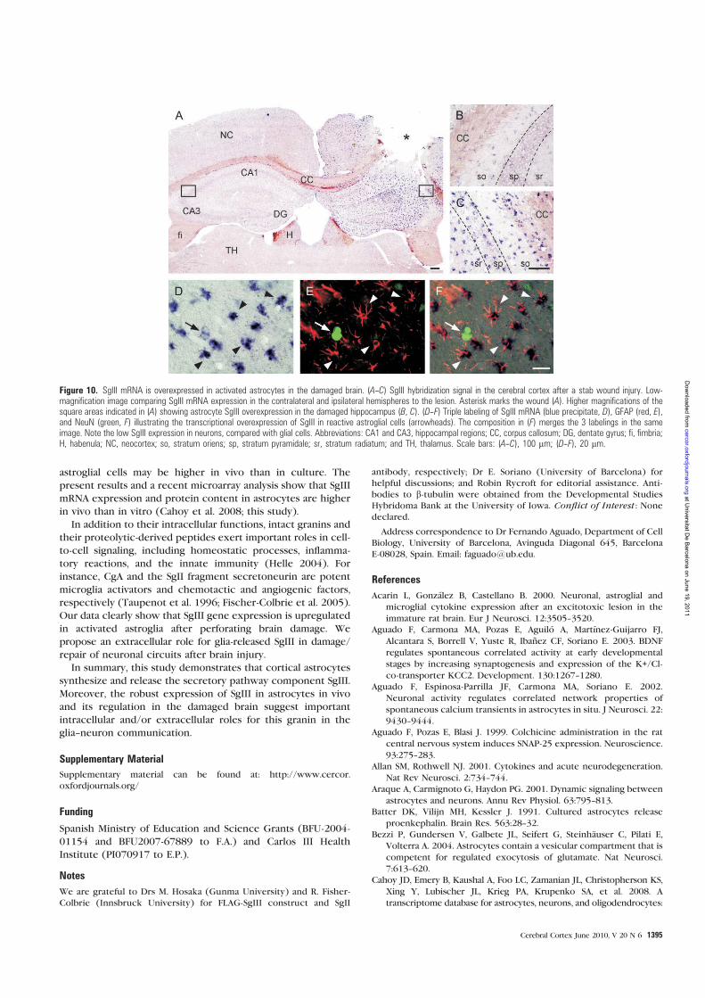
astroglial cells may be higher in vivo than in culture. The
present results and a recent microarray analysis show that SgIII
mRNA expression and protein content in astrocytes are higher
in vivo than in vitro (Cahoy et al. 2008; this study).
In addition to their intracellular functions, intact granins and
their proteolytic-derived peptides exert important roles in cell-
to-cell signaling, including homeostatic processes, inflamma-
tory reactions, and the innate immunity (Helle 2004). For
instance, CgA and the SgII fragment secretoneurin are potent
microglia activators and chemotactic and angiogenic factors,
respectively (Taupenot et al. 1996; Fischer-Colbrie et al. 2005).
Our data clearly show that SgIII gene expression is upregulated
in activated astroglia after perforating brain damage. We
propose an extracellular role for glia-released SgIII in damage/
repair of neuronal circuits after brain injury.
In summary, this study demonstrates that cortical astrocytes
synthesize and release the secretory pathway component SgIII.
Moreover, the robust expression of SgIII in astrocytes in vivo
and its regulation in the damaged brain suggest important
intracellular and/or extracellular roles for this granin in the
glia--neuron communication.
Supplementary Material
Supplementary material can be found at: http://www.cercor.
oxfordjournals.org/
Funding
Spanish Ministry of Education and Science Grants (BFU-2004-
01154 and BFU2007-67889 to F.A.) and Carlos III Health
Institute (PI070917 to E.P.).
Notes
We are grateful to Drs M. Hosaka (Gunma University) and R. Fisher-
Colbrie (Innsbruck University) for FLAG-SgIII construct and SgII
antibody, respectively; Dr E. Soriano (University of Barcelona) for
helpful discussions; and Robin Rycroft for editorial assistance. Anti-
bodies to b-tubulin were obtained from the Developmental Studies
Hybridoma Bank at the University of Iowa. Conflict of Interest : Nonedeclared.
Address correspondence to Dr Fernando Aguado, Department of Cell
Biology, University of Barcelona, Avinguda Diagonal 645, Barcelona
E-08028, Spain. Email: [email protected].
References
Acarin L, Gonzalez B, Castellano B. 2000. Neuronal, astroglial and
microglial cytokine expression after an excitotoxic lesion in the
immature rat brain. Eur J Neurosci. 12:3505--3520.
Aguado F, Carmona MA, Pozas E, Aguilo A, Martınez-Guijarro FJ,
Alcantara S, Borrell V, Yuste R, Ibanez CF, Soriano E. 2003. BDNF
regulates spontaneous correlated activity at early developmental
stages by increasing synaptogenesis and expression of the K+/Cl-co-transporter KCC2. Development. 130:1267--1280.
Aguado F, Espinosa-Parrilla JF, Carmona MA, Soriano E. 2002.
Neuronal activity regulates correlated network properties of
spontaneous calcium transients in astrocytes in situ. J Neurosci. 22:
9430--9444.
Aguado F, Pozas E, Blasi J. 1999. Colchicine administration in the rat
central nervous system induces SNAP-25 expression. Neuroscience.
93:275--283.
Allan SM, Rothwell NJ. 2001. Cytokines and acute neurodegeneration.
Nat Rev Neurosci. 2:734--744.
Araque A, Carmignoto G, Haydon PG. 2001. Dynamic signaling between
astrocytes and neurons. Annu Rev Physiol. 63:795--813.
Batter DK, Vilijn MH, Kessler J. 1991. Cultured astrocytes release
proenkephalin. Brain Res. 563:28--32.
Bezzi P, Gundersen V, Galbete JL, Seifert G, Steinhauser C, Pilati E,
Volterra A. 2004. Astrocytes contain a vesicular compartment that is
competent for regulated exocytosis of glutamate. Nat Neurosci.
7:613--620.
Cahoy JD, Emery B, Kaushal A, Foo LC, Zamanian JL, Christopherson KS,
Xing Y, Lubischer JL, Krieg PA, Krupenko SA, et al. 2008. A
transcriptome database for astrocytes, neurons, and oligodendrocytes:
Figure 10. SgIII mRNA is overexpressed in activated astrocytes in the damaged brain. (A--C) SgIII hybridization signal in the cerebral cortex after a stab wound injury. Low-magnification image comparing SgIII mRNA expression in the contralateral and ipsilateral hemispheres to the lesion. Asterisk marks the wound (A). Higher magnifications of thesquare areas indicated in (A) showing astrocyte SgIII overexpression in the damaged hippocampus (B, C). (D--F) Triple labeling of SgIII mRNA (blue precipitate, D), GFAP (red, E),and NeuN (green, F) illustrating the transcriptional overexpression of SgIII in reactive astroglial cells (arrowheads). The composition in (F) merges the 3 labelings in the sameimage. Note the low SgIII expression in neurons, compared with glial cells. Abbreviations: CA1 and CA3, hippocampal regions; CC, corpus callosum; DG, dentate gyrus; fi, fimbria;H, habenula; NC, neocortex; so, stratum oriens; sp, stratum pyramidale; sr, stratum radiatum; and TH, thalamus. Scale bars: (A--C), 100 lm; (D--F), 20 lm.
Cerebral Cortex June 2010, V 20 N 6 1395
at Universitat D
e Barcelona on June 19, 2011
cercor.oxfordjournals.orgD
ownloaded from
a new resource for understanding brain development and function. J
Neurosci. 28:264--278.
Calegari F, Coco S, Taverna E, Bassetti M, Verderio C, Corradi N,
Matteoli M, Rosa P. 1999. A regulated secretory pathway in cultured
hippocampal astrocytes. J Biol Chem. 274:22539--22547.
Christopherson KS, Ullian EM, Stokes CC, Mullowney CE, Hell JW,
Agah A, Lawler J, Mosher DF, Bornstein P, Barres BA. 2005.
Thrombospondins are astrocyte-secreted proteins that promote
CNS synaptogenesis. Cell. 120:421--433.
Coppinger JA, Cagney G, Toomey S, Kislinger T, Belton O,
McRedmond JP, Cahill DJ, Emili A, Fitzgerald DJ, Maguire PB.
2004. Characterization of the proteins released from activated
platelets leads to localization of novel platelet proteins in human
atherosclerotic lesions. Blood. 103:2096--2104.
Cortes R, Ceccatelli S, Schalling M, Hokfelt T. 1990. Differential effects
of intracerebroventricular colchicine administration on the expres-
sion of mRNAs for neuropeptides and neurotransmitter enzymes,
with special emphasis on galanin: an in situ hybridization study.
Synapse. 6:369--391.
Fiacco TA, Agulhon C, McCarthy KD. 2009. Sorting out astrocyte phys-
iology from pharmacology. Annu Rev Pharmacol Toxicol. 49:151--174.
Fischer-Colbrie R, Kirchmair R, Kahler CM, Wiedermann CJ, Saria A.
2005. Secretoneurin: a new player in angiogenesis and chemotaxis
linking nerves, blood vessels and the immune system. Curr Protein
Pept Sci. 6:373--385.
Fischer-Colbrie R, Kirchmair R, Schobert A, Olenik C, Meyer DK,
Winkler H. 1993. Secretogranin II is synthesized and secreted in
astrocyte cultures. J Neurochem. 60:2312--2314.
Fischer-Colbrie R, Laslop A, Kirchmair R. 1995. Secretogranin II:
molecular properties, regulation of biosynthesis and processing to
the neuropeptide secretoneurin. Prog Neurobiol. 46:49--70.
Goss JR, O’Malley ME, Zou L, Styren SD, Kochanek PM, DeKosky ST. 1998.
Astrocytes are the major source of nerve growth factor upregulation
following traumatic brain injury in the rat. Exp Neurol. 149:301--309.
Han L, Suda M, Tsuzuki K, Wang R, Ohe Y, Hirai H, Watanabe T,
Takeuchi T, Hosaka M. 2008. A large form of secretogranin III
functions as a sorting receptor for chromogranin A aggregates in
PC12 cells. Mol Endocrinol. 22:1935--1949.
Helle KB. 2004. The granin family of uniquely acidic proteins of the
diffuse neuroendocrine system: comparative and functional aspects.
Biol Rev Camb Philos Soc. 79:769--794.
Holthuis JC, Jansen EJ, Martens GJ. 1996. Secretogranin III is a sulfated
protein undergoing proteolytic processing in the regulated secre-
tory pathway. J Biol Chem. 271:17755--17760.
Holthuis JC, Martens GJ. 1996. The neuroendocrine proteins secretog-
ranin II and III are regionally conserved and coordinately expressed
with proopiomelanocortin in Xenopus intermediate pituitary.
J Neurochem. 66:2248--2256.
Hosaka M, Suda M, Sakai Y, Izumi T, Watanabe T, Takeuchi T. 2004.
Secretogranin III binds to cholesterol in the secretory granule mem-
brane as an adapter for chromogranin A. J Biol Chem. 279:3627--3634.
Hosaka M, Watanabe T, Sakai Y, Kato T, Takeuchi T. 2005. Interaction
between secretogranin III and carboxypeptidase E facilitates prohor-
mone sorting within secretory granules. J Cell Sci. 118:4785--4795.
Hosaka M, Watanabe T, Sakai Y, Uchiyama Y, Takeuchi T. 2002.
Identification of a chromogranin A domain that mediates binding to
secretogranin III and targeting to secretory granules in pituitary
cells and pancreatic beta-cells. Mol Biol Cell. 13:3388--3399.
Jahnke GD, Brunssen S, Maier WE, Harry GJ. 2001. Neurotoxicant-
induced elevation of adrenomedullin expression in hippocampus
and glia cultures. J Neurosci Res. 66:464--474.
Kim T, Tao-Cheng JH, Eiden LE, Loh YP. 2001. Chromogranin A, an ‘‘on/
off’’ switch controlling dense-core secretory granule biogenesis.
Cell. 106:499--509.
Krzan M, Stenovec M, Kreft M, Pangrsic T, Grilc S, Haydon PG, Zorec R.
2003. Calcium-dependent exocytosis of atrial natriuretic peptide
from astrocytes. J Neurosci. 23:1580--1583.
Lein ES, Hawrylycz MJ, Ao N, Ayres M, Bensinger A, Bernard A, Boe AF,
Boguski MS, Brockway KS, Byrnes EJ, et al. 2007. Genome-wide atlas
of gene expression in the adult mouse brain. Nature. 445:
168--176.
Levey AI, Bolam JP, Rye DB, Hallanger AE, Demuth RM, Mesulam MM,
Wainer BH. 1986. A light and electron microscopic procedure for
sequential double antigen localization using diaminobenzidine and
benzidine dihydrochloride. J Histochem Cytochem. 34:1449--1457.
Majdoubi ME, Metz-Boutigue MH, Garcia-Sablone P, Theodosis DT,
Aunis D. 1996. Immunocytochemical localization of chromogranin
A in the normal and stimulated hypothalamo-neurohypophysial
system of the rat. J Neurocytol. 25:405--416.
Martı E, Ferrer I, Blasi J. 2001. Differential regulation of chromogranin A,
chromogranin B and secretoneurin protein expression after transient
forebrain ischemia in the gerbil. Acta Neuropathol. 101:159--166.
Mathewson AJ, Berry M. 1985. Observations on the astrocyte response
to a cerebral stab wound in adult rats. Brain Res. 327:61--69.
Montana V, Malarkey EB, Verderio C, Matteoli M, Parpura V. 2006.
Vesicular transmitter release from astrocytes. Glia. 54:700--715.
Montero-Hadjadje M, Vaingankar S, Elias S, Tostivint H, Mahata SK,
Anouar Y. 2008. Chromogranins A and B and secretogranin II:
evolutionary and functional aspects. Acta Physiol (Oxf). 192:309--324.
Nie XJ, Olsson Y. 1996. Endothelin peptides in brain diseases. Rev
Neurosci. 7:177--186.
Ottiger HP, Battenberg EF, Tsou AP, Bloom FE, Sutcliffe JG. 1990.
1B1075: a brain- and pituitary-specific mRNA that encodes a novel
chromogranin/secretogranin-like component of intracellular
vesicles. J Neurosci. 10:3135--3147.
Paco S, Margelı MA, Olkkonen VM, Imai A, Blasi J, Fischer-Colbrie R,
Aguado F. 2009. Regulation of exocytotic protein expression and
Ca2+-dependent peptide secretion in astrocytes. J Neurochem.
110:143--156.
Panatier A, Theodosis DT, Mothet JP, Touquet B, Pollegioni L,
Poulain DA, Oliet SH. 2006. Glia-derived D-serine controls NMDA
receptor activity and synaptic memory. Cell. 125:775--784.
Pascual O, Casper KB, Kubera C, Zhang J, Revilla-Sanchez R, Sul JY,
TakanoH,MossSJ,McCarthyK,HaydonPG.2005.Astrocyticpurinergic
signaling coordinates synaptic networks. Science. 310:113--116.
Perea G, Araque A. 2007. Astrocytes potentiate transmitter release at
single hippocampal synapses. Science. 317:1083--1086.
Pirker S, Czech T, Baumgartner C, Maier H, Novak K, Furtinger S,
Fischer-Colbrie R, Sperk G. 2001. Chromogranins as markers of
altered hippocampal circuitry in temporal lobe epilepsy. Ann
Neurol. 50:216--226.
Prasad P, Yanagihara AA, Small-Howard AL, Turner H, Stokes AJ. 2008.
Secretogranin III directs secretory vesicle biogenesis in mast cells in
a manner dependent upon interaction with chromogranin A. J
Immunol. 181:5024--5034.
Ramamoorthy P, Whim MD. 2008. Trafficking and fusion of neuropep-
tide Y-containing dense-core granules in astrocytes. J Neurosci.
28:13815--13827.
Ridet JL, Privat A, Malhotra SK, Gage FH. 1997. Reactive astrocytes:
cellular and molecular cues to biological function. Trends Neurosci.
20:570--577.
Shyu WC, Lin SZ, Chiang MF, Chen DC, Su CY, Wang HJ, Liu RS, Tsai CH,
Li H. 2008. Secretoneurin promotes neuroprotection and neuronal
plasticity via the Jak2/Stat3 pathway in murine models of stroke. J
Clin Invest. 118:133--148.
Simon JP, Bader MF, Aunis D. 1988. Secretion from chromaffin cells is
controlled by chromogranin A-derived peptides. Proc Natl Acad Sci
USA. 85:1712--1716.
Sofroniew MV. 2005. Reactive astrocytes in neural repair and pro-
tection. Neuroscientist. 11:400--407.
Takahashi K, Nakayama M, Totsune K, Murakami O, Sone M,
Kitamuro T, Yoshinoya A, Shibahara S. 2000. Increased secretion
of adrenomedullin from cultured human astrocytes by cytokines. J
Neurochem. 74:99--103.
Takeuchi T, Hosaka M. 2008. Sorting mechanism of peptide hormones
and biogenesis mechanism of secretory granules by secretogranin
III, a cholesterol-binding protein, in endocrine cells. Curr Diabetes
Rev. 4:31--38.
Taupenot L, Ciesielski-Treska J, Ulrich G, Chasserot-Golaz S, Aunis D,
Bader MF. 1996. Chromogranin A triggers a phenotypic trans-
formation and the generation of nitric oxide in brain microglial
cells. Neuroscience. 72:377--389.
1396 Secretogranin III in Astrocytes d Paco et al.
at Universitat D
e Barcelona on June 19, 2011
cercor.oxfordjournals.orgD
ownloaded from
Taupenot L, Harper KL, O’Connor DT. 2003. The chromogranin-
secretogranin family. N Engl J Med. 348:1134--1149.
Vilijn MH, Das B, Kessler JA, Fricker LD. 1989. Cultured astrocytes and
neurons synthesize and secrete carboxypeptidase E, a neuropeptide-
processing enzyme. J Neurochem. 53:1487--1493.
Volterra A, Meldolesi J. 2005. Astrocytes, from brain glue to
communication elements: the revolution continues. Nat Rev
Neurosci. 6:626--640.
Winkler H, Fischer-Colbrie R. 1992. The chromogranins A and B: the
first 25 years and future perspectives. Neuroscience. 49:497--528.
Winsky-Sommerer R, Benjannet S, Rovere C, Barbero P, Seidah NG,
Epelbaum J, Dournaud P. 2000. Regional and cellular localization of
the neuroendocrine prohormone convertases PC1 and PC2 in the
rat central nervous system. J Comp Neurol. 424:439--460.
Woulfe J, Deng D, Munoz D. 1999. Chromogranin A in the central
nervous system of the rat: pan-neuronal expression of its mRNA and
selective expression of the protein. Neuropeptides. 33:285--300.
Zhang Z, Chen G, Zhou W, Song A, Xu T, Luo Q, Wang W, Gu XS,
Duan S. 2007. Regulated ATP release from astrocytes through
lysosome exocytosis. Nat Cell Biol. 8:945--953.
Cerebral Cortex June 2010, V 20 N 6 1397
at Universitat D
e Barcelona on June 19, 2011
cercor.oxfordjournals.orgD
ownloaded from

113
En aquest apartat es discuteixen de forma integrada tots els resultats inclosos en els tres
treballs presentats, juntament altres dades obtingudes durant el transcurs de la tesi doctoral
que encara no estan publicades i són preliminars.
En els terminals sinàptics el complex SNARE està format per una reacció
estequiomètrica 1:1:1 entre VAMP-2, SNAP25 i Stx1 (Jahn i Scheller, 2006). Però existeixen
diverses isoformes de les SNARE amb propietats estructurals similars i capaces de substituir les
altres en la formació del complex de fusió (Fasshauer, 1999), donant la possibilitat de la
formació de combinacions alternatives de proteïnes en altres tipus de cèl·lules secretores
especialitzades. En aquest treball s’identifiquen les proteïnes que formen part de la
maquinària exocítica en dos tipus cel·lulars glials en cultiu: els astròcits i la micròglia.
1.1. Astròcits
Característicament, les principals isoformes de les famílies R- i Q-SNARE i de Munc18
detectades en astròcits són diferents de les expressades en neurones. Mentre que les
neurones expressen sintaxina1, Munc18a, SNAP25 i VAMP-2 en astròcits les formes
majoritàries són sintaxina4, Munc18c, SNAP23 i VAMP-3, igual que en les línies tumorals C6 i
U373 que deriven de cèl·lules astroglials.
La prevalença de la Q-SNARE SNAP23 en astròcits està d’acord amb estudis previs
(Hepp et al., 1999) i dóna suport a la idea que substitueix funcionalment SNAP25 en cèl·lules
no endocrines. Les isoformes implicades en la neurosecreció depenent de calci sintaxina1,
Munc18a i VAMP-2 estan presents tant en neurones com en astròcits. En astròcits, però,
només s’expressa la variant 1B de sintaxina1, en concordança amb la distribució heterogènia
descrita per les isoformes de sintaxina1 (Ruiz-Montasell et al., 1996). Aquests resultats s’han
corroborat recentment in vivo, on s’ha detectat l’expressió de sintaxina1 en astròcits de
l’hipocamp (Schubbert et al., 2011).
A més de les proteïnes implicades en la exocitosi depenent de calci, els astròcits
expressen de forma abundant sintaxina4, sintaxina2, Munc18c i VAMP-3. Malgrat que el
significat funcional d’aquestes proteïnes exocítiques ubiqües en el SNC no és gaire conegut,
en altres tipus cel·lulars no neuronals se’ls ha assignat un paper en la secreció depenent
d’estímul (Flaumenhaf, 2003; Imai et al., 2004). També podria ser que la presència de diverses
isoformes d’SNAREs reflecteixi la coexistència de diferents orgànuls responsables de l’exocitosi
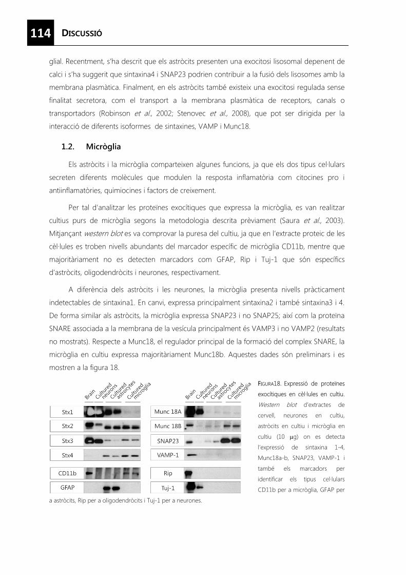
114
glial. Recentment, s’ha descrit que els astròcits presenten una exocitosi lisosomal depenent de
calci i s’ha suggerit que sintaxina4 i SNAP23 podrien contribuir a la fusió dels lisosomes amb la
membrana plasmàtica. Finalment, en els astròcits també existeix una exocitosi regulada sense
finalitat secretora, com el transport a la membrana plasmàtica de receptors, canals o
transportadors (Robinson et al., 2002; Stenovec et al., 2008), que pot ser dirigida per la
interacció de diferents isoformes de sintaxines, VAMP i Munc18.
1.2. Micròglia
Els astròcits i la micròglia comparteixen algunes funcions, ja que els dos tipus cel·lulars
secreten diferents molècules que modulen la resposta inflamatòria com citocines pro i
antiinflamatòries, quimiocines i factors de creixement.
Per tal d’analitzar les proteïnes exocítiques que expressa la micròglia, es van realitzar
cultius purs de micròglia segons la metodologia descrita prèviament (Saura et al., 2003).
Mitjançant western blot es va comprovar la puresa del cultiu, ja que en l’extracte proteic de les
cèl·lules es troben nivells abundants del marcador específic de micròglia CD11b, mentre que
majoritàriament no es detecten marcadors com GFAP, Rip i Tuj-1 que són específics
d’astròcits, oligodendròcits i neurones, respectivament.
A diferència dels astròcits i les neurones, la micròglia presenta nivells pràcticament
indetectables de sintaxina1. En canvi, expressa principalment sintaxina2 i també sintaxina3 i 4.
De forma similar als astròcits, la micròglia expressa SNAP23 i no SNAP25; així com la proteïna
SNARE associada a la membrana de la vesícula principalment és VAMP3 i no VAMP2 (resultats
no mostrats). Respecte a Munc18, el regulador principal de la formació del complex SNARE, la
micròglia en cultiu expressa majoritàriament Munc18b. Aquestes dades són preliminars i es
mostren a la figura 18.
FFIGURA18. Expressió de proteïnes
exocítiques en cèl·lules en cultiu.
Western blot d’extractes de
cervell, neurones en cultiu,
astròcits en cultiu i micròglia en
cultiu (10 �g) on es detecta
l’expressió de sintaxina 1-4,
Munc18a-b, SNAP23, VAMP-1 i
també els marcadors per
identificar els tipus cel·lulars
CD11b per a micròglia, GFAP per
a astròcits, Rip per a oligodendròcits i Tuj-1 per a neurones.
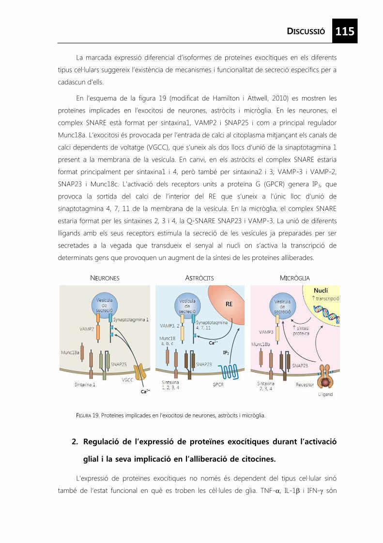
115
La marcada expressió diferencial d’isoformes de proteïnes exocítiques en els diferents
tipus cel·lulars suggereix l’existència de mecanismes i funcionalitat de secreció específics per a
cadascun d’ells.
En l’esquema de la figura 19 (modificat de Hamilton i Attwell, 2010) es mostren les
proteïnes implicades en l’exocitosi de neurones, astròcits i micròglia. En les neurones, el
complex SNARE està format per sintaxina1, VAMP2 i SNAP25 i com a principal regulador
Munc18a. L’exocitosi és provocada per l’entrada de calci al citoplasma mitjançant els canals de
calci dependents de voltatge (VGCC), que s’uneix als dos llocs d’unió de la sinaptotagmina 1
present a la membrana de la vesícula. En canvi, en els astròcits el complex SNARE estaria
format principalment per sintaxina1 i 4, però també per sintaxina2 i 3; VAMP-3 i VAMP-2,
SNAP23 i Munc18c. L’activació dels receptors units a proteïna G (GPCR) genera IP3, que
provoca la sortida del calci de l’interior del RE que s’uneix a l’únic lloc d’unió de
sinaptotagmina 4, 7, 11 de la membrana de la vesícula. En la micròglia, el complex SNARE
estaria format per les sintaxines 2, 3 i 4, la Q-SNARE SNAP23 i VAMP-3. La unió de diferents
lligands amb els seus receptors estimula la secreció de les vesícules ja preparades per ser
secretades a la vegada que transdueix el senyal al nucli on s’activa la transcripció de
determinats gens que provoquen un augment de la síntesi de les proteïnes alliberades.
FFIGURA 19. Proteïnes implicades en l’exocitosi de neurones, astròcits i micròglia.
L’expressió de proteïnes exocítiques no només és dependent del tipus cel·lular sinó
també de l’estat funcional en què es troben les cèl·lules de glia. TNF-�, IL-1� i IFN-� són
NEEURONES ASSTRÒCITS MIICRÒGLIA
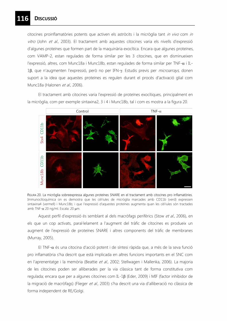
116
citocines proinflamatòries potents que activen els astròcits i la micròglia tant in vivo com in
vitro (John et al., 2003). El tractament amb aquestes citocines varia els nivells d’expressió
d’algunes proteïnes que formen part de la maquinària exocítica. Encara que algunes proteïnes,
com VAMP-2, estan regulades de forma similar per les 3 citocines, que en disminueixen
l’expressió, altres, com Munc18a i Munc18b, estan regulades de forma similar per TNF-� i IL-
1�, que n’augmenten l’expressió, però no per IFN-�. Estudis previs per microarrays, donen
suport a la idea que aquestes proteïnes es regulen durant el procés d’activació glial com
Munc18a (Halonen et al., 2006).
El tractament amb citocines varia l’expressió de proteïnes exocítiques, principalment en
la micròglia, com per exemple sintaxina2, 3 i 4 i Munc18b, tal i com es mostra a la figura 20.
FFIGURA 20. La micròglia sobreexpressa algunes proteïnes SNARE en el tractament amb citocines pro inflamatòries. Immunocítoquímica on es demostra que les cèl·lules de micròglia marcades amb CD11b (verd) expressen sintaxina4 (vermell) i Munc18b; i que l’expressió d’aquestes proteïnes augmenta quan les cèl·lules són tractades amb TNF-� 20 ng/ml. Escala: 20 �m.
Aquest perfil d’expressió és semblant al dels macròfags perifèrics (Stow et al., 2006), en
els que un cop activats, paral·lelament a l’augment del tràfic de citocines es produeix un
augment de l’expressió de proteïnes SNARE i altres components del tràfic de membranes
(Murray, 2005).
El TNF-� és una citocina d’acció potent i de síntesi ràpida que, a més de la seva funció
pro inflamatòria s’ha descrit que està implicada en altres funcions importants en el SNC com
en l’aprenentatge i la memòria (Beattie et al., 2002; Stellwagen i Mallenka, 2006). La majoria
de les citocines poden ser alliberades per la via clàssica tant de forma constitutiva com
regulada; encara que per a algunes citocines com IL-1� (Eder, 2009) i MIF (factor inhibidor de
la migració de macròfags) (Flieger et al., 2003) s’ha descrit una via d’alliberació no clàssica de
forma independent de RE/Golgi.
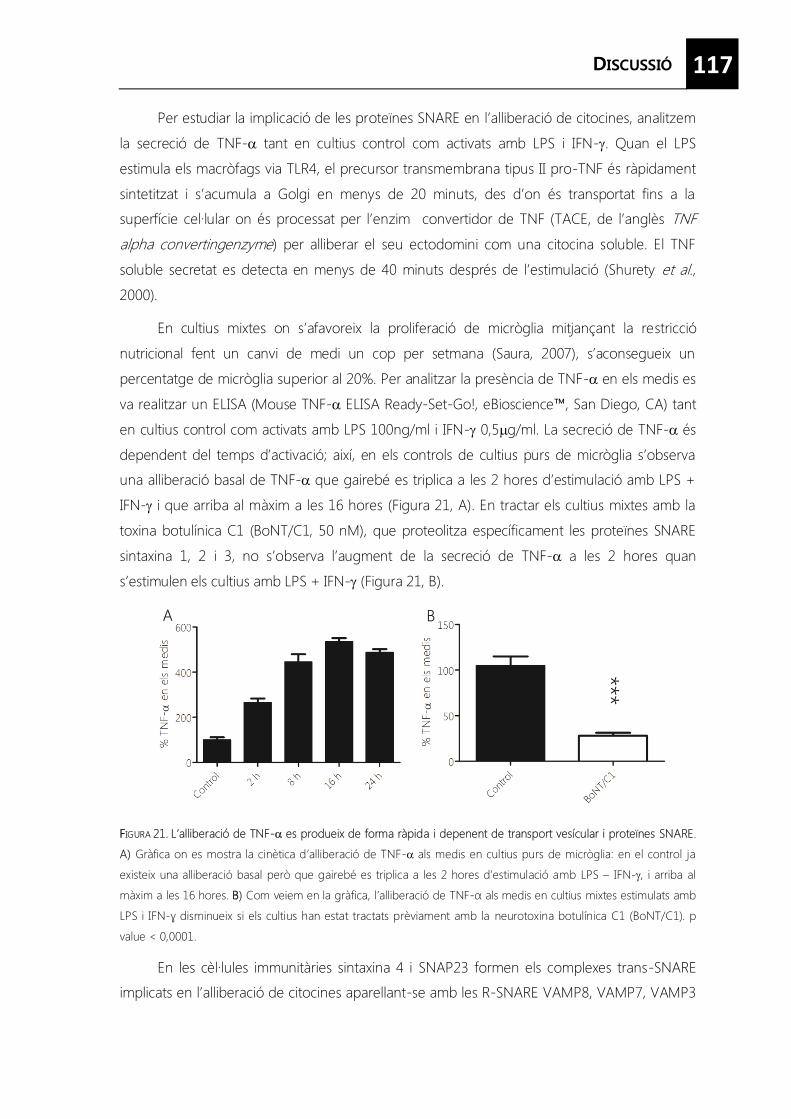
117
Per estudiar la implicació de les proteïnes SNARE en l’alliberació de citocines, analitzem
la secreció de TNF-� tant en cultius control com activats amb LPS i IFN-�. Quan el LPS
estimula els macròfags via TLR4, el precursor transmembrana tipus II pro-TNF és ràpidament
sintetitzat i s’acumula a Golgi en menys de 20 minuts, des d’on és transportat fins a la
superfície cel·lular on és processat per l’enzim convertidor de TNF (TACE, de l’anglès TNF alpha convertingenzyme) per alliberar el seu ectodomini com una citocina soluble. El TNF
soluble secretat es detecta en menys de 40 minuts després de l’estimulació (Shurety et al., 2000).
En cultius mixtes on s’afavoreix la proliferació de micròglia mitjançant la restricció
nutricional fent un canvi de medi un cop per setmana (Saura, 2007), s’aconsegueix un
percentatge de micròglia superior al 20%. Per analitzar la presència de TNF-� en els medis es
va realitzar un ELISA (Mouse TNF-� ELISA Ready-Set-Go!, eBioscience™, San Diego, CA) tant
en cultius control com activats amb LPS 100ng/ml i IFN-� 0,5�g/ml. La secreció de TNF-� és
dependent del temps d’activació; així, en els controls de cultius purs de micròglia s’observa
una alliberació basal de TNF-� que gairebé es triplica a les 2 hores d’estimulació amb LPS +
IFN-� i que arriba al màxim a les 16 hores (Figura 21, A). En tractar els cultius mixtes amb la
toxina botulínica C1 (BoNT/C1, 50 nM), que proteolitza específicament les proteïnes SNARE
sintaxina 1, 2 i 3, no s’observa l’augment de la secreció de TNF-� a les 2 hores quan
s’estimulen els cultius amb LPS + IFN-��(Figura 21, B).
FFIGURA 21. L’alliberació de TNF-� es produeix de forma ràpida i depenent de transport vesícular i proteïnes SNARE.
A) Gràfica on es mostra la cinètica d’alliberació de TNF-� als medis en cultius purs de micròglia: en el control ja
existeix una alliberació basal però que gairebé es triplica a les 2 hores d’estimulació amb LPS – IFN-�, i arriba al
màxim a les 16 hores. BB) Com veiem en la gràfica, l’alliberació de TNF-α als medis en cultius mixtes estimulats amb
LPS i IFN-ɣ disminueix si els cultius han estat tractats prèviament amb la neurotoxina botulínica C1 (BoNT/C1). p
value < 0,0001.
En les cèl·lules immunitàries sintaxina 4 i SNAP23 formen els complexes trans-SNARE
implicats en l’alliberació de citocines aparellant-se amb les R-SNARE VAMP8, VAMP7, VAMP3
***
A B
118
i VAMP2 dels grànuls de secreció (Stanley i Lacy, 2010). La toxina BoNT/C1 no proteolitza
sintaxina4, sinó principalment sintaxina1 i també sintaxina2 i 3, però bloqueja de forma molt
efectiva l’alliberació de TNF-� en les cèl·lules de glia. Tenint en compte que les possibles
dianes de la toxina en les astròcits són sintaxina1, 2 i 3, i en la micròglia només sintaxina2 i 3, i
que les primeres cèl·lules en activar-se i les que produeixen principalment TNF-� són les
cèl·lules de la micròglia, aquests resultats suggereixen que l’alliberació de TNF-� es realitza
mitjançant les proteïnes SNARE sintaxina2 i 3 en les cèl·lules glials. Malgrat això, són necessaris
més experiments amb tècniques més específiques, com siRNA o dominants negatius, per
determinar exactament quines isoformes de les proteïnes SNARE estan implicades en
l’alliberació de la secreció regulada de TNF-��en les cèl·lules glials.
Per estudiar la secreció regulada per calci en astròcits in vitro, s’han buscat les
condicions en les que els astròcits presenten un fenotip diferenciat semblant al que presenten
in vivo. El tractament amb anàlegs del cAMP produeix canvis dramàtics en la morfologia i
fisiologia dels astròcits, proporcionant un fenotip diferenciat en cultiu apte per realitzar estudis
de secreció regulada de pèptids. Tanmateix, encara existeix certa controvèrsia a la literatura si
aquests canvis són deguts a l’adquisició d’un fenotip reactiu (Fedoroff et al., 1984; Bochelen et
al., 2000) o un fenotip més madur, diferenciat (McManus et al., 1999).
Mitjançant l’estudi amb microarrays s’ha determinat que els anàlegs de cAMP
disminueixen la transcripció de RNA implicats en la proliferació i el desenvolupament.
Contràriament, els gens típicament relacionats amb els astròcits madurs com els del suport
metabòlic i estructural de les neurones i el control de l’homeòstasi iònica estan
sobreexpressats en el tractament amb cAMP. Aquests resultats juntament amb l’observació
que el transcriptoma global dels astròcits en cultiu tractats amb cAMP s’assembla més al perfil
del transcriptoma dels astròcits madurs i no dels astròcits en desenvolupament aïllats en fresc,
suggereix que el tractament amb cAMP indueix diferenciació (Cahoy et al., 2008). També el
tractament amb cAMP indueix la down-regulació de gens relacionats distintivament amb
l’activació astroglial com, la hiperplàsia, la reorganització del citoesquelet, el
desencadenament de les reaccions immunològiques i la formació de la cicatriu glial (Sofroniev,
2009). Aquests resultats estan en concordança amb estudis previs de microarrays on els
astròcits tractats amb citocines incrementen l’expressió dels gens implicats en l’astrogliosi
(Halonen et al., 2006; Falsig et al., 2006; John et al., 2003). Els resultats obtinguts mostren que
119
el transcriptoma dels astròcits en cultiu tractats amb 8Br-cAMP és més semblant al de les
cèl·lules in vivo, la qual cosa indica que la senyalització del cAMP confereix un fenotip in vivo
reprimint l’activació i promovent la diferenciació.
Un nombre creixent de treballs ha demostrat que els astròcits presenten una via
d’alliberació de gliotransmissors depenent de calci (Volterra i Meldonesi, 2005). Els últims anys,
s’han identificat diversos pèptids sintetitzats i alliberats pels astròcits que regulen diferents
funcions neuronals.
Els astròcits alliberen de forma depenent de calci el pèptid ANP expressat de forma
exògena (Krzan et al., 2003) i nosaltres hem determinat que la seva secreció augmenta en el
tractament amb anàlegs del cAMP. Aquesta secreció regulada per calci de l’ANP en astròcits
no només s’ha determinat en astròcits en cultiu sinó també en seccions d’hipocamp (Potokar
et al., 2009).
El pèptid endogensecretogranina II presenta una secreció en astròcits en cultiu
extremadament baixa (Fischer-Colbrie, 1993; Calegari et al., 1999), encara que és
suficientment alta per ser detectada mitjançant western blot. El seu contingut augmenta en
astròcits en cultiu tractats amb 8Br-cAMP, consistentment amb la presència del element
funcional de resposta a cAMP (CRE) en el seu promotor (Taupenot et al., 2003). En els
astròcits tractats amb 8Br-cAMP es pot detectar l’alliberació de la SgII induïda per un
increment de calci en el medi de cultiu als 5 min. La dependència dels nivells de calci s’ha
demostrat posteriorment abolint l’efecte de la ionomicina/TPA amb el quelant de calci
intracel·lular BAPTA-AM.
En el tractament amb 8Br-cAMP es realitzen efectes oposats al tractament amb
citocines sobre la regulació de les proteïnes exocítiques en astròcits en cultiu augmentant
l’expressió de les proteïnes clàssicament implicades en la secreció regulada com sintaxina1,
Munc 18a i VAMP-2. Aquests canvis podrien explicar l’augment de l’alliberació de pèptids,
encara que la proteïna més regulada per aquest tractament, VAMP-2, no es troba
majoritàriament en els grànuls que contenen els pèptids, on podria estar substituïda per una
altra VAMP. L’augment d’altres proteïnes exocítiques com sintaxina1, Munc18a o
sinaptotagmina IV, podrien estar relacionades amb l’augment de l’alliberació dels pèptids
induïda per calci en les cèl·lules tractades.
També el tractament dels cultius d’astròcits amb la toxina botulínica BoNT/C1 afecta
l’alliberació de SgII. La toxina BoNT/C1 és una metal·loproteasa que, mitjançant la proteòlisi
específica de sintaxina1, 2, 3 i SNAP25, produeix un bloqueig de la secreció dependent de
SNAREs (Humeau et al., 2000). En neurones, la toxina entra a l’interior cel·lular mitjançant la
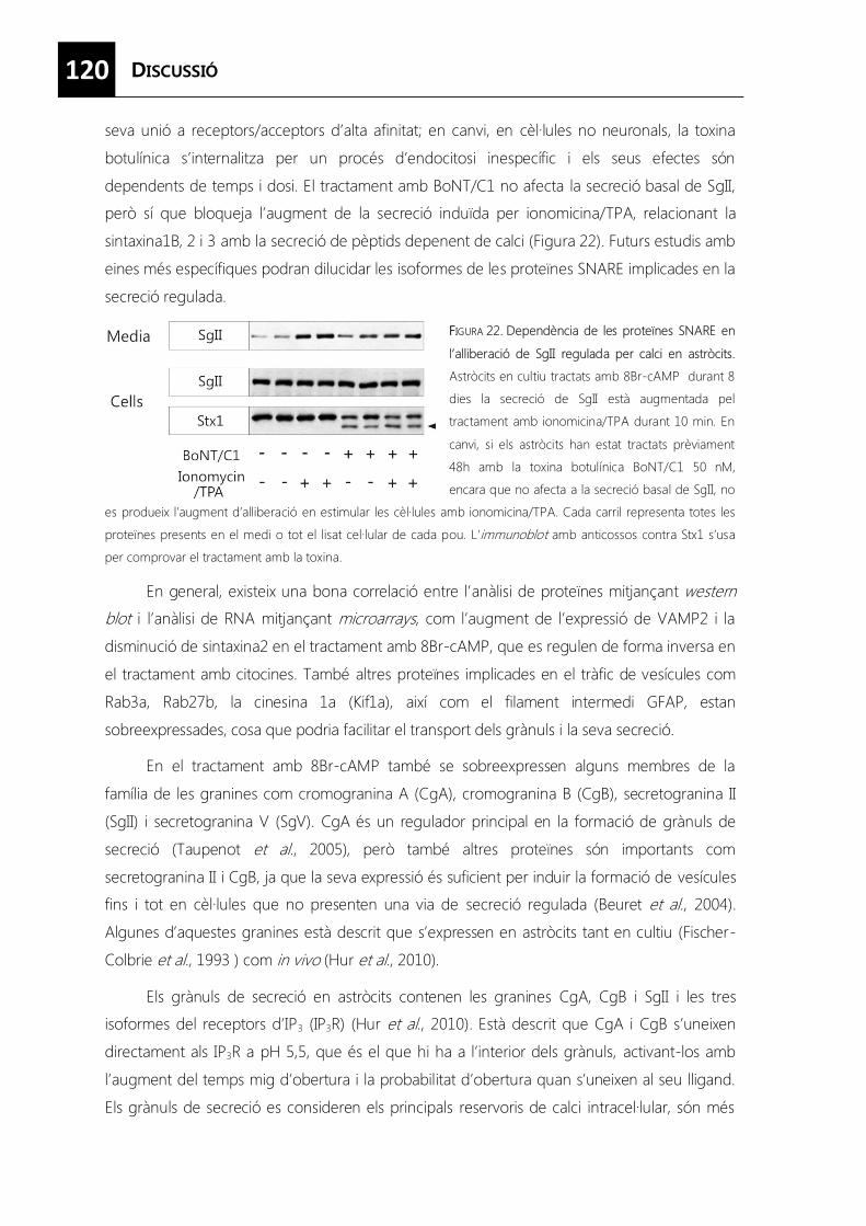
120
seva unió a receptors/acceptors d’alta afinitat; en canvi, en cèl·lules no neuronals, la toxina
botulínica s’internalitza per un procés d’endocitosi inespecífic i els seus efectes són
dependents de temps i dosi. El tractament amb BoNT/C1 no afecta la secreció basal de SgII,
però sí que bloqueja l’augment de la secreció induïda per ionomicina/TPA, relacionant la
sintaxina1B, 2 i 3 amb la secreció de pèptids depenent de calci (Figura 22). Futurs estudis amb
eines més específiques podran dilucidar les isoformes de les proteïnes SNARE implicades en la
secreció regulada.
FFIGURA 22. Dependència de les proteïnes SNARE en
l’alliberació de SgII regulada per calci en astròcits.
Astròcits en cultiu tractats amb 8Br-cAMP durant 8
dies la secreció de SgII està augmentada pel
tractament amb ionomicina/TPA durant 10 min. En
canvi, si els astròcits han estat tractats prèviament
48h amb la toxina botulínica BoNT/C1 50 nM,
encara que no afecta a la secreció basal de SgII, no
es produeix l’augment d’alliberació en estimular les cèl·lules amb ionomicina/TPA. Cada carril representa totes les
proteïnes presents en el medi o tot el lisat cel·lular de cada pou. L’immunoblot amb anticossos contra Stx1 s’usa
per comprovar el tractament amb la toxina.
En general, existeix una bona correlació entre l’anàlisi de proteïnes mitjançant western blot i l’anàlisi de RNA mitjançant microarrays, com l’augment de l’expressió de VAMP2 i la
disminució de sintaxina2 en el tractament amb 8Br-cAMP, que es regulen de forma inversa en
el tractament amb citocines. També altres proteïnes implicades en el tràfic de vesícules com
Rab3a, Rab27b, la cinesina 1a (Kif1a), així com el filament intermedi GFAP, estan
sobreexpressades, cosa que podria facilitar el transport dels grànuls i la seva secreció.
En el tractament amb 8Br-cAMP també se sobreexpressen alguns membres de la
família de les granines com cromogranina A (CgA), cromogranina B (CgB), secretogranina II
(SgII) i secretogranina V (SgV). CgA és un regulador principal en la formació de grànuls de
secreció (Taupenot et al., 2005), però també altres proteïnes són importants com
secretogranina II i CgB, ja que la seva expressió és suficient per induir la formació de vesícules
fins i tot en cèl·lules que no presenten una via de secreció regulada (Beuret et al., 2004).
Algunes d’aquestes granines està descrit que s’expressen en astròcits tant en cultiu (Fischer-
Colbrie et al., 1993 ) com in vivo (Hur et al., 2010).
Els grànuls de secreció en astròcits contenen les granines CgA, CgB i SgII i les tres
isoformes del receptors d’IP3 (IP3R) (Hur et al., 2010). Està descrit que CgA i CgB s’uneixen
directament als IP3R a pH 5,5, que és el que hi ha a l’interior dels grànuls, activant-los amb
l’augment del temps mig d’obertura i la probabilitat d’obertura quan s’uneixen al seu lligand.
Els grànuls de secreció es consideren els principals reservoris de calci intracel·lular, són més
121
sensibles perquè requereixen concentracions més baixes d’IP3 per activar-se i a més, poden
provocar augments de calci a nivell local, relacionant-los directament amb el control de
l’exocitosi. Per tant, els augments d’expressió de CgA i CgB en el tractament amb anàlegs de
l’AMPc podria augmentar els nivells d’exocitosi.
En un treball recent es descriu un fenotip astrocític on estan augmentats els grànuls de
secreció i l’expressió dels seus marcadors quan els nivells d’expressió del factor de transcripció
REST són baixos (Prada et al., 2011). Degut a que enel nostre anàlisi de microarrays no
s’observen variacions en els nivells d’expressió de REST en el tractament amb 8Br-cAMP, els
efectes del cAMP en la maduració dels astròcits podrien ser independents de REST.
Originàriament, la secretogranina III va ser identificada en neurones i les cèl·lules
endocrines de la hipòfisi (Ottiger et al., 1990). Més recentment s’ha descrit l’expressió de SgIII
en altres tipus cel·lulars que presenten una via de secreció regulada com els mastòcits (Prasad
et al., 2008) i les cèl·lules PC12 (Han, 2008). En aquest treball s’ha determinat que els astròcits
expressen nivells alts tant de mRNA com de la proteïna de SgIII. Aquests resultats coincideixen
amb la hibridació in situ que es troba a l’Allen Brain Atlas, on el marcatge del mRNA de SgIII
es troba tant en neurones com en cèl·lules glials (Lein et al., 2007). Mitjançant un marcatge
doble, es mostra que l’expressió de SgIII en cèl·lules no neuronals correspon gairebé de forma
exclusiva a diferents poblacions d’astròcits, en els quals varia la intensitat de l’expressió.
Mentre que l’expressió de SgIII és alta en els astròcits corticals i la glia de Bergmann, és molt
baixa en els astròcits situats en la substància blanca.
En astròcits en cultiu, ja s’havien descrit alguns dels components típics de la via de
secreció de neurotransmissors, com els transportadors vesiculars i les proteïnes exocítiques.
Però en astròcits in vivo, no s’han pogut identificar totes aquestes proteïnes. En canvi, els
nostres resultats impliquen la SgIII en la via de secreció en astròcits in vivo.
La SgIII que es detecta en els astròcits presenta un pes molecular superior a la SgIII que
es troba en els neurones, similar a la SgIII recentment detectada a les cèl·lules PC12 i a les
glàndules adrenals (Han et al., 2008). Això és a causa que els astròcits no expressen les
convertases PC1/3 i PC2 que són les encarregades de processar les prohormones (Winsky-
Sommerer et al., 2000). De manera similar, altres substrats de les convertases com la
secretogranina II i la proencefalina tampoc són processades en els astròcits (Batter et al., 1991;
122
Fischer-Colbrie et al., 1993). Malgrat això, falta determinar si la forma no processada de SgIII
té un paper diferencial o si és processada extracel·lularment per donar lloc a pèptids bioactius.
La interacció de la SgIII amb la carboxipeptidasa E té un paper fonamental en la
classificació dels pèptids cap als grànuls de secreció (Hosaka, 2005). Com ja hem descrit
anteriorment, els astròcits presenten els components moleculars necessaris per a l’alliberació
de grànuls de secreció i poden alliberar pèptids d’una manera depenent de calci (Vilijn et al.,
1989; Batter et al., 1991; Takahashi et al., 2000; Krzan et al., 2003; Ramamoorthy i Whim,
2008), per tant, suposem que SgIII realitza una funció en la via de secreció de pèptids en els
astròcits. Tanmateix, el baix contingut de SgIII a l’interior de les cèl·lules i la seva abundància
en el medi extracel·lular indica que és ràpidament alliberada a l’exterior cel·lular, però s’ha
demostrat que petites quantitats de SgIII són suficients per direccionar gran quantitat de
pèptids als grànuls de secreció a les cèl·lules PC12 (Han et al., 2008). Els astròcits expressen
petites quantitats de CgA, un factor d’agregació clau per a la biogènesi dels grànuls que
interacciona amb SgIII, fet que podria explicar la baixa retenció de SgIII en els astròcits
(Majdoubi et al., 1996; Woulfe et al., 1999); i a més a més, els astròcits presenten una
alliberació basal bastant elevada i una baixa alliberació estimulada comparada amb altres
cèl·lules secretores.
A més de les funcions intracel·lulars, les granines i els seus pèptids tenen funcions
fonamentals en la comunicació cèl·lula-cèl·lula, incloent processos homeostàtics, reaccions
inflamatòries i la immunitat innata (Helle, 2004). Per exemple, la CgA i la secretoneurina, un
pèptid derivat de la SgII, són potents activadors de la micròglia i factors quimiotàctics i
angiogènics, respectivament (Taupenot et al., 1996; Fischer-Colbrie et al., 2005).
La regulació de les granines clàssiques en el cervell s’ha estudiat en diferents condicions
(Winklerand Fischer-Colbrie, 1992; Fischer-Colbrie et al., 1995). CgA, CgB i SgII/secretoneurina
es regulen de forma diferencial en diferents poblacions neuronals després de diverses
alteracions cerebrals, com en l’isquèmia o en l’epilèpsia (Martí et al., 2001; Pirker et al., 2001),
però no en cèl·lules no-neuronals. El que hem descrit és la regulació de la SgIII, tant a nivell de
mRNA com de proteïna, en astròcits reactius in vivo després d’una lesió traumàtica al cervell.
El que s’observa és l’acumulació de la proteïna SgIII en el citoplasma dels astròcits, encara que
caldrien més experiments per comprovar quina és la causa d’aquest efecte.
123
També veiem que la regulació transcripcional de les diferents granines en astròcits és
diferencial. Així com veiem que SgII augmenta la seva transcripció quan augmenten els nivell
intracel·lulars de cAMP, la SgIII augmenta quan es produeix una lesió traumàtica, on els
astròcits es tornen reactius i sobreexpressen una gran varietat de pèptids, com per exemple,
hormones peptídiques, factors de creixement i citocines (Nie i Olsson, 1996; Goss et al. 1998;
Acarin et al., 2000; Jahnke et al., 2001). Aquesta regulació coordinada de la SgIII i la secreció
de pèptids està reforçada per estudis previs on es descriu una regulació paral·lela de SgIII i el
precursor de la propiomelanocortinaen la hipòfisi intermèdia de Xenopus en l’adaptació al
color, (Holthuis i Martens, 1996). De manera que hipotetitzem un paper important en el tràfic
de pèptids en astròcits reactius in vivo.
5.1. En humans
A causa d’aquesta funcionalitat potencial de SgIII tant en la secreció de pèptids en
astròcits com en la regulació de la seva expressió durant l’activació glial, es va iniciar un estudi
d’expressió en malalties neurodegeneratives en humans on es produeix gliosi.
En malalties neurodegeneratives com l’Alzheimer i el Parkinson (Delacourt, 1990; Pike et
al., 1995; Knott et al., 2002) i després de l’exposició a substàncies tòxiques com 1-metil-4-
fenil-1,2,3,6-tetrahidropiridina, la 6-hidroxidopamina (6-OHDA) i l’àcid kaínic es produeix
l’astrocitosi reactiva en el SNC.
S’estima que en l’actualitat 24 milions de persones en el món pateixen demència, sent la
més comú la malaltia d’Alzheimer (Ballard et al., 2011). Aquest malaltia és un desordre
neurodegeneratiu progressiu que es caracteritza per l’acumulació anormal de proteïnes en el
sistema nerviós. Quan augmenta la concentració, ja sigui per sobreproducció o falta
d’eliminació, el �-amiloide s’agrega formant oligòmers, protofibril·les, fibril·les i finalment
plaques amiloides a l’espai extracel·lular. Les proteïnes Tau i �-sinucleina també es poden
auto-agregar formant oligòmers i grans inclusions a l’interior de les neurones, conegudes com
cabdells neurofibril·lars i cossos de Lewis respectivament. Totes aquestes lesions promouen
l’activació de les cèl·lules de la micròglia i els astròcits (Mucke, 2009).
A causa de l’elevada prevalença i l’astrocitosi present, usem la malaltia d’Alzheimer com
a model d’astrocitosi per estudiar la regulació de l’expressió de SgIII en humans.
Mitjançant immunocitoquímica en el cervell humà adult, la SgIII es detecta
principalment en les capes superiors de l’escorça, en l’hilus del gir dentat i a la capa piramidal
de la regió CA3 de l’hipocamp. A més, també es detecta en axons varicosos i en els somes
d’algunes neurones, piramidals i no piramidals, de la regió CA1 de l’hipocamp. També
124
cèl·lules de morfologia astrocitària a la substància blanca i a la superfície del cervell expressen
SgIII, però no de forma tan abundant com en ratolins.
En pacients d’Alzheimer, mitjançant immunocitoquímica s’observa un canvi en la
distribució de SgIII respecte dels individus control. SgIII es troba a les plaques senils
identificades pel marcatge amb �-amiloide, mentre que desapareix del soma d’algunes
poblacions neuronals, encara que no s’observen canvis en el contingut total d’aquesta
proteïna a l’escorça. Mitjançant doble marcatge, tal i com s’observa a la figura 23, SgIII a les
plaques senils es troba associada a les neurites distròfiques i als astròcits. Aquest són resultats
preliminars que coincideixen amb els resultats previs obtinguts en ratolins on SgIII s’acumula
en el citoplasma dels astròcits, suggerint que SgIII podria participar en els mecanismes
inflamatoris i degeneratius de la malaltia d’Alzheimer.
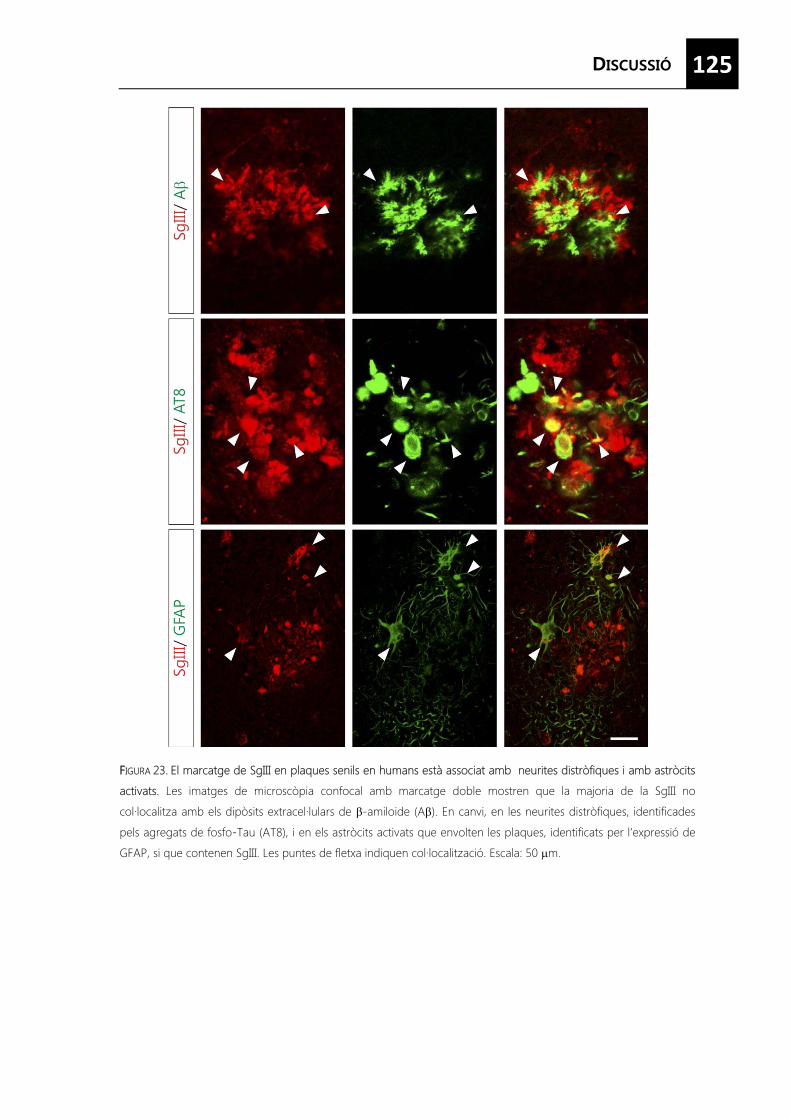
125
FFIGURA 23. El marcatge de SgIII en plaques senils en humans està associat amb neurites distròfiques i amb astròcits
activats. Les imatges de microscòpia confocal amb marcatge doble mostren que la majoria de la SgIII no
col·localitza amb els dipòsits extracel·lulars de �-amiloide (A�). En canvi, en les neurites distròfiques, identificades
pels agregats de fosfo-Tau (AT8), i en els astròcits activats que envolten les plaques, identificats per l’expressió de
GFAP, si que contenen SgIII. Les puntes de fletxa indiquen col·localització. Escala: 50 �m.


129
1. Neurones, astròcits i micròglia expressen patrons diferencials de proteïnes
que mitjancen els processos d’exocitosi.
2. Isoformes de sintaxines i els seus reguladors Munc18 són diferencialment
sobreexpressats durant l’activació glial mitjançada per agents proinflamatoris
amb un possible paper en l’alliberació de citocines.
3. La senyalització mitjançada per la via del cAMP restringeix les característiques
d’immaduresa i activació dels astròcits en cultiu i promou la seva maduració.
4. La diferenciació dels astròcits per cAMP augmenta els nivells d’expressió de
proteïnes de la via de secreció regulada per calci promovent l’alliberació
evocada de pèptids.
5. Els astròcits in vivo e in vitro presenten una forma no processada de
secretogranina III, un receptor de direccionament a grànuls de secreció.
6. L’activació astroglial in vivo en lesions traumàtiques indueix la sobreexpressió
de secretogranina III, el que suggereix un paper repressor o potenciador del
dany cerebral.


133
Abbott NJ, Rönnbäck L, Hansson E. Astrocyte-endothelial interactions at the blood-brain
barrier. Nat Rev Neurosci. 77(1): 41-53 (2006)
Acarin L, González B, Catellano B. Neuronal, astroglial and microglial cytokine expression after
an excitotoxic lesion in the immature rat brain. Eur J Neurosci. 112(10):3 505-20 (2000)
Aguado F, Espinosa-Parrilla JF, Carmona MA, Soriano E. Neuronal activity regulates correlated
network properties of spontaneous calcium transients in astrocytes in situ.J Neurosci.
22(21): 9430-44 (2002)
Allen NJ i Barres BA. Neuroscience: Glia- more than just brain glue. Nature. 4457(7230): 675-
677 (2009)
Araque A, Carmignoto G, Haydon PG. Dynamic signaling between astrocytes and neurons. Annu Rev Physiol. 663:795-813 (2001)
Araque A, Parpura V, Sanzgiri RP, Haydon PG. Tripartite synapses: glia, the unacknowledged
partner. Trends Neurosci. 222(5): 208-215 (1999)
Attwell D, Buchan AM, Charpak S, Lauritzen M, Macvicar BA, Newman EA. Glial and neuronal
control of brain blood flow. Nature. 4468(7321): 232-243 (2010)
Auld DS, Robitaille R. Glial cells and neurotransmission: an inclusive view of synaptic
function.Neuron. 440(2):389-400 (2003)
Ballard C, Gauthier S, Corbett A, Brayne C, Aarsland D, Jones E. Alzheimer’s disease. Lancet.
377(9770): 1019-1031 (2011)
Barres BA. The mistery and magic of glia: a perspective on their roles in health and disease.
Neuron. 660(3): 430-440 (2008)
Batter DK, Vilijn MH, Kessler J. Cultured astrocytes release proenkephalin. 1991. Brain Res.
563(1-2): 28-32 (1991)
Beattie EC, Stellwagen D, Morishita W, Bresnahan JC, Ha BK, Von Zastrow M, Beattie MS,
Mallenka RC. Control of synaptic strength by glial TNF alpha. Science. 2295: 2282-2285
(2002)
Benado A, Nasagi-Ativa Y, Dagi-Eisenberg R. Protein trafficking in immune cells. Immunology.
214(7): 507-525 (2009)
A
B
134
Bergami M, Santi S, Formaggio E, Cagnoli C, Verderio C, Blum R, Berninger B, Matteoli M,
Canossa M. Uptake and recycling of pro-BDNF for transmitter-induced secretion by
cortical astrocytes. J Cell Biol. 1183: 213-221 (2008)
Bergersen LH i Gundersen V. Morphological evidence for vesicular glutamate release from
astrocytes. Neuroscience. 1158: 260-265 (2009)
Bessis A, Béchade C, Bernard D, Roumier A. Microglial control of neuronal death and synaptic
properties. Glia. 555(3): 233-238 (2007)
Beuret N, Stettler H, Renold A, Rutishauser J, Spiess M. Expression of regulated secretory
proteins is suficient to generate granule-like structures in constitutively secreting cells. J
Biol Chem. 2279: 20242-20249 (2004)
Bezzi P, Gundersen V, Galbete JL, Seifert G, Steinhauser C, Pilati E, Volterra A. Astrocytes
contain a vesicular compartment that is competent for regulated exocytosis of
glutamate. Nat Neurosci. 77: 613-620 (2004)
Blazquez M and Shennan KI. Basic mechanisms of secretion: sorting into the regulated
secretory pathway. Biochem Cell Biol. 778: 181-191 (2000)
Blott EJ i Griffiths GM. Secretory lysosomes. Nat Rev Mol Cell Biol. 33(2): 122-131 (2002)
Blott EJ, Bossi G, Clark R, Zvelebil M, Griffiths GM. Fas ligand is targeted to secretory lysosomes
via a proline-rich domain in its cytoplasmic tail. J Cell Sci.114: 2045-2416 (2001)
Bochelen D, Langley K, Adamczyk M, Kupferberg A, Hor F, Vincendon G, Mersel M. 7beta-
hydroxysterol is cytotoxic to neonatal rat astrocytes in primary culture when cAMP
levels are increased. J Neurosci Res. 662: 99-111 (2000)
Bonanomi D, Benfenati F, Valtorta F. Protein sorting in the synaptic vesicle life cycle. Prog
Neurobiol. 880(4): 177-217 (2006)
Bonifacio JS i Glick BS. The mecanisms of vesicle budding and fusion. Cell. 116: 153-166 (2004)
Borges R, Díaz-Vera J, Domínguez N, Arnau MR, Machado JD. Chromogranins as regulators
of exocytosis. J Neurochem. 1114: 335-343 (2010)
Bradl M i Lassmann H. Oligodendrocytes: biology and pathology. Acta Neuropathol. 1119(1):
37- 53 (2010)
Braulke T i Bonifacino JS. Sorting of lysosomal proteins. Biochim Biophys Acta. 11793(4): 605-
614 (2009)
135
Buffo A, Rolando C, Ceruti S. Astrocytes in the damaged brain: molecular and cellular insights
into their reactive response and healing potential. Biochem Pharmacol. 779(2): 77-89
(2010)
Bushong EA, Martone ME, Ellisman MH. Maturation of astrocyte morphology and the
establishment of astrocyte domains during postnatal hippocampal development. Int J
Dev Neurosci.22(2): 73-86 (2004)
Cahoy JD, Emery B, Kaushal A, Foo LC, Zamanian JL, Christopherson KS, Xing Y, Lubischer JL,
Krieg PA, Krupenko SA, Thompson WJ, Barres BA.A transcriptome database for
astrocytes, neurons, and oligodendrocytes: a new resource for understanding brain
development and function.J Neurosci. 228(1):264-78. (2008)
Calegari F, Coco S, Taverna E, Bassetti M, Verderio C, Corradi N, Matteoli M, Rosa P.A
regulated secretory pathway in cultured hippocampal astrocytes.J Biol Chem.
274(32):22539-47 (1999)
Carmona MA, Murai KK, Wang L, Roberts AJ, Pasquale EB. Glial ephrin-A3 regulates
hippocampal dendritic spine morphology and glutamate transport. Proc Natl Acad Sci
USA. 1106(3): 12524-9 (2009)
Charles P, Hernandez MP, Stankoff B, Aigrot MS, Colin C, Rougon C, Zalc B, Lubetzki C.
Negative regulation of central nervous system myelination by polysialylated-neural cell
adhesion molecule. Proc Natl Acad Sci. 997(13): 7585-7590 (2000)
Chen XK, Xiong YF, Zhou Z. “Kiss-and-run” exocytosis in astrocytes. Neuroscientist. 112(5): 375-
378 (2006)
Chieregatti E, Chicka MC, Chapman ER, Baldini G. SNAP-23 functions in docking/fusion of
granules at low Ca2+.Mol Biol Cell. 115(4):1918-30 (2004)
Christopherson KS, Ullian EM, Stokes CC, Mullowney CE, Hell JW, Agah A, Lawler J, Mosher DF,
Bornstein P and Barres, BA. Thrombospondins are astrocyte-secreted proteins that
promote CNS synaptogenesis. Cell. 1120(3): 421-433 (2005)
Coco S, Calegari F, Pravettoni E, Pozzi D, Taverna E, Rosa P, Matteoli M, Verderio C. Storage
and release of ATP from astrocytes in culture. J Biol Chem. 2278: 1354-1362 (2003)
C
136
Daginakatte GC, Gadzinski A, Emnett RJ, Stark JL, Gonzales ER, Yan P, Lee JM, Cross AH,
Gutmann DH. Expression profiling identifies a molecular signature of reactive
astrocytes stimulated by cyclic AMP or proinflammatory cytokines. Exp Neurol. 2210:
261-267 (2008)
Dancourt J i Barlowe C. Protein sorting receptors in the early secretory pathway. Annu Rev
Biochem. 779: 777-802 (2010)
De Matteis MA and Luini A. Exiting the Golgi complex. Nat Rev Mol Cell Biol. 99(4): 273-284
(2008)
Delacourt A. General and dramatic glial reaction in Alzheimer brains.Neurology. 440(1):33-7
(1990)
Dideakos JD i Reudelhuber TL. Sending proteins to the dense core secretory granules: still a
lot to sort out. J Cell Biol. 999: 97-104 (2007)
Dowell JA, Johnson JA, Li J. Identification of astrocyte secreted proteins with a combination of
shotgun proteomics and bioinformatics. J Proteome Res. 88: 4135-4143 (2009)
Duan S, Anderson CM, Keung EC, Chen Y, Swanson RA. P2X7 receptor-mediated release of
excitatory amino acids from astrocytes. J Neurosci. 223: 1320-1328 (2003)
Dugan LL, Kim JS, Zhang Y, Bart RD, Sun Y, Holtzman DM, Gutmann DH. (1999). Differential
effects of cAMP in neurons and astrocytes. Role of B-raf. J Biol Chem. 2274: 25842-
25848.
Eder C. Mechanisms of interleukin-1 beta release. Immunobiology. 2214: 543-553 (2009)
Faden, AI. Experimental neurobiology of central nervous system trauma. Crit Rev Neurobiol.
7(3-4):175-86 (1993)
Falsig, J., Pörzgen, P., Lund, S., Schrattenholz, A., and Leist, M. The inflammatory transcriptome
of reactive murine astrocytes and implications for their innate immune function. J
Neurochem. 996: 893-907 (2006)
Fasshauer D, Antonin W, Margittai M, Pabst S, Jahn R. Mixed and non-cognate SNARE
complexes. Characterization of assembly and biophysical properties.J Biol Chem.
274(22):15440-6 (1999)
D
EF
137
Fasshauser D, Sutton RB, Brunger AT, Jahn R. Conserved structural features of the synaptic
fusion complex: SNARE proteins reclassified as Q- and R-SNAREs. Proc Natl Acad Sci
USA.995: 15781-15786 (1998)
Fedoroff S, McAuley WA, Houle JD, Devon RM. Astrocyte cell lineage. V. Similarity of
astrocytes that form in the presence of dBcAMP in cultures to reactive astrocytes in
vivo. J Neurosci Res.12: 14-27 (1984)
Fellin T. Communication between neurons and astrocytes: relevance to the modulation of
synaptic and network activity. J Neurochem. 1108(3): 533-544 (2009)
Fischer-Colbrie R, Kirchmair R, Kähler CM, Wiedermann CJ, Saria A. Secretoneurin: a new
player in angiogenesis and chemotaxis linking nerves, blood vessels and the immune
system. Curr Protein Pept Sci. 66(4):373-85 (2005)
Fischer-Colbrie R, Kirchmair R, Schobert A, Olenik C, Meyer DK, Winkler H. Secretogranin II is
synthesized and secreted in astrocyte cultures. J Neurochem. 660(6):2312-4 (1993)
Fischer-Colbrie R, Laslop A, Kirchmair R. Secretogranin II: molecular properties, regulation of
biosynthesis and processing to the neuropeptide secretoneurin. Prog Neurobiol.
46(1):49-70 (1995)
Flaumenhaft R. Molecular basis of platelet granule secretion. Arterioscler Thromb Vasc Biol.
23(7):1152-60 (2003)
Flieger O, Engling A, Bucala R, Lue H, Nickel W, Bernhagen J. Regulated secretion of
macrophage migration inhibitory factor is mediated by a non-classical pathway
involving an ABC transporter. FEBS Lett. 5551: 78-86 (2003)
Freeman MR. Specification and morphogenesis of astrocytes. Science. 3330(6005): 774-778
(2010)
Goss JR, O'Malley ME, Zou L, Styren SD, Kochanek PM, DeKosky ST.Astrocytes are the major
source of nerve growth factor upregulation following traumatic brain injury in the rat.
Exp Neurol. 149(2):301-9 (1998)
Greco TM, Seeholzer SH, Mak A, Spruce L, Ischiropoulos H. Quantitative mass spectrometry-
based proteomics reveals the dynamic range of primary mouse astrocyte protein
secretion. J Proteome Res. 77: 2764-2774 (2010)
Guillemot F. Cell fate specification in the mammalian telencephalon. Prog Neurobiol. 883(1):
37-52 (2007)
G
138
Halassa MM, Fellin T, Takano H, Dong JH, Haydon PG. Synaptic islands defined by the territory
of a single astrocyte. J. Neurosci. 227: 6473-6477 (2007)
Halonen SK, Woods T, McInnerney K, Weiss LM. Microarray analysis of IFN-gamma response
genes in astrocytes. J Neuroimmunol. 1175: 19-30 (2006)
Hamilton NB i Attwell D. Do astrocytes really exocytose neurotransmitters? Nat Rev
Neurosci.11(4): 227-238 (2010)
Hampton DW, Rhodes KE, Zhao C, Franklin RJM, Fawcett JW. The responses of
oligodendrocyte precursor cells, astrocytes and microglia to a cortical stab injury, in
the brain. Neuroscience. 1127 (4): 813-820 (2004)
Han L, Suda M, Tsuzuki K, Wang R, Ohe Y, Hirai H, Watanabe T, Takeuchi T, Hosaka M. A
large form of secretogranin III functions as a sorting receptor for chromogranin A
aggregates in PC12 cells. Mol Endocrinol. 222(8):1935-49 (2008)
Hepp R, Perraut M, Chasserot-Golaz S, Galli T, Aunis D, Langley K, Grant NJ. Cultured glial
cells express the SNAP-25 analogue SNAP-23. Glia. 227(2):181-7 (1999)
Herx LM i Yong VW. Interleukin-1 beta is required for the early evolution of reactive
astrogliosis following CNS lesion. J Neuropathol Exp Neurol. 660: 961-971 (2001)
Hewett JA. Determinants of regional and local diversity within the astroglial lineage of the
normal central nervous system. J Neurochem. 1110(6): 1717-1736 (2009)
Hirabayashi Y, Suzki N, Tsuboi M, Endo TA, Toyoda T, Shinga J, Koseki H, Vidal M, Gotoh
Y.Polycomb limits the neurogenic competence of neural precursor cells to promote
astrogenic fate transition. Neuron. 63: 600-613 (2009)
Hochstim C, Deneen B, Lukaszewicz A, Zhou Q, Anderson DJ. Identification of positionally
distinct astrocyte subtypes whose identities are specified by a homeodomain code.
Cell. 133: 510-522 (2008)
Holthuis JC, Martens GJ. The neuroendocrine proteins secretogranin II and III are regionally
conserved and coordinately expressed with proopiomelanocortin in Xenopus
intermediate pituitary. J Neurochem. 666(6): 2248-56 (1996)
Hosaka M, Watanabe T, Sakai Y, Kato T, Takeuchi T. Interaction between secretogranin III and
carboxypeptidase E facilitates prohormone sorting within secretory granules. J Cell Sci.
118: 4785-4795 (2005)
H
139
Houades V, Koulakoff A, Ezan P, Seif I, Giaume C. Gap junction-mediated astrocytic networks
in the mouse barrel cortex. J Neurosci. 228(20): 5207–5217 (2008)
Humeau Y, Doussau F, Grant NJ, Poulain B. How botulinum and tetanus neurotoxins block
neurotransmitter release. Biochimie. 882(5): 427-46 (2000)
Huneycutt BS, Benveniste EN. Regulation of astrocyte cell biology by the cAMP/protein kinase
A signaling pathway. Adv Neuroimmunol. 55(3): 261-9 (1995)
Hur YS, Kim KD, Paek SH, Yoo SH. Evidence for the existence of secretory granules (dense-
core vesicle)-based inositol 1,4,5-triphosphate-dependent Ca2+ signaling system in
astrocytes. PLoS One. 55(8), e11973 (2010)
Imai A, Nashida T, Shimomura H. Roles of Munc18-3 in amylase release from rat parotid
acinar cells. Arch Biochem Biophys. 4422(2): 175-82 (2004)
Jahn R i Scheller RH. SNAREs-engines for membrane fusion. Nat Rev Mol Cell Biol. 7(9): 631-
43 (2006)
Jahn R i Sudhof TC. Membrane fusion and exocytosis. Annu Rev Biochem. 68: 863-911 (1999)
Jahnke GD, Brunssen S, Maier WE, Harry GJ. Neurotoxicant-induced elevation of
adrenomedullin expression in hippocampus and glia cultures. J Neurosci Res. 66(3):
464-74 (2001)
Jaiswal JK, Fix M, Takano T, Nedergaard M, Simon SM. Resolving vesicle fusion from lysis to
monitor calcium-triggered lysosomal exocytosis in astrocytes. Proc Natl Acad Sci USA.
104(35): 14151-14156 (2007)
Jean Y, Lercher L, Dreyfus C. Glutamate elicits release of BDNF from basal forebrain of
astrocytes in a process dependent on metabotropic receptors and the PLC pathway.
Neuron Glia Biol. 44, 35-42 (2008)
John GR, Lee SC, Brosnan CF. Cytokines: powerful regulators of glial cell activation.
Neuroscientist. 99: 10-22 (2003)
John GR, Lee SC, Song X, Rivieccio M, Brosnan CF. IL-1-regulated responses in astrocytes:
relevance to injury and recovery. Glia. 449: 161-176. (2005)
Jourdain P, Bergersen LH, Bhaukaurally K, Bezzi P, Santello M, Domercq M, Matute C, Tonello
F, Gundersen V, Volterra A. Glutamate exocytosis from astrocytes controls synaptic
strength. Nature Neurosci. 110: 331-339 (2007)
I J
140
Kandel ER, Schwartz JH, Jesell TM. Principles of Neuroscience 4th ed. McGraw-Hill. New York
(2000)
Keene SD, Greco TM, Parastatidis I, Lee SH, Hughes EG, Balice-Gordon RJ, Speicher DW,
ischiropoulos H. Mass spectrometric and computational analysis of cytokine-induced
alterations in the astrocyte secretome. Proteomics. 99: 768-782 (2009)
Kettenmann H i Ransom BR. Neuroglia, 2nd ed. Oxford University Press, Oxford, 2004
Kimelberg HK, Goderie SK, Higman S, Pang S, Waniewski RA. Swelling-induced release of
glutamate, aspartate and taurine from astrocytes cultures. J Neurosci. 10: 1583-1591
(1990)
Knott C, Stern G, Kingsbury A, Welcher AA, Wilkin GP. Elevated glial brain-derived
neurotrophic factor in Parkinson's diseased nigra. Parkinsonism Relat Disord. 88(5):329-
41 (2002)
Koch C i Laurent G. Complexity and the nervous system. Science. 2284(5411): 96-98 (1999)
Kofuji P i Newman EA. Potassium buffering in the central nervous system. Neuroscience.
129(4): 1045-1056 (2004)
Koshimizu H, Kim T, Cawley NX, Loh YP. Chromogranin A: A new proposal for trafficking,
processing and induction of granule biogenesis. Regul Pept. 1165(1): 95-101 (2010)
Kriegstein A i Alvarez-Buylla A. The glial nature of embryonic and adult neural stem cells.
Annu Rev Neurosci. 332: 149-84 (2009)
Krzan M, Stenovec M, Kreft M, Pangrsic T, Grilc S, Haydon PG, Zorec R. Calcium-dependent
exocytosis of atrial natriuretic peptide from astrocytes. J Neurosci. 223(5): 1580-3 (2003)
Lastres-Becker I, Fernández-Pérez A, Cebolla B, Vallejo M. Pituitary adenylate cyclase-
activating polypeptide stimulates glial fibrillary acidic protein gene expression in
cortical precursor cells by activating Ras and Rap1. Mol Cell Neurosci. 339: 291-301
(2008)
Lee S, Park JY, Lee WH, Kim H, Park HC, Mori K, Suk K. Lipocalin-2 is an autocrine mediator of
reactive astrocytosis. J Neurosci. 229(1): 234-239 (2009)
Lein ES, Hawrylycz MJ, Ao N, Ayres M, Bensinger A, Bernard A, Boe AF, Boguski MS, Brockway
KS, Byrnes EJ, Chen L, Chen L, Chen TM, Chin MC, Chong J, Crook BE, Czaplinska A,
K
L

141
Dang CN, Datta S, Dee NR, Desaki AL, Desta T, Diep E, Dolbeare TA, Donelan MJ,
Dong HW, Dougherty JG, Duncan BJ, Ebbert AJ, Eichele G, Estin LK, Faber C, Facer BA,
Fields R, Fischer SR, Fliss TP, Frensley C, Gates SN, Glattfelder KJ, Halverson KR, Hart
MR, Hohmann JG, Howell MP, Jeung DP, Johnson RA, Karr PT, Kawal R, Kidney JM,
Knapik RH, Kuan CL, Lake JH, Laramee AR, Larsen KD, Lau C, Lemon TA, Liang AJ, Liu
Y, Luong LT, Michaels J, Morgan J, Morgan RJ, Mortrud MT, Mosqueda NF, Ng LL, Ng
R, Orta GJ, Overly CC, Pak TH, Parry SE, Pathak SD, Pearson OC, Puchalski RB, Riley ZL,
Rockett HR, Rowland SA, Royall JJ, Ruiz MJ, Sarno NR, Schaffnit K, Shapovalova NV,
Sivisay T, Slaughterbeck CR, Smith SC, Smith KA, Smith BI, Sodt AJ, Stewart NN, Stumpf
KR, Sunkin SM, Sutram M, Tam A, Teemer CD, Thaller C, Thompson CL, Varnam LR,
Visel A, Whitlock RM, Wohnoutka PE, Wolkey CK, Wong VY, Wood M, Yaylaoglu MB,
Young RC, Youngstrom BL, Yuan XF, Zhang B, Zwingman TA, Jones AR. Genome-wide
atlas of gene expression in the adult mouse brain. Nature. 4445: 168-176.(2007)
Li D, Ropert N, Koulakoff A, Giaume C, Oheim M. Lysosomes are the major vesicular
compartment undergoing Ca2+-regulated exocytosis from cortical astrocytes. J
Neurosci. 228(30): 7648-7658 (2008)
Liberto CM, Albrecht PJ, Herx LM, Yong VW, Levison SW. Pro-regenerative properties of
cytokine-activated astrocytes. J Neurochem. 889, 1092-1100 (2004)
Liu Y, Rao MS. Glial progenitors in the CNS and possible lineage relationships among them.
Biol Cell. 996(4): 279-90 (2004)
Majdoubi ME, Metz-Boutigue MH, Garcia-Sablone P, Theodosis DT, Aunis D.
Immunocytochemical localization of chromogranin A in the normal and stimulated
hypothalamo-neurohypophysial system of the rat. J Neurocytol. 225(7):405-16 (1996)
Malarkey EB i Parpura V. Mechanisms of glutamate release from astrocytes. Neurochem Int.
52: 142-154 (2008)
Malsam J, Kreye S, Söllner TH. Membrane fusion: SNAREs and regulation. Cell Mol Life Sci. 665:
2814-2832 (2008)
Martineau M, Galli T, Baux G, Mothet JP. Confocal imaging and tracking of the exocytotic
routes for D-serine mediated transmission. Glia. 556: 1271-1284 (2008)
Matyash V i Kettenmann H. Heterogeneity in astrocyte morphology and physiology. Brain Res
Rev. 663(1-2): 2-10 (2010)
M
142
McManus MF, Chen LC, Vallejo I, Vallejo M.Astroglial differentiation of cortical precursor cells
triggered by activation of the cAMP-dependent signaling pathway. J Neurosci. 119:
9004-9015 (1999)
Meeuwsen S, Persoon-Deen C, Bsibsi M, Ravid R, van Noort JM. Cytokine, chemokine and
growth factor gene profiling of cultured human astrocytes after exposure to
proinflammatory stimuli. Glia. 443: 243-253 (2003)
Miller FD i Gauthier AS. Timing is everything: making neurons versus glia in the developing
cortex. Neuron. 54: 357-369 (2007)
Mingorance A, Solé M, Munetón V, Martínez A, Nieto-Sampedro M, Soriano E, del Río JA.
Regeneration of lesioned entorhino-hippocampal axons in vitro by combined
degradation of inhibitory proteoglycans and blockade of Nogo-66/NgR signaling.
FASEB J. 220(3): 491-3 (2006)
Montana V, Malarkey EB, Verderio C, Matteoli M, Parpura V. Vesicular transmitter release from
astrocytes. Glia. 554(7):700-15 (2006)
Moore NH, Costa LG, Schaffer SA, Goodlett DR, Guizzetti M. Shotgun proteomics implicates
extracellular matrix proteins and protease systems in neuronal development induced
by astrocyte cholinergic stimulation. J Neurochem. 1108(4): 891-908 (2008)
Mothet JP, Pollegoni L, Ouanounou G, Martineau M, Fossier P, Baux G. Glutamate receptor
activation triggers a calcium-dependent and SNARE-dependent release of the
gliotransmitter D-serine. Proc Natl Acad Sci USA. 1102: 50606-50611 (2005)
Mucke L. Neuroscience: Alzheimer's disease. Nature. 4461(7266):895-7 (2009)
Muotri AR i Gage FH. Generation of neuronal variability and complexity. Nature. 4441(7097):
1087- 1093 (2006)
Murray RZ, Wylie FG, Khromykh T, Hume DA, Stow JL. Syntaxin 6 and Vti1b form a novel
SNARE complex, which is up-regulated in activated macrophages to facilitate
exocytosis of tumor necrosis factor-alpha. J Biol Chem. 2280: 10478-10483 (2005)
Nakashima K, Takizawa T, Ochiai W, Yanagisawa M, Hisatsune T, Nakafuku M, Miyazono K,
Kishimoto T, Kageyama R, Taga T. BMP2-mediated alteration in the developmental
pathway of fetal mouse brain cells from neurogenesis to astrocytogenesis. Proc Natl
Acad Sci USA. 998(10): 5868-5873 (2001)
N
143
Nash B, Ioannidou K, Barnett SC. Astrocyte phenotypes and their relationship to myelination. J
Anat. 2219(1): 44-52 (2010)
Nave KA. Myelination and support of axonal integrity by glia. Nature. 4468(7321): 244-252
(2010)
Nickel W. Pathways of unconventional protein secretion. Curr Opin Biotechnol. 221(5): 621-626
(2010)
Nie XJ i Olsson Y. Endothelin peptides in brain diseases. Rev Neurosci. 7(3):177-86 (1996)
Nieweg K, Schaller H, Pfrieger FW. Marked differences in cholesterol synthesis between
neurons and glial cells from postnatal rats. J Neurochem. 1109(1): 125-134 (2009)
Nishiyama A, Komitova M, Suzuki R, Zhu X. Polydendrocytes (NG2 cells): multifunctional cells
with lineage plasticity. Nat Rev Neurosci. 110(1): 9- 22 (2009)
Nishiyama A, Yang Z, Butt A. Astrocytes and NG2-glia: what’s in a name? J Anat. 2207(6): 687-
693 (2005)
Oberheim NA, Wang X, Goldman S, Nedegaard M. Astrocytic complexity distinguishes the
human brain. Trends Neurosci. 229: 547-553 (2006)
Olson JK and Miller SD. Microglia initiate central nervous sytem innate and adaptive immune
responses through multiple TLRs. J Immunol. 1173(6): 3916-3924 (2004)
Osborne KD, Lee W, Malarkey EB, Irving AJ, Parpura V. Dynamic imaging of cannabinoid
receptor 1 vesicular trafficking in cultured astrocytes. ASN Neuro. 11(5):283-296 (2009)
Ottiger HP, Battenberg EF, Tsou AP, Bloom FE, Sutcliffe JG. 1B1075: a brain- and pituitary-
specific mRNA that encodes a novel chromogranin/secretogranin-like component of
intracellular vesicles.J Neurosci. 110(9):3135-47 (1990)
Palop JJ, Chin J, Mucke L. A network dysfunction perspective on neurodegenerative diseases.
Nature. 4443(7113): 768- 773 (2006)
Pangrsic T, Potokar M, Stenovec M, Kreft M, Fabbretti E, Nistri A, Pryazhnikov E, Khiroug L,
Giniatullin R, Zorec R. Exocytotic release of ATP from cultured astrocytes. J Biol Chem.
282: 28749-28758 (2007)
Park JJ i Loh YP. How peptide hormone vesicles are transported to the secretion site for
exocytosis. Mol Endocrinol. 222(12): 2583-2595 (2008)
O
P
144
Park JJ, Cawley NX, Loh YP. Carboxypeptidase E cytoplasmic tail-driven vesicle transport is key
for activity-dependent secretion of peptide hormones. Mol Endocrinol. 222: 989-1005
(2008)
Parpura V i Zorec R. Gliotransmission: Exocytotic release from astrocytes. Brain Res Rev. 63:
83-92 (2010)
Parpura V, Basarsky TA, Liu F, Jeftinija K, Jeftinija S, Haydon PG. Glutamate-mediated
astrocyte-neuron signalling. Nature. 3369: 744-747 (1994)
Perea G i Araque A. Astrocytes potentiate transmitter release at single hippocampal synapses.
Science. 3317: 1083-1086 (2007)
Perea G i Araque A. GLIA modulates synaptic transmission. Brain Res Rev. 663: 93-102 (2010)
Peters A. A fourth type of neuroglial cell in the adult central nervous system. J neurocytol.
33(3): 345-357 (2004)
Pike CJ, Cummings BJ, Cotman CW. Early association of reactive astrocytes with senile plaques
in Alzheimer's disease. Exp Neurol. 1132(2):172-9 (1995)
Ponnambalam S i Baldwin SA. Constitutive protein secretion from the trans-Golgi network to
the plasma membrane. Mol Membr Biol. 20: 129-139 (2003)
Potokar M, Stenovec M, Kreft M, Gabrijel M, Zorec R. Physiopathologic dynamics of vesicle
traffic in astrocytes. Histol Histopathol. 226: 277-284 (2011)
Prada I, Marchaland J, Podini P, Magrassi L, D'Alessandro R, Bezzi P, Meldolesi J.REST/NRSF
governs the expression of dense-core vesicle gliosecretion in astrocytes. J Cell Biol.
193(3):537-49 (2011)
Ramamoorthy P i Whim MD. Trafficking and fusion of neuropeptide Y-containing dense-core
granules in astrocytes. J Neurosci. 228(51): 13815-13827 (2008)
Ransohoff RM i Cardona AE. The myeloid cells of the central nervous system parenchima.
Nature. 4468: 253-262 (2010)
Reyes RC i Parpura V. The trinity of Ca2+ sources for the exocytotic glutamate release from
astrocytes. Neurochem Int. 555(1-3): 2-8 (2009)
Robel S, Berninger B, Götz M. The stem cell potential of glia: lessons from reactive gliosis. Nat
Rev Neurosci. 112(2): 88-104 (2011)
Robinson MS. Adaptable adaptors for coated vesicles. Trends Cell Biol. 114(4): 167-174 (2004)
R
145
Rosenberg SS, Kelland EE, Tokar E, De la Torre AR, Chan JR. The geometric and spatial
constraints of the microenvironment induce oligodendrocyte differentiation. Proc Natl
Acad Sci. 1105(38): 14662-14667 (2008)
Rowitch DH i Kriegstein AR. Developmental genetics of vertebrate glial-cell specification.
Nature. 4468: 214-222 (2010)
Ruiz-Montasell B, Aguado F, Majó G, Chapman ER, Canals JM, Marsal J, Blasi J. Differential
distribution of syntaxin isoforms 1A and 1B in the rat central nervous system. Eur J
Neurosci. 88(12):2544-52 (1996)
Sakry D, Karram K, Trotter K. Synapses between NG2 glia and neurons. J Anat. 2219 (1): 2-7
(2011)
Saura J, Tusell JM, Serratosa J. High-yield isolation of murine microglia by mild trypsinization.
Glia. 444 (3): 183-189 (2003)
Saura J. Microglial cells in astroglial cultures: a cautionary note. J Neuroinflammation. 44: 26-37
(2007)
Schubert V, Bouvier D, Volterra A. SNARE protein expression in synaptic terminals and
astrocytes in the adult hippocampus: A comparative analysis. Glia. doi:
10.1002/glia.21190. [Epub ahead of print] (2011)
Shiga H, Murakami J, Nagao T, Tanaka M, Kawahara K, Matsuoka I, Ito E. Glutamate release
from astrocytes is stimulated via the appearance of exocytosis during cyclic AMP-
induced morphologic changes. J Neurosci Res. 884: 338-347 (2006)
Shurety W, Merino-Trigo A, Brown D, Hume DA, Stow JL. Localization and trans-Golgi
trafficking of tumor necrosis factor alpha in macrophages. J Interferon Cytokine Res.
20: 427-438 (2000)
Silver J i Miller JH. Regeneration beyond the glial scar. Nature Rev Neurosci. 5(2): 146-156
(2004)
Slezak M, Pfrieger FW. New roles for astrocytes: regulation of CNS synaptogenesis.Trends
Neurosci. 226(10): 531-5 (2003)
Smith HS. Activated microglia in nociception. Pain Physician. 113(3):295-304 (2010)
Sofroniew M i Vinters HV. Astrocytes: biology and pathology. Acta Neuropathol. 1119: 7-35
(2010)
S
146
Sofroniew MV. Molecular dissection of reactive astrogliosis and glial scar formation. Trends
Neurosci. 332: 638-647 (2009)
Stanley AC i Lacy P. Pathways for cytokine secretion. Physiology (Bethesda). 225(4): 218-29
(2010)
Stellwagen D i Mallenka RC. Synaptic scaling mediated by glial TNF-alpha. Nature. 4440: 1054-
1059 (2006)
Stenmark H. Rab GTPases as coordinators of vesicle traffic. Nat Rev Mol Cell Biol. 110(8): 513-
525 (2009)
Stenovec M, Kreft M, Grilc S, Pangrisc T, Zorec R. EAAT2 density at the astrocyte plasma
membrane and Ca2+-regulated exocytosis. Mol Memb Biol. 225: 203-215 (2008)
Stevens B, Allen NJ, Vazquez LE, Howell GR, Christopherson KS, Nouri N, Micheva KD,
Mehalow AK, Huberman AD, Stafford B, Sher A, Litke AM, Lambris JD, Smith SJ, John
SW, Barres BA.The classical complement cascade mediates CNS synapse elimination.
Cell. 1131(6): 1164-1178 (2007)
Stow JL, Low PC, Offenhäuser C, Sangermani D. Cytokine secretion in macrophages and other
cells: pathways and mediators. Immunology. 214(7): 601-612 (2009)
Stow JL, Manderson AP, Murray RZ. SNAREing immunity: the role of SNAREs in the immune
system. Nat Rev Immunol. 66(12): 919-29 (2006)
Szatkowsky M, Barbour B, Attwell D. Non-vesicular release of glutamate from glial cells by
reversed electrogenic glutamate uptake. Nature. 3348: 443-446 (1990)
Takahashi K, Nakayama M, Totsune K, Murakami O, Sone M, Kitamuro T, Yoshinoya A,
Shibahara S. Increased secretion of adrenomedullin from cultured human astrocytes
by cytokines. J Neurochem. 774(1): 99-103 (2000)
Takano T, Kang J, Jaiswal JK, Simon SM, Lin JH, Yu Y, Li Y, Yang J, Dienel G, Zielke HR,
Nedegaard M. Receptor-mediated glutamate release from volume sensitive channels
in astrocytes. Proc Natl Acad Sci USA. 1102: 16466-16471 (2005)
Taupenot L, Ciesielski-Treska J, Ulrich G, Chasserot-Golaz S, Aunis D, Bader MF. Chromogranin
A triggers a phenotypic transformation and the generation of nitric oxide in brain
microglial cells. Neuroscience. 772(2): 377-89 (1996)
T
147
Taupenot L, Harper KL, O’Connor DT. Role of H+-ATPase-mediated acidification in sorting
and release of the regulated secretory protein chromogranin A: evidence for a
vesiculogenic function. J Biol Chem. 2280: 3885-3897 (2005)
Tsang MC, Lo AC, Cheung PT, Chung SS, Chung SK. Perinatal hypoxia-/ ischemia-induced
endothelin-1 mRNA in astrocyte-like and endothelial cells. NeuroReport. 112(10): 2265-
2270 (2001)
Verkhratsky A i Parpura V. Recent advances in (patho)physiology of astroglia. Acta Pharmacol
Sin. 331(9): 1044-1054 (2010)
Verkhratsky A. Physiology of neuronal-glial networking. Neurochem Int. 557: 332-343 (2010)
Vilijn MH, Das B, Kessler JA, Fricker LD. Cultured astrocytes and neurons synthesize and
secrete carboxypeptidase E, a neuropeptide-processing enzyme. J Neurochem. 553(5):
1487-93 (1989)
Volterra A i Meldolesi J. Astrocytes, from brain glue to communication elements: the
revolution continues. Nat Rev Neurosci. 66(8): 626-640 (2005)
Warr O, Takahashi M, Attwell D. Modulation of extracellular glutamate concentration in rat
brain slices by cystine-glutamate exchange. J Physiol. 5514: 783-793 (1999)
Wigley R, Hamilton N, Nishiyama A, Kirchhoff F, Butt AM. Morphological and physiological
interacctions of NG2-glia with astrocyte and neurons. J Anat. 2210(6): 661- 670 (2007)
Winkler H and Fischer-Colbrie R.The chromogranins A and B: the first 25 years and future
perspectives.Neuroscience. 449(3):497-528 (1992)
Winsky-Sommerer R, Benjannet S, Rovère C, Barbero P, Seidah NG, Epelbaum J, Dournaud P.
Regional and cellular localization of the neuroendocrine prohormone convertases PC1
and PC2 in the rat central nervous system. J Comp Neurol. 424(3): 439-60 (2000)
Woulfe J, Deng D, Munoz D. Chromogranin A in the central nervous system of the rat: pan-
neuronal expression of its mRNA and selective expression of the protein. Neuropeptides. 333(4):285-300 (1999)
Yang I, Han SJ, Kaur G, Crane C, Parsa AT. The role of microglia in central nervous system
immunity and glioma immunology. J Clin Neurosci. 117(1): 6-10 (2010)
V
W
Y
148
Ye ZC, Wyeth MS, Baltan-Tekkok S, Ransom BR. Functional hemmichannels in astrocytes: a
novel mechanism of glutamate release. J Neurosci. 223: 3588-3596 (2003)
Yoo SH. Secretory granules in inositol 1,4,5-triphosphate-dependent Ca2+ signaling in the
cytoplasm of neuroendocrine cells. FASEB J. 224: 653-664 (2010)
Zhang Z, Chen G, Zhou W, Song A, Xu T, Luo Q, Wang W, Gu XS, Duan S. Regulated ATP
release from astrocytes through lysosome exocytosis. Nat Cell Biol. 99(8): 945-953
(2007)
Zhang X, Odom DT, Koo SH, Conkright MD, Canettieri G, Best J, Chen H, Jenner R,
Herbolsheimer E, Jacobsen E, Kadam S, Ecker JR, Emerson B, Hogenesch JB, Unterman
T, Young RA, Montminy M. Genome-wide analysis of cAMP-response element
binding protein occupancy, phosphorylation, and target gene activation in human
tissues. Proc Natl Acad Sci USA. 1102: 4459-4464 (2005)
Zhang Y, Luo Y, Zhai Q, Ma L, Dorf ME. Negative role of cAMP-dependent protein kinase A in
RANTES-mediated transcription of proinflammatory mediators through Raf. FASEB J.
17: 734-736 (2003)
Z

151
La tesi de Sonia Paco Mercader es basa en els articles següents:
Article 1:
CCyclic AMP signaling represses activation and promotes differentiation of astrocyte
transcriptome
Paco S, Hummel M, Pla V, Sumoy L, Aguado F.
En preparació.
Article 2:
Regulation of exocytotic protein expression and Ca2+-dependent peptide secretion in
astrocytes.
Paco S, Margelí MA, Olkkonen VM, Imai A, Blasi J, Fischer-Colbrie R, Aguado F.
J Neurochem. 2009 Jul;110(1):143-56.
Índex d’impacte: 3,999
Article 3:
Secretogranin III is an astrocyte granin that is overexpressed in reactive glia.
Paco S, Pozas E, Aguado F.
Cereb Cortex. 2010 Jun;20(6):1386-97.
Índex d’impacte: 6,979
152
Fernando Aguado Tomás, director de la tesi doctoral de Sonia Paco Mercader,
certifica que el doctorand ha contribuït als treballs recollits en els tres articles de la
següent manera:
En los tres artículos incluidos en la tesis doctoral de Sonia Paco Mercader (dos
ya publicados y uno pendiente de publicación), la doctoranda ha realizado la gran
mayoría del trabajo experimental desarrollado. La labor realizada incluye
experimentos tanto de cultivos celulares como de animales, la aplicación de técnicas
bioquímicas y de biología celular y molecular y el uso de aplicaciones de análisis de
microarrays. Es de señalar que muchas de las técnicas realizadas fueron puestas a
punto por la doctoranda para su utilización en el transcurso de la tesis doctoral. Por
otro lado, Sonia Paco Mercader ha participado activamente en el diseño de los
experimentos, la interpretación de los resultados y la escritura de los manuscritos. Por
todo ello, certifico la máxima responsabilidad de la doctoranda en cada uno de los
tres trabajos presentados, como queda reflejado en su autoría de primer firmante.
���������
�
�������� �� �� �������� ���������� �� ����� ����� ����������������� ��������� ����� �������� ��
��������������������������������������������������������������������������������������
����������������� ������������� ����������������� ����������!��������� �������������������
����������������������������������������"�
�� ��� ����� ��� #$%� ��&���'�� �� ������� ����� ���������������� �� ������� (����� ������ ����
������� ���������� ��� ��� ����������� �� ���� ��� ���� ����� �� ��� ��������� ���!� ���� ���� �����) ��
����� *�&��������+������� ���������������������� ���������������+�������� ����+����� ��,�����
&������������������)�������������������-�������������������������.�������������
��-����������������������)������������������������.��� ������������� ��������������/��
���!�������0�����)������������0�����������
���������� ����� ��������������1����2�����0�������)���������)���� �� �����������������������
����� ����������
��������������������������������$��������2��3����������0������������)�����������������#��4�
)5������6������(����&/�������2�����)���#���7�89���)��
���������� �����#��:�� ���2���������-������ ������� �(������ ���9����� ����� 9�����;�+�������������
���� ����� ������ �*� ������������ ���� ���� ��������� ����� ��� ����������� 0������ � ����� ����
������������� ;�+����� ����� ��0���� ���� ������� ���� ���������� ���� ����'��� ���� ����)����'��� ����
�����������������������������������+��������<���������������=����������������������������
����������� ������������!����������������*�
��������������� ���������������������2���������!����3�����>������&����������?����������#������
�������� ��������������������@�������������������� �����������������>�;�����(��������&�����
��������(��������������������������������������������*������?���������������������������������
��������������������� �� ����������������������������� ������������������������+�����
����0���������������������������������������������������+*�
6����� �� ��� ����� ���� �������� �� ��� A$� �� ���� ������� ������������� ������� ��������� ��
������������������������������0����0������
���� ����� ������ �� ��� ����� �� B�������������� �� �C�������� D���� 9���� �� �����
������������������2���������������������������������������������� ���������������E����������
��������D��������������
1��/� ��������� ���� ������� �� ��� ������ �� ��� 2���������� ���� �����.������ �������������� 2���� ����
�������������������������������������� ����������������������������A������������������*�