Embed Size (px)
Citation preview

Mechanics of Breathing 1 and 2 Linda Costanzo, Ph.D. OBJECTIVES:
After studying this lecture, the student should understand:
1. The definition of compliance and its relationship to elastance. 2. How lung compliance is measured and the explanation of hysteresis. 3. How compliance of the chest wall can be demonstrated by a pneumothorax. 4. How to interpret the pressure-volume curves for the lung, chest wall, and
combined lung-chest wall system. 5. How lung diseases such as emphysema and fibrosis alter lung compliance and the
resulting effects on the pressure-volume curves. 6. The concept of surface tension in alveoli and the role of surfactant. 7. The factors that determine and alter airway resistance. 8. Changes in alveolar, intrapleural, and transmural pressures during the normal
breathing cycle. 9. Alveolar, intrapleural, and transmural pressures during forced expiration, and the
effect of emphysema on these pressures.
I. MUSCLES OF RESPIRATION
A. Inspiration The most important muscle of inspiration is the diaphragm. It is a thin, dome-shaped muscle that inserts into the lower ribs and is supplied by the phrenic nerve. When the diaphragm contracts (as directed by phrenic nerve activity), it forces the abdominal contents down and forward, and the volume of the chest cavity is increased. In normal tidal breathing, the excursion of the diaphragm is very slight, about 1 cm. (During heavy breathing, however, the diaphragm moves greater distances, up to 10 cm.) During exercise, the external intercostal muscles and accessory muscles are used for inspiration.
B. Expiration During normal tidal breathing, expiration is passive. The lungs and chest wall are elastic structures and naturally “want” to return to their resting positions after being expanded during inspiration. During exercise, expiration becomes active, utilizing the abdominal muscles and the internal intercostal muscles.

II. COMPLIANCE (inverse of elastance!) Compliance describes the distensibility of the respiratory structures. It says how much the volume of a structure changes for a given change in pressure (ΔV/ΔP). If compliance is high, the volume changes a lot; if compliance is low, the volume changes little. A point that hangs students up is that compliance is not elasticity, it is the inverse of elasticity (or elastance). The more elastic fibers in a structure (e.g., the alveoli), the greater its tendency to “spring back” and the stiffer and less compliant it is (like a thick rubber band). The fewer elastic fibers in a structure, the less its tendency to spring back and the more compliant it is. You must get this! High compliance = less elastic recoil; low compliance = more elastic recoil. A. How lung pressures are expressed
We will now discuss compliance of lung structures, or how their volume changes as a function of pressure. The pressure term needs clarification, as it can be expressed in several different ways. It can mean pressure inside a structure (e.g., the lungs), pressure outside a structure, or even the pressure difference across a structure, which is called transmural pressure. For example, transpulmonary pressure (the pressure difference across the lungs), is the difference between intra-alveolar pressure and intrapleural pressure. (The intrapleural ‘space’ lies between the alveoli and the chest wall.) By convention, lung pressures are referred to atmospheric pressure, where atmospheric pressure (PB) is called “zero.” For example, if alveolar pressure is equal to atmospheric, it is said to be zero; if it is higher than atmospheric, it is positive, and if it is lower than atmospheric, it is negative.
B. Compliance of the lungs The lungs are both compliant and elastic. They must be compliant to fill with air during inspiration. They must be elastic to recoil and push air out during expiration. The compliance of the lungs is demonstrated by an isolated lung in a jar.

Figure 1.
In the figure, lung volume is expressed as a function of pressure. Compliance is the slope of the relationship (i.e., ΔV/ΔP). The space outside the lung is analogous to intrapleural pressure in vivo. In the experiment, the lungs and airways are open to the atmosphere and the pressure inside the lungs is equal to atmospheric. Pressure outside the lung is varied with a pump to simulate changes in intrapleural pressure. The volume of the lung is measured at different pressures. When the outside pressure is made more negative (i.e., lower than atmospheric), the lung inflates and its volume increases. When the outside pressure is made less negative, the lung deflates and its volume decreases. A sequence of inflation and deflation creates a pressure-volume loop. The inspiration (filling) limb of the lung’s pressure-volume loop has a different slope (compliance) than the expiration limb (called hysteresis). For a given outside (intrapleural) pressure, the volume of the lung is higher during expiration than during inspiration. Thus, lung compliance is higher during expiration than during inspiration. Lung compliance is

typically measured on the expiration limb because the inspiration limb flattens at highest volumes (when lung tissue is already maximally stretched out). Why does hysteresis occur? The slopes of the inspiration and expiration curves are compliance, and compliance is an intrinsic property of the lung. Since it’s the same lung, why would compliance be higher when we expire than when we inspire? The answer is surface tension. In the air-filled lung, there are strong intermolecular forces between liquid molecules lining alveoli. During inspiration, one begins at low lung volume where the liquid molecules are close together and strongly attracted to each other – to inflate the lung, one must break these intermolecular forces; thus, it is harder to inflate the lung than would be expected based on compliance alone. For expiration, one begins at high lung volume where liquid molecules are far apart and intermolecular forces needn’t be broken. Thus, the observed compliance curve during inspiration is determined both by intrinsic compliance and by surface tension; the observed compliance curve during expiration is determined only by intrinsic compliance (i.e., the “real” compliance).
C. Compliance of the chest wall
Figure 2.
The figure shows the relationship between the lungs and the chest wall. The conducting airways are shown as a single tube, and the gas exchange region is shown as a single alveolus. The intrapleural space between the lungs and chest wall is exaggerated. Like the lungs, the chest wall also is

compliant, and its compliance is demonstrated by introducing air into the intrapleural space (pneumothorax).
Figure 3.
Intrapleural pressure is normally negative. This negative intrapleural pressure is created by two opposing elastic forces pulling on the intrapleural space: the lungs, with their elastic properties, tend to collapse; the chest wall, with its elastic properties, tends to spring out. When these two opposing forces pull on the intrapleural space, a negative pressure is created. In turn, this negative intrapleural pressure opposes the natural tendency of the lungs to collapse and the chest wall to spring out; that is, the negative intrapleural pressure is responsible for preventing the lungs from collapsing and the chest wall from springing out. When a sharp object punctures the intrapleural space, air is introduced into the space (pneumothorax) and intrapleural pressure becomes equal to atmospheric pressure; thus, instead of its normal negative value, intrapleural pressure becomes zero, which has two predictable consequences. (1) There is no longer a negative intrapleural pressure to hold the lungs open, and the lungs collapse. (2) There is no longer a negative intrapleural pressure to keep the chest wall from expanding, and the chest wall springs out.

D. Pressure-volume Curves for the Lungs, Chest wall, and Combined Lung and Chest Wall
Figure 4.
Pressure-volume curves are shown for the lungs alone (lung in a jar), chest wall alone, and combined lung and chest-wall system. (The curve for the chest wall alone is obtained by subtraction of the lung curve from the combined lung and chest-wall curve.) The curve for the combined lung and chest-wall system is obtained by having a trained subject breathe in and out of a spirometer. The subject inspires or expires to a given volume. The spirometer valve is then closed and, as the subject relaxes his respiratory muscles, his airway pressure is measured (called relaxation pressure). In this way, values for airway pressure are obtained at a series of static volumes of the combined lung and chest-wall system. When the volume is functional residual capacity (FRC), airway pressure is zero and equal to atmospheric pressure. At volumes less than FRC, airway pressures are negative (less volume, less pressure). At volumes higher than FRC, airway pressures are positive (more volume, more pressure). The slope of each curve is compliance. Note that the compliance of the chest wall alone is similar to the compliance of the lungs alone (the slopes are the same). However, the compliance of the combined lung and chest-wall system is less than that of either structure alone (i.e., the combined lung and chest wall curve is “flatter”). Visualize one balloon (the lungs) inside another balloon (the chest wall). Alone, each balloon is compliant, but the combined system (balloon within balloon) is less compliant and

harder to expand. To interpret these curves, begin at the volume called FRC, the equilibrium volume of the combined lung and chest-wall system. FRC is the volume present in the lungs after a person has expired a normal tidal breath. Then compare the graphs at volumes less than FRC and volumes greater than FRC.
1. Volume is FRC. When the volume is FRC, the combined lung and chest-wall system is at equilibrium. Airway pressure is equal to atmospheric pressure, which is called zero. At FRC, because they are elastic structures, the lungs “want” to collapse and the chest wall “wants” to expand. If these elastic forces were unopposed, the structures would do exactly that! However, at FRC, the collapsing force on the lungs is exactly equal to the expanding force on the chest wall, as shown by the equidistant arrows, and the combined lung and chest-wall system neither tends to collapse or expand.
2. Volume less than FRC. When the volume in the system is less than FRC (i.e., the subject forcibly expires into the spirometer), there is less volume in the lungs and the collapsing elastic force of the lungs is smaller. The expanding force on the chest wall is greater, however, and the combined lung and chest wall system “wants” to expand. (Notice on the graph that, at volumes less than FRC, the collapsing force on the lungs is smaller than the expanding force on the chest wall, and the combined system tends to expand.)
3. Volume greater than FRC. When the volume in the system is greater than FRC (i.e., the subject inspires from the spirometer), there is more volume in the lungs and the collapsing (elastic) force of the lungs is greater. The expanding force on the chest wall is smaller, however, and the combined lung and chest-wall system “wants” to collapse. (Notice on the graph that, at volumes greater than FRC, the collapsing force on the lungs is greater than the expanding force on the chest wall, and the overall system tends to collapse. At highest lung volumes, both the lungs and the chest wall “want” to collapse [the chest wall curve has crossed the vertical axis] and, there is a very large collapsing force on the combined system.)

E. Changes in Lung Compliance in Disease
Figure 5.
Lung compliance changes in disease, changing the slopes of the relationships. For convenience, each component of the system is shown on a separate graph (i.e., chest wall alone, lung alone, and combined lung and chest wall). The chest wall alone is included only for completeness, since its compliance is not altered by disease. The solid lines in each graph gives the normal relationships. The dashed and dotted lines show the effects of disease.

1. Emphysema (increased lung compliance). Emphysema is associated with loss of elastic fibers in the lungs. As a result, the compliance of the lungs increases. (Recall again, the inverse relationship between elastance and compliance.) An increase in compliance is associated with an increased (steeper) slope of the volume versus pressure curve for the lung. At a given volume, the collapsing (elastic recoil) force on the lungs is decreased. At the original value for FRC, the tendency for the lungs to collapse is now less than the tendency of the chest wall to expand, and these opposing forces will no longer be balanced. In order for the opposing forces to be balanced, volume must be added to the lungs to increase their collapsing force. Thus, the combined lung and chest-wall system seeks a new higher FRC, where the two opposing forces can be balanced; the new intersection point, where airway pressure is zero, is increased. The patient with emphysema breathes at higher lung volumes (in recognition of the higher FRC) and has a barrel-shaped chest.
2. Fibrosis (decreased lung compliance). Fibrosis is a restrictive disease associated with stiffer lung tissues and decreased compliance. There is decreased slope of the volume versus pressure curve for the lung. At the original FRC, the tendency of the lungs to collapse is greater than the tendency of the chest wall to expand, and the opposing forces will no longer be balanced. To reestablish the balance, the lung and chest-wall system will seek a new lower FRC; the new intersection point, where airway pressure is zero, is decreased.
III. SURFACE TENSION
A. Law of LaPlace Surface tension of a sphere (such as an alveolus) is given by the Law of LaPlace: P = 2T r where P is collapsing pressure on the alveolus (or pressure required to keep the alveolus open), T is surface tension, and r is radius of the alveolus. The alveoli are lined with a film of liquid and intermolecular attractive forces create a surface tension, which creates a pressure that tends to

collapse the alveoli. Because alveoli are small, there is a potential problem in keeping them open. (According to LaPlace, the smaller the radius, the higher the collapsing pressure.) Alveoli could solve this problem by having large radii; however, large radii means reduced surface area, which is bad for gas exchange. Surfactant to the rescue!
B. Surfactant is synthesized by type II alveolar cells. Surfactant reduces surface tension of alveoli, thus reducing the collapsing pressure of small alveoli. The most important constituent of surfactant is dipalmitoyl phosphatidylcholine (DPPC). Molecules of DPPC are amphoteric and align themselves on the alveolar surface with their hydrophobic portions attracted to each other and their hydrophilic portions repelled from each other. In this way, DPPC breaks up the liquid molecules that were responsible for high alveolar surface tension. When surfactant is present, surface tension and collapsing pressure are reduced and small alveoli can be kept open.
In neonatal respiratory distress syndrome, there is deficiency of surfactant (Surfactant begins appearing at gestational week 24 and is almost always present by gestational week 35. Thus, infants born between weeks 24 and 35 have uncertain surfactant status.) In the absence of surfactant, small alveoli have increased surface tension and collapsing pressure and will collapse (called atelectasis). Absence of surfactant also decreases lung compliance. Collapsed alveoli are not ventilated (they are too hard to expand) and, therefore, do not participate in gas exchange. You will learn that this is called a shunt, which causes hypoxemia (a decrease in arterial PO2).
IV. RESISTANCE OF AIRWAYS
A. Ohm’s Law There is a relationship between airflow, pressure, and resistance that is analogous to the relationship between blood flow, pressure, and resistance in the cardiovascular system, i.e., Ohm’s law: Q = ΔP R where Q is airflow, ΔP is pressure difference between mouth (or nose) and alveoli, and R is resistance of airways. The driving force for airflow is the pressure difference.

Resistance of the airways is given by Poiseuille’s law: R = 8 η l π r4 where R is airway resistance, η is viscosity of air, l is length of airway, and r is radius of the airway. The most important aspect of Poiseuille is the inverse dependence of resistance on r4. The smaller the airway, the higher the resistance; the larger the airway, the lower the resistance. Incidentally, medium-sized bronchi are the site of highest airway resistance. (Although it would seem that smaller airways would have higher resistance, they do not because they are arranged in parallel.)
B. What changes airway resistance?
1. Autonomic nervous system. As you have heard, bronchial smooth muscle has autonomic innervation.
a. Parasympathetic stimulation, via muscarinic receptors, causes constriction of bronchial smooth muscle, decreased radius, and increased resistance to airflow.
b. Sympathetic stimulation, via β2 receptors, causes dilation of bronchial smooth muscle, increased radius, and decreased resistance to airflow. The β2 receptors also are activated by circulating catecholamines (epinephrine) and β2-adrenergic agonists (isoproterenol), which are useful in dilating the airways in asthma.
2. Lung disease. Increased airway resistance is a feature of obstructive lung diseases such as asthma and chronic obstructive pulmonary disease (COPD, a combination of emphysema and chronic bronchitis). Increased airway resistance causes decreased airflow; this is especially a problem during expiration, which is normally passive. (Later, we will discuss why emphysema, a disease of increased compliance, causes increased airway resistance and obstruction, a point that is not intuitively evident.)
3. Lung volume. You may be surprised to hear that lung volume affects resistance of airways. Lung tissue exerts radial traction, or pull, on airways. High lung volume exerts more traction and

decreases airway resistance. Low lung volume exerts less traction and increases airway resistance. That persons with asthma breathe at high lung volumes (higher FRC) is helpful, as it partially offsets the high airway resistance associated with their disease.
V. THE BREATHING CYCLE: describes the changes in lung volumes and pressures that occur during the cycle of inspiration, expiration, inspiration....etc.
A. Normal breathing. In normal breathing, the volume inspired and expired is tidal volume. For the figures below, recall that pressures are referred to atmospheric, which is called “zero.” Transmural pressures across the lungs are shown by the open arrows and, by convention, are calculated as alveolar (or airway) pressure minus intrapleural pressure; the magnitude of the transmural pressure is shown above the open arrow. For example, if alveolar pressure is zero and intrapleural pressure is -5 cm H2O, then transmural pressure is +5 cm H2O (0 - [-5] = +5). If transmural pressure is positive, the structure is open; if transmural pressure is negative, the structure collapses. For all phases of the normal breathing cycle, despite changes in alveolar, airway, and intrapleural pressure, transmural pressure across lungs and airways is always positive; thus, these structures remain open.

Figure 6.Figure 5-13 Volumes and pressures during the normal breathing cycle. Intrapleural pressure and alveolar pressure are referred to atmospheric pressure. Letters A to D correspond to phases of the breathing cycle in Figure 5-14.
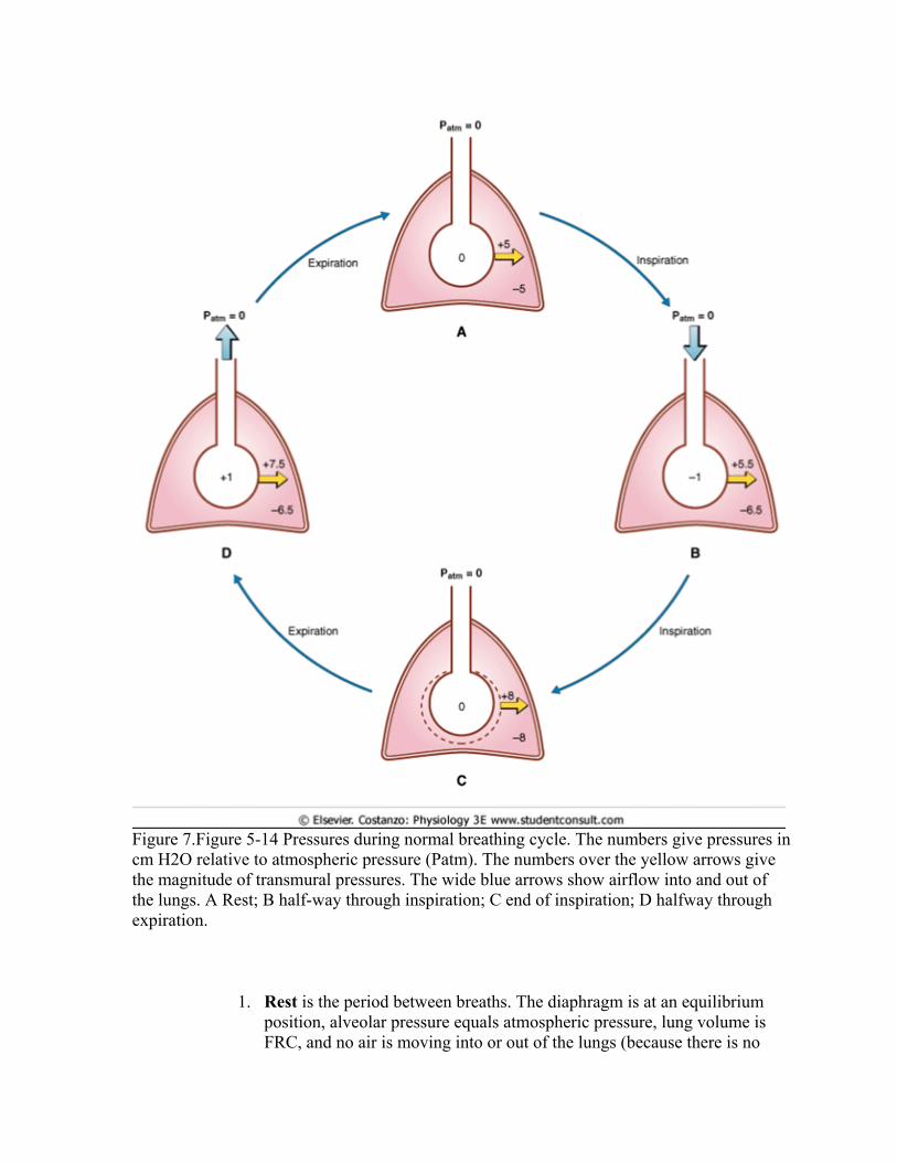
Figure 7.Figure 5-14 Pressures during normal breathing cycle. The numbers give pressures in cm H2O relative to atmospheric pressure (Patm). The numbers over the yellow arrows give the magnitude of transmural pressures. The wide blue arrows show airflow into and out of the lungs. A Rest; B half-way through inspiration; C end of inspiration; D halfway through expiration.
1. Rest is the period between breaths. The diaphragm is at an equilibrium position, alveolar pressure equals atmospheric pressure, lung volume is FRC, and no air is moving into or out of the lungs (because there is no

pressure difference between the alveoli and the atmosphere. Intrapleural pressure is negative (-5 cm H2O); as explained, the lungs and the chest wall pull against the intrapleural space to create this negative intrapleural pressure. Transmural pressures across the lungs and airways are positive and, therefore, these structures are open.
2. During inspiration, the diaphragm contracts (as directed by the phrenic nerve) and the volume of the thorax increases. Lung volume increases, lung pressure decreases (Boyle’s law), and both alveolar and airway pressures become negative (less than atmospheric). A pressure gradient is now created between the atmosphere and the lungs, and air flows into the lungs down this gradient. The volume inspired is tidal volume, so at the peak of inspiration lung volume is FRC + tidal volume. At the peak of inspiration, intrapleural pressure is more negative than at rest (-8 cm H2O) because thoracic volume has increased and pulls more on the intrapleural space. The extent that intrapleural pressure changes during inspiration is used to measure a parameter called dynamic compliance of the lungs (ΔV/ΔP, where ΔP is the change in intrapleural pressure). For example, if dynamic compliance of the lungs is 0.3 L/cm H2O and a person inspires a tidal volume of 600 ml, intrapleural pressure will decrease by 2 cm H2O during the inspiration (e.g,. from -5 to -7 cm H2O).
3. Expiration is normally passive. Elastic recoil forces in the lungs compress the air in the lungs and raise the pressure. Alveolar pressure becomes higher than atmospheric pressure and this pressure gradient drives air out of the lung. The volume expired is tidal volume. Following expiration lung volume returns to FRC and intrapleural pressure returns to -5 cm H2O.
B. Forced expiration. In forced expiration, expiratory muscles are used to forcibly breath out as much as possible. Because of the effort involved, lung and airway pressures are made more positive than in normal expiration, and intrapleural pressure (normally negative) is also made positive.
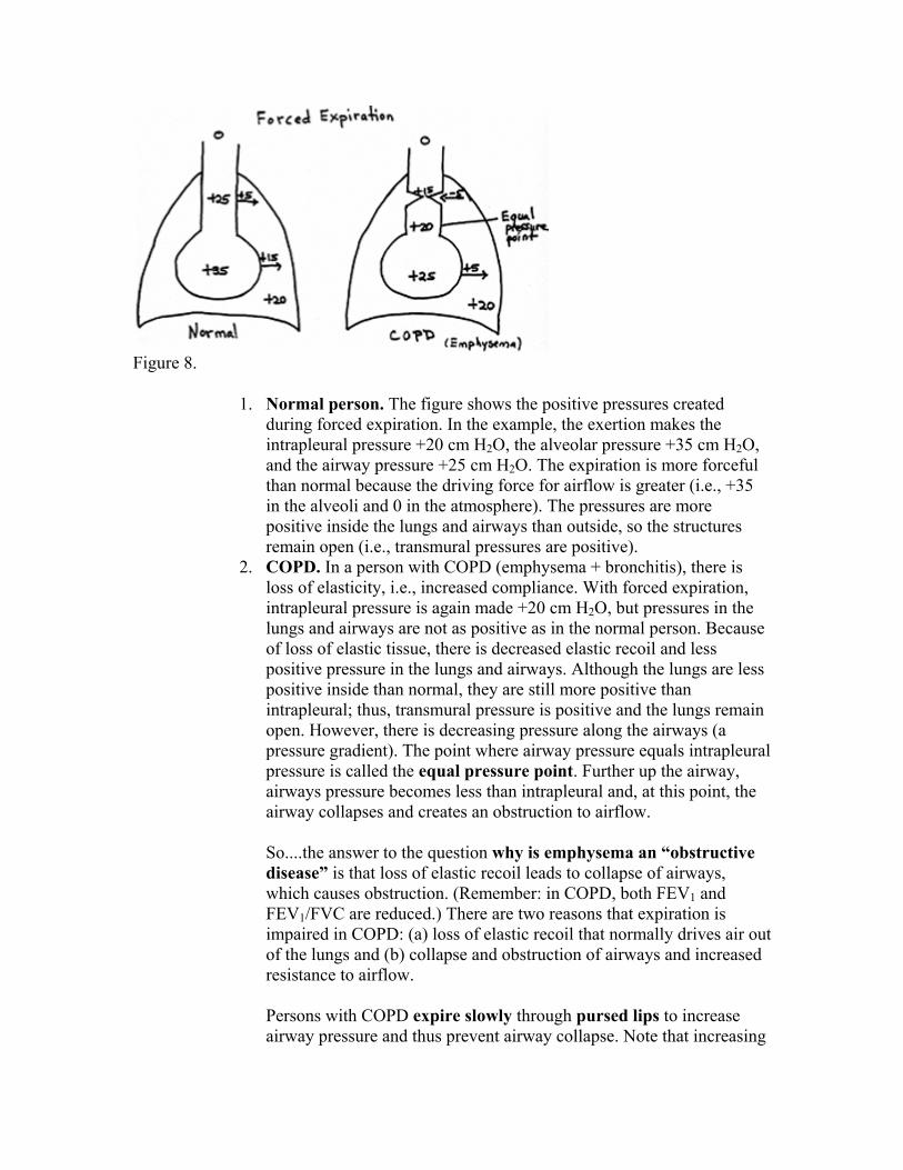
Figure 8.
1. Normal person. The figure shows the positive pressures created during forced expiration. In the example, the exertion makes the intrapleural pressure +20 cm H2O, the alveolar pressure +35 cm H2O, and the airway pressure +25 cm H2O. The expiration is more forceful than normal because the driving force for airflow is greater (i.e., +35 in the alveoli and 0 in the atmosphere). The pressures are more positive inside the lungs and airways than outside, so the structures remain open (i.e., transmural pressures are positive).
2. COPD. In a person with COPD (emphysema + bronchitis), there is loss of elasticity, i.e., increased compliance. With forced expiration, intrapleural pressure is again made +20 cm H2O, but pressures in the lungs and airways are not as positive as in the normal person. Because of loss of elastic tissue, there is decreased elastic recoil and less positive pressure in the lungs and airways. Although the lungs are less positive inside than normal, they are still more positive than intrapleural; thus, transmural pressure is positive and the lungs remain open. However, there is decreasing pressure along the airways (a pressure gradient). The point where airway pressure equals intrapleural pressure is called the equal pressure point. Further up the airway, airways pressure becomes less than intrapleural and, at this point, the airway collapses and creates an obstruction to airflow. So....the answer to the question why is emphysema an “obstructive disease” is that loss of elastic recoil leads to collapse of airways, which causes obstruction. (Remember: in COPD, both FEV1 and FEV1/FVC are reduced.) There are two reasons that expiration is impaired in COPD: (a) loss of elastic recoil that normally drives air out of the lungs and (b) collapse and obstruction of airways and increased resistance to airflow. Persons with COPD expire slowly through pursed lips to increase airway pressure and thus prevent airway collapse. Note that increasing

the effort for expiration doesn’t help, since effort makes both intrapleural and alveolar/airway pressure more positive.
IV. SUMMARY OF LUNG MECHANICS IN DISEASES
Disease FEV1 FVC FEV1/FVC FRC (and residual volume)
Peak Expiratory Flow Rate
Asthma (obstructive) ↓↓ ↓ ↓ ↑ ↓ COPD (obstructive) ↓↓ ↓ ↓ ↑ ↓ Fibrosis (restrictive) ↓ ↓↓ ↑ ↓ ↑ (or --)

VI. PRACTICE QUESTIONS
1. Compared to restrictive lung disease, obstructive lung disease has a lower:
A. Vital capacity B. FVC C. FEV1 D. FEV1/FVC E. Tidal volume
2. In persons with fibrosis, at usual (normal) values for FRC,
A. The collapsing force on the lungs is greater than the expanding
force on the chest wall. B. The expanding force on the lungs is greater than the collapsing
force on the chest wall. C. There is a collapsing force on both the lungs and the chest wall. D. The collapsing force on the lungs is smaller than the expanding
force on the chest wall. E. The collapsing force on the lungs is equal to the expanding force
on the chest wall.
3. In persons with emphysema, in order to balance the collapsing forces on the lungs and chest wall:
A. FRC increases B. FRC decreases C. FRC can remain at its usual value, but residual volume decreases D. Tidal volume increases E. FVC increases
4. Which of the following pairs of pressures would cause collapse of the
structure?
A. Intra-alveolar pressure = +5 cm H2O; intrapleural pressure = -5 cm H2O
B. Intra-airway pressure = +20 cm H2O; intrapleural pressure = +15 cm H2O
C. Intra-alveolar pressure = 0; intrapleural pressure = -5 cm H2O D. Intra-airway pressure = +15 cm H2O; intrapleural pressure = +20
cm H2O
EXPLANATIONS

1. Answer = D. Vital capacity and FEV1 are decreased in both. It’s the ratio of FEV1/FVC that is clearly lower in obstructive than restrictive.
2. Answer = A.
3. Answer = A
4. Answer = D. Easy, but I forced you to practice and visualize.