Embed Size (px)
Citation preview
Biochimica et Biophysica Acta 1784 (2008) 1908–1917
Contents lists available at ScienceDirect
Biochimica et Biophysica Acta
j ourna l homepage: www.e lsev ie r.com/ locate /bbapap
Mechanism of the chemical step for the guanosine triphosphate (GTP) hydrolysiscatalyzed by elongation factor Tu
B.L. Grigorenko a, M.S. Shadrina a, I.A. Topol b, J.R. Collins b, A.V. Nemukhin a,c,⁎a Department of Chemistry, M.V. Lomonosov Moscow State University, Leninskie Gory, 1/3, Moscow, 119991, Russian Federationb Advanced Biomedical Computing Center, Advanced Technology Program, SAIC-Frederick Inc., NCI-Frederick, Frederick, MD 21702-1201, USAc N.M. Emanuel Institute of Biochemical Physics, Russian Academy of Sciences, ul. Kosygina, 4, Moscow, 119334, Russian Federation
⁎ Corresponding author. Department of Chemistry, MUniversity, Leninskie Gory, 1/3, Moscow, 119991+74949391096; fax: +74959390283.
E-mail addresses: [email protected], anemukhin
1570-9639/$ – see front matter © 2008 Elsevier B.V. Aldoi:10.1016/j.bbapap.2008.08.003
a b s t r a c t
a r t i c l e i n f oArticle history:
Elongation factor Tu (EF-Tu) Received 25 April 2008Received in revised form 12 July 2008Accepted 4 August 2008Available online 16 August 2008Keywords:EF-Tu proteinGTP hydrolysisMolecular modelingEnzymatic mechanism
, the protein responsible for delivering aminoacyl-tRNAs (aa-tRNAs) to ribosomalA site during translation, belongs to the group of guanosine-nucleotide (GTP/GDP) binding proteins. Its active‘on’-state corresponds to the GTP-bound form, while the inactive ‘off’-state corresponds to the GDP-boundform. In this work we focus on the chemical step, GTP+H2O→GDP+Pi, of the hydrolysis mechanism. Weapply molecular modeling tools including molecular dynamics simulations and the combined quantummechanical–molecular mechanical calculations for estimates of reaction energy profiles for two possiblearrangements of switch II regions of EF-Tu. In the first case we presumably mimic binding of the ternarycomplex EF-Tu·GTP·aa-tRNA to the ribosome and allow the histidine (His85) side chain of the protein toapproach the reaction active site. In the second case, corresponding to the GTP hydrolysis by EF-Tu alone, theside chain of His85 stays away from the active site, and the chemical reaction GTP+H2O→GDP+Pi proceedswithout participation of the histidine but through water molecules. In agreement with the experimentalobservations which distinguish rate constants for the fast chemical reaction in EF-Tu·GTP·aa-tRNA·ribosomeand the slow spontaneous GTP hydrolysis in EF-Tu, we show that the activation energy barrier for the firstscenario is considerably lower compared to that of the second case.
© 2008 Elsevier B.V. All rights reserved.
1. Introduction
The hydrolysis cycle of guanosine triphosphate (GTP) provides auniversal switch mechanism in controlling numerous cellular func-tions [1–4]. One of the important guanosine-nucleotide bindingproteins, elongation factor EF-Tu, is responsible for delivering allelongator aminoacyl-tRNAs (aa-tRNAs) to the ribosomal A site duringtranslation. The protein consists of a Ras-like domain (the G-domain)that binds GTP (or guanosine diphosphate, GDP) and two β-barreldomains which, together with the G-domain, account for interactionswith the acceptor domain of aa-tRNA. EF-Tu, GTP and aa-tRNA form astable ternary complex that binds to the ribosome. Codon–anticodoninteraction leads to conformational changes in EF-Tu and to theformation of the activated GTPase state of the ribosome·EF-Tu·aa-tRNAcomplex which is followed by the intrinsic chemical reaction of GTPhydrolysis. Subsequent conformational changes in EF-Tu from theGTP- to GDP-bound form lead to the aa-tRNA release while EF-Tu·GDPleaves the ribosome. Elongation factor EF-Ts catalyzes reconstructionof the active form EF-Tu·GTP.
.V. Lomonosov Moscow State, Russian Federation. Tel.:
@yahoo.com (A.V. Nemukhin).
l rights reserved.
The complete kinetic mechanism of EF-Tu binding of aa-tRNA tothe E. coli ribosome was described in Ref. [5]. The chemical reaction ofGTP hydrolysis leading to GDP and inorganic phosphate Pi
GTPþH2OYGDPþ Pi ð1Þ
by EF-Tu itself is known to be slow while its complex with theribosome performs very efficiently, enhancing the reaction rate about105 fold [6]. In this work we focus only on this chemical step of themechanism following our previous studies of GTP hydrolysis by theRas protein [7,8] and of adenosine triphosphate (ATP) hydrolysis bymyosin [9].
The reaction scheme of the hydrolysis of GTP by the protein matrix(M) may be described by the following scheme
Md GTP X1M4d GTP X
2M4d GDPd Pi X
3M44d GDPþ Pi ð2Þ
It includes the following stages: (1) active site reorganizationleading to formation of the enzyme-substrate (ES) complex M⁎· GTP,(2) formation of the ternary product complex M⁎· GDP·Pi through thechemical transition state (TS), and (3) product (Pi) release. Rearrange-ments of the protein structure occur in steps 1 and 3. Our quantumbased modeling refers to stage 2 in Eq. (2), while molecular dynamicssimulations refer to the protein reorganization in step 1.
1909B.L. Grigorenko et al. / Biochimica et Biophysica Acta 1784 (2008) 1908–1917
By performing molecular modeling we address, in particular, theimportant question of the role the histidine side chain His85 (or His84in another numbering) plays in the chemical step of GTP hydrolysis byEF-Tu. This problem was discussed in the literature [10–13] inrelevance to site-directed mutational studies. In particular, thesestudies revealed that replacement of this residue with Ala consider-ably reduced the rate constant of GTP hydrolysis [13], whilereplacement with Gln showed only a slightly reduced rate of intrinsichydrolysis reaction compared to the native protein [11–13]. However,analysis of the available crystal structures from the PDB archive [14] ofEF-Tu complexed with GTP analogs 1EXM [15], 1EFT [16], indicatesthat in these structures His85 points away from the active site of theenzyme. Moreover, the side chains of Val20 and Ile61 forming the so-called ‘hydrophobic gate’ prevent His85 to approach the active site.
According to the working hypotheses discussed in Ref. [13] theribosome stimulates the intrinsic GTP hydrolysis by 5 orders ofmagnitude by promoting the conformational reorganization thatfavors His85 approach to the active site. This suggestion is supportedby the observation that His85 is a part of the switch II region whichendures the greatest reorganization on the way from the GTP-boundto the GDP-bound form of EF-Tu and, therefore, re-orientation ofHis85 during conformational changes in EF-Tu is structurally possible.Secondly, the crystal structure (PDBID: 1HA3) of the complex betweenEF-Tu⁎·GDP and aurodox is reported [17] in which EF-Tu is believed tobe in the GTP-bound form. In this structure, the His85 side chain isoriented toward the nucleotide-binding site and the flexible chain ofresidues 53–64, including Ile61, does not prevent His85 fromapproaching the active site. Our simulations described below are inaccord with this hypothesis of direct involvement of His85 in thehydrolysis reaction and provide detailed visualization of the reactionmechanism.
More generally, in this work we address the question of themechanism of the intrinsic chemical reaction in enzymatic hydrolysisof nucleoside triphosphates which is still under active debate [3,18–31]. The key issue is an assignment of the reaction mechanism to acertain type depending on the nature of a transition state. On oneextreme, a fully dissociative transition state for the GTP hydrolysis isrepresented by an intermediate that shows bond cleavage betweenthe γ-phosphate group and GDP. On the other extreme, a fullyassociative transition state is represented by a penta-coordinatedintermediate that shows no β–γ bridge bond cleavage but significantamount of bond formation between the incoming lytic watermoleculeand the γ-phosphate of GTP. It should be noted that none of theavailable experiments can determine whether the mechanism isassociative or dissociative [32]. Another popular model suggests thatthe substrate GTP itself, specifically, γ-phosphate group of GTP, servesas the general base in its own hydrolysis [33,34].
Our previous simulations of the intrinsic chemical reaction of theGTP hydrolysis in the Ras protein either by itself [7] or in the Ras–RasGAP complex [8], of the ATP hydrolysis in myosin [9], and of themethyl triphosphate hydrolysis in water [35,36] performed in thecombined quantum mechanical–molecular mechanical (QM/MM)approximation allow us to conclude that in all enzymatic reactionsthe reasonable minimum energy profiles with low activation energiesare consistent with the following reaction route. In all cases a lowactivation energy cleavage of the Pγ–Oβγ bond in nucleotidetriphospate and separation of the γ-phosphate from nucleotidediphosphate occurs through a planar arrangement of the PγO3
− groupin TS with almost unaltered lytic water molecule. Formation of theinorganic phosphate results as a consequence of proton transfersmediated by the neighboring molecular groups of the protein matrix.In Ras and Ras–RasGAP catalyzed hydrolysis of GTP, the Gln61 sidechain serves as a crucial mediator. Therefore, when proceeding tosimulations of GTP hydrolysis in EF-Tu we expected to find similarfeatures of the reaction mechanism with an active participation ofHis85 in the case of rapid enzymatic reaction.
2. Methods
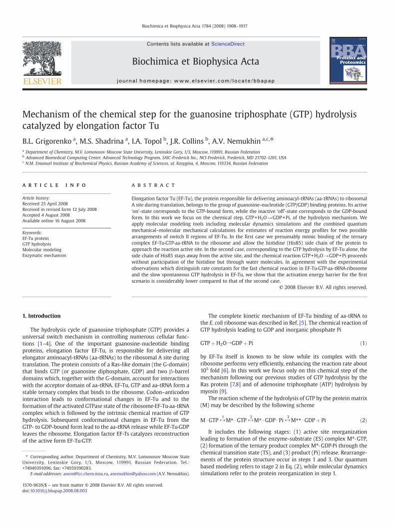
We started simulations from the coordinates of heavy atoms in thecrystal structures PDBID:1EFT solved at the resolution 2.50 Å [16] ofEF-Tu from Thermus aquaticus and PDBID:1EXM solved at theresolution 1.70 Å [15] of EF-Tu from Thermus thermophilus. Thesecrystals accommodate the slowly hydrolizable GTP analog, 5′-guanylylimidodiphosphate (GMPPNP), containing the NH groupinstead of the bridging βγ-oxygen atom. Fig. 1 shows the positionsof GTP analog, GMPPNP, magnesium cation, His85, and the side chainsof the ‘hydrophobic gate’ Val20 and Ile61. The distance between Nδ ofHis85 and Pγ of GMPPNP constitutes 8.5 Å in PDBID:1EXM, as shownin Fig. 1, and 7.1 Å in PDBID:1EFT (in the latter case, a slightly differentorientation of the His85 side chain occurs).
At the first stages of the work we used the computer packageNAMD2 [37] of molecular dynamics (MD) simulations to model thestructures of enzyme-substrate (ES) complexes for subsequentquantum based calculations. The native GTP molecule was re-constructed from its analog GMPPNP, the coordinates of hydrogenatoms were introduced, and the protein was solvated by watermolecules. The model for the ES complex included 6348 protein andligand atoms, 108 crystal water molecules and 4047 solvent watermolecules. The CHARMM force field parameters [38] extended withthose for GTP from Ref. [39] were used in MD simulations. Initially, weoptimized positions of water molecules by the minimum energycriterion with the fixed positions of protein and ligand atoms. Thenthe constraints were lifted, and positions of all atoms were optimized.The MD runs were performed for the model system within arectangular box with dimensions 61×72.5×48 Å3 with the imposedperiodic boundary conditions. No constraints were imposed on thebond lengths allowing a 1 fs time step for integration at temperature300 K.
When starting from coordinates of the crystal structures PDBID:1EFT or PDBID: 1EXM in MD simulations we always arrived at thestructure (called Hisout below) with the remote position of the His85side chain relative to the γ-phosphate group of GTP. In thisarrangement, His85 side chain could not approach the active sitedue to steric hindrance of the hydrophobic gate of Val20 and Ile61.Then we prepared manually another model structure (called Hisinbelow) by re-orienting the His85 side chain toward the γ-phosphategroup of GTP following themotifs of the crystal structure PDBID: 1HA3[17]. For such re-arrangement we temporarily moved apart theresidues of the hydrophobic gate and shifted the residues of switchII. Then we restored the hydrogen bond network and re-optimizedatomic coordinates. A fairly long MD trajectory (700 ps) executed forthis structure showed the His85 side chain remained close to theactive site. Rout mean square fluctuations of Cα protein atoms duringthe MD trajectory was found to be relatively small, typically less than1.2 Å indicating the relative stability of the system.
The coordinates obtained in preliminary MD simulations for bothmodel systems, Hisout and Hisin, were used as an initial guess for morerigorous calculations of equilibrium geometry parameters by usingthe combined quantum mechanical–molecular mechanical (QM/MM)method [40]. To ensure that the QM/MM optimization resulted inminimum energy points corresponding to the stable ES complexes ofHisout and Hisin structures we numerously varied the starting sets ofcoordinates around those obtained in preliminaryMD simulations andrepeated QM/MM minimization calculations until the lowest energystructures were reached.
In QM/MM calculations we used the same version of the theory asin previous studies of enzymatic GTP and ATP hydrolysis [7–9], i.e., theflexible effective fragment variant [41,42] of the effective fragmentpotential QM/MM [43] method. This is an approach which allows oneto perform calculations close to an ab initio QM treatment of the entiremolecular system. Molecular groups assigned to the MM part arerepresented by effective fragments which introduce their electrostatic
Fig. 1. The fragment of the crystal structure PDBID:1EXM (15) showing the positions of GTP analog, GMPPNP, magnesium cation, His85, and the side chains of the ‘hydrophobic gate’Val20 and Ile61. Here and below the carbon atoms are distinguished by green, oxygen atoms by red, nitrogen atoms by blue, phosphorus atoms by brown, magnesium by magenta.
1910 B.L. Grigorenko et al. / Biochimica et Biophysica Acta 1784 (2008) 1908–1917
potentials expanded up to octupoles as one-electron contributions tothe quantum Hamiltonian. These potentials, as well as contributionsfrom interactions of effective fragments with the QM region, areobtained in preliminary quantum chemical calculations for separatedfragments by using ab initio electron densities. The exchange-repulsion potentials to be combined with the electrostatic terms arealso created in preliminary ab initio calculations. Thus, all empiricalparameters are entirely within the MM subsystem. In the flexibleeffective fragment version [41,42] the fragment–fragment interactionsare modeled by the force field parameters from the AMBER library[44]. The computer program used in the simulations is based on theGAMESS(US) [45] (more specifically, its Intel-specific version, PCGAMESS [46]) quantum chemistry package and on the TINKER [47]molecular modeling system.
In this application we performed partitioning of the model systeminto QM and MM parts as follows. All phosphate groups of fullyunprotonated GTP molecule, the lytic water molecule, the magnesiumcation, and the side chain of His85were assigned to the QM subsystemin the case of the Hisin structure, while for calculations for the Hisoutstructure, an additional water molecule was introduced to the QMsubsystem instead of the His85 side chain. In total, 33 atomsconstituted the quantum subsystem and 1556 atoms combined to473 effective fragments formed the MM subsystem for the Hisinstructure. Correspondingly, 24 atoms in the QM part and 1481 MMatoms combined to 448 effective fragments were considered for theHisout structure.
The simulations included scans of the composite multidimen-sional QM/MM potential energy surface in the regions wherechemical bonds or hydrogen bonds could be cleaved or formed. Asa result, the basins around presumable stationary points werespecified for more careful calculations of the local minima or saddlepoints. The stationary points were located by unconstrainedminimizations (for local minima) or by constrained minimizations(for saddle points) of the QM/MM energy. The location of a transitionstates (TS) was determined based on the criterion that the gradient
of the constrained internal coordinate along an assumed reactionpath must change its sign at the presumed TS. The internalcoordinates of all atoms in the QM subsystem and positions ofeffective fragments in the MM subsystem including all watermolecules were optimized. Positions of remote effective fragmentsat distance greater 210 Å from the reaction center were kept fixedas in the crystal structure.
As in our previous simulations of enzymatic GTP and ATPhydrolysis [7–9], quantum calculations were carried out by usingthe Hartree-Fock approach in the QM part. The polarized “LANL2DZdpECP” basis set (and the corresponding pseudopotential for phosphorus[48]) was used for all atoms except magnesium. For magnesium, thestandard 6-31G basis was employed. It should be noted that multipleminimum energy points could be located in geometry optimizations.We attempted to overcome this difficulty by performing in each casenumerous selections of the starting sets of coordinates for minimiza-tion until the lowest energy was reached under the condition that thehydrogen bond network in the immediate vicinity of the active site.Below we shall use the notations Hisin and Hisout for the QM/MMoptimized model structures.
3. Results
3.1. The reaction path for the Hisin model structure
Fig. 2 illustrates the geometry configuration of the ES complex asoptimized in QM/MM calculations. We show the immediateparticipants of the chemical reaction, the phosphate groups of GTPand the lytic water molecule, Wat1. The latter is oriented in aperfect position for the in-line attack on γ-phosphate by thehydrogen bond network which includes the side chain of His85. Theequilibrium distance between oxygen from the lytic water oxygenand γ-phosphorus, 3.1 Å, is typical for ES complexes for theenzymatic hydrolysis of nucleotide triphosphates [7–9,31]. Thecharacteristic 6-fold coordination shell of magnesium cation is
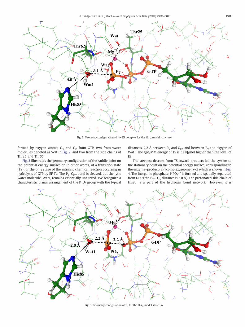
Fig. 2. Geometry configuration of the ES complex for the Hisin model structure.
1911B.L. Grigorenko et al. / Biochimica et Biophysica Acta 1784 (2008) 1908–1917
formed by oxygen atoms: Oγ and Oβ from GTP, two from watermolecules denoted as Wat in Fig. 2, and two from the side chains ofThr25 and Thr65.
Fig. 3 illustrates the geometry configuration of the saddle point onthe potential energy surface or, in other words, of a transition state(TS) for the only stage of the intrinsic chemical reaction occurring inhydrolysis of GTP by EF-Tu. The Pγ–Oβγ bond is cleaved, but the lyticwater molecule, Wat1, remains essentially unaltered. We recognize acharacteristic planar arrangement of the PγO3 group with the typical
Fig. 3. Geometry configuration of TS
distances, 2.2 Å between Pγ and Oβγ and between Pγ and oxygen ofWat1. The QM/MM energy of TS is 32 kJ/mol higher than the level ofES.
The steepest descent from TS toward products led the system tothe stationary point on the potential energy surface, corresponding tothe enzyme–product (EP) complex, geometry of which is shown in Fig.4. The inorganic phosphate, HPO4
2− is formed and spatially separatedfrom GDP (the Pγ–Oβγ distance is 3.0 Å). The protonated side chain ofHis85 is a part of the hydrogen bond network. However, it is
for the Hisin model structure.
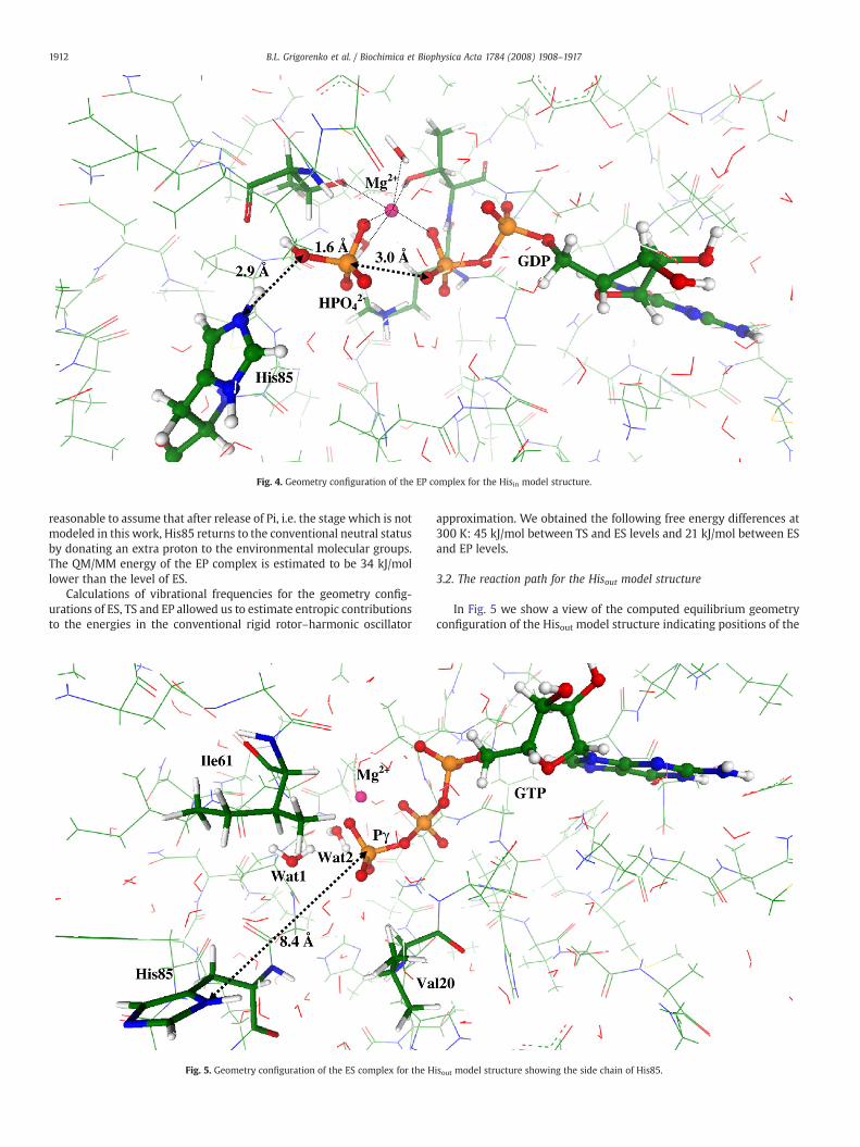
Fig. 4. Geometry configuration of the EP complex for the Hisin model structure.
1912 B.L. Grigorenko et al. / Biochimica et Biophysica Acta 1784 (2008) 1908–1917
reasonable to assume that after release of Pi, i.e. the stage which is notmodeled in this work, His85 returns to the conventional neutral statusby donating an extra proton to the environmental molecular groups.The QM/MM energy of the EP complex is estimated to be 34 kJ/mollower than the level of ES.
Calculations of vibrational frequencies for the geometry config-urations of ES, TS and EP allowed us to estimate entropic contributionsto the energies in the conventional rigid rotor–harmonic oscillator
Fig. 5. Geometry configuration of the ES complex for the H
approximation. We obtained the following free energy differences at300 K: 45 kJ/mol between TS and ES levels and 21 kJ/mol between ESand EP levels.
3.2. The reaction path for the Hisout model structure
In Fig. 5 we show a view of the computed equilibrium geometryconfiguration of the Hisout model structure indicating positions of the
isout model structure showing the side chain of His85.
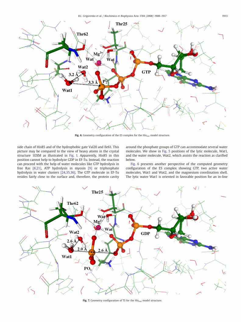
Fig. 6. Geometry configuration of the ES complex for the Hisout model structure.
1913B.L. Grigorenko et al. / Biochimica et Biophysica Acta 1784 (2008) 1908–1917
side chain of His85 and of the hydrophobic gate Val20 and Ile61. Thispicture may be compared to the view of heavy atoms in the crystalstructure 1EXM as illustrated in Fig. 1. Apparently, His85 in thisposition cannot help to hydrolyze GDP in EF-Tu. Instead, the reactioncan proceed with the help of water molecules like GTP hydrolysis infree Ras [8,21], ATP hydrolysis in myosin [9] or triphosphatehydrolysis in water clusters [24,35,36]. The GTP molecule in EF-Turesides fairly close to the surface and, therefore, the protein cavity
Fig. 7. Geometry configuration of TS
around the phosphate groups of GTP can accommodate several watermolecules. We show in Fig. 5 positions of the lytic molecule, Wat1,and the water molecule, Wat2, which assists the reaction as clarifiedbelow.
Fig. 6 presents another perspective of the computed geometryconfiguration of the ES complex showing GTP, two active watermolecules, Wat1 and Wat2, and the magnesium coordination shell.The lytic water Wat1 is oriented in favorable position for an in-line
for the Hisout model structure.
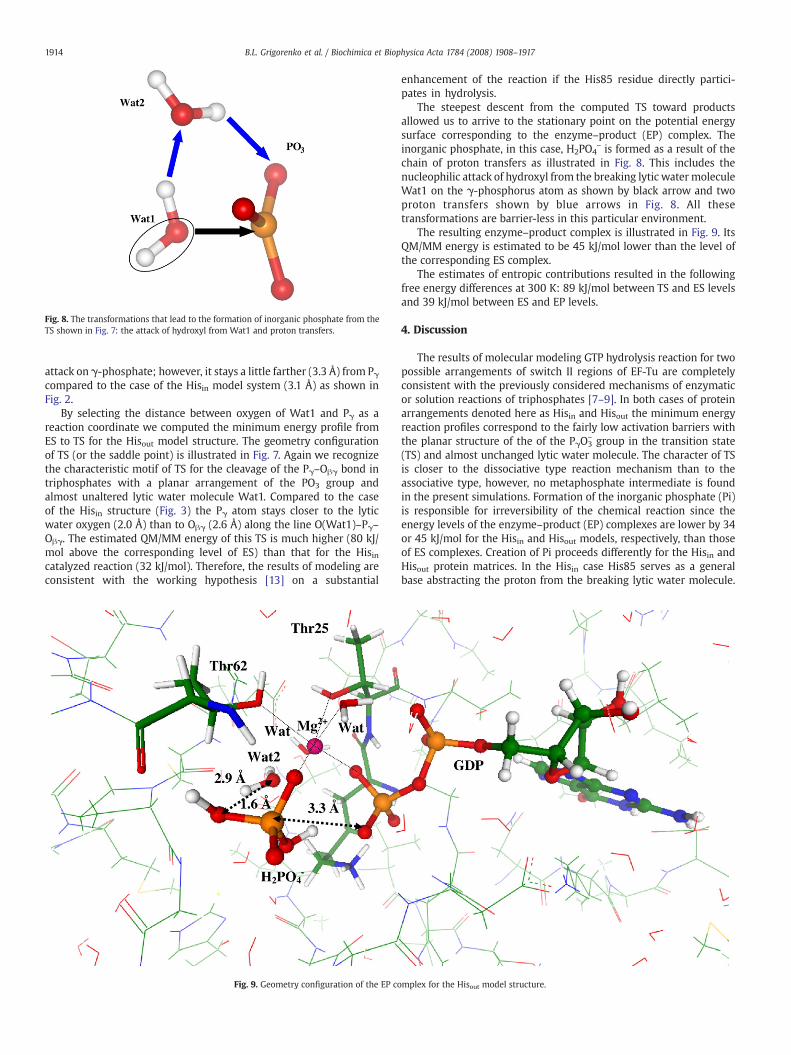
Fig. 8. The transformations that lead to the formation of inorganic phosphate from theTS shown in Fig. 7: the attack of hydroxyl from Wat1 and proton transfers.
1914 B.L. Grigorenko et al. / Biochimica et Biophysica Acta 1784 (2008) 1908–1917
attack on γ-phosphate; however, it stays a little farther (3.3 Å) from Pγcompared to the case of the Hisin model system (3.1 Å) as shown inFig. 2.
By selecting the distance between oxygen of Wat1 and Pγ as areaction coordinate we computed the minimum energy profile fromES to TS for the Hisout model structure. The geometry configurationof TS (or the saddle point) is illustrated in Fig. 7. Again we recognizethe characteristic motif of TS for the cleavage of the Pγ–Oβγ bond intriphosphates with a planar arrangement of the PO3 group andalmost unaltered lytic water molecule Wat1. Compared to the caseof the Hisin structure (Fig. 3) the Pγ atom stays closer to the lyticwater oxygen (2.0 Å) than to Oβγ (2.6 Å) along the line O(Wat1)–Pγ–Oβγ. The estimated QM/MM energy of this TS is much higher (80 kJ/mol above the corresponding level of ES) than that for the Hisincatalyzed reaction (32 kJ/mol). Therefore, the results of modeling areconsistent with the working hypothesis [13] on a substantial
Fig. 9. Geometry configuration of the EP co
enhancement of the reaction if the His85 residue directly partici-pates in hydrolysis.
The steepest descent from the computed TS toward productsallowed us to arrive to the stationary point on the potential energysurface corresponding to the enzyme–product (EP) complex. Theinorganic phosphate, in this case, H2PO4
− is formed as a result of thechain of proton transfers as illustrated in Fig. 8. This includes thenucleophilic attack of hydroxyl from the breaking lytic water moleculeWat1 on the γ-phosphorus atom as shown by black arrow and twoproton transfers shown by blue arrows in Fig. 8. All thesetransformations are barrier-less in this particular environment.
The resulting enzyme–product complex is illustrated in Fig. 9. ItsQM/MM energy is estimated to be 45 kJ/mol lower than the level ofthe corresponding ES complex.
The estimates of entropic contributions resulted in the followingfree energy differences at 300 K: 89 kJ/mol between TS and ES levelsand 39 kJ/mol between ES and EP levels.
4. Discussion
The results of molecular modeling GTP hydrolysis reaction for twopossible arrangements of switch II regions of EF-Tu are completelyconsistent with the previously considered mechanisms of enzymaticor solution reactions of triphosphates [7–9]. In both cases of proteinarrangements denoted here as Hisin and Hisout the minimum energyreaction profiles correspond to the fairly low activation barriers withthe planar structure of the of the PγO3
− group in the transition state(TS) and almost unchanged lytic water molecule. The character of TSis closer to the dissociative type reaction mechanism than to theassociative type, however, no metaphosphate intermediate is foundin the present simulations. Formation of the inorganic phosphate (Pi)is responsible for irreversibility of the chemical reaction since theenergy levels of the enzyme–product (EP) complexes are lower by 34or 45 kJ/mol for the Hisin and Hisout models, respectively, than thoseof ES complexes. Creation of Pi proceeds differently for the Hisin andHisout protein matrices. In the Hisin case His85 serves as a generalbase abstracting the proton from the breaking lytic water molecule.
mplex for the Hisout model structure.
1915B.L. Grigorenko et al. / Biochimica et Biophysica Acta 1784 (2008) 1908–1917
In the case of the Hisout conformation formation of Pi results as aconsequence of proton transfers mediated by the water moleculesinside the EF-Tu protein cavity.
The results are consistent with the experimental observationsaccording to which GTP hydrolysis in EF-Tu, with the His85 side chainfar apart the active site, is very slow, about 5×10⁻⁵ s, while thehydrolysis rate in the activated by the ribosome ternary complex EF-Tu·GTP·aa-tRNA, and hence with the His85 side chain staying close tothe active site, is considerably higher. Our QM/MM calculations resultin the substantial decrease of the activation barrier in case of activeparticipation of His85 in the reaction compared to the case of GTPhydrolysis by water molecules inside the protein cavity.
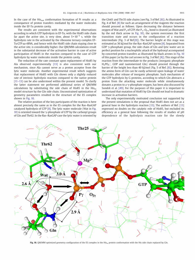
The reduction of the rate constant upon replacement of His85 byAla observed experimentally [13] is also consistent with ourmechanism, since Ala cannot serve as a proton acceptor from thelytic water molecule. Another experimental result which suggeststhat replacement of His85 with Gln shows only a slightly reducedrate of intrinsic hydrolysis reaction compared to the native protein[11–13] can be also understood within the present model. To clarifythe latter statement we performed additional series of QM/MMcalculations by substituting the side chain of His85 in the Hisinmodel structure by the Gln side chain. Unconstrained optimization ofgeometry parameters resulted in the structure of the ES complexshown in Fig. 10.
The relative position of the key participants of the reaction is herealmost precisely the same as in the ES complex for the Ras–RasGAPcatalyzed hydrolysis of GTP [8]. The lytic water molecule (Wat in Fig.10) is oriented toward the γ-phosphate of GTP by the carbonyl groupsof Gln and Thr62. In the Ras–RasGAP case the lytic water is oriented by
Fig. 10. QM/MM optimized geometry configuration of the ES complex in th
the Gln61 and Thr35 side chains (see Fig. 3 of Ref. [8]). As illustrated inFig. 4 of Ref. [8] for such an arrangement of the reagents the reactionshould proceed as follows. Upon decreasing the distance betweenoxygen ofWat and Pγ of GTP, the Pγ–Oβγ bond is cleaved (as illustratedby the red thick arrow in Fig. 10), the system overcomes the firsttransition state and occurs in the configuration of a reactionintermediate (Fig. 3 of Ref.[8]). The barrier height at this stage wasestimated as 36 kJ/mol for the Ras–RasGAP system [8]. Separated fromGDP γ-phosphate group, the side chain of Gln and lytic water are inperfect position for a nucleophilic attack of the hydroxyl accompaniedby concerted proton transfers as illustrated by black arrows in Fig. 10of this paper (or by the curl arrows in Fig. 3 of Ref. [8]). This stage of thereaction from the intermediate to the products (inorganic phosphateH2PO4
−, GDP and tautomerized Gln) should proceed through thebarrier of the height less than 40 kJ/mol (Fig. 5 of Ref. [8]). Restoringthe amino form of Gln can be easily achieved upon leakage of watermolecules after release of inorganic phosphate. Such mechanism ofthe GTP hydrolysis by G-proteins, according to which Gln abstracts aproton from the attacking water molecule while simultaneouslydonates a proton to a γ-phosphate oxygen, has been also discussed bySondek et al. [49]. For the purposes of this paper it is important tounderstand that mutation of His85 by Gln should not lead to dramaticincrease in activation barriers.
The only experimentally motivated conclusion not supported bythe present simulations is the proposal that His85 does not act as ageneral base in the hydrolysis reaction [13]. The authors of Ref. [13]expressed no doubts on the catalytic role of His85, but excluded itsefficiency as a general base following the results of studies of pHdependence of the hydrolysis reaction rate for the slowly
e Hisin protein conformation with the His side chain replaced by Gln.
1916 B.L. Grigorenko et al. / Biochimica et Biophysica Acta 1784 (2008) 1908–1917
hydrolizable GTP analog, mant-GTPγS. Within the range of 6.5–8.5pH units compared to the usual experimental conditions at pH=7.5the reaction rate showed no dependence on pH which was atvariance with the expected behavior if a general base with a pKa ofabout 7, presumably assigned to His85, were involved in catalysis.Instead, the authors of Ref. [13] proposed that the catalytic role ofHis85 is to stabilize reaction TS by hydrogen bonding to the lyticwater molecule or the γ-phosphate group of GTP. We may argue,firstly, that mant-GTPγS is not a full substitute of natural GTP and,secondly, that the pKa value of His85 buried inside the protein cavityfor the case of the Hisin structure may lie outside the studied rangeof 6.5–8.5 pH units. It seems difficult to specify another role of His85in stabilizing the reaction TS at the mechanistic level. Apparently,His85 is a part of hydrogen bond networks in geometry configura-tions of all stationary points: ES (Fig. 2), TS (Fig. 3) and EP (Fig. 4). Itclearly stabilizes the TS (Fig. 3) by fixing the lytic water moleculeWat1 by hydrogen bonds, but not the γ-phosphate group of GTP.However in order to complete the reaction by formation of inorganicphosphate and to arrive to the configuration of the enzyme–productcomplex His85 accepts the proton. The calculation results indicatethat the conformation of the EP complex (Fig. 4) with the protonatedHis85 corresponds to the true minimum energy point with theenergy level lower than that of the ES complex. Therefore, molecularmodeling provides a direct evidence of such a developmentcompared to indirect evaluations based on solution pKa values forHis.
5. Conclusion
The results of molecular modeling provide support to the workinghypotheses [13] that the ribosome stimulates the intrinsic GTPhydrolysis by the EF-Tu protein. This is achieved by promoting theconformational reorganization that favors the side chain of His85approach to the active site. The simulated reaction routes for the Hisinand Hisout protein conformations are characterized by distinctiveenergy profiles with the considerably lower activation barrier for theHisin scenario.
Acknowledgements
This work was partly supported by the grant from the RussianFoundation for Basic Research (project #07-03-00060). We thank thestaff and administration of the Advanced Biomedical ComputingCenter for their support for this project. This project has been fundedin whole or in part with federal funds from the National CancerInstitute, National Institutes of Health, under contract N01-CO-12400.The content of this publication does not necessarily reflect the viewsor policies of the Department of Health and Human Services, nor doesmention of trade names, commercial products, or organization implyendorsement by the U.S. Government.
References
[1] H.R. Bourne, D.A. Sanders, F. McCormick, The GTPase superfamily: a conservedswitch for diverse cell functions, Nature 348 (1990) 125–131.
[2] S.R. Sprang, G protein mechanisms: insights from structural analysis, Annu. Rev.Biochem. 66 (1997) 639–672.
[3] G. Li, X.C. Zhang, GTP hydrolysis mechanism of Ras-like GTPases, J. Mol. Biol. 340(2004) 921–932.
[4] S.R. Sprang, Z. Chen, X. Du, Structural basis of effector regulation and signaltermination in heterotrimeric Gα proteins, Adv. Protein Chem. 74 (2007) 1–65.
[5] T. Pape, W. Wintermeyer, M. Rodnina, Complete kinetic mechanism of elongationfactor Tu-dependent binding of aminiacyl-tRNA to the A site of the E. coliribosome, EMBO J. 17 (1998) 7490–7497.
[6] M.V. Rodnina, T. Pape, R. Fricke, W. Wintermeyer, Elongation factor Tu, a GTPasetriggered by codon recognition on the ribosome: mechanism and GTP consump-tion, Biochem. Cell Biol. 73 (1995) 1221–1227.
[7] B.L. Grigorenko, A.V. Nemukhin, I.A. Topol, R.E. Cachau, S.K. Burt, QM/MMmodeling the Ras-GAP catalyzed hydrolysis of guanosine triphosphate, Proteins:Struct. Funct. Bioinf. 60 (2005) 495–503.
[8] B.L. Grigorenko, M.S. Shadrina, I.A. Topol, S.K. Burt, A.V. Nemukhin, Mechanisms ofguanosine triphosphate hydrolysis by Ras and Ras-GAP proteins as rationalized byab initio QM/MM simulations, Proteins: Struct. Funct. Bioinf. 66 (2007) 456–466.
[9] B.L. Grigorenko, A.V. Rogov, I.A. Topol, S.K. Burt, H.M. Martinez, A.V. Nemukhin,Mechanism of themyosin catalyzed hydrolysis of ATP as rationalized bymolecularmodeling, Proc. Natl. Acad. Sci. U. S. A. 104 (2007) 7057–7061.
[10] R.H. Cool, A. Parmeggiani, Substitution of histidine-84 and the GTPase mechanismof elongation factor Tu, Biochemistry 30 (1991) 362–366.
[11] G. Scarano, I.M. Krab, V. Bocchini, A. Parmeggiani, Relevance of histidine-84 in theelongation factor Tu GTPase activity and in poly(Phe) synthesis: its substitution byglutamine and alanine, FEBS Lett. 365 (1995) 214–218.
[12] W. Zeidler, C. Egle, S. Ribeiro, A. Wagner, V. Katunin, R. Kreutzer, M. Rodnina, W.Wintermeyer, M. Sprinzl, Site-directed mutagenesis of Thermus thermophiluselongation factor Tu. Replacement of His85, Asp81 and Arg300, Eur. J. Biochem.229 (1995) 596–604.
[13] T. Daviter, H.-J. Wieden, M.V. Rodnina, Essential role of histidine 84 in elongationfactor Tu for the chemical step of GTP hydrolysis on the ribosome, J. Mol. Biol. 332(2003) 689–699.
[14] H.M. Berman, J. Westbrook, Z. Feng, G. Gilliland, T.N. Bhat, H. Weissig, I.N.Shindyalov, P.E. Bourne, The protein data bank, Nucleic Acids Res. 28 (2000)235–242.
[15] R. Hilgenfeld, J.R. Mesters, T. Hogg, Insights into the GTPase mechanism of EF-Tufrom structural studies, Ribosome: Struct., Funct., Antibiot. Cell Interact. 28 (2000)347–357.
[16] M. Kjeldgaard, P. Nissen, S. Thirup, J. Nyborg, The crystal structure of elongationfactor EF-Tu from Thermus aquaticus in the GTP conformation, Structure 1 (1993)35–50.
[17] L. Vogeley, G.J. Palm, J.R. Mesters, R. Hilgenfeld, Conformational change ofelongation factor Tu (EF-Tu) induced by antibiotic binding. Crystal structure of thecomplex between EF-Tu⁎·GDP and aurodox, J. Biol. Chem. 276 (2001)17149–17155.
[18] R. Langen, T. Schweins, A. Warshel, On the mechanism of guanosine triphosphatehydrolysis in ras p21 proteins, Biochemistry 31 (1992) 8691–8696.
[19] T. Schweins, R. Langen, A. Warshel, Why have mutagenesis studies not located thegeneral base in ras p21? Nat. Struct. Biol. 1 (1994) 476–484.
[20] K.A. Maegley, S.J. Admiraal, D. Herschlag, Ras-catalyzed hydrolysis of GTP: a newperspective frommodel studies, Proc. Natl. Acad. Sci. U. S. A. 93 (1996) 8160–8166.
[21] A.J. Scheidig, C. Burmester, R.G. Goody, The pre-hydrolysis state of p21ras incomplex with GTP: new insights into the role of water molecules in the GTPhydrolysis reaction of ras-like proteins, Structure 7 (1999) 1311–1324.
[22] T.M. Glennon, J. Villa, A. Warshel, How does GAP catalyze the GTP reaction of Ras?:a computer simulation study, Biochemistry 39 (2000) 9641–9651.
[23] D. Katagiri, M. Hata, T. Itoh, S. Neya, T. Hoshino, Atomic-scale mechanism of theGTP → GDP hydrolysis reaction by the Giα1 protein, J. Phys. Chem. B 107 (2003)3278–3283.
[24] Y.N. Wang, I.A. Topol, J.R. Collins, S.K. Burt, Theoretical studies on the hydrolysis ofmono-phosphate in gas phase and aqueous solution, J. Am. Chem. Soc. 125 (2003)13265–13273.
[25] I.A. Topol, R.E. Cachau, A.V. Nemukhin, B.L. Grigorenko, S.K. Burt, Quantumchemical modeling of the GTP hydrolysis by the RAS–GAP protein complex,Biochim. Biophys. Acta 1700 (2004) 125–136.
[26] A. Shurki, A. Warshel, Why does the Ras switch “break” by oncogenic mutations?Proteins: Struct. Funct. Bioinf. 55 (2004) 1–10.
[27] G. Li, Q. Cui, Mechanochemical coupling in myosin: a theoretical analysis withmolecular dynamics and combined QM/MM reaction path calculations, J. Phys.Chem. B 108 (2004) 3342–3357.
[28] S. Schwarzl, J.C. Smith, S. Fisher, Insights into the chemomechanical coupling ofthe myosin motor from simulation of its ATP hydrolysis mechanism, Biochemistry45 (2006) 5830–5847.
[29] C. Kőtting, M. Blessonohl, Y. Suveydis, R.C. Goody, A. Wittinghofer, K. Gerwert, Aphosphoryl transfer intermediate in the GTPase reaction of Ras in complexwith itsGTPase-activating protein, Proc. Natl. Acad. Sci. U. S. A. 103 (2006) 13911–13916.
[30] A. Wittinghofer, Phosphoryl transfer in Ras proteins, conclusive or elusive? TrendsBiochem. Sci. 31 (2006) 20–23.
[31] H. Te Heesen, K. Gerwert, J. Schlitter, Role of the arginine finger in Ras·RasGAPrevealed by QM/MM calculations, FEBS Lett. 581 (2007) 5677–5684.
[32] J. Åqvist, K. Kolmodin, J. Florian, A. Warshel, Mechanistic alternatives in phosphatemonoester hydrolysis: what conclusions can be drawn from available experi-mental data? Chem. Biol. 6 (1999) R71–R80.
[33] M. Kosloff, Z. Selinger, Substrate assisted catalysis — application to G proteins,Trends Biochem. Sci. 26 (2001) 257–262.
[34] S. Pasqualato, J. Cherfils, Crystallographic evidence for substrate-assisted GTPhydrolysis by a small GTP binding protein, Structure 13 (2005) 533–540.
[35] B.L. Grigorenko, A.V. Rogov, A.V. Nemukhin, On the mechanism of triphosphatehydrolysis in aqueous solution: QM/MM simulations in water clusters, J. Phys.Chem. B 110 (2006) 4407–4412.
[36] A.V. Rogov, B.L. Grigorenko, A.V. Bochenkova, A.A. Granovsky, A.V. Nemukhin, Therole of magnesium in hydrolysis reaction of triphosphates in water: modeling bythe quantum mechanical–molecular mechanical method, Moscow Univ. Chem.Bull. 62 (2007) 123–127.
[37] L. Kale, R. Skeel, M. Bhandarkar, R. Brunner, A. Gursoy, N. Krawetz, J. Phillips, A.Shinozaki, K. Varadarajan, K. Schulten, NAMD2: greater scalability for parallelmolecular dynamics, J. Comp. Phys. 151 (1999) 283–312.
[38] B.R. Brooks, R.E. Bruccoleri, B.D. Olafson, D.S. States, S. Swaminathan, M. Karplus,CHARMM: a program for macromolecular energy, minimization, and dynamicscalculations, J. Comp. Chem. 4 (1983) 187–217.
1917B.L. Grigorenko et al. / Biochimica et Biophysica Acta 1784 (2008) 1908–1917
[39] A.D. MacKerell Jr., D. Bashford, M. Bellott, R.L. Dunbrack Jr., J.D. Evanseck, M.J. Field,S. Fisher, J. Gao, H. Guo, S. Ha, D. Joseph-McCarthy, L. Kuchnir, K. Kuczera, F.T.K. Lau,C. Mattos, S. Michnick, T. Ngo, D.T. Nguyen, B. Prodhom,W.E. Reiher III, B. Roux, M.Schlenkrich, J.C. Smith, R. Stote, J. Straub, M. Watanabe, J. Wiorkiewicz-Kuczera, D.Yin, M. Karplus, All-atom empirical potential for molecular modeling anddynamics studies of proteins, J. Phys. Chem. B 102 (1998) 3586–3616.
[40] A. Warshel, M. Levitt, Theoretical studies of enzymatic reactions: dielectric,electrostatic and steric stabilization of the carbonium ion in the reaction oflyzozyme, J. Mol. Biol. 103 (1976) 227–249.
[41] B.L. Grigorenko, A.V. Nemukhin, I.A. Topol, S.K. Burt, Modeling of biomolecularsystems with the quantum mechanical and molecular mechanical method basedon the effective fragment potential technique: proposal of flexible fragments, J.Phys. Chem. A 106 (2002) 10663–10672.
[42] A.V. Nemukhin, B.L. Grigorenko, I.A. Topol, S.K. Burt, Flexible effective fragmentQM/MM method: validation through the challenging tests, J. Comput. Chem. 24(2003) 1410–1420.
[43] M.S. Gordon, M.A. Freitag, P. Bandyopadhyay, J.H. Jensen, V. Kairys, W.J. Stevens,The effective fragment potential method: a QM-based MM approach to modelingenvironmental effects in chemistry, J. Phys. Chem. A 105 (2001) 293–307.
[44] W.D. Cornell, P. Cieplak, C.I. Bayly, I.R. Gould, K.M. Merz, D.M. Ferguson, D.C.Spellmeyer, T. Fox, J.W. Caldwell, P.A. Kollman, A second generation force field forthe simulation of proteins, nuclear acids, and organic molecules, J. Am. Chem. Soc.117 (1995) 5179–5197.
[45] M.W. Schmidt, K.K. Baldridge, J.A. Boatz, S.T. Elbert, M.S. Gordon, J.H. Jensen, S.Koseki, N. Matsunaga, K.A. Nguyen, S.J. Su, T.L. Windus, M. Dupuis, J.A.Montgomery, General atomic and molecular electronic structure system, J.Comp. Chem. 14 (1993) 1347–1363.
[46] A.V. Nemukhin, B.L. Grigorenko, A.A. Granovsky, Molecular modeling with the PCGAMESS package: from diatomicmolecules to enzymes, MoscowUniv. Chem. Bull.45 (2004) 75–102.
[47] P. Ren, J.W. Ponder, Polarizable atomic multipole water model for molecularmechanics simulation, J. Phys. Chem. B 107 (2003) 5933–5947.
[48] P.J. Hay, W.R. Wadt, Ab initio effective core potentials for molecularcalculations. Potentials for main group elements Na to Bi, J. Chem. Phys. 82(1985) 284–298.
[49] J. Sondek, D.G. Lambright, J.P. Noel, H.E. Hamm, P.B. Sigler, GTPase mechanism of Gproteins from the 1.7-Å crystal structure of transducin α GDP AlF4−, Nature 372(1994) 276–279.


![Structure of guanosine-3[prime],5[prime]-cytidine](https://img.pdfslide.net/doc/110x75/6185f12859d7806a1a3467d8/structure-of-guanosine-3prime5prime-cytidine-.jpg)