Embed Size (px)
Citation preview
CLINICAL MICROBIOLOGY REVIEWS, July 2011, p. 469–489 Vol. 24, No. 30893-8512/11/$12.00 doi:10.1128/CMR.00064-10Copyright © 2011, American Society for Microbiology. All Rights Reserved.
Mechanisms of Obligatory Intracellular Infection withAnaplasma phagocytophilum
Yasuko Rikihisa*Department of Veterinary Biosciences, College of Veterinary Medicine, The Ohio State University, Columbus, Ohio
INTRODUCTION .......................................................................................................................................................469Anaplasma phagocytophilum....................................................................................................................................469
Classification .......................................................................................................................................................469Morphology ..........................................................................................................................................................470Natural reservoir.................................................................................................................................................470Strain variations .................................................................................................................................................470
Human Granulocytic Anaplasmosis and Diseases in Domestic Animals .......................................................472HGA ......................................................................................................................................................................472Diseases in domestic animals ...........................................................................................................................472
GENOMIC FEATURES AND GENE REGULATION OF A. PHAGOCYTOPHILUM ......................................473Genomic Features ...................................................................................................................................................473Major Surface Antigen P44 ...................................................................................................................................474Type IV Secretion System......................................................................................................................................475Two-Component System and Transcriptional Regulation ................................................................................475
REPLICATION OF A. PHAGOCYTOPHILUM IN EUKARYOTIC CELLS.......................................................475Intracellular Developmental Cycle .......................................................................................................................475Host Cell Receptor, Site of Entry, and Internalization Signal ........................................................................476Anaplasma Inclusion Biogenesis............................................................................................................................477
Contribution of host cells ..................................................................................................................................477Modification of the inclusion membrane by anaplasma proteins ......................................................477
SUBVERSION OF NEUTROPHIL ANTIMICROBIAL DEFENSES..................................................................477Downregulation of Reactive Oxygen Species Generation..................................................................................478Inhibition of Host Cell Apoptosis.........................................................................................................................480Subversion of Autophagy .......................................................................................................................................480Other Host Cell Signals Activated and Required for A. phagocytophilum Infection .....................................481
Tyrosine kinase activation .................................................................................................................................481ERK1/2 activation...............................................................................................................................................481
PATHOGENESIS AND IMMUNE RESPONSES..................................................................................................481Generation of Chemokines/Cytokines ..................................................................................................................481Roles of IFN-� and IL-8 ........................................................................................................................................482
TICK FACTORS REQUIRED FOR TRANSMISSION ........................................................................................483CONCLUSIONS .........................................................................................................................................................484ACKNOWLEDGMENTS ...........................................................................................................................................484REFERENCES ............................................................................................................................................................484
INTRODUCTION
In the early 1990s, an unknown febrile tick-borne illness,now called human granulocytic anaplasmosis (HGA) (57), ac-companied with granulocytic inclusions of bacteria closely re-lated to the veterinary pathogens Ehrlichia phagocytophilumand E. equi, all of which are now called Anaplasma phagocyto-philum, was discovered in Minnesota and Wisconsin (14, 48).This seminal discovery led to the successful isolation and cul-tivation of A. phagocytophilum (80) and an understanding ofclinical, immunological, and pathological characteristics ofHGA and the disease distribution, tick vector species, and wildanimal reservoirs. Alongside these discoveries, progresses have
been made toward an understanding of the mechanisms bywhich this pathogen can invade and proliferate inside neutro-phils, primary host defensive cells, to cause disease. Genomesequences of A. phagocytophilum (61) and the vector tickIxodes scapularis and the development of new methods andapproaches have been expanding our ability to investigate thisextraordinary pathogen and to determine bacterial and hostfactors critical for its invasion, survival, persistence, and trans-mission from infected ticks to mammalian host cells.
Anaplasma phagocytophilum
Classification. A. phagocytophilum includes the newly dis-covered human pathogen whose original name was the hu-man granulocytic ehrlichiosis (HGE) agent (14, 48) andpreviously known ruminant (Cytoecetes phagocytophila andEhrlichia phagocytophila) and equine (Ehrlichia equi) patho-gens (57). A. phagocytophilum belongs to the family Anaplas-
* Mailing address: Department of Veterinary Biosciences, Collegeof Veterinary Medicine, The Ohio State University, 1925 Coffey Road,Columbus, OH 43210. Phone: (614) 292-5661. Fax: (614) 292-6473.E-mail: [email protected].
469
on April 8, 2020 by guest
http://cmr.asm
.org/D
ownloaded from

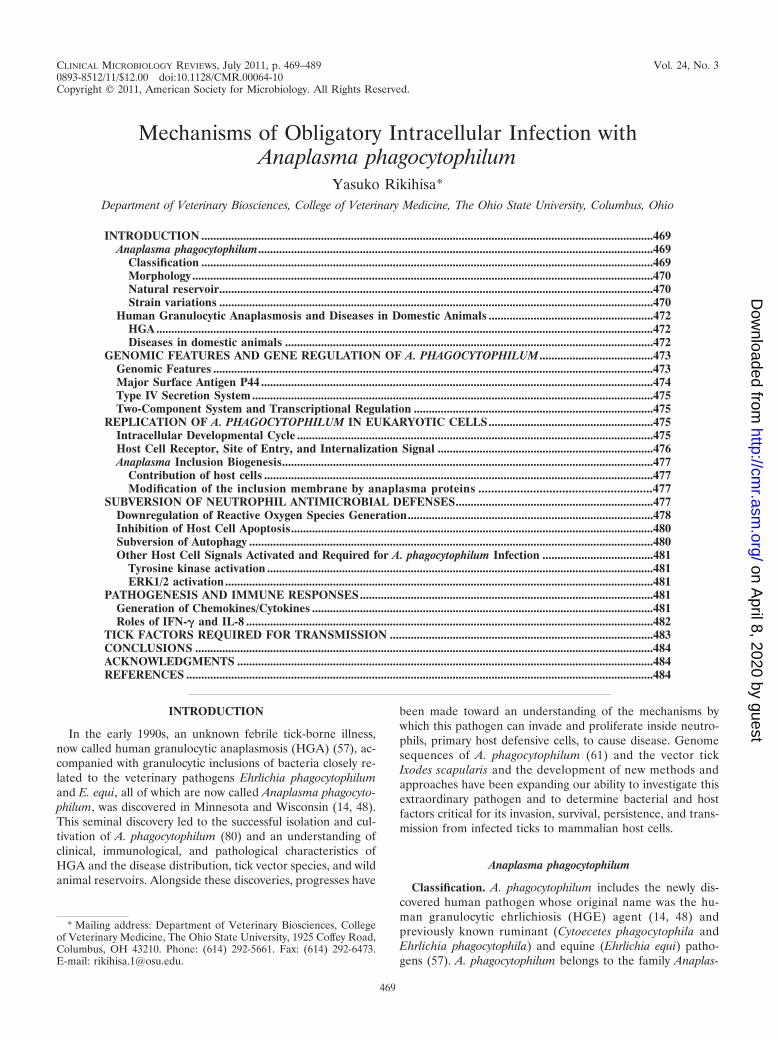
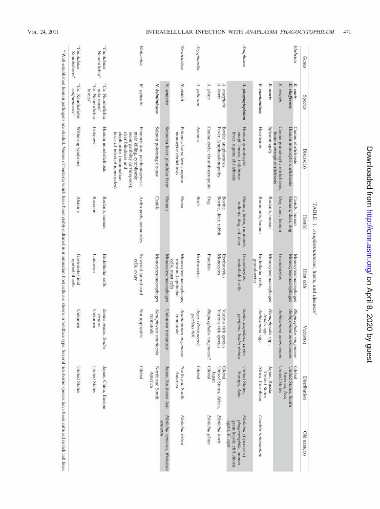
mataceae, in the order Rickettsiales and the class Alphaproteo-bacteria (57). The family Anaplasmataceae includes fivewell-known genera, Ehrlichia, Anaplasma, Neorickettsia, Aegyp-tianella, and Wolbachia, and two less-well-studied genera,“Candidatus Neoehrlichia” and “Candidatus Xenohaliotis.” Allof these genera infect specific invertebrate hosts (ticks, insects,trematodes, nematodes, or mollusks) that are abundant in na-ture. Unlike Neorickettsia and Wolbachia spp., which can betransmitted through generations of invertebrate hosts, Ana-plasma and Ehrlichia cannot effectively pass from adult ticks tooffspring (transovarial passage) (139, 206). All genera exceptWolbachia and “Candidatus Xenohaliotis” are known to infectvertebrates (mammals or birds). Vertebrate infection can beacute or chronic and may result in fatality. The bacteria infectspecific host cell types within vertebrates, usually cells ofhematopoietic origin, such as neutrophils, monocytes/mac-rophages, platelets, red blood cells, or endothelial cells.Characteristics of members of the family Anaplasmataceaeare summarized in Table 1. A. phagocytophilum is one of fourspecies belonging to the genus Anaplasma, which have differ-ent host cell specificities. For A. phagocytophilum, primary hostcells are granulocytes, and endothelial cells are also infected(48, 80, 92, 142, 197, 235). The phylogenetic relationship of A.phagocytophilum to other members of the genus Anaplasmaand members of the family Anaplasmataceae is shown in Fig. 1.
Morphology. A. phagocytophilum is a small Gram-negativepleomorphic coccus enveloped by two membranes, as are othermembers of the family Anaplasmataceae. The bacterial size isgenerally 0.4 to 1.3 �m, but the bacteria can be as large as 2�m. The outer membrane of the bacterium is often ruffled,which creates an irregular periplasmic space, and there is nocapsule layer. Fine DNA strands and ribosomes are distinctlyseen within the bacteria (185, 199, 235). Unlike members of thefamily Rickettsiaceae, which escape from phagosomes and rep-licate directly within the cytoplasm of eukaryotes, members ofthe family Anaplasmataceae replicate in membrane-bound vac-uoles (referred to as inclusions or parasitophorous vacuoles)within the cytoplasm of eukaryotic host cells. The bacteria maybe tightly packed inside inclusions in part due to a loss ofpeptidoglycan and lipopolysaccharide (LPS) (130). The losspermits the bacteria to squeeze within a limited intravacuolarspace while maintaining the plasticity of the infected granulo-cytes that is required for capillary circulation.
Gram staining is not suitable to visualize intracellular bac-teria because of a lack of contrast against the host cytoplasm.Romanowsky staining is generally used, usually with a quickmethod such as Diff-Quik. This approach stains the bacteriapurple, which allows the visualization of characteristic mul-berry-like bacterial clumps called morulae. (The term “mo-rula” is derived from the Latin term “morus,” which meansmulberry.) Morulae are usually 1.5 to 2.5 �m in diameter butcan be as large as 6 �m (185).
Natural reservoir. A. phagocytophilum DNA has been de-tected in several species of Ixodes ticks (I. scapularis, I. pacifi-cus, I. spinipalpis, I. ricinus, I. persulcatus, and I. ovatus) in theUnited States, Europe, and Asia (37, 38a, 170, 171, 193, 216).Naturally infected ticks were shown to transmit A. phagocyto-philum to naïve mammals (63, 192, 207). Once ticks acquire thebacterium from infected mammals through a blood meal, thebacterium is maintained from the larva or nymph stage to adult
stages of metamorphosis and is transmitted to mammals dur-ing the next blood meal (169, 216, 250). Since there is noevidence of transovarial (from adult ticks to eggs) transmis-sion, larvae do not transmit the bacterium to mammals, butinfected nymphs and adult ticks do. The mammalian reservoirfor A. phagocytophilum infection within the United Statesincludes white-footed mice (Peromyscus leucopus), raccoons(Procyon lotor), gray squirrels (Sciurus carolinensis), gray foxes(Urocyon cinereoargenteus), and redwood chipmunks (69, 127,165, 216). A variety of other wild animals are also implicated asreservoirs (68; reviewed recently in references 208 and 218).Although Ixodes ticks often feed on white-tailed deer, the deerare infected with the Ap-Variant 1 strain of A. phagocytophi-lum, rather than with the human strain, in the United States(146). Diverse A. phagocytophilum strains are also found inanimals and ticks in Europe, Japan, and Russia (109, 111, 151,160, 170, 203, 238, 239), where HGA has been rarely reported.These findings imply that the zoonosis potential of A. phago-cytophilum depends not only on the transmissibility, habitats,and population density of ticks and infected mammals (90) butalso on the genetic variations of A. phagocytophilum. A primarynatural tick-mammalian transmission cycle of A. phagocytophi-lum interlacing with bacterial strain diversity and host suscep-tibility is depicted in Fig. 2. The transovarial transmission of A.phagocytophilum variants occurs in Dermacentor albipictus (19).Thus, it is possible that there are atypical systems such as D.albipictus feeding into the normal Ixodes infection cycle.
Strain variations. All A. phagocytophilum isolates appear tohave serological cross-reactivity. In mammals, the most com-mon constitutively produced antigens are 42- to 49-kDa pro-teins, which are expressed on the bacterial outer membrane(11, 56, 105, 111a, 239, 249, 251). The proteins are encoded bythe p44 (also called msp2) gene family. Serological tests aregenerally group specific and cannot be used to distinguishindividual strains. A. phagocytophilum strains show a minordegree of variation in the nucleotide sequence in 16S rRNAand groESL. The p44, p44ESup1 (omp-1N), msp2 (differentfrom p44), and ankA genes contain major strain variation (23,44, 61, 67, 111, 134, 135, 150, 153, 203, 224, 239). These andpotentially other genes may allow more detailed comparisonsamong strains. For example, diverse ankA sequences are foundin naturally infected ticks and wild deer, including those thathave not been detected in humans or domestic animals (203,224).
Several studies reported that A. phagocytophilum strains dif-fer in host infectivity. For example, strain Ap-Variant 1 fromI. scapularis ticks infects goat and deer but not mice (146, 148,149). A Californian strain infectious to equines was not infec-tious to ruminants (205). A. phagocytophilum strains from wildrodents were reported to differ in horse infectivity (68). Al-though several human, ruminant, and equine strains are culti-vated in ISE6 and/or IDE8 or other tick cell lines (80, 147, 159,234), so far, only human isolates have been directly cultured byusing HL-60 cells, a human promyelocytic leukemia cell line.Recently, new strains of A. phagocytophilum from Chinesesheep and wild rodents were cultured in HL-60 cells afterinitial passage in BALB/c mice (246). The bacterial factors thatdetermine virulence, host mammal species specificity, and cul-tivability are not known. Arthropod-borne diseases, includingA. phagocytophilum infection, are on the rise, probably because
470 RIKIHISA CLIN. MICROBIOL. REV.
on April 8, 2020 by guest
http://cmr.asm
.org/D
ownloaded from
TA
BL
E1.
Anaplasm
ataceae,hosts,anddiseases
a
Genus
SpeciesD
isease(s)H
ost(s)H
ostcells
Vector(s)
Distribution
Old
name(s)
Ehrlichia
E.canis
Canine
ehrlichiosisC
anids,human
Monocytes/m
acrophagesR
hipicephalussanguineus
Global
E.chaffeensis
Hum
anm
onocyticehrlichiosis
Hum
an,deer,dogM
onocytes/macrophages
Am
blyomm
aam
ericanumU
nitedStates,South
Am
erica,Asia
E.ew
ingiiC
aninegranulocytic
ehrlichiosis,hum
anew
ingiiehrlichiosisD
og,deer,human
Granulocytes
Am
blyomm
aam
ericanumU
nitedStates
E.m
urisSplenom
egalyR
odents,human
Monocytes/m
acrophagesH
emaphysalis
spp.,Ixodes
spp.Japan,R
ussia,U
nitedStates
E.rum
inantiumH
eartwater
Rum
inants,human
Endothelialcells,granulocytes
Am
blyomm
aspp.
Africa,C
aribbeanC
owdria
ruminantium
Anaplasm
aA
.phagocytophilumH
uman
granulocyticanaplasm
osis,tick-bornefever,equine
ehrlichiosis
Hum
an,horse,ruminants,
rodents,dog,cat,deerG
ranulocytes,endothelialcells
Ixodesscapularis,Ixodes
pacificus,Ixodesricinus
United
States,E
urope,Asia
Ehrlichia
(Cytoecetes)
phagocytophila,human
granulocyticehrlichiosis
agent,E.equi
A.m
arginaleB
ovineanaplasm
osisB
ovineE
rythrocytesV
arioustick
speciesG
lobalA
.bovisF
ever,lymphoadenopathy
Bovine,deer,rabbit
Monocytes
Various
tickspecies
United
States,Africa,
JapanE
hrlichiabovis
A.platys
Canine
cyclicthrom
bocytopeniaD
ogPlatelets
Rhipicephalus
sanguineus?G
lobalE
hrlichiaplatys
Aegyptianella
A.pallorum
Anem
iaB
irdsE
rythrocytesA
rgas(P
ersicargus)persicus
tickG
lobal
Neorickettsia
N.risticii
Potomac
horsefever,equine
monocytic
ehrlichiosisH
orseM
onocytes/macrophages,
intestinalepithelialcells,m
astcells
Acanthatrium
oregonensetrem
atodeN
orthand
SouthA
merica
Ehrlichia
risticii
N.sennetsu
Sennetsufever,glandular
feverH
uman
Monocytes/m
acrophagesU
nknown
trematode
Japan,SoutheastA
siaE
hrlichiasennetsu,R
ickettsiasennetsu
N.helm
inthoecaSalm
onpoisoning
diseaseC
anidsM
onocytes/macrophages
Nanophyetus
salmincola
trematode
North
andSouth
Am
erica
Wolbachia
W.pipientis
Fem
inization,parthenogenesis,m
alekilling,cytoplasm
icincom
patibility(arthropods);
riverblindness
andelephantitis
(mam
malian
hostsof
infectednem
atodes)
Arthropods,nem
atodesSyncytiallateralcord
cells,ovaryN
otapplicable
Global
“CandidatusN
eoehrlichia”“C
a.Neoehrlichia
mikurensis”
Hum
anneoehrlichiosis
Rodents,hum
anE
ndothelialcellsIxodes
ovatus,Ixodesricinus
Japan,China,E
urope
“Ca.N
eoehrlichialotoris”
Unknow
nR
accoonU
nknown
Unknow
nU
nitedStates
“CandidatusX
enohaliotis”“C
a.Xenohaliotis
californiensis”W
itheringsyndrom
eA
baloneG
astrointestinalepithelialcells
Unknow
nU
nitedStates
aW
ell-establishedhum
anpathogens
areshaded.N
ames
ofbacteria
which
havebeen
stablycultured
inm
amm
alianhostcells
areshow
nin
boldfacetype.Severaltick-borne
specieshave
beencultured
intick
celllines.
VOL. 24, 2011 INTRACELLULAR INFECTION WITH ANAPLASMA PHAGOCYTOPHILUM 471
on April 8, 2020 by guest
http://cmr.asm
.org/D
ownloaded from
of changes in human activities and climate (27, 77, 84). Theresults of molecular studies on A. phagocytophilum variants willshed light on the evolution, population dynamics, and ecologyof naturally occurring A. phagocytophilum strains and will helpidentify risks for outbreaks of zoonosis and important veteri-nary diseases.
Human Granulocytic Anaplasmosis and Diseases inDomestic Animals
HGA. HGA is characterized by fever; chills; headache; my-algia; hematological abnormalities, including leucopenia andthrombocytopenia; and increased serum aminotransferaseliver enzyme activity, which suggests mild to moderate liverinjury (3, 15, 58, 59). Patients generally respond to doxycycline,the treatment of choice. Although fluoroquinolones are activein vitro against A. phagocytophilum, it causes a relapse of in-fection in severe combined immunodeficiency mice and HGApatients and is not recommended (237). The case-fatality rateis 0.7% in the Midwestern United States, and the risk of fa-tality is greater when therapy initiation is delayed, when pa-tients are elderly, and when patients have complicating oppor-tunistic infections and/or antecedent medical conditions, suchas diabetes mellitus or immunocompromise (13, 59). In a caseseries, illness associated with HGA in the Northeastern UnitedStates was more mild than that originally described in theMidwestern United States (3), suggesting strain-dependent vir-ulence. The median age of patients with HGA is 50 to 60 years,higher than that associated with other tick-borne diseases (forexample, the median age of patients with Lyme disease isaround 40 years).
The number of documented cases of HGA has increasedevery year since 1999, the year when the disease becamenotifiable in the United States (176), and 1,161 cases of A.phagocytophilum infection were reported in 2009 (47). Num-bers of HGA cases and A. phagocytophilum sero- or DNA-positive clinical cases have also been increasing in Europeand Asia (7, 17, 123, 170a, 175, 182, 200, 247). Minnesota,Wisconsin, New York, and Massachusetts remain states withthe highest incidence of HGA (47). In areas of Wisconsin
and New York where the disease is endemic, seroepidemio-logical data suggest that many infections go unrecognized,and it was estimated that 15% and 36% of the populations,respectively, have been infected (2, 16). Results from PCRassays and serological tests suggest that HGA is a majorcause of unexplained fever during the tick season in Wis-consin (26). Most cases of HGA occur in the same statesthat have high incidences of Lyme disease and human babe-siosis; this is because Ixodes ticks also transmit Borreliaburgdorferi, the causative agent of Lyme disease, and Babe-sia species, the causative agent of babesiosis. Simultaneousinfection with A. phagocytophilum and B. burgdorferi in hu-mans has been reported (4, 17, 123, 162). Therefore, thechoice of antimicrobial agent needs to take into accountwhether a patient has a combined infection. For example,amoxicillin can be used to treat early-stage Lyme disease,although it is not effective as a treatment for HGA.
Although the primary mode of transmission of A. phagocy-tophilum to humans is from an infected tick, the transmissionof the bacteria can occur perinatally (97) and nosocomially(247). A. phagocytophilum in infected human blood specimensis viable at 4°C for up to 18 days (108), and transmission hasoccurred through blood transfusion (46) and contact with theblood of infected mammals (18).
Diseases in domestic animals. Clinical signs of granulocyticanaplasmosis in domestic animals (horse, dog, and ruminants)include fever accompanied by leucopenia and thrombocytope-nia. Opportunistic infections may also occur (reviewed in ref-erences 43, 196, 233). Other signs of disease in animals includedepression and anorexia in horses and dogs and limb edemaand ataxia in horses. A. phagocytophilum strains that werepreviously known as Cytoecetes phagocytophila or Ehrlichiaphagocytophila cause tick-borne fever in ruminants, mainly inWestern Europe (66, 82, 102). Strains that were previouslyknown as Ehrlichia equi or the HGE agent cause granulocyticehrlichiosis in equines (also known as equine granulocytic ana-plasmosis) (7, 30, 86, 112, 142, 205) and in canines (also knownas canine granulocytic anaplasmosis, distinct from Ehrlichiaewingii infection) (7, 85, 183, 188; reviewed in reference 43).Equine and canine granulocytic ehrlichioses have been found
FIG. 1. Phylogram of members of the genus Anaplasma in the family Anaplasmataceae. The genus Anaplasma is highlighted in gray.Phylogenetic trees were constructed based on 16S rRNA sequence alignment by the Clustal W method using the MegAlign program from theLasergene package. GenBank accession numbers are shown in parentheses.
472 RIKIHISA CLIN. MICROBIOL. REV.
on April 8, 2020 by guest
http://cmr.asm
.org/D
ownloaded from
in broad distributions across the United States, Canada, SouthAmerica, and Europe. Feline granulocytic anaplasmosis isless common but has been increasingly reported in Europeand the United States (8, 28, 31, 122, 202).
The present review primarily describes our current under-standing of the molecular mechanisms of intracellular infec-tion by A. phagocytophilum isolates from HGA patients in theUnited States, since A. phagocytophilum from other sourceshas not been sufficiently studied in this aspect. For clinical andlaboratory diagnosis, treatment, epidemiology, and the preven-tion of HGA, please refer to recent reviews and guidelines (15,59, 218, 226, 236).
GENOMIC FEATURES AND GENE REGULATIONOF A. PHAGOCYTOPHILUM
Genomic Features
The genome size (1.47 Mb) of A. phagocytophilum strainHZ, isolated from a patient in the state of New York (199), isapproximately one-quarter of the size of the Escherichia coligenome, the model prokaryote. The number of open readingframes (ORFs) (1,369) is also about one-quarter of that foundin E. coli (61). The G-C content of the DNA in A. phagocyto-philum strain HZ is 41.6 mol%, which is lower than the E. coli
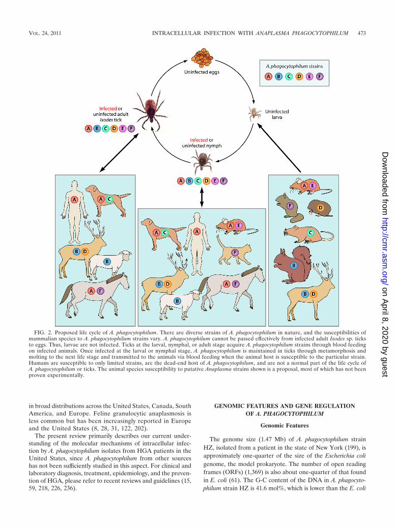
FIG. 2. Proposed life cycle of A. phagocytophilum. There are diverse strains of A. phagocytophilum in nature, and the susceptibilities ofmammalian species to A. phagocytophilum strains vary. A. phagocytophilum cannot be passed effectively from infected adult Ixodes sp. ticksto eggs. Thus, larvae are not infected. Ticks at the larval, nymphal, or adult stage acquire A. phagocytophilum strains through blood feedingon infected animals. Once infected at the larval or nymphal stage, A. phagocytophilum is maintained in ticks through metamorphosis andmolting to the next life stage and transmitted to the animals via blood feeding when the animal host is susceptible to the particular strain.Humans are susceptible to only limited strains, are the dead-end host of A. phagocytophilum, and are not a normal part of the life cycle ofA. phagocytophilum or ticks. The animal species susceptibility to putative Anaplasma strains shown is a proposal, most of which has not beenproven experimentally.
VOL. 24, 2011 INTRACELLULAR INFECTION WITH ANAPLASMA PHAGOCYTOPHILUM 473
on April 8, 2020 by guest
http://cmr.asm
.org/D
ownloaded from
average G-C content (�50 mol%) but higher than the �29.1%of Rickettsia prowazekii (10). No plasmids, intact prophages, ortransposable elements are present. The genome of A. phago-cytophilum has numerous repeats (12.7% of the genome), asseen in over 100 p44 (msp2) genes, type IV secretion (T4S)genes, and genes containing tandem repeats (61, 207, 248).Among genes that are conspicuously absent from A. phagocy-tophilum are those genes required for the biosynthesis of lipo-polysaccharide and peptidoglycan (61, 130). Approximately747 ORFs (55%) have an assigned function, encoding mostlyhousekeeping genes found in other bacteria (61), and 82 ORFsencode conserved hypothetical proteins (meaning that they arefound in other bacteria as well but do not have an assignedfunction). Approximately 458 ORFs encode unique hypothet-ical proteins that are not found in other organisms. A recentglobal proteomic analysis revealed a total of 1,212 A. phago-cytophilum proteins in infected human leukocytes, represent-ing 89.3% of the predicted bacterial proteomes. Nearly allbacterial proteins (�99%) with known functions are expressed,whereas only approximately 80% of “hypothetical” proteinswere detected in infected human cells (132).
Several hypothetical proteins in A. phagocytophilum werefound to be surface-exposed proteins, inclusion membraneproteins, and T4S substrates that confer unique phenotypes tothis bacterium (74, 99, 100, 166, 195, 210). A. phagocytophilumand Rickettsia prowazekii share 469 genes (61), and both bac-teria cannot use glucose as a carbon or energy source. How-ever, unlike Rickettsia, A. phagocytophilum has a partial glycol-ysis pathway starting with fructose 1,6-biphosphate. Like R.prowazekii, A. phagocytophilum has pathways for aerobic res-piration, including the metabolism of pyruvate, the tricarbox-ylic acid cycle, and the electron transport chain. Unlike R.prowazekii, however, A. phagocytophilum does not encode acytochrome d-type oxidase (cydAB [cytochrome d ubiquinoloxidase]), which has a high affinity for oxygen that is useful formicroaerophilic respiration; A. phagocytophilum does not haveATP/ADP translocase, which assists in moving ADP out of thebacterial inner membrane while assisting ATP to enter theinner bacterial membrane to supply the bacteria with host-synthesized ATP; and A. phagocytophilum contains genes forthe biosynthesis of all of the necessary nucleotides and mostvitamins and cofactors, including biotin, folate, flavin adeninedinucleotide (FAD), NAD, coenzyme A, thiamine, and proto-heme, which might benefit the tick vector. Selected character-
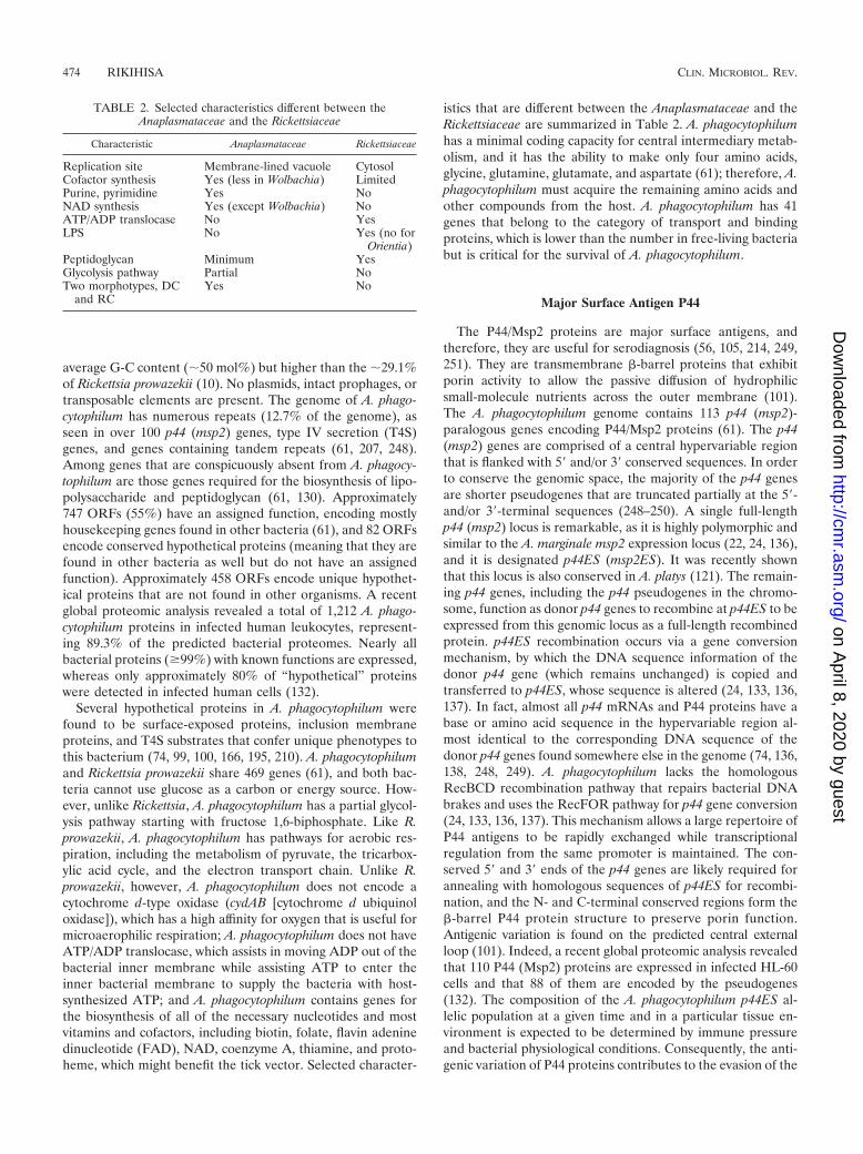
istics that are different between the Anaplasmataceae and theRickettsiaceae are summarized in Table 2. A. phagocytophilumhas a minimal coding capacity for central intermediary metab-olism, and it has the ability to make only four amino acids,glycine, glutamine, glutamate, and aspartate (61); therefore, A.phagocytophilum must acquire the remaining amino acids andother compounds from the host. A. phagocytophilum has 41genes that belong to the category of transport and bindingproteins, which is lower than the number in free-living bacteriabut is critical for the survival of A. phagocytophilum.
Major Surface Antigen P44
The P44/Msp2 proteins are major surface antigens, andtherefore, they are useful for serodiagnosis (56, 105, 214, 249,251). They are transmembrane �-barrel proteins that exhibitporin activity to allow the passive diffusion of hydrophilicsmall-molecule nutrients across the outer membrane (101).The A. phagocytophilum genome contains 113 p44 (msp2)-paralogous genes encoding P44/Msp2 proteins (61). The p44(msp2) genes are comprised of a central hypervariable regionthat is flanked with 5� and/or 3� conserved sequences. In orderto conserve the genomic space, the majority of the p44 genesare shorter pseudogenes that are truncated partially at the 5�-and/or 3�-terminal sequences (248–250). A single full-lengthp44 (msp2) locus is remarkable, as it is highly polymorphic andsimilar to the A. marginale msp2 expression locus (22, 24, 136),and it is designated p44ES (msp2ES). It was recently shownthat this locus is also conserved in A. platys (121). The remain-ing p44 genes, including the p44 pseudogenes in the chromo-some, function as donor p44 genes to recombine at p44ES to beexpressed from this genomic locus as a full-length recombinedprotein. p44ES recombination occurs via a gene conversionmechanism, by which the DNA sequence information of thedonor p44 gene (which remains unchanged) is copied andtransferred to p44ES, whose sequence is altered (24, 133, 136,137). In fact, almost all p44 mRNAs and P44 proteins have abase or amino acid sequence in the hypervariable region al-most identical to the corresponding DNA sequence of thedonor p44 genes found somewhere else in the genome (74, 136,138, 248, 249). A. phagocytophilum lacks the homologousRecBCD recombination pathway that repairs bacterial DNAbrakes and uses the RecFOR pathway for p44 gene conversion(24, 133, 136, 137). This mechanism allows a large repertoire ofP44 antigens to be rapidly exchanged while transcriptionalregulation from the same promoter is maintained. The con-served 5� and 3� ends of the p44 genes are likely required forannealing with homologous sequences of p44ES for recombi-nation, and the N- and C-terminal conserved regions form the�-barrel P44 protein structure to preserve porin function.Antigenic variation is found on the predicted central externalloop (101). Indeed, a recent global proteomic analysis revealedthat 110 P44 (Msp2) proteins are expressed in infected HL-60cells and that 88 of them are encoded by the pseudogenes(132). The composition of the A. phagocytophilum p44ES al-lelic population at a given time and in a particular tissue en-vironment is expected to be determined by immune pressureand bacterial physiological conditions. Consequently, the anti-genic variation of P44 proteins contributes to the evasion of the
TABLE 2. Selected characteristics different between theAnaplasmataceae and the Rickettsiaceae
Characteristic Anaplasmataceae Rickettsiaceae
Replication site Membrane-lined vacuole CytosolCofactor synthesis Yes (less in Wolbachia) LimitedPurine, pyrimidine Yes NoNAD synthesis Yes (except Wolbachia) NoATP/ADP translocase No YesLPS No Yes (no for
Orientia)Peptidoglycan Minimum YesGlycolysis pathway Partial NoTwo morphotypes, DC
and RCYes No
474 RIKIHISA CLIN. MICROBIOL. REV.
on April 8, 2020 by guest
http://cmr.asm
.org/D
ownloaded from

host immune system and the persistence of A. phagocytophiluminfection.
Type IV Secretion System
In order to secrete proteins, enzymes, DNA, or toxins fromthe cytoplasm to the exterior of bacteria across the cytoplasmicmembrane, bacteria use at least six distinct extracellular pro-tein secretion systems, referred to as type I to type VI secretionsystems (172). Homologs of type I secretion system (T1SS) andT4SS components were found in the A. phagocytophilum ge-nome (61). The T4SS forms a transmembrane channel com-posed of a multiprotein complex, which delivers the effectorprotein and DNA, and a nucleoprotein complex in an ATP-dependent manner. Recently, T4SSs have been classified intofour groups: groups F, P, I, and GI (107). A. phagocytophilumhas P-T4SSs (previously known as type IVA, the prototypeencoded by plasmid pTi of Agrobacterium tumefaciens). Inter-estingly, despite the genome size reduction, A. phagocytophi-lum has up to eight nonidentical copies of these genes. Thegenes are distributed into three major genomic islands: sodB-virB3-virB4-virB6-1-virB6-2-virB6-3-virB6-4, virB8-1-virB9-1-virB10-virB11-virD4, and virB2-1-virB2-2-virB2-3-virB2-4-virB2-5-virB2-6-virB2-7-virB2-8-virB4-2. Between these genomic islandsare virB8-2, virB9-2, and a putative virB7 (no ORF numberassigned; positions 1033978 to 1034181). The significance ofthe duplicated virB genes is currently unknown, but distinctsets of virB2 genes, which encode a surface-exposed pilus, areexpressed in ticks and mammalian cell cultures (164). It iscritical to identify substrates that are delivered by the T4SS tounderstand its role in bacterial infection. So far, two T4SSsubstrates, ankyrin-repeat-rich protein A (AnkA) and Ana-plasma translocated substrate 1 (Ats-1) (128, 166), have beenidentified and are discussed below. Unlike several other bac-teria, genes encoding these proteins are not closely associatedwith virB or virD genomic islands; therefore, a global genomicsearch is required to find remaining potential substrates, as wasperformed for Ats-1 (166).
Two-Component System and Transcriptional Regulation
Two-component systems (TCSs) serve as a basic stimulus-response-coupling mechanism to allow organisms to sense andrespond to changes under many different environmental con-ditions (145). They typically consist of an inner membrane-bound histidine kinase that senses a specific environmentalstimulus and a corresponding response regulator that mediatesthe cellular response, mostly through the differential expres-sion of target genes. The A. phagocytophilum genome encodesthree pairs of TCSs: PleC/PleD, CckA/CtrA, and NtrY/NtrX(49). PleC, CckA, and NtrY are sensor histidine kinases whichare essential for infection (49). CtrA, NtrX, and PleD areresponse regulators. CtrA and NtrX have a C-terminal DNA-binding domain and are believed to function as transcriptionfactors. PleD has a C-terminal GGDEF domain associatedwith a diguanylate cyclase that produces cyclic di-GMP (c-di-GMP) (120). The expression of PleC and PleD is upregulatedduring an exponential growth stage (120). A hydrophobic c-di-GMP analog, 2�-O-di(tert-butyldimethylsilyl)-c-di-GMP, canserve as a functional antagonist in well-defined c-di-GMP-
regulated phenomena in Salmonella enterica serovar Typhi-murium, including cellulose synthesis, clumping, and theupregulation of csgD and adrA mRNAs (119). The analogcompetitively inhibits c-di-GMP binding to recombinant PleDand to native proteins of A. phagocytophilum and inhibits theinfection of HL-60 cells with A. phagocytophilum (120).
The differential expression of A. phagocytophilum genes inHL-60 cells and ISE6 tick cells (164, 228) and the temporaltranscriptional and protein expression profiles during A.phagocytophilum infection in HL-60 cells and neutrophils (120,128, 166, 167) imply that the host environment and bacterialconcentration regulate the transcription of A. phagocytophi-lum. However, genes encoding the nutritional stress responseprotein RelA/SpoT and proteins required for the biosynthesisof a quorum-sensing pheromone have not been found in A.phagocytophilum. A. phagocytophilum encodes only two sigmafactor homologs: a constitutive factor, �70, and a single alter-native factor, �32 (RpoH). The paucity of alternative sigmafactors suggests that A. phagocytophilum relies on the regula-tion of constitutive �70-type promoters by transcription factorsand posttranscriptional regulation.
Mass spectrometry analysis of the DNA affinity-purifiedA. phagocytophilum protein identified a 12.5-kDa hypotheticalprotein (GenBank accession no. YP_505110.1). We named thisprotein A. phagocytophilum expression regulator (ApxR).ApxR is an unique DNA-binding protein that can regulatethe transcription of downstream genes in A. phagocytophilum(229). Interestingly, ApxR is upregulated in HL-60 cells,whereas a putative transcriptional regulator, Tr1, a protein of21 kDa which is predicted to have a DNA-binding motif, isupregulated in ISE6 tick cell cultures, suggesting that theseproteins might be involved in the expression of mammal- andtick-specific genes (164, 228). Both ApxR and Tr1 homologsare uniquely evolved in members of the family Anaplasmata-ceae but are not found in the family Rickettsiaceae or in otherbacterial families. In infected ticks, other than the data avail-able from transcript analyses of p44, tr1, msp2, and apxR (63,134, 228, 250), little information is available on the expressionof A. phagocytophilum genes.
REPLICATION OF A. PHAGOCYTOPHILUM INEUKARYOTIC CELLS
Intracellular Developmental Cycle
Electron microscopy identified distinct morphotypes of A.phagocytophilum inside neutrophil inclusions from infectedsheep (235). Two morphotypes of bacteria can also be ob-served in both mammalian and tick cell cultures, a larger re-ticulate (RC) form and a smaller dense-core (DC) form thatcontains condensed protoplasm (158, 185, 223), and are con-sidered to be normal developmental stages similar to those ofother intracellular bacteria, including Chlamydia species (157)and Coxiella burnetii (91). In addition, a variety of aberrantbacterial forms have been reported (158, 185, 223). The bio-logical significance of these aberrant forms is unknown, butthey may reflect an unfavorable or unbalanced physiologicalstate of the bacteria. Electron microscopy indicated that onlythe DC form binds to HL-60 cells (223), but both the DC andRC forms bind to and enter ISE6 tick cells (158). Only the DC
VOL. 24, 2011 INTRACELLULAR INFECTION WITH ANAPLASMA PHAGOCYTOPHILUM 475
on April 8, 2020 by guest
http://cmr.asm
.org/D
ownloaded from
form binds to Chinese hamster ovary cells that are transfectedto express P-selectin glycoprotein ligand 1 (PSGL-1), and bind-ing is inhibited by the preincubation of the cells with an anti-PSGL-1 antibody (223). Troese and Carlyon (223) reportedthat internalized DC bacteria transition to RC bacteria inHL-60 cells within 12 h and that the RC bacteria initiatereplication. By 24 h, large numbers of RC bacteria can beobserved within individual inclusions, and by 36 h, the reinfec-tion of already infected cells occurs, with individual, vacuole-enclosed DC and RC bacteria within the same cell (223).Although the proposed developmental cycle of the DC and RCforms morphologically resembles that of Chlamydia species,genes encoding histone H1-like proteins involved in chromatincondensation in Chlamydia (88) have not been identified in A.phagocytophilum.
Immunofluorescence showed that only small (�1-�m) bac-teria corresponding to the DC form bind to and enter humanperipheral blood neutrophils (167). In HL-60 cells, however,either small or large (�1-�m) bacteria are taken up, and mostlarge bacteria, but not most small bacteria, are rapidly deliv-ered to lysosome-associated membrane protein 1 (LAMP1)-positive compartments (167), implying that different develop-mental stages of A. phagocytophilum have distinct surface orbiological properties that allow them enter host cells throughdifferent receptors and internalization pathways. Except for aT4S apparatus protein, VirB9 (167), molecular markers thatdifferentiate the DC form from the RC form have not beenreported.
The ultrastructures of the DC and RC forms of A. marginaleand Ehrlichia muris in tick tissues have been reported (117,186). A. phagocytophilum has been detected in the salivaryglands of experimentally infected ticks and in field-derivedadult deer ticks as Feulgen-positive inclusions (216), but thebacterial ultrastructure in ticks remains to be studied.
Host Cell Receptor, Site of Entry, and Internalization Signal
A. phagocytophilum is maintained mostly in circulating ma-ture neutrophils, rather than in peripheral tissues, includinghematopoietic tissue (25, 94, 243), although A. phagocytophi-lum can infect bone marrow progenitors and endothelial cells(92, 116). While skin lesions have not been reported for HGA,during field transmission from ticks to lambs, A. phagocytophi-lum is found primarily in association with neutrophils andmacrophages in skin lesions, suggesting the role of peripheralleukocytes in tick transmission (83).
The ability to attach to and enter susceptible host cells isessential for infection. Cell lines, rather than human neutro-phils, have been used in most studies of the binding and inter-nalization of A. phagocytophilum. The tetrasaccharide sialylLewisx (sLex) on P-selectin glycoprotein ligand 1 (PSGL-1) isrequired for the binding and infection of HL-60 cells by A.phagocytophilum (81, 93, 190, 191, 201, 244). Similar to thebinding of P-selectin, the binding of A. phagocytophilum toHL-60 cells activates the PSGL-1 signaling pathway, whichresults in the tyrosine phosphorylation of ROCK1, an effectorkinase of the GTPase RhoA by spleen tyrosine kinase (Syk).PSGL-1-blocking antibodies and Syk small interfering RNA(siRNA) inhibit ROCK1 phosphorylation in A. phagocytophi-lum-infected HL-60 cells (220). The treatment of HL-60 cells
with piceatannol, a Syk inhibitor, or the knockdown of eitherSyk or ROCK1 impairs infection by A. phagocytophilum (220).ROCK1 is a serine/threonine kinase and is a key regulator ofactin organization, suggesting that bacterial binding to PSGL-1activates actin cytoskeleton reorganization through ROCK1activation to facilitate bacterial invasion.
A naturally occurring subpopulation of A. phagocytophilum,termed NCH-1A2, has been selected through cultivation insLex-defective HL-60 cells (HL-60 A2 cells). NCH-1A2 bindsto HL-60 cells in a sialic acid- and PSGL-1-independent man-ner (190, 201). In agreement with the PSGL-1 independence,Syk is also not essential for NCH-1A2 infection of HL-60 cells(191). Whether NCH-1A2 is a genetic or phenotypic variantand whether an alternative receptor exists are unknown. None-theless, these studies suggested that A. phagocytophilum caninfect HL-60 cells through at least two independent receptorsand signaling pathways.
PSGL-1 is not required for infection of mice becauseA. phagocytophilum binds effectively to PSGL-1�/� murineneutrophils, and it infects PSGL-1�/� mice. Fucosyl trans-ferases are, however, required for infection of mice, becausethe binding of A. phagocytophilum to Fuc-TIV�/�/Fuc-TVII�/� neutrophils and the infection of Fuc-TIV�/�/Fuc-TVII�/� mice are significantly reduced (40). Tick colonizationwith A. phagocytophilum was recently shown to involve 1,3-fucosylation on N-glycan, as determined by using the I. ricinustick embryonic cell line IRE/CTVM19 and siRNA in I. scapu-laris ticks (179). The binding and/or infection of A. phagocyto-philum appears to involve the major surface proteins Msp2(P44), Asp55, and Asp62, as shown by the results of neutral-ization studies (74, 173, 230). It is important to identify thebacterial ligands that interact with PSGL-1 and other potentialreceptors to elucidate the specific ligand-receptor interactionsthat mediate bacterial attachment and entry.
Entry and intracellular infection by A. phagocytophilum re-quire lipid rafts, which are signaling platforms (131). Caveo-lin-1, which is concentrated in lipid microdomains (9), wascolocalized with early inclusions of A. phagocytophilum inHL-60 cells (131). Whether caveolin-1 is required for infectionby A. phagocytophilum is unknown. Glycosylphosphatidylinosi-tol (GPI)-anchored proteins (GAPs) and flotillin 1, both ofwhich are associated with lipid rafts, are required for bindingand infection by A. phagocytophilum (131, 240). In contrast,clathrin, a protein involved in endocytosis, is not associatedwith the internalization of A. phagocytophilum (131, 154). A.phagocytophilum concentrates in lipid rafts, but the signalingcascades are still poorly understood. One signal shown to berapidly induced upon A. phagocytophilum binding is the phos-phorylation of protein tyrosine, which is required for internal-ization and infection with A. phagocytophilum (103, 128, 131,220). Tyrosine-phosphorylated proteins and phospholipase C2 (PLC-2) accumulate in A. phagocytophilum-infected cellswithin 5 min of infection (131). ROCK1 is tyrosine phosphor-ylated after A. phagocytophilum infection, although it is notknown how long after infection this occurs (220). In addition,the tyrosine phosphorylation of AnkA, a bacterial T4S effectorprotein, occurs 5 min after infection of HL-60 cells (103).Tyrosine-phosphorylated proteins recruit active signaling mol-ecules to create multiprotein signaling networks (177). In or-der to understand how bacteria enter, it is necessary to eluci-
476 RIKIHISA CLIN. MICROBIOL. REV.
on April 8, 2020 by guest
http://cmr.asm
.org/D
ownloaded from
date the downstream signaling pathways that become activeafter tyrosine phosphorylation.
Anaplasma Inclusion Biogenesis
Contribution of host cells. A. phagocytophilum multipliesstrictly within membrane-bound inclusions. As bacteria divideand proliferate, the inclusions expand to occupy most of thecytoplasm of infected cells. The characteristics of the inclu-sions, including the membrane and luminal components, areunique and largely undetermined, and they appear to evolveover time as the A. phagocytophilum infection cycle progresses.A recent global proteomic analysis revealed an upregulation ofhuman proteins involved mostly in cytoskeleton components,vesicular trafficking, cell signaling, and energy metabolism inHL-60 cells infected with A. phagocytophilum compared withuninfected cells (132).
Neutrophils kill invading microorganisms by sequesteringvital nutrients (e.g., iron) or by fusing the phagosomes thatcontain the invading microorganisms with granules that con-tain both antimicrobial peptides (e.g., defensins and ly-sozymes) and lysosomal hydrolytic enzymes (52). A. phago-cytophilum interferes with vesicular trafficking to avoidlysosomes. The A. phagocytophilum inclusion compartmentdoes not resemble an endosome because it lacks transferrinreceptor (TfR), early endosomal antigen 1, Rab5, -adaptin,clathrin heavy chain, and annexins I, II, IV, and VI (154). Theinclusion compartment also is not acidic and lacks V-typeH� ATPase, and it does not acquire the late endosomal/lysosomal markers, including myeloperoxidase, CD63, andLAMP1 (154, 231). The inclusion also avoids fusion with se-cretory vesicles and specific and tertiary granules harboringNADPH oxidase (39, 104, 154). Additionally, unlike Chla-mydia species (89), the growth of A. phagocytophilum is notimpaired by brefeldin A (BFA), the Golgi apparatus-destabi-lizing agent, and the A. phagocytophilum inclusions do notacquire the Golgi vesicular marker �-coat protein (�-COP) orC6NBD {6-[N-(7-nitro-2,1,3-benzoxadiazol-4-yl) amino]}-sphigomyelin (154). Certain A. phagocytophilum inclusionscontain major histocompatibility complex class I (MHCI) andMHCII, and these may be involved in bacterial antigen pre-sentation (154). A. phagocytophilum inclusions were reportedto colocalize with green fluorescent protein (GFP)-Rab4A,GFP-Rab10, GFP-Rab11A, GFP-Rab14, RFP-Rab22A, GFP-Rab35 (all of which regulate endocytic recycling), and GFP-Rab1 (98). Interestingly, the treatment of infected cells withtetracycline for 30 min results in the loss of these GFP-Rabproteins and the acquisition of GFP-Rab5, GFP-Rab7, and thelysosomal markers (78, 98), suggesting that the inhibition ofthe endosome-lysosome pathway requires new bacterial pro-tein synthesis. Several indicators of early autophagosomeswere detected in A. phagocytophilum replicative inclusions, in-cluding a double-lipid bilayer membrane and the colocalizationof GFP-tagged LC3 and Beclin 1, the human homologs of theyeast Saccharomyces cerevisiae autophagy-related proteinsAtg8 and Atg6, respectively (168). A. phagocytophilumautophagosomes do not mature into degradative autolyso-somes. The inhibition of the autophagosomal pathway by3-methyladenine (3-MA), an inhibitor of class III phosphati-
dylinositol 3-kinase (PI3KC3), inhibits A. phagocytophilum in-fection by preventing bacterial growth but not bacterial inter-nalization (168).
A. phagocytophilum requires cholesterol for survival and in-fection, and unlike Escherichia coli, A. phagocytophilum is ca-pable of incorporating a substantial amount of host cholesterolinto its membrane (130). The expansion of the inclusion mem-brane during infection also requires a lipid source, includingcholesterol. In fact, A. phagocytophilum infection is facilitatedin mice with high blood cholesterol levels (243). Infection withA. phagocytophilum results in an upregulation of host cell cho-lesterol levels and transportation to the inclusion (241). Thisoccurs by the upregulation of low-density lipoprotein (LDL)receptor and LDL vesicular transport, not by de novo biosyn-thesis in host cells, and it requires bacterial protein synthesis(241). Cholesterol is then incorporated into the anaplasmamembrane. Importantly, the inhibition of the LDL cholesteroluptake pathway blocks bacterial infection (241).
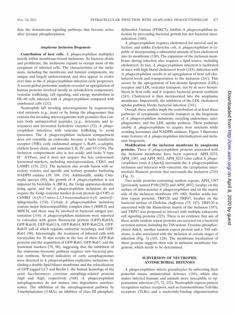
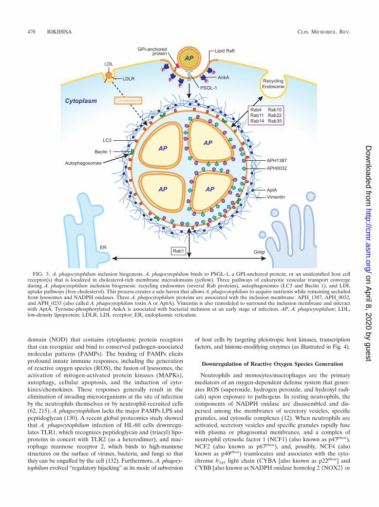
Overall, these studies imply the contribution of at least threepathways of cytoplasmic vesicular transport in the biogenesisof A. phagocytophilum inclusions: recycling endosomes, auto-phagosomes, and the LDL uptake pathway. These pathwaysenable A. phagocytophilum to acquire host nutrients whileavoiding lysosomes and NADPH oxidases. Figure 3 illustratessome features of A. phagocytophilum internalization and inclu-sion biogenesis.
Modification of the inclusion membrane by anaplasmaproteins. Three A. phagocytophilum proteins associated withthe inclusion membrane have been reported: APH_0233,APH_1387, and APH_0032. APH_0233 (also called A. phago-cytophilum toxin A [AptA]) surrounds the A. phagocytophiluminclusion and interacts with vimentin, the cytoplasmic inter-mediate filament protein that surrounds the inclusion (210)(Fig. 3).
Two acidic proteins containing tandem repeats, APH_1387(previously named P100 [207]) and APH_0032, localize on thesurface of intravacuolar A. phagocytophilum and on the matrixside of the inclusion membrane (99, 100). Similar acidic tan-dem repeat proteins, TRP120 and TRP47, localize on thebacterial surface of Ehrlichia chaffeensis (75, 187). TRP120 isassociated with the filamentous matrix of the inclusion (187),and TRP47 was proposed to interact with multiple eukaryoticcell signaling proteins (225). There is no evidence that any ofthese acidic tandem repeat proteins are secreted via a bacterialsecretion system, including the T4S system. Tyrosine-phosphor-ylated AnkA, another tandem repeat protein and a T4S sub-strate, is also associated with the inclusion at certain stages ofinfection (Fig. 3) (103, 128). The membrane localization ofthese proteins suggests their role in inclusion membrane bio-genesis, which needs to be determined.
SUBVERSION OF NEUTROPHILANTIMICROBIAL DEFENSES
A. phagocytophilum infects granulocytes by subverting theirpowerful innate antimicrobial defenses (194), which alsomakes infected humans and animals more susceptible to op-portunistic infection (13, 72, 232). Neutrophils express patternrecognition surface receptors, such as transmembrane Toll-likereceptors (TLRs) and a nucleotide-binding oligomerization
VOL. 24, 2011 INTRACELLULAR INFECTION WITH ANAPLASMA PHAGOCYTOPHILUM 477
on April 8, 2020 by guest
http://cmr.asm
.org/D
ownloaded from
domain (NOD) that contains cytoplasmic protein receptorsthat can recognize and bind to conserved pathogen-associatedmolecular patterns (PAMPs). The binding of PAMPs elicitsprofound innate immune responses, including the generationof reactive oxygen species (ROS), the fusion of lysosomes, theactivation of mitogen-activated protein kinases (MAPKs),autophagy, cellular apoptosis, and the induction of cyto-kines/chemokines. These responses generally result in theelimination of invading microorganisms at the site of infectionby the neutrophils themselves or by neutrophil-recruited cells(62, 215). A. phagocytophilum lacks the major PAMPs LPS andpeptidoglycan (130). A recent global proteomics study showedthat A. phagocytophilum infection of HL-60 cells downregu-lates TLR1, which recognizes peptidoglycan and (triacyl) lipo-proteins in concert with TLR2 (as a heterodimer), and mac-rophage mannose receptor 2, which binds to high-mannosestructures on the surface of viruses, bacteria, and fungi so thatthey can be engulfed by the cell (132). Furthermore, A. phagocy-tophilum evolved “regulatory hijacking” as its mode of subversion
of host cells by targeting pleiotropic host kinases, transcriptionfactors, and histone-modifying enzymes (as illustrated in Fig. 4).
Downregulation of Reactive Oxygen Species Generation
Neutrophils and monocytes/macrophages are the primarymediators of an oxygen-dependent defense system that gener-ates ROS (superoxide, hydrogen peroxide, and hydroxyl radi-cals) upon exposure to pathogens. In resting neutrophils, thecomponents of NADPH oxidase are disassembled and dis-persed among the membranes of secretory vesicles, specificgranules, and cytosolic complexes (12). When neutrophils areactivated, secretory vesicles and specific granules rapidly fusewith plasma or phagosomal membranes, and a complex ofneutrophil cytosolic factor 1 (NCF1) (also known as p47phox),NCF2 (also known as p67phox), and, possibly, NCF4 (alsoknown as p40phox) translocates and associates with the cyto-chrome b245 light chain (CYBA [also known as p22phox] andCYBB [also known as NADPH oxidase homolog 2 {NOX2} or
FIG. 3. A. phagocytophilum inclusion biogenesis. A. phagocytophilum binds to PSGL-1, a GPI-anchored protein, or an unidentified host cellreceptor(s) that is localized in cholesterol-rich membrane microdomains (yellow). Three pathways of eukaryotic vesicular transport convergeduring A. phagocytophilum inclusion biogenesis: recycling endosomes (several Rab proteins), autophagosomes (LC3 and Beclin 1), and LDLuptake pathways (free cholesterol). This process creates a safe haven that allows A. phagocytophilum to acquire nutrients while remaining secludedfrom lysosomes and NADPH oxidases. Three A. phagocytophilum proteins are associated with the inclusion membrane: APH_1387, APH_0032,and APH_0233 (also called A. phagocytophilum toxin A or AptA). Vimentin is also remodeled to surround the inclusion membrane and interactwith AptA. Tyrosine-phosphorylated AnkA is associated with bacterial inclusion at an early stage of infection. AP, A. phagocytophilum; LDL,low-density lipoprotein; LDLR, LDL receptor; ER, endoplasmic reticulum.
478 RIKIHISA CLIN. MICROBIOL. REV.
on April 8, 2020 by guest
http://cmr.asm
.org/D
ownloaded from

gp91phox]) (55). Unlike E. coli, A. phagocytophilum does notinduce ROS upon interactions with human or murine neutro-phils (21, 32, 104, 155, 227). A. phagocytophilum also does notinduce the assembly of the NADPH oxidase subunits in thebacterial inclusion membranes in HL-60 cells (39, 104, 156). Inagreement with these reports, the course of A. phagocytophi-lum infection in CYBB�/� mice does not differ from that inwild-type mice, indicating that NADPH oxidase does not helpclear infection (20). This is not because A. phagocytophilum isresistant to ROS, since it is easily killed upon exposure to ROS(129). On the other hand, Carlyon et al. reported that neutro-phil NADPH oxidase is activated when exposed to A. phago-cytophilum, and this group proposed that one of the primarymechanisms by which A. phagocytophilum is protected fromROS is the scavenging of exogenous O2
� (that cannot readilydiffuse through the bacterial cytoplasmic membrane) (39).
Once A. phagocytophilum binds to human neutrophils, theneutrophils become refractory even to powerful exogenousstimuli, such as phorbol myristate acetate (PMA), LPS, E. coli,formyl-methionyl-leucyl-phenylalanine (FMLP), or Fc-Oxy-burst immune complexes, indicating that this is active ROSinhibition by the bacteria (21, 155, 156, 227). Inhibition isspecific to neutrophils, because human monocytes can respondto exogenous stimuli in the presence of A. phagocytophilum
(155). A. phagocytophilum decreases protein levels ofCYBA, but not other components of NADPH oxidase(CYBB, NCF1, NCF2, and NCF4), in human neutrophilswithin 1 h after exposure to the bacteria (156). Periodateoxidation and a heat-sensitive component, rather than Ana-plasma protein synthesis or viability, are required for the inhi-bition of O2
� generation in neutrophils (32, 155, 156). Cellcontact with bacteria is presumably necessary, because theinhibitory factor does not diffuse through a 0.45-�m filter invitro (155). Overall, these studies suggest that the destabiliza-tion of the complex of CYBB and CYBA (cytochrome b245) isinvolved in the inhibitory effects of A. phagocytophilum on O2
�
generation by human neutrophils.Not only preformed CYBA, as described above, but also the
mRNA expression of Rac2 is downregulated in HL-60 cellsand neutrophils after A. phagocytophilum infection (41). CYBBmRNA and surface protein levels are downregulated in in-fected HL-60 cells (21). CYBB downregulation seems to be apart of a generalized downregulation of host immune re-sponses. In A. phagocytophilum strain Webster, a large propor-tion of AnkA, the bacterial T4S substrate, is contained withinthe nucleus and was proposed to induce CYBB downregula-tion (45, 71, 174). AnkA reportedly binds to a broad range oftargets in the nucleus, including several nuclear proteins,
FIG. 4. A. phagocytophilum “regulatory hijacking.” A. phagocytophilum dysregulates host cellular regulatory networks by targeting pleiotropichost kinases, transcription factors, and histone-modifying enzymes. In particular, A. phagocytophilum activates at least three distinct signalingpathways involving ERK1/2, Abl, and phosphoinositide 3-kinases (PI-3K). Some of the early phosphorylation substrates identified during A.phagocytophilum infection are the bacterial T4S substrate AnkA, which binds SHP-1 through its SH2 domain, and Abi-1, which activates Abl-1.A. phagocytophilum also upregulates nuclear cathepsin L, which cleaves CDP, and CDP binds to the promoter regions of several genes todownregulate gene transcription, including the genes for the transcription factors PU.1, c/EBPε, and IRF-1. The expression of HDAC1 isupregulated, and HDAC1 binds to the promoter region of target genes to suppress transcription. CDP, CCAAT displacement protein; c/EBPε,CCAAT enhancer binding protein epsilon; IRF-1: interferon regulatory factor 1; HDAC1, histone deacetylase 1; DEFAS, human -defensin 5;BPI, bactericidal/permeability-increasing protein; LYZ, lysozyme; GNLY, granulysin; DCD, dermcidin; HNP, human neutrophil peptide 1. 1,activated/upregulated; 2, inhibited/downregulated.
VOL. 24, 2011 INTRACELLULAR INFECTION WITH ANAPLASMA PHAGOCYTOPHILUM 479
on April 8, 2020 by guest
http://cmr.asm
.org/D
ownloaded from
the internucleosomal region of chromosomes in HL-60 cells,ATC-rich sequences, and the transcriptional regulation regionsof the CYBB locus (71, 174). Other researchers reported thatthe downregulation of the CYBB gene is associated with anincreased binding of the repressive CCAAT displacement pro-tein (CDP) to the promoter of the CYBB gene and with de-creased levels of interferon regulatory factor 1 (IRF1) and thePU.1 protein in infected HL-60 cells (222). The relevance ofthese observations of HL-60 cells to human neutrophils is,however, questionable, since in human neutrophils during A.phagocytophilum infection, mRNA amounts of NADPH oxi-dase, including CYBB and Rac2, remain unchanged or areupregulated (32). Recently, A. phagocytophilum infection wasshown to activate nuclear cathepsin L and trigger the cleavageof CDP, leading to increased DNA binding (221). A. phagocy-tophilum infection also enhances the binding of CDP to thepromoters of the genes for human neutrophil peptide 1 andC/EBPε, two molecules that are important for neutrophil de-fense and maturation. Enhanced CDP activity, therefore, glob-ally influences neutrophil function (221). The downregulationof the transcription factors IRF and PU.1 also globally influ-ences neutrophil function. In addition, A. phagocytophilum in-fection of THP-1 cells, an acute monocytic leukemia cell line,increases the expression, activity, and binding of histonedeacetylase 1 (HDAC1) (70). HDAC1 overexpression en-hances infection, whereas the inhibition of HDAC1 by phar-macological agents or siRNA decreases the bacterial load (70).Since transcriptional patterns and regulations differ betweenimmortal cell lines and neutrophil, it is important to test theinvolvement of cathepsin L, PU.1, IRF1, and HDAC1 in A.phagocytophilum-infected human neutrophils. Collectively,however, these data, as well as data from many microarraystudies that are not elaborated in this review, suggest that A.phagocytophilum survives by modulating critical cell signalingmechanisms involved in phagocytic activation and the differ-entiation of infected neutrophils (Fig. 4).
Inhibition of Host Cell Apoptosis
Apoptosis, the process of programmed cell death, not only isimportant for removing damaged or no-longer-needed cellsbut also is an important innate cellular immune mechanism forkilling intracellular pathogens. Particularly, peripheral bloodneutrophils are already in the process of spontaneous apop-tosis and readily accelerate the apoptosis in response to infec-tion with microbes (54). A. phagocytophilum infection inhibitsboth spontaneous and induced apoptosis of isolated peripheralblood human neutrophils for up to 48 or 96 h, and this allowsthe bacteria sufficient time for replication, which takes places�24 h after infection (76, 245). ROS limit the life span ofneutrophils by activating death receptor signaling (204). Inagreement with this, A. phagocytophilum itself does not induceNADPH oxidase activation in human and murine neutrophils(21, 32, 104, 155). A. phagocytophilum prevents human neutro-phils from reducing mRNA for the antiapoptotic bcl-2 familymember bfl-1 (A1), losing mitochondrial membrane potentialand activating caspase-3 (73, 76). Several microarray studiesshowed that A. phagocytophilum infection upregulates the ex-pression of the antiapoptotic bcl-2 family members (32, 125,180). A. phagocytophilum infection blocks the clustering of Fas
on the cell surface during spontaneous neutrophil apoptosis(73). Furthermore, A. phagocytophilum blocks the anti-Fas-in-duced programmed cell death of human neutrophils (32, 73). A.phagocytophilum infection also inhibits the cleavage of pro-caspase-8, the activation of caspase-8, and the cleavage of Bid, amolecule that links the intrinsic and extrinsic pathways of apop-tosis (73). Likewise, A. phagocytophilum infection inhibits thetranslocation of proapoptotic Bax to mitochondria, the activationof caspase-9, the activation of the initiator caspase in the intrinsicpathway, and the degradation of X-chromosome-linked inhibitorof apoptosis protein (XIAP), a potent caspase inhibitor (200a). Inaddition, the transcription of numerous genes related to apoptosisand differentiation is modulated in human neutrophils infectedwith A. phagocytophilum (32, 125). Choi et al. proposed that A.phagocytophilum inhibits human neutrophil apoptosis through theactivation of p38 MAPK (50).
Anaplasma translocated factor 1 (Ats-1) was recently shownto be responsible in part for the antiapoptotic effects of A.phagocytophilum (166). Ats-1 is secreted by the T4SS andmoves across two bacterial membranes and the inclusion mem-brane to the host cell cytoplasm, where it localizes in themitochondrial matrix of infected human neutrophils, HL-60cells, RF/6A monkey endothelial cells, HeLa cells, and evenyeast cells. Ats-1 contains a cleavable N-terminal mitochondrion-targeting presequence, which directs the bacterial proteinacross two mitochondrial membranes by the mitochondrialprotein transport system. This is an intrinsic property of Ats-1,because the in vitro-synthesized Ats-1 protein translocates intoisolated mitochondria in a cell-free system (Fig. 4) (166). Mi-tochondrion-translocated Ats-1 inhibits etoposide-inducedapoptosis in mammalian cells. Ats-1 also inhibits the dockingof human Bax to mitochondria and the subsequent apoptosisof yeast, which lacks Bcl-2 members, after the induction of thehuman Bax protein (166).
Subversion of Autophagy
Autophagy is an essential eukaryotic cellular process to se-quester and digest undesirable intracellular objects, includingintracellular pathogens, to protect the whole organism andthus is considered one of the important innate immune re-sponse mechanisms (152). Several hallmarks of early autophago-somes are found in A. phagocytophilum replicative inclusions,including two lipid bilayers and colocalization with two criticalautophagy proteins: LC3 (human homolog of yeast autophagy-related gene 8 [Atg8]) and Beclin 1 (human homolog of yeastAtg6) (168). The level of the membrane-associated form ofLC3, LC3-II, is increased during A. phagocytophilum infection(Fig. 4). In agreement with the lack of lysosomal fusion foundin previous studies (154, 231), A. phagocytophilum does notmature into autolysosomes. Importantly, instead of beingkilled, autophagosome formation favors A. phagocytophiluminfection: the stimulation of autophagy by rapamycin enhancesA. phagocytophilum infection (168). Furthermore, autophagy isrequired for A. phagocytophilum infection, because the inhibi-tion of the autophagosomal pathway by 3-methyladenine (3-MA), an inhibitor of the class III phosphatidylinositol 3-kinase(PI3KC3), inhibits A. phagocytophilum infection (168). Inter-estingly, 3-MA does not inhibit the internalization of A. phago-cytophilum but prevents its growth. This inhibition is reversible:
480 RIKIHISA CLIN. MICROBIOL. REV.
on April 8, 2020 by guest
http://cmr.asm
.org/D
ownloaded from

after the removal of 3-MA, bacteria resume replication (168).These data suggest that A. phagocytophilum subverts and ac-tively induces cellular autophagy to remodel the host cell cy-toplasm to make space available for their growth as well as torecycle host membranes and nutrients for bacterial growth.Taken together, A. phagocytophilum subverts two importantinnate immune mechanisms of neutrophils, apoptosis and au-tophagy, by inhibiting and inducing, respectively, to keep thehost cell alive and create a safe haven.
Other Host Cell Signals Activated and Required forA. phagocytophilum Infection
Tyrosine kinase activation. A. phagocytophilum infection ofhuman leukocytes requires protein tyrosine kinase activity, sincebacterial infection is inhibited by the nonspecific protein tyrosinekinase inhibitor genistein and the host cytoplasmic delivery ofantiphosphotyrosine antibody by the Chariot-mediated proteintransfection system (128). The bacterial AnkA protein is the pre-dominant phosphotyrosine protein in A. phagocytophilum-infected HL-60 cells and peripheral blood neutrophils (103, 128).The tyrosine phosphorylation of AnkA becomes evident as earlyas 5 min after A. phagocytophilum binds to host cells; bacterialinternalization is not required for this early phosphorylationevent, and AnkA is progressively phosphorylated throughout theintracellular growth of bacteria (103, 128). Host cytoplasmicallydelivered AnkA is required for A. phagocytophilum infection, asshown by Chariot-mediated anti-AnkA delivery (128). The N-terminal two-thirds of AnkA contains approximately 11 ankyrinrepeats, and the C terminus of AnkA contains 6 to 7 tandemtyrosine phosphorylation sites (198). In A. phagocytophilum-infected HL-60 cells, the tyrosine phosphorylation of AnkA oc-curs after secretion into the host cytoplasm by two nonreceptortyrosine kinases, Src and Abelson leukemia (Abl), which impliesthat these kinases are activated rapidly upon bacterial binding(Fig. 4) (103, 128). Following phosphorylation by Src, AnkAbinds to the SH2 domains of the nonreceptor tyrosine phos-phatase Src homology protein 1 (SHP-1) (103). SHP-1 gener-ally interacts with and dephosphorylates a wide spectrum ofphosphoproteins, and it primarily downregulates cellular acti-vation (184). AnkA forms a complex with Abl-1 via Abl inter-actor 1 (Abi-1), activates Abl-1 kinase, and is phosphorylatedby Abl-1 (128). Abl kinase activity is essential for A. phagocy-tophilum infection, as shown by a study using the Abl kinase-specific inhibitor STI571 (also known as imatinib mesylate[Gleevec]) or Abl-1 siRNA (128). In addition, as describedabove (see “Host cell receptor, site of entry, and internaliza-tion signal”), Syk is activated to tyrosine phosphorylateROCK1 in a PSGL-1-dependent manner during A. phagocyto-philum infection (220). Thus, multiple host tyrosine kinases areactivated during A. phagocytophilum infection, and specific in-hibitors of the tyrosine kinases, such as imatinib mesylate,currently used to treat patients with Bcr-Abl-positive chronicmyelogenous leukemia, may be useful in reducing HGA clinicalsigns. To fully understand the roles of tyrosine kinase activation inA. phagocytophilum infection, further studies to determine thesetyrosine kinase and phosphatase activation mechanisms and thedownstream targets are needed.
ERK1/2 activation. Extracellular signal-regulated kinases(ERKs), also called mitogen-activated protein kinases, are
widely expressed intracellular protein kinases that communi-cate a signal from a receptor on the surface of the cell to theDNA in the nucleus of the cell. The ERK/MAPK pathway,particularly ERK2, is activated in A. phagocytophilum-infectedhuman neutrophils at 3 h after infection (126). ERK1/2 acti-vation by A. phagocytophilum is evident in HL-60 cells at themid-exponential growth stage, and activation is required forinfection (241). The ectopic expression of the A. phagocytophi-lum AptA protein activates ERK1/2 in HL-60 cells (Fig. 4)(210). AptA interacts with the intermediate filament proteinvimentin, which is essential for A. phagocytophilum-inducedERK1/2 activation and infection (210). Thus, AptA is anotherbacterial protein that activates the pleiotropic kinase pathwayin the host. The molecular events linking cell surface receptorsto the activation of ERKs are complex. The primary ERKactivation pathway is the Ras GTP-binding protein activationof another protein kinase, Raf-1, which phosphorylates a“MAPK kinase,” which then phosphorylates ERKs (Ras/Raf/ERK signaling cascade). It was found that the Ras prenylationinhibitor manumycin A effectively inhibits A. phagocytophiluminfection (242). However, interestingly, manumycin A can di-rectly inactivate the bacterium, resulting in reduced infectionand ERK1/2 activation. Thus, the manumycin group of drugsmay have a therapeutic potential for HGA (242).
PATHOGENESIS AND IMMUNE RESPONSES
Generation of Chemokines/Cytokines
Major clinical signs of HGA are fever, headache, myalgias,chills, and various combinations of leucopenia, anemia, andthrombocytopenia (3, 14). The pathogenesis of HGA is poorlyunderstood; however, the small amounts of bacteria detectedin the blood of patients and animals suggest that the clinicaldisease is mediated by proinflammatory cytokines. Accordingto a single report available on cytokine expression in HGApatients, only gamma interferon (IFN-) and interleukin-10(IL-10) protein concentrations are elevated in sera from pa-tients with acute infection versus sera from convalescent pa-tients or healthy subjects (60). Concentrations of tumornecrosis factor alpha (TNF-), IL-1�, and IL-4 are not ele-vated (60). A. phagocytophilum is a natural equine pathogenknown to cause equine ehrlichiosis (140, 142), and A. phago-cytophilum strains isolated from HGA patients can infect andcause clinical disease in horses; the horse is therefore consid-ered a valuable animal model of HGA (141). Four horsesinfected by intravenous inoculation with A. phagocytophilum orby the attachment of infected ticks showed an upregulation ofmRNA expression levels of IL-1� and TNF- in peripheralblood leukocytes (PBLs), and three of the four horses showedan upregulation of the mRNA expression level of IL-8. Onehorse showed weak mRNA expressions of IFN-, IL-10, andIL-12 p35. None of the horses had detectable mRNA expres-sion levels of IL-2, IL-4, IL-6, or IL-12 p40 (112). A. phagocy-tophilum is also a natural pathogen of ruminants, and concen-trations of TNF- and nitrate in ovine sera are significantlyincreased in sheep infected with A. phagocytophilum (79).More studies are certainly needed to understand the roles ofcytokines/chemokines in HGA pathogenesis in vivo.
In vitro, A. phagocytophilum and the recombinant P44 pro-
VOL. 24, 2011 INTRACELLULAR INFECTION WITH ANAPLASMA PHAGOCYTOPHILUM 481
on April 8, 2020 by guest
http://cmr.asm
.org/D
ownloaded from

tein induce rapid and strong mRNA expressions of IL-1�,TNF-, and IL-6 by human PBLs and monocytes within 2 h.The protein secretion of these proinflammatory cytokines oc-curs within 24 h of induction (113, 114). The addition of A.phagocytophilum to normal ovine PBLs enhances the in vitroproduction of TNF- and nitric oxide (79). In contrast, onlyIL-1� is upregulated in human neutrophils, and none of theseproinflammatory cytokines is upregulated in HL-60 cells (113).mRNAs for IL-8, IL-10, IFN-, IL-2, and transforming growthfactor � (TGF-�) are not consistently upregulated in humanPBLs after incubation with A. phagocytophilum for 2 h (113).These studies indicate that human monocytes, rather than hu-man neutrophils, are responsible for proinflammatory cytokineproduction. Akkoyunlu et al. (6) reported that IL-8 is pro-duced by retinoic acid-treated HL-60 cells after 24 h of cultureand by A. phagocytophilum-infected human neutrophils after7 h of culture, suggesting a relatively slow IL-8 induction. Thatsame study did not detect IL-1, IL-1�, or TNF- in theculture supernatants of retinoic acid-treated or nontreatedHL-60 cells 6 days after the addition of A. phagocytophilum tocultures. Klein et al. reported that the levels of IL-8 and otherchemokines, but not IL-1, IL-6, or TNF-, are significantlyproduced by infected, dimethyl sulfoxide-treated HL-60 cellsor human bone marrow cells 24 h after infection (115). Amicroarray analysis showed that numerous cytokines, chemo-kines, and some of their receptors are induced in human neu-trophils exposed to A. phagocytophilum for 6 to 9 h: TNF-,IL-1�, IL-1ε, IL-6, CXCL1, CXCL2, CXCL3, CCL3, CCL4,CCL20, CD54, IL-1RN, IL-1R1, and orosomucoid (an acute-phase protein); however, IL-8 (CXCL8) is not strongly inducedin human neutrophils. That same study showed that live bac-teria induce a stronger response than heat-killed bacteria, sug-gesting a requirement for bacterial metabolism and growth(32). Another microarray study showed that several chemo-kines, cytokines, and their receptors are upregulated 1 h afterinfection in neutrophils and 2 h after infection in HL-60 cells(126). In contrast, a different microarray study did not detectan upregulation of cytokines/chemokines in HL-60 cells 3 daysafter infection with A. phagocytophilum (53) or in NB4 cells 4 hafter infection (180). The secretion of IL-8 and CCL20 byneutrophils can be detected 4 and 7 h, respectively, after in-fection with A. phagocytophilum (6, 209). Although the level ofIL-8 transcription is relatively low or undetectable in infectedneutrophils (32, 209), IL-8 protein levels are significantly in-creased. It is important, therefore, to confirm results frommRNA analyses with data on protein levels and function.Overall, these studies indicate that A. phagocytophilum inducesweak and/or delayed proinflammatory cytokine and chemokineresponses by human neutrophils in vitro, which may be causedby the A. phagocytophilum-induced alteration of intracellularsignaling and may aid bacterial infection.
The induction of proinflammatory cytokines by humanmonocytes in response to LPS involves the activation of MAPKand NF-�B (87). In agreement with the delayed and weakproinflammatory cytokine induction, human neutrophils donot show an activation of p38 MAPK or NF-�B within 1 h afterinfection with A. phagocytophilum in vitro; in contrast, humanPBLs (buffy coat cells) and isolated human monocytes do showan activation of p38 MAPK and NF-�B within 1 h after incu-bation with A. phagocytophilum (114). This is also in agreement
with strong IL-1�, TNF-, and IL-6 responses by human PBLsand monocytes (114). Lee et al. showed that uninfected and A.phagocytophilum-infected human neutrophils produce little ac-tivation of p38 MAPK 1 h after infection and a subsequentdownregulation of p38 MAPK 3 h after infection (126), cor-roborating data from a previous study (114). In contrast, ac-cording to data reported by Choi et al. (50), both uninfectedhuman neutrophils and A. phagocytophilum-infected neutro-phils 3 h after exposure show a profound activation of p38MAPK, at a level similar to that observed after E. coli LPSstimulation. After 18 h of exposure, p38 MAPK is continuouslyupregulated in infected or LPS-stimulated neutrophils, but it isdownregulated in uninfected neutrophils (50). The strong ac-tivation of p38 MAPK in uninfected neutrophils suggests anonspecific, background stimulation of neutrophils, so thesedata should be interpreted cautiously. Nonetheless, the resultsindicate that A. phagocytophilum infection dose not activatep38 MAPK in human neutrophils compared to uninfected neu-trophils at 3 h and between 3 and 18 h postexposure in vitro.
Since A. phagocytophilum lacks LPS and peptidoglycans,other PAMPs, such as lipoproteins, heat shock proteins, orunidentified molecules of A. phagocytophilum, may stimulatemonocytes to induce proinflammatory cytokines. A globalproteomic analysis demonstrated that the 15 predicted lipopro-teins are expressed by A. phagocytophilum in HL-60 cells (132).The activation of NF-�B in monocytes by A. phagocytophiluminfection was confirmed by using mouse macrophages in vitro,and TLR2, which can recognize bacterial lipoproteins, is in-volved in this effect (51). The lack of rapid activation in hostneutrophils may be due to active suppression by A. phagocyto-philum, since human neutrophils have functional TLR2. Pro-inflammatory cytokines generated by monocytes and perhapsother leukocytes, but not neutrophils, are expected to contrib-ute to bacterial clearance, HGA pathogenesis, and diseasemanifestation. In fact, neutrophils do not seem to act as effi-cient killer cells during A. phagocytophilum infection, becausemice deficient for antimicrobial effectors of neutrophils, suchas myeloperoxidase, granulocyte elastase, and cathepsin G,were fully competent in pathogen elimination (29).
Roles of IFN-� and IL-8
In immunocompetent mice (C3H/HeN, C3H/HeJ, C57BL/6,BALB/c, DBA/2, and CD1 strains), A. phagocytophilum causesa subclinical infection that peaks 4 to 7 days after infection andthen spontaneously declines (5, 6, 34, 36, 94, 96, 143, 144, 213,217). Despite the lack of clear clinical signs, infected mice dodevelop anemia, leucopenia, and thrombocytopenia, similar toHGA in humans. The mouse model of HGA is therefore usefulto determine the suppression mechanisms of A. phagocytophilumproliferation and the causes of hematological abnormalities but isnot useful for studying the pathogenesis of overt disease.
C3H mice that received an intraperitoneal inoculation of A.phagocytophilum showed a significant elevation in splenic IL-12and IFN- mRNA levels and serum IFN- protein levels 2 daysafter infection (5). IFN-�/� mice showed a significant in-crease in the number of infected neutrophils 5 and 8 days afterinfection, but rickettsemia was resolved after 2 weeks, similarto what was observed for wild-type mice (5). Using severalgene-deficient mouse strains, one study showed that IFN-
482 RIKIHISA CLIN. MICROBIOL. REV.
on April 8, 2020 by guest
http://cmr.asm
.org/D
ownloaded from
production by natural killer (NK) cells is important for initialdefense, and subsequent bacterial clearance is strictly depen-dent on CD4� T cells, but perforin, Fas/FasL, and major Th1cytokines, such as IL-12, IFN-, and monocyte chemoattrac-tant protein 1 (MCP-1), do not appear to be important (29). Inanother study, IFN- secretion by an NKT cell was proposedto be important, as the administration of alpha-galactosyl-ceramide, an NKT cell agonist, increased IFN- secretion andprotected mice from A. phagocytophilum infection (178). c-JunNH2-terminal kinase 2 knockout (JNK2�/�) mice producedincreased levels of IFN- after A. phagocytophilum challengeand were more refractory to infection with A. phagocytophilumthan wild-type mice (178). A recent study showed that anapoptotic speck-like protein with a caspase-activating recruit-ing domain (ASC/PYCARD) and caspase-1, but not theMyD88 or mitogen-activated kinase kinase 3 (MKK3) path-way, are critical for the IFN--mediated control of A. phago-cytophilum infection (181). The Nod-like receptor (NLR) fam-ily member IPAF/NLRC4, but not NALP3/NLRP3, is partiallyrequired for IFN- production in response to A. phagocytophi-lum infection. This is in stark contrast to infection with mono-cyte-tropic Ehrlichia muris, which causes chronic mouse infec-tion, whereby MyD88�/� mice showed enhanced infection andreduced IFN- production in correlation with markedly lowerlevels of IL-12 production by dendritic cells (118).
CD11b/CD18 also appears to be important for early bacte-rial clearance and killing in vivo because the infection ofCD11b/CD18 knockout mice results in an initial increase inthe bacterial burden compared with that of wild-type mice(33). Similarly, in vitro culturing of neutrophils from in-fected CD11b/CD18 knockout mice resulted in a marked in-crease in bacterial proliferation compared with neutrophilsfrom congenic controls (33).
Furthermore, impaired IFN- intracellular signaling was re-ported for HL-60 cells infected with A. phagocytophilum; un-like the impaired IFN- signaling observed after E. chaffeensisinfection (124), the defect of IFN- signaling observed after A.phagocytophilum infection is not at the level of STAT1 phos-phorylation but at the level of the binding of phosphorylatedSTAT1 to the promoter of the gene for interferon regulatoryfactor 1 (222). Infected neutrophils show a marked inhibitionin the secretion of the IFN--inducible chemokines IP-10/CXCL10 and MIG/CXCL9 (222). More recently, however,Bussmeyer et al. (38) reported that A. phagocytophilum-in-fected human neutrophils showed a decrease in the IFN--induced phosphorylation of STAT1, a decrease in the level ofexpression of the IFN- receptor -chain CD119, and an en-hancement in the expression of suppressor of cytokine signal-ing 1 (SOCS1) and SOCS3. Overall, these studies suggest thatIFN- production by NK and NKT cells is important for pro-tection from A. phagocytophilum infection. However, A. phago-cytophilum counteracts IFN- signaling in infected host cells.
IL-8 is a chemokine that induces neutrophil migration tosites of infection. A. phagocytophilum induces IL-8 productionand upregulates the expression of its receptor CXCR2 (IL-8receptor) in neutrophils and in a promyelocytic (HL-60) cellline that has been treated with retinoic acid to induce differ-entiation along the neutrophil lineage (6). The recombinantP44 protein also induces IL-8 secretion in neutrophils and instimulated HL-60 cells (6). Human neutrophils migrate toward
A. phagocytophilum-infected cells in a chemotaxis chamber as-say, and this movement can be blocked with antibodies to IL-8(6). Immunocompetent and severe combined immunodeficientmice that are administered CXCR2 antisera are resistant to A.phagocytophilum infection, as are CXCR2�/� mice (6). Highblood cholesterol levels facilitate A. phagocytophilum infectionand upregulate the expression of both macrophage inflamma-tory protein 2 (MIP-2) (the mouse IL-8 homolog) and CXCR2in mice (243). Thus, IL-8 and CXCR2 appear to enhance A.phagocytophilum infection.
A. phagocytophilum infection in mice induces the delayed andweak expression of TNF- and IL-6 and significant expression ofMIP-2, KC (mouse IL-8 homologs), and JE (mouse MCP-1 ho-molog) in the bone marrow (106), similar to that observed forhuman bone marrow or HL-60 cells (115). As the exposure ofbone marrow cells to chemokines (e.g., IL-8 and MIP-1) sup-presses the proliferation of myeloid progenitor cells (35), alter-ations in chemokine levels in the bone marrow were pro-posed to be responsible in part for the hematologicalabnormality that accompanies A. phagocytophilum infection(106).
TICK FACTORS REQUIRED FOR TRANSMISSION
The tick is an essential biological vector in which A. phago-cytophilum replicates and persists through molting betweensequential mammalian transmissions (95, 110). The recent de-velopment of RNA interference (RNAi) techniques for ticks(189) led to several novel discoveries regarding the role of tickproteins in A. phagocytophilum. One of the salivary gland pro-teins from the I. scapularis tick, salp16, is induced during tickengorgement and is required for infection with A. phagocyto-philum (211). This is because A. phagocytophilum migratesnormally from A. phagocytophilum-infected mice to the gut ofengorging salp16-deficient ticks, but up to 90% of the bacteriathat enter the ticks are unable to successfully infect the salivaryglands of I. scapularis ticks (211). As in mammalian hosts, A.phagocytophilum selectively modulates tick signaling by alter-ing protein phosphorylation to aid A. phagocytophilum withinticks. Unlike in human cells, in tick cells A. phagocytophiluminfection induces dramatic morphological changes by chang-ing the G-actin/F-actin ratio, which is mediated by actinphosphorylation in a p21-activated kinase (IPAK1)-depen-dent manner (212). G-protein-coupled-receptor G� sub-units stimulate IPAK1 through the activation of phosphoino-sitide 3-kinase. Importantly, the silencing of ipak1 using RNAitreatment reduces actin phosphorylation and A. phagocytophi-lum acquisition by ticks. Interestingly, phosphorylated actintranslocates into tick cell nuclei, enhances the binding of theTATA box protein to RNA polymerase II (RNAPII), andselectively promotes the expression of salp16, a gene crucialfor the survival of A. phagocytophilum (212). Those authorsreported that actin phosphorylation was not induced in A.phagocytophilum-infected primary cultures of human neutro-phils, suggesting that this is a specific signaling pathway acti-vated in ticks by A. phagocytophilum. It is possible that salp16may not be only tick gene activated by this pathway, and theactivation of phosphoinositide 3-kinase may have pleiotropiceffects on tick cells. It remains to be determined how A. phago-cytophilum activates tick cell phosphoinositide 3-kinase.
VOL. 24, 2011 INTRACELLULAR INFECTION WITH ANAPLASMA PHAGOCYTOPHILUM 483
on April 8, 2020 by guest
http://cmr.asm
.org/D
ownloaded from
A. phagocytophilum infection upregulates the expression of1,3-fucosyltransferases in ticks, and gene silencing signifi-cantly reduces the colonization of ticks. The acquisition, butnot the transmission, of A. phagocytophilum is inhibited when1,3-fucosyltransferases are silenced during tick feeding (179).A. phagocytophilum therefore uses 1,3-fucose to colonizeticks, as in mammalian hosts.
Ticks need to endure desiccation, extreme environmentaltemperatures, and starvation, etc., throughout their life cycle.Interestingly, the presence of A. phagocytophilum in I. scapu-laris ticks increases their ability to survive in the cold by up-regulating an I. scapularis antifreeze glycoprotein (IAFGP) asdemonstrated via RNAi knockdown studies (163). IAFGP in-creases the viability of yeast cells in cold temperatures (163).The increased survival of infected ticks allows the bacteria topersist in the environment. Because ticks are not dependent onA. phagocytophilum for survival, but A. phagocytophilum infec-tion appears to benefit ticks, these data suggest that A. phago-cytophilum is considered “a facultative symbiont” for ticks.Overall, A. phagocytophilum modulates tick cells, but the in-fection does not seem to harm ticks, and especially the pro-longed survival of infected ticks is advantageous for A. phago-cytophilum to persist in nature and disseminate.
CONCLUSIONS
Through intensive research efforts on host-pathogen inter-actions, A. phagocytophilum emerged as an important modelpathogen that can subvert the host innate immune response,particularly the antimicrobial mechanisms of neutrophils.Despite of the lack of effective genetic tools to analyze A.phagocytophilum, many fundamental questions concerningpathogen-host interactions could have been approached byusing cell biological techniques, the targeted knockdown ofgenes in mammalian cell culture and in ticks, and knockoutmouse strains. These studies would benefit from collaborativeefforts of rickettsiologists and experts in the respective fields ofeukaryotic cell biology and immunology. The Himar trans-posase system has been developed to randomly insert genesinto the A. phagocytophilum genome (65), and studies using thecloned mutants are expected to generate a wealth of newinformation. Recently, the entire plasmid (4.5 kb) was insertedinto the Anaplasma marginale chromosome by a single homol-ogous crossover event not mediated by the transposase (64),suggesting the possibility of transforming A. phagocytophilumby using homologous recombination. The ongoing use of bac-terial and tick genomics will no doubt continue to uncovernovel virulence effectors of A. phagocytophilum and tick vectorbiology, which will deepen our understanding of the molecularpathogenesis of A. phagocytophilum infection.
ACKNOWLEDGMENTS
I acknowledge Tim Vojt for assistance in preparing the illustrationsand Sam R. Telford III, Tufts Commings School of Veterinary Med-icine, for helpful comments on tick transmission.
The work in my laboratory was supported by NIH grantsR01AI030010, R01AI054476, and R01AI047885.
REFERENCES
1. Reference deleted.2. Aguero-Rosenfeld, M. E., et al. 2002. Seroprevalence of antibodies that
react with Anaplasma phagocytophila, the agent of human granulocyticehrlichiosis, in different populations in Westchester County, New York.J. Clin. Microbiol. 40:2612–2615.
3. Aguero-Rosenfeld, M. E., et al. 1996. Human granulocytic ehrlichiosis: acase series from a medical center in New York State. Ann. Intern. Med.125:904–908.
4. Ahkee, S., and J. Ramirez. 1996. A case of concurrent Lyme meningitis withehrlichiosis. Scand. J. Infect. Dis. 28:527–528.
5. Akkoyunlu, M., and E. Fikrig. 2000. Gamma interferon dominates the murinecytokine response to the agent of human granulocytic ehrlichiosis and helps tocontrol the degree of early rickettsemia. Infect. Immun. 68:1827–1833.
6. Akkoyunlu, M., S. E. Malawista, J. Anguita, and E. Fikrig. 2001. Exploi-tation of interleukin-8-induced neutrophil chemotaxis by the agent of hu-man granulocytic ehrlichiosis. Infect. Immun. 69:5577–5588.
7. Alberti, A., et al. 2005. Anaplasma phagocytophilum, Sardinia, Italy. Emerg.Infect. Dis. 11:1322–1324.
8. Alves, A. S., et al. 2009. Evidence of Bartonella spp., Rickettsia spp. andAnaplasma phagocytophilum in domestic, shelter and stray cat blood andfleas, Portugal. Clin. Microbiol. Infect. 15(Suppl. 2):1–3.
9. Anderson, R. G. 1998. The caveolae membrane system. Annu. Rev.Biochem. 67:199–225.
10. Andersson, S. G., et al. 1998. The genome sequence of Rickettsia prowazekiiand the origin of mitochondria. Nature 396:133–140.
11. Asanovich, K. M., et al. 1997. Antigenic diversity of granulocytic Ehrlichiaisolates from humans in Wisconsin and New York and a horse in California.J. Infect. Dis. 176:1029–1034.
12. Babior, B. M. 2004. NADPH oxidase. Curr. Opin. Immunol. 16:42–47.13. Bakken, J. S., and J. S. Dumler. 2000. Human granulocytic ehrlichiosis.
Clin. Infect. Dis. 31:554–560.14. Bakken, J. S., et al. 1994. Human granulocytic ehrlichiosis in the upper
Midwest United States. A new species emerging? JAMA 272:212–218.15. Bakken, J. S., and S. Dumler. 2008. Human granulocytic anaplasmosis.
Infect. Dis. Clin. North Am. 22:433–448.16. Bakken, J. S., et al. 1998. Seroprevalence of human granulocytic ehrlichio-
sis among permanent residents of northwestern Wisconsin. Clin. Infect.Dis. 27:1491–1496.
17. Bakken, J. S., J. Krueth, R. L. Tilden, J. S. Dumler, and B. E. Kristiansen.1996. Serological evidence of human granulocytic ehrlichiosis in Norway.Eur. J. Clin. Microbiol. Infect. Dis. 15:829–832.
18. Bakken, J. S., et al. 1996. Exposure to deer blood may be a cause of humangranulocytic ehrlichiosis. Clin. Infect. Dis. 23:198.
19. Baldridge, G. D., et al. 2009. Transovarial transmission of Francisella-likeendosymbionts and Anaplasma phagocytophilum variants in Dermacentoralbipictus (Acari: Ixodidae). J. Med. Entomol. 46:625–632.
20. Banerjee, R., J. Anguita, and E. Fikrig. 2000. Granulocytic ehrlichiosis inmice deficient in phagocyte oxidase or inducible nitric oxide synthase.Infect. Immun. 68:4361–4362.
21. Banerjee, R., J. Anguita, D. Roos, and E. Fikrig. 2000. Infection by theagent of human granulocytic ehrlichiosis prevents the respiratory burst bydown-regulating gp91phox. J. Immunol. 164:3946–3949.
22. Barbet, A. F., A. Lundgren, J. Yi, F. R. Rurangirwa, and G. H. Palmer.2000. Antigenic variation of Anaplasma marginale by expression of MSP2mosaics. Infect. Immun. 68:6133–6138.
23. Barbet, A. F., et al. 2006. Structure of the expression site reveals globaldiversity in MSP2 (P44) variants in Anaplasma phagocytophilum. Infect.Immun. 74:6429–6437.
24. Barbet, A. F., et al. 2003. Expression of multiple outer membrane proteinsequence variants from a single genomic locus of Anaplasma phagocytophi-lum. Infect. Immun. 71:1706–1718.
25. Bayard-McNeeley, M., et al. 2004. In vivo and in vitro studies on Anaplasmaphagocytophilum infection of the myeloid cells of a patient with chronicmyelogenous leukaemia and human granulocytic ehrlichiosis. J. Clin.Pathol. 57:499–503.
26. Belongia, E. A., et al. 2001. Tickborne infections as a cause of nonspecificfebrile illness in Wisconsin. Clin. Infect. Dis. 32:1434–1439.
27. Beugnet, F., and J. L. Marie. 2009. Emerging arthropod-borne diseases ofcompanion animals in Europe. Vet. Parasitol. 163:298–305.
28. Billeter, S. A., J. A. Spencer, B. Griffin, C. C. Dykstra, and B. L. Blagburn.2007. Prevalence of Anaplasma phagocytophilum in domestic felines in theUnited States. Vet. Parasitol. 147:194–198.
29. Birkner, K., et al. 2008. The elimination of Anaplasma phagocytophilumrequires CD4� T cells, but is independent of Th1 cytokines and a widespectrum of effector mechanisms. Eur. J. Immunol. 38:3395–3410.
30. Bjoersdorff, A., B. Bagert, R. F. Massung, A. Gusa, and I. Eliasson. 2002.Isolation and characterization of two European strains of Ehrlichia phago-cytophila of equine origin. Clin. Diagn. Lab. Immunol. 9:341–343.
31. Bjoersdorff, A., L. Svendenius, J. H. Owens, and R. F. Massung. 1999.Feline granulocytic ehrlichiosis—a report of a new clinical entity and char-acterisation of the infectious agent. J. Small Anim. Pract. 40:20–24.
32. Borjesson, D. L., et al. 2005. Insights into pathogen immune evasion mech-anisms: Anaplasma phagocytophilum fails to induce an apoptosis differen-tiation program in human neutrophils. J. Immunol. 174:6364–6372.
484 RIKIHISA CLIN. MICROBIOL. REV.
on April 8, 2020 by guest
http://cmr.asm
.org/D
ownloaded from

33. Borjesson, D. L., S. I. Simon, E. Hodzic, C. M. Ballantyne, and S. W. Barthold.2002. Kinetics of CD11b/CD18 up-regulation during infection with the agent ofhuman granulocytic ehrlichiosis in mice. Lab. Invest. 82:303–311.
34. Borjesson, D. L., S. I. Simon, F. Tablin, and S. W. Barthold. 2001. Throm-bocytopenia in a mouse model of human granulocytic ehrlichiosis. J. Infect.Dis. 184:1475–1479.
35. Broxmeyer, H. E., et al. 1993. Comparative analysis of the human macro-phage inflammatory protein family of cytokines (chemokines) on prolifer-ation of human myeloid progenitor cells. Interacting effects involving sup-pression, synergistic suppression, and blocking of suppression. J. Immunol.150:3448–3458.
36. Bunnell, J. E., E. R. Trigiani, S. R. Srinivas, and J. S. Dumler. 1999.Development and distribution of pathologic lesions are related to immunestatus and tissue deposition of human granulocytic ehrlichiosis agent-in-fected cells in a murine model system. J. Infect. Dis. 180:546–550.
37. Burkot, T. R., et al. 2001. Use of a sentinel host system to study the questingbehavior of Ixodes spinipalpis and its role in the transmission of Borreliabissettii, human granulocytic ehrlichiosis, and Babesia microti. Am. J. Trop.Med. Hyg. 65:293–299.
38. Bussmeyer, U., et al. 2010. Impairment of gamma interferon signaling inhuman neutrophils infected with Anaplasma phagocytophilum. Infect. Im-mun. 78:358–363.
38a.Cao, W. C., et al. 2003. Prevalence of Anaplasma phagocytophila and Bor-relia burgdorferi in Ixodes persulcatus ticks from northeastern China. Am. J.Trop. Med. Hyg. 68:547–550.
39. Carlyon, J. A., D. Abdel-Latif, M. Pypaert, P. Lacy, and E. Fikrig. 2004.Anaplasma phagocytophilum utilizes multiple host evasion mechanisms tothwart NADPH oxidase-mediated killing during neutrophil infection. In-fect. Immun. 72:4772–4783.
40. Carlyon, J. A., et al. 2003. Murine neutrophils require alpha1,3-fucosylationbut not PSGL-1 for productive infection with Anaplasma phagocytophilum.Blood 102:3387–3395.
41. Carlyon, J. A., W. T. Chan, J. Galan, D. Roos, and E. Fikrig. 2002. Re-pression of rac2 mRNA expression by Anaplasma phagocytophila is essen-tial to the inhibition of superoxide production and bacterial proliferation.J. Immunol. 169:7009–7018.
42. Reference deleted.43. Carrade, D. D., J. E. Foley, D. L. Borjesson, and J. E. Sykes. 2009. Canine
granulocytic anaplasmosis: a review. J. Vet. Intern. Med. 23:1129–1141.44. Casey, A. N., et al. 2004. Groupings of highly similar major surface protein
(p44)-encoding paralogues: a potential index of genetic diversity amongstisolates of Anaplasma phagocytophilum. Microbiology 150:727–734.
45. Caturegli, P., et al. 2000. ankA: an Ehrlichia phagocytophila group geneencoding a cytoplasmic protein antigen with ankyrin repeats. Infect. Im-mun. 68:5277–5283.
46. CDC. 2008. Anaplasma phagocytophilum transmitted through blood transfu-sion—Minnesota, 2007. MMWR Morb. Mortal. Wkly. Rep. 57:1145–1148.
47. CDC. 2010. Final 2009 reports of nationally notifiable infectious diseases.MMWR Morb. Mortal. Wkly. Rep. 59:1025–1039.
48. Chen, S. M., J. S. Dumler, J. S. Bakken, and D. H. Walker. 1994. Identi-fication of a granulocytotropic Ehrlichia species as the etiologic agent ofhuman disease. J. Clin. Microbiol. 32:589–595.
49. Cheng, Z., Y. Kumagai, M. Lin, C. Zhang, and Y. Rikihisa. 2006. Intra-leukocyte expression of two-component systems in Ehrlichia chaffeensis andAnaplasma phagocytophilum and effects of the histidine kinase inhibitorclosantel. Cell. Microbiol. 8:1241–1252.
50. Choi, K. S., J. T. Park, and J. S. Dumler. 2005. Anaplasma phagocytophilumdelay of neutrophil apoptosis through the p38 mitogen-activated proteinkinase signal pathway. Infect. Immun. 73:8209–8218.
51. Choi, K. S., D. G. Scorpio, and J. S. Dumler. 2004. Anaplasma phagocyto-philum ligation to Toll-like receptor (TLR) 2, but not to TLR4, activatesmacrophages for nuclear factor-kappa B nuclear translocation. J. Infect.Dis. 189:1921–1925.
52. Cohen, M. S. 1994. Molecular events in the activation of human neutrophilsfor microbial killing. Clin. Infect. Dis. 18(Suppl. 2):S170–S179.
53. de la Fuente, J., et al. 2005. Gene expression profiling of human promy-elocytic cells in response to infection with Anaplasma phagocytophilum.Cell. Microbiol. 7:549–559.
54. DeLeo, F. R. 2004. Modulation of phagocyte apoptosis by bacterial patho-gens. Apoptosis 9:399–413.
55. DeLeo, F. R., L. A. Allen, M. Apicella, and W. M. Nauseef. 1999. NADPHoxidase activation and assembly during phagocytosis. J. Immunol. 163:6732–6740.
56. Dumler, J. S., et al. 1995. Serologic cross-reactions among Ehrlichia equi,Ehrlichia phagocytophila, and human granulocytic Ehrlichia. J. Clin. Micro-biol. 33:1098–1103.
57. Dumler, J. S., et al. 2001. Reorganization of genera in the families Rick-ettsiaceae and Anaplasmataceae in the order Rickettsiales: unification ofsome species of Ehrlichia with Anaplasma, Cowdria with Ehrlichia andEhrlichia with Neorickettsia, descriptions of six new species combinationsand designation of Ehrlichia equi and ‘HGE agent’ as subjective synonymsof Ehrlichia phagocytophila. Int. J. Syst. Evol. Microbiol. 51:2145–2165.
58. Dumler, J. S., et al. 2005. Human granulocytic anaplasmosis and Anaplasmaphagocytophilum. Emerg. Infect. Dis. 11:1828–1834.
59. Dumler, J. S., J. E. Madigan, N. Pusterla, and J. S. Bakken. 2007. Ehr-lichioses in humans: epidemiology, clinical presentation, diagnosis, andtreatment. Clin. Infect. Dis. 45(Suppl. 1):S45–S51.
60. Dumler, J. S., E. R. Trigiani, J. S. Bakken, M. E. Aguero-Rosenfeld, andG. P. Wormser. 2000. Serum cytokine responses during acute human gran-ulocytic ehrlichiosis. Clin. Diagn. Lab. Immunol. 7:6–8.
61. Dunning Hotopp, J. C., et al. 2006. Comparative genomics of emerginghuman ehrlichiosis agents. PLoS Genet. 2:e21.
62. Ekman, A. K., and L. O. Cardell. 2010. The expression and function ofNod-like receptors in neutrophils. Immunology 130:55–63.
63. Felek, S., S. Telford III, R. C. Falco, and Y. Rikihisa. 2004. Sequenceanalysis of p44 homologs expressed by Anaplasma phagocytophilum in in-fected ticks feeding on naive hosts and in mice infected by tick attachment.Infect. Immun. 72:659–666.
64. Felsheim, R. F., et al. 2010. Transformation of Anaplasma marginale. Vet.Parasitol. 167:167–174.
65. Felsheim, R. F., et al. 2006. Transformation of Anaplasma phagocytophilum.BMC Biotechnol. 6:42.
66. Foggie, A. 1949. Studies on tick-borne fever in sheep. J. Gen. Microbiol. 3:v–vi.67. Foley, J. E., N. C. Nieto, A. Barbet, and P. Foley. 2009. Antigen diversity in
the parasitic bacterium Anaplasma phagocytophilum arises from selectively-represented, spatially clustered functional pseudogenes. PLoS One 4:e8265.
68. Foley, J. E., et al. 2009. Distinct ecologically relevant strains of Anaplasmaphagocytophilum. Emerg. Infect. Dis. 15:842–843.
69. Gabriel, M. W., R. N. Brown, J. E. Foley, J. M. Higley, and R. G. Botzler. 2009.Ecology of Anaplasma phagocytophilum infection in gray foxes (Urocyoncinereoargenteus) in northwestern California. J. Wildl. Dis. 45:344–354.
70. Garcia-Garcia, J. C., N. C. Barat, S. J. Trembley, and J. S. Dumler. 2009.Epigenetic silencing of host cell defense genes enhances intracellular sur-vival of the rickettsial pathogen Anaplasma phagocytophilum. PLoS Pathog.5:e1000488.
71. Garcia-Garcia, J. C., K. E. Rennoll-Bankert, S. Pelly, A. M. Milstone, andJ. S. Dumler. 2009. Silencing of host cell CYBB gene expression by thenuclear effector AnkA of the intracellular pathogen Anaplasma phagocyto-philum. Infect. Immun. 77:2385–2391.
72. Garyu, J. W. A., K.-S. Choi, D. J. Grab, and J. S. Dumler. 2005. Defectivephagocytosis in Anaplasma phagocytophilum-infected neutrophils. Infect.Immun. 73:1187–1190.
73. Ge, Y., and Y. Rikihisa. 2006. Anaplasma phagocytophilum delays sponta-neous human neutrophil apoptosis by modulation of multiple apoptoticpathways. Cell. Microbiol. 8:1406–1416.
74. Ge, Y., and Y. Rikihisa. 2007. Identification of novel surface proteins ofAnaplasma phagocytophilum by affinity purification and proteomics. J. Bac-teriol. 189:7819–7828.
75. Ge, Y., and Y. Rikihisa. 2007. Surface-exposed proteins of Ehrlichiachaffeensis. Infect. Immun. 75:3833–3841.
76. Ge, Y., K. Yoshiie, F. Kuribayashi, M. Lin, and Y. Rikihisa. 2005. Ana-plasma phagocytophilum inhibits human neutrophil apoptosis via upregula-tion of bfl-1, maintenance of mitochondrial membrane potential and pre-vention of caspase 3 activation. Cell. Microbiol. 7:29–38.
77. Gilbert, L. 2010. Altitudinal patterns of tick and host abundance: a poten-tial role for climate change in regulating tick-borne diseases? Oecologia162:217–225.
78. Gokce, H. I., G. Ross, and Z. Woldehiwet. 1999. Inhibition of phagosome-lysosome fusion in ovine polymorphonuclear leucocytes by Ehrlichia(Cytoecetes) phagocytophila. J. Comp. Pathol. 120:369–381.
79. Gokce, H. I., and Z. Woldehiwet. 2002. Production of tumour necrosisfactor-alpha (TNF-alpha) and reactive nitrogen intermediates by ovineperipheral blood leucocytes stimulated by Ehrlichia (Cytoecetes) phagocyto-phila. J. Comp. Pathol. 126:202–211.
80. Goodman, J. L., et al. 1996. Direct cultivation of the causative agent ofhuman granulocytic ehrlichiosis. N. Engl. J. Med. 334:209–215.
81. Goodman, J. L., C. M. Nelson, M. B. Klein, S. F. Hayes, and B. W. Weston.1999. Leukocyte infection by the granulocytic ehrlichiosis agent is linked toexpression of a selectin ligand. J. Clin. Invest. 103:407–412.
82. Gordon, W. S., A. Brownlee, D. R. Wilson, and J. MacLeod. 1932. “Tick-bornefever” (a hitherto undescribed disease of sheep). J. Comp. Pathol.Ther. 65:301–307.
83. Granquist, E. G., et al. 2010. A morphological and molecular study ofAnaplasma phagocytophilum transmission events at the time of Ixodes rici-nus tick bite. Acta Vet. Scand. 52:43.
84. Gray, J. S., H. Dautel, A. Estrada-Pena, O. Kahl, and E. Lindgren. 2009.Effects of climate change on ticks and tick-borne diseases in Europe. In-terdiscip. Perspect. Infect. Dis. 2009:593232.
85. Greig, B., K. M. Asanovich, P. J. Armstrong, and J. S. Dumler. 1996.Geographic, clinical, serologic, and molecular evidence of granulocyticehrlichiosis, a likely zoonotic disease, in Minnesota and Wisconsin dogs.J. Clin. Microbiol. 34:44–48.
86. Gribble, D. H. 1969. Equine ehrlichiosis. J. Am. Vet. Med. Assoc. 155:462–469.
VOL. 24, 2011 INTRACELLULAR INFECTION WITH ANAPLASMA PHAGOCYTOPHILUM 485
on April 8, 2020 by guest
http://cmr.asm
.org/D
ownloaded from
87. Guha, M., and N. Mackman. 2001. LPS induction of gene expression inhuman monocytes. Cell. Signal. 13:85–94.
88. Hackstadt, T., W. Baehr, and Y. Ying. 1991. Chlamydia trachomatis devel-opmentally regulated protein is homologous to eukaryotic histone H1. Proc.Natl. Acad. Sci. U. S. A. 88:3937–3941.
89. Hackstadt, T., M. A. Scidmore, and D. D. Rockey. 1995. Lipid metabolismin Chlamydia trachomatis-infected cells: directed trafficking of Golgi-de-rived sphingolipids to the chlamydial inclusion. Proc. Natl. Acad. Sci.U. S. A. 92:4877–4881.
90. Halos, L., et al. 2010. Ecological factors characterizing the prevalence ofbacterial tick-borne pathogens in Ixodes ricinus ticks in pastures and wood-lands. Appl. Environ. Microbiol. 76:4413–4420.
91. Heinzen, R. A., T. Hackstadt, and J. E. Samuel. 1999. Developmentalbiology of Coxiella burnettii. Trends Microbiol. 7:149–154.
92. Herron, M. J., M. E. Ericson, T. J. Kurtti, and U. G. Munderloh. 2005. Theinteractions of Anaplasma phagocytophilum, endothelial cells, and humanneutrophils. Ann. N. Y. Acad. Sci. 1063:374–382.
93. Herron, M. J., et al. 2000. Intracellular parasitism by the human granulo-cytic ehrlichiosis bacterium through the P-selectin ligand, PSGL-1. Science288:1653–1656.
94. Hodzic, E., et al. 2001. Infection of mice with the agent of human granu-locytic ehrlichiosis after different routes of inoculation. J. Infect. Dis. 183:1781–1786.
95. Hodzic, E., et al. 1998. Acquisition and transmission of the agent of humangranulocytic ehrlichiosis by Ixodes scapularis ticks. J. Clin. Microbiol. 36:3574–3578.
96. Hodzic, E., et al. 1998. Granulocytic ehrlichiosis in the laboratory mouse. J.Infect. Dis. 177:737–745.
97. Horowitz, H. W., et al. 1998. Perinatal transmission of the agent of humangranulocytic ehrlichiosis. N. Engl. J. Med. 339:375–378.
98. Huang, B., et al. 2010. The Anaplasma phagocytophilum-occupied vacuoleselectively recruits Rab-GTPases that are predominantly associated withrecycling endosomes. Cell. Microbiol. 12:1292–1307.
99. Huang, B., et al. 2010. Anaplasma phagocytophilum APH_0032 is expressedlate during infection and localizes to the pathogen-occupied vacuolar mem-brane. Microb. Pathog. 49:273–284.
100. Huang, B., et al. 2010. Anaplasma phagocytophilum APH_1387 is expressedthroughout bacterial intracellular development and localizes to the patho-gen-occupied vacuolar membrane. Infect. Immun. 78:1864–1873.
101. Huang, H., X. Wang, T. Kikuchi, Y. Kumagai, and Y. Rikihisa. 2007. Porinactivity of Anaplasma phagocytophilum outer membrane fraction and puri-fied P44. J. Bacteriol. 189:1998–2006.
102. Hudson, J. R. 1950. The recognition of tick-borne fever as a disease ofcattle. Br. Vet. J. 106:3–17.
103. Ijdo, J., A. C. Carlson, and E. L. Kennedy. 2007. Anaplasma phagocytophi-lum AnkA is tyrosine-phosphorylated at EPIYA motifs and recruits SHP-1during early infection. Cell. Microbiol. 9:1284–1296.
104. Ijdo, J., and A. C. Mueller. 2004. Neutrophil NADPH oxidase is reduced atthe Anaplasma phagocytophilum phagosome. Infect. Immun. 72:5392–5401.
105. Ijdo, J. W., C. Wu, L. A. Magnarelli, and E. Fikrig. 1999. Serodiagnosis ofhuman granulocytic ehrlichiosis by a recombinant HGE-44-based enzyme-linked immunosorbent assay. J. Clin. Microbiol. 37:3540–3544.
106. Johns, J. L., K. C. Macnamara, N. J. Walker, G. M. Winslow, and D. L.Borjesson. 2009. Infection with Anaplasma phagocytophilum induces mul-tilineage alterations in hematopoietic progenitor cells and peripheral bloodcells. Infect. Immun. 77:4070–4080.
107. Juhas, M., D. W. Crook, and D. W. Hood. 2008. Type IV secretion systems:tools of bacterial horizontal gene transfer and virulence. Cell. Microbiol.10:2377–2386.
108. Kalantarpour, F., I. Chowdhury, G. P. Wormser, and M. E. Aguero-Rosen-feld. 2000. Survival of the human granulocytic ehrlichiosis agent underrefrigeration conditions. J. Clin. Microbiol. 38:2398–2399.
109. Katargina, O., et al. 2011. Identification of Anaplasma phagocytophilum intick populations in Estonia, European part of Russia and Belarus. Clin.Microbiol. Infect. doi:10.1111/j.1469-0691.2010.03457.x.
110. Katavolos, P., P. M. Armstrong, J. E. Dawson, and S. R. Telford III. 1998.Duration of tick attachment required for transmission of granulocyticehrlichiosis. J. Infect. Dis. 177:1422–1425.
111. Kawahara, M., et al. 2006. Novel genetic variants of Anaplasma phagocytophilum,Anaplasma bovis, Anaplasma centrale, and a novel Ehrlichia sp. in wild deer andticks on two major islands in Japan. Appl. Environ. Microbiol. 72:1102–1109.
111a.Kim, H. Y., and Y. Rikihisa. 1998. Characterization of monoclonal anti-bodies to the 44-kilodalton major outer membrane protein of the humangranulocytic ehrlichiosis agent. J. Clin. Microbiol. 36:3278–3884.
112. Kim, H. Y., J. Mott, N. Zhi, T. Tajima, and Y. Rikihisa. 2002. Cytokine geneexpression by peripheral blood leukocytes in horses experimentally infectedwith Anaplasma phagocytophila. Clin. Diagn. Lab. Immunol. 9:1079–1084.
113. Kim, H. Y., and Y. Rikihisa. 2000. Expression of interleukin-1beta, tumornecrosis factor alpha, and interleukin-6 in human peripheral blood leuko-cytes exposed to human granulocytic ehrlichiosis agent or recombinantmajor surface protein P44. Infect. Immun. 68:3394–3402.
114. Kim, H. Y., and Y. Rikihisa. 2002. Roles of p38 mitogen-activated protein
kinase, NF-kappaB, and protein kinase C in proinflammatory cytokine mRNAexpression by human peripheral blood leukocytes, monocytes, and neutrophilsin response to Anaplasma phagocytophila. Infect. Immun. 70:4132–4141.
115. Klein, M. B., S. Hu, C. C. Chao, and J. L. Goodman. 2000. The agent ofhuman granulocytic ehrlichiosis induces the production of myelosuppress-ing chemokines without induction of proinflammatory cytokines. J. Infect.Dis. 182:200–205.
116. Klein, M. B., J. S. Miller, C. M. Nelson, and J. L. Goodman. 1997. Primarybone marrow progenitors of both granulocytic and monocytic lineages aresusceptible to infection with the agent of human granulocytic ehrlichiosis. J.Infect. Dis. 176:1405–1409.
117. Kocan, K. M., et al. 1992. Development of Anaplasma marginale in maleDermacentor andersoni transferred from parasitemic to susceptible cattle.Am. J. Vet. Res. 53:499–507.
118. Koh, Y. S., J. E. Koo, A. Biswas, and K. S. Kobayashi. 2010. MyD88-dependent signaling contributes to host defense against ehrlichial infection.PLoS One 5:e11758.
119. Kumagai, Y., J. Matsuo, Y. Hayakawa, and Y. Rikihisa. 2010. Cyclic di-GMP signaling regulates invasion of Ehrlichia chaffeensis into human mono-cytes. J. Bacteriol. 192:4122–4133.
120. Lai, T. H., Y. Kumagai, M. Hyodo, Y. Hayakawa, and Y. Rikihisa. 2009. TheAnaplasma phagocytophilum PleC histidine kinase and PleD diguanylatecyclase two-component system and role of cyclic di-GMP in host cell infec-tion. J. Bacteriol. 191:693–700.
121. Lai, T. H., N. G. Orellana, Y. Yuasa, and Y. Rikihisa. 2011. Cloning of themajor outer membrane protein expression locus in Anaplasma platys andseroreactivity of a species-specific antigen. J. Bacteriol. 193:2924–2930.
122. Lappin, M. R., et al. 2004. Molecular and serologic evidence of Anaplasmaphagocytophilum infection in cats in North America. J. Am. Vet. Med.Assoc. 225:893–896, 879.
123. Lebech, A. M., et al. 1998. Immunoserologic evidence of human granulo-cytic ehrlichiosis in Danish patients with Lyme neuroborreliosis. Scand. J.Infect. Dis. 30:173–176.
124. Lee, E. H., and Y. Rikihisa. 1998. Protein kinase A-mediated inhibition ofgamma interferon-induced tyrosine phosphorylation of Janus kinases andlatent cytoplasmic transcription factors in human monocytes by Ehrlichiachaffeensis. Infect. Immun. 66:2514–2520.
125. Lee, H. C., and J. L. Goodman. 2006. Anaplasma phagocytophilum causes globalinduction of antiapoptosis in human neutrophils. Genomics 88:496–503.
126. Lee, H. C., M. Kioi, J. Han, R. K. Puri, and J. L. Goodman. 2008. Ana-plasma phagocytophilum-induced gene expression in both human neutro-phils and HL-60 cells. Genomics 92:144–151.
127. Levin, M. L., W. L. Nicholson, R. F. Massung, J. W. Sumner, and D. Fish.2002. Comparison of the reservoir competence of medium-sized mammalsand Peromyscus leucopus for Anaplasma phagocytophilum in Connecticut.Vector Borne Zoonotic Dis. 2:125–136.
128. Lin, M., A. den Dulk-Ras, P. J. Hooykaas, and Y. Rikihisa. 2007. Ana-plasma phagocytophilum AnkA secreted by type IV secretion system istyrosine phosphorylated by Abl-1 to facilitate infection. Cell. Microbiol.9:2644–2657.
129. Lin, M., and Y. Rikihisa. 2007. Degradation of p22phox and inhibition ofsuperoxide generation by Ehrlichia chaffeensis in human monocytes. Cell.Microbiol. 9:861–874.
130. Lin, M., and Y. Rikihisa. 2003. Ehrlichia chaffeensis and Anaplasma phago-cytophilum lack genes for lipid A biosynthesis and incorporate cholesterolfor their survival. Infect. Immun. 71:5324–5331.
131. Lin, M., and Y. Rikihisa. 2003. Obligatory intracellular parasitism by Ehrlichiachaffeensis and Anaplasma phagocytophilum involves caveolae and glycosyl-phosphatidylinositol-anchored proteins. Cell. Microbiol. 5:809–820.
132. Lin, M., et al. 2011. Global proteomic analysis of two tick-borne emergingzoonotic agents: Anaplasma phagocytophilum and Ehrlichia chaffeensis.Front. Microbiol. Cell. Infect. Microbiol. 2:24.
133. Lin, Q., and Y. Rikihisa. 2005. Establishment of cloned Anaplasma phago-cytophilum and analysis of p44 gene conversion within an infected horse andinfected SCID mice. Infect. Immun. 73:5106–5114.
134. Lin, Q., et al. 2004. Anaplasma phagocytophilum has a functional msp2 genethat is distinct from p44. Infect. Immun. 72:3883–3889.
135. Lin, Q., Y. Rikihisa, R. F. Massung, Z. Woldehiwet, and R. C. Falco. 2004.Polymorphism and transcription at the p44-1/p44-18 genomic locus in Ana-plasma phagocytophilum strains from diverse geographic regions. Infect.Immun. 72:5574–5581.
136. Lin, Q., Y. Rikihisa, N. Ohashi, and N. Zhi. 2003. Mechanisms of variable p44expression by Anaplasma phagocytophilum. Infect. Immun. 71:5650–5661.
137. Lin, Q., C. Zhang, and Y. Rikihisa. 2006. Analysis of involvement of theRecF pathway in p44 recombination in Anaplasma phagocytophilum and inEscherichia coli by using a plasmid carrying the p44 expression and p44donor loci. Infect. Immun. 74:2052–2062.
138. Lin, Q., et al. 2002. Analysis of sequences and loci of p44 homologs ex-pressed by Anaplasma phagocytophila in acutely infected patients. J. Clin.Microbiol. 40:2981–2988.
139. Long, S. W., et al. 2003. Evaluation of transovarial transmission and transmis-
486 RIKIHISA CLIN. MICROBIOL. REV.
on April 8, 2020 by guest
http://cmr.asm
.org/D
ownloaded from
sibility of Ehrlichia chaffeensis (Rickettsiales: Anaplasmataceae) inAmblyomma americanum (Acari: Ixodidae). J. Med. Entomol. 40:1000–1004.
140. Madigan, J. E., J. E. Barlough, J. S. Dumler, N. S. Schankman, and E.DeRock. 1996. Equine granulocytic ehrlichiosis in Connecticut caused by anagent resembling the human granulocytotropic ehrlichia. J. Clin. Microbiol.34:434–435.
141. Madigan, J. E., et al. 1995. Transmission and passage in horses of the agentof human granulocytic ehrlichiosis. J. Infect. Dis. 172:1141–1144.
142. Madigan, J. E., and D. Gribble. 1987. Equine ehrlichiosis in northernCalifornia: 49 cases (1968-1981) J. Am. Vet. Med. Assoc. 190:445–448.
143. Martin, M. E., J. E. Bunnell, and J. S. Dumler. 2000. Pathology, immuno-histology, and cytokine responses in early phases of human granulocyticehrlichiosis in a murine model. J. Infect. Dis. 181:374–378.
144. Martin, M. E., K. Caspersen, and J. S. Dumler. 2001. Immunopathologyand ehrlichial propagation are regulated by interferon-gamma and inter-leukin-10 in a murine model of human granulocytic ehrlichiosis. Am. J.Pathol. 158:1881–1888.
145. Mascher, T., J. D. Helmann, and G. Unden. 2006. Stimulus perception inbacterial signal-transducing histidine kinases. Microbiol. Mol. Biol. Rev.70:910–938.
146. Massung, R. F., et al. 2005. Anaplasma phagocytophilum in white-taileddeer. Emerg. Infect. Dis. 11:1604–1606.
147. Massung, R. F., et al. 2006. Isolation of Anaplasma phagocytophilum strainAp-variant 1 in a tick-derived cell line. Ann. N. Y. Acad. Sci. 1078:541–544.
148. Massung, R. F., T. N. Mather, and M. L. Levin. 2006. Reservoir compe-tency of goats for the Ap-variant 1 strain of Anaplasma phagocytophilum.Infect. Immun. 74:1373–1375.
149. Massung, R. F., T. N. Mather, R. A. Priestley, and M. L. Levin. 2003.Transmission efficiency of the AP-variant 1 strain of Anaplasma phagocy-tophila. Ann. N. Y. Acad. Sci. 990:75–79.
150. Massung, R. F., et al. 2000. Sequence analysis of the ank gene of granulo-cytic ehrlichiae. J. Clin. Microbiol. 38:2917–2922.
151. Masuzawa, T., I. G. Kharitonenkov, Y. Okamoto, T. Fukui, and N. Ohashi.2008. Prevalence of Anaplasma phagocytophilum and its coinfection withBorrelia afzelii in Ixodes ricinus and Ixodes persulcatus ticks inhabiting TverProvince (Russia)—a sympatric region for both tick species. J. Med. Mi-crobiol. 57:986–991.
152. Mitroulis, I., et al. 2010. Regulation of the autophagic machinery in humanneutrophils. Eur. J. Immunol. 40:1461–1472.
153. Morissette, E., et al. 2009. Diversity of Anaplasma phagocytophilum strains,USA. Emerg. Infect. Dis. 15:928–931.
154. Mott, J., R. E. Barnewall, and Y. Rikihisa. 1999. Human granulocyticehrlichiosis agent and Ehrlichia chaffeensis reside in different cytoplasmiccompartments in HL-60 cells. Infect. Immun. 67:1368–1378.
155. Mott, J., and Y. Rikihisa. 2000. Human granulocytic ehrlichiosis agentinhibits superoxide anion generation by human neutrophils. Infect. Immun.68:6697–6703.
156. Mott, J., Y. Rikihisa, and S. Tsunawaki. 2002. Effects of Anaplasma phago-cytophila on NADPH oxidase components in human neutrophils and HL-60cells. Infect. Immun. 70:1359–1366.
157. Moulder, J. W. 1985. Comparative biology of intracellular parasitism. Mi-crobiol. Rev. 49:298–337.
158. Munderloh, U. G., et al. 1999. Invasion and intracellular development ofthe human granulocytic ehrlichiosis agent in tick cell culture. J. Clin. Mi-crobiol. 37:2518–2524.
159. Munderloh, U. G., et al. 1996. Isolation of the equine granulocytic ehrlichiosisagent, Ehrlichia equi, in tick cell culture. J. Clin. Microbiol. 34:664–670.
160. Murase, Y., et al. 2011. Molecular detection of Anaplasma phagocytophilumin cattle and Ixodes persulcatus ticks. Vet. Microbiol. 149:504–507.
161. Reference deleted.162. Nadelman, R. B., et al. 1997. Simultaneous human granulocytic ehrlichiosis
and Lyme borreliosis. N. Engl. J. Med. 337:27–30.163. Neelakanta, G., H. Sultana, D. Fish, J. F. Anderson, and E. Fikrig. 2010.
Anaplasma phagocytophilum induces Ixodes scapularis ticks to express anantifreeze glycoprotein gene that enhances their survival in the cold. J. Clin.Invest. 120:3179–3190.
164. Nelson, C. M., et al. 2008. Whole genome transcription profiling of Ana-plasma phagocytophilum in human and tick host cells by tiling array analysis.BMC Genomics 9:364.
165. Nieto, N. C., and J. E. Foley. 2009. Reservoir competence of the redwoodchipmunk (Tamias ochrogenys) for Anaplasma phagocytophilum. VectorBorne Zoonotic Dis. 9:573–577.
166. Niu, H., V. Kozjak-Pavlovic, T. Rudel, and Y. Rikihisa. 2010. Anaplasmaphagocytophilum Ats-1 is imported into host cell mitochondria and inter-feres with apoptosis induction. PLoS Pathog. 6:e1000774.
167. Niu, H., Y. Rikihisa, M. Yamaguchi, and N. Ohashi. 2006. Differentialexpression of VirB9 and VirB6 during the life cycle of Anaplasma phago-cytophilum in human leucocytes is associated with differential binding andavoidance of lysosome pathway. Cell. Microbiol. 8:523–534.
168. Niu, H., M. Yamaguchi, and Y. Rikihisa. 2008. Subversion of cellularautophagy by Anaplasma phagocytophilum. Cell. Microbiol. 10:593–605.
169. Ogden, N. H., K. Bown, B. K. Horrocks, Z. Woldehiwet, and M. Bennett.
1998. Granulocytic Ehrlichia infection in ixodid ticks and mammals inwoodlands and uplands of the U.K. Med. Vet. Entomol. 12:423–429.
170. Ohashi, N., et al. 2005. Anaplasma phagocytophilum-infected ticks, Japan.Emerg. Infect. Dis. 11:1780–1783.
170a.Oteo, J. A., and P. Brouqui. 2005. Ehrlichiosis and human anaplasmosis.Enferm. Infecc. Microbiol. Clin. 23:375–380.
171. Pancholi, P., et al. 1995. Ixodes dammini as a potential vector of humangranulocytic ehrlichiosis. J. Infect. Dis. 172:1007–1012.
172. Papanikou, E., S. Karamanou, and A. Economou. 2007. Bacterial proteinsecretion through the translocase nanomachine. Nat. Rev. Microbiol.5:839–851.
173. Park, J., K. S. Choi, and J. S. Dumler. 2003. Major surface protein 2 ofAnaplasma phagocytophilum facilitates adherence to granulocytes. Infect.Immun. 71:4018–4025.
174. Park, J., K. J. Kim, K. S. Choi, D. J. Grab, and J. S. Dumler. 2004.Anaplasma phagocytophilum AnkA binds to granulocyte DNA and nuclearproteins. Cell. Microbiol. 6:743–751.
175. Park, J. H., E. J. Heo, K. S. Choi, J. S. Dumler, and J. S. Chae. 2003.Detection of antibodies to Anaplasma phagocytophilum and Ehrlichiachaffeensis antigens in sera of Korean patients by Western immunoblottingand indirect immunofluorescence assays. Clin. Diagn. Lab. Immunol. 10:1059–1064.
176. Patsy, A., et al. 2010. Summary of notifiable diseases—United States, 2008.MMWR Morb. Mortal. Wkly. Rep. 57:1–94.
177. Pawson, T. 1995. Protein modules and signalling networks. Nature 373:573–580.178. Pedra, J. H., et al. 2008. c-Jun NH2-terminal kinase 2 inhibits gamma
interferon production during Anaplasma phagocytophilum infection. Infect.Immun. 76:308–316.
179. Pedra, J. H., et al. 2010. Fucosylation enhances colonization of ticks byAnaplasma phagocytophilum. Cell. Microbiol. 12:1222–1234.
180. Pedra, J. H., B. Sukumaran, J. A. Carlyon, N. Berliner, and E. Fikrig. 2005.Modulation of NB4 promyelocytic leukemic cell machinery by Anaplasmaphagocytophilum. Genomics 86:365–377.
181. Pedra, J. H., et al. 2007. ASC/PYCARD and caspase-1 regulate the IL-18/IFN-gamma axis during Anaplasma phagocytophilum infection. J. Immunol.179:4783–4791.
182. Petrovec, M., et al. 1997. Human disease in Europe caused by a granulocyticEhrlichia species. J. Clin. Microbiol. 35:1556–1559.
183. Poitout, F. M., J. K. Shinozaki, P. J. Stockwell, C. J. Holland, and S. K.Shukla. 2005. Genetic variants of Anaplasma phagocytophilum infectingdogs in Western Washington State. J. Clin. Microbiol. 43:796–801.
184. Poole, A. W., and M. L. Jones. 2005. A SHPing tale: perspectives on theregulation of SHP-1 and SHP-2 tyrosine phosphatases by the C-terminaltail. Cell. Signal. 17:1323–1332.
185. Popov, V. L., et al. 1998. Ultrastructural differentiation of the genogroups inthe genus Ehrlichia. J. Med. Microbiol. 47:235–251.
186. Popov, V. L., et al. 2007. Ultrastructural evidence of the ehrlichial devel-opmental cycle in naturally infected Ixodes persulcatus ticks in the course ofcoinfection with Rickettsia, Borrelia, and a flavivirus. Vector Borne Zoo-notic Dis. 7:699–716.
187. Popov, V. L., X. Yu, and D. H. Walker. 2000. The 120 kDa outer membraneprotein of Ehrlichia chaffeensis: preferential expression on dense-core cellsand gene expression in Escherichia coli associated with attachment andentry. Microb. Pathog. 28:71–80.
188. Pusterla, N., et al. 1997. Granulocytic ehrlichiosis in two dogs in Switzer-land. J. Clin. Microbiol. 35:2307–2309.
189. Ramakrishnan, V. G., M. N. Aljamali, J. R. Sauer, and R. C. Essenberg.2005. Application of RNA interference in tick salivary gland research.J. Biomol. Tech. 16:297–305.
190. Reneer, D. V., et al. 2006. Characterization of a sialic acid- and P-selectinglycoprotein ligand-1-independent adhesin activity in the granulocytotropicbacterium Anaplasma phagocytophilum. Cell. Microbiol. 8:1972–1984.
191. Reneer, D. V., M. J. Troese, B. Huang, S. A. Kearns, and J. A. Carlyon.2008. Anaplasma phagocytophilum PSGL-1-independent infection does notrequire Syk and leads to less-efficient AnkA delivery. Cell. Microbiol. 10:1827–1838.
192. Reubel, G. H., R. B. Kimsey, J. E. Barlough, and J. E. Madigan. 1998.Experimental transmission of Ehrlichia equi to horses through naturallyinfected ticks (Ixodes pacificus) from Northern California. J. Clin. Micro-biol. 36:2131–2134.
193. Richter, P. J., Jr., et al. 1996. Ixodes pacificus (Acari: Ixodidae) as a vectorof Ehrlichia equi (Rickettsiales: Ehrlichieae). J. Med. Entomol. 33:1–5.
194. Rikihisa, Y. 2006. Ehrlichia subversion of host innate responses. Curr.Opin. Microbiol. 9:95–101.
195. Rikihisa, Y. 2010. Molecular events involved in cellular invasion by Ehrlichiachaffeensis and Anaplasma phagocytophilum. Vet. Parasitol. 167:155–166.
196. Rikihisa, Y. 2004. Rickettsial diseases, p. 96–109. In S. M. Reed, W. Bayly,and D. C. Sellon (ed.), Equine internal medicine, 2nd ed. W. B. Saunders,Philadelphia, PA.
197. Rikihisa, Y. 1991. The tribe Ehrlichieae and ehrlichial diseases. Clin. Mi-crobiol. Rev. 4:286–308.
VOL. 24, 2011 INTRACELLULAR INFECTION WITH ANAPLASMA PHAGOCYTOPHILUM 487
on April 8, 2020 by guest
http://cmr.asm
.org/D
ownloaded from

198. Rikihisa, Y., and M. Lin. 2010. Anaplasma phagocytophilum and Ehrlichia chaffeen-sis type IV secretion and Ank proteins. Curr. Opin. Microbiol. 13:59–66.
199. Rikihisa, Y., et al. 1997. Ultrastructural and antigenic characterization of agranulocytic ehrlichiosis agent directly isolated and stably cultivated from apatient in New York State. J. Infect. Dis. 175:210–213.
200. Ruscio, M., and M. Cinco. 2003. Human granulocytic ehrlichiosis in Italy:first report on two confirmed cases. Ann. N. Y. Acad. Sci. 990:350–352.
200a.Salvesen, G. S., and C. S. Duckett. 2002. IAP proteins: blocking the road todeath’s door. Nat. Rev. Mol. Cell Biol. 3:401–410.
201. Sarkar, M., D. V. Reneer, and J. A. Carlyon. 2007. Sialyl-Lewis x-indepen-dent infection of human myeloid cells by Anaplasma phagocytophilumstrains HZ and HGE1. Infect. Immun. 75:5720–5725.
202. Schaarschmidt-Kiener, D., F. Graf, F. D. von Loewenich, and W. Muller.2009. Anaplasma phagocytophilum infection in a cat in Switzerland.Schweiz. Arch. Tierheilkd. 151:336–341. (In German.)
203. Scharf, W., et al. 2011. Distinct host species correlate with Anaplasmaphagocytophilum ankA gene clusters. J. Clin. Microbiol. 49:790–796.
204. Scheel-Toellner, D., et al. 2004. Reactive oxygen species limit neutrophil lifespan by activating death receptor signaling. Blood 104:2557–2564.
205. Stannard, A. A., D. H. Gribble, and R. S. Smith. 1969. Equine ehrlichiosis:a disease with similarities to tick-borne fever and bovine petechial fever.Vet. Rec. 84:149–150.
206. Stich, R. W., et al. 1989. Transstadial and attempted transovarial transmis-sion of Anaplasma marginale by Dermacentor variabilis. Am. J. Vet. Res.50:1377–1380.
207. Storey, J. R., et al. 1998. Molecular cloning and sequencing of three gran-ulocytic Ehrlichia genes encoding high-molecular-weight immunoreactiveproteins. Infect. Immun. 66:1356–1363.
208. Stuen, S. 2007. Anaplasma phagocytophilum—the most widespread tick-borneinfection in animals in Europe. Vet. Res. Commun. 31(Suppl. 1):79–84.
209. Sukumaran, B., J. A. Carlyon, J. L. Cai, N. Berliner, and E. Fikrig. 2005.Early transcriptional response of human neutrophils to Anaplasma phago-cytophilum infection. Infect. Immun. 73:8089–8099.
210. Sukumaran, B., et al. 2011. Anaplasma phagocytophilum AptA modulatesErk1/2 signalling. Cell. Microbiol. 13:47–61.
211. Sukumaran, B., et al. 2006. An Ixodes scapularis protein required for sur-vival of Anaplasma phagocytophilum in tick salivary glands. J. Exp. Med.203:1507–1517.
212. Sultana, H., et al. 2010. Anaplasma phagocytophilum induces actin phos-phorylation to selectively regulate gene transcription in Ixodes scapularisticks. J. Exp. Med. 207:1727–1743.
213. Sun, W., et al. 1997. Immunization against the agent of human granulocyticehrlichiosis in a murine model. J. Clin. Invest. 100:3014–3018.
214. Tajima, T., et al. 2000. Comparison of two recombinant major outer mem-brane proteins of the human granulocytic ehrlichiosis agent for use in anenzyme-linked immunosorbent assay. Clin. Diagn. Lab. Immunol. 7:652–657.
215. Takeda, K., T. Kaisho, and S. Akira. 2003. Toll-like receptors. Annu. Rev.Immunol. 21:335–376.
216. Telford, S. R., III, et al. 1996. Perpetuation of the agent of human granu-locytic ehrlichiosis in a deer tick-rodent cycle. Proc. Natl. Acad. Sci. U. S. A.93:6209–6214.
217. Telford, S. R., and J. E. Dawson. 1996. Persistent infection of C3h/HeJ miceby Ehrlichia chaffeensis. Vet. Microbiol. 52:103–112.
218. Thomas, R. J., J. S. Dumler, and J. A. Carlyon. 2009. Current managementof human granulocytic anaplasmosis, human monocytic ehrlichiosis andEhrlichia ewingii ehrlichiosis. Expert Rev. Anti Infect. Ther. 7:709–722.
219. Reference deleted.220. Thomas, V., and E. Fikrig. 2007. Anaplasma phagocytophilum specifically
induces tyrosine phosphorylation of ROCK1 during infection. Cell. Micro-biol. 9:1730–1737.
221. Thomas, V., S. Samanta, and E. Fikrig. 2008. Anaplasma phagocytophilumincreases cathepsin L activity, thereby globally influencing neutrophil func-tion. Infect. Immun. 76:4905–4912.
222. Thomas, V., S. Samanta, C. Wu, N. Berliner, and E. Fikrig. 2005. Ana-plasma phagocytophilum modulates gp91phox gene expression through al-tered interferon regulatory factor 1 and PU.1 levels and binding of CCAATdisplacement protein. Infect. Immun. 73:208–218.
223. Troese, M. J., and J. A. Carlyon. 2009. Anaplasma phagocytophilum dense-cored organisms mediate cellular adherence through recognition of humanP-selectin glycoprotein ligand 1. Infect. Immun. 77:4018–4027.
224. von Loewenich, F. D., et al. 2003. High diversity of ankA sequences ofAnaplasma phagocytophilum among Ixodes ricinus ticks in Germany. J. Clin.Microbiol. 41:5033–5040.
225. Wakeel, A., J. A. Kuriakose, and J. W. McBride. 2009. An Ehrlichiachaffeensis tandem repeat protein interacts with multiple host targets in-volved in cell signaling, transcriptional regulation, and vesicle trafficking.Infect. Immun. 77:1734–1745.
226. Walker, D. H., et al. 2000. Consensus on diagnosis of human ehrlichioses.ASM News 66:287–290.
227. Wang, T., et al. 2002. Superoxide anion production during Anaplasmaphagocytophila infection. J. Infect. Dis. 186:274–280.
228. Wang, X., Z. Cheng, C. Zhang, T. Kikuchi, and Y. Rikihisa. 2007. Ana-plasma phagocytophilum p44 mRNA expression is differentially regulated inmammalian and tick host cells: involvement of the DNA binding proteinApxR. J. Bacteriol. 189:8651–8659.
229. Wang, X., T. Kikuchi, and Y. Rikihisa. 2007. Proteomic identification of anovel Anaplasma phagocytophilum DNA binding protein that regulates aputative transcription factor. J. Bacteriol. 189:4880–4886.
230. Wang, X., T. Kikuchi, and Y. Rikihisa. 2006. Two monoclonal antibodieswith defined epitopes of P44 major surface proteins neutralize Anaplasmaphagocytophilum by distinct mechanisms. Infect. Immun. 74:1873–1882.
231. Webster, P., J. W. IJdo, L. M. Chicoine, and E. Fikrig. 1998. The agent ofhuman granulocytic ehrlichiosis resides in an endosomal compartment.J. Clin. Invest. 101:1932–1941.
232. Woldehiwet, Z. 1987. The effects of tick-borne fever on some functions ofpolymorphonuclear cells of sheep. J. Comp. Pathol. 97:481–485.
233. Woldehiwet, Z. 2010. The natural history of Anaplasma phagocytophilum.Vet. Parasitol. 167:108–122.
234. Woldehiwet, Z., et al. 2002. Cultivation of an ovine strain of Ehrlichiaphagocytophila in tick cell cultures. J. Comp. Pathol. 127:142–149.
235. Woldehiwet, Z., and G. R. Scott. 1982. Stages in the development of Cy-toecetes phagocytophila, the causative agent of tick-borne fever. J. Comp.Pathol. 92:469–474.
236. Wormser, G. P., et al. 2006. The clinical assessment, treatment, and pre-vention of Lyme disease, human granulocytic anaplasmosis, and babesiosis:clinical practice guidelines by the Infectious Diseases Society of America.Clin. Infect. Dis. 43:1089–1134.
237. Wormser, G. P., et al. 2006. Dissociation between inhibition and killing bylevofloxacin in human granulocytic anaplasmosis. Vector Borne ZoonoticDis. 6:388–394.
238. Wuritu, Gaowa, F. Kawamori, M. Aochi, T. Masuda, and N. Ohashi. 2009.Characterization of p44/msp2 multigene family of Anaplasma phagocytophi-lum from two different tick species, Ixodes persulcatus and Ixodes ovatus, inJapan. Jpn. J. Infect. Dis. 62:142–145.
239. Wuritu, et al. 2009. Structural analysis of a p44/msp2 expression site ofAnaplasma phagocytophilum in naturally infected ticks in Japan. J. Med.Microbiol. 58:1638–1644.
240. Xiong, Q., and Y. Rikihisa. 2010. Flotillin-1 is required for Anaplasmaphagocytophlium infection of mammalian host cells, abstr. B-2538. Abstr.110th Gen. Meet. Am. Soc. Microbiol., San Diego, CA.
241. Xiong, Q., M. Lin, and Y. Rikihisa. 2009. Cholesterol-dependent Ana-plasma phagocytophilum exploits the low-density lipoprotein uptake path-way. PLoS Pathog. 5:e1000329.
242. Xiong, Q., and Y. Rikihisa. 2011. The prenylation inhibitor manumycin Areduces the viability of Anaplasma phagocytophilum. J. Med. Microbiol.60:744–749.
243. Xiong, Q., X. Wang, and Y. Rikihisa. 2007. High-cholesterol diet facilitatesAnaplasma phagocytophilum infection and up-regulates macrophage in-flammatory protein-2 and CXCR2 expression in apolipoprotein E-deficientmice. J. Infect. Dis. 195:1497–1503.
244. Yago, T., et al. 2003. Structurally distinct requirements for binding ofP-selectin glycoprotein ligand-1 and sialyl Lewis x to Anaplasma phagocy-tophilum and P-selectin. J. Biol. Chem. 278:37987–37997.
245. Yoshiie, K., H. Y. Kim, J. Mott, and Y. Rikihisa. 2000. Intracellular infec-tion by the human granulocytic ehrlichiosis agent inhibits human neutrophilapoptosis. Infect. Immun. 68:1125–1133.
246. Zhan, L., et al. 2010. Anaplasma phagocytophilum from rodents and sheep,China. Emerg. Infect. Dis. 16:764–768.
247. Zhang, L., et al. 2008. Nosocomial transmission of human granulocyticanaplasmosis in China. JAMA 300:2263–2270.
248. Zhi, N., N. Ohashi, and Y. Rikihisa. 1999. Multiple p44 genes encodingmajor outer membrane proteins are expressed in the human granulocyticehrlichiosis agent. J. Biol. Chem. 274:17828–17836.
249. Zhi, N., et al. 1998. Cloning and expression of the 44-kilodalton major outermembrane protein gene of the human granulocytic ehrlichiosis agent andapplication of the recombinant protein to serodiagnosis. J. Clin. Microbiol.36:1666–1673.
250. Zhi, N., et al. 2002. Transcript heterogeneity of the p44 multigene family ina human granulocytic ehrlichiosis agent transmitted by ticks. Infect. Immun.70:1175–1184.
251. Zhi, N., Y. Rikihisa, H. Y. Kim, G. P. Wormser, and H. W. Horowitz. 1997.Comparison of major antigenic proteins of six strains of the human gran-ulocytic ehrlichiosis agent by Western immunoblot analysis. J. Clin. Micro-biol. 35:2606–2611.
488 RIKIHISA CLIN. MICROBIOL. REV.
on April 8, 2020 by guest
http://cmr.asm
.org/D
ownloaded from
Yasuko Rikihisa received M.S. and Ph.D.degrees from the University of Tokyo, To-kyo, Japan. She then took postdoctoraltraining at Harvard Medical School, study-ing Orientia tsutsugamushi intracellular in-fection mechanisms. She is currently a Pro-fessor of Microbiology at The Ohio StateUniversity. She has a long-standing re-search interest in members of the familyAnaplasmataceae, particularly those whichcause human granulocytic anaplasmosis,human monocytic ehrlichiosis, canine ehrlichiosis, and Potomachorse fever. She is a fellow of American Academy of Microbiologyand the American Association for the Advancement of Science.
VOL. 24, 2011 INTRACELLULAR INFECTION WITH ANAPLASMA PHAGOCYTOPHILUM 489
on April 8, 2020 by guest
http://cmr.asm
.org/D
ownloaded from