Embed Size (px)
Citation preview
Universidade do Porto Faculdade de Engenharia da Universidade do Porto
Instituto de Ciências Biomédicas Abel Salazar National Institute for Materials Science (Tsukuba, Japan)
Mechanotransduction in cardiac stem cells:
role of YAP/TAZ in the cellular response to the
microenvironment
Diogo Miguel Mosqueira Alves Moreira da Silva
Mestrado Integrado em Bioengenharia – ramo Biotecnologia Molecular
Supervision: Dr. Giancarlo Forte and Dr. Perpétua do Ó
Porto, July 2012

ii
MECHANOTRANSDUCTION IN CARDIAC STEM CELLS:
ROLE OF YAP/TAZ IN THE CELLULAR RESPONSE TO THE
MICROENVIRONMENT Dissertation for Master Degree in Bioengineering – Molecular Biotechnology branch
Workplace: Smart Biomaterials Group, Biomaterials Unit, International Center for Materials Nanoarchitectonics (MANA), National Institute for Materials Science (NIMS) – Tsukuba, Japan
Supervision: Giancarlo Forte1, PhD Perpétua Pinto do Ó2,3, PhD
Afilliation: 1 - Smart Biomaterials Group, Biomaterials Unit, International Center for Materials Nanoarchitectonics (MANA), National Institute for Materials Science (NIMS) – Tsukuba, Japan 2- INEB – Instituto de Engenharia Biomédica, Porto, Portugal 3- ICBAS – Instituto de Ciências Biomédicas de Abel Salazar, Porto, Portugal
Category: 1- MANA scientist and NIMS senior researcher 2- Assistant investigator 3- Affiliate professor
The present work was funded by the Japan Society for the Promotion of Science (JSPS) through
the ‘‘Funding Program for World-Leading Innovative R&D on Science and Technology (FIRST
Program) , “MANA Grand Challenge Program 2012”, the project PTDC/SAU-ORG/118297/2010
and in the framework of the project PEst-C/SAU/LA0002/2011 from the Portuguese
Foundation for Science and Technology (FCT), Quadro de Referência Estratégico Nacional
(QREN) and FEDER funds through the Programa Operacional Factores de Competitividade –
COMPETE. DMS was the recipient of NIMS Internship Program Fellowship and PPÓ was
granted by Ciência2007.
Aprovado em provas públicas pelo júri
Presidente: Prof. Dr. Alexandre Quintanilha (IBMC/UP)
Arguente: Dr. João Bettencourt Relvas (IBMC/UP)
Orientador: Dra. Perpétua Pinto-do-Ó (INEB/ICBAS/UP)
iii
Acknowledgments
This section is to thank all those who helped me throughout my academic path, not
only for this thesis, but also for the whole Master programme.
First of all, I would like to express my deepest gratitude to Prof. Perpétua Pinto do Ó,
for all the dedication she showed since we first met. I have learned a lot from our collaboration
in these last three years, in all the different projects I could work with you as my supervisor.
Your never-ending efforts in getting me the opportunity to work with you at INEB, and further
enrich my research experience by sending me to Japan will never be forgotten. You have
greatly inspired me and even influenced the hard choices I had to make and also shaped my
determination towards the future.
Dr. Giancarlo Forte has also greatly contributed to my background as a bioengineer by
receiving me in Japan, acting not only as a great supervisor but mostly as a friend I hope to
keep in the future. Your enthusiasm is incredibly contagious, and I thank you for all you had to
endure to receive me in your lab (the famous Japanese bureaucracy really is demanding but it
works) and for everything we have been through together, both professionally and personally.
Next, I would like to acknowledge all the other supervisors I worked with during these
5 years (I realize now they are quite a few). As such, I would like to thank Prof. Diamantino
Freitas (FEUP), Dr. Paulo Pereira (IBMC), Prof. Ülo Langel (SU), Prof. Nuno Azevedo
(LEPAE/FEUP) and Dr. Takao Aoyagi (NIMS) and as already mentioned Prof. Perpétua do Ó
(INEB) and Dr. Giancarlo Forte (NIMS). All the respective groups led by these supervisors also
provided a great work environment, so my thanks to all them, especially Dr. Diana Nascimento,
Ana Freire and Mariana Valente at INEB; mina svenska vänner: Henrik Helmfors, Andrés
Alarcón, Staffan Lindberg, Dr. Kariem Ezzat, Dr. Oana Tudoran and Daisy Helmqvist på
Stockholms Universitet; miei amici italiani Dr. Stefania Pagliari and Sara Romanazzo and 日本
人の友達 (Nihon-jin no tomodachi): Takaharu Okada, Dr. Koichiro Uto and Dr. Janice Tam at
NIMS.
Moreover, all my professors and colleagues in the Master program in Bioengineering,
especially the ones in Molecular Biotechnology branch, had an important contribution in
making me the bioengineer I aspire to be. You have not only contributed to a great learning
(and sometimes rather funny!) environment but also provided some friendly competition. So
my thanks to all of you, especially my previous workmates and friends: Andreia Silva, Ana
Catarina Fonseca, José Pedro Quintanilha and Diogo Rodrigues.

iv
Last but not least, I would like to thank my whole family who has been behind me all
the way, ever since I started to study. The love and support you have given me has been
unequaled and words cannot express how thankful I am to all of you. Por isso agradeço à
minha mãe, ao meu pai, à Eva e ao Zé, aos meus primos: João, Marta, Ana, Rui, Leonor e tios/
padrinhos, bem como à minha avó. Para finalizar um agradecimento muito especial à Sara que
sempre foi o meu pilar emocional, tão importante para que eu chegasse onde estou hoje
tendo a certeza que tomei as decisões correctas, sempre com o teu apoio incondicional. A tua
dedicação, lealdade e amor nunca serão esquecidos e serão certamente retribuídos.
To finish, I would like to generalize these acknowledgements in a nerdy/funny way
(which has been my hallmark for quite some time), by relating them with my thesis: thank all
of you for presenting all the extracellular cues needed for me to differentiate into a
Bioengineer!
v
Abstract
Cardiac diseases represent the first cause of death worldwide. Stem cell-based therapies
constitute a promising therapeutic approach, yet the first attempts of clinical translation have
met modest success. This is partly due to the limited understanding of biological mechanisms
mediating interaction of cells with the surrounding microenvironment, be it their native milieu,
or the one presented in therapeutic strategies. This dissertation addresses this issue by
studying the effect of mechano-structural cues on cardiac stem/progenitor cell fate
determination, via two transcriptional modulators, YAP and YAZ, implicated in
mechanotransduction pathways. YAP/TAZ regulation and activity were studied in a murine Sca-
1+ cardiac stem/progenitor cell line (cMPCSca-1), by the identification of intracellular localization
of these proteins, along with processes in which they are involved. It was concluded that
YAP/TAZ are influenced by a plethora of different extracellular cues, being negatively regulated
by confluence and requiring actin cytoskeleton for nuclear localization, which was enhanced by
suppressing ROCK signaling and cell tension. Furthermore, YAP/TAZ nuclear localization
gradually increases with stiffness of the interacting surface. Cell shape also regulates YAP/TAZ
localization in cMPCSca-1: round cells display cytoplasmic expression whereas spread
morphologies correlate with nuclear expression. Dynamic changes in stiffness and
nanotopography of the surfaces trigger YAP/TAZ nuclear localization. Moreover, YAP/TAZ were
shown to be involved in several cellular processes such as proliferation, cardiac commitment
and differentiation, adhesion and gene expression. Altogether, YAP/TAZ are mechanical
sensors involved in cardiac stem/progenitor cell sensing of the microenvironment, responding
to several mechano-structural cues and controlling important cellular responses related with
stem cell fate determination.
Key words: mechanotransduction; YAP/TAZ; cardiac stem/progenitor cells; extracellular
microenvironment; mechano-structural cues
vi
Resumo
Doenças cardíacas representam a principal causa de morte a nível mundial. Terapias baseadas
em células estaminais constituem uma abordagem terapêutica promissora, embora o seu
sucesso na cliníca tenha sido modesto. Tal deve-se a um conhecimento limitado dos
mecanismos biológicos que medeiam a interacção de células com o microambiente
extracelular, quer o meio nativo onde residem, quer o apresentado numa estratégia
terapêutica. Nesta tese aborda-se esta questão pelo estudo do efeito de factores mecano-
estruturais na determinação do destino celular de células estaminais/progenitoras cardíacas,
através de dois moduladores transcricionais, YAP e TAZ, implicados em vias de
mecanotransdução. A regulação e actividade de YAP/TAZ foram estudadas numa linha celular
representativa de células estaminais/ progenitoras cardíacas Sca-1+ (cMPCSca-1), através da
identificação da localização intracelular destas proteínas, bem como dos processos em que
estão involvidas. Concluiu-se que YAP/TAZ são influenciados por uma série de diferentes
factores extracelulares, sendo negativamente regulados pela confluência celular e requerendo
citoesqueleto de actina para localização nuclear, que é favorecida ao inibir vias mediadas por
ROCK e tensão celular. Além disso, a localização nuclear de YAP/TAZ aumenta gradualmente
com a dureza da superfície com a qual as células interagem. A morfologia celular também
regula a localização de YAP/TAZ em cMPCSca-1: células arredondadas apresentam expressão
citoplasmática ao passo que as estendidas exibem nuclear. Alterações dinâmicas da rigidez e
nanotopografia das superfícies activam a localização nuclear de YAP/TAZ. Além disso, mostrou-
se que YAP/TAZ estavam involvidos em vários processos celulares como proliferação,
comprometimento cardíaco e diferenciação, adesão e expressão génica. Concluíndo, YAP/TAZ
são sensores mecânicos involvidos na perceção do microambiente das células estaminais
/progenitoras cardíacas, respondendo a vários factores mecano-estruturais e controlando
respostas celulares importantes, relacionadas com a determinação do destino celular.
Palavras-chave: mecanotransdução; YAP/TAZ; células estaminais/progenitoras cardíacas;
microambiente extracelular; factores mecanoestruturais
vii
Table of contents
Acknowledgments .................................................................................................... ii
Abstract ................................................................................................................... v
Resumo ................................................................................................................... vi
List of Figures and Tables ......................................................................................... ix
List of Abbreviations ................................................................................................ xi
1) Introduction ......................................................................................................... 1
1.1) Cardiac diseases and tissue engineering strategies ........................................................... 1
1.1.1) Scaffolds and bioreactors used in cardiac tissue engineering .................................... 1
1.1.2) Stem cell sources for cardiac tissue engineering ........................................................ 3
1.2) Stem cell fate determination ............................................................................................. 6
1.2.1) Overview ..................................................................................................................... 6
1.2.2) Influence of mechano-structural factors .................................................................... 8
1.3) Mechanotransduction signaling pathways ...................................................................... 10
1.3.1) Integrins as mechanoreceptors ................................................................................ 10
1.3.2) Focal adhesions as mechanochemical signaling complexes ..................................... 11
1.3.3) Actin cytoskeleton as a global signal integrator ....................................................... 11
1.3.4) Nuclear shuttling proteins......................................................................................... 12
1.4) YAP and TAZ transcriptional modulators ......................................................................... 14
1.4.1) Structural characterization ....................................................................................... 14
1.4.2) Function in different cell types ................................................................................. 15
1.4.3) Regulation by cellular pathways ............................................................................... 15
1.4.4) Role in mechanotransduction ................................................................................... 16
1.5) Objective of the experimental work ................................................................................ 18
2) Materials and Methods ...................................................................................... 19
2.1) Cell culture, transfection and treatment with pharmacological inhibitors ..................... 19
2.2) In vitro wound healing assay ............................................................................................ 20
2.3) Immunofluorescence staining and confocal microscopy ................................................. 20
2.4) Protein extraction, cell fractioning and Western Blot ..................................................... 20
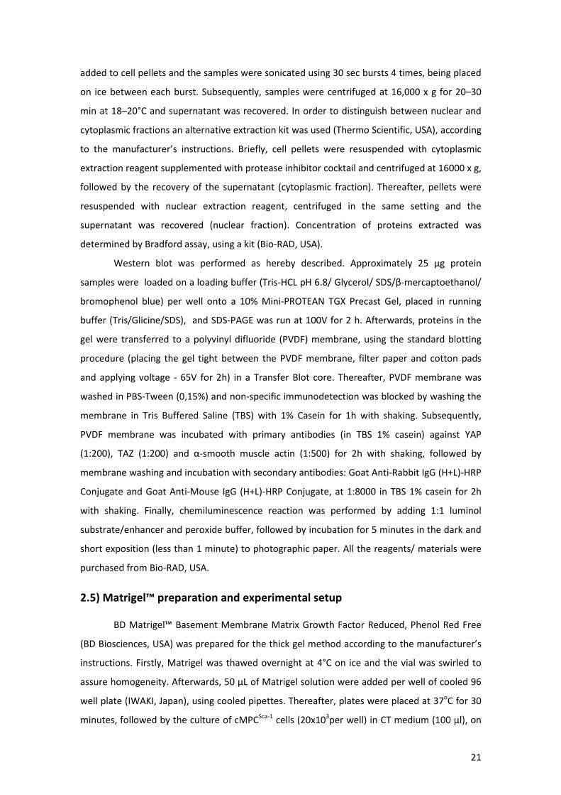
2.5) Matrigel™ preparation and experimental setup ............................................................. 21
2.6) Polyacrylamide gels preparation...................................................................................... 22
2.7) Poly-ε-caprolactone film preparation and stiffness control ............................................ 23
2.8) Alamar Blue® assay ........................................................................................................... 24
2.9) RNA extraction and real time PCR array .......................................................................... 25
viii
2.10) Biostatistical analysis ..................................................................................................... 25
3) Results ............................................................................................................... 26
3.1) YAP/TAZ characterization and regulation by confluence and migration ......................... 26
3.2) Signaling pathways involved in YAP/TAZ localization and activity .................................. 28
3.3) Role of YAP/TAZ in CPC proliferation ............................................................................... 33
3.4) Role of cell tension and YAP/TAZ in CPC cardiac commitment ....................................... 35
3.5) YAP/TAZ activity in substrates displaying different stiffness ........................................... 37
3.5.1) Activity on soft substrates: Matrigel™ ...................................................................... 37
3.5.2) Activity of YAP/TAZ on physiologically relevant surfaces: coated polyacrylamide gels
with controlled stiffness. ..................................................................................................... 39
3.5.3) Activity of YAP/TAZ on stiff surfaces (PCL films) ....................................................... 44
3.6) Activity of YAP/TAZ in response to dynamic changes in surface stiffness ....................... 46
3.7) Influence of cell shape in YAP/TAZ activity ...................................................................... 48
3.8) Differential gene expression with surface stiffness and YAP/TAZ supression ................. 52
4) Conclusions and discussion ................................................................................. 54
4.1) Overview of YAP/TAZ regulation and activity .................................................................. 54
4.2) Future perspectives.......................................................................................................... 56
5) References ......................................................................................................... 62
ix
List of Figures and Tables Figure 1 – Current strategies for cardiac tissue engineering. ....................................................... 5
Figure 2- Stem cell fate determination ......................................................................................... 7
Figure 3 – Mechano-structural factors influence stem cell fate. .................................................. 9
Figure 4 – General mechanotransduction pathways.. ................................................................ 13
Figure 5 – YAP and YAZ structure. ............................................................................................... 14
Figure 6 - Regulation and functions of YAP/TAZ transcriptional modulators ............................. 17
Figure 7– Polyacrylamide gels preparation ................................................................................. 23
Figure 8 - Preparation of cross-linked PCLs ................................................................................. 24
Figure 9 - YAP and TAZ display an overlapping signal both in cytoplasmic and nuclear
expression in cMPCSca-1 ................................................................................................................ 26
Figure 10 – YAP colocalizes with GATA-4 and phosphorylated paxillin in cMPCSca-1 ................... 27
Figure 11 - YAP/TAZ activity is regulated by confluence but not by migration in cMPCSca-1 ....... 27
Figure 12 - Percentage of cMPCSca-1 expressing nuclear YAP in different conditions .................. 28
Figure 13 – Effect of pharmacological inhibitors on cMPCSca-1 morphology and focal adhesion
organization. ............................................................................................................................... 29
Figure 14– Effect of pharmacological inhibitors of defined signaling pathways involved in cell
mechano-transduction on cMPC Sca-1 cells.. ................................................................................ 30
Figure 15 – Effect of pharmacological inhibitors on YAP/TAZ nuclear activity on cMPCSca-1 ..... 30
Figure 16- Percentage of cMPCSca-1 expressing nuclear YAP when treated with pharmacological
inhibitors. .................................................................................................................................... 31
Figure 17 - Analysis of YAP expression in cMPCSca-1 by Western Blot. ........................................ 32
Figure 18- Influence of pharmacological inhibitors’ treatment on cMPCSca-1 migration and zyxin
nuclear shuttling.. ....................................................................................................................... 33
Figure 19- Western Blot for YAP and TAZ expression in control cMPCSca-1 and silenced cells.. .. 34
Figure 20- Role of YAP/TAZ in CPC proliferation ......................................................................... 34
Figure 21 – Role of tension and YAP/TAZ on CPC cardiac commitment ..................................... 36
Figure 22– YAP/TAZ localization in cMPCSca-1 grown on soft substrates ..................................... 37
Figure 23 – Regulation of YAP/TAZ in cMPCSca-1 vasculogenesis, on soft substrates .................. 39
Figure 24 – YAP/TAZ activity on cMPCSca-1 seeded onto polyacrylamide gels. ............................ 41
Figure 25 – Percentage of cMPCSca-1 expressing nuclear YAP or TAZ when seeded onto
polyacrylamide gels coated with either collagen or fibronectin................................................. 42
Figure 26 – Role of YAP/TAZ in cell adhesion onto PA gels. ........................................................ 43
x
Figure 27 – Influence of YAP/TAZ in cellular adhesion to substrates displaying different
stiffnesses .................................................................................................................................... 44
Figure 28 – Activity of YAP/TAZ on stiff PCL films. ...................................................................... 45
Figure 29 – Percentage of cells expressing nuclear YAP when seeded onto PCL films exhibiting
different stiffnesses. .................................................................................................................... 46
Figure 30– YAP/TAZ activity in dynamic surface changes. .......................................................... 47
Figure 31 – Role of YAP/TAZ during dynamic changes of the surface where cMPCSca-1 is seeded.
..................................................................................................................................................... 48
Figure 32 – cMPCSca-1 shape when seeded onto micropatterned surfaces displaying different
adhesive areas. ............................................................................................................................ 49
Figure 33 - Regulation of YAP/TAZ activity by cell shape. ........................................................... 50
Figure 34 -Regulation of cMPCSca-1 cardiac commitment and focal adhesion maturarion by cell
shape. .......................................................................................................................................... 50
Figure 35 – Role of YAP/TAZ on TBX5 activation and vinculin maturarion in micropatterened
surfaces ....................................................................................................................................... 51
Figure 36 - Role of YAP/TAZ in mechanotransduction of cardiac stem/progenitor cells. .......... 56
Table 1 – Differences in gene expression between YAP/TAZ-/- and YAP/TAZ+/+ cMPCSca-1, both
seeded onto TCPS. ....................................................................................................................... 52
Table 2 – Differences in gene expression between cells seeded onto Pa gels displaying 0,5 KPa
and 40 KPa stiffness, respectively. .............................................................................................. 53
Table 3 – Proposed future studies - purpose and strategy. ........................................................ 60

xi
List of Abbreviations
CVD- cardiovascular disease
MI- myocardial infarction
CTE- cardiac tissue engineering
ECM- Extracellular matrix
PEG - poly (ethylene glycol)
MSC - mesenchymal stem cells
ES - embryonic stem
CPC- cardiac stem/progenitor cells
Lin - lineage markers
Sca-1 - Stem Cell Antigen-1
DMSO - dimethyl sulfoxide
PGS - poly glycerol sebacate
PGA - poly glycolic acid
PLA - poly lactic acid
FA- focal adhesions
FAK - focal adhesion kinase
PY- phosphotyrosine
MLC- myosin light chain
MLCK - myosin light chain kinase
ROCK - Rho kinase
YAP - Yes-associated protein
TAZ - transcriptional co-activator with PDZ-binding motif
EMT - epithelial to mesenchymal transition
PBS - Phosphate buffer saline
PA - Polyacrylamide
PCL - Poly-ε-caprolactone
SEM - standard error of mean
TCPS -tissue culture polystyrene


1
1) Introduction
1.1) Cardiac diseases and tissue engineering strategies
Cardiac diseases represent a dominant cause of death worldwide, including low and
middle income countries1. In fact, in 2005, the total number of cardiovascular disease (CVD)
fatalities (mainly coronary heart disease, stroke, and rheumatic heart disease) had increased
globally to 17.5 million from 14.4 million in 1990. The World Health Organization estimates
there will be about 20 million CVD casualties in 2015, accounting for 30 percent of all deaths
worldwide2. Thus, CVD is today the largest single contributor to global mortality and is
predicted to dominate mortality trends in the future3. One of the most common cardiac
pathologies is ischaemic heart disease, which is characterized by reduced blood supply to the
heart muscle. Obstruction of coronary arteries leads to myocardial infarction (MI) with the
associated loss of cardiomyocytes4. MI typically results in fibrotic scar formation and
permanently impaired cardiac function because the myocardial tissue exhibits a very limited
regenerative capacity5.
Currently, the only suitable treatment for heart failure is cardiac transplant, which is
restricted by the shortage of organ donors for transplantation and the process of graft
rejection over time, with the subsequent need for immunosuppressants6. Therefore, new
therapeutic strategies addressing this problem are eagerly sought. As such, cardiac tissue
engineering (CTE) may provide a novel approach to treat heart disease, by replacing, repairing
or regenerating the damaged myocardium using tissue- and/or cell-based strategies6.
In the classical tissue engineering approach, different cell types are combined with
scaffolds or hydrogels (that may present cells growth factors along with mechanical support)
and cultivated in bioreactors6. As such, in order to achieve a successful therapeutic outcome, it
is necessary to optimize all these parameters so that cells are efficiently integrated in the
scaffold prior to transplantation, and their phenotype is adequate for the desired therapy.
Alternatively, when scaffold-free approaches are used, a right number of cells have to be
delivered to the injured site, and cell viability needs to be maintained in order to accomplish
tissue repair/regeneration.
1.1.1) Scaffolds and bioreactors used in cardiac tissue engineering
The scaffolds so far proposed for CTE are very diverse, reflecting the lack of success
achieved so far, which led to the investigation of alternatives. Both naturally-derived and

2
synthetic scaffolds have been suggested as suitable cell delivery systems for CTE
applications5,6. Natural scaffolds include polymers such as alginate7, chitosan8, collagen-based9,
fibrin10, gelatin11 and glycosaminoglycan12, and fibrous or porous scaffolds (that aim at
mimicking the nanoscale structure of the native extracellular matrix (ECM) where cells are
embedded) composed of collagen, elastin, fibronectin and laminin fibers or sponges6.
Synthetic scaffolds typically include poly (glycerol sebacate)(PGS), poly glycolic acid (PGA) and
poly lactic acid (PLA) 6. Moreover, engineered cardiac tissue has also been generated using
scaffolds derived from decellularized native tissues, resulting in acellular structures that
preserve the architecture of native ECM of the heart13. Alternatively, functional engineered
cardiac tissue can be generated by a scaffold-free approach, for instance by a cell sheet
strategy wherein individual cell monolayers or cell sheets were stacked to create thick cardiac
tissue14. Furthermore, cells can be injected intravenously into coronary arteries or directly into
the myocardium. However, this approach revealed several adverse effects15, e.g. extensive cell
loss through the vasculature, very low efficiency of engraftment16 and survival in the
inflammatory environment of infarcted myocardium. Moreover, it has been reported that a
major fraction (approx. 90%) of the delivered cells vanish within a week17.
However, the native myocardium displays a very complex macro- to nano-scale
structural organization, which is essential to proper cardiac function6. Some researchers have
tackled this problem by focusing on the functional improvement of engineered cardiac tissue
by mimicking the aligned structure of the native myocardium18. Topographical cues can
influence properties of cardiomyocytes, including cell attachment, cell hypertrophy,
binucleation, remodelling of ion channels, release of atrial natriuretic peptide, biomechanical
stresses, and structural remodeling19. In order to provide such anisotropic cues, different
strategies have been explored such as nanopatterning of poly (ethylene glycol) (PEG)
hydrogels20, rotary spinning of polymer nanofibers21, and stamping of ECM proteins in lanes on
thin films and hydrogels19. Other scaffolds have been designed with controllable stiffness and
anisotropy, suitable for cardiac tissue engineering22. Micro- and nanoscale techniques were
even used to create stem cell niches to regulate their differentiation towards the myocardial
lineage23,24.
Cells seeded in scaffolds have also been cultured in dynamic conditions using
bioreactors able to provide mechanical stimuli favorable to the desired phenotype and
enhance oxygen flow and the removal of catabolic products from scaffold inner layers. For this,
different setups were used such as perfusion bioreactors that provide adequate oxygen supply
to the scaffold, otherwise impaired in static conditions25. Furthermore, since previous studies
demonstrated the relevance of physical stimuli for the morphology, mechanical properties and
3
function of engineered cardiac tissues6, cyclic mechanical stimulation was used to create thick
cardiac tissues that were implanted in a rat MI model26. In addition, it is essential that cells
within engineered cardiac tissues are capable of synchronously responding to electrical pacing
in order to develop proper excitation-contraction coupling27. As such, bioreactors that provide
electrical field stimulation (e.g. with byphasical pulses28) were used to induce synchronous
beating of cardiomyocytes in engineered cardiac tissues27.
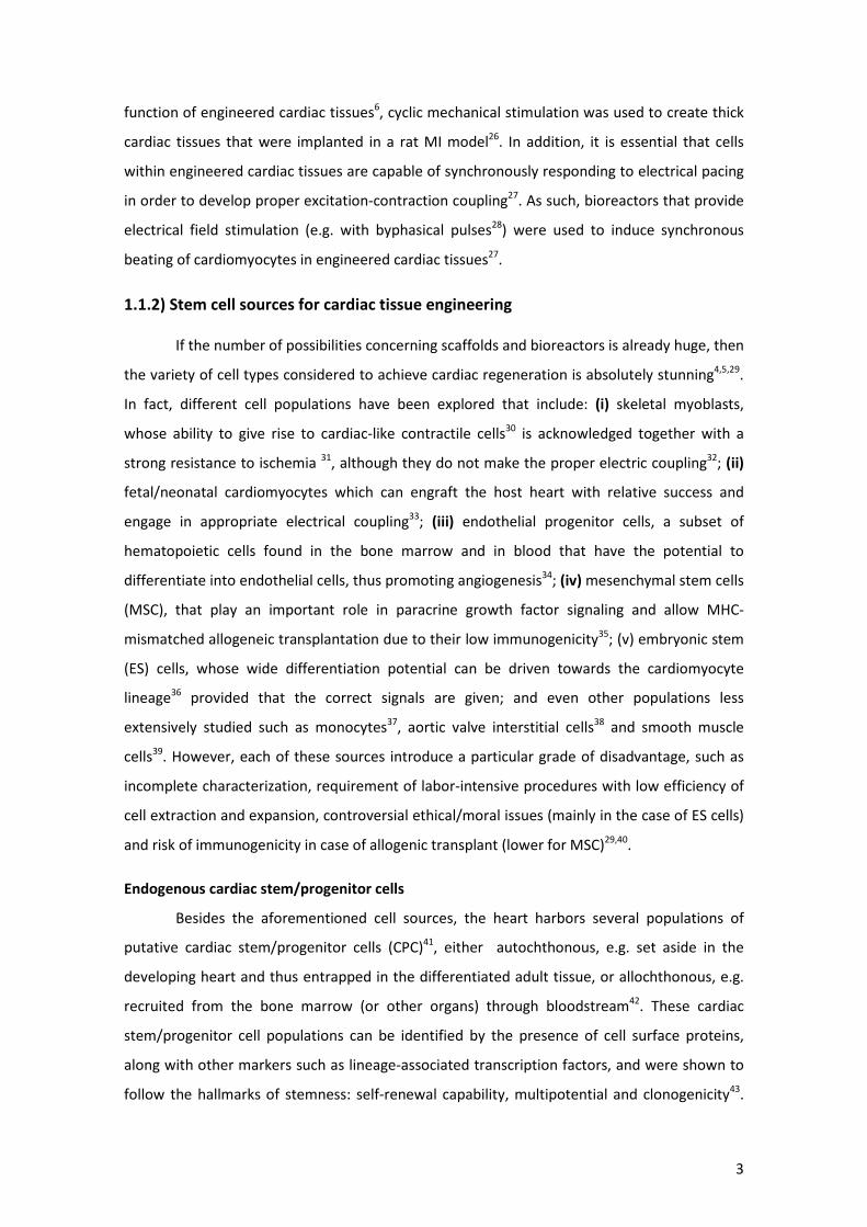
1.1.2) Stem cell sources for cardiac tissue engineering
If the number of possibilities concerning scaffolds and bioreactors is already huge, then
the variety of cell types considered to achieve cardiac regeneration is absolutely stunning4,5,29.
In fact, different cell populations have been explored that include: (i) skeletal myoblasts,
whose ability to give rise to cardiac-like contractile cells30 is acknowledged together with a
strong resistance to ischemia 31, although they do not make the proper electric coupling32; (ii)
fetal/neonatal cardiomyocytes which can engraft the host heart with relative success and
engage in appropriate electrical coupling33; (iii) endothelial progenitor cells, a subset of
hematopoietic cells found in the bone marrow and in blood that have the potential to
differentiate into endothelial cells, thus promoting angiogenesis34; (iv) mesenchymal stem cells
(MSC), that play an important role in paracrine growth factor signaling and allow MHC-
mismatched allogeneic transplantation due to their low immunogenicity35; (v) embryonic stem
(ES) cells, whose wide differentiation potential can be driven towards the cardiomyocyte
lineage36 provided that the correct signals are given; and even other populations less
extensively studied such as monocytes37, aortic valve interstitial cells38 and smooth muscle
cells39. However, each of these sources introduce a particular grade of disadvantage, such as
incomplete characterization, requirement of labor-intensive procedures with low efficiency of
cell extraction and expansion, controversial ethical/moral issues (mainly in the case of ES cells)
and risk of immunogenicity in case of allogenic transplant (lower for MSC)29,40.
Endogenous cardiac stem/progenitor cells
Besides the aforementioned cell sources, the heart harbors several populations of
putative cardiac stem/progenitor cells (CPC)41, either autochthonous, e.g. set aside in the
developing heart and thus entrapped in the differentiated adult tissue, or allochthonous, e.g.
recruited from the bone marrow (or other organs) through bloodstream42. These cardiac
stem/progenitor cell populations can be identified by the presence of cell surface proteins,
along with other markers such as lineage-associated transcription factors, and were shown to
follow the hallmarks of stemness: self-renewal capability, multipotential and clonogenicity43.
4
Moreover, contrarily to what was previously thought, the heart is not a terminally
differentiated organ, i.e. moderate renewal of cardiomyocytes throughout life was already
reported44–46. Nevertheless, this process is very slow46, and in pathological conditions such as
MI, limited numbers of cardiac stem cells, scar formation, an unfavorable milieu associated
with the injured tissue (inflammatory environment and lack of vascular supply), along with
continuous contractile activity of the injured heart may all limit myocardial repair and
regeneration47.
Several CPC populations have been identified within the mammalian heart, and will be
briefly described in this section. (i) Cells expressing the LIM-homeodomain transcription factor
Islet-1 (Isl-1+), that can be identified and isolated from embryonic and post-natal mouse, rat
and human myocardium and were shown to expand in vitro on a cardiac mesenchymal feeder
layer and differentiate into functional cardiomyocytes48. (ii) Cells expressing the Stem Cell
Factor (SCF) receptor c-kit and lacking mature lineage markers (Lin) comprise another CPC
population. Lin- c-Kit+ CPC are self-renewing, clonogenic, and multipotent, giving rise to
myocytes, smooth muscle cells, and endothelial cells. When injected into an ischemic heart,
these cells or their clonal progeny reconstitute contractile myocardium43,49. (iii) CPC expressing
Stem Cell Antigen-1 (Sca-1+) constitute another important cell subset50. The latter can be
isolated from adult mouse hearts (where cardiomyocytes comprise 20–30% of the total
cellular fraction, the remainder including fibroblasts, vascular smooth muscle, and endothelial
cells) by immunomagnetic sorting techniques51. Sca-1+ CPC express early cardiac
transcriptional regulators (e.g. GATA-4, MEF-2c and TEF-1) but do not display cardiac structural
proteins, e.g. α and β myosin heavy chain and cardiac actin51. In vitro differentiation into
cardiomyocytes is modest and, as with the c-Kit+ CPCs, appears to require prior treatment with
demethylating agents such as cytosine analog 5-azacytidine (5-Aza) and dimethyl sulfoxide
(DMSO). Overall, only a small subpopulation of cells express cardiac transcription factors,
display sarcomeric structures, and form spontaneously beating cardiomyocytes with calcium
transients51. Nevertheless, when freshly isolated Sca-1+ cells were injected intravenously into
mice after ischemia-reperfusion, they were shown to target the border zone of the injured
myocardium and differentiate into cardiomyocytes, expressing important cardiac markers such
as sarcomeric α-actin, cardiac Troponin I, and connexin-43, with and without fusing with host
cells52. The presence of a resident cell population having similar characteristics has been
recently described in the human heart53. (iv)An additional multipotent and clonogenic cell
population has been identified within the myocardium as well as in other tissues by their
ability to efflux toxic compounds like Hoechst and rhodamine dyes. This so-called side
population (SP), represent 2% of total cardiac cells and express Sca-1, ATP-binding cassettes
5
(Abcg2), and multi drug resistance-1 (MDR-1), but not c-Kit, CD34, and CD45 (hematopoietic
markers). When injected into an injured heart, SP cells could differentiate into cardiomyocytes,
endothelial cells, or smooth muscle cells54. (v) Finally, CPCs can be clonally expanded from
human myocardial biopsies, as these cells are spontaneously shed from human surgical
specimens and murine heart samples in primary culture and self-organize into spontaneously
beating clusters (cardiospheres). Cells from cardiospheres differentiate in co-culture with
neonatal rat ventricular cardiomyocytes and, when implanted into a model of mouse
infarction, they establish functional cell–cell connections and differentiate into the three main
cardiac lineages (cardiomyocytes, endothelial cells, smooth muscle cells)55.
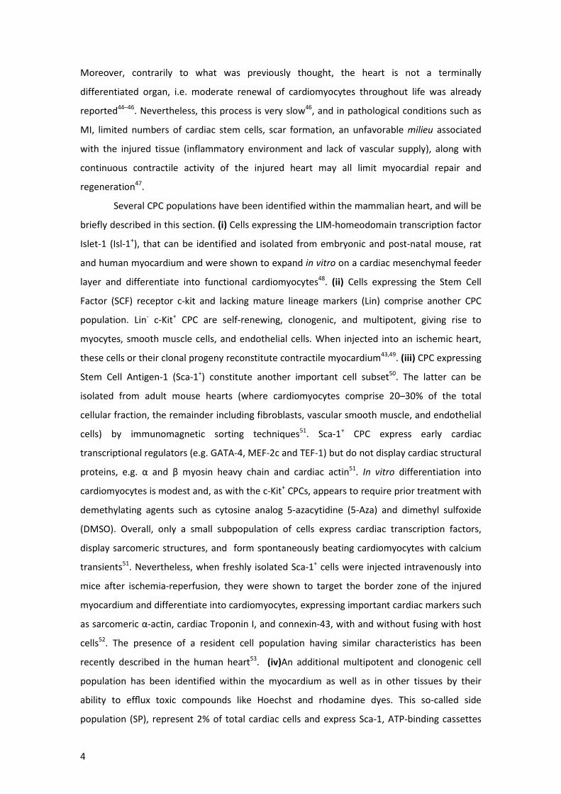
Current strategies for cardiac tissue engineering are summarized in Figure 1.
Figure 1 – Current strategies for cardiac tissue engineering. Cells from different sources are harvested, purified and expanded in culture, and seeded into appropriate scaffolds inside bioreactors that provide several types of stimulation to enhance cell proliferation and integration with the scaffold. Alternatively, scaffold-free systems can be used such as direct injection of cells (intrapericardial, intravenously, among other routes of delivery) and cell sheets. Finally, cells are implanted into the heart.
6
1.2) Stem cell fate determination
1.2.1) Overview
Even though the different CTE strategies explored above seem promising, there are
still serious challenges that need to be solved before translation into clinical practice56,57. The
most obvious is the choice of the appropriate cell source for cell-based therapy. Thus far,
autologous bone marrow derived stem cells, skeletal myoblast, MSCs, and circulating blood-
derived progenitor cells have been used in human clinical trials57 and others using c-Kit+ cardiac
resident stem cells58, or CPC derived from cardiospheres59 are currently in progress60. As stated
above, all the sources currently explored have disadvantages that need to be overcome for a
successful and sustainable cell-therapy, e.g. need to obtain large cell numbers, immunological
rejection in case of allogeneic settings; autologous sources might also be compromised by low
“off-the-shelf” availability57. Furthermore, some bioengineering roadblocks need to be
addressed in order to improve existing CTE strategies, such as (i) insufficient scaffold seeding
with non-proliferative cells (i.e. cardiomyocytes), to the point of tissue-like cell densities17; (ii)
insufficient nutrient delivery to the inner layers of the engineered bio-constructs, for which
perfusion bioreactors are being developed61 and (iii) cell delivery with sufficient engraftment
rates, low death rates of transplanted cells62 and suitable mechanical properties of the scaffold
and/or delivery system56. Finally, the limited understanding of the mechanisms that modulate
the repair of the myocardium, likely related to the control/ modulation of the stem cell fate,
might explain the modest success of the existing strategies57. This problem comprises the
central topic of this thesis and will be explored in the sections below.
It has been proposed that tissue-resident stem cell fate determination (e.g. cell
response - proliferation, self-renewal, differentiation, migration) to different stimuli is
influenced by the concerted integration of multiple parameters. These include overall
extracellular matrix (ECM) composition, along with biochemical (cytokines, hormones, growth
factors), physico-chemical (oxygen tension, pH, temperature) and mechano-structural
(stiffness, rugosity, porosity) signals coming from the microenvironment in which stem cells
reside42 (Figure 2A). Therefore, a complete understanding of the composition of the cardiac
natural microenvironment in physiological conditions as well as insight on how it is affected by
pathological events is required. Most studies agree that progenitor cells (single or clustered)
are settled in specific anatomic and functional locations (niches)42, that mediate signals
maintaining stem cell self-renewal and multipotency or inducing their commitment in
response to tissue specific needs63. It is believed that the heart stem/progenitor cell niches
have only functional and temporal dimensions and are characterized by a dynamically

7
symmetric array of signals delivered by neighboring non-progenitor cells (including fibroblasts
and cardiomyocytes) and ECM soluble and insoluble components (mostly collagen) (Figure 2B).
It has been proposed that stem cells are inherently unstable (i.e., prone to chaotically adopt
multiple phenotypes) and only a complex array of opposite symmetric signals maintains
progenitor cells in a metastable quiescent state, even for long periods of time, preventing
aging processes and preserving their multipotency44. A very small modification in local or long-
range extracellular signaling can attract progenitor cells towards one of many possible states.
Once the symmetry of signals maintaining the metastable equilibrium has been broken by
internal and/or external perturbations, progenitor cells engage in differentiating pathways and
tissue assembly processes in an environment characterized by the aforementioned
parameters, changing with time to generate a dynamic multicomponent template of
microenvironmental symmetry42 (Figure 2C).
Figure 2- Stem cell fate determination. A- CPC fate determination results from the concerted integration of different parameters: biochemical, physico-chemical and mechano-structural factors. B- The native microenvironment of the heart comprehends different cell types such as cardiac stem/progenitor cells, fibroblasts, smooth muscle cells, endothelial cells, and cardiomyocytes, embedded in ECM rich in collagen. It presents different cues to CPC. C- The microenvironment of the heart dynamically changes with time either maintainting CPC in a metastable quiescent state or inducing different cellular responses such as migration proliferation and differentiation. Adapted from
42.
A B
C
8
This concept has been demonstrated in vitro, as the differentiation of different CPC
populations to cardiomyocytes was shown to result from a fine-tuned combination of specific
biological and physical factors, including scaffold geometry and stiffness64,65, rather than solely
biological factors. Since the work developed for this thesis is focused on the influence of
mechano-structural parameters in the determination of CPC fate, these factors will receive
special attention.
1.2.2) Influence of mechano-structural factors
Several mechano-structural factors, including physical signals such as tensile,
compressive, shear, osmotic, and fluid stresses often arising from interactions with the ECM,
control stem cell fate66. Cell shape has been pointed out as a regulator of stem cell fate as, for
instance, growth and differentiation of capillary endothelial cells are in part regulated by ECM-
induced changes in cell shape67. Cell shape can be modulated by several approaches such as
artificial matrices, type of culture system (flattened in 2D vs. round in 3D) and micropatterned
surfaces where the area of cell attachment is controlled (by selective coating with extracellular
matrix proteins in restricted areas)66,68 (Figure 3A). Another example illustrating the relevance
of this parameter is the control of MSC lineage commitment between adipogenic and
osteogenic phenotype in this micropatterning approach69 (different shapes favor alternative
phenotypes).
Another relevant parameter is surface/ECM nanotopography as cells have the ability to
sense micro-and even nanoscale geometric cues from their environment, such as differences
in molecular conformation, surface topography or roughness and fiber diameter66. In fact, the
nanoscale geometry and size of the features of the ECM may have significant effects on a
number of cell properties, such as attachment/adhesion, migration, and proliferation,
although the molecular mechanisms associated with these processes remain to be
elucidated66. As illustrated in Figure 3B, changes in the nanotopography of the substrate may
influence adhesion by altering the degree of cell spreading, along with clustering of cell surface
receptors (integrins) and other cell-adhesion molecules70.
Importantly, the stiffness of the ECM/ surface is also a relevant mechanical parameter
sensed by the cells and modulating stem cell fate. Cells that attach to a substrate have been
shown to exert contractile forces, resulting in tensile stresses in the cytoskeleton71. The
relationship between these forces and the mechanical stiffness, or elasticity, of the ECM may
have a major influence on cell behavior such as migration, apoptosis, and proliferation66. In the
case of stem cell fate, the most illustrative example is the modulation of lineage commitment
and differentiation of MSC by changes in stiffness of the surface where cells were seeded.

9
Human MSC were shown to specify lineage and commit to phenotypes with extreme
sensitivity to tissue-level elasticity, as soft matrices that mimic brain are neurogenic, stiffer
matrices that mimic muscle favor myogenic commitment, and rigid matrices that mimic
collagenous bone proved to be osteogenic72,73.
Finally, other mechanical factors play a role in cell fate determination, as cells are
constantly exposed to a variety of mechanical stimuli through the actions of muscle forces,
gravity, blood flow, and other physical processes74 (Figure 3C).
Figure 3 – Mechano-structural factors influence stem cell fate. A- Cell shape, modulates cellular morphology and actin cytoskeleton tension with downstream responses controlling cell fate. B- Surface/ ECM nanotopography is also a relevant factor as it leads to clustering of integrins and subsequent alterations in cell signaling. C- Cells are exposed to a variety of physical signals including tensile, compressive, shear, osmotic and fluid stresses. These, along with ECM/ surface stiffness induce different degrees of ECM stretching and tension of the cytoskeleton and nucleus through focal adhesions, generating different cell responses such as modulation the activity of osmotically sensitive ion channels. Adapted from
66 and
125.
A
B
C

10
Several examples of mechanical modulation of biological responses can be presented:
dynamic mechanical compression can significantly increase the expression of chondrocytic
markers (e.g., Sox-9, type II collagen, and aggrecan) in bone-marrow-derived MSCs
encapsulated in a hydrogel75. However, changes in mechanical parameters of the environment
affect other factors as well, resulting in complex physical environments that consist of time-
varying stress, strain, fluid flow, and pressure and, potentially, other biophysical changes such
as osmotic pressure which may alter the structure of ECM proteins and the activity of soluble
growth factors and cytokines. As such, it is difficult to isolate the in vivo effects of mechanical
force from indirect effects associated with mechanically driven changes in adhesive cues and/
or paracrine signaling and, as described earlier, subsequent changes in cell shape66.
1.3) Mechanotransduction signaling pathways
Taking into account all the aforementioned mechano-structural factors, a central
question is to understand how cells can perceive such mechanical stimuli in order to generate
an appropriate response. This process is termed mechanotransduction and encompasses the
molecular mechanisms by which cells sense and respond to mechanical changes74. Central
pathways and players will be explored in this section.
1.3.1) Integrins as mechanoreceptors
Travelling from the outside to the inside of the cells, first-line players in
mechanotransduction are transmembrane adhesion receptors that mediate mechanical
coupling between ECM and the cytoskeleton. In this field, integrins are considered
mechanoreceptors of excellence76 as, unlike other receptors, when applied with direct
mechanical stress using magnetic twisting cytometry techniques, cell stiffness (elastic
modulus) increases in direct proportion as the level of stress that was raised71,77 Integrins are
heterodimeric transmembrane proteins formed by an α and a β chain that require Mg2+ or Ca2+
to bind to the ECM proteins78. Integrin regulation and function is very complex as it comprises
a multitude of different families and because they are involved in different cellular process. Yet
for the purpose of the herein thesis, relevance is mainly given to the role of integrins in
mechanotransduction. As such, integrins act as bidirectional signal transducers: interactions
with cytoskeletal adaptor proteins that associate with the cytoplasmic tails of integrins control
the ligand-binding activity of the receptors and, conversely, the extracellular ligand-binding,
clustering or pulling on integrins triggers the recruitment of cytoskeletal adaptor proteins79,80.
11
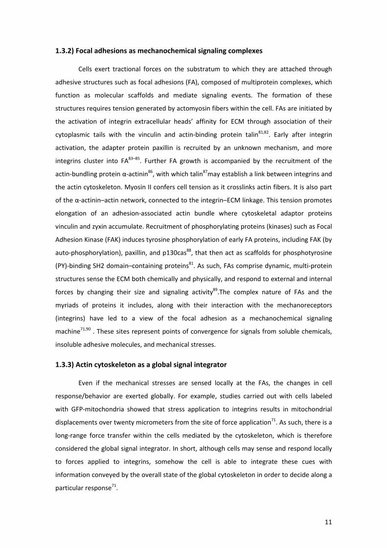
1.3.2) Focal adhesions as mechanochemical signaling complexes
Cells exert tractional forces on the substratum to which they are attached through
adhesive structures such as focal adhesions (FA), composed of multiprotein complexes, which
function as molecular scaffolds and mediate signaling events. The formation of these
structures requires tension generated by actomyosin fibers within the cell. FAs are initiated by
the activation of integrin extracellular heads’ affinity for ECM through association of their
cytoplasmic tails with the vinculin and actin-binding protein talin81,82. Early after integrin
activation, the adapter protein paxillin is recruited by an unknown mechanism, and more
integrins cluster into FA83–85. Further FA growth is accompanied by the recruitment of the
actin-bundling protein α-actinin86, with which talin87may establish a link between integrins and
the actin cytoskeleton. Myosin II confers cell tension as it crosslinks actin fibers. It is also part
of the α-actinin–actin network, connected to the integrin–ECM linkage. This tension promotes
elongation of an adhesion-associated actin bundle where cytoskeletal adaptor proteins
vinculin and zyxin accumulate. Recruitment of phosphorylating proteins (kinases) such as Focal
Adhesion Kinase (FAK) induces tyrosine phosphorylation of early FA proteins, including FAK (by
auto-phosphorylation), paxillin, and p130cas88, that then act as scaffolds for phosphotyrosine
(PY)-binding SH2 domain–containing proteins81. As such, FAs comprise dynamic, multi-protein
structures sense the ECM both chemically and physically, and respond to external and internal
forces by changing their size and signaling activity89.The complex nature of FAs and the
myriads of proteins it includes, along with their interaction with the mechanoreceptors
(integrins) have led to a view of the focal adhesion as a mechanochemical signaling
machine71,90 . These sites represent points of convergence for signals from soluble chemicals,
insoluble adhesive molecules, and mechanical stresses.
1.3.3) Actin cytoskeleton as a global signal integrator
Even if the mechanical stresses are sensed locally at the FAs, the changes in cell
response/behavior are exerted globally. For example, studies carried out with cells labeled
with GFP-mitochondria showed that stress application to integrins results in mitochondrial
displacements over twenty micrometers from the site of force application71. As such, there is a
long-range force transfer within the cells mediated by the cytoskeleton, which is therefore
considered the global signal integrator. In short, although cells may sense and respond locally
to forces applied to integrins, somehow the cell is able to integrate these cues with
information conveyed by the overall state of the global cytoskeleton in order to decide along a
particular response71.
12
FAs are generally linked by actin stress fibers that are in a state of balanced isometric
contraction, as some proteins such as non-muscle myosin II crosslink actin fibers conferring
them additional stability and tension91. The force transmitted to sites of adhesion derives from
the interaction of myosin II with actin filaments that attach to these sites. Myosin II activity is
regulated by myosin light-chain (MLC) phosphorylation, which is either directly positively
regulated by MLC kinase (MLCK) or Rho kinase (ROCK) or negatively regulated by MLC
phosphatase, which is itself phosphorylated and inhibited by ROCK. MLC phosphorylation
activates myosin, resulting in increased contractility and transmission of tension to sites of
adhesion92. Interplay of all these pathways and signaling proteins (along with other structures
not reviewed in this thesis such as microtubules) makes mechanotransduction a very complex
field so that more in depth studies are required in order to fully understand cell mechano-
biology. As an example, clustering of integrins into focal adhesions and focal complexes is
regulated by the actin cytoskeleton. In turn, actin dynamics are governed by Rho family
GTPases. Integrin-mediated adhesion activates these GTPases, triggering assembly of stress
fibers80.
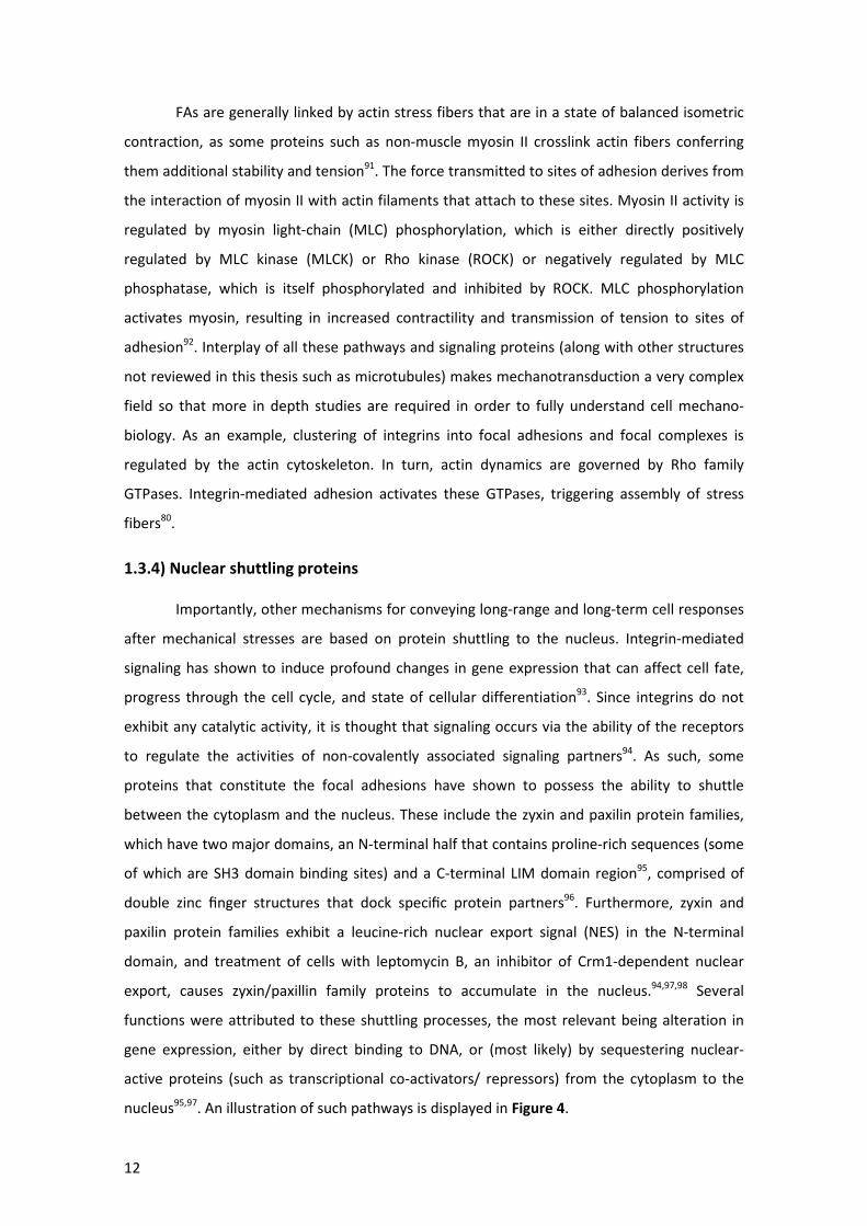
1.3.4) Nuclear shuttling proteins
Importantly, other mechanisms for conveying long-range and long-term cell responses
after mechanical stresses are based on protein shuttling to the nucleus. Integrin-mediated
signaling has shown to induce profound changes in gene expression that can affect cell fate,
progress through the cell cycle, and state of cellular differentiation93. Since integrins do not
exhibit any catalytic activity, it is thought that signaling occurs via the ability of the receptors
to regulate the activities of non-covalently associated signaling partners94. As such, some
proteins that constitute the focal adhesions have shown to possess the ability to shuttle
between the cytoplasm and the nucleus. These include the zyxin and paxilin protein families,
which have two major domains, an N-terminal half that contains proline-rich sequences (some
of which are SH3 domain binding sites) and a C-terminal LIM domain region95, comprised of
double zinc finger structures that dock specific protein partners96. Furthermore, zyxin and
paxilin protein families exhibit a leucine-rich nuclear export signal (NES) in the N-terminal
domain, and treatment of cells with leptomycin B, an inhibitor of Crm1-dependent nuclear
export, causes zyxin/paxillin family proteins to accumulate in the nucleus.94,97,98 Several
functions were attributed to these shuttling processes, the most relevant being alteration in
gene expression, either by direct binding to DNA, or (most likely) by sequestering nuclear-
active proteins (such as transcriptional co-activators/ repressors) from the cytoplasm to the
nucleus95,97. An illustration of such pathways is displayed in Figure 4.
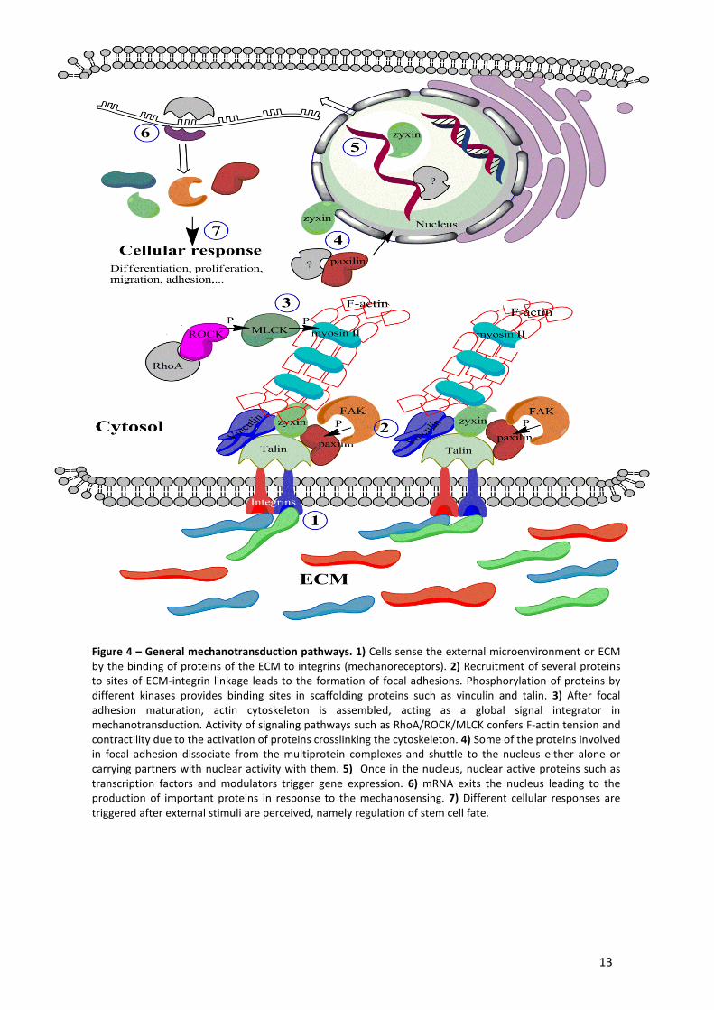
13
Figure 4 – General mechanotransduction pathways. 1) Cells sense the external microenvironment or ECM by the binding of proteins of the ECM to integrins (mechanoreceptors). 2) Recruitment of several proteins to sites of ECM-integrin linkage leads to the formation of focal adhesions. Phosphorylation of proteins by different kinases provides binding sites in scaffolding proteins such as vinculin and talin. 3) After focal adhesion maturation, actin cytoskeleton is assembled, acting as a global signal integrator in mechanotransduction. Activity of signaling pathways such as RhoA/ROCK/MLCK confers F-actin tension and contractility due to the activation of proteins crosslinking the cytoskeleton. 4) Some of the proteins involved in focal adhesion dissociate from the multiprotein complexes and shuttle to the nucleus either alone or carrying partners with nuclear activity with them. 5) Once in the nucleus, nuclear active proteins such as transcription factors and modulators trigger gene expression. 6) mRNA exits the nucleus leading to the production of important proteins in response to the mechanosensing. 7) Different cellular responses are triggered after external stimuli are perceived, namely regulation of stem cell fate.

14
In addition, other mechanotransduction pathways exist based on conformational
changes of cell surface receptors mediated by stretching induced by external forces. These
include direct stretching of integrins, deformation of gap junctions containing calcium-sensitive
stretch receptors, alteration of ion channel permeability on the cell membrane and activation
of growth factor receptor signaling cascades in the absence of the ligands99. They converge on
the activation of phosphorelay systems (Mitogen-activated protein (MAP) kinases), with
subsequent downstream changes in gene expression. However, because the latter do not
constitute the aim of this thesis so they will not be further reviewed100.
1.4) YAP and TAZ transcriptional modulators
1.4.1) Structural characterization
Identifying key molecules involved in stem cell determination is crucial for achieving a
higher understanding of stem cell biology to be harnessed in tissue engineering therapies. In
this context, Yes-associated protein (YAP) and highly related (structurally and functionally)
transcriptional co-activator with PDZ-binding motif (TAZ) appear to be promising candidates101.
Structurally, both TAZ and YAP contain (i) a 14-3-3 binding motif; (ii) a single or duplicated WW
domains (1 in TAZ, 2 in YAP); (iii) an extended coiled-coiled region within a larger
transcriptional regulatory domain; (iv) multiple sites of phosphorylation, and (v) a C-terminal
motif that can interact with PDZ domain-containing proteins102, as represented in Figure 5. The
WW domains of TAZ and YAP bind strongly to the sequence motif Pro-Pro-X-Tyr, which can be
found within the regulatory regions of a large number of transcription factors, suggesting that
TAZ and YAP may function as general transcriptional modulators during the execution of many
developmental programs101,102. The C-terminus of YAP/TAZ likely interacts directly with core
transcriptional machinery to stimulate gene expression. Moreover, the PDZ motifs allow
interaction of YAP/TAZ with several transcription factors such as: Runx2 (runt-related
transcription factor 2 103, PPAR (peroxisome proliferator-activated receptor104), TBX5 (T-box
transcription factor 5, TBX5)105 , TEADs (TEA domain family members106), with subsequent
transcriptional modulating activity.
Figure 5 – YAP and YAZ structure. Both proteins have WW domains, an extended coiled-coil region, multiple sites of phosphorylation, a 14-3-3 binding motif and a C-terminal motif that can interact with PDZ domain-containing proteins. Adapted from
102
15
1.4.2) Function in different cell types
The discovery of the YAP/TAZ interactome led to the finding of the involvement of
these molecules in different processes, either acting as transducers of Hippo pathway or
responding to other stimuli. These include: (i) stem cell fate determination: favoring MSC
osteogenic differentiation rather than adipogenic104 and enhancing MyoD-induced myogenic
differentiation in myoblasts107; (ii) stem cell proliferation: inducing expansion of intestinal stem
cells108, restraining cardiomyocyte proliferation and heart size109 and maintaining human
embryonic stem (hES) cell self-renewal110; (iii) in cancer: overexpression of YAP/TAZ in normal
mammary cells causes morphologic changes characteristic of tumorigenesis, promoting cell
migration and invasion111, with enhanced proliferation and epithelial to mesenchymal
transition (EMT)112,113. Moreover, the physiological function of TAZ could be investigated in
vivo by the generation of knockout mice114,115, which, surprisingly, just suffer a minor skeletal
defect though TAZ plays a crucial role in MSC differentiation. Nonetheless, TAZ knockout (KO)
mice develop two severe abnormalities: polycystic kidney disease and emphysema, implicating
TAZ role in renal and lung development114,115.
1.4.3) Regulation by cellular pathways
Regulation of YAP/TAZ activity is extremely complex due to the involvement of several
signaling pathways and external stimuli, and is represented in Figure 6. YAP/TAZ nuclear
activity is negatively regulated by the Hippo tumor supressor pathway, composed by a kinase
cascade wherein MST1/2 (macrophage stimulating 1 /2), complexed with its regulatory subunit
SAV1, phosphorylates and activates LATS1/2 in complex with its regulatory subunit MOB1,
resulting in phosphorylation of YAP/TAZ116. When phosphorylated, YAP/TAZ bind to 14-3-3
proteins and remain sequestered in the cytoplasm112, thus cannot go to the nucleus where
transcriptional regulation occurs. On the other hand, dephosphorylation of YAP/TAZ by PP1
(phosphoprotein phosphatase 1) coupled with ASPP2 (ankyrin repeat-containing, SH3 domain-
containing, and proline-rich region-containing protein 2) prevents 14-3-3 sequestration and
enhance YAP/TAZ nuclear levels, activating these proteins117. Highlighting the complexity of
this regulation, α-catenin prevents YAP/TAZ dephosphorylation by PP1/ASPP2, negatively
regulating YAP/TAZ activity,e.g. impairing epidermal stem cell proliferation118. Furthermore,
YAP/TAZ protein stability is controlled by a phosphodegron recognized by the F-box protein β-
TrCP (beta-Transducin repeat containing protein) and ubiquitylated by the SCF/CRL1β-TrCP E3
ligase. Phosphorylation of a phosphodegron YAP/TAZ by LATS primes it for further
phosphorylation by CK1ε and subsequent binding by β -TrCP, leading to YAP/TAZ proteasomal

16
degradation119,120. Cell confluence is a known negative regulator of YAP/TAZ activity in
vitro, both by activating Hippo pathway, and through formation of protein complexes in tight
or adherens junctions (structures connecting adjacent cells), where interacting partners of
YAP/TAZ such as Angiomiotin121 and zonula occludens proteins122 can retain the proteins in
their vicinity impairing their activity in the nucleus. Nevertheless, a cytoplasmic function of
YAP/TAZ has also been documented, due to their interaction (via PDZ domain) with proteins
belonging to other pathways, such as in Wnt signaling123,124.
1.4.4) Role in mechanotransduction
Importantly, YAP and TAZ activity has also been shown to be modulated by external
mechanical contexts. In fact, Dupont et al125 have studied extensively the role of YAP/TAZ in
mechanotransduction, and concluded that in hMSC, YAP/TAZ are predominantly nuclear when
cells are seeded onto stiffer substrates, while cells on softer substrates display cytoplasmic
YAP/TAZ. Importantly, inhibition of Rho or disruption of actin cytoskeleton by pharmacological
treatment inhibited YAP/TAZ activity on stiff substrates, demonstrating their requirement for
YAP/TAZ to act as mediators of mechanical signals. In this stem cell type, both active ROCK and
non-muscle myosin II- mediated cell tension were required for YAP/TAZ to be nuclear, as
shown by treatment with the respective inhibitors. Furthermore, the response of YAP/TAZ to
the matrix rigidity seems to be independent from the Hippo pathway, pointing out the
existence of an independent mechanism125. Cell shape has shown to modulate YAP/TAZ
intracellular localization and activity, as concluded through the use of micropatterned
substrates with either large islands that cause individual cells to adopt a flat, spread
morphology or small islands that induce a round, compact shape. These experiments
revealed that spread-out cells have higher levels of nuclear YAP than round cells125. In
addition, cell attachment also interferes with YAP/TAZ activity, as detached cells have lower
YAP/TAZ activity126. Moreover, actin cytoskeleton has shown to regulate YAP/TAZ activity, as
expected by its involvement in mechanotransduction, where it is considered the global signal
integrator. Experiments in mammalian cells showed that the amount of F-actin was
proportionally correlated with YAP/TAZ activity: (i) treatment of cells with Cytochalasin D
inhibits F-actin polymerization and prevents YAP/TAZ translocation to the nucleus127; (ii) F-
actin stress fibers are abundant in cells at low cell density and rare in high-density cultures
and flat cells have more stress fibers than round cells, consistent with the observed
YAP/TAZ activity in confluence vs. non-confluence and in different cell shapes. Altogether,
YAP/TAZ respond to different conditions of the extracellular environment and its regulation
comprises several mechanisms, as shown in Figure 6.
17
Cytoplasm
Nucleus
Cell membrane
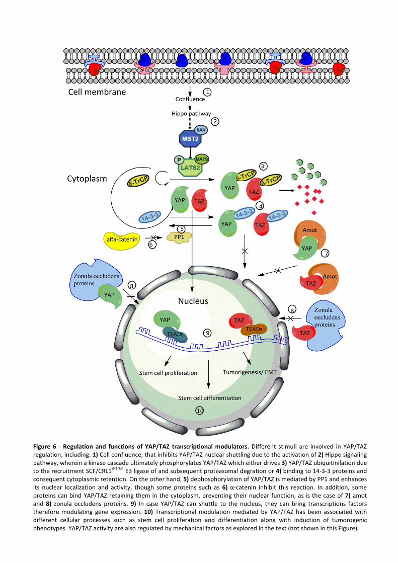
Figure 6 - Regulation and functions of YAP/TAZ transcriptional modulators. Different stimuli are involved in YAP/TAZ regulation, including: 1) Cell confluence, that inhibits YAP/TAZ nuclear shuttling due to the activation of 2) Hippo signaling pathway, wherein a kinase cascade ultimately phosphorylates YAP/TAZ which either drives 3) YAP/TAZ ubiquitinilation due to the recruitment SCF/CRL1
β-TrCP E3 ligase of and subsequent proteasomal degration or 4) binding to 14-3-3 proteins and
consequent cytoplasmic retention. On the other hand, 5) dephosphorylation of YAP/TAZ is mediated by PP1 and enhances its nuclear localization and activity, though some proteins such as 6) α-catenin inhibit this reaction. In addition, some proteins can bind YAP/TAZ retaining them in the cytoplasm, preventing their nuclear function, as is the case of 7) amot and 8) zonula occludens proteins. 9) In case YAP/TAZ can shuttle to the nucleus, they can bring transcriptions factors therefore modulating gene expression. 10) Transcriptional modulation mediated by YAP/TAZ has been associated with different cellular processes such as stem cell proliferation and differentiation along with induction of tumorogenic phenotypes. YAP/TAZ activity are also regulated by mechanical factors as explored in the text (not shown in this Figure).
18
1.5) Objective of the experimental work
Taking into account the reviewed topics in this introductory section, the aim of the
experimental work developed for this dissertation is the dissection of molecular pathways
associated with YAP/TAZ nuclear shuttling in mechanotransduction, in the cardiac
stem/progenitor cell context. Factors like elasticity, nanorugosity and three-dimensional pore
geometry are known to affect cell cytoskeleton organization, which is responsible of a shift in
intracellular signal transduction and, eventually, in changes in gene activation. Therefore, it is
conceivable that the expression of genes involved in stem/progenitor cell maintenance,
survival or differentiation could be switched off or on in response to slight changes in the
mechano-physical signals coming from the microenvironment. As such, the knowledge
obtained with this work is expected to shed some light into cardiac stem cell fate
determination in relation with YAP/TAZ activity as a transducer of these mechanostructural
effects. Bearing this in mind, one central question to be addressed is “how can cells perceive
their microenvironment?”
In brief, this work was performed in the cardiac context by the use of a cardiac
progenitor cell line characterized by the expression of Sca-1 cell surface protein hereafter
referred to as cMPCSca-1(Freire AG, Nascimento DS, Forte G et al., manuscript). The
experimental plan was conceived to address how is YAP/TAZ activity regulated in
mechanotansduction, in terms of substrate stiffness, nanotopography, cell shape and
interacting proteins. As such, nuclear vs. cytoplasmic expression of YAP/TAZ was evaluated,
with subsequent downstream responses related with cardiac stem/progenitor cell biology such
as proliferation and differentiation/commitment to mature cardiac cell types, resorting to
immunostaining relevant markers, Western blot and real time PCR for assessing protein and
gene expression. Influence of migration in YAP/TAZ activity in cMPCSca-1 was also evaluated
using the in vitro wound healing assay, whereas involvement in cell adhesion was assessed
using Alamar blue® assay. Involvement of signaling pathways into YAP/TAZ activity was also
sought, and was analyzed by the use of specific chemical inhibitors.
19
2) Materials and Methods
2.1) Cell culture, transfection and treatment with pharmacological inhibitors
Murine Sca-1+ cardiac stem/progenitor cell line (cMPCSca-1) were grown in DMEM:F12
1:1 mixture with 15mM HEPES and L-Glutamine (Lonza, Switzerland) supplemented with 10%
Fetal Bovine Serum (FBS) (Equitech-BioInc., Kerrville, TX), 200 U/ml Penicillin and 200 µg/ml
streptomycin (Invitrogen, USA), ascorbic acid (50µg/ml) (WAKO chemicals, Japan), 0,2% EGM-2
basal medium (Lonza, Switzerland) and 0,1 mM MEM Non-essential aminoacids (Invitrogen,
USA), hereafter referred to as CT medium. Human mesenchymal stem cells (hMSC) were
grown in Mesenchymal stem cell growth medium (MSCGM) composed of MSC Basal medium
(Lonza, Switzerland), supplemented with Single Quots Kit (Lonza, Switzerland).
For the experiments done in tissue culture polystyrene (TCPS), cMPCSca-1 were
detached using 0.25% Trypsin 1mM EDTA (Nacalai Tesque, Japan) and subcultured in CT
medium onto plates of different sizes: 8 chamber-slides (BD Biosciences, USA), 24 well plates
(IWAKI, Japan), 6-well plates (Nuclon Delta,USA) or micropatterned adhesive islands slides
(Cytoo, USA). This was done either in non-confluent (7,5 x10³ cells/cm2) or confluent
conditions (25x103 cells/cm2). For cMPCSca-1 seeding onto polyacrylamide gels or poly ε-
caprolactone films, low cell adhesion 24 well plates (IWAKI, Japan) were used after pre-coating
of gels or films with CT medium for 2h. The cells were analyzed at 3h, 5h, 8h and 24h.
For the short-interfering RNA (siRNA) transfections, 7,5x10³ cells/cm2 were seeded in
antibiotic-free CT medium onto 24 well plates, 18h prior to transfection. Cells were washed
with PBS 1h before the transfection, YAP siRNA duplexes (1 µg/ well) and transfection reagent
were mixed in a 1:1 (v/v) ratio and added at 10% in transfection medium. All these reagents
were purchased from Santa Cruz Biotechnology, USA. Thereafter, growth medium was
replaced with transfection medium containing siRNA complexes comprising 20% of the final
volume. After 7h, CT medium supplemented with 2x normal FBS and antibiotics concentration
was added to the cells. Thereafter, cMPCSca-1 were incubated for 24h, followed by replacement
of transfection medium by regular growth medium. YAP and TAZ expression was subsequently
assayed by Western Blot 24h after last medium replacement, and/or cMPCSca-1 YAP/TAZ -/- were
subcultured afterwards.
Cytochalasin D (1mg/ml) (Sigma, USA), Y27632 (50µM) (WAKO chemicals, Japan) and
Blebbistatin (50µM) pharmacological inhibitors (Merck Biosciences, USA) were dissolved in PBS
and added to growth medium (10% of the final treatment volume), for 4h. Leptomycin B
(10ng/ml) (Sigma, USA) was dissolved in 70% methanol (Wako chemicals, Japan) whereas
okadaic acid (0,7 nM) (Sigma, USA) was dissolved in DMSO (Wako chemicals, Japan). Both were
20
added 100x concentrated to the medium (1% of the final treatment volume), for 2h.
Appropriate controls were performed using methanol and dimethyl sulfoxide alone.
2.2) In vitro wound healing assay
In order to analyze the effect of migration in regulating YAP/TAZ localization and
activity, an established in vitro migration assay was performed. Herein, cells are grown until
confluence, followed by a scratch (wound) made with a pipette tip on the cell monolayer,
allowing cells to migrate and close the wound. Different time points (2h, 4h and 8h) were
analyzed
2.3) Immunofluorescence staining and confocal microscopy
Cells seeded on every setup (TCPS well plates, polyacrylamide gels, PCL films,
micropatterned surfaces) were fixed with 4% paraformaldehyde (Sigma-Aldrich, USA) in PBS
for 15 minutes at room temperature, and permeabilized with 0.1% Triton X-100 (Sigma-
Aldrich, USA) in PBS for 2 minutes at room temperature. Depending on the marker analyzed,
cells were incubated for 1,5h with primary antibodies against: YAP (1:200), TAZ (1:200),
Vinculin (1:100); 405-conjugated Alexa Fluor® Zyxin (1:100), phosphoPaxillin (1:150)
(Invitrogen, USA), GATA-4 (1:200), TBX5 (1:300), Ki67 (1:100) (Abcam, UK). Unless stated
otherwise in the text above, all antibodies were purchased from Santa Cruz Biotechnology,
USA. The appropriate fluorophore-conjugated secondary antibodies (1:300) were as follows:
Alexa fluor 488 goat anti mouse; Alexa fluor 488 goat anti rabbit; Alexa fluor 546 goat anti
mouse; Alexa fluor 546 goat anti rabbit; Alexa fluor 488 donkey anti goat; Alexa fluor 546
donkey anti goat. All antibodies were dissolved in bovine serum albumin (Wako chemicals,
Japan) 1% in PBS, to block unspecific binding. Nuclei were counterstained with 4-6-diamidino-
2-phenylindole (DAPI, Sigma-Aldrich, USA). Moreover, filamentous actin was stained using
rhodamine phalloidin (1:500, Invitrogen, USA). The images were taken using a Leica DMRB
microscope equipped with a digital camera or using a confocal laser scanner microscope (LEICA
SF5), after excitation at 405 nm, 488 nm, and 543 nm wavelengths for blue, green, and red
channels acquisition, respectively. All the pictures obtained were processed both with Image J
and Adobe Photoshop CS5 (USA).
2.4) Protein extraction, cell fractioning and Western Blot
Protein extraction of total cell lysates was performed using a Ready prep extraction kit
(Bio-RAD, USA), according to the manufacturer’s instructions. In short, ready prep protein
extraction buffer supplemented with protease inhibitor cocktail (Thermo Scientific, USA) was
21
added to cell pellets and the samples were sonicated using 30 sec bursts 4 times, being placed
on ice between each burst. Subsequently, samples were centrifuged at 16,000 x g for 20–30
min at 18–20°C and supernatant was recovered. In order to distinguish between nuclear and
cytoplasmic fractions an alternative extraction kit was used (Thermo Scientific, USA), according
to the manufacturer’s instructions. Briefly, cell pellets were resuspended with cytoplasmic
extraction reagent supplemented with protease inhibitor cocktail and centrifuged at 16000 x g,
followed by the recovery of the supernatant (cytoplasmic fraction). Thereafter, pellets were
resuspended with nuclear extraction reagent, centrifuged in the same setting and the
supernatant was recovered (nuclear fraction). Concentration of proteins extracted was
determined by Bradford assay, using a kit (Bio-RAD, USA).
Western blot was performed as hereby described. Approximately 25 µg protein
samples were loaded on a loading buffer (Tris-HCL pH 6.8/ Glycerol/ SDS/β-mercaptoethanol/
bromophenol blue) per well onto a 10% Mini-PROTEAN TGX Precast Gel, placed in running
buffer (Tris/Glicine/SDS), and SDS-PAGE was run at 100V for 2 h. Afterwards, proteins in the
gel were transferred to a polyvinyl difluoride (PVDF) membrane, using the standard blotting
procedure (placing the gel tight between the PVDF membrane, filter paper and cotton pads
and applying voltage - 65V for 2h) in a Transfer Blot core. Thereafter, PVDF membrane was
washed in PBS-Tween (0,15%) and non-specific immunodetection was blocked by washing the
membrane in Tris Buffered Saline (TBS) with 1% Casein for 1h with shaking. Subsequently,
PVDF membrane was incubated with primary antibodies (in TBS 1% casein) against YAP
(1:200), TAZ (1:200) and α-smooth muscle actin (1:500) for 2h with shaking, followed by
membrane washing and incubation with secondary antibodies: Goat Anti-Rabbit IgG (H+L)-HRP
Conjugate and Goat Anti-Mouse IgG (H+L)-HRP Conjugate, at 1:8000 in TBS 1% casein for 2h
with shaking. Finally, chemiluminescence reaction was performed by adding 1:1 luminol
substrate/enhancer and peroxide buffer, followed by incubation for 5 minutes in the dark and
short exposition (less than 1 minute) to photographic paper. All the reagents/ materials were
purchased from Bio-RAD, USA.
2.5) Matrigel™ preparation and experimental setup
BD Matrigel™ Basement Membrane Matrix Growth Factor Reduced, Phenol Red Free
(BD Biosciences, USA) was prepared for the thick gel method according to the manufacturer’s
instructions. Firstly, Matrigel was thawed overnight at 4°C on ice and the vial was swirled to
assure homogeneity. Afterwards, 50 μL of Matrigel solution were added per well of cooled 96
well plate (IWAKI, Japan), using cooled pipettes. Thereafter, plates were placed at 37oC for 30
minutes, followed by the culture of cMPCSca-1 cells (20x103per well) in CT medium (100 µl), on
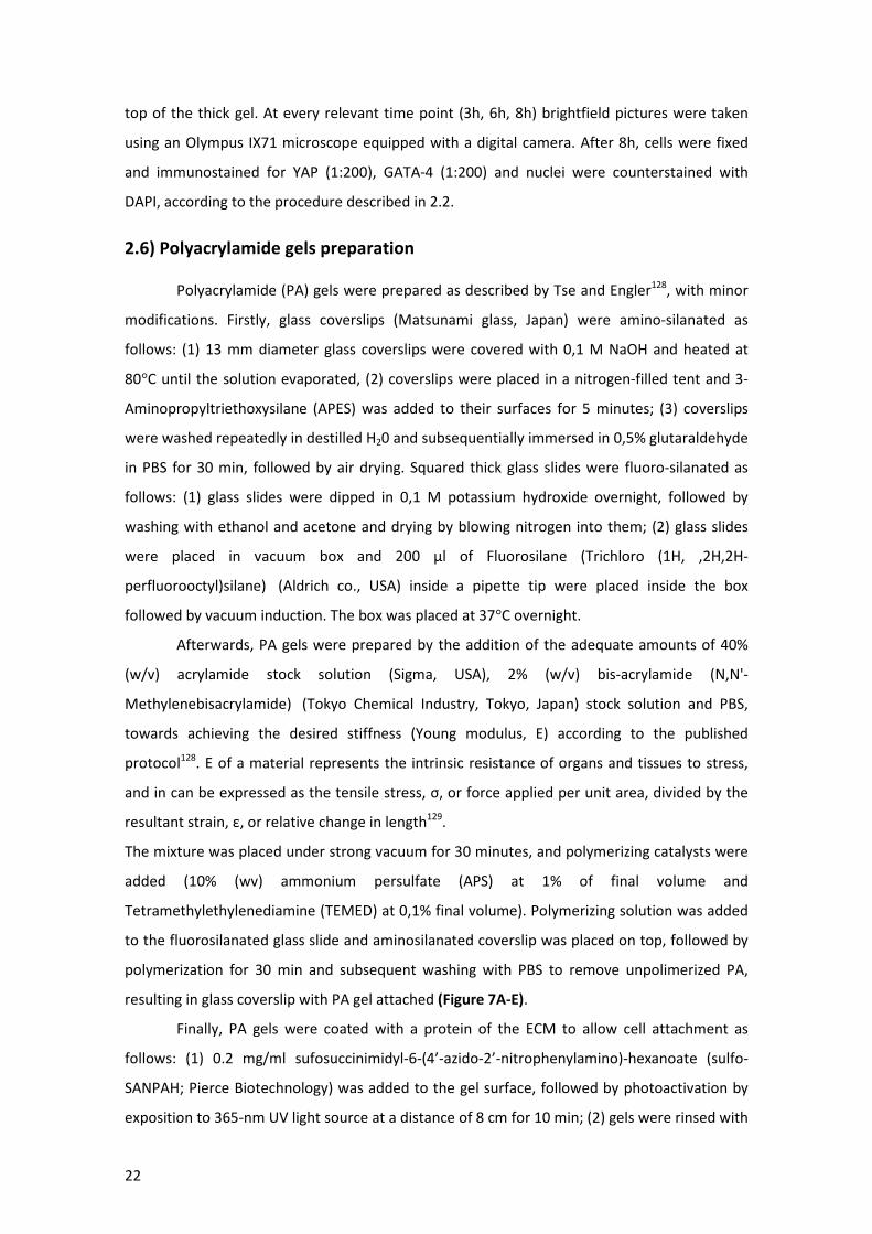
22
top of the thick gel. At every relevant time point (3h, 6h, 8h) brightfield pictures were taken
using an Olympus IX71 microscope equipped with a digital camera. After 8h, cells were fixed
and immunostained for YAP (1:200), GATA-4 (1:200) and nuclei were counterstained with
DAPI, according to the procedure described in 2.2.
2.6) Polyacrylamide gels preparation
Polyacrylamide (PA) gels were prepared as described by Tse and Engler128, with minor
modifications. Firstly, glass coverslips (Matsunami glass, Japan) were amino-silanated as
follows: (1) 13 mm diameter glass coverslips were covered with 0,1 M NaOH and heated at
80°C until the solution evaporated, (2) coverslips were placed in a nitrogen-filled tent and 3-
Aminopropyltriethoxysilane (APES) was added to their surfaces for 5 minutes; (3) coverslips
were washed repeatedly in destilled H20 and subsequentially immersed in 0,5% glutaraldehyde
in PBS for 30 min, followed by air drying. Squared thick glass slides were fluoro-silanated as
follows: (1) glass slides were dipped in 0,1 M potassium hydroxide overnight, followed by
washing with ethanol and acetone and drying by blowing nitrogen into them; (2) glass slides
were placed in vacuum box and 200 µl of Fluorosilane (Trichloro (1H, ,2H,2H-
perfluorooctyl)silane) (Aldrich co., USA) inside a pipette tip were placed inside the box
followed by vacuum induction. The box was placed at 37°C overnight.
Afterwards, PA gels were prepared by the addition of the adequate amounts of 40%
(w/v) acrylamide stock solution (Sigma, USA), 2% (w/v) bis-acrylamide (N,N'-
Methylenebisacrylamide) (Tokyo Chemical Industry, Tokyo, Japan) stock solution and PBS,
towards achieving the desired stiffness (Young modulus, E) according to the published
protocol128. E of a material represents the intrinsic resistance of organs and tissues to stress,
and in can be expressed as the tensile stress, σ, or force applied per unit area, divided by the
resultant strain, ε, or relative change in length129.
The mixture was placed under strong vacuum for 30 minutes, and polymerizing catalysts were
added (10% (wv) ammonium persulfate (APS) at 1% of final volume and
Tetramethylethylenediamine (TEMED) at 0,1% final volume). Polymerizing solution was added
to the fluorosilanated glass slide and aminosilanated coverslip was placed on top, followed by
polymerization for 30 min and subsequent washing with PBS to remove unpolimerized PA,
resulting in glass coverslip with PA gel attached (Figure 7A-E).
Finally, PA gels were coated with a protein of the ECM to allow cell attachment as
follows: (1) 0.2 mg/ml sufosuccinimidyl-6-(4’-azido-2’-nitrophenylamino)-hexanoate (sulfo-
SANPAH; Pierce Biotechnology) was added to the gel surface, followed by photoactivation by
exposition to 365-nm UV light source at a distance of 8 cm for 10 min; (2) gels were rinsed with
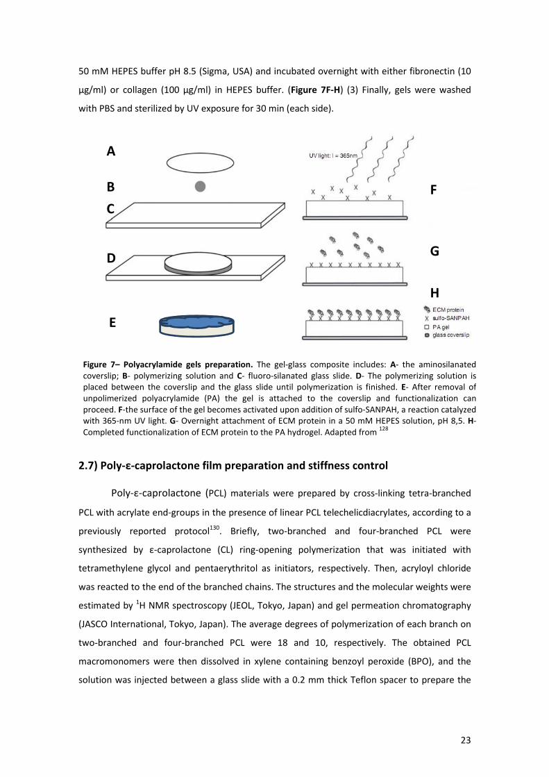
23
50 mM HEPES buffer pH 8.5 (Sigma, USA) and incubated overnight with either fibronectin (10
µg/ml) or collagen (100 µg/ml) in HEPES buffer. (Figure 7F-H) (3) Finally, gels were washed
with PBS and sterilized by UV exposure for 30 min (each side).
2.7) Poly-ε-caprolactone film preparation and stiffness control
Poly-ε-caprolactone (PCL) materials were prepared by cross-linking tetra-branched
PCL with acrylate end-groups in the presence of linear PCL telechelicdiacrylates, according to a
previously reported protocol130. Briefly, two-branched and four-branched PCL were
synthesized by ε-caprolactone (CL) ring-opening polymerization that was initiated with
tetramethylene glycol and pentaerythritol as initiators, respectively. Then, acryloyl chloride
was reacted to the end of the branched chains. The structures and the molecular weights were
estimated by 1H NMR spectroscopy (JEOL, Tokyo, Japan) and gel permeation chromatography
(JASCO International, Tokyo, Japan). The average degrees of polymerization of each branch on
two-branched and four-branched PCL were 18 and 10, respectively. The obtained PCL
macromonomers were then dissolved in xylene containing benzoyl peroxide (BPO), and the
solution was injected between a glass slide with a 0.2 mm thick Teflon spacer to prepare the
A
B
C
D
E
F
G
H
Figure 7– Polyacrylamide gels preparation. The gel-glass composite includes: A- the aminosilanated coverslip; B- polymerizing solution and C- fluoro-silanated glass slide. D- The polymerizing solution is placed between the coverslip and the glass slide until polymerization is finished. E- After removal of unpolimerized polyacrylamide (PA) the gel is attached to the coverslip and functionalization can proceed. F-the surface of the gel becomes activated upon addition of sulfo-SANPAH, a reaction catalyzed with 365-nm UV light. G- Overnight attachment of ECM protein in a 50 mM HEPES solution, pH 8,5. H-Completed functionalization of ECM protein to the PA hydrogel. Adapted from
128
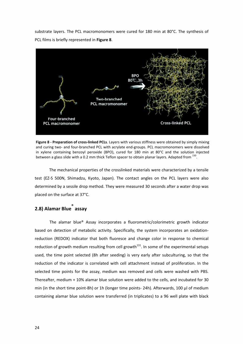
24
substrate layers. The PCL macromonomers were cured for 180 min at 80°C. The synthesis of
PCL films is briefly represented in Figure 8.
The mechanical properties of the crosslinked materials were characterized by a tensile
test (EZ-S 500N, Shimadzu, Kyoto, Japan). The contact angles on the PCL layers were also
determined by a sessile drop method. They were measured 30 seconds after a water drop was
placed on the surface at 37°C.
2.8) Alamar Blue® assay
The alamar blue® Assay incorporates a fluorometric/colorimetric growth indicator
based on detection of metabolic activity. Specifically, the system incorporates an oxidation-
reduction (REDOX) indicator that both fluoresce and change color in response to chemical
reduction of growth medium resulting from cell growth131. In some of the experimental setups
used, the time point selected (8h after seeding) is very early after subculturing, so that the
reduction of the indicator is correlated with cell attachment instead of proliferation. In the
selected time points for the assay, medium was removed and cells were washed with PBS.
Thereafter, medium + 10% alamar blue solution were added to the cells, and incubated for 30
min (in the short time point-8h) or 1h (longer time points- 24h). Afterwards, 100 µl of medium
containing alamar blue solution were transferred (in triplicates) to a 96 well plate with black
Figure 8 - Preparation of cross-linked PCLs. Layers with various stiffness were obtained by simply mixing and curing two- and four-branched PCL with acrylate end-groups. PCL macromonomers were dissolved in xylene containing benzoyl peroxide (BPO), cured for 180 min at 80°C and the solution injected between a glass slide with a 0.2 mm thick Teflon spacer to obtain planar layers. Adapted from
150.
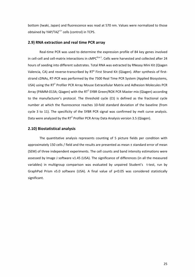
25
bottom (Iwaki, Japan) and fluorescence was read at 570 nm. Values were normalized to those
obtained by YAP/TAZ+/+ cells (control) in TCPS.
2.9) RNA extraction and real time PCR array
Real-time PCR was used to determine the expression profile of 84 key genes involved
in cell-cell and cell-matrix interactions in cMPCSca-1. Cells were harvested and collected after 24
hours of seeding into different substrates. Total RNA was extracted by RNeasy Mini Kit (Qiagen
Valencia, CA) and reverse-transcribed by RT² First Strand Kit (Qiagen). After synthesis of first-
strand cDNAs, RT-PCR was performed by the 7500 Real Time PCR System (Applied Biosystems,
USA) using the RT2 Profiler PCR Array Mouse Extracellular Matrix and Adhesion Molecules PCR
Array (PAMM-013A; Qiagen) with the RT2 SYBR Green/ROX PCR Master mix (Qiagen) according
to the manufacturer’s protocol. The threshold cycle (Ct) is defined as the fractional cycle
number at which the fluorescence reaches 10-fold standard deviation of the baseline (from
cycle 3 to 11). The specificity of the SYBR PCR signal was confirmed by melt curve analysis.
Data were analyzed by the RT2 Profiler PCR Array Data Analysis version 3.5 (Qiagen).
2.10) Biostatistical analysis
The quantitative analysis represents counting of 5 picture fields per condition with
approximately 150 cells / field and the results are presented as mean ± standard error of mean
(SEM) of three independent experiments. The cell counts and band intensity estimations were
assessed by Image J software v1.45 (USA). The significance of differences (in all the measured
variables) in multigroup comparison was evaluated by unpaired Student’s t-test, run by
GraphPad Prism v5.0 software (USA). A final value of p<0.05 was considered statistically
significant.

26
Figure 9 - YAP and TAZ display an overlapping signal both in cytoplasmic and nuclear expression in
cMPCSca-1
. As such, YAP and TAZ activity was considered redundant throughout this thesis. Nuclei were counterstained with DAPI.
3) Results
3.1) YAP/TAZ characterization and regulation by confluence and migration
Since YAP/TAZ has never been studied in cardiac stem/progenitor cells, a preliminary
characterization of its activity under conditions already explored in other cell types has been
carried out. To rule out different activities/mechanisms between YAP and TAZ both proteins
were co-immunostained, as shown in Figure 9. YAP and TAZ were shown to display an
overlapping signal, both when present in the cytoplasm and in the nucleus. Thus, due to this
overlapping expression pattern, activity of both proteins was inferred from the readout of one
of them, in the remainder of this thesis. Nevertheless, that was further verified for each
experimental setting. Additionally, relevance of YAP/TAZ in the CPC context was firstly
analyzed by co-staining YAP with the transcription factor GATA-4 (Figure 10A), revealing co-
localization of both proteins, thus rendering GATA-4 a possible interacting partner (given that
YAP/TAZ is a transcriptional co-activator and GATA-4 a transcriptional factor). Moreover, co-
localization of YAP with a nuclear shuttling protein paxillin (phosphorylated) was also found,
hinting into the mechanism of YAP nuclear translocation and/or interaction with nuclear
shuttling proteins (Figure 10B).
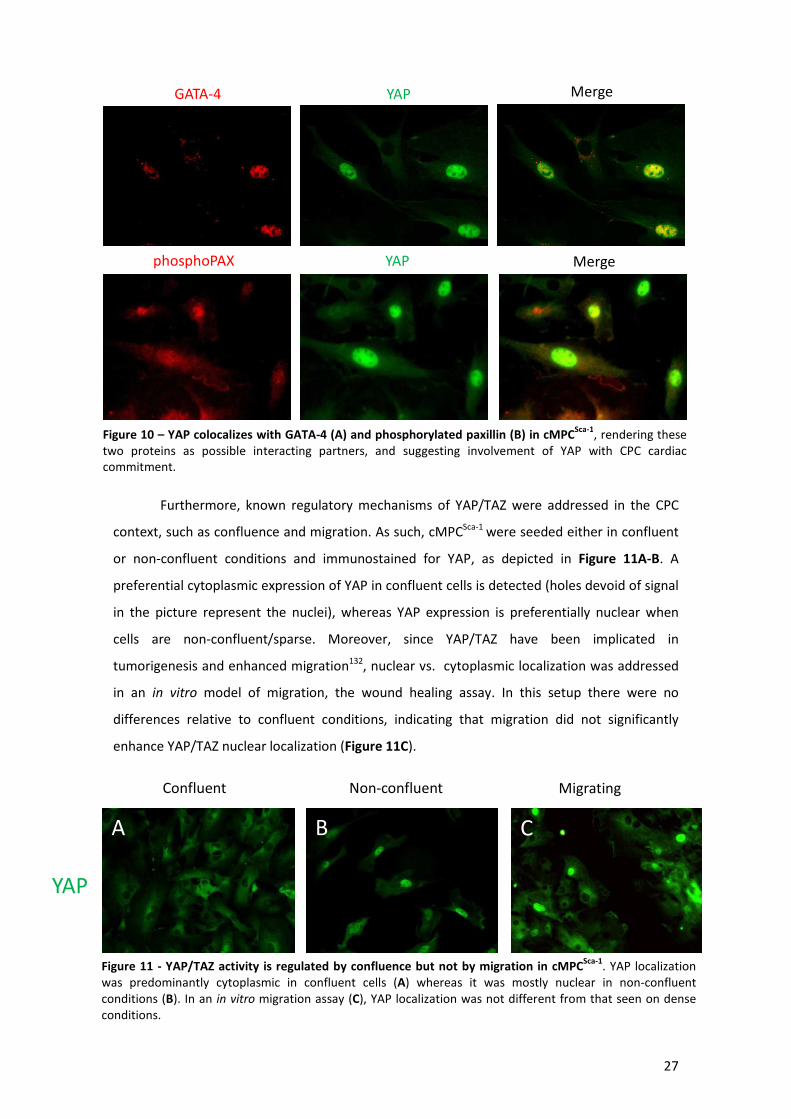
27
Furthermore, known regulatory mechanisms of YAP/TAZ were addressed in the CPC
context, such as confluence and migration. As such, cMPCSca-1 were seeded either in confluent
or non-confluent conditions and immunostained for YAP, as depicted in Figure 11A-B. A
preferential cytoplasmic expression of YAP in confluent cells is detected (holes devoid of signal
in the picture represent the nuclei), whereas YAP expression is preferentially nuclear when
cells are non-confluent/sparse. Moreover, since YAP/TAZ have been implicated in
tumorigenesis and enhanced migration132, nuclear vs. cytoplasmic localization was addressed
in an in vitro model of migration, the wound healing assay. In this setup there were no
differences relative to confluent conditions, indicating that migration did not significantly
enhance YAP/TAZ nuclear localization (Figure 11C).
GATA-4 YAP Merge
phosphoPAX YAP Merge
Figure 10 – YAP colocalizes with GATA-4 (A) and phosphorylated paxillin (B) in cMPCSca-1
, rendering these two proteins as possible interacting partners, and suggesting involvement of YAP with CPC cardiaccommitment.
YAP
Confluent Non-confluent Migrating WH)
A B C
Figure 11 - YAP/TAZ activity is regulated by confluence but not by migration in cMPCSca-1
. YAP localization was predominantly cytoplasmic in confluent cells (A) whereas it was mostly nuclear in non-confluent conditions (B). In an in vitro migration assay (C), YAP localization was not different from that seen on dense conditions.
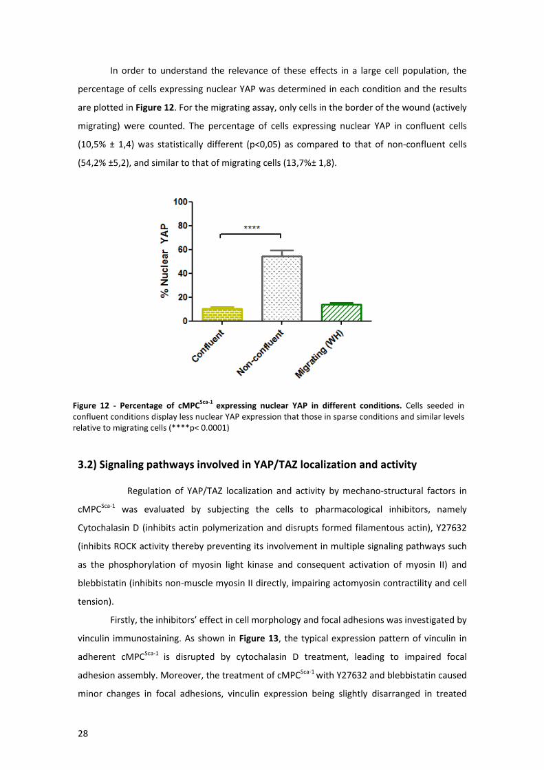
28
In order to understand the relevance of these effects in a large cell population, the
percentage of cells expressing nuclear YAP was determined in each condition and the results
are plotted in Figure 12. For the migrating assay, only cells in the border of the wound (actively
migrating) were counted. The percentage of cells expressing nuclear YAP in confluent cells
(10,5% ± 1,4) was statistically different (p<0,05) as compared to that of non-confluent cells
(54,2% ±5,2), and similar to that of migrating cells (13,7%± 1,8).
3.2) Signaling pathways involved in YAP/TAZ localization and activity
Regulation of YAP/TAZ localization and activity by mechano-structural factors in
cMPCSca-1 was evaluated by subjecting the cells to pharmacological inhibitors, namely
Cytochalasin D (inhibits actin polymerization and disrupts formed filamentous actin), Y27632
(inhibits ROCK activity thereby preventing its involvement in multiple signaling pathways such
as the phosphorylation of myosin light kinase and consequent activation of myosin II) and
blebbistatin (inhibits non-muscle myosin II directly, impairing actomyosin contractility and cell
tension).
Firstly, the inhibitors’ effect in cell morphology and focal adhesions was investigated by
vinculin immunostaining. As shown in Figure 13, the typical expression pattern of vinculin in
adherent cMPCSca-1 is disrupted by cytochalasin D treatment, leading to impaired focal
adhesion assembly. Moreover, the treatment of cMPCSca-1 with Y27632 and blebbistatin caused
minor changes in focal adhesions, vinculin expression being slightly disarranged in treated
Figure 12 - Percentage of cMPCSca-1
expressing nuclear YAP in different conditions. Cells seeded in confluent conditions display less nuclear YAP expression that those in sparse conditions and similar levels relative to migrating cells (****p< 0.0001)
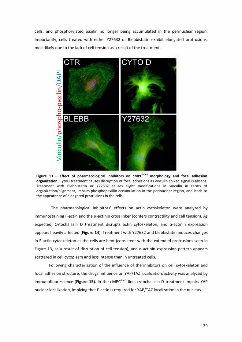
29
cells, and phosphorylated paxilin no longer being accumulated in the perinuclear region.
Importantly, cells treated with either Y27632 or Blebbistatin exhibit elongated protrusions,
most likely due to the lack of cell tension as a result of the treatment.
The pharmacological inhibitors’ effects on actin cytoskeleton were analyzed by
immunostaining F-actin and the α-actinin crosslinker (confers contractility and cell tension). As
expected, Cytochalasin D treatment disrupts actin cytoskeleton, and α-actinin expression
appears heavily affected (Figure 14). Treatment with Y27632 and blebbistatin induces changes
in F-actin cytoskeleton as the cells are bent (consistent with the extended protrusions seen in
Figure 13, as a result of disruption of cell tension), and α-actinin expression pattern appears
scattered in cell cytoplasm and less intense than in untreated cells.
Following characterization of the influence of the inhibitors on cell cytoskeleton and
focal adhesion structure, the drugs’ influence on YAP/TAZ localization/activity was analyzed by
immunofluorescence (Figure 15). In the cMPCSca-1 line, cytochalasin D treatment impairs YAP
nuclear localization, implying that F-actin is required for YAP/TAZ localization in the nucleus.
Vin
culin
/ph
osp
ho
-pax
ilin
/DA
PI
Figure 13 – Effect of pharmacological inhibitors on cMPCSca-1
morphology and focal adhesion
organization. CytoD treatment causes disruption of focal adhesions as vinculin spiked signal is absent. Treatment with Blebbistatin or Y72632 causes slight modifications in vinculin in terms of organization/alignment, impairs phosphopaxillin accumulation in the perinuclear region, and leads to the appearance of elongated protrusions in the cells.
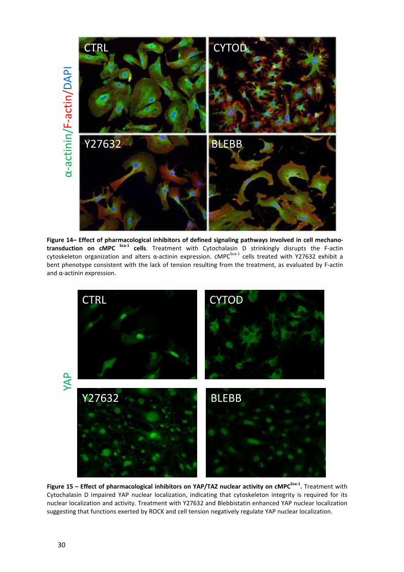
30
CTRL CYTOD
Y27632 BLEBB
YAP
Figure 15 – Effect of pharmacological inhibitors on YAP/TAZ nuclear activity on cMPCSca-1
. Treatment with Cytochalasin D impaired YAP nuclear localization, indicating that cytoskeleton integrity is required for its nuclear localization and activity. Treatment with Y27632 and Blebbistatin enhanced YAP nuclear localization suggesting that functions exerted by ROCK and cell tension negatively regulate YAP nuclear localization.
Figure 14– Effect of pharmacological inhibitors of defined signaling pathways involved in cell mechano-
transduction on cMPC Sca-1
cells. Treatment with Cytochalasin D strinkingly disrupts the F-actin cytoskeleton organization and alters α-actinin expression. cMPC
Sca-1 cells treated with Y27632 exhibit a
bent phenotype consistent with the lack of tension resulting from the treatment, as evaluated by F-actin and α-actinin expression.
CTRL CYTOD
Y27632 BLEBB
α-a
ctin
in/F
-act
in/D
AP
I
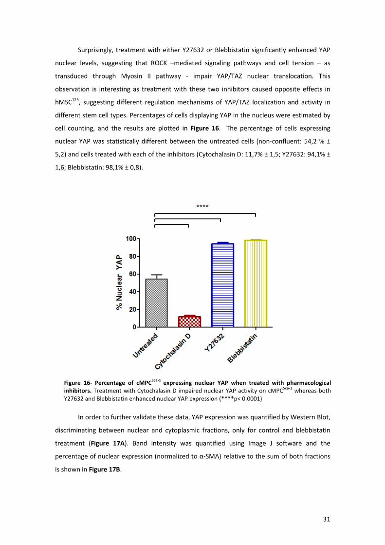
31
Surprisingly, treatment with either Y27632 or Blebbistatin significantly enhanced YAP
nuclear levels, suggesting that ROCK –mediated signaling pathways and cell tension – as
transduced through Myosin II pathway - impair YAP/TAZ nuclear translocation. This
observation is interesting as treatment with these two inhibitors caused opposite effects in
hMSC125, suggesting different regulation mechanisms of YAP/TAZ localization and activity in
different stem cell types. Percentages of cells displaying YAP in the nucleus were estimated by
cell counting, and the results are plotted in Figure 16. The percentage of cells expressing
nuclear YAP was statistically different between the untreated cells (non-confluent: 54,2 % ±
5,2) and cells treated with each of the inhibitors (Cytochalasin D: 11,7% ± 1,5; Y27632: 94,1% ±
1,6; Blebbistatin: 98,1% ± 0,8).
In order to further validate these data, YAP expression was quantified by Western Blot,
discriminating between nuclear and cytoplasmic fractions, only for control and blebbistatin
treatment (Figure 17A). Band intensity was quantified using Image J software and the
percentage of nuclear expression (normalized to α-SMA) relative to the sum of both fractions
is shown in Figure 17B.
Figure 16- Percentage of cMPCSca-1
expressing nuclear YAP when treated with pharmacological
inhibitors. Treatment with Cytochalasin D impaired nuclear YAP activity on cMPCSca-1
whereas both Y27632 and Blebbistatin enhanced nuclear YAP expression (****p< 0.0001)
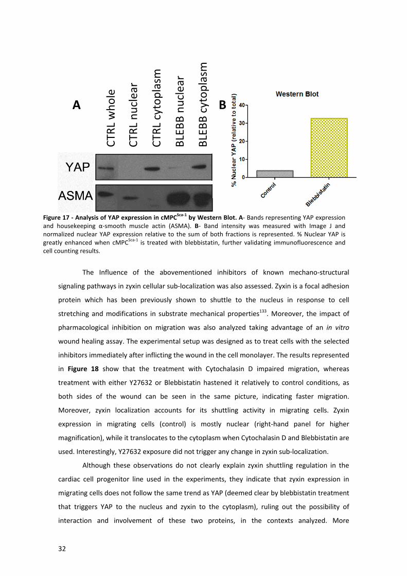
32
The Influence of the abovementioned inhibitors of known mechano-structural
signaling pathways in zyxin cellular sub-localization was also assessed. Zyxin is a focal adhesion
protein which has been previously shown to shuttle to the nucleus in response to cell
stretching and modifications in substrate mechanical properties133. Moreover, the impact of
pharmacological inhibition on migration was also analyzed taking advantage of an in vitro
wound healing assay. The experimental setup was designed as to treat cells with the selected
inhibitors immediately after inflicting the wound in the cell monolayer. The results represented
in Figure 18 show that the treatment with Cytochalasin D impaired migration, whereas
treatment with either Y27632 or Blebbistatin hastened it relatively to control conditions, as
both sides of the wound can be seen in the same picture, indicating faster migration.
Moreover, zyxin localization accounts for its shuttling activity in migrating cells. Zyxin
expression in migrating cells (control) is mostly nuclear (right-hand panel for higher
magnification), while it translocates to the cytoplasm when Cytochalasin D and Blebbistatin are
used. Interestingly, Y27632 exposure did not trigger any change in zyxin sub-localization.
Although these observations do not clearly explain zyxin shuttling regulation in the
cardiac cell progenitor line used in the experiments, they indicate that zyxin expression in
migrating cells does not follow the same trend as YAP (deemed clear by blebbistatin treatment
that triggers YAP to the nucleus and zyxin to the cytoplasm), ruling out the possibility of
interaction and involvement of these two proteins, in the contexts analyzed. More
Figure 17 - Analysis of YAP expression in cMPCSca-1
by Western Blot. A- Bands representing YAP expression and housekeeping α-smooth muscle actin (ASMA). B- Band intensity was measured with Image J and normalized nuclear YAP expression relative to the sum of both fractions is represented. % Nuclear YAP is greatly enhanced when cMPC
Sca-1 is treated with blebbistatin, further validating immunofluorescence and
cell counting results.
CTR
L w
ho
le
CTR
L n
ucl
ear
CTR
L cy
top
lasm
BLE
BB
nu
clea
r
BLE
BB
cyt
op
lasm
A B

33
interestingly, these results suggest that the localization of YAP/TAZ and zyxin is differentially
regulated in migrating cardiac stem/progenitor cells.
3.3) Role of YAP/TAZ in CPC proliferation
In order to investigate the relevance of YAP/TAZ in the context of Sca-1+ cardiac
stem/progenitor cells, its suppression was achieved using siRNA-mediated mRNA silencing. As
shown in Figure 19, both YAP and TAZ were silenced by the siRNA directed to YAP, accounting
for the high similarity in mRNA sequence between both these players.
Subsequently, by exploiting cells’ knock-down for YAP/TAZ expression, the relevance of
such proteins in different cellular processes was assessed. Firstly, the role of YAP/TAZ in CPC
proliferation was evaluated by immunostaining of Ki67, a known marker of cell proliferation134.
As shown in Figure 20A, the expression of Ki67 is significantly higher in YAP/TAZ-/- cells, the
Figure 18- Influence of pharmacological inhibitors’ treatment on cMPCSca-1
migration and zyxin nuclear
shuttling. Treatment with Cytochalasin D impaired migration whereas cells treated with either Y27632 or blebbistatin migrated faster relatively to untreated cells. Zyxin localization is influenced by the treatment as well, as shown in detailed pictures in the right hand side: zyxin is predominantly nuclear in untreated cells and cMPC
Sca-1treated with Y27632 and cytoplasmic in cells treated with Cytochalasin D or
blebbistatin.

34
percentages of nuclear ki67+ cells in control and YAP/TAZ -/- cells being 26,6 ±2,1% and
54,6±5,5%, respectively (Figure 20B).
ki67 DAPI Merged
Co
ntr
ol
YAP/
TAZ
-/-
A
B
Figure 20- Role of YAP/TAZ in CPC proliferation. Immunostaining of ki67 (A) resulted in a higher expression in YAP/TAZ -/- cells, as quantified and plotted (B). **** p<0.0001
Figure 19- Western Blot for YAP
and TAZ expression in control
cMPCSca-1
and silenced cells.
Successful knock-down of both YAP and TAZ expression is achieved by siRNA directed against YAP mRNA.
35
3.4) Role of cell tension and YAP/TAZ in CPC cardiac commitment
Since YAP/TAZ were found to co-localize with the early cardiac transcription factor
GATA-4 in cMPCSca-1 (Figure 10), and an effect of cell tension release in triggering nuclear
shuttling of such factors was demonstrated, the involvement of myosin II pathway in CPC
cardiac commitment was investigated. GATA-4 was immunostained in control and YAP/TAZ-/-
cells treated with blebbistatin to inhibit Myosin II activity (thus cell tension) (Figure 21A).
Nuclear GATA-4+ cells were quantified by immunofluorescence, yielding the following results:
control: 23,4%± 1,2; YAP/TAZ -/-: 5,7% ± 1,1; control + blebbistatin: 46,6% ± 3,7; YAP/TAZ -/- +
blebbistatin 4,3% ± 0,3) (Figure 21B).
The results suggest that YAP/TAZ is needed for GATA-4 nuclear localization in the
cMPCSca-1 cardiac progenitor cell-line model. Interestingly, nuclear GATA-4 expression seems to
be enhanced when cell tension is released. These results indicate YAP/TAZ as a relevant factor
in the transcriptional modulation of CPC commitment in response to a mechano-structural
cues arising from the extracellular environment. Nonetheless, controlled differentiation
experiments will be necessary to confirm the data.
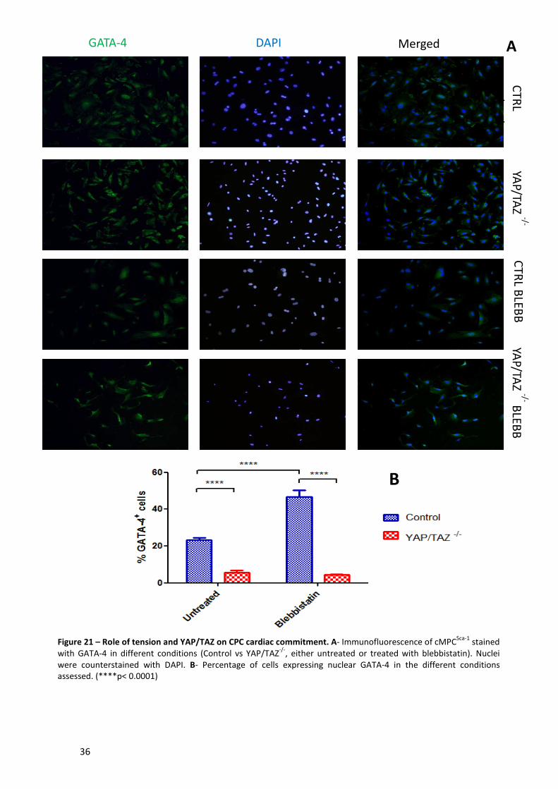
36
CTR
L u
ntreat
YAP/TA
Z -/- C
TRL B
LEBB
YA
P/TAZ -/- B
LEBB
GATA-4 DAPI Merged A
B
Figure 21 – Role of tension and YAP/TAZ on CPC cardiac commitment. A- Immunofluorescence of cMPCSca-1
stained with GATA-4 in different conditions (Control vs YAP/TAZ
-/-, either untreated or treated with blebbistatin). Nuclei
were counterstained with DAPI. B- Percentage of cells expressing nuclear GATA-4 in the different conditions assessed. (****p< 0.0001)
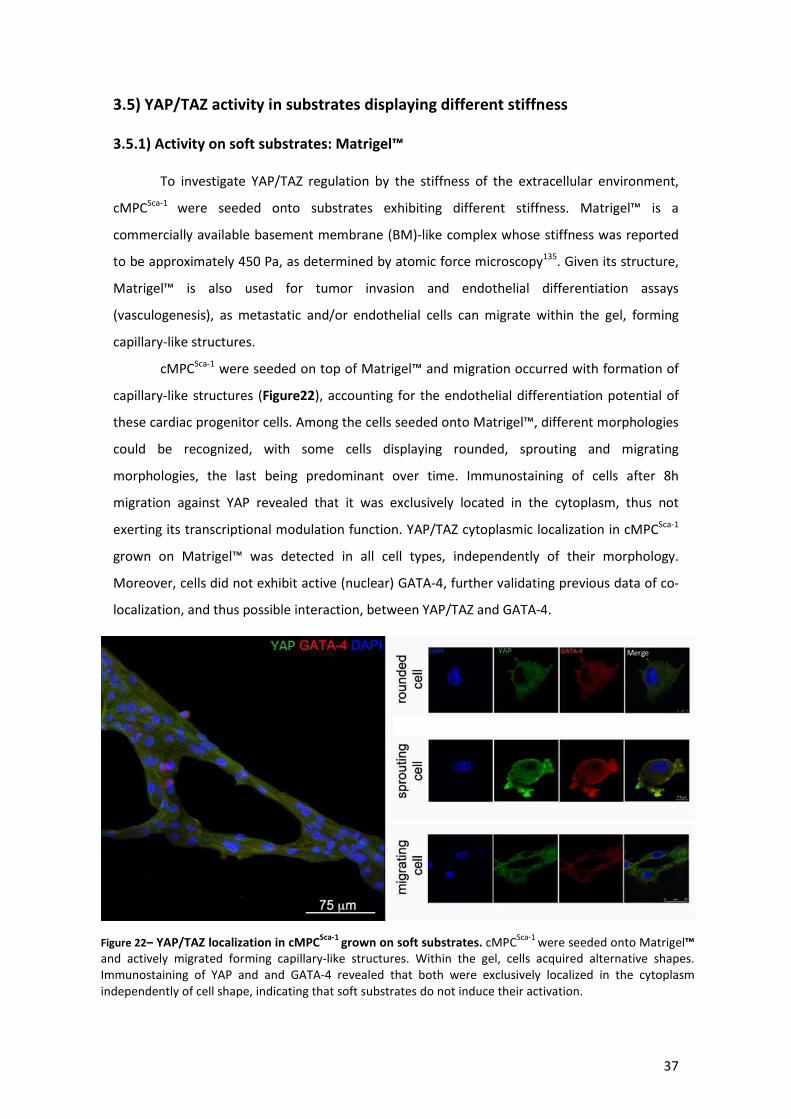
37
3.5) YAP/TAZ activity in substrates displaying different stiffness
3.5.1) Activity on soft substrates: Matrigel™
To investigate YAP/TAZ regulation by the stiffness of the extracellular environment,
cMPCSca-1 were seeded onto substrates exhibiting different stiffness. Matrigel™ is a
commercially available basement membrane (BM)-like complex whose stiffness was reported
to be approximately 450 Pa, as determined by atomic force microscopy135. Given its structure,
Matrigel™ is also used for tumor invasion and endothelial differentiation assays
(vasculogenesis), as metastatic and/or endothelial cells can migrate within the gel, forming
capillary-like structures.
cMPCSca-1 were seeded on top of Matrigel™ and migration occurred with formation of
capillary-like structures (Figure22), accounting for the endothelial differentiation potential of
these cardiac progenitor cells. Among the cells seeded onto Matrigel™, different morphologies
could be recognized, with some cells displaying rounded, sprouting and migrating
morphologies, the last being predominant over time. Immunostaining of cells after 8h
migration against YAP revealed that it was exclusively located in the cytoplasm, thus not
exerting its transcriptional modulation function. YAP/TAZ cytoplasmic localization in cMPCSca-1
grown on Matrigel™ was detected in all cell types, independently of their morphology.
Moreover, cells did not exhibit active (nuclear) GATA-4, further validating previous data of co-
localization, and thus possible interaction, between YAP/TAZ and GATA-4.
Figure 22– YAP/TAZ localization in cMPCSca-1
grown on soft substrates. cMPCSca-1
were seeded onto Matrigel™and actively migrated forming capillary-like structures. Within the gel, cells acquired alternative shapes. Immunostaining of YAP and and GATA-4 revealed that both were exclusively localized in the cytoplasm independently of cell shape, indicating that soft substrates do not induce their activation.
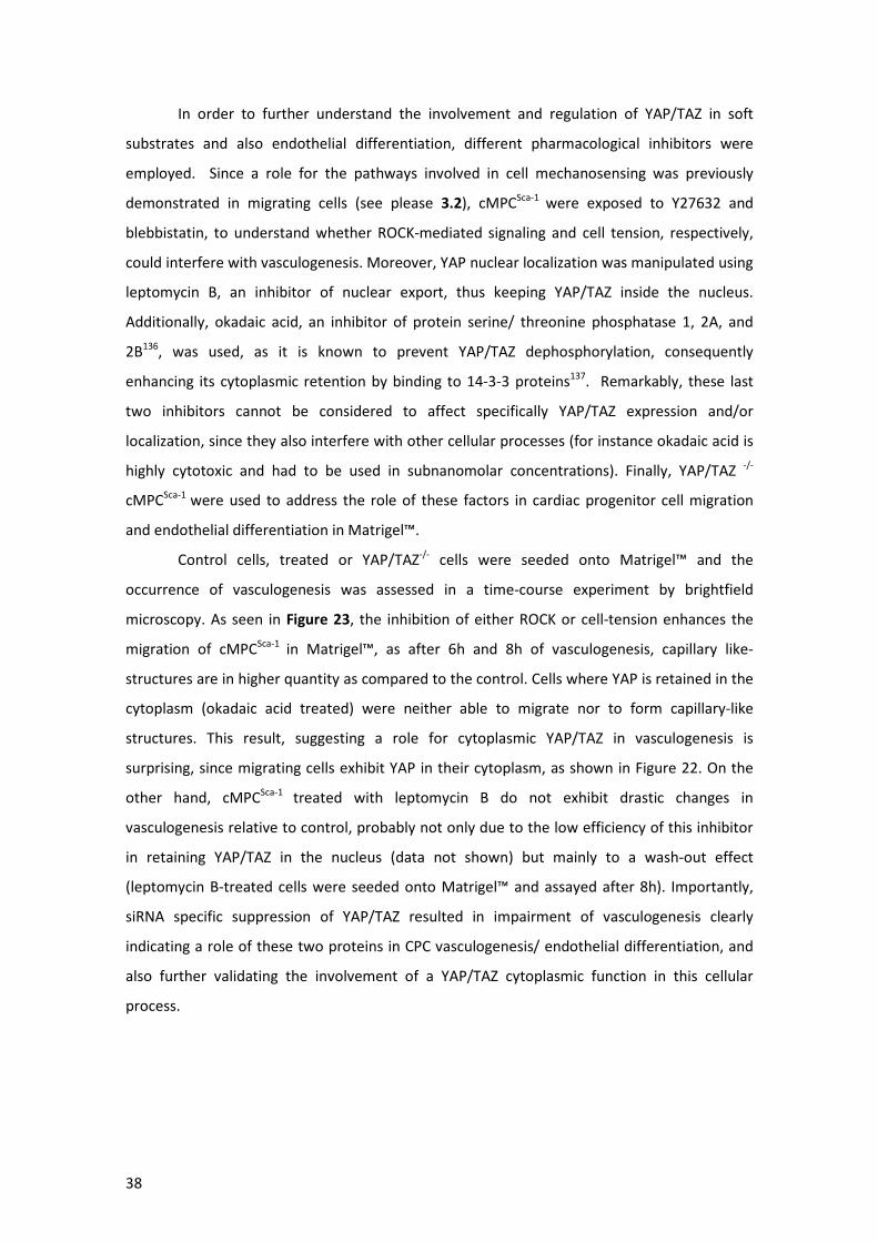
38
In order to further understand the involvement and regulation of YAP/TAZ in soft
substrates and also endothelial differentiation, different pharmacological inhibitors were
employed. Since a role for the pathways involved in cell mechanosensing was previously
demonstrated in migrating cells (see please 3.2), cMPCSca-1 were exposed to Y27632 and
blebbistatin, to understand whether ROCK-mediated signaling and cell tension, respectively,
could interfere with vasculogenesis. Moreover, YAP nuclear localization was manipulated using
leptomycin B, an inhibitor of nuclear export, thus keeping YAP/TAZ inside the nucleus.
Additionally, okadaic acid, an inhibitor of protein serine/ threonine phosphatase 1, 2A, and
2B136, was used, as it is known to prevent YAP/TAZ dephosphorylation, consequently
enhancing its cytoplasmic retention by binding to 14-3-3 proteins137. Remarkably, these last
two inhibitors cannot be considered to affect specifically YAP/TAZ expression and/or
localization, since they also interfere with other cellular processes (for instance okadaic acid is
highly cytotoxic and had to be used in subnanomolar concentrations). Finally, YAP/TAZ -/-
cMPCSca-1 were used to address the role of these factors in cardiac progenitor cell migration
and endothelial differentiation in Matrigel™.
Control cells, treated or YAP/TAZ-/- cells were seeded onto Matrigel™ and the
occurrence of vasculogenesis was assessed in a time-course experiment by brightfield
microscopy. As seen in Figure 23, the inhibition of either ROCK or cell-tension enhances the
migration of cMPCSca-1 in Matrigel™, as after 6h and 8h of vasculogenesis, capillary like-
structures are in higher quantity as compared to the control. Cells where YAP is retained in the
cytoplasm (okadaic acid treated) were neither able to migrate nor to form capillary-like
structures. This result, suggesting a role for cytoplasmic YAP/TAZ in vasculogenesis is
surprising, since migrating cells exhibit YAP in their cytoplasm, as shown in Figure 22. On the
other hand, cMPCSca-1 treated with leptomycin B do not exhibit drastic changes in
vasculogenesis relative to control, probably not only due to the low efficiency of this inhibitor
in retaining YAP/TAZ in the nucleus (data not shown) but mainly to a wash-out effect
(leptomycin B-treated cells were seeded onto Matrigel™ and assayed after 8h). Importantly,
siRNA specific suppression of YAP/TAZ resulted in impairment of vasculogenesis clearly
indicating a role of these two proteins in CPC vasculogenesis/ endothelial differentiation, and
also further validating the involvement of a YAP/TAZ cytoplasmic function in this cellular
process.

39
3.5.2) Activity of YAP/TAZ on physiologically relevant surfaces: coated
polyacrylamide gels with controlled stiffness.
Since the origin of cMPCSca-1 cells is related to the cardiac muscle, it is important to
evaluate the cellular response when these cells are seeded onto scaffolds displaying stiffness
values similar to that of the muscle. At the physiologically appropriate strains, the degree of
stiffness (Young modulus, E) varies dramatically between tissues: brain (Ebrain ∼ 0.1-1 kPa) is
clearly softer than striated skeletal muscle (Emuscle ∼ 8-17 kPa), which is more compliant than
precalcified bone (Eprecalcified bone = 25-40 kPa)72.
As such, YAP/TAZ activity was explored in cMPCSca-1 cells grown onto surfaces
displaying physiological stiffness values, by the use of polyacrylamide gels with tunable
stiffness. Moreover, the biological effect of extracellular matrix proteins on YAP/TAZ
expression was studied by coating these gels with fibronectin or collagen I. Therefore, cMPCSca1
cells were seeded onto PA gels and immunostained against YAP and TAZ by using two different
antibodies after 24h, as shown in Figure 24A (collagen coating) and Figure 24B (fibronectin
coating). The cells positive for YAP or TAZ were counted and expressed as a percentage of total
cells (Figure 25). As shown in Figure 24, cMPCSca-1 cells are sensitive to substrate stiffness,
responding by activating YAP/TAZ on stiffer substrates (higher than 10kPa), for collagen coated
gels (Figure 25A). However, in gels coated with fibronectin, YAP/TAZ expression seems less
Figure 23 – Regulation of YAP/TAZ in cMPCSca-1
vasculogenesis, on soft substrates. Inhibition of either cell tension or ROCK by blebbistatin or Y27632 treatments (respectively) enhances cMPC
Sca-1 vasculogenesis, as seen by the greater number of capillary-like
structures in the first two conditions. Retaining YAP/TAZ in the cytoplasm (okadaic acid treatment) greatly impairs vasculogenesis while keeping them in the nucleus (Leptomycin B treatment) has minor effects. Importantly, specific suppression of YAP and TAZ greatly inhibits cMPC
Sca-1 vasculogenisis indicating a role of YAP/TAZ in endothelial differentiation.

40
sensitive to stiffness changes, as the percentage of cells expressing these proteins in the
nucleus does not increase gradually with stiffness (Figure 25B). This indicates that, apart from
the mechanical properties of the ECM, its composition is also a parameter to consider in
cardiac progenitor cell mechanosensing. Hence, the concept that the regulation of cell fate
depends on several factors of different nature (biological, mechano-structural and physico-
chemical) is further validated. Moreover, because hMSC were shown to respond to stiffness
under different thresholds (TAZ is cytoplasmic at 0,7kPa and nuclear at 40kPa), differences in
mechanosensing between different stem cell populations might be inferred by these results.
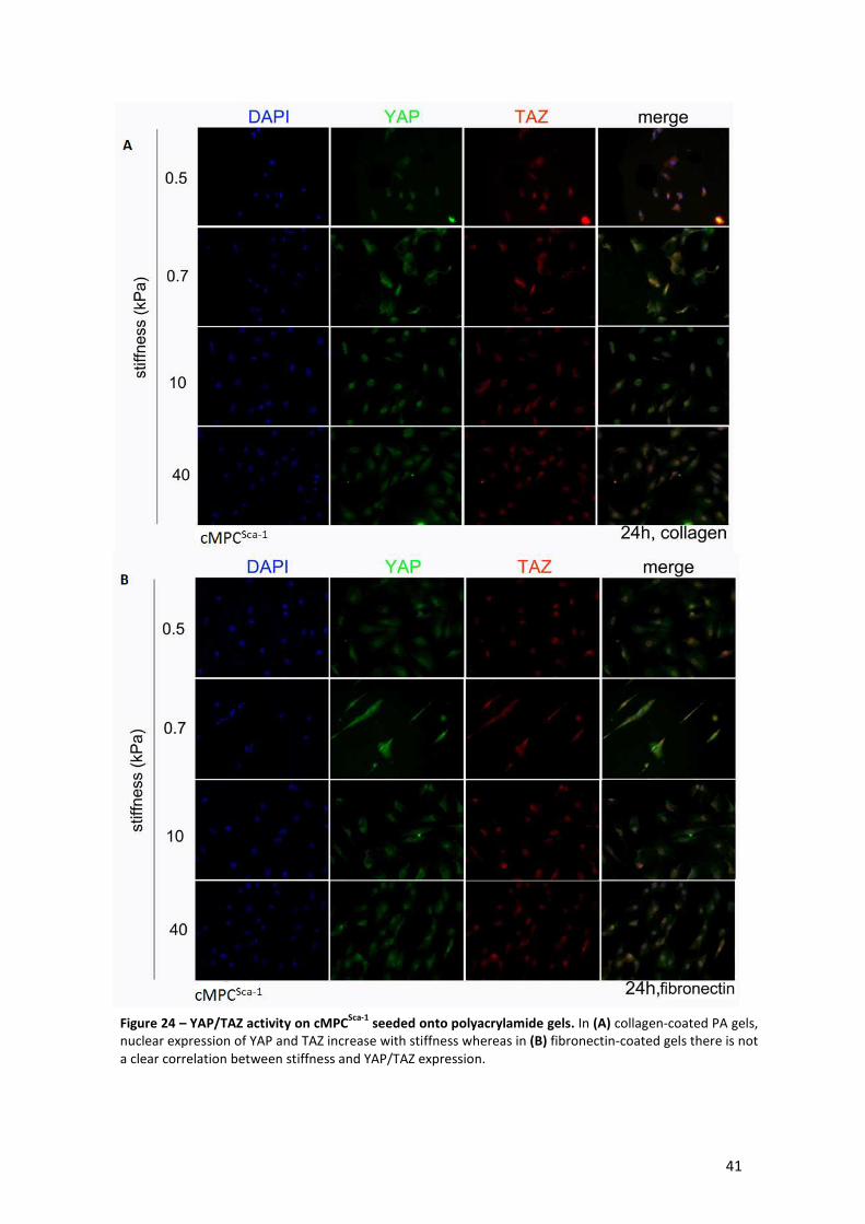
41
A
B
Figure 24 – YAP/TAZ activity on cMPCSca-1
seeded onto polyacrylamide gels. In (A) collagen-coated PA gels, nuclear expression of YAP and TAZ increase with stiffness whereas in (B) fibronectin-coated gels there is not a clear correlation between stiffness and YAP/TAZ expression.
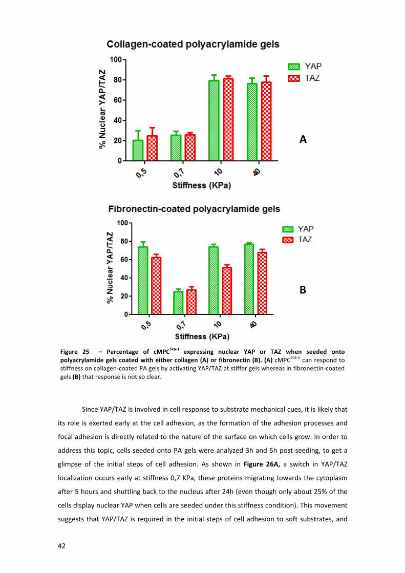
42
Since YAP/TAZ is involved in cell response to substrate mechanical cues, it is likely that
its role is exerted early at the cell adhesion, as the formation of the adhesion processes and
focal adhesion is directly related to the nature of the surface on which cells grow. In order to
address this topic, cells seeded onto PA gels were analyzed 3h and 5h post-seeding, to get a
glimpse of the initial steps of cell adhesion. As shown in Figure 26A, a switch in YAP/TAZ
localization occurs early at stiffness 0,7 KPa, these proteins migrating towards the cytoplasm
after 5 hours and shuttling back to the nucleus after 24h (even though only about 25% of the
cells display nuclear YAP when cells are seeded under this stiffness condition). This movement
suggests that YAP/TAZ is required in the initial steps of cell adhesion to soft substrates, and
A
B
Figure 25 – Percentage of cMPCSca-1
expressing nuclear YAP or TAZ when seeded onto
polyacrylamide gels coated with either collagen (A) or fibronectin (B). (A) cMPCSca-1
can respond to stiffness on collagen-coated PA gels by activating YAP/TAZ at stiffer gels whereas in fibronectin-coated gels (B) that response is not so clear.
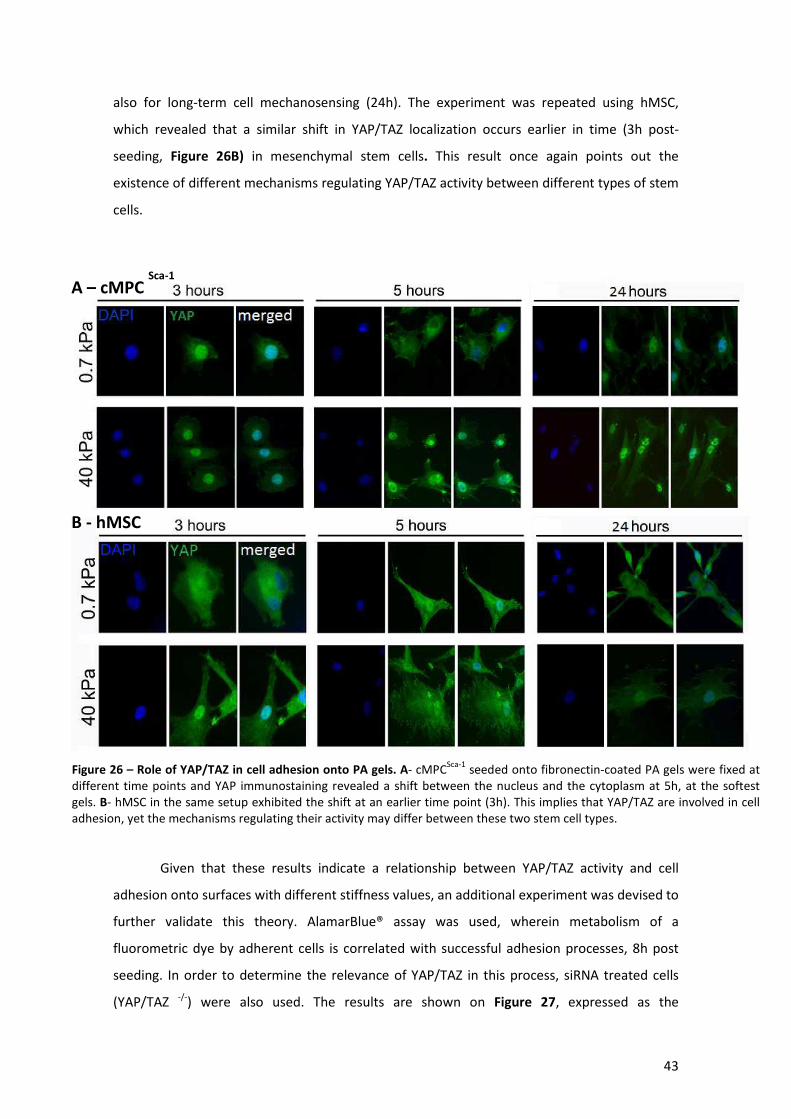
43
also for long-term cell mechanosensing (24h). The experiment was repeated using hMSC,
which revealed that a similar shift in YAP/TAZ localization occurs earlier in time (3h post-
seeding, Figure 26B) in mesenchymal stem cells. This result once again points out the
existence of different mechanisms regulating YAP/TAZ activity between different types of stem
cells.
Given that these results indicate a relationship between YAP/TAZ activity and cell
adhesion onto surfaces with different stiffness values, an additional experiment was devised to
further validate this theory. AlamarBlue® assay was used, wherein metabolism of a
fluorometric dye by adherent cells is correlated with successful adhesion processes, 8h post
seeding. In order to determine the relevance of YAP/TAZ in this process, siRNA treated cells
(YAP/TAZ -/-) were also used. The results are shown on Figure 27, expressed as the
B - hMSC
A – cMPC Sca-1
Figure 26 – Role of YAP/TAZ in cell adhesion onto PA gels. A- cMPCSca-1
seeded onto fibronectin-coated PA gels were fixed at different time points and YAP immunostaining revealed a shift between the nucleus and the cytoplasm at 5h, at the softest gels. B- hMSC in the same setup exhibited the shift at an earlier time point (3h). This implies that YAP/TAZ are involved in cell adhesion, yet the mechanisms regulating their activity may differ between these two stem cell types.
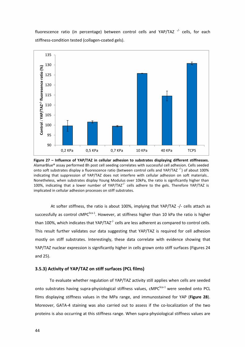
44
fluorescence ratio (in percentage) between control cells and YAP/TAZ -/- cells, for each
stiffness-condition tested (collagen-coated gels).
At softer stiffness, the ratio is about 100%, implying that YAP/TAZ -/- cells attach as
successfully as control cMPCSca-1. However, at stiffness higher than 10 kPa the ratio is higher
than 100%, which indicates that YAP/TAZ-/- cells are less adherent as compared to control cells.
This result further validates our data suggesting that YAP/TAZ is required for cell adhesion
mostly on stiff substrates. Interestingly, these data correlate with evidence showing that
YAP/TAZ nuclear expression is significantly higher in cells grown onto stiff surfaces (Figures 24
and 25).
3.5.3) Activity of YAP/TAZ on stiff surfaces (PCL films)
To evaluate whether regulation of YAP/TAZ activity still applies when cells are seeded
onto substrates having supra-physiological stiffness values, cMPCSca-1 were seeded onto PCL
films displaying stiffness values in the MPa range, and immunostained for YAP (Figure 28).
Moreover, GATA-4 staining was also carried out to assess if the co-localization of the two
proteins is also occurring at this stiffness range. When supra-physiological stiffness values are
90
95
100
105
110
115
120
125
130
135
0,2 KPa 0,5 KPa 0,7 KPa 10 KPa 40 KPa TCPS
Co
ntr
ol
: Y
AP
/TA
Z-/
-fl
uo
rese
nce
ra
tio
(%
)
Figure 27 – Influence of YAP/TAZ in cellular adhesion to substrates displaying different stiffnesses.
AlamarBlue® assay performed 8h post cell seeding correlates with successful cell adhesion. Cells seeded onto soft substrates display a fluorescence ratio (between control cells and YAP/TAZ
-/-) of about 100%
indicating that suppression of YAP/TAZ does not interfere with cellular adhesion on soft materials.. Nonetheless, when substrates display Young Modulus over 10kPa, the ratio is significantly higher than 100%, indicating that a lower number of YAP/TAZ
-/- cells adhere to the gels. Therefore YAP/TAZ is
implicated in cellular adhesion processes on stiff substrates.
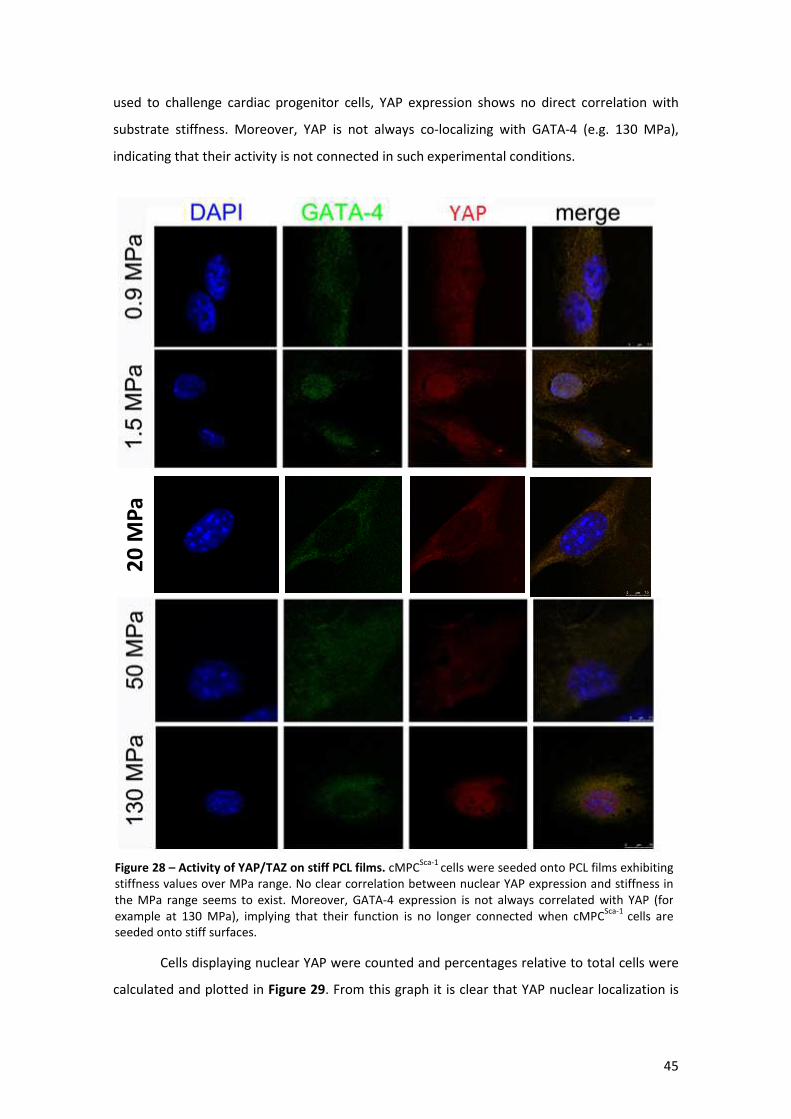
45
used to challenge cardiac progenitor cells, YAP expression shows no direct correlation with
substrate stiffness. Moreover, YAP is not always co-localizing with GATA-4 (e.g. 130 MPa),
indicating that their activity is not connected in such experimental conditions.
Cells displaying nuclear YAP were counted and percentages relative to total cells were
calculated and plotted in Figure 29. From this graph it is clear that YAP nuclear localization is
20
MP
a
Figure 28 – Activity of YAP/TAZ on stiff PCL films. cMPCSca-1
cells were seeded onto PCL films exhibiting stiffness values over MPa range. No clear correlation between nuclear YAP expression and stiffness in the MPa range seems to exist. Moreover, GATA-4 expression is not always correlated with YAP (for example at 130 MPa), implying that their function is no longer connected when cMPC
Sca-1 cells are
seeded onto stiff surfaces.
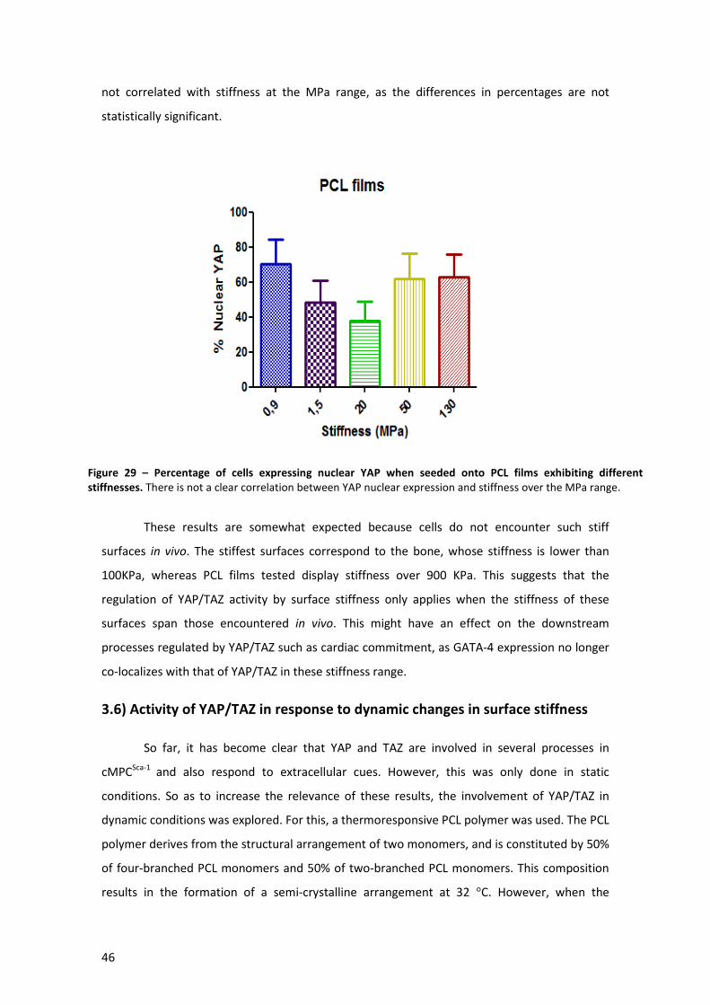
46
not correlated with stiffness at the MPa range, as the differences in percentages are not
statistically significant.
These results are somewhat expected because cells do not encounter such stiff
surfaces in vivo. The stiffest surfaces correspond to the bone, whose stiffness is lower than
100KPa, whereas PCL films tested display stiffness over 900 KPa. This suggests that the
regulation of YAP/TAZ activity by surface stiffness only applies when the stiffness of these
surfaces span those encountered in vivo. This might have an effect on the downstream
processes regulated by YAP/TAZ such as cardiac commitment, as GATA-4 expression no longer
co-localizes with that of YAP/TAZ in these stiffness range.
3.6) Activity of YAP/TAZ in response to dynamic changes in surface stiffness
So far, it has become clear that YAP and TAZ are involved in several processes in
cMPCSca-1 and also respond to extracellular cues. However, this was only done in static
conditions. So as to increase the relevance of these results, the involvement of YAP/TAZ in
dynamic conditions was explored. For this, a thermoresponsive PCL polymer was used. The PCL
polymer derives from the structural arrangement of two monomers, and is constituted by 50%
of four-branched PCL monomers and 50% of two-branched PCL monomers. This composition
results in the formation of a semi-crystalline arrangement at 32 °C. However, when the
Figure 29 – Percentage of cells expressing nuclear YAP when seeded onto PCL films exhibiting different
stiffnesses. There is not a clear correlation between YAP nuclear expression and stiffness over the MPa range.

47
temperature is shifted to 37°C, structural rearrangements occur turning the polymer into a
complete amorphous structure, thereby decreasing its stiffness. Using this system, both the
nanotopography and the stiffness are changed with temperature, providing an interesting
model to test cell responses to dynamic changes.
Thus, cMPCSca-1 were seeded onto this material at 32°C overnight and then moved to
37°C to induce the shift in substrate elasticity. Thereafter, YAP was immunostained and
localization compared to non-shifting control (TCPS). Pictures are shown in Figure 30A, and
percentage of cells expressing nuclear YAP were calculated by manual cell counting and
plotted in Figure30B.
YAP DAPI Merge
TC
PS
Co
ntr
ol
PC
L sw
itch
A
B
Figure 30– YAP/TAZ activity in dynamic surface changes. A - cMPCSca-1
were seeded onto a temperature responsive PCL polymer and YAP nuclear localization was assessed by immunofluorescence. B- Percentage of cells expressing nuclear YAP is higher when the material where they were seeded change its nanotopography and stiffness (PCL) in response to temperature, relative to non-changing TCPS control.

48
In static conditions, the percentage of cells expressing nuclear YAP when seeded onto
PCL films ranges from around 50 to 75% (Figure 29B). This number is enhanced to over 95%
when cells suffer a dynamic change, indicating that cells can sense the change in stiffness and
nanotopography imposed by the temperature responsive polymer. To prove that YAP/TAZ is
involved in this process, Alamar Blue assay was used to assess adhesion of cMPCSca-1 exposed
to the dynamic change, in comparison to YAP/TAZ-/- cells. Data obtained were represented as a
ratio between fluorescence measured after the dynamic change and that measured before it
(Figure 31). Since this ratio is less than 100% for YAP/TAZ-/- cells it can be inferred that some
cells have detached from the polymer, contrarily to what happened in control cells. This
indicates that YAP/TAZ is needed during dynamic changes (in this case stiffness and
nanotopography) of the surface where cMPCSca-1 are seeded, participating in the maintenance
of proper cell attachment.
3.7) Influence of cell shape in YAP/TAZ activity
Study of the regulation of YAP/TAZ by cell shape can be performed by the use of
micropatterned surfaces portraying different adhesive areas (islands coated with fibronectin).
As shown in Figure 32, these areas induce the cells to acquire different cell shapes, either
round (due to area constraint) in the smaller areas or flattened in the larger areas. Moreover,
this system allows single cell level studies avoiding the impact of cell-to-cell contact.
80
90
100
110
120
130
Control YAP/TAZ -/-
Flu
ore
sce
nce
aft
er/
be
fore
sw
itch
Alamar Blue Dynamic change
Figure 31 – Role of YAP/TAZ during dynamic changes of the surface where cMPCSca-1
is seeded. Alamar blue assay was used in order to quantify differences in cell proliferation/ adhesion between control and YAP/TAZ
-/- cMPC
Sca-1. Ratio between measured fluorescence after the dynamic change and before it is
lower than 100% in YAP/TAZ-/-
cells, as opposing to control cMPCSca-1
. This implies that YAP/TAZ is needed for proper cell adhesion when dynamic changes (stiffness and nanotopography) are imposed to the surface where cMPC
Sca-1 are seeded.
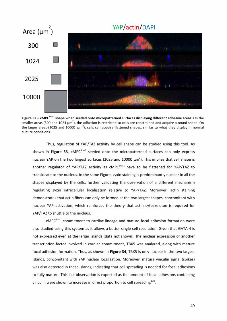
49
Thus, regulation of YAP/TAZ activity by cell shape can be studied using this tool. As
shown in Figure 33, cMPCSca-1 seeded onto the micropatterned surfaces can only express
nuclear YAP on the two largest surfaces (2025 and 10000 µm2). This implies that cell shape is
another regulator of YAP/TAZ activity as cMPCSca-1 have to be flattened for YAP/TAZ to
translocate to the nucleus. In the same Figure, zyxin staining is predominantly nuclear in all the
shapes displayed by the cells, further validating the observation of a different mechanism
regulating zyxin intracellular localization relative to YAP/TAZ. Moreover, actin staining
demonstrates that actin fibers can only be formed at the two largest shapes, concomitant with
nuclear YAP activation, which reinforces the theory that actin cytoskeleton is required for
YAP/TAZ to shuttle to the nucleus.
cMPCSca-1 commitment to cardiac lineage and mature focal adhesion formation were
also studied using this system as it allows a better single cell resolution. Given that GATA-4 is
not expressed even at the larger islands (data not shown), the nuclear expression of another
transcription factor involved in cardiac commitment, TBX5 was analyzed, along with mature
focal adhesion formation. Thus, as shown in Figure 34, TBX5 is only nuclear in the two largest
islands, concomitant with YAP nuclear localization. Moreover, mature vinculin signal (spikes)
was also detected in these islands, indicating that cell spreading is needed for focal adhesions
to fully mature. This last observation is expected as the amount of focal adhesions containing
vinculin were shown to increase in direct proportion to cell spreading138.
Area (µm2)
300
1024
2025
10000
YAP/actin/DAPI
Figure 32 – cMPCSca-1
shape when seeded onto micropatterned surfaces displaying different adhesive areas. On the smaller areas (300 and 1024 µm
2), the adhesion is restricted so cells are constrained and acquire a round shape. On
the larger areas (2025 and 10000 µm2), cells can acquire flattened shapes, similar to what they display in normal
culture conditions.
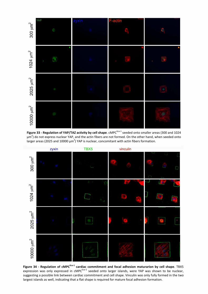
50
Figure 33 - Regulation of YAP/TAZ activity by cell shape. cMPCSca-1
seeded onto smaller areas (300 and 1024 µm
2) do not express nuclear YAP, and the actin fibers are not formed. On the other hand, when seeded onto
larger areas (2025 and 10000 µm2) YAP is nuclear, concomitant with actin fibers formation.
Figure 34 - Regulation of cMPCSca-1
cardiac commitment and focal adhesion maturarion by cell shape. TBX5 expression was only expressed in cMPC
Sca-1 seeded onto larger islands, were YAP was shown to be nuclear,
suggesting a possible link between cardiac commitment and cell shape. Vinculin was only fully formed in the two largest islands as well, indicating that a flat shape is required for mature focal adhesion formation.
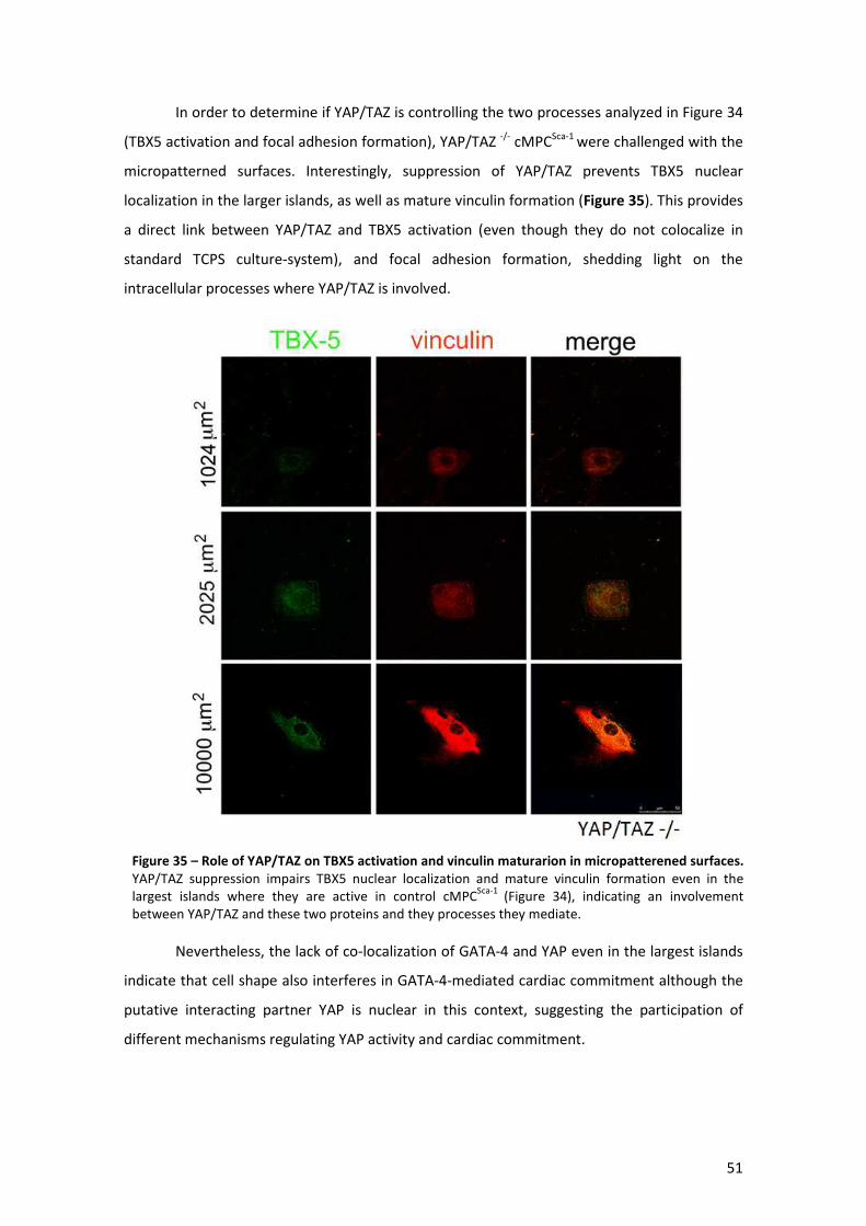
51
In order to determine if YAP/TAZ is controlling the two processes analyzed in Figure 34
(TBX5 activation and focal adhesion formation), YAP/TAZ -/- cMPCSca-1 were challenged with the
micropatterned surfaces. Interestingly, suppression of YAP/TAZ prevents TBX5 nuclear
localization in the larger islands, as well as mature vinculin formation (Figure 35). This provides
a direct link between YAP/TAZ and TBX5 activation (even though they do not colocalize in
standard TCPS culture-system), and focal adhesion formation, shedding light on the
intracellular processes where YAP/TAZ is involved.
Nevertheless, the lack of co-localization of GATA-4 and YAP even in the largest islands
indicate that cell shape also interferes in GATA-4-mediated cardiac commitment although the
putative interacting partner YAP is nuclear in this context, suggesting the participation of
different mechanisms regulating YAP activity and cardiac commitment.
Figure 35 – Role of YAP/TAZ on TBX5 activation and vinculin maturarion in micropatterened surfaces.
YAP/TAZ suppression impairs TBX5 nuclear localization and mature vinculin formation even in the largest islands where they are active in control cMPC
Sca-1 (Figure 34), indicating an involvement
between YAP/TAZ and these two proteins and they processes they mediate.
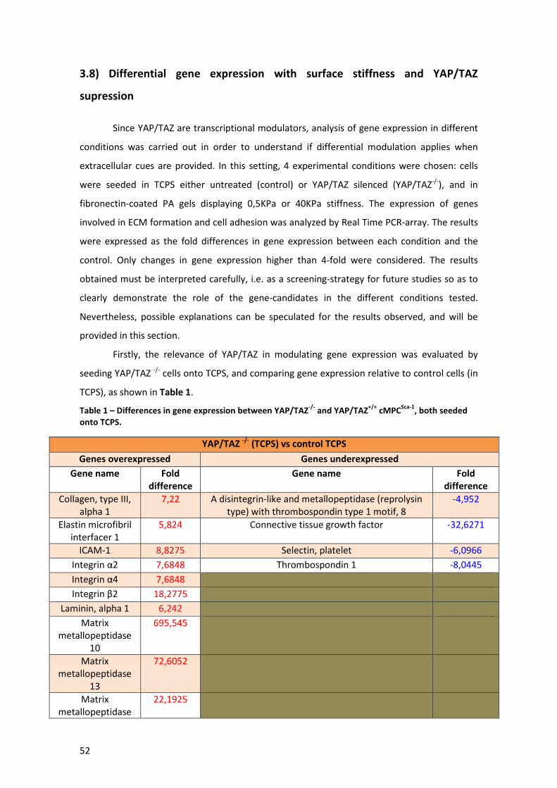
52
3.8) Differential gene expression with surface stiffness and YAP/TAZ
supression
Since YAP/TAZ are transcriptional modulators, analysis of gene expression in different
conditions was carried out in order to understand if differential modulation applies when
extracellular cues are provided. In this setting, 4 experimental conditions were chosen: cells
were seeded in TCPS either untreated (control) or YAP/TAZ silenced (YAP/TAZ-/-), and in
fibronectin-coated PA gels displaying 0,5KPa or 40KPa stiffness. The expression of genes
involved in ECM formation and cell adhesion was analyzed by Real Time PCR-array. The results
were expressed as the fold differences in gene expression between each condition and the
control. Only changes in gene expression higher than 4-fold were considered. The results
obtained must be interpreted carefully, i.e. as a screening-strategy for future studies so as to
clearly demonstrate the role of the gene-candidates in the different conditions tested.
Nevertheless, possible explanations can be speculated for the results observed, and will be
provided in this section.
Firstly, the relevance of YAP/TAZ in modulating gene expression was evaluated by
seeding YAP/TAZ -/- cells onto TCPS, and comparing gene expression relative to control cells (in
TCPS), as shown in Table 1.
Table 1 – Differences in gene expression between YAP/TAZ-/-
and YAP/TAZ+/+
cMPCSca-1
, both seeded
onto TCPS.
YAP/TAZ -/-
(TCPS) vs control TCPS
Genes overexpressed Genes underexpressed
Gene name Fold
difference
Gene name Fold
difference
Collagen, type III, alpha 1
7,22 A disintegrin-like and metallopeptidase (reprolysin type) with thrombospondin type 1 motif, 8
-4,952
Elastin microfibril interfacer 1
5,824 Connective tissue growth factor -32,6271
ICAM-1 8,8275 Selectin, platelet -6,0966
Integrin α2 7,6848 Thrombospondin 1 -8,0445
Integrin α4 7,6848
Integrin β2 18,2775
Laminin, alpha 1 6,242
Matrix metallopeptidase
10
695,545
Matrix metallopeptidase
13
72,6052
Matrix metallopeptidase
22,1925

53
15
Matrix metallopeptidase 3
162,2416
Synaptotagmin I 29,692
The high number of genes being differentially expressed between these conditions
further validates the role of YAP/TAZ as a transcriptional modulator on cMPCSca-1. These
differences span integrin composition, matrix metallopeptidase and also some proteins of the
ECM, being upregulated in YAP/TAZ -/- cells. Regarding that, suppression of YAP/TAZ has been
implicated in impaired vinculin maturation, so it is likely that, since cells cannot strongly attach
to a surface, they tend to migrate, thereby enhancing expression of matrix metallopeptidases.
Nonetheless, this putative explanation needs to be confirmed in order to obtain greater
understanding of YAP/TAZ relevance in adhesion and migration in terms of gene expression.
In order to understand if cMPCSca-1 can perceive subtler changes in stiffness, differences
in gene expression between cells seeded onto 0,5 and 40KPa were analyzed, as shown in
Table2.
Table 2 – Differences in gene expression between cells seeded onto Pa gels displaying 0,5 KPa and 40
KPa stiffness, respectively.
These data demonstrates that cMPCSca-1 can sense different stiffness of the
extracellular environment where it is embedded, further validating results obtained for
regulation of YAP/TAZ activation by small differences stiffness. The main differences
concerned matrix metallopeptidase expression which was different between cells seeded onto
surfaces exhibiting different stiffness.
Albeit preliminary, these results confirm that YAP/TAZ have a role in cell adhesion and
matrix sensing, modulating gene expression for generating appropriate cell responses.
0,5 KPa vs 40 KPa
Genes overexpressed Genes underexpressed
Gene name Fold difference
Gene name Fold difference
Contactin 1 5,0771 Laminin, alpha 3 -4,7437
Ectonucleoside triphosphate diphosphohydrolase 1
7,9118 Matrix metallopeptidase 10 -14,2808
ICAM-1 23,3282 Matrix metallopeptidase 3 -4,3954
Matrix metallopeptidase 14 (membrane-inserted)
4,3289 Matrix metallopeptidase 1a (interstitial collagenase)
-8,9755
Matrix metallopeptidase 15 13,306 Matrix metallopeptidase 3 -11,5195
Matrix metallopeptidase 9 -8,6099
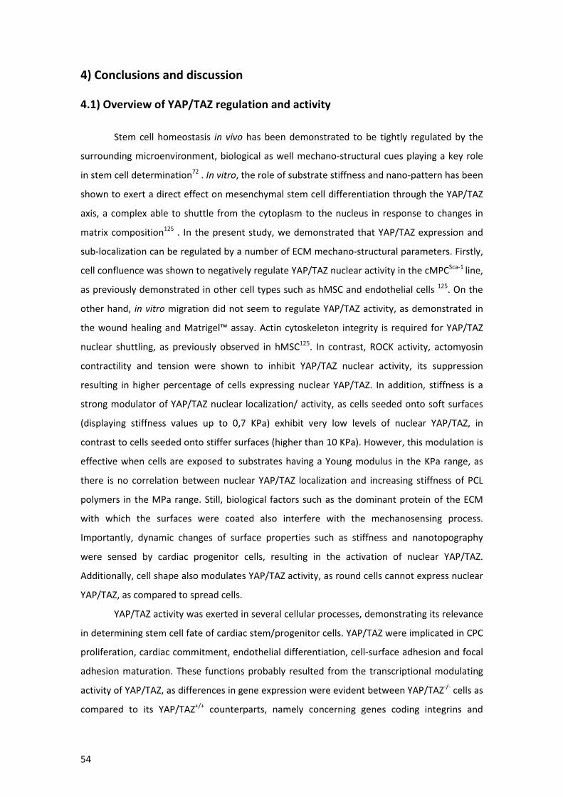
54
4) Conclusions and discussion
4.1) Overview of YAP/TAZ regulation and activity
Stem cell homeostasis in vivo has been demonstrated to be tightly regulated by the
surrounding microenvironment, biological as well mechano-structural cues playing a key role
in stem cell determination72 . In vitro, the role of substrate stiffness and nano-pattern has been
shown to exert a direct effect on mesenchymal stem cell differentiation through the YAP/TAZ
axis, a complex able to shuttle from the cytoplasm to the nucleus in response to changes in
matrix composition125 . In the present study, we demonstrated that YAP/TAZ expression and
sub-localization can be regulated by a number of ECM mechano-structural parameters. Firstly,
cell confluence was shown to negatively regulate YAP/TAZ nuclear activity in the cMPCSca-1 line,
as previously demonstrated in other cell types such as hMSC and endothelial cells 125. On the
other hand, in vitro migration did not seem to regulate YAP/TAZ activity, as demonstrated in
the wound healing and Matrigel™ assay. Actin cytoskeleton integrity is required for YAP/TAZ
nuclear shuttling, as previously observed in hMSC125. In contrast, ROCK activity, actomyosin
contractility and tension were shown to inhibit YAP/TAZ nuclear activity, its suppression
resulting in higher percentage of cells expressing nuclear YAP/TAZ. In addition, stiffness is a
strong modulator of YAP/TAZ nuclear localization/ activity, as cells seeded onto soft surfaces
(displaying stiffness values up to 0,7 KPa) exhibit very low levels of nuclear YAP/TAZ, in
contrast to cells seeded onto stiffer surfaces (higher than 10 KPa). However, this modulation is
effective when cells are exposed to substrates having a Young modulus in the KPa range, as
there is no correlation between nuclear YAP/TAZ localization and increasing stiffness of PCL
polymers in the MPa range. Still, biological factors such as the dominant protein of the ECM
with which the surfaces were coated also interfere with the mechanosensing process.
Importantly, dynamic changes of surface properties such as stiffness and nanotopography
were sensed by cardiac progenitor cells, resulting in the activation of nuclear YAP/TAZ.
Additionally, cell shape also modulates YAP/TAZ activity, as round cells cannot express nuclear
YAP/TAZ, as compared to spread cells.
YAP/TAZ activity was exerted in several cellular processes, demonstrating its relevance
in determining stem cell fate of cardiac stem/progenitor cells. YAP/TAZ were implicated in CPC
proliferation, cardiac commitment, endothelial differentiation, cell-surface adhesion and focal
adhesion maturation. These functions probably resulted from the transcriptional modulating
activity of YAP/TAZ, as differences in gene expression were evident between YAP/TAZ-/- cells as
compared to its YAP/TAZ+/+ counterparts, namely concerning genes coding integrins and
55
different matrix metallopeptidases. Differences in gene expression between cardiac progenitor
cells seeded onto surfaces displaying diverse stiffness were also evident, a total of 20 genes
being identified as candidates for differential mechanosensing activities, in terms of cell
adhesion molecules and proteins of the ECM. A proposed model depicting regulatory
mechanisms and activities involved with YAP/TAZ function in cardiac stem/progenitor cells is
represented in Figure 36.
Altogether, YAP/TAZ appear to constitute mechanical sensors involved in cardiac
stem/progenitor cell sensing of the microenvironment, responding to mechano-structural cues
and controlling important cellular responses related with stem cell fate determination.
Figure 36 - Role of YAP/TAZ in mechanotransduction of cardiac stem/progenitor cells. Regulation of YAP/TAZ is made at different levels such as confluence, actin cytoskeleton and cell tension, stiffness of interfacing surface and its dominant extracellular protein, cell shape and dynamic changes of substrate where cells are seeded. In CPC, YAP/TAZ is involved in cell proliferation, cardiac commitment/ differentiation, and adhesion, modulating gene expression concerning at least matrix metallopeptidases and integrins.
56
Results in the herein study are rather interesting when compared with findings
reported by others. For instance, cardiac stem/progenitor cells and mesenchymal stem cells
both seem to rely on YAP/TAZ as a mechanosensor, yet with different regulation mechanisms.
For instance, ROCK-mediated signaling and cell tension have opposite effects on these two
stem cell systems as, contrarily to its action in the cardiac system, these elements are required
to activate YAP/TAZ nuclear localization/activity in hMSC125. Moreover, YAP/TAZ nuclear
shuttling occurs at different time points (in early adhesion steps) in the two cell types.
Furthermore, YAP/TAZ response to stiffness has slightly different thresholds, as in hMSC
YAP/TAZ displays a nuclear expression at 40 KPa whereas in cardiac progenitor cells this event
takes place at 10KPa. Cell shape and spreading appears to exert similar responses in both
stem/progenitor cell types.
YAP/TAZ are known to act as transcriptional co-activators in the nucleus, thus requiring
additional factors to exert their function on gene expression. The interaction of YAP/TAZ with
nuclear shuttling proteins was not fully determined, yet (phosphorylated) paxillin is a putative
partner as concluded by co-localization experiments. On the contrary zyxin does not seem to
be a suitable partner for YAP/TAZ, since different intracellular localizations were detected in
response to pharmacological inhibitors in the in vitro wound healing migration assay.
A novelty in this study was the demonstration that suppression of cell tension can
induce CPC cardiac commitment in a mechanism dependent on YAP/TAZ activity. This result
states that a mechano-structural cue can effectively control stem cell fate determination, and
points at YAP/TAZ as one of the mechanotransducer involved in this mechanism, highlighting
its importance in the stem cell field. In the hMSC context, however, cytoskeletal tension and
actomyosin contractility were shown to promote osteogenesis in tri-dimensional microcarrier
system139, further demonstrating the importance of mechano-structural factors as regulators
of stem cell fate determination.
Importantly, since YAP/TAZ have shown to be involved in CPC proliferation, cardiac
commitment and endothelial differentiation, it is plausible that these two proteins are key
molecular switches, redirecting CPC fate either to self-renewal or differentiation into different
lineages, in response to mechano-structural factors characteristic of the microenvironment in
which CPC reside. This knowledge could be harnessed to improve existing CTE strategies.
4.2) Future perspectives
Despite the fact that this study already contemplates a large scope of events involved
in YAP/TAZ regulation and activity, taking the work developed for this MSc. Degree dissertation
57
to the verge of publication, several experiments could still be performed in order to further
validate/ optimize some of the observations reported.
First of all, it would be interesting to perform immunohistochemistry studies on
murine heart tissue, so as to understand if YAP and/or TAZ are expressed in different resident
putative cardiac stem/progenitor cell subsets, i.e. Sca-1+ , c-Kit+ or Isl-1+ CPCs.
Secondly, confirmation of cell counting experiments with Western Blot should be
finished, providing a more sensitive and accurate method to assess YAP/TAZ nuclear activity.
Thirdly, the use of additional pharmacological inhibitors could further validate the data
acquired so far concerning YAP/TAZ regulation by mechanical forces. For instance, thrombin
has shown to increase cell tension concomitant with myosin II phosphorylation. Treatment
with thrombin could be anticipated to produce the opposite effects induced by blebbistatin,
and therefore confirm tension as a powerful modulator of YAP/TAZ activity140.
Fourthly, additional migration experiments could be performed in order to further
investigate the role of YAP/TAZ activation during cell migration. This assertion is due to the fact
that migration is involved in many processes including tissue repair and regeneration92,
wherein involvement of actin cytoskeleton dynamics and myosin II is well studied91 , and could
thus provide interesting models for studying mechanotransduction pathways in CPC. As such,
further dissection of molecular mechanisms activated during CPC migration could provide
useful data for the purpose of scaffold implantation/seeding, possibly ameliorating therapeutic
outcomes for cell-based therapies.
Moreover, the reported co-localization and regulation of YAP and GATA-4 is an
interesting process, deserving future studies. Protein interaction should be confirmed,
considering that our co-immunoprecipitation attempts were so far less succeeded. Thus, this
experiment should be completed in order to confirm the interaction, as it further validates the
data acquired in this thesis.
RNAi experiments provided very important results. It would be interesting to
discriminate between the effects mediated by YAP and TAZ alone, by the use of specific
suppression of each protein, either by the use of more specific siRNA sequences or lentiviral-
transfection of shRNA for either YAP or TAZ. This strategy would allow understanding the
relative contribution of each protein for the cellular responses reported in this study.
Next, even though a broad range of stiffnesses was analyzed in this work, the chemical
composition of the materials used differed between the Pa range (Matrigel), kPA range
(Polyacrylamide) and MPa range (Poly-ε-caprolactone). In order to solve this issue, the same
surface composition should be used throughout all the stiffness range, yet, to the best of our
knowledge, such material does not exist. Nevertheless, attempts were made towards reducing
58
the contribution of biological factors, such as the use of growth factor-reduced Matrigel, and
determination of the impact of biological factors in mechanosensing of PA gels. Still, additional
experiments could be performed in order to address this problem, such as the use of other
hydrogels comprising different chemical compositions such as agarose-based in order to
further validate stiffness-specific sensing by cardiac progenitor cells, regardless of the chemical
composition of the surface.
Dynamic changes were evaluated in this thesis, but more stimuli could be used in order
to challenge the mechanosensing of cells by YAP/TAZ. In the experimental setup used,
nanotopography but mostly stiffness are modulated by temperature. As such, only one of the
mechanostructural parameters should be changed at a time. For this purpose materials (PCL
polymers) that only change their nanotopography have already been developed, so these
should be used as well, in order to provide a more detailed understanding of the external cues
modulating YAP/TAZ activity.
The influence of cell shape on YAP/TAZ nuclear activity was also studied, yet some
questions remain to be elucidated. This is so because measurements of tension of the different
adhesive areas of the used micropatterned plates were previously performed, revealing that
tension increases proportionally with adhesion area141. As such, the used system does not
solely contemplate varying shapes, but also tension, a factor known to be relevant in
modulating YAP/TAZ activity. However, since cells seeded in the larger islands (acquiring
flattened shape) displayed positive nuclear YAP whereas tension negatively regulates nuclear
YAP in cTERT, this possibly implies that cell shape is a stronger cue than tension in modulating
YAP/TAZ nuclear activity.
Real time PCR gene array experiments provided candidate genes being differentially
expressed due to regulation by stiffness changes and as a result of YAP/TAZ suppression. These
results are of difficult interpretation due to the complexity of the analyzed genes. For instance,
matrix metallopeptidases are known to be regulated by several mechanical forces, including
tensile strain, dynamic compression and hydrostatic pressure142. Thus, as mentioned in the
results section, more thorough experiments should be performed, at the single gene level
(such as siRNA-mediated suppression), in order to clearly demonstrate their relevance in
mechanotransduction of stiffness changes and absence of YAP/TAZ activity. Moreover, since
some results pointed out involvement of YAP/TAZ in several responses such as cardiac
commitment by activation of transcription factor GATA-4, additional genes should be studied,
such as the transcription factors involved in stem cell commitment, along with other markers
correlated with differentiation. As such, high-throughput experiments aiming at the detection
59
of protein interactions between YAP and transcription factors could also be performed, such as
protein microarrays, yeast two hybrid or phage display systems.
In order to reach a higher understanding of the molecular pathways involved in
YAP/TAZ mechanotransduction in CPC, participation of additional proteins in YAP/TAZ activity
should be investigated. In this sense, several candidates arise, namely other cytoskeletal
protein kinases (a group in which studied myosin light chain kinase is included), such as titin,
which is known to be involved in mechanosensing, as mechanical modulation of its catalytic
activity might allow cytoskeletal signaling proteins to create feedback mechanisms between
cytoskeletal tension generation or cellular remodeling143. Moreover, lessons from the
cytoskeleton and focal adhesion rearrangements during migration, as well as interplay of
microtubules and Rho GTPase signalling in cell polarization and motility can be useful for
understanding how YAP/TAZ involvement in focal adhesion maturation is regulated. Thus, the
regulation of YAP/TAZ activity by microtubules and Rho GTPases could constitute a topic for
further studies, for which pharmacological inhibitors could be used (such as nocodazole for
disrupting microtubules144). Moreover, focal adhesion kinase (FAK) is a central player in focal
adhesion maturation, and has been implicated in cardiac development, playing a critical role at
the cellular level in the responses of cardiomyocytes and cardiac fibroblasts to biomechanical
stress and to hypertrophic agonists such as angiotensin II and endothelin145. FAK has also been
shown to be essential for angiogenesis in the embryo and cardiomyocyte response to pressure
overload in adult mice146. As such, modulation of YAP/TAZ activity by FAK is also a possible
relevant study. In addition, integrins are the first line of mechanosensing so should also be
studied, for example by the generation of chimeric structures allowing the identification of the
subtype of monomers involved in particular mechanotransduction signaling pathways.
Furthermore, since YAP is the transducer of Hippo signaling, an important pathway
regulating organ size control147 in multiple species, it would be interesting to understand
whether Hippo signaling is behind most of the regulatory processes of YAP/TAZ. Independence
of Hippo pathway in YAP/TAZ regulation in hMSC has been demonstrated125, so the same
experimental framework could be applied to the cardiac field. This could be explored by
making CPC insensitive to Hippo pathway (for example by siRNA-mediated suppression of its
kinase LATS1/2 which acts directly on YAP/TAZ) and then check if cardiac progenitor cells
respond to the presented stimuli (confluence, tension inhibition, etc.) in the same manner
comparing with normal cells. Finally, interaction of YAP/TAZ with established signaling
pathways modulating cardiac stem cell fate determination (such as Notch148 and Wnt149) would
be an interesting topic to address in future studies.
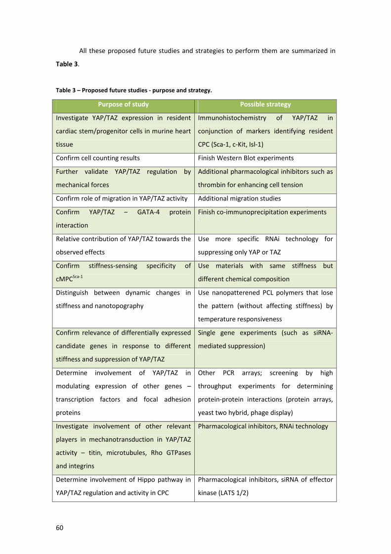
60
All these proposed future studies and strategies to perform them are summarized in
Table 3.
Table 3 – Proposed future studies - purpose and strategy.
Purpose of study Possible strategy
Investigate YAP/TAZ expression in resident
cardiac stem/progenitor cells in murine heart
tissue
Immunohistochemistry of YAP/TAZ in
conjunction of markers identifying resident
CPC (Sca-1, c-Kit, Isl-1)
Confirm cell counting results Finish Western Blot experiments
Further validate YAP/TAZ regulation by
mechanical forces
Additional pharmacological inhibitors such as
thrombin for enhancing cell tension
Confirm role of migration in YAP/TAZ activity Additional migration studies
Confirm YAP/TAZ – GATA-4 protein
interaction
Finish co-immunoprecipitation experiments
Relative contribution of YAP/TAZ towards the
observed effects
Use more specific RNAi technology for
suppressing only YAP or TAZ
Confirm stiffness-sensing specificity of
cMPCSca-1
Use materials with same stiffness but
different chemical composition
Distinguish between dynamic changes in
stiffness and nanotopography
Use nanopatterened PCL polymers that lose
the pattern (without affecting stiffness) by
temperature responsiveness
Confirm relevance of differentially expressed
candidate genes in response to different
stiffness and suppression of YAP/TAZ
Single gene experiments (such as siRNA-
mediated suppression)
Determine involvement of YAP/TAZ in
modulating expression of other genes –
transcription factors and focal adhesion
proteins
Other PCR arrays; screening by high
throughput experiments for determining
protein-protein interactions (protein arrays,
yeast two hybrid, phage display)
Investigate involvement of other relevant
players in mechanotransduction in YAP/TAZ
activity – titin, microtubules, Rho GTPases
and integrins
Pharmacological inhibitors, RNAi technology
Determine involvement of Hippo pathway in
YAP/TAZ regulation and activity in CPC
Pharmacological inhibitors, siRNA of effector
kinase (LATS 1/2)

61
An additional degree of relevance would also be given to this work if the results would
be extended to human cardiac stem/progenitor cells of the same population (Sca-1+).
Despite the fact that the work developed for this thesis and the knowledge generated
from it comprise a fundamental study in biology, the information acquired might be important
to design applications useful for clinical purposes. As such, future stem cell-based therapies
targeting the heart and relying on the use of bioengineered scaffolds should contemplate
YAP/TAZ as modulators of stem/progenitor cell fate, possibly by triggering appropriate cellular
responses towards achieving the desired outcome.
Investigate interaction of YAP/TAZ with
established pathways controlling CPC fate –
Notch and Wnt
Pharmacological inhibitors, RNAi technology
62
5) References
1. Fuster, V. & Kelly, B. B. Chapter 2 - Epidemiology of Cardiovascular Disease. Promoting Cardiovascular Health in the developing world: A critical challenge to
achieve global health 49–123 (2010).
2. WHO. 2005. Preventing chronic diseases: A vital investment. Available at <http://www.who.int/chp/chronic_> (acessed in July 5th 2012)
3. World health statistics 2009. Geneva: World Health Organization.
4. Segers, V. F. M. & Lee, R. T. Stem-cell therapy for cardiac disease. Nature 451, 937–42 (2008).
5. Zammaretti, P. & Jaconi, M. Cardiac tissue engineering: regeneration of the wounded heart. Current opinion in biotechnology 15, 430–4 (2004).
6. Chiu, L. L. Y., Iyer, R. K., Reis, L. A., Nunes, S. S. & Radisic, M. Cardiac tissue engineering: current state and perspectives. Frontiers in Bioscience 17, 1533–1550 (2012).
7. Dar, A., Shachar, M., Leor, J. & Cohen, S. Optimization of cardiac cell seeding and distribution in 3D porous alginate scaffolds. Biotechnology and bioengineering 80, 305–12 (2002).
8. Deng, C. et al. A collagen–chitosan hydrogel for endothelial differentiation and angiogenesis. Tissue Engineering Part A 16, 3099–3109 (2010).
9. Watfchow, J. Three-dimensional cardiomyocytes muscle model reconstitution in a collagen system of embryonic matrix : a new heart. Matrix 683–694
10. Christman, K. L. et al. Injectable fibrin scaffold improves cell transplant survival, reduces infarct expansion, and induces neovasculature formation in ischemic myocardium. Journal of the American College of Cardiology 44, 654–60 (2004).
11. Li, M., Guo, Y., Wei, Y., MacDiarmid, A. G. & Lelkes, P. I. Electrospinning polyaniline-contained gelatin nanofibers for tissue engineering applications. Biomaterials 27, 2705–15 (2006).
12. Xiang, Z., Liao, R., Kelly, M. & Spector, M. Collagen-GAG scaffolds grafted onto myocardial infarcts in a rat model: a delivery vehicle for mesenchymal stem cells. Tissue Engineering 12, 2467–78 (2006).
13. Ott, H. C. et al. Perfusion-decellularized matrix: using nature’s platform to engineer a bioartificial heart. Nature medicine 14, 213–21 (2008).

63
14. Furuta, A. et al. Pulsatile cardiac tissue grafts using a novel three-dimensional cell sheet manipulation technique functionally integrates with the host heart, in vivo. Circulation research 98, 705–12 (2006).
15. Abdel-Latif, A. et al. Adult Bone Marrow–Derived Cells for Cardiac Repair A Systematic Review and Meta-analysis. Archives of internal medicine 167, 989–997 (2007).
16. Wollert, K. C. & Drexler, H. Clinical applications of stem cells for the heart. Circulation research 96, 151–63 (2005).
17. Laflamme, M. a & Murry, C. E. Regenerating the heart. Nature biotechnology 23, 845–56 (2005).
18. Kim, D.-H. et al. Nanoscale cues regulate the structure and function of macroscopic cardiac tissue constructs. Proceedings of the National Academy of
Sciences of the United States of America 107, 565–70 (2010).
19. Chung, C. et al. Hypertrophic phenotype in cardiac cell assemblies solely by structural cues and ensuing self-organization. FASEB journal 25, 851–62 (2011).
20. Badie, N. & Bursac, N. Novel micropatterned cardiac cell cultures with realistic ventricular microstructure. Biophysical journal 96, 3873–85 (2009).
21. Badrossamay, M. R., McIlwee, H. A., Goss, J. a & Parker, K. K. Nanofiber assembly by rotary jet-spinning. Nano letters 10, 2257–61 (2010).
22. Engelmayr Jr, G. C. et al. Accordion-like honeycombs for tissue engineering of cardiac anisotropy. Nature Materials 7, 1003–1010 (2009).
23. Taya, C. Y. et al. Micropatterned matrix directs differentiation of human mesenchymal stem cells towards myocardial lineage. Experimental Cell Research 316, 1159–68 (2010).
24. Sasaki, D. et al. Mass preparation of size-controlled mouse embryonic stem cell aggregates and induction of cardiac differentiation by cell patterning method. Biomaterials 30, 4384–9 (2009).
25. Radisic, M. et al. Oxygen gradients correlate with cell density and cell viability in engineered cardiac tissue. Biotechnology and bioengineering 93, 332–43 (2006).
26. Zimmermann, W.-H. et al. Engineered heart tissue grafts improve systolic and diastolic function in infarcted rat hearts. Nature medicine 12, 452–8 (2006).
27. Tandon, N. et al. Electrical stimulation systems for cardiac tissue engineering. Nature Protocols 4, 155–173 (2009).
64
28. Jacot, J. G., Martin, J. C. & Hunt, D. L. Biphasic electrical field stimulation aids in tissue engineering of multicell-type cardiac organoids. Tissue Engineering Part A 43, 1–13 (2011).
29. Beitnes, J. O., Lunde, K., Brinchmann, J. E. & Aakhus, S. Stem cells for cardiac repair in acute myocardial infarction. Cell 1015–1025 (2011).
30. Reinecke, H., Minami, E., Poppa, V. & Murry, C. E. Evidence for fusion between cardiac and skeletal muscle cells. Circulation research 94, e56–60 (2004).
31. Winitsky, S. O. et al. Adult murine skeletal muscle contains cells that can differentiate into beating cardiomyocytes in vitro. PLoS biology 3, e87 (2005).
32. Chen, M. Q. et al. Modeling Conduction in Host-Graft Interactions between Stem Cell Grafts and Cardiomyocytes. Engineering in Medicine and Biology
Society. 2009, 6014–7 (2009).
33. Reinecke, H., Zhang, M., Bartosek, T. & Murry, C. Survival, integration, and differentiation of cardiomyocyte grafts a study in normal and injured rat hearts. Circulation 100, 193–202 (1999).
34. Young, P. P., Vaughan, D. E. & Hatzopoulos, A. K. Biologic properties of endothelial progenitor cells and their potential for cell therapy. Progress in
cardiovascular diseases 49, 421–9 (2007).
35. Miyahara, Y. et al. Monolayered mesenchymal stem cells repair scarred myocardium after myocardial infarction. Nature medicine 12, 459–65 (2006).
36. Tomescot, A. et al. Differentiation in vivo of cardiac committed human embryonic stem cells in postmyocardial infarcted rats. Stem cells (Dayton, Ohio) 25, 2200–5 (2007).
37. Radisic, M. et al. High-density seeding of myocyte cells for cardiac tissue engineering. Biotechnology and bioengineering 82, 403–14 (2003).
38. Bertipaglia, B. et al. Cell Characterization of Porcine Aortic Valve and Decellularized Leaflets Repopulated With Aortic Valve Interstitial Cells : The VESALIO Project (Vitalitate Exornatum Succedaneum Aorticum Labore Ingenioso Obtenibitur ). Annals of Thoracic Surgery, The (2003).
39. Matsubayashi, K. et al. Improved left ventricular aneurysm repair with bioengineered vascular smooth muscle grafts. Circulation 108, 219–25 (2003).
40. Fahime, E. E. & Tremblay, J. Chapter 2- Myocardial Regeneration:Which Cell and Why. Stem Cell Therapy and Tissue Engineering for Cardiovascular Repair 25–35 (2006).
65
41. Messina, E., Giacomello, A. & Marbán, E. Chapter 7 - Endogenous Stem Cells. Stem Cells and Myocardial Regeneration 83–115 (2007).
42. Di Nardo, P., Forte, G., Ahluwalia, A. & Minieri, M. Cardiac progenitor cells: potency and control. Journal of cellular physiology 224, 590–600 (2010).
43. Bearzi, C. et al. Human cardiac stem cells. Cancer 104, (2007).
44. Anversa, P., Kajstura, J., Leri, A. & Bolli, R. Life and death of cardiac stem cells: a paradigm shift in cardiac biology. Circulation 113, 1451–63 (2006).
45. Quaini, F. et al. Chimerism of the transplanted heart. New England Journal of
Medicine 346, 5–15 (2002).
46. Bergmann, O. et al. Evidence for cardiomyocyte renewal in humans. Science
(New York, N.Y.) 324, 98–102 (2009).
47. Leri, A., Kajstura, J. A. N. & Anversa, P. Cardiac Stem Cells and Mechanisms of Myocardial Regeneration. Physiological Reviews 1373–1416 (2005).
48. Laugwitz, K., Moretti, A. & Lam, J. Postnatal isl1 1 cardioblasts enter fully differentiated cardiomyocyte lineages. 440, 433–440 (2003).
49. Beltrami, A. P. et al. Adult Cardiac Stem Cells Are Multipotent and Support Myocardial Regeneration. Evaluation 114, 763–776 (2009).
50. Holmes, C. & Stanford, W. L. Concise review: stem cell antigen-1: expression, function, and enigma. Stem cells (Dayton, Ohio) 25, 1339–47 (2007).
51. Oh, H. et al. Cardiac progenitor cells from adult myocardium : Homing, differentiation , and fusion after infarction. October (2003).
52. Wang, X. et al. The role of the sca-1+/CD31- cardiac progenitor cell population in postinfarction left ventricular remodeling. Stem cells (Dayton, Ohio) 24, 1779–88 (2006).
53. Smits, A. M. et al. Human cardiomyocyte progenitor cells differentiate into functional mature cardiomyocytes: an in vitro model for studying human cardiac physiology and pathophysiology. Nature protocols 4, 232–43 (2009).
54. Oyama, T. et al. Cardiac side population cells have a potential to migrate and differentiate into cardiomyocytes in vitro and in vivo. Journal of Cell Biology 176, 329–41 (2007).
55. Messina, E. et al. Isolation and expansion of adult cardiac stem cells from human and murine heart. Circulation research 95, 911–21 (2004).
66
56. Zimmermann, W.-H. et al. Heart muscle engineering: an update on cardiac muscle replacement therapy. Cardiovascular research 71, 419–29 (2006).
57. Simpson, D. & Jr, S. C. D. Lost in Translation : What is Limiting Cardiomyoplasty and Can Tissue Engineering Help? Cell Research 4, 210–223 (2011).
58. Bolli, R. et al. Cardiac stem cells in patients with ischaemic cardiomyopathy (SCIPIO): initial results of a randomised phase 1 trial. The Lancet 378, 1847–1857 (2011).
59. Makkar, R. R. et al. Intracoronary cardiosphere-derived cells for heart regeneration after myocardial infarction (CADUCEUS): a prospective, randomised phase 1 trial. Lancet 379, 895–904 (2012).
60. Mishra, R. et al. Characterization and functionality of cardiac progenitor cells in congenital heart patients. Circulation 123, 364–73 (2011).
61. Radisic, M. et al. Medium perfusion enables engineering of compact and contractile cardiac tissue. American journal of physiology. Heart and circulatory
physiology 286, H507–16 (2004).
62. Zhang, M. et al. Cardiomyocyte grafting for cardiac repair: graft cell death and anti-death strategies. Journal of molecular and cellular cardiology 33, 907–21 (2001).
63. Scadden, D. T. The stem-cell niche as an entity of action. Nature 441, 1075–9 (2006).
64. Forte, G. et al. Criticality of the biological and physical stimuli array inducing resident cardiac stem cell determination. Stem cells (Dayton, Ohio) 26, 2093–103 (2008).
65. Pagliari, S. et al. Cooperation of biological and mechanical signals in cardiac progenitor cell differentiation. Advanced materials 23, 514–8 (2011).
66. Guilak, F., Cohen, D. M., Estes, B. T., Gimble, J. M. & Chen, C. S. Control of stem cell fate by physical interaction with the extracellular matrix. Cell Stem Cell 5, 17–26 (2009).
67. Ingber, D. Extracellular matrix and cell shape: Potential control points for inhibition of angiogenesis. Journal of Cellular Biochemistry 47, 236–241 (1997).
68. Chen, C. S., Mrksich, M., Huang, S., Whitesides, G. M. & Ingber, D. E. Micropatterned surfaces for control of cell shape, position, and function. Biotechnology progress 14, 356–63 (1998).
67
69. McBeath, R., Pirone, D. M., Nelson, C. M., Bhadriraju, K. & Chen, C. S. Cell shape, cytoskeletal tension, and RhoA regulate stem cell lineage commitment. Developmental cell 6, 483–95 (2004).
70. Arnold, M. et al. Activation of integrin function by nanopatterned adhesive interfaces. Chemphyschem : a European journal of chemical physics and physical
chemistry 5, 383–8 (2004).
71. Ingber, D. E. The mechanochemical basis of cell and tissue regulation. Mechanics
& chemistry of biosystems : MCB 1, 53–68 (2004).
72. Engler, A. J., Sen, S., Sweeney, H. L. & Discher, D. E. Matrix elasticity directs stem cell lineage specification. Cell 126, 677–89 (2006).
73. Even-Ram, S., Artym, V. & Yamada, K. M. Matrix control of stem cell fate. Cell 126, 645–7 (2006).
74. Ingber, D. Mechanobiology and diseases of mechanotransduction. Annals of
Medicine 35, 564–577 (2003).
75. Terraciano, V. et al. Differential response of adult and embryonic mesenchymal progenitor cells to mechanical compression in hydrogels. Stem cells (Dayton,
Ohio) 25, 2730–8 (2007).
76. Katsumi, A., Orr, a W., Tzima, E. & Schwartz, M. A. Integrins in mechanotransduction. The Journal of biological chemistry 279, 12001–4 (2004).
77. Matthews, B. D. et al. Mechanical properties of individual focal adhesions probed with a magnetic microneedle. Biochemical and biophysical research
communications 313, 758–64 (2004).
78. Hynes, R. O. The extracellular matrix: not just pretty fibrils. Science (New York,
N.Y.) 326, 1216–9 (2009).
79. Huveneers, S. & Danen, E. H. J. Adhesion signaling - crosstalk between integrins, Src and Rho. Journal of cell science 122, 1059–69 (2009).
80. Schoenwaelder, S. M. & Burridge, K. Bidirectional signaling between the cytoskeleton and integrins. Current opinion in cell biology 11, 274–86 (1999).
81. Pasapera, A. M., Schneider, I. C., Rericha, E., Schlaepfer, D. D. & Waterman, C. M. Myosin II activity regulates vinculin recruitment to focal adhesions through FAK-mediated paxillin phosphorylation. The Journal of cell biology 188, 877–90 (2010).
82. Tadokoro, S. et al. Talin binding to integrin beta tails: a final common step in integrin activation. Science (New York, N.Y.) 302, 103–6 (2003).
68
83. Laukaitis, C. M., Webb, D. J., Donais, K. & Horwitz, a F. Differential dynamics of alpha 5 integrin, paxillin, and alpha-actinin during formation and disassembly of adhesions in migrating cells. The Journal of cell biology 153, 1427–40 (2001).
84. Webb, D. J. et al. FAK-Src signalling through paxillin, ERK and MLCK regulates adhesion disassembly. Nature cell biology 6, 154–61 (2004).
85. Wiseman, P. W. et al. Spatial mapping of integrin interactions and dynamics during cell migration by image correlation microscopy. Journal of cell science 117, 5521–34 (2004).
86. Choi, C. K. et al. Actin and alpha-actinin orchestrate the assembly and maturation of nascent adhesions in a myosin II motor-independent manner. Nature cell biology 10, 1039–50 (2008).
87. Lee, H.-S. et al. Characterization of an actin-binding site within the talin FERM domain. Journal of molecular biology 343, 771–84 (2004).
88. Ballestrem, C. et al. Molecular mapping of tyrosine-phosphorylated proteins in focal adhesions using fluorescence resonance energy transfer. Journal of cell
science 119, 866–75 (2006).
89. Wolfenson, H., Bershadsky, A., Henis, Y. I. & Geiger, B. Actomyosin-generated tension controls the molecular kinetics of focal adhesions. Journal of cell science 124, 1425–32 (2011).
90. Alenghat, F. J. & Ingber, D. E. Mechanotransduction: all signals point to cytoskeleton, matrix, and integrins. Science’s STKE : signal transduction
knowledge environment 2002, pe6 (2002).
91. Parsons, J. T., Horwitz, A. R. & Schwartz, M. A. Cell adhesion: integrating cytoskeletal dynamics and cellular tension. Molecular Cell 11, 633–643 (2010).
92. Ridley, A. J. et al. Cell migration: integrating signals from front to back. Science
(New York, N.Y.) 302, 1704–9 (2003).
93. Clark, E. & Brugge, J. Integrins and signal transduction pathways: the road taken. Science 268, 233–9 (1995).
94. Nix, D. & Beckerle, M. C. Nuclear-cytoplasmic shuttling of the focal contact protein, zyxin: a potential mechanism for communication between sites of cell adhesion and the nucleus. The Journal of cell biology 138, 1139–47 (1997).
95. Hervy, M., Hoffman, L. & Beckerle, M. C. From the membrane to the nucleus and back again: bifunctional focal adhesion proteins. Current opinion in cell
biology 18, 524–32 (2006).

69
96. Kadrmas, J. & Beckerle, M. The LIM domain: from the cytoskeleton to the nucleus. Nature Reviews Molecular Cell Biology 5, 920–31 (2004).
97. Wang, Y. & Gilmore, T. D. Zyxin and paxillin proteins: focal adhesion plaque LIM domain proteins go nuclear. Biochimica et Biophysica Acta (BBA) - Molecular Cell
Research 1593, 115–120 (2003).
98. Dong, J.-M., Lau, L.-S., Ng, Y.-W., Lim, L. & Manser, E. Paxillin nuclear-cytoplasmic localization is regulated by phosphorylation of the LD4 motif: evidence that nuclear paxillin promotes cell proliferation. The Biochemical
journal 418, 173–84 (2009).
99. Silver, F. & Siperko, L. Mechanosensing and mechanochemical transduction: how is mechanical energy sensed and converted into chemical energy in an extracellular matrix? Critical Reviews in Biomedical Engineering 31, 255–331 (2003).
100. Silver, F. H., DeVore, D. & Siperko, L. M. Role of mechanophysiology in aging of ECM: effects of changes in mechanochemical transduction. Journal of applied
physiology (Bethesda, Md. : 1985) 95, 2134–41 (2003).
101. Liu, C., Huang, W. & Lei, Q. Regulation and function of the TAZ transcription co-activator. Journal of Biochemistry and Molecular Biology 2, 247–256 (2011).
102. Hong, J. & Yaffe, M. B. TAZ, a β-Catenin-Like Molecule That Regulates Mesenchymal Stem Cell Differentiation. Cell Cycle 5, 176–179 (2006).
103. Cui, C. B., Cooper, L. F., Yang, X., Karsenty, G. & Aukhil, I. Transcriptional coactivation of bone-specific transcription factor Cbfa1 by TAZ. Molecular and
cellular biology 23, 1004–13 (2003).
104. Hong, J.-H. et al. TAZ, a transcriptional modulator of mesenchymal stem cell differentiation. Science (New York, N.Y.) 309, 1074–8 (2005).
105. Murakami, M., Nakagawa, M., Olson, E. N. & Nakagawa, O. A WW domain protein TAZ is a critical coactivator for TBX5, a transcription factor implicated in Holt-Oram syndrome. Proceedings of the National Academy of Sciences of the
United States of America 102, 18034–9 (2005).
106. Mahoney, W. M., Hong, J.-H., Yaffe, M. B. & Farrance, I. K. G. The transcriptional co-activator TAZ interacts differentially with transcriptional enhancer factor-1 (TEF-1) family members. The Biochemical journal 388, 217–25 (2005).
107. Jeong, H. et al. TAZ as a novel enhancer of MyoD-mediated myogenic differentiation. The FASEB journal : official publication of the Federation of
American Societies for Experimental Biology 24, 3310–20 (2010).

70
108. Staley, B. K. & Irvine, K. D. Warts and Yorkie mediate intestinal regeneration by influencing stem cell proliferation. Current biology : CB 20, 1580–7 (2010).
109. Heallen, T. et al. Hippo pathway inhibits Wnt signaling to restrain cardiomyocyte proliferation and heart size. Science (New York, N.Y.) 332, 458–61 (2011).
110. Varelas, X. et al. TAZ controls Smad nucleocytoplasmic shuttling and regulates human embryonic stem-cell self-renewal. Nature cell biology 10, 837–48 (2008).
111. Chan, S. W. et al. A role for TAZ in migration, invasion, and tumorigenesis of breast cancer cells. Cancer research 68, 2592–8 (2008).
112. Lei, Q.-Y. et al. TAZ promotes cell proliferation and epithelial-mesenchymal transition and is inhibited by the hippo pathway. Molecular and cellular biology 28, 2426–36 (2008).
113. Cordenonsi, M. et al. The Hippo transducer TAZ confers cancer stem cell-related traits on breast cancer cells. Cell 147, 759–72 (2011).
114. Hossain, Z. et al. Glomerulocystic kidney disease in mice with a targeted inactivation of Wwtr1. Proceedings of the National Academy of Sciences of the
United States of America 104, 1631–6 (2007).
115. Makita, R. et al. Multiple renal cysts, urinary concentration defects, and pulmonary emphysematous changes in mice lacking TAZ. American journal of
physiology. Renal physiology 294, F542–53 (2008).
116. Edgar, B. From cell structure to transcription: Hippo forges a new path. Cell 124, 267–73 (2006).
117. Liu, C.-Y. et al. PP1 cooperates with ASPP2 to dephosphorylate and activate TAZ. The Journal of biological chemistry 286, 5558–66 (2011).
118. Schlegelmilch, K. et al. Yap1 acts downstream of α-catenin to control epidermal proliferation. Cell 144, 782–95 (2011).
119. Liu, C.-Y. et al. The hippo tumor pathway promotes TAZ degradation by phosphorylating a phosphodegron and recruiting the SCF{beta}-TrCP E3 ligase. The Journal of biological chemistry 285, 37159–69 (2010).
120. Zhao, B., Li, L., Tumaneng, K., Wang, C.-Y. & Guan, K.-L. A coordinated phosphorylation by Lats and CK1 regulates YAP stability through SCF(beta-TRCP). Genes & development 24, 72–85 (2010).
121. Chan, S. W. et al. Hippo pathway-independent restriction of TAZ and YAP by angiomotin. The Journal of biological chemistry 286, 7018–26 (2011).
71
122. Remue, E. et al. TAZ interacts with zonula occludens-1 and -2 proteins in a PDZ-1 dependent manner. FEBS letters 584, 4175–80 (2010).
123. Hergovich, A. & Hemmings, B. a TAZ-mediated crosstalk between Wnt and Hippo signaling. Developmental cell 18, 508–9 (2010).
124. Varelas, X. et al. The Hippo pathway regulates Wnt/beta-catenin signaling. Developmental cell 18, 579–91 (2010).
125. Dupont, S. et al. Role of YAP/TAZ in mechanotransduction. Nature 474, 179–83 (2011).
126. Schroeder, M. C. & Halder, G. Regulation of the Hippo pathway by cell architecture and mechanical signals. Seminars in cell & developmental biology 1–9 (2012).
127. Casella, J., Flanagan, M. & Lin, S. Cytochalasin D inhibits actin polymerization and induces depolymerization of actin filaments formed during platelet shape change. Nature 293, 302–305 (1981).
128. Tse, J. R. & Engler, A. J. Preparation of hydrogel substrates with tunable mechanical properties. Current protocols in cell biology / editorial board, Juan S.
Bonifacino ... [et al.] Chapter 10, Unit 10.16 (2010).
129. Discher, D. E., Janmey, P. & Wang, Y.-L. Tissue cells feel and respond to the stiffness of their substrate. Science (New York, N.Y.) 310, 1139–43 (2005).
130. Ebara, M., Uto, K., Idota, N., Hoffman, J. M. & Aoyagi, T. Shape-Memory Surface with Dynamically Tunable Nano-Geometry Activated by Body Heat. Advanced
Materials 24, 273–278 (2012).
131. Lancaster, M. V. & Fields, R. Antibiotic and Cytotoxic Drug Susceptibility Assays using Resazurin and Poising Agents. U.S. Patent No. 5,501,959 (1996).
132. Zhang, H. et al. TEAD transcription factors mediate the function of TAZ in cell growth and epithelial-mesenchymal transition. The Journal of biological
chemistry 284, 13355–62 (2009).
133. Wójtowicz, A. et al. Zyxin mediation of stretch-induced gene expression in human endothelial cells. Circulation research 107, 898–902 (2010).
134. Scholzen, T. & Gerdes, J. The Ki-67 Protein : From the Known and Unknown. Journal of Cellular Physiology 182, 311–322 (2000).
135. Soofi, S., Last, J., Liliensiek, S., Nealey, P. & Murphy, C. The elastic modulus of MatrigelTM as determined by atomic force microscopy. Journal of Structural
Biology 167, 216–219 (2010).
72
136. Bialojan, C. & Takai, a Inhibitory effect of a marine-sponge toxin, okadaic acid, on protein phosphatases. Specificity and kinetics. The Biochemical journal 256, 283–90 (1988).
137. Liu, C.-Y. et al. PP1 cooperates with ASPP2 to dephosphorylate and activate TAZ. The Journal of biological chemistry 286, 5558–66 (2011).
138. Chen, C. S., Alonso, J. L., Ostuni, E., Whitesides, G. M. & Ingber, D. E. Cell shape provides global control of focal adhesion assembly. Biochemical and biophysical
research communications 307, 355–61 (2003).
139. Tseng, P.-C. et al. Spontaneous osteogenesis of MSCs cultured on 3D microcarriers through alteration of cytoskeletal tension. Biomaterials 33, 556–64 (2012).
140. Goeckeler, Z. M., Bridgman, P. C. & Wysolmerski, R. B. Nonmuscle myosin II is responsible for maintaining endothelial cell basal tone and stress fiber integrity. 26506, 994–1006 (2008).
141. Wang, N., Ostuni, E., Whitesides, G. M. & Ingber, D. E. Micropatterning tractional forces in living cells. Cell motility and the cytoskeleton 52, 97–106 (2002).
142. Blain, E. Mechanical regulation of matrix metalloproteinases. Frontiers in
Bioscience 12, 507–527 (2007).
143. Gautel, M. Cytoskeletal protein kinases: titin and its relations in mechanosensing. Pflügers Archiv : European journal of physiology 462, 119–34 (2011).
144. Modrianský, M. & Dvorák, Z. Microtubule disruptors and their interaction with biotransformation enzymes. Biomedical papers of the Medical Faculty of the
University Palacký, Olomouc, Czechoslovakia 149, 213–5 (2005).
145. Franchini, K. G., Clemente, C. F. M. Z. & Marin, T. M. Focal adhesion kinase signaling in cardiac hypertrophy and failure. Brazilian journal of medical and
biological research 42, 44–52 (2009).
146. Vadali, K., Cai, X. & Schaller, M. D. Focal adhesion kinase: an essential kinase in the regulation of cardiovascular functions. IUBMB life 59, 709–16 (2007).
147. Zhao, B., Tumaneng, K. & Guan, K.-L. The Hippo pathway in organ size control, tissue regeneration and stem cell self-renewal. Nature cell biology 13, 877–83 (2011).
148. Collesi, C., Zentilin, L., Sinagra, G. & Giacca, M. Notch1 signaling stimulates proliferation of immature cardiomyocytes. The Journal of cell biology 183, 117–28 (2008).

73
149. Koyanagi, M. et al. Non-canonical Wnt signaling enhances differentiation of human circulating progenitor cells to cardiomyogenic cells. The Journal of
biological chemistry 280, 16838–42 (2005).
150. Forte, G. et al. Substrate stiffness modulates gene expression and phenotype in neonatal cardiomyocytes in vitro. Tissue Engineering In press, (2012).


![The role of YAP/TAZ activity in cancer metabolic reprogramming · 2018. 9. 3. · YAP/TAZ functions as tumor suppressors [11, 12]. Im-munohistochemical staining revealed that the](https://img.pdfslide.net/doc/110x75/60b83e231181bf12a21ddfce/the-role-of-yaptaz-activity-in-cancer-metabolic-reprogramming-2018-9-3-yaptaz.jpg)