Embed Size (px)
Citation preview
MEDIASTINUMMEDIASTINUM
14.12.08 .14.12.08 .
Dr.KvkDr.Kvk
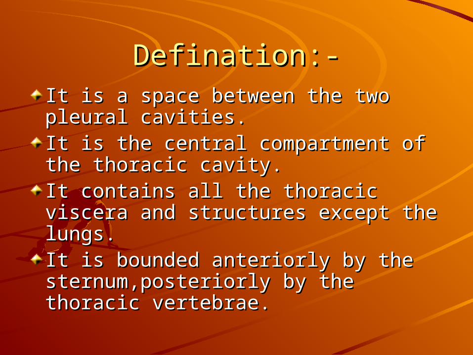
Defination:-Defination:-It is a space between the two pleural It is a space between the two pleural cavities.cavities.It is the central compartment of the It is the central compartment of the thoracic cavity.thoracic cavity.It contains all the thoracic viscera It contains all the thoracic viscera and structures except the lungs.and structures except the lungs.It is bounded anteriorly by the It is bounded anteriorly by the sternum,posteriorly by the thoracic sternum,posteriorly by the thoracic vertebrae.vertebrae.

DIVISIONS OF MEDIASTINUMDIVISIONS OF MEDIASTINUMIt is divided by an immaginary plane It is divided by an immaginary plane passing through the sternal angle passing through the sternal angle anteriorly and posteriorly by the anteriorly and posteriorly by the intervertebral discs between T4 &T5.intervertebral discs between T4 &T5.
This plane is called transverse thoracic This plane is called transverse thoracic plane.plane.
Superior mediastinum lies above this Superior mediastinum lies above this plane.plane.
Inferior mediastinum lies below this plane.Inferior mediastinum lies below this plane.
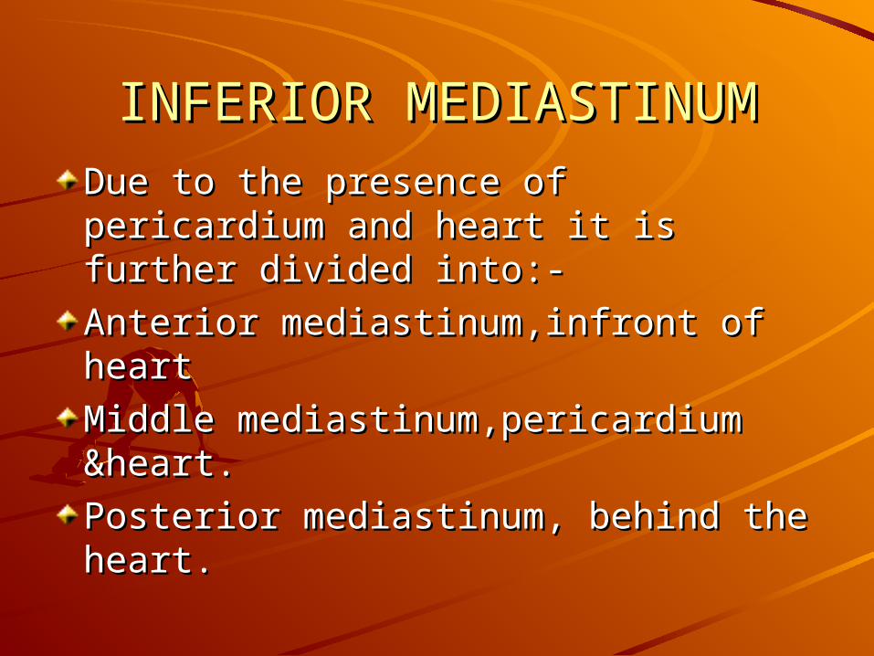
INFERIOR MEDIASTINUMINFERIOR MEDIASTINUM
Due to the presence of pericardium Due to the presence of pericardium and heart it is further divided into:-and heart it is further divided into:-
Anterior mediastinum,infront of heartAnterior mediastinum,infront of heart
Middle mediastinum,pericardium Middle mediastinum,pericardium &heart.&heart.
Posterior mediastinum, behind the Posterior mediastinum, behind the heart.heart.
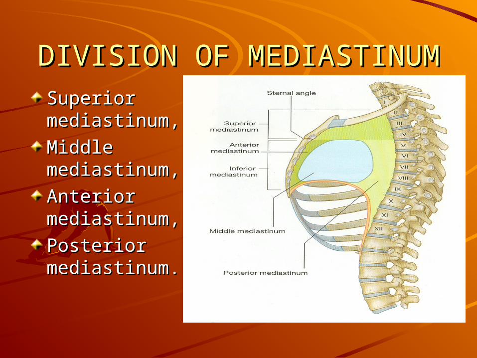
DIVISION OF MEDIASTINUMDIVISION OF MEDIASTINUMSuperior Superior mediastinum,mediastinum,
Middle Middle mediastinum,mediastinum,
Anterior Anterior mediastinum,mediastinum,
Posterior Posterior mediastinum.mediastinum.
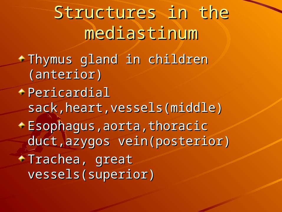
Structures in the mediastinumStructures in the mediastinum
Thymus gland in children (anterior)Thymus gland in children (anterior)
Pericardial Pericardial sack,heart,vessels(middle)sack,heart,vessels(middle)
Esophagus,aorta,thoracic Esophagus,aorta,thoracic duct,azygos vein(posterior)duct,azygos vein(posterior)
Trachea, great vessels(superior)Trachea, great vessels(superior)

SUPERIOR MEDIASTINUMSUPERIOR MEDIASTINUMIt lies above the It lies above the transverse thoracic transverse thoracic plane.plane.
It contains aortic It contains aortic arches, thymus, arches, thymus, terminal part of terminal part of S.V.C.,brachiocephS.V.C.,brachiocephalic veins,Trachea alic veins,Trachea and esophagus.and esophagus.
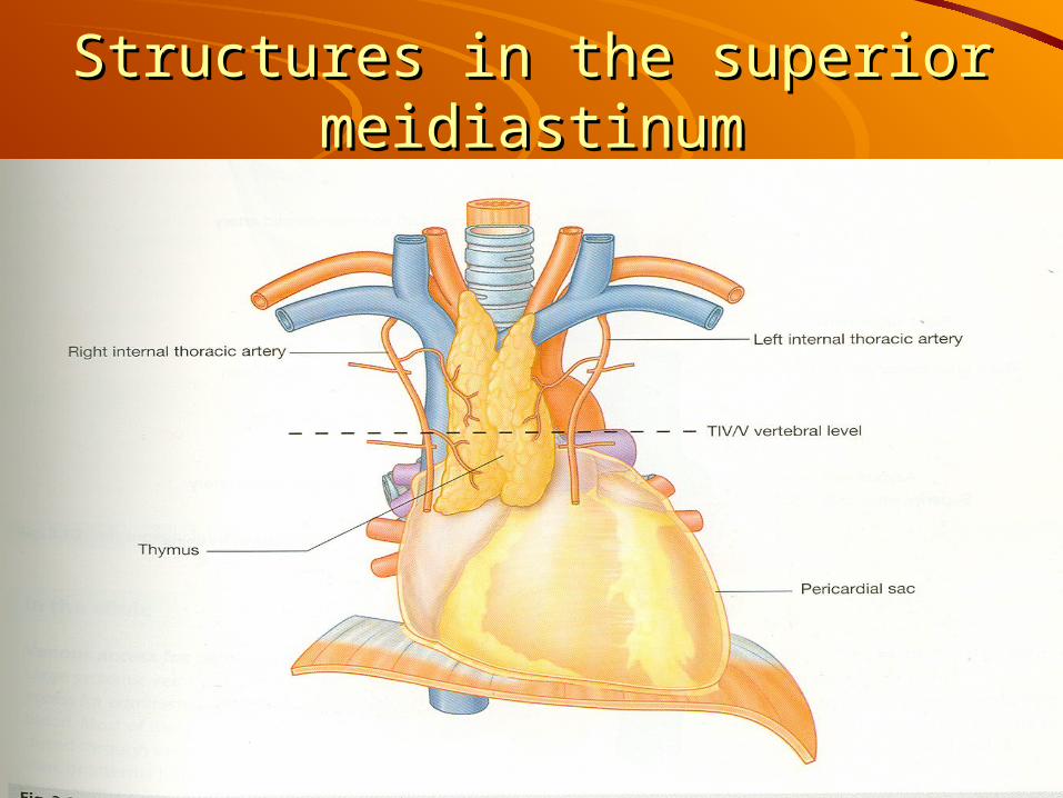
Structures in the superior Structures in the superior meidiastinummeidiastinum

Thymus Thymus Thymus is located Thymus is located in the anterior in the anterior mediastinum mediastinum between the between the pericardium and pericardium and sternumsternumThymus is present Thymus is present only in childrenonly in childrenAfter puberty After puberty thymus thymus degenaratesdegenarates

Structures in the posterior Structures in the posterior mediastinummediastinum
ANTERIOR MEDIASTINUMANTERIOR MEDIASTINUMIt contains loose areolar connective It contains loose areolar connective tissue,tissue,Lymphatic vessels,Lymphatic vessels,2-3 anterior mediastinal lymph 2-3 anterior mediastinal lymph nodes.nodes.Branches of internal mammary Branches of internal mammary artery,artery,Pulmonary arteries,pulmonary veins,Pulmonary arteries,pulmonary veins,Phrenic nerves.Phrenic nerves.
POSTERIOR MEDIASTINUMPOSTERIOR MEDIASTINUM
Thoracic part of aorta,Thoracic part of aorta,
Azygous and hemiazygous veins,Azygous and hemiazygous veins,
Vagus and splancnic nerves,Vagus and splancnic nerves,
Esophagus,Esophagus,
Thoracic duct and lymph nodes.Thoracic duct and lymph nodes.
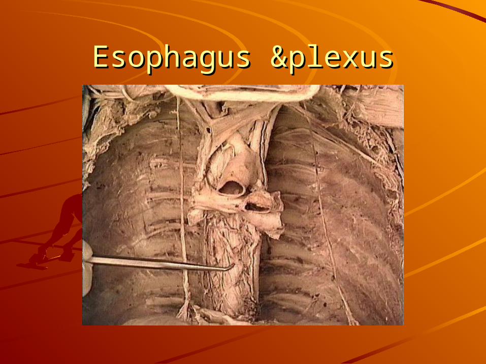
Esophagus &plexusEsophagus &plexus
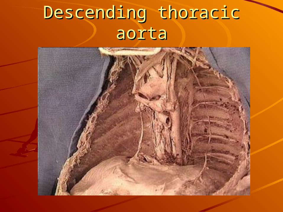
Descending thoracic aortaDescending thoracic aorta

APPLIED ANATOMYAPPLIED ANATOMYTumours arising from thymus,Thymoma Tumours arising from thymus,Thymoma compresses the mediastinum.compresses the mediastinum.Pancoast tumour may compress the Pancoast tumour may compress the sympathetic ganglion and cause Horner’s sympathetic ganglion and cause Horner’s syndrome.syndrome.Pericardial effusion-cardiac tamponade Pericardial effusion-cardiac tamponade due to pericardial effusion.due to pericardial effusion.Hilar group of lymph nodes enlargment Hilar group of lymph nodes enlargment leads to dysphagia and dyspnoea.leads to dysphagia and dyspnoea.Compression over recurrent laryngeal Compression over recurrent laryngeal nerves lead to hoarseness of voice.nerves lead to hoarseness of voice.
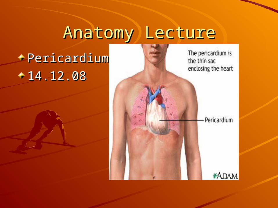
Anatomy LectureAnatomy Lecture
Pericardium Pericardium
14.12.0814.12.08
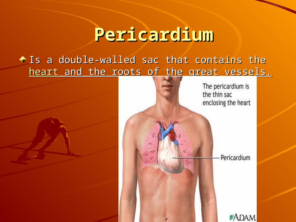
PericardiumPericardiumIs a double-walled sac that contains the Is a double-walled sac that contains the heartheart and and the roots of the great vessels.the roots of the great vessels.

Layers of pericardiumLayers of pericardium

PERICARDIUMPERICARDIUMPeri= aroundPeri= around
Cardium= heartCardium= heart
It is a fibro-serous sacIt is a fibro-serous sac
which surround thewhich surround the
Heart and the roots Heart and the roots
of the large vesselsof the large vessels
It consist of:It consist of:
1- The fibrous pericardium1- The fibrous pericardium
2-The serous pericardium2-The serous pericardium
two layer:two layer:
parietal layer parietal layer
Visceral layerVisceral layer
3- the sinuses of the pericardium3- the sinuses of the pericardium
(transverse – oblique )(transverse – oblique )
FIBROUS &SEROUS FIBROUS &SEROUS PERICARDIUMPERICARDIUM
Fibrous pericardium is made up of Fibrous pericardium is made up of dense conective tissuedense conective tissue
Inferiorly is connected to diaphragm Inferiorly is connected to diaphragm by percardio phrenic ligamentby percardio phrenic ligament
Interiorly connected to the sternum by Interiorly connected to the sternum by sterno pericardial ligament sterno pericardial ligament
Superiorly,completely fused with the Superiorly,completely fused with the roots of the great vessels of the heart roots of the great vessels of the heart
PLUERA&PERICARDIUMPLUERA&PERICARDIUM
Most of the pericardium surface is Most of the pericardium surface is covered pluera the lung except and covered pluera the lung except and area posterior to sternum at the level area posterior to sternum at the level of of 44thth 5 5thth costal cartilage . costal cartilage .
AnteriorlyAnteriorly related to anterior related to anterior mediastinum and thymus in childrenmediastinum and thymus in children
Posteriorly Posteriorly related to related to esophagus,descenting aortaesophagus,descenting aorta
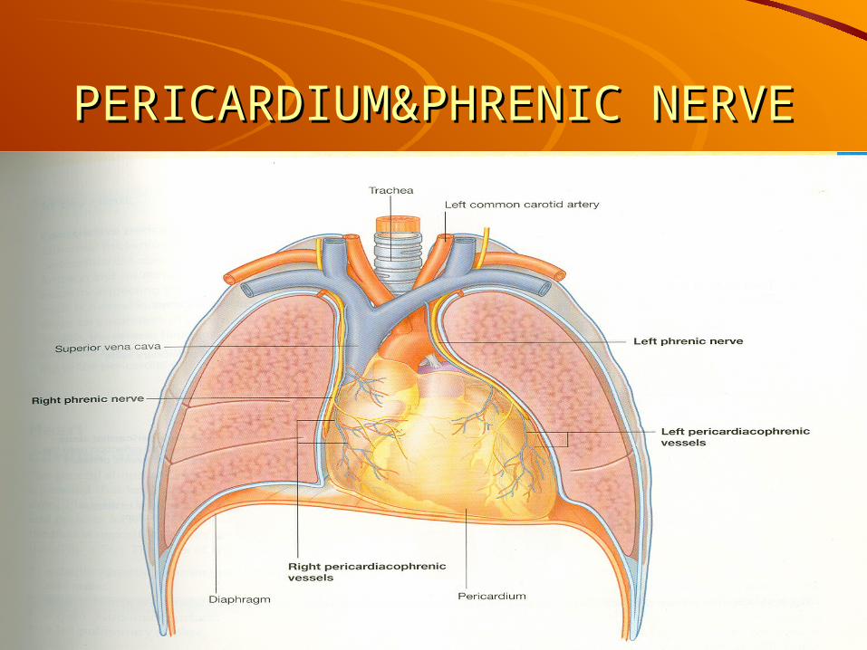
PERICARDIUM&PHRENIC PERICARDIUM&PHRENIC NERVENERVE


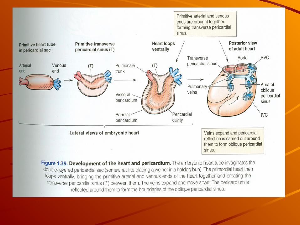
Formation of pericardial cavityFormation of pericardial cavity
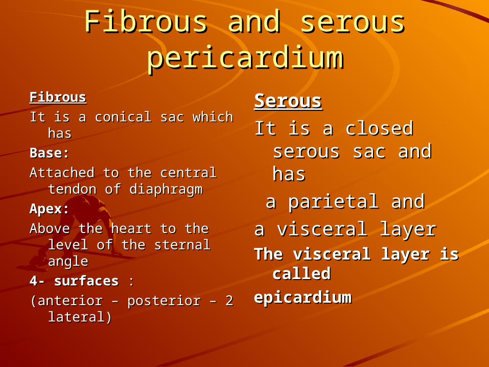
Fibrous and serous pericardiumFibrous and serous pericardiumFibrousFibrous
It is a conical sac which has It is a conical sac which has
Base:Base:
Attached to the central Attached to the central tendon of diaphragmtendon of diaphragm
Apex:Apex:
Above the heart to the level Above the heart to the level of the sternal angleof the sternal angle
4- surfaces4- surfaces : :
(anterior – posterior – 2 (anterior – posterior – 2 lateral)lateral)
SerousSerous
It is a closed serous It is a closed serous sac and hassac and has
a parietal and a parietal and
a visceral layera visceral layerThe visceral layer is The visceral layer is
called called
epicardiumepicardium
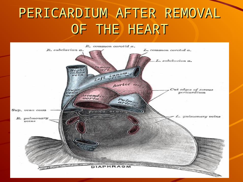
PERICARDIUM AFTER PERICARDIUM AFTER REMOVAL OF THE HEARTREMOVAL OF THE HEART
InnervationsInnervations
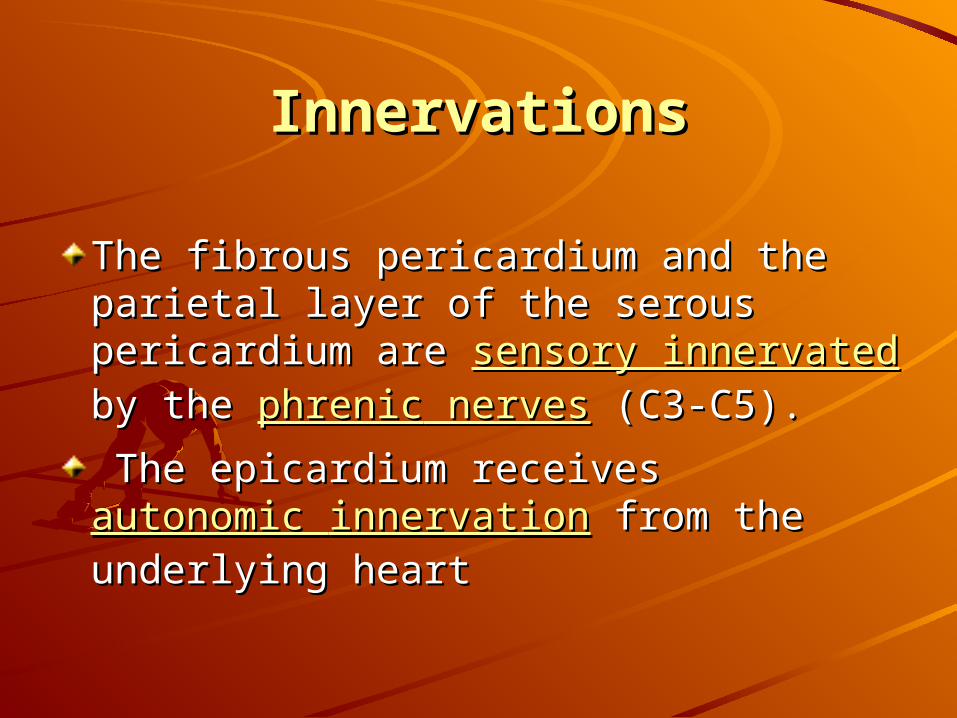
The fibrous pericardium and the The fibrous pericardium and the parietal layer of the serous parietal layer of the serous pericardium are pericardium are sensory innervatedsensory innervated by the by the phrenicphrenic nerves nerves (C3-C5). (C3-C5).
The epicardium receives The epicardium receives autonomic autonomic innervationinnervation from the underlying from the underlying heartheart
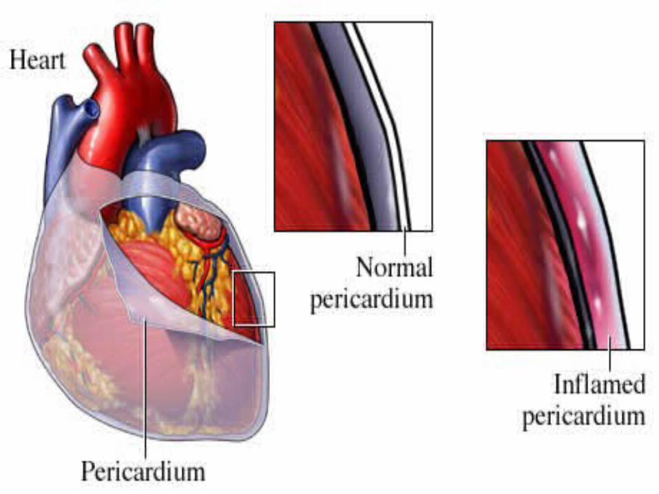
DiseasesDiseases
PericarditisPericarditis is is inflammationinflammation of the of the pericardium. It can cause fluid to pericardium. It can cause fluid to build up in the sac (build up in the sac (pericardial effusionpericardial effusion).).
Excessive amounts of fluid may lead Excessive amounts of fluid may lead to to cardiac cardiac tamponadetamponade
Pericardio centhesis removal of fluid Pericardio centhesis removal of fluid from the pericardial cavity by from the pericardial cavity by inserting a needle inserting a needle

THANKYOU THANKYOU
Dr.KUMARDr.KUMAR
Associate Professor Associate Professor


![[14.12.08] 행렬대수(ShaderStudy)](https://img.pdfslide.net/doc/110x75/55a0a1a31a28abec2f8b47ca/141208-shaderstudy.jpg)