Embed Size (px)
Citation preview
Research ArticleMelanomas and Dysplastic Nevi Differ in EpidermalCD1c+ Dendritic Cell Count
Grzegorz Dyduch1 Katarzyna Ewa Tyrak2 Anna Glajcar1 Joanna Szpor1
Magdalena Ulatowska-BiaBas1 and Krzysztof OkoN1
1Chair of Pathomorphology Faculty of Medicine Jagiellonian University Medical College Grzegorzecka 16 31-351 Krakow Poland2II Chair of Internal Medicine Faculty of Medicine Jagiellonian University Medical College Skawinska 8 31-066 Krakow Poland
Correspondence should be addressed to Krzysztof Okon mpokoncyf-kredupl
Received 18 December 2016 Revised 30 January 2017 Accepted 6 February 2017 Published 26 February 2017
Academic Editor Vasiliki Galani
Copyright copy 2017 Grzegorz Dyduch et al This is an open access article distributed under the Creative Commons AttributionLicense which permits unrestricted use distribution and reproduction in any medium provided the original work is properlycited
Background Dendritic cells could be involved in immune surveillance of highly immunogenic tumors such as melanoma Theirrole in the progression melanocytic nevi to melanoma is however a matter of controversyMethodsThe number of dendritic cellswithin epidermis in peritumoral zone and within the lesion was counted on slides immunohistochemically stained for CD1aCD1c DC-LAMP and DC-SIGN in 21 of dysplastic nevi 27 in situ melanomas and 21 invasive melanomas Results We found asignificant difference in the density of intraepidermal CD1c+ cells between the examined lesions the mean CD1c cell count was700mm2 for invasive melanomas 294 for in situ melanomas and 1335 for dysplastic nevi The differences between dysplasticnevi and melanoma in situ as well as between dysplastic nevi and invasive melanoma were significant There was no correlation innumber of positively stained cells between epidermis and dermis We did not observe any intraepidermal DC-LAMP+ cells neitherin melanoma in situ nor in invasive melanoma as well as any intraepidermal DC-SIGN+ cells in dysplastic nevi Conclusion It wasshown that the number of dendritic cells differs between dysplastic nevi in situ melanomas and invasive melanomas This couldeventually suggest their participation in the development of melanoma
1 Introduction
Cutaneousmalignantmelanoma is an aggressive human neo-plasm characterized by constantly increasing prevalencerates and very high mortality Additionally despite advancesin melanoma treatment the available therapeutic agents arestill not fully satisfactory for patients with advanced stages ofthe tumor These facts are prompting scientists to search fornew predictive markers and potential targets for melanomatherapy Cutaneous malignant melanoma is considered to beone of the most immunogenic tumors [1] and it is sugges-ted that circulating immunemarkers could correlate with theoverall prognosis of the patient [2] Dendritic cells (DCs)acting as professional antigen-presenting cells play a key rolein the tumor-associated immunological reactions They arefound in peripheral tissues and in immunological organsdisplaying considerable heterogeneity in phenotype locationand function [3ndash6] On the basis of cell surface markers
and intracellular molecules different subtypes of skin DCs(Langerhans cells dermal DC plasmacytoid DC and inflam-matory dendritic epidermal cells) can be distinguished Thesurface receptors of different DCs subtypes determine theirability to recognize capture and present tumor-associatedantigens to naıve T cells in the context of major histocompat-ibility molecules (MHCs) providing a bridge between innateand adaptive immune responses [3 7] However not onlydo immature DCs fail to stimulate naıve T cells to developinto effective CD4+ or CD8+ lymphocytes but they alsoactivate CD4+ CD25+ regulatory T cells which enhance theunresponsiveness of effector T cells in the tumor micro-environment [8]
The role of DCs in development and propagation of vari-ous human cancers including breast colon esophageal lungand oral carcinoma [9ndash14] has been previously describedHowever the role of dendritic cells and their cutaneoussubtypes in the evolution of primary malignant melanoma
HindawiBioMed Research InternationalVolume 2017 Article ID 6803756 7 pageshttpsdoiorg10115520176803756
2 BioMed Research International
Table 1 Antibodies used in the study
Primary antibody Clone Dilution Antigen retrieval Incubation time ProducerCD1a MTB1 1 10 Citrate Overnight Novocastra (Leica Biosystems Germany)CD1c 5B8 1 200 EDTA 30min Abcam UKDC-LAMP Rabbit polyclonal 1 50 EDTA 30min Novus USADC-SIGN 5D7 1 50 EDTA 30min Abcam UK
remains to be confirmed as there are very few publicationsaddressing this issue In the previous studies the differencesin distribution and phenotype of dendritic cells in intra- andperitumoral area have been observedMost of the DC subsetsfound in melanoma cell nests were immature suggestingdefects in maturation process of melanoma-associated den-dritic cells [15ndash18] Moreover the degree of infiltration bycertain DCs has been considered to inversely correlate withmelanoma thickness [16]
Beside their possible prognostic value evaluation ofdendritic cells might prove useful in designing new cell-based immunotherapy for patientsrsquo with advanced stages ofcutaneous melanoma [19] The advances in identificationof tumor antigens facilitated the development of targetedtreatment and immunomodulation therapy including theuse of patientrsquos dendritic cells Such treatment is based onincreasing the capacity of the immune system to inducetumor regression where dendritic cells play a crucial rolestimulating T cells to effective response against tumor-associated antigens [19 20] Over the last few years ex vivo-generated DCs [21] and in vivo-DC-targeting [22] have beenwidely investigated as a potential therapeutic vaccine invarious types of cancers [23] In humans Food and DrugAdministration has already approved the targeted DC-basedimmune therapy (sipuleucel-T vaccine) for prostate cancer[6]
In the present study we aimed to investigate the densityof dendritic cells expressing CD1a CD1c DC-SIGN andDC-LAMP CD1a and CD1c are nonclassicalMHC class I antigens[24] the expression of CD1a is largely confined to DCs of thehuman epidermis whereas the presence of CD1c has beenobserved in both dermal and epidermal cells [23] DC-SIGNis the C-type lectin receptor regulating adhesion processessuch as DC trafficking transient T-cell binding and antigencapture [21 25] The expression of DC-LAMP a lysosome-associated membrane glycoprotein defines mature dendriticcells The samples were obtained from 69 patients with oneof the following diagnosis dysplastic nevus melanoma insitu and invasive melanoma (level III or IV according toClark scale) DC density results were evaluated with regardto the type of the melanocytic lesion dendritic cellsrsquo surfacemarkers and maturation status as well as their associationwith clinical characteristic of a patient The obtained resultsmight prove useful in explaining the prognostic and pre-dictive significance of dendritic cells infiltrating differentmelanocytic lesions
2 Materials and Methods
21 Tissue Specimens We analysed 69 cutaneous samplesobtained between 2005 and 2014 from patients who were
diagnosed with dysplastic nevus melanoma in situ orinvasive melanoma The paraffin-embedded samples wereobtained from the archives of the Department of Pathomor-phology Data concerning localization of the lesions as well aspatientsrsquo sex and age were collected from the referrals for thehistopathological examination stored in the database of theDepartment of Pathomorphology
22 Immunohistochemistry Immunohistochemistrywas per-formed using the following monoclonal antibodies CD1aCD1c DC-LAMP and DC-SIGN Primary antibodies anddilution as well as the retrieval procedure used in our studyare summarized in Table 1 Cutaneous tissue samples werestained manually and processed according to the protocolused on a routine basis in the laboratory of the Departmentof Pathomorphology The selected paraffin-embedded tissueblocks were cut into 4 120583m thick sections mounted onSuperFrost glass slides (Thermo Scientific USA) and driedin an incubator for 12 hours in 34∘CThe obtained slides weredeparaffinized dehydrated and then incubated in 3 H
2O2
solution for 10 minutes to block endogenous peroxidaseactivity Antigen retrieval was performed by immersing theslides in citrate buffer (pH 60 001M) or EDTA (pH 80001M) and subjecting them to 97∘C in a water bath for 30minutes
Polyclonal secondary antibodies conjugated to horse-radish peroxidase (HRP) enzyme (Ultra Vision LPValueDet-ection System HRP Polymer Lab Vision Thermo Scien-tific USA) were applied to visualize the obtained antigen-antibody complexes using DAB (331015840-diaminobenzidine) aschromogen Cell nuclei were stained with hematoxylin toenhance contrast in tissue sections
23 Evaluation of Immunostaining Quantitative assessmentof each DC subset was performed in light microscopy on thebasis of the numbers of positively stained cells (membraneand cytoplasmic brown staining) The slides were firstlyexamined at lowmagnification (10x lens) to select ldquohot spotsrdquoof positively stained cells Secondly the number of positivelystained cells was counted at high magnification (40x lens)and expressed per 1mm2 The number of dendritic cells wasevaluated in epidermis and in dermis separately for eachmonoclonal antibody independently in every cutaneous sam-ple In epidermis cells were counted above the tumor tissuein dermis in 05mm band of peritumoral tissue or beneathepidermis (in situ lesions) In dysplastic nevi and invasivemelanoma cases positively stained cells were also countedin within and between nests of melanocytes (intratumorallocation) The obtained results were entered into MicrosoftOffice Excel Spreadsheet (Microsoft Corporation USA)
BioMed Research International 3
Invasive melanoma In situ melanoma Dysplastic nevus
0
5
10
15
20
25
30
35
Mean
NS
minus5
p = 0031
p = 00003
Mean plusmn SD
Mean plusmn SE
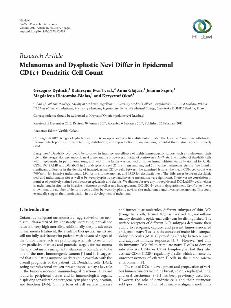
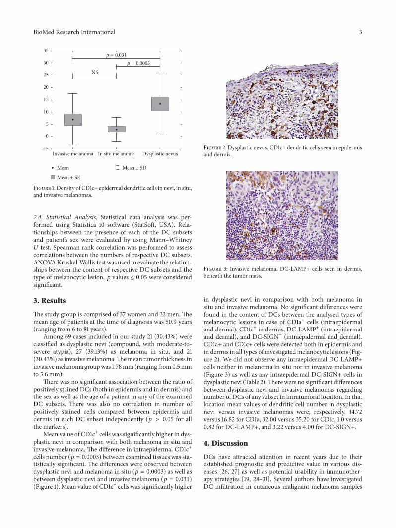
Figure 1 Density of CD1c+ epidermal dendritic cells in nevi in situand invasive melanomas
24 Statistical Analysis Statistical data analysis was per-formed using Statistica 10 software (StatSoft USA) Rela-tionships between the presence of each of the DC subsetsand patientrsquos sex were evaluated by using MannndashWhitneyU test Spearman rank correlation was performed to assesscorrelations between the numbers of respective DC subsetsANOVAKruskal-Wallis test was used to evaluate the relation-ships between the content of respective DC subsets and thetype of melanocytic lesion 119901 values le 005 were consideredsignificant
3 Results
The study group is comprised of 37 women and 32 men Themean age of patients at the time of diagnosis was 509 years(ranging from 6 to 81 years)
Among 69 cases included in our study 21 (3043) wereclassified as dysplastic nevi (compound with moderate-to-severe atypia) 27 (3913) as melanoma in situ and 21(3043) as invasivemelanomaThemean tumor thickness ininvasivemelanoma groupwas 178mm (ranging from05mmto 56mm)
There was no significant association between the ratio ofpositively stained DCs (both in epidermis and in dermis) andthe sex as well as the age of a patient in any of the examinedDC subsets There was also no correlation in number ofpositively stained cells compared between epidermis anddermis in each DC subset independently (119901 gt 005 for allthe markers)
Mean value of CD1c+ cells was significantly higher in dys-plastic nevi in comparison with both melanoma in situ andinvasive melanoma The difference in intraepidermal CD1c+cells number (119901 = 00003) between examined tissues was sta-tistically significant The differences were observed betweendysplastic nevi and melanoma in situ (119901 = 00003) as well asbetween dysplastic nevi and invasive melanoma (119901 = 0031)(Figure 1) Mean value of CD1c+ cells was significantly higher
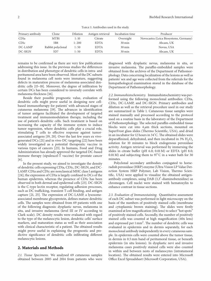
Figure 2 Dysplastic nevus CD1c+ dendritic cells seen in epidermisand dermis
Figure 3 Invasive melanoma DC-LAMP+ cells seen in dermisbeneath the tumor mass
in dysplastic nevi in comparison with both melanoma insitu and invasive melanoma No significant differences werefound in the content of DCs between the analysed types ofmelanocytic lesions in case of CD1a+ cells (intraepidermaland dermal) CD1c+ in dermis DC-LAMP+ (intraepidermaland dermal) and DC-SIGN+ (intraepidermal and dermal)CD1a+ and CD1c+ cells were detected both in epidermis andin dermis in all types of investigatedmelanocytic lesions (Fig-ure 2) We did not observe any intraepidermal DC-LAMP+cells neither in melanoma in situ nor in invasive melanoma(Figure 3) as well as any intraepidermal DC-SIGN+ cells indysplastic nevi (Table 2)Therewere no significant differencesbetween dysplastic nevi and invasive melanomas regardingnumber of DCs of any subset in intratumoral location In thatlocation mean values of dendritic cell number in dysplasticnevi versus invasive melanomas were respectively 1472versus 1682 for CD1a 3200 versus 3520 for CD1c 10 versus082 for DC-LAMP+ and 322 versus 400 for DC-SIGN+
4 Discussion
DCs have attracted attention in recent years due to theirestablished prognostic and predictive value in various dis-eases [26 27] as well as potential usability in immunother-apy strategies [19 28ndash31] Several authors have investigatedDC infiltration in cutaneous malignant melanoma samples
4 BioMed Research International
Table 2 Number of dendritic cells in epidermal and dermal compartment of dysplastic nevi and in situ and invasive melanomas
Epidermal DermalMean Range SD 119901 Mean Range SD 119901
CD1aInvasive melanoma 3824 0ndash80 2163
02808 (NS)
2255 0ndash53 1479
02434 (NS)In situ melanoma 3584 15ndash70 1325 1632 3ndash39 1085Dysplastic nevus 4329 18ndash74 1492 1529 1ndash43 1127All cases 3923 0ndash80 1710 1803 0ndash53 1265
CD1cInvasive melanoma 700 0ndash36 1055
00003
5181 6ndash96 3079
00559 (NS)In situ melanoma 294 0ndash17 496 3356 0ndash151 3721Dysplastic nevus 1335 2ndash45 1244 4000 8ndash172 3508All cases 792 0ndash45 1070 4224 0ndash172 3456
DC-LAMPInvasive melanoma 000 0-0 000
03588 (NS)
126 0ndash10 262
02456 (NS)In situ melanoma 000 0-0 000 077 0ndash14 302Dysplastic nevus 005 0-1 022 085 0ndash7 201All cases 002 0-1 013 095 0ndash14 257
DC-SIGNInvasive melanoma 010 0ndash2 045
05867 (NS)
1189 0ndash39 1354
02495 (NS)In situ melanoma 026 0ndash5 115 453 0ndash26 819Dysplastic nevus 000 0-0 000 424 0ndash32 731All cases 012 0ndash5 069 671 0ndash39 1034
showing decreased number in advanced stages of the tumor[16 17 32] The others have noted a correlation of theplasmacytoid DCs subset with tumor thickness and poorprognosis [15 33] and a positive correlation of density of DC-LAMP+mature DCs with survival in univariate analysis [15]Nonetheless there are very few up-to-date studies investigat-ing the significance of DCs in neoplastic melanocytic skinlesions quantifying various subsets and relative contributionof epidermal and dermal DCs to host immunosurveillanceLiterature is also lacking in data concerning the infiltrationof dysplastic nevi by dendritic cells
In our cohort of melanocytic lesion samples we founda statistically significant increase in the number of CD1c-positive cells in the epidermis of melanoma in situ andinvasive tumors when compared with dysplastic nevi Otherauthors have also obtained similar results showing thedepletion of intraepidermal DCs in melanoma [17 32]However it should be mentioned that these other studieslooked at the DC number in general as at that time the avail-able immunochemistry was insufficient to precisely quantifydistinct DC subsets The reason why these differences arerestricted to epidermis might be partly explained by distinctorigin and features of DCs in respective layers of the skinLangerhans cells (LCs) representing the DC populationfound in epidermis are considered to be of nonlymphoidtissue origin while dermal DCs derive from bone marrowand blood-borne precursors [34]The lack of blood vessels inepidermis together with the presence of basement membranemay also be contributory Moreover it is suggested that somemelanoma-derived factors might adversely affect epidermalLangerhans cellsrsquo migration differentiation and function
[32] For instance TGF-beta and IL-10 found in melanomacell nests have been described to reduce the development ofDCs [34 35] In addition necrosis regression or scarringof the tumor in more advanced stages may contribute to thedepletion of DCs in epidermis overlying this type of malig-nant skin lesions [32] Our finding indicates that epidermalarea of melanomas might not be adequately equipped withCD1c-positive DC subset and thereby might be unable toelicit an appropriate antitumor immune response Howeverwhether the CD1c-positive cells depletion occurs duringthe initiation promotion or progression of the tumor callsneeds further elucidation There is also little available datain the literature with regard to CD1c antibody and the exactpathophysiological significance of this observation remainsto be established
CD1a-positive DCs have been widely investigated in var-ious human cancers with results varying between papers Wedid not observe any difference in CD1a-positive cells densitybetween epidermis anddermis in any of the investigated typesof melanocytic lesions However Ladanyi et al have shownreduced number of intraepidermal CD1a-positive Langer-hans cells overlying melanomas suggesting that it could beexplained by the fact that even nonulcerated epidermis abovemelanoma is often atrophic or hyperkeratotic It has also beenreported that infiltration of CD1a-positive DC within tumorcell nests and peritumoral area correlates with decreasedtumor thickness and the radial growth phase of melanomasrespectively [16] Vermi et al observed the significant increasein CD1a-positive cells accumulation in primary cutaneousmelanoma comparedwith normal skin andmelanocytic neviin melanoma samples immature CD1a-positive DCs were
BioMed Research International 5
mainly found in the dermis surrounding the tumor in closevicinity of lymphoid cell aggregates [18] Recently presenceof CD1a-positive DCs around the tumor cell nests was alsoobserved to correlate with the absence of hematogenousspread in malignant melanoma of the skin [36] In otherhumanmalignancies the decreased number of CD1a-positiveDCs in tumor area is often associated with the progression ofthe disease and unfavourable prognosis [11 14]
DC-SIGN-positive DCs have recently emerged asvery potent cells shaping immune responses by mediatingimmune recognition and cell adhesion [37] Despite thefact that our study did not reveal any statistically significantdifferences in the density and distribution of this subsetnoteworthy is very low epidermal count of DC-SIGN-positive DCs as we have observed a few of them in onlytwo samples among all the investigated cases We think thatthis subset of DCs requires further investigation on a largersample of cases especially as data concerning this markerin melanocytic lesions is scarce and ambiguous AlthoughDC-SIGN antibody is generally classified as nonspecificmarker of DC maturation [38] in one study on malignantmelanoma the expression of DC-SIGN and DC-LAMP wasfound to be mutually exclusive indicating that DC-SIGNis expressed only by immature DCs At the same timeDC-SIGN-positive dermal DCs were noted to encompassdistinct subset compared to immature CD1a-positive DCsIt should also be born in mind that DC-SIGN antibody maystain positively other immune cells such as certain subsetsof macrophages [18]
We have also noted the lack of mature DC-LAMP-positive dendritic cells in epidermis of melanoma in situ andinvasive tumors and very low number of them in dermalcompartment This is in agreement with findings from otherstudies concerning various cancers which highlight a sup-pressive tumor influence on DCs function as the infiltratingcells in advanced stages of the disease are mostly immatureand thus unable to present effectively tumor-derived antigensand mediate antitumor response [39ndash41] The content ofDC-LAMP-positive DCs in primary cutaneous melanomawas also found to be rather scarce and mostly confinedto the peritumoral lymphocyte infiltrate [15] Mature DCswere observed to form clusters with T cells which could bean evidence of a still ongoing immune response in tumormicroenvironment It has also been shown that the degreeof DC-LAMP+ cells infiltration inversely correlates withmelanoma thickness and is associated with patientsrsquo survival[16]
The present study investigated only some problemsconcerning the density of DCs in dysplastic and malig-nant melanocytic lesions It is still unclear whether thedifferences in DC content and distribution patterns are acause or a result of neoplastic processes Another interestingquestion may arise with regard to the DCs subsets whosedensity and location do not differ significantly betweendistinct melanocytic lesions regardless of whether theyare of malignant nature or not Do DCs fail to serve aspart of host immune system control due to some impair-ment of functioning or should it be rather attributable tothe low immunogenicity of melanoma cells Undoubtedly
a thorough analysis of dendritic cell family including a vari-ety of markers and double staining procedures is mandatoryto draw valid conclusions on the development of immunityin the carcinogenesis of malignant melanoma Moreoveranalysis of a large number of melanoma samples alongwith the prospective study of clinical data could offer betterunderstanding of predictive and prognostic significance oftumor-infiltrating DCs Although our study requires furthervalidation we believe that it sheds new light on the roleof DCs in dysplastic and neoplastic cutaneous lesions andthus brings added value to the optimal patient care anddevelopment of novel immunotherapy strategies
Competing Interests
The authors declare that there is no conflict of interestsregarding the publication of this paper
References
[1] A Ladanyi ldquoPrognostic and predictive significance of immunecells infiltrating cutaneous melanomardquo Pigment Cell andMelanoma Research vol 28 no 5 pp 490ndash500 2015
[2] M Neagu C Constantin and S Zurac ldquoImmune parameters inthe prognosis and therapy monitoring of cutaneous melanomapatients Experience role and limitationsrdquo BioMed ResearchInternational vol 2013 Article ID 107940 13 pages 2013
[3] J Banchereau and R M Steinman ldquoDendritic cells and thecontrol of immunityrdquo Nature vol 392 no 6673 pp 245ndash2521998
[4] R Bonasio and U H von Andrian ldquoGeneration migrationand function of circulating dendritic cellsrdquo Current Opinion inImmunology vol 18 no 4 pp 503ndash511 2006
[5] K Shortman and Y-J Liu ldquoMouse and human dendritic cellsubtypesrdquoNature Reviews Immunology vol 2 no 3 pp 151ndash1612002
[6] F Benencia L Sprague J McGinty M Pate and M Muc-cioli ldquoDendritic cells the tumor microenvironment and thechallenges for an effective antitumor vaccinationrdquo Journal ofBiomedicine and Biotechnology vol 2012 Article ID 425476 15pages 2012
[7] C A Janeway Jr and R Medzhitov ldquoInnate immune recogni-tionrdquo Annual Review of Immunology vol 20 pp 197ndash216 2002
[8] K Mahnke E Schmitt L Bonifaz A H Enk and H JonuleitldquoImmature but not inactive the tolerogenic function of imma-ture dendritic cellsrdquo Immunology and Cell Biology vol 80 no5 pp 477ndash483 2002
[9] D Bell P Chomarat D Broyles et al ldquoIn breast carcinoma tis-sue immature dendritic cells reside within the tumor whereasmature dendritic cells are located in peritumoral areasrdquo Journalof Experimental Medicine vol 190 no 10 pp 1417ndash1425 1999
[10] T Schwaab J E Weiss A R Schned and RJ J Barth ldquoDen-dritic cell infiltration in colon cancerrdquo Journal of Immunother-apy vol 24 no 2 pp 130ndash137 2001
[11] L Lu K Pan H-X Zheng et al ldquoIL-17A promotes immune cellrecruitment in human esophageal cancers and the infiltratingdendritic cells represent a positive prognosticmarker for patientsurvivalrdquo Journal of Immunotherapy vol 36 no 8 pp 451ndash4582013
6 BioMed Research International
[12] R Remark C Becker J E Gomez et al ldquoThe non-small celllung cancer immune contexture a major determinant of tumorcharacteristics and patient outcomerdquo American Journal of Res-piratory and Critical Care Medicine vol 191 no 4 pp 377ndash3902015
[13] T E Reichert C Scheuer R Day W Wagner and T L White-side ldquoThe number of intratumoral dendritic cells and 120577-chainexpression in T cells as prognostic and survival biomarkers inpatients with oral carcinomardquo Cancer vol 91 no 11 pp 2136ndash2147 2001
[14] J Upadhyay N N Rao and R B Upadhyay ldquoA comparativeanalysis of langerhans cell in oral epithelial dysplasia and oralsquamous cell carcinoma using antibody CD-1ardquo Journal ofCancer Research andTherapeutics vol 8 no 4 pp 591ndash597 2012
[15] T O Jensen H Schmidt H J Moslashller et al ldquoIntratumoralneutrophils and plasmacytoid dendritic cells indicate poorprognosis and are associated with pSTAT3 expression in AJCCstage III melanomardquo Cancer vol 118 no 9 pp 2476ndash24852012
[16] A Ladanyi J Kiss B Somlai et al ldquoDensity of DC-LAMP+mature dendritic cells in combination with activated T lympho-cytes infiltrating primary cutaneousmelanoma is a strong inde-pendent prognostic factorrdquo Cancer Immunology Immunother-apy vol 56 no 9 pp 1459ndash1469 2007
[17] K Toriyama D-RWen E Paul and A J Cochran ldquoVariationsin the distribution frequency and phenotype of langerhanscells during the evolution of malignant melanoma of the skinrdquoJournal of Investigative Dermatology vol 100 no 3 pp S269ndashS273 1993
[18] W Vermi R Bonecchi F Facchetti et al ldquoRecruitment ofimmature plasmacytoid dendritic cells (plasmacytoid mono-cytes) and myeloid dendritic cells in primary cutaneousmelanomasrdquo Journal of Pathology vol 200 no 2 pp 255ndash2682003
[19] E Castillo-Montiel J C Chimal-Eguıa I Tello G Pinon-ZarateMHerrera-Enrıquez andA E Castell-Rodrıguez ldquoEn-hancing dendritic cell immunotherapy for melanoma using asimple mathematical modelrdquo Theoretical Biology and MedicalModelling vol 12 article 11 2015
[20] S Van Lint S Wilgenhof C Heirman et al ldquoOptimized den-dritic cell-based immunotherapy for melanoma the TriMix-formulardquo Cancer Immunology Immunotherapy vol 63 no 9pp 959ndash967 2014
[21] T B H Geijtenbeek R Torensma S J Van Vliet et alldquoIdentification of DC-SIGN a novel dendritic cell-specificICAM-3 receptor that supports primary immune responsesrdquoCell vol 100 no 5 pp 575ndash585 2000
[22] R M Steinman ldquoDecisions about dendritic cells past presentand futurerdquo Annual Review of Immunology vol 30 pp 1ndash222012
[23] J T Elder N J Reynolds K D Cooper C E M Griffiths BD Hardas and P A Bleicher ldquoCD1 gene expression in humanskinrdquo Journal of Dermatological Science vol 6 no 3 pp 206ndash213 1993
[24] M C Udey ldquoSkin dendritic cells in immunity and autoimmu-nityrdquo Journal of Investigative Dermatology Symposium Proceed-ings vol 9 no 1 pp 15ndash17 2004
[25] T B H Geijtenbeek A Engering and Y Van Kooyk ldquoDC-SIGN a C-type lectin on dendritic cells that unveils manyaspects of dendritic cell biologyrdquo Journal of Leukocyte Biologyvol 71 no 6 pp 921ndash931 2002
[26] G La Rocca R Anzalone S Corrao et al ldquoCD1a down-regu-lation in primary invasive ductal breast carcinoma may pre-dict regional lymph node invasion and patient outcomerdquoHistopathology vol 52 no 2 pp 203ndash212 2008
[27] C Ayari H Larue H Hovington et al ldquoHigh level of maturetumor-infiltrating dendritic cells predicts progression tomuscleinvasion in bladder cancerrdquoHuman Pathology vol 44 no 8 pp1630ndash1637 2013
[28] M Lepore C De Lalla L Mori P Dellabona G De Liberoand G Casorati ldquoTargeting leukemia by CD1c-restricted T cellsspecific for a novel lipid antigenrdquo OncoImmunology vol 4 no3 Article ID e970463 2015
[29] R L Prue F Vari K J Radford et al ldquoA phase i clinical trialof CD1c (BDCA-1)+ dendritic cells pulsed with HLA-Alowast0201peptides for immunotherapy of metastatic hormone refractoryprostate cancerrdquo Journal of Immunotherapy vol 38 no 2 pp71ndash76 2015
[30] S M Keon M S Ruiz S Gazzaniga and R Wainstok ldquoDen-dritic cell-based vaccination in cancer therapeutic implicationsemerging frommurinemodelsrdquo Frontiers in Immunology vol 6article 243 2015
[31] L Qiu J Li S Yu et al ldquoA novel cancer immunotherapy basedon the combination of a synthetic carbohydrate-pulsed den-dritic cell vaccine and glycoengineered cancer cellsrdquoOncotargetvol 6 no 7 pp 5195ndash5203 2015
[32] M A Stene M Babajanians S Bhuta and A J CochranldquoQuantitative alterations in cutaneous langerhans cells duringthe evolution of malignant melanoma of the skinrdquo Journal ofInvestigative Dermatology vol 91 no 2 pp 125ndash128 1988
[33] C Aspord M-T Leccia J Charles and J Plumas ldquoPlas-macytoid dendritic cells support melanoma progression bypromoting Th2 and regulatory immunity through OX40L andICOSLrdquoCancer Immunology Research vol 1 no 6 pp 402ndash4152013
[34] C T Conrad N R Ernst W Dummer E B Brocker andJ C Becker ldquoDifferential expression of transforming growthfactor 1205731 and interleukin 10 in progressing and regressing areasof primary melanomardquo Journal of Experimental and ClinicalCancer Research vol 18 no 2 pp 225ndash232 1999
[35] A H Enk H Jonuleit J Saloga and J Knop ldquoDendriticcells as mediators of tumor-induced tolerance in metastaticmelanomardquo International Journal of Cancer vol 73 no 3 pp309ndash316 1997
[36] E Emri K Egervari T Varvolgyi et al ldquoCorrelation amongmetallothionein expression intratumoural macrophage infil-tration and the risk of metastasis in human cutaneous malig-nant melanomardquo Journal of the European Academy of Derma-tology and Venereology vol 27 no 3 pp e320ndashe327 2013
[37] JDenDunnen S I Gringhuis andT BHGeijtenbeek ldquoInnatesignaling by the C-type lectin DC-SIGN dictates immuneresponsesrdquo Cancer Immunology Immunotherapy vol 58 no 7pp 1149ndash1157 2009
[38] N Karthaus R Torensma and J Tel ldquoDeciphering the messagebroadcast by tumor-infiltrating dendritic cellsrdquo American Jour-nal of Pathology vol 181 no 3 pp 733ndash742 2012
[39] J Tabarkiewicz P Rybojad A Jablonka and J Rolinski ldquoCD1c+and CD303+ dendritic cells in peripheral blood lymph nodesand tumor tissue of patients with non-small cell lung cancerrdquoOncology Reports vol 19 no 1 pp 237ndash243 2008
BioMed Research International 7
[40] F O Nestle G Burg J Fah T Wrone-Smith and B J Nicko-loff ldquoHuman sunlight-induced basal-cell-carcinoma-associateddendritic cells are deficient in T cell co-stimulatory moleculesand are impaired as antigen-presenting cellsrdquoAmerican Journalof Pathology vol 150 no 2 pp 641ndash651 1997
[41] D I Gabrilovich J Corak I F Ciernik D Kavanaugh and DP Carbone ldquoDecreased antigen presentation by dendritic cellsin patients with breast cancerrdquo Clinical Cancer Research vol 3no 3 pp 483ndash490 1997
Submit your manuscripts athttpswwwhindawicom
Stem CellsInternational
Hindawi Publishing Corporationhttpwwwhindawicom Volume 2014
Hindawi Publishing Corporationhttpwwwhindawicom Volume 2014
MEDIATORSINFLAMMATION
of
Hindawi Publishing Corporationhttpwwwhindawicom Volume 2014
Behavioural Neurology
EndocrinologyInternational Journal of
Hindawi Publishing Corporationhttpwwwhindawicom Volume 2014
Hindawi Publishing Corporationhttpwwwhindawicom Volume 2014
Disease Markers
Hindawi Publishing Corporationhttpwwwhindawicom Volume 2014
BioMed Research International
OncologyJournal of
Hindawi Publishing Corporationhttpwwwhindawicom Volume 2014
Hindawi Publishing Corporationhttpwwwhindawicom Volume 2014
Oxidative Medicine and Cellular Longevity
Hindawi Publishing Corporationhttpwwwhindawicom Volume 2014
PPAR Research
The Scientific World JournalHindawi Publishing Corporation httpwwwhindawicom Volume 2014
Immunology ResearchHindawi Publishing Corporationhttpwwwhindawicom Volume 2014
Journal of
ObesityJournal of
Hindawi Publishing Corporationhttpwwwhindawicom Volume 2014
Hindawi Publishing Corporationhttpwwwhindawicom Volume 2014
Computational and Mathematical Methods in Medicine
OphthalmologyJournal of
Hindawi Publishing Corporationhttpwwwhindawicom Volume 2014
Diabetes ResearchJournal of
Hindawi Publishing Corporationhttpwwwhindawicom Volume 2014
Hindawi Publishing Corporationhttpwwwhindawicom Volume 2014
Research and TreatmentAIDS
Hindawi Publishing Corporationhttpwwwhindawicom Volume 2014
Gastroenterology Research and Practice
Hindawi Publishing Corporationhttpwwwhindawicom Volume 2014
Parkinsonrsquos Disease
Evidence-Based Complementary and Alternative Medicine
Volume 2014Hindawi Publishing Corporationhttpwwwhindawicom
2 BioMed Research International
Table 1 Antibodies used in the study
Primary antibody Clone Dilution Antigen retrieval Incubation time ProducerCD1a MTB1 1 10 Citrate Overnight Novocastra (Leica Biosystems Germany)CD1c 5B8 1 200 EDTA 30min Abcam UKDC-LAMP Rabbit polyclonal 1 50 EDTA 30min Novus USADC-SIGN 5D7 1 50 EDTA 30min Abcam UK
remains to be confirmed as there are very few publicationsaddressing this issue In the previous studies the differencesin distribution and phenotype of dendritic cells in intra- andperitumoral area have been observedMost of the DC subsetsfound in melanoma cell nests were immature suggestingdefects in maturation process of melanoma-associated den-dritic cells [15ndash18] Moreover the degree of infiltration bycertain DCs has been considered to inversely correlate withmelanoma thickness [16]
Beside their possible prognostic value evaluation ofdendritic cells might prove useful in designing new cell-based immunotherapy for patientsrsquo with advanced stages ofcutaneous melanoma [19] The advances in identificationof tumor antigens facilitated the development of targetedtreatment and immunomodulation therapy including theuse of patientrsquos dendritic cells Such treatment is based onincreasing the capacity of the immune system to inducetumor regression where dendritic cells play a crucial rolestimulating T cells to effective response against tumor-associated antigens [19 20] Over the last few years ex vivo-generated DCs [21] and in vivo-DC-targeting [22] have beenwidely investigated as a potential therapeutic vaccine invarious types of cancers [23] In humans Food and DrugAdministration has already approved the targeted DC-basedimmune therapy (sipuleucel-T vaccine) for prostate cancer[6]
In the present study we aimed to investigate the densityof dendritic cells expressing CD1a CD1c DC-SIGN andDC-LAMP CD1a and CD1c are nonclassicalMHC class I antigens[24] the expression of CD1a is largely confined to DCs of thehuman epidermis whereas the presence of CD1c has beenobserved in both dermal and epidermal cells [23] DC-SIGNis the C-type lectin receptor regulating adhesion processessuch as DC trafficking transient T-cell binding and antigencapture [21 25] The expression of DC-LAMP a lysosome-associated membrane glycoprotein defines mature dendriticcells The samples were obtained from 69 patients with oneof the following diagnosis dysplastic nevus melanoma insitu and invasive melanoma (level III or IV according toClark scale) DC density results were evaluated with regardto the type of the melanocytic lesion dendritic cellsrsquo surfacemarkers and maturation status as well as their associationwith clinical characteristic of a patient The obtained resultsmight prove useful in explaining the prognostic and pre-dictive significance of dendritic cells infiltrating differentmelanocytic lesions
2 Materials and Methods
21 Tissue Specimens We analysed 69 cutaneous samplesobtained between 2005 and 2014 from patients who were
diagnosed with dysplastic nevus melanoma in situ orinvasive melanoma The paraffin-embedded samples wereobtained from the archives of the Department of Pathomor-phology Data concerning localization of the lesions as well aspatientsrsquo sex and age were collected from the referrals for thehistopathological examination stored in the database of theDepartment of Pathomorphology
22 Immunohistochemistry Immunohistochemistrywas per-formed using the following monoclonal antibodies CD1aCD1c DC-LAMP and DC-SIGN Primary antibodies anddilution as well as the retrieval procedure used in our studyare summarized in Table 1 Cutaneous tissue samples werestained manually and processed according to the protocolused on a routine basis in the laboratory of the Departmentof Pathomorphology The selected paraffin-embedded tissueblocks were cut into 4 120583m thick sections mounted onSuperFrost glass slides (Thermo Scientific USA) and driedin an incubator for 12 hours in 34∘CThe obtained slides weredeparaffinized dehydrated and then incubated in 3 H
2O2
solution for 10 minutes to block endogenous peroxidaseactivity Antigen retrieval was performed by immersing theslides in citrate buffer (pH 60 001M) or EDTA (pH 80001M) and subjecting them to 97∘C in a water bath for 30minutes
Polyclonal secondary antibodies conjugated to horse-radish peroxidase (HRP) enzyme (Ultra Vision LPValueDet-ection System HRP Polymer Lab Vision Thermo Scien-tific USA) were applied to visualize the obtained antigen-antibody complexes using DAB (331015840-diaminobenzidine) aschromogen Cell nuclei were stained with hematoxylin toenhance contrast in tissue sections
23 Evaluation of Immunostaining Quantitative assessmentof each DC subset was performed in light microscopy on thebasis of the numbers of positively stained cells (membraneand cytoplasmic brown staining) The slides were firstlyexamined at lowmagnification (10x lens) to select ldquohot spotsrdquoof positively stained cells Secondly the number of positivelystained cells was counted at high magnification (40x lens)and expressed per 1mm2 The number of dendritic cells wasevaluated in epidermis and in dermis separately for eachmonoclonal antibody independently in every cutaneous sam-ple In epidermis cells were counted above the tumor tissuein dermis in 05mm band of peritumoral tissue or beneathepidermis (in situ lesions) In dysplastic nevi and invasivemelanoma cases positively stained cells were also countedin within and between nests of melanocytes (intratumorallocation) The obtained results were entered into MicrosoftOffice Excel Spreadsheet (Microsoft Corporation USA)
BioMed Research International 3
Invasive melanoma In situ melanoma Dysplastic nevus
0
5
10
15
20
25
30
35
Mean
NS
minus5
p = 0031
p = 00003
Mean plusmn SD
Mean plusmn SE
Figure 1 Density of CD1c+ epidermal dendritic cells in nevi in situand invasive melanomas
24 Statistical Analysis Statistical data analysis was per-formed using Statistica 10 software (StatSoft USA) Rela-tionships between the presence of each of the DC subsetsand patientrsquos sex were evaluated by using MannndashWhitneyU test Spearman rank correlation was performed to assesscorrelations between the numbers of respective DC subsetsANOVAKruskal-Wallis test was used to evaluate the relation-ships between the content of respective DC subsets and thetype of melanocytic lesion 119901 values le 005 were consideredsignificant
3 Results
The study group is comprised of 37 women and 32 men Themean age of patients at the time of diagnosis was 509 years(ranging from 6 to 81 years)
Among 69 cases included in our study 21 (3043) wereclassified as dysplastic nevi (compound with moderate-to-severe atypia) 27 (3913) as melanoma in situ and 21(3043) as invasivemelanomaThemean tumor thickness ininvasivemelanoma groupwas 178mm (ranging from05mmto 56mm)
There was no significant association between the ratio ofpositively stained DCs (both in epidermis and in dermis) andthe sex as well as the age of a patient in any of the examinedDC subsets There was also no correlation in number ofpositively stained cells compared between epidermis anddermis in each DC subset independently (119901 gt 005 for allthe markers)
Mean value of CD1c+ cells was significantly higher in dys-plastic nevi in comparison with both melanoma in situ andinvasive melanoma The difference in intraepidermal CD1c+cells number (119901 = 00003) between examined tissues was sta-tistically significant The differences were observed betweendysplastic nevi and melanoma in situ (119901 = 00003) as well asbetween dysplastic nevi and invasive melanoma (119901 = 0031)(Figure 1) Mean value of CD1c+ cells was significantly higher
Figure 2 Dysplastic nevus CD1c+ dendritic cells seen in epidermisand dermis
Figure 3 Invasive melanoma DC-LAMP+ cells seen in dermisbeneath the tumor mass
in dysplastic nevi in comparison with both melanoma insitu and invasive melanoma No significant differences werefound in the content of DCs between the analysed types ofmelanocytic lesions in case of CD1a+ cells (intraepidermaland dermal) CD1c+ in dermis DC-LAMP+ (intraepidermaland dermal) and DC-SIGN+ (intraepidermal and dermal)CD1a+ and CD1c+ cells were detected both in epidermis andin dermis in all types of investigatedmelanocytic lesions (Fig-ure 2) We did not observe any intraepidermal DC-LAMP+cells neither in melanoma in situ nor in invasive melanoma(Figure 3) as well as any intraepidermal DC-SIGN+ cells indysplastic nevi (Table 2)Therewere no significant differencesbetween dysplastic nevi and invasive melanomas regardingnumber of DCs of any subset in intratumoral location In thatlocation mean values of dendritic cell number in dysplasticnevi versus invasive melanomas were respectively 1472versus 1682 for CD1a 3200 versus 3520 for CD1c 10 versus082 for DC-LAMP+ and 322 versus 400 for DC-SIGN+
4 Discussion
DCs have attracted attention in recent years due to theirestablished prognostic and predictive value in various dis-eases [26 27] as well as potential usability in immunother-apy strategies [19 28ndash31] Several authors have investigatedDC infiltration in cutaneous malignant melanoma samples
4 BioMed Research International
Table 2 Number of dendritic cells in epidermal and dermal compartment of dysplastic nevi and in situ and invasive melanomas
Epidermal DermalMean Range SD 119901 Mean Range SD 119901
CD1aInvasive melanoma 3824 0ndash80 2163
02808 (NS)
2255 0ndash53 1479
02434 (NS)In situ melanoma 3584 15ndash70 1325 1632 3ndash39 1085Dysplastic nevus 4329 18ndash74 1492 1529 1ndash43 1127All cases 3923 0ndash80 1710 1803 0ndash53 1265
CD1cInvasive melanoma 700 0ndash36 1055
00003
5181 6ndash96 3079
00559 (NS)In situ melanoma 294 0ndash17 496 3356 0ndash151 3721Dysplastic nevus 1335 2ndash45 1244 4000 8ndash172 3508All cases 792 0ndash45 1070 4224 0ndash172 3456
DC-LAMPInvasive melanoma 000 0-0 000
03588 (NS)
126 0ndash10 262
02456 (NS)In situ melanoma 000 0-0 000 077 0ndash14 302Dysplastic nevus 005 0-1 022 085 0ndash7 201All cases 002 0-1 013 095 0ndash14 257
DC-SIGNInvasive melanoma 010 0ndash2 045
05867 (NS)
1189 0ndash39 1354
02495 (NS)In situ melanoma 026 0ndash5 115 453 0ndash26 819Dysplastic nevus 000 0-0 000 424 0ndash32 731All cases 012 0ndash5 069 671 0ndash39 1034
showing decreased number in advanced stages of the tumor[16 17 32] The others have noted a correlation of theplasmacytoid DCs subset with tumor thickness and poorprognosis [15 33] and a positive correlation of density of DC-LAMP+mature DCs with survival in univariate analysis [15]Nonetheless there are very few up-to-date studies investigat-ing the significance of DCs in neoplastic melanocytic skinlesions quantifying various subsets and relative contributionof epidermal and dermal DCs to host immunosurveillanceLiterature is also lacking in data concerning the infiltrationof dysplastic nevi by dendritic cells
In our cohort of melanocytic lesion samples we founda statistically significant increase in the number of CD1c-positive cells in the epidermis of melanoma in situ andinvasive tumors when compared with dysplastic nevi Otherauthors have also obtained similar results showing thedepletion of intraepidermal DCs in melanoma [17 32]However it should be mentioned that these other studieslooked at the DC number in general as at that time the avail-able immunochemistry was insufficient to precisely quantifydistinct DC subsets The reason why these differences arerestricted to epidermis might be partly explained by distinctorigin and features of DCs in respective layers of the skinLangerhans cells (LCs) representing the DC populationfound in epidermis are considered to be of nonlymphoidtissue origin while dermal DCs derive from bone marrowand blood-borne precursors [34]The lack of blood vessels inepidermis together with the presence of basement membranemay also be contributory Moreover it is suggested that somemelanoma-derived factors might adversely affect epidermalLangerhans cellsrsquo migration differentiation and function
[32] For instance TGF-beta and IL-10 found in melanomacell nests have been described to reduce the development ofDCs [34 35] In addition necrosis regression or scarringof the tumor in more advanced stages may contribute to thedepletion of DCs in epidermis overlying this type of malig-nant skin lesions [32] Our finding indicates that epidermalarea of melanomas might not be adequately equipped withCD1c-positive DC subset and thereby might be unable toelicit an appropriate antitumor immune response Howeverwhether the CD1c-positive cells depletion occurs duringthe initiation promotion or progression of the tumor callsneeds further elucidation There is also little available datain the literature with regard to CD1c antibody and the exactpathophysiological significance of this observation remainsto be established
CD1a-positive DCs have been widely investigated in var-ious human cancers with results varying between papers Wedid not observe any difference in CD1a-positive cells densitybetween epidermis anddermis in any of the investigated typesof melanocytic lesions However Ladanyi et al have shownreduced number of intraepidermal CD1a-positive Langer-hans cells overlying melanomas suggesting that it could beexplained by the fact that even nonulcerated epidermis abovemelanoma is often atrophic or hyperkeratotic It has also beenreported that infiltration of CD1a-positive DC within tumorcell nests and peritumoral area correlates with decreasedtumor thickness and the radial growth phase of melanomasrespectively [16] Vermi et al observed the significant increasein CD1a-positive cells accumulation in primary cutaneousmelanoma comparedwith normal skin andmelanocytic neviin melanoma samples immature CD1a-positive DCs were
BioMed Research International 5
mainly found in the dermis surrounding the tumor in closevicinity of lymphoid cell aggregates [18] Recently presenceof CD1a-positive DCs around the tumor cell nests was alsoobserved to correlate with the absence of hematogenousspread in malignant melanoma of the skin [36] In otherhumanmalignancies the decreased number of CD1a-positiveDCs in tumor area is often associated with the progression ofthe disease and unfavourable prognosis [11 14]
DC-SIGN-positive DCs have recently emerged asvery potent cells shaping immune responses by mediatingimmune recognition and cell adhesion [37] Despite thefact that our study did not reveal any statistically significantdifferences in the density and distribution of this subsetnoteworthy is very low epidermal count of DC-SIGN-positive DCs as we have observed a few of them in onlytwo samples among all the investigated cases We think thatthis subset of DCs requires further investigation on a largersample of cases especially as data concerning this markerin melanocytic lesions is scarce and ambiguous AlthoughDC-SIGN antibody is generally classified as nonspecificmarker of DC maturation [38] in one study on malignantmelanoma the expression of DC-SIGN and DC-LAMP wasfound to be mutually exclusive indicating that DC-SIGNis expressed only by immature DCs At the same timeDC-SIGN-positive dermal DCs were noted to encompassdistinct subset compared to immature CD1a-positive DCsIt should also be born in mind that DC-SIGN antibody maystain positively other immune cells such as certain subsetsof macrophages [18]
We have also noted the lack of mature DC-LAMP-positive dendritic cells in epidermis of melanoma in situ andinvasive tumors and very low number of them in dermalcompartment This is in agreement with findings from otherstudies concerning various cancers which highlight a sup-pressive tumor influence on DCs function as the infiltratingcells in advanced stages of the disease are mostly immatureand thus unable to present effectively tumor-derived antigensand mediate antitumor response [39ndash41] The content ofDC-LAMP-positive DCs in primary cutaneous melanomawas also found to be rather scarce and mostly confinedto the peritumoral lymphocyte infiltrate [15] Mature DCswere observed to form clusters with T cells which could bean evidence of a still ongoing immune response in tumormicroenvironment It has also been shown that the degreeof DC-LAMP+ cells infiltration inversely correlates withmelanoma thickness and is associated with patientsrsquo survival[16]
The present study investigated only some problemsconcerning the density of DCs in dysplastic and malig-nant melanocytic lesions It is still unclear whether thedifferences in DC content and distribution patterns are acause or a result of neoplastic processes Another interestingquestion may arise with regard to the DCs subsets whosedensity and location do not differ significantly betweendistinct melanocytic lesions regardless of whether theyare of malignant nature or not Do DCs fail to serve aspart of host immune system control due to some impair-ment of functioning or should it be rather attributable tothe low immunogenicity of melanoma cells Undoubtedly
a thorough analysis of dendritic cell family including a vari-ety of markers and double staining procedures is mandatoryto draw valid conclusions on the development of immunityin the carcinogenesis of malignant melanoma Moreoveranalysis of a large number of melanoma samples alongwith the prospective study of clinical data could offer betterunderstanding of predictive and prognostic significance oftumor-infiltrating DCs Although our study requires furthervalidation we believe that it sheds new light on the roleof DCs in dysplastic and neoplastic cutaneous lesions andthus brings added value to the optimal patient care anddevelopment of novel immunotherapy strategies
Competing Interests
The authors declare that there is no conflict of interestsregarding the publication of this paper
References
[1] A Ladanyi ldquoPrognostic and predictive significance of immunecells infiltrating cutaneous melanomardquo Pigment Cell andMelanoma Research vol 28 no 5 pp 490ndash500 2015
[2] M Neagu C Constantin and S Zurac ldquoImmune parameters inthe prognosis and therapy monitoring of cutaneous melanomapatients Experience role and limitationsrdquo BioMed ResearchInternational vol 2013 Article ID 107940 13 pages 2013
[3] J Banchereau and R M Steinman ldquoDendritic cells and thecontrol of immunityrdquo Nature vol 392 no 6673 pp 245ndash2521998
[4] R Bonasio and U H von Andrian ldquoGeneration migrationand function of circulating dendritic cellsrdquo Current Opinion inImmunology vol 18 no 4 pp 503ndash511 2006
[5] K Shortman and Y-J Liu ldquoMouse and human dendritic cellsubtypesrdquoNature Reviews Immunology vol 2 no 3 pp 151ndash1612002
[6] F Benencia L Sprague J McGinty M Pate and M Muc-cioli ldquoDendritic cells the tumor microenvironment and thechallenges for an effective antitumor vaccinationrdquo Journal ofBiomedicine and Biotechnology vol 2012 Article ID 425476 15pages 2012
[7] C A Janeway Jr and R Medzhitov ldquoInnate immune recogni-tionrdquo Annual Review of Immunology vol 20 pp 197ndash216 2002
[8] K Mahnke E Schmitt L Bonifaz A H Enk and H JonuleitldquoImmature but not inactive the tolerogenic function of imma-ture dendritic cellsrdquo Immunology and Cell Biology vol 80 no5 pp 477ndash483 2002
[9] D Bell P Chomarat D Broyles et al ldquoIn breast carcinoma tis-sue immature dendritic cells reside within the tumor whereasmature dendritic cells are located in peritumoral areasrdquo Journalof Experimental Medicine vol 190 no 10 pp 1417ndash1425 1999
[10] T Schwaab J E Weiss A R Schned and RJ J Barth ldquoDen-dritic cell infiltration in colon cancerrdquo Journal of Immunother-apy vol 24 no 2 pp 130ndash137 2001
[11] L Lu K Pan H-X Zheng et al ldquoIL-17A promotes immune cellrecruitment in human esophageal cancers and the infiltratingdendritic cells represent a positive prognosticmarker for patientsurvivalrdquo Journal of Immunotherapy vol 36 no 8 pp 451ndash4582013
6 BioMed Research International
[12] R Remark C Becker J E Gomez et al ldquoThe non-small celllung cancer immune contexture a major determinant of tumorcharacteristics and patient outcomerdquo American Journal of Res-piratory and Critical Care Medicine vol 191 no 4 pp 377ndash3902015
[13] T E Reichert C Scheuer R Day W Wagner and T L White-side ldquoThe number of intratumoral dendritic cells and 120577-chainexpression in T cells as prognostic and survival biomarkers inpatients with oral carcinomardquo Cancer vol 91 no 11 pp 2136ndash2147 2001
[14] J Upadhyay N N Rao and R B Upadhyay ldquoA comparativeanalysis of langerhans cell in oral epithelial dysplasia and oralsquamous cell carcinoma using antibody CD-1ardquo Journal ofCancer Research andTherapeutics vol 8 no 4 pp 591ndash597 2012
[15] T O Jensen H Schmidt H J Moslashller et al ldquoIntratumoralneutrophils and plasmacytoid dendritic cells indicate poorprognosis and are associated with pSTAT3 expression in AJCCstage III melanomardquo Cancer vol 118 no 9 pp 2476ndash24852012
[16] A Ladanyi J Kiss B Somlai et al ldquoDensity of DC-LAMP+mature dendritic cells in combination with activated T lympho-cytes infiltrating primary cutaneousmelanoma is a strong inde-pendent prognostic factorrdquo Cancer Immunology Immunother-apy vol 56 no 9 pp 1459ndash1469 2007
[17] K Toriyama D-RWen E Paul and A J Cochran ldquoVariationsin the distribution frequency and phenotype of langerhanscells during the evolution of malignant melanoma of the skinrdquoJournal of Investigative Dermatology vol 100 no 3 pp S269ndashS273 1993
[18] W Vermi R Bonecchi F Facchetti et al ldquoRecruitment ofimmature plasmacytoid dendritic cells (plasmacytoid mono-cytes) and myeloid dendritic cells in primary cutaneousmelanomasrdquo Journal of Pathology vol 200 no 2 pp 255ndash2682003
[19] E Castillo-Montiel J C Chimal-Eguıa I Tello G Pinon-ZarateMHerrera-Enrıquez andA E Castell-Rodrıguez ldquoEn-hancing dendritic cell immunotherapy for melanoma using asimple mathematical modelrdquo Theoretical Biology and MedicalModelling vol 12 article 11 2015
[20] S Van Lint S Wilgenhof C Heirman et al ldquoOptimized den-dritic cell-based immunotherapy for melanoma the TriMix-formulardquo Cancer Immunology Immunotherapy vol 63 no 9pp 959ndash967 2014
[21] T B H Geijtenbeek R Torensma S J Van Vliet et alldquoIdentification of DC-SIGN a novel dendritic cell-specificICAM-3 receptor that supports primary immune responsesrdquoCell vol 100 no 5 pp 575ndash585 2000
[22] R M Steinman ldquoDecisions about dendritic cells past presentand futurerdquo Annual Review of Immunology vol 30 pp 1ndash222012
[23] J T Elder N J Reynolds K D Cooper C E M Griffiths BD Hardas and P A Bleicher ldquoCD1 gene expression in humanskinrdquo Journal of Dermatological Science vol 6 no 3 pp 206ndash213 1993
[24] M C Udey ldquoSkin dendritic cells in immunity and autoimmu-nityrdquo Journal of Investigative Dermatology Symposium Proceed-ings vol 9 no 1 pp 15ndash17 2004
[25] T B H Geijtenbeek A Engering and Y Van Kooyk ldquoDC-SIGN a C-type lectin on dendritic cells that unveils manyaspects of dendritic cell biologyrdquo Journal of Leukocyte Biologyvol 71 no 6 pp 921ndash931 2002
[26] G La Rocca R Anzalone S Corrao et al ldquoCD1a down-regu-lation in primary invasive ductal breast carcinoma may pre-dict regional lymph node invasion and patient outcomerdquoHistopathology vol 52 no 2 pp 203ndash212 2008
[27] C Ayari H Larue H Hovington et al ldquoHigh level of maturetumor-infiltrating dendritic cells predicts progression tomuscleinvasion in bladder cancerrdquoHuman Pathology vol 44 no 8 pp1630ndash1637 2013
[28] M Lepore C De Lalla L Mori P Dellabona G De Liberoand G Casorati ldquoTargeting leukemia by CD1c-restricted T cellsspecific for a novel lipid antigenrdquo OncoImmunology vol 4 no3 Article ID e970463 2015
[29] R L Prue F Vari K J Radford et al ldquoA phase i clinical trialof CD1c (BDCA-1)+ dendritic cells pulsed with HLA-Alowast0201peptides for immunotherapy of metastatic hormone refractoryprostate cancerrdquo Journal of Immunotherapy vol 38 no 2 pp71ndash76 2015
[30] S M Keon M S Ruiz S Gazzaniga and R Wainstok ldquoDen-dritic cell-based vaccination in cancer therapeutic implicationsemerging frommurinemodelsrdquo Frontiers in Immunology vol 6article 243 2015
[31] L Qiu J Li S Yu et al ldquoA novel cancer immunotherapy basedon the combination of a synthetic carbohydrate-pulsed den-dritic cell vaccine and glycoengineered cancer cellsrdquoOncotargetvol 6 no 7 pp 5195ndash5203 2015
[32] M A Stene M Babajanians S Bhuta and A J CochranldquoQuantitative alterations in cutaneous langerhans cells duringthe evolution of malignant melanoma of the skinrdquo Journal ofInvestigative Dermatology vol 91 no 2 pp 125ndash128 1988
[33] C Aspord M-T Leccia J Charles and J Plumas ldquoPlas-macytoid dendritic cells support melanoma progression bypromoting Th2 and regulatory immunity through OX40L andICOSLrdquoCancer Immunology Research vol 1 no 6 pp 402ndash4152013
[34] C T Conrad N R Ernst W Dummer E B Brocker andJ C Becker ldquoDifferential expression of transforming growthfactor 1205731 and interleukin 10 in progressing and regressing areasof primary melanomardquo Journal of Experimental and ClinicalCancer Research vol 18 no 2 pp 225ndash232 1999
[35] A H Enk H Jonuleit J Saloga and J Knop ldquoDendriticcells as mediators of tumor-induced tolerance in metastaticmelanomardquo International Journal of Cancer vol 73 no 3 pp309ndash316 1997
[36] E Emri K Egervari T Varvolgyi et al ldquoCorrelation amongmetallothionein expression intratumoural macrophage infil-tration and the risk of metastasis in human cutaneous malig-nant melanomardquo Journal of the European Academy of Derma-tology and Venereology vol 27 no 3 pp e320ndashe327 2013
[37] JDenDunnen S I Gringhuis andT BHGeijtenbeek ldquoInnatesignaling by the C-type lectin DC-SIGN dictates immuneresponsesrdquo Cancer Immunology Immunotherapy vol 58 no 7pp 1149ndash1157 2009
[38] N Karthaus R Torensma and J Tel ldquoDeciphering the messagebroadcast by tumor-infiltrating dendritic cellsrdquo American Jour-nal of Pathology vol 181 no 3 pp 733ndash742 2012
[39] J Tabarkiewicz P Rybojad A Jablonka and J Rolinski ldquoCD1c+and CD303+ dendritic cells in peripheral blood lymph nodesand tumor tissue of patients with non-small cell lung cancerrdquoOncology Reports vol 19 no 1 pp 237ndash243 2008
BioMed Research International 7
[40] F O Nestle G Burg J Fah T Wrone-Smith and B J Nicko-loff ldquoHuman sunlight-induced basal-cell-carcinoma-associateddendritic cells are deficient in T cell co-stimulatory moleculesand are impaired as antigen-presenting cellsrdquoAmerican Journalof Pathology vol 150 no 2 pp 641ndash651 1997
[41] D I Gabrilovich J Corak I F Ciernik D Kavanaugh and DP Carbone ldquoDecreased antigen presentation by dendritic cellsin patients with breast cancerrdquo Clinical Cancer Research vol 3no 3 pp 483ndash490 1997
Submit your manuscripts athttpswwwhindawicom
Stem CellsInternational
Hindawi Publishing Corporationhttpwwwhindawicom Volume 2014
Hindawi Publishing Corporationhttpwwwhindawicom Volume 2014
MEDIATORSINFLAMMATION
of
Hindawi Publishing Corporationhttpwwwhindawicom Volume 2014
Behavioural Neurology
EndocrinologyInternational Journal of
Hindawi Publishing Corporationhttpwwwhindawicom Volume 2014
Hindawi Publishing Corporationhttpwwwhindawicom Volume 2014
Disease Markers
Hindawi Publishing Corporationhttpwwwhindawicom Volume 2014
BioMed Research International
OncologyJournal of
Hindawi Publishing Corporationhttpwwwhindawicom Volume 2014
Hindawi Publishing Corporationhttpwwwhindawicom Volume 2014
Oxidative Medicine and Cellular Longevity
Hindawi Publishing Corporationhttpwwwhindawicom Volume 2014
PPAR Research
The Scientific World JournalHindawi Publishing Corporation httpwwwhindawicom Volume 2014
Immunology ResearchHindawi Publishing Corporationhttpwwwhindawicom Volume 2014
Journal of
ObesityJournal of
Hindawi Publishing Corporationhttpwwwhindawicom Volume 2014
Hindawi Publishing Corporationhttpwwwhindawicom Volume 2014
Computational and Mathematical Methods in Medicine
OphthalmologyJournal of
Hindawi Publishing Corporationhttpwwwhindawicom Volume 2014
Diabetes ResearchJournal of
Hindawi Publishing Corporationhttpwwwhindawicom Volume 2014
Hindawi Publishing Corporationhttpwwwhindawicom Volume 2014
Research and TreatmentAIDS
Hindawi Publishing Corporationhttpwwwhindawicom Volume 2014
Gastroenterology Research and Practice
Hindawi Publishing Corporationhttpwwwhindawicom Volume 2014
Parkinsonrsquos Disease
Evidence-Based Complementary and Alternative Medicine
Volume 2014Hindawi Publishing Corporationhttpwwwhindawicom
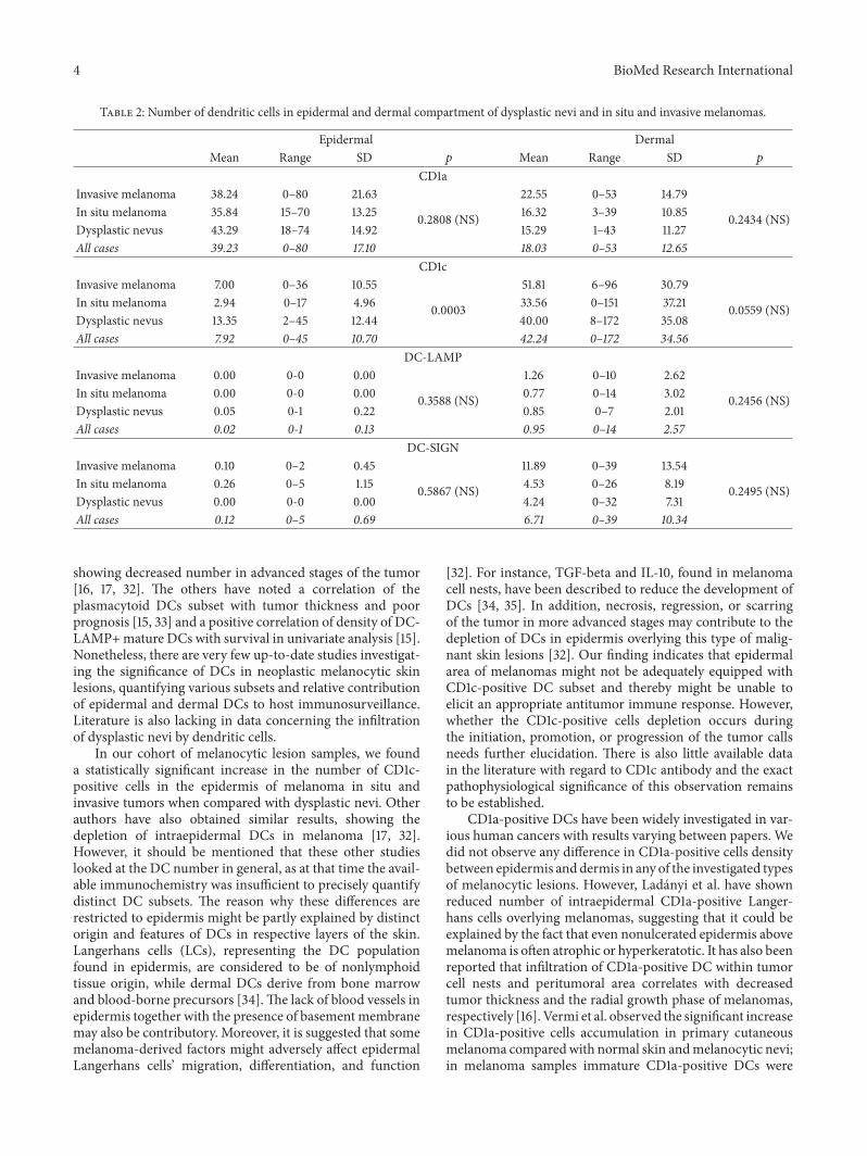
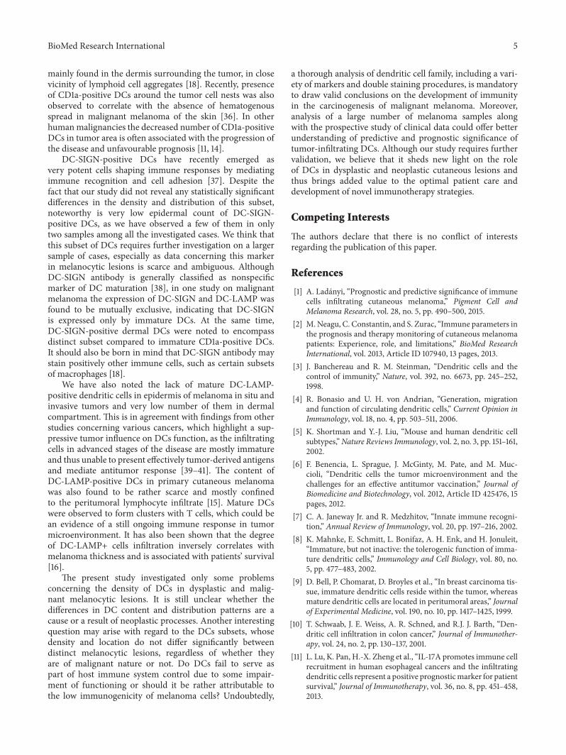
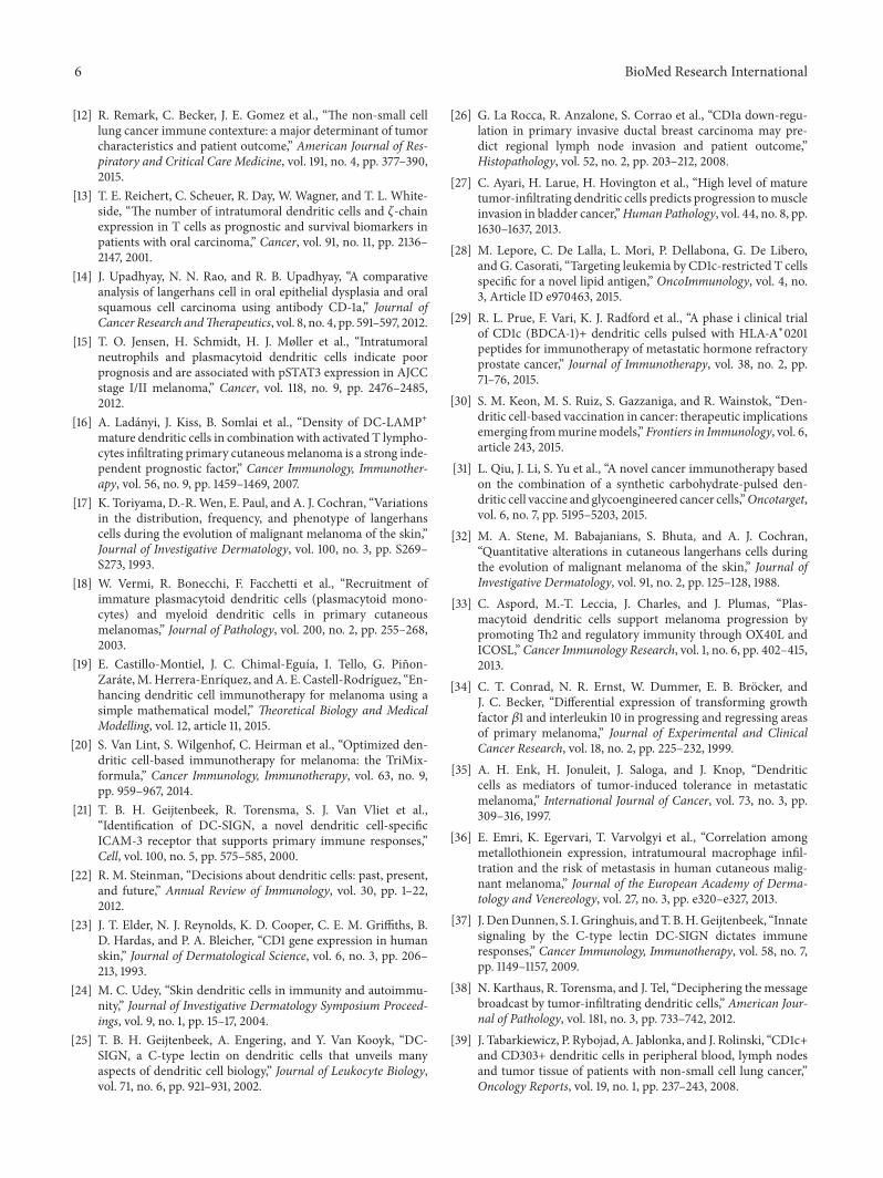