Embed Size (px)
Citation preview
University of New MexicoUNM Digital Repository
Psychology ETDs Electronic Theses and Dissertations
9-16-2014
Memory Profiles in Schizophrenia: ANeuropsychological Comparison with TemporalLobe EpilepsyS. Laura Lundy
Follow this and additional works at: https://digitalrepository.unm.edu/psy_etds
This Dissertation is brought to you for free and open access by the Electronic Theses and Dissertations at UNM Digital Repository. It has beenaccepted for inclusion in Psychology ETDs by an authorized administrator of UNM Digital Repository. For more information, please [email protected].
Recommended CitationLundy, S. Laura. "Memory Profiles in Schizophrenia: A Neuropsychological Comparison with Temporal Lobe Epilepsy." (2014).https://digitalrepository.unm.edu/psy_etds/84
S. Laura Lundy Candidate
Psychology
Department
This dissertation is approved, and it is acceptable in quality and form for publication:
Approved by the Dissertation Committee:
Ronald A. Yeo, Ph.D. , Chairperson
Robert J. Thoma, Ph.D.
Derek Hamilton, Ph.D.
Steven Verney, Ph.D.
MEMORY PROFILES IN SCHIZOPHRENIA:
A NEUROPSYCHOLOGICAL COMPARISON WITH
TEMPORAL LOBE EPILEPSY
by
S. LAURA LUNDY
B.S., Psychology, Duke University, 1998
M.S., Psychology, University of New Mexico, 2007
DISSERTATION
Submitted in Partial Fulfillment of the
Requirements for the Degree of
Doctor of Philosophy
Psychology
The University of New Mexico
Albuquerque, New Mexico
December, 2011
iii
MEMORY PROFILES IN SCHIZOPHRENIA: A NEUROPSYCHOLOGICAL
COMPARISON WITH TEMPORAL LOBE EPILEPSY
by
S. Laura Lundy
B.S., Psychology, Duke University, 1998
M.S., Psychology, University of New Mexico, 2007
Ph.D., Clinical Psychology, University of New Mexico, 2011
ABSTRACT
Previous research has demonstrated various neuroanatomical and
neuropsychological abnormalities in schizophrenia. Although results vary depending on
population characteristics, medication status, imaging methodology, and choice of
cognitive assessment measures, overall results suggest decreased cerebral volume within
frontal and temporal lobes and in individual structures, particularly hippocampus, within
these regions; abnormal connectivity within fronto-temporal networks; and deficits in
executive function, working memory, processing speed, and memory, with verbal
memory deficits more reliably demonstrated than non-verbal impairment. In particular,
left-hemisphere hippocampal-dependent verbal memory impairments have been proposed
to be a core feature of schizophrenia, as these deficits are reliably demonstrated in first-
episode, medication-naïve patients, chronic in- and outpatients, and at-risk populations
such as individuals with prodromal schizophrenia symptoms and first-degree relatives of
patients with schizophrenia. Verbal memory deficits have also been reliably
demonstrated in patients with left-hemisphere temporal lobe epilepsy (TLE), and those
who have undergone temporal lobe resection (TLR). Given the known localization of
structural abnormalities in TLR groups and their relation to deficits in memory, it seemed
reasonable to compare memory and other neuropsychological functions in schizophrenia
iv
and TLR groups to determine whether similar profiles emerged which might provide
additional evidence of significant left-hemisphere involvement in the development and
maintenance of schizophrenia. A comprehensive neuropsychological battery was
administered to a total of 43 schizophrenia, left and right TLR, and control participants.
Consistent with hypotheses, the schizophrenia and left TLR groups performed worse than
controls on verbal memory, and the right TLR group performed worse than controls on
non-verbal memory tasks, with a trend in the predicted direction for the schizophrenia
group. Patients with schizophrenia also performed worse than controls on working
memory, motor skills, and processing speed, as predicted. Hypotheses were not supported
regarding overall memory profiles: the left and right TLR groups showed the expected
interactive performance on verbal versus non-verbal memory, but the schizophrenia
group was not found to have a memory profile similar to that of the left TLR group.
Overall, results suggested that the cognitive profile of schizophrenia may best be
represented as a complex interaction pattern rather than a hemisphere-specific model as
seen in TLE.
v
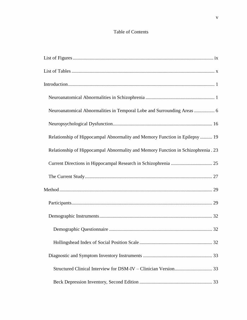
Table of Contents
List of Figures .................................................................................................................... ix
List of Tables ...................................................................................................................... x
Introduction ......................................................................................................................... 1
Neuroanatomical Abnormalities in Schizophrenia ......................................................... 1
Neuroanatomical Abnormalities in Temporal Lobe and Surrounding Areas ................. 6
Neuropsychological Dysfunction.................................................................................. 16
Relationship of Hippocampal Abnormality and Memory Function in Epilepsy .......... 19
Relationship of Hippocampal Abnormality and Memory Function in Schizophrenia . 23
Current Directions in Hippocampal Research in Schizophrenia .................................. 25
The Current Study ......................................................................................................... 27
Method .............................................................................................................................. 29
Participants .................................................................................................................... 29
Demographic Instruments ............................................................................................. 32
Demographic Questionnaire ..................................................................................... 32
Hollingshead Index of Social Position Scale ............................................................ 32
Diagnostic and Symptom Inventory Instruments ......................................................... 33
Structured Clinical Interview for DSM-IV – Clinician Version ............................... 33
Beck Depression Inventory, Second Edition ............................................................ 33
vi
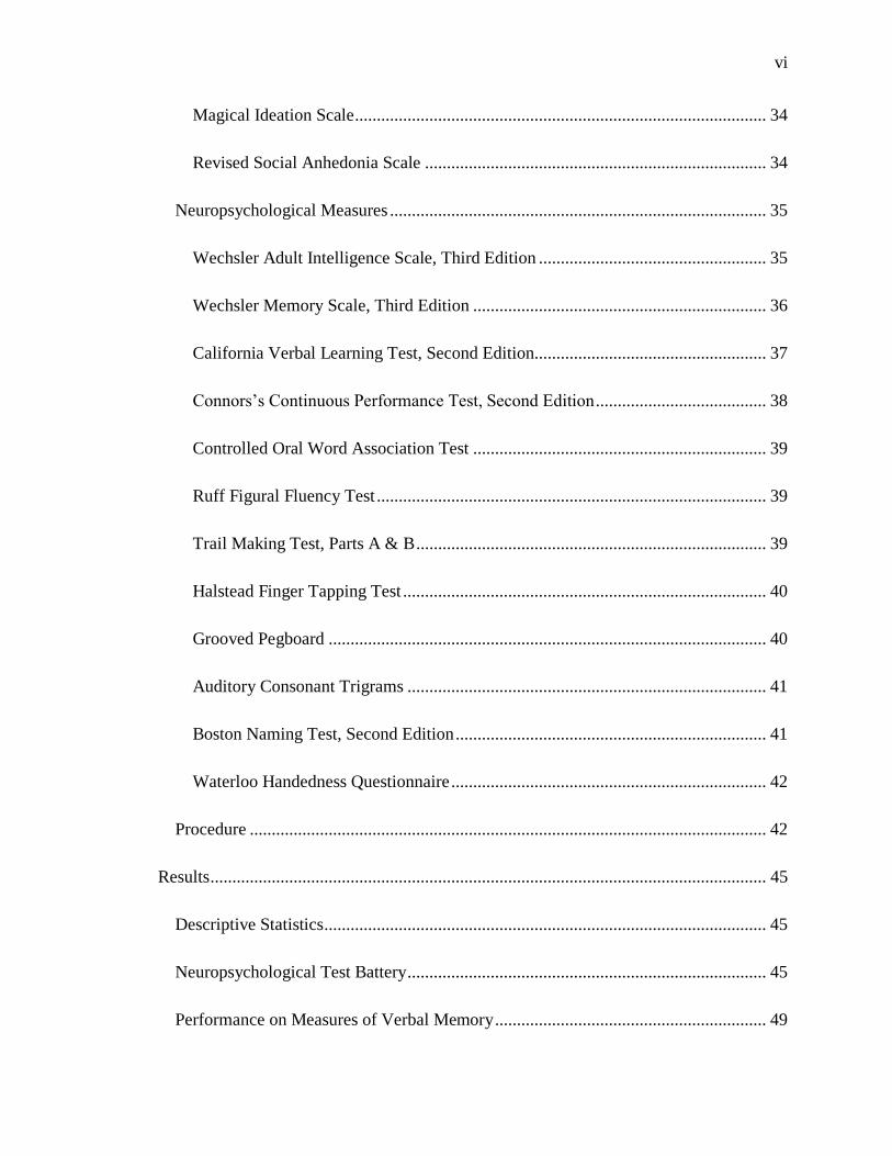
Magical Ideation Scale .............................................................................................. 34
Revised Social Anhedonia Scale .............................................................................. 34
Neuropsychological Measures ...................................................................................... 35
Wechsler Adult Intelligence Scale, Third Edition .................................................... 35
Wechsler Memory Scale, Third Edition ................................................................... 36
California Verbal Learning Test, Second Edition..................................................... 37
Connors‟s Continuous Performance Test, Second Edition ....................................... 38
Controlled Oral Word Association Test ................................................................... 39
Ruff Figural Fluency Test ......................................................................................... 39
Trail Making Test, Parts A & B ................................................................................ 39
Halstead Finger Tapping Test ................................................................................... 40
Grooved Pegboard .................................................................................................... 40
Auditory Consonant Trigrams .................................................................................. 41
Boston Naming Test, Second Edition ....................................................................... 41
Waterloo Handedness Questionnaire ........................................................................ 42
Procedure ...................................................................................................................... 42
Results ............................................................................................................................... 45
Descriptive Statistics ..................................................................................................... 45
Neuropsychological Test Battery .................................................................................. 45
Performance on Measures of Verbal Memory .............................................................. 49
vii
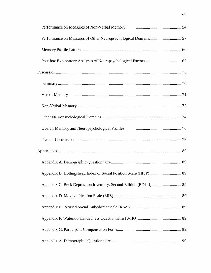
Performance on Measures of Non-Verbal Memory...................................................... 54
Performance on Measures of Other Neuropsychological Domains .............................. 57
Memory Profile Patterns ............................................................................................... 60
Post-hoc Exploratory Analyses of Neuropsychological Factors .................................. 67
Discussion ......................................................................................................................... 70
Summary ....................................................................................................................... 70
Verbal Memory ............................................................................................................. 71
Non-Verbal Memory ..................................................................................................... 73
Other Neuropsychological Domains ............................................................................. 74
Overall Memory and Neuropsychological Profiles ...................................................... 76
Overall Conclusions ...................................................................................................... 79
Appendices ........................................................................................................................ 89
Appendix A. Demographic Questionnaire .................................................................... 89
Appendix B. Hollingshead Index of Social Position Scale (HISP) .............................. 89
Appendix C. Beck Depression Inventory, Second Edition (BDI-II) ............................ 89
Appendix D. Magical Ideation Scale (MIS) ................................................................. 89
Appendix E. Revised Social Anhedonia Scale (RSAS)................................................ 89
Appendix F. Waterloo Handedness Questionnaire (WHQ) .......................................... 89
Appendix G. Participant Compensation Form .............................................................. 89
Appendix A. Demographic Questionnaire .................................................................... 90
viii
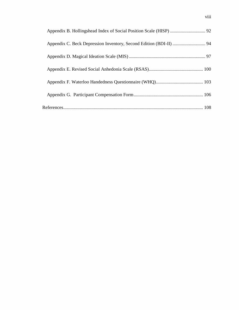
Appendix B. Hollingshead Index of Social Position Scale (HISP) .............................. 92
Appendix C. Beck Depression Inventory, Second Edition (BDI-II) ............................ 94
Appendix D. Magical Ideation Scale (MIS) ................................................................. 97
Appendix E. Revised Social Anhedonia Scale (RSAS).............................................. 100
Appendix F. Waterloo Handedness Questionnaire (WHQ) ........................................ 103
Appendix G. Participant Compensation Form ........................................................... 106
References ....................................................................................................................... 108
ix
List of Figures
Figure 1 Performance on Individual Measures of Verbal Memory by Group.. ............... 53
Figure 2 Performance on Individual Measures of Non-Verbal Memory by Group ......... 58
Figure 3 Memory Profiles by Group. ............................................................................... 64
Figure 4 Performance on Memory Composites by Group. .............................................. 65
Figure 5 Performance on Memory Composites by Group.. ............................................. 66
x
List of Tables
Table 1. Surgical Details for TLR Patients ....................................................................... 30
Table 2. Demographic Data by Group .............................................................................. 31
Table 3. Emotional/Symptom and Overall Intellectual Functioning Data by Group ....... 46
Table 4. Individual Variables within Neuropsychological Composite Factors and Their
Abbreviations .................................................................................................................... 47
Table 5. Verbal Memory Outcomes by Group ................................................................. 51
Table 6. Nonverbal Memory Outcomes by Group ........................................................... 56
Table 7. Pearson's Correlations Between Years of Education, Socioeconomic Status, and
Neuropsychological Composite Scores ............................................................................ 59
Table 8. Non-Memory Neuropsychological Composite Outcomes by Group ................. 61
Table 9. Individual Variables within Immediate and Delayed Verbal and Nonverbal
Memory Composite Factors and Their Abbreviations...................................................... 62
Table 10. Standardized Coefficients and Correlations of Neuropsychological Predictor
Variables with the Two Discriminant Functions .............................................................. 68
1
Introduction
Schizophrenia is classified as a psychotic disorder in the Diagnostic and
Statistical Manual of Mental Disorders (DSM-IV-TR; American Psychiatric Association,
2000.). It is characterized by the presence of various combinations of emotional
dysfunction, such as alogia, inappropriate affect, anhedonia, and impaired volition;
perceptual abnormalities, such as delusions and hallucinations; thought and speech
dysfunction, such as tangentiality, loose associations, and incoherence; disorganized
behavior, such as hoarding useless objects or dressing inappropriately; and catatonic
behavior, such as echolalia or posturing. In the majority of cases, schizophrenia is a
chronic and severely debilitating disorder, impacting individuals‟ ability to obtain
competitive employment, maintain social connections, and perform activities of daily
living sufficiently to live independently. Given the severe nature of schizophrenic
symptomatology and outcomes, it is not surprising that much research has been focused
on elucidating the etiology, course, and nature of specific deficits in this disorder.
Neuroanatomical Abnormalities in Schizophrenia
Neuroimaging studies have consistently demonstrated neuroanatomical
abnormalities in schizophrenia. Many brain regions, considered both as discrete areas and
nodes within networks of interconnected structures, have been implicated. Depending
upon the clinical characteristics (e.g., first-episode vs. chronic) of the population of study,
some findings are more reliably demonstrated than others. Moreover, with the advent of
magnetic resonance imaging (MRI), more subtle neuroanatomical differences can be
observed than was previously possible with computed technology (CT) scanning
(Seidman, Faraone, Goldstein, Goodman, Kremen, et al., 1999). Shenton and colleagues
2
(2001) recently published a review of 193 peer-reviewed magnetic resonance imaging
(MRI) studies of structural abnormalities in schizophrenia. These reviews included
studies of whole-brain, ventricular system, frontal lobe, temporal lobe, thalamus, and
other brain regions. Of 50 studies of whole-brain volume, only 11 (22%) reported
differences between patients with schizophrenia and healthy control subjects (e.g.,
Wright et al., 2000, in which patients were reported to have overall cerebral volume two
percent smaller than controls); however, they did note that one study (Jacobsen, Giedd &
Vaituzis, 1996) found smaller brain volumes in patients with childhood-onset
schizophrenia, suggesting that reduced overall brain volume may be related to more
severe genetic or environmental neurodevelopmental anomalies resulting in early onset of
symptoms. In general, however, most MRI studies do not report differences in whole
brain volume in schizophrenia versus healthy controls (e.g., McCarley, Wible, Frumin,
Hirayasu, Levitt, et al., 1999).
Ventricular dilation is another finding commonly attributed to schizophrenia.
Thirty-three MRI studies of the third ventricle were included in Shenton et al.‟s (2001)
review. Of these, 24 (73%) reported enlarged volume, which may be related to the
reduction in thalamic volume inconsistently reported in schizophrenia, as these structures
are located close to each other. Of five studies assessing the fourth ventricle, only one
(20%) noted increased volume in schizophrenia. The clinical implications of enlarged
fourth ventricle are unclear. The lateral ventricles have received much more attention in
schizophrenia research, given their proximity to temporal lobe structures thought to play
a central role in the development and maintenance of schizophrenic symptoms, and are
discussed in the next section (see below).
3
The parietal lobe, although part of the heteromodal association cortex with
interconnections to prefrontal and temporal lobe structures (Pearlson, Petty, Ross, &
Tien, 1996) and associated with functions such as language and attention which are
known to be impaired in schizophrenia (e.g., Bilder et al., 2000), has not been thoroughly
studied with regard to schizophrenia. In their review, Shenton et al. (2001) only found 15
MRI studies of the parietal lobe, nine (60%) of which reported positive findings.
Methodological differences among these studies may be partially responsible for
discrepant findings, as methods of measurement vary (e.g., whole-lobe versus
regional/functional subdivisions) and functional left-greater-than-right asymmetry, which
is present in healthy brains and necessary for intact language development, has not
always been evaluated. Nonetheless, some interesting relationships have been noted.
Studies have shown a reversal of the normal left-greater-than-right ratio of angular gyrus
(part of the inferior parietal lobule [IPL] particularly important for language
comprehension) volume in male, but not female, patients with schizophrenia compared to
controls (Frederikse, Lu, Aylward, Barta, Sharma, et al., 2000; Niznikiewicz, Donnino,
McCarley, Nestor, Iosifescu, et al. 2000). One study also showed correlations between
IPL and interconnected prefrontal (superior and inferior frontal gyrus and orbital gyrus)
and temporal (anterior superior temporal gyrus [STG], amygdala, and hippocampus)
cortices, providing evidence that the heteromodal association cortex may be affected in
schizophrenia (Niznikiewicz et al. 2000).
The frontal lobes, including prefrontal cortex (PFC), are known to be essential for
cognitive and behavioral regulation (Kolb & Whishaw, 2009) and have been fairly
extensively researched regarding their roles in the development and/or maintenance of
4
schizophrenia. Shenton and colleagues (2001) reviewed 50 MRI studies of the anatomy
of the frontal lobes in schizophrenia, of which 30 (60%) reported positive findings.
However, it is important to note that most studies have measured the frontal lobe as one
structure, rather than parceling out specific frontal and prefrontal regions, such as
orbitofrontal and dorsolateral prefrontal cortices, which are known to have functionally
different connectivities involved in various aspects of cognition. Thus, measurement of
the entire frontal lobe may obscure more specific, localized differences. Nevertheless,
results of studies of entire-lobe structure have suggested abnormalities in prefrontal-
temporal connectivity. For example, in a study by Wible et al. (1995), no differences
between male patients with schizophrenia (presenting with mainly positive symptoms)
and male healthy controls were found in volume of PFC as a whole, but left prefrontal
gray matter volume was found to correlate significantly with reduced volume of the left
hippocampal-amygdala complex, left STG, and left parahippocampal complex in the
schizophrenia group only. Breier et al. (1992) measured amygdala/hippocampus and PFC
volumes in patients with chronic schizophrenia versus healthy controls. They found that
patients had smaller right and left overall amygdala/hippocampal complex volumes as
well as smaller right and left prefrontal volumes. Within the amygdala/hippocampal
complex, they reported smaller right and left amygdala and left hippocampal volumes in
the patient group. They further reported reduced prefrontal white matter in the patient
group, with right-hemisphere prefrontal white matter highly correlated with right
amygdala/hippocampal volume, suggesting the presence of abnormal corticolimbic
connectivity in schizophrenia. Further evidence for abnormal prefrontal-limbic
connectivity, particularly in the left hemisphere, in schizophrenia is suggested by a study
5
which reported a correlation between left hippocampal volume reduction and decreased
cerebral blood flow to the dorsolateral (DL) PFC during an executive working memory
task in the affected twin of monozygotic twins discordant for schizophrenia (Weinberger,
Berman, Suddath, & Torrey, 1992).
Although few studies have measured subregions within the frontal lobe, those that
have tended to report abnormalities in volume and connectivity as well. For example,
Buchanan and colleagues (1998) divided the PFC into superior, inferior, middle, and
orbital regions based on anatomical landmarks and found volumetric reductions of the
right and left inferior prefrontal gray matter (primarily composed of Broca‟s area, which
projects to DLPFC, STG, and other heteromodal areas which have been implicated in the
pathogenesis of schizophrenia) in a mixed-gender sample of schizophrenia patients
compared to controls. In a study conducted by Goldstein et al. (1999) comparing frontal
lobe regions in patients with chronic schizophrenia to healthy controls, patients were
found to have bilaterally reduced, though more so on the left, DLPFC areas, and reduced
right orbitofrontal PFC area. Gur and colleagues (2000a) compared prefrontal volumes in
a group of 29 neuroleptic-naive (16 men and 13 women) and 41 previously treated
patients (24 men and 17 women) compared to healthy controls. They found DLPFC
volume reductions of 9% in male patients and 11% in female patients, a dorsomedial PFC
volume reduction of 9% in male patients only, and orbitofrontal volume reductions only
in women (23% and 10% for lateral and medial regions, respectively). Importantly, when
they compared the neuroleptic-naïve versus medicated patients, there were no significant
differences in volume in any brain region, suggesting that PFC reductions are not simply
a result of chronic medication. Other studies, however, have reported negative findings
6
regarding frontal lobe volumes, even when subregions were delineated and measured
separately. For example, Baaré et al. (1999) evaluated left and right dorsolateral, medial,
and orbital prefrontal gray and white matter volumes in patients with schizophrenia
versus healthy controls. They reported no significant group differences in any region of
interest (ROI), though they noted that volumes tended to be smaller in patients as a
group. However, they did find significant correlations between reduced left and right
prefrontal gray matter volume and performance on measures of verbal and visual memory
in the patient group, suggesting that even in the absence of outright structural
abnormalities, frontal lobe dysfunction likely contributes to the cognitive deficits seen in
schizophrenia. In particular, this study highlights the association between dysfunction in
PFC and memory functioning, which is known to be dependent on structures of the
medial temporal lobe (Squire & Zola-Morgan, 1991).
Neuroanatomical Abnormalities in Temporal Lobe and Surrounding Areas
Fifty-five MRI studies of the lateral ventricular system were included in the
Shenton et al. (2001) review; the lateral ventricles are considered important for
schizophrenia research because they surround temporal lobe structures such as
hippocampus and amygdala that have been implicated in impaired cognitive functioning
in this group (see below). Of these 55 studies, 44 (80%) reported enlarged lateral
ventricles in schizophrenia. For example, Andreasen et al. (1990) reported increased
lateral ventricular volume in patients with schizophrenia, particularly in male
participants. Lauriello et al. (1997) reported increased lateral and third ventricular volume
in patients with chronic schizophrenia, regardless of gender. Kelsoe and colleagues
(1988) found that lateral ventricular volume was increased by 62% in schizophrenic

7
subjects compared to healthy controls. Furthermore, of the 11 studies reviewed which did
not report overall lateral ventricle enlargement, many noted enlargement in the temporal
horn portion of the lateral ventricles, particularly in the left hemisphere. For example,
Shenton and colleagues (1992) found that the volume of the left, but not right, lateral
ventricle was enlarged in patients with schizophrenia, and that left temporal horn
volumes were 180% larger, and the right temporal horn was 74% larger, in patients with
schizophrenia than in healthy controls. In a study of first-episode schizophrenic patients,
Bogerts et al. (1990) found that overall, patients had larger anterior temporal horn
volumes than controls, and that female patients had larger left, but not right, total
temporal horn volumes than controls. While findings of lateral ventricle enlargement are
highly consistent in the schizophrenia literature, these abnormalities also occur frequently
in other neurological disorder such as Parkinson‟s disease, Huntington‟s disease, and
Alzheimer‟s dementia, and are thus not specific to the schizophrenia process (e.g.,
Apostolova et al., 2010). Nonetheless, enlargement of the ventricular space, especially in
the left hemisphere, surrounding temporal lobe structures which have reliably been
implicated in behavioral and cognitive dysfunction in schizophrenia suggests
neurodevelopmental or neurodegenerative processes related to this region (Shenton et al.,
2001).
The temporal lobe is comprised of two main divisions: lateral, or neocortical, and
medial. The lateral temporal lobe structures include the STG (further broken down into
the planum temporale and Heschl‟s gyrus, or transverse temporal gyrus), middle temporal
gyrus, and inferior temporal gyrus. The medial temporal lobe structures include the
hippocampus, amygdala, and parahippocampal gyrus (as well as the entorhinal cortex,
8
which is neuroanatomically continuous with the parahippocampal gyrus). Abnormalities
in the temporal lobes have long been posited to be related to the behavioral and cognitive
sequelae of schizophrenia [e.g., Kraepelin (1971), and Southard (1915), who believed
that auditory hallucinations were the result of temporal lobe abnormalities]. Structural
abnormalities in the temporal lobe and surrounding regions have been of particular
interest in schizophrenia research, given the location of primary auditory cortex on
Heschl‟s gyrus and the involvement of limbic system structures, and their connections to
frontal and prefrontal cortex, in the formation of memory and language processing
functions, which have been shown to be impaired in schizophrenia (e.g., Hoff et al.,
1999). Shenton et al., (2001), for example, posited that hallucinations and cognitive
deficits in schizophrenia are associated with abnormalities in medial temporal lobe
structures (e.g., hippocampus, amygdala, and parahippocampal gyrus) involved in
encoding and retrieval of memories. Lateral temporal lobe structures, such as the STG,
have also been shown to be associated with hallucinations (e.g., Barta, Pearlson, Powers,
Richards, & Tune, 1990; Penfield & Perot, 1963). It is not surprising, then, that much
focus has been directed at investigating the dysmorphology and dysfunction of the
temporal lobes in schizophrenia research.
In Shenton et al.‟s (2001) review, 31 (61%) of the 51 MRI studies reporting
whole-lobe volumes noted smaller overall temporal lobes in the schizophrenia compared
to control group. For example, Bogerts and colleagues (1990) reported a 9% reduction in
whole temporal lobe volume in male patients with first-episode schizophrenia compared
to healthy controls, though no differences were noted in female subjects. Other studies
have reported temporal lobe volume reductions only in the left hemisphere (e.g., Gur,
9
Cowell, Turetsky, Gallacher, Cannon, et al. 1998; Turetsky, Cowell, Gur, Grossman,
Shtasel, et al., 1995), and in the left hemisphere only in male patients (e.g., Bryant,
Buchanan, Vladar, Breier, & Rothman, 1999). Regarding lateral temporal lobe structures,
most studies have focused on assessing the STG and its divisions. Overall, the majority of
studies report volume reduction in schizophrenia. In fact, of the 12 MRI studies of gray
matter volume in STG included in Shenton et al.‟s (2001) review, 100% reported reduced
volume (of 15 studies measuring STG gray and white matter, 10 [67%] reported positive
findings). For example, Gur and colleagues (2000b) reported an 11.5% decrease in STG
volume in males with schizophrenia, but not in females. Another study reported reduced
STG gray matter in a heterogeneous (i.e., mixed-subtype) group of inpatients with
chronic schizophrenia compared to healthy controls (Zipursky, Marsh, Lim, DeMent,
Shear, et al., 1994). Holinger et al. (1999) investigated STG volumes in left-handed males
with schizophrenia, and found that patients had bilaterally smaller gray matter volumes in
the posterior STG (16% smaller on the right, 15% smaller on the left), and smaller total
right STG than controls. This study highlights the importance of assessing handedness
and hemispheric asymmetry in volumetric MRI research, as results may differ between
left- and right-handed patients. The planum temporale (PT), another lateral temporal lobe
structure important for language processing, has been investigated in schizophrenia
research as well. A recent meta-analysis of PT volumes in schizophrenia concluded that
although methodological differences in measurement likely obscured some results, strong
evidence is nonetheless found for larger right PT in patients, resulting in a reversal of the
developmentally-normal left-greater-than-right asymmetry in this region (Shapleske,
Rossell, Woodruff, & David, 1999). In the Shenton et al. (2001) review, six of ten (60%)
10
studies of PT reported differences in asymmetry between patients and healthy controls
(e.g., Barta, Pearlson, Brill, Royall, McGilchrist, et al., 1997; Petty, Barta, Pearlson,
McGilchrist, Lewis, et al., 1995). Importantly, abnormalities in PT volume and
asymmetry may be related to abnormalities in the heteromodal association cortex, which
would strengthen the argument that frontal-parietal-temporal networks are disturbed in
schizophrenia (Pearlson, Petty, Ross, & Tien, 1996).
In addition, much recent research has employed a voxel-based morphometry
(VBM) approach to analyzing differences in cerebral volumes, in which concentration or
density of gray matter (GM) is assessed relative to the concentration of other types of
tissue within the brain. VBM has been hailed as a more reliable and unbiased method for
analyzing volumetric differences in gray (GM) and white (WM) matter than conventional
analyses of regions of interest (ROIs), as it utilizes an automated algorithm for mapping
spatially normalized images from structural MRIs into standardized stereotactic space,
thereby eliminating user error in delineation of ROIs and the bias inherent in specifying a
priori ROIs rather than examining all regions as potentially significant (Segall et al.,
2009). VBM studies tend to demonstrate significantly lower gray matter (GM) densities
in schizophrenia-spectrum groups compared to healthy control groups, particularly in the
temporal lobe. For example, in a recent meta-analysis of 15 studies which employed
VBM analysis of brain tissue in schizophrenia, significant gray and white matter density
differences were reported in 50 regions of interest (ROIs). One of the most consistent
differences which emerged in the meta-analysis was within the left STG, which had a
lower relative density in schizophrenia versus in healthy controls (Honea, Crow,
Passingham, & Mackay, 2005). Interestingly, VBM studies have uncovered significant
11
differences in STG in schizophrenia-spectrum disorders that were not found using
conventional ROI analysis (e.g., Giuliani, Calhoun, Pearlson, Francis, & Buchanan, 2005;
Kubicki et al., 2002). Likewise, in a recent multisite VBM study, patients with
schizophrenia-spectrum disorders (including schizophrenia, schizoaffective disorder, and
schizophreniform disorder) were found to have a 5% reduction in GM density in the STG
compared to healthy controls (Segall et al., 2009).
Structural abnormalities of the tissue of the medial temporal lobe are among the
most robust neuroanatomical findings in schizophrenia research (Heckers, 2001). A
number of MRI studies have reported smaller volume of the medial temporal lobe overall
in schizophrenia (Bogerts et al., 1993; Gur et al., 2000b; McCarley et al., 1999; Wright et
al., 2000), particularly in the left hemisphere (DeLisi et al., 1991; Shenton et al., 1992).
In addition, VBM studies of schizophrenia have reported lower densities of gray matter in
the medial temporal lobe overall compared to healthy controls. For example, in a meta-
analysis conducted by Honea and colleagues (2005), nine of fourteen studies which
assessed the temporal lobe reported reduced medial temporal lobe densities. Eight of
these studies reported differences in the left hemisphere, with the remaining study
(Wright et al., 1999) reporting a trend in this direction, while only three reported right-
hemisphere differences.
As with the frontal lobe, however, parceling out the contributions of separate
medial temporal lobe structures may be more informative for schizophrenia research than
assessing the medial temporal lobe as a single unit. An important consideration in
interpreting the results of studies of medial temporal lobe structure abnormalities is that
although the hippocampus and amygdala are functionally different, they are
12
neuroanatomically continuous and it is somewhat difficult to reliably dissect these
structures on MRI slices in order to evaluate their separate volumes. Thus many studies
measure volumes of the hippocampal/amygdala complex as a whole, which may obscure
more localized findings. Regardless, of the 49 studies included in Shenton et al.‟s (2001)
review which evaluated medial temporal lobe structures separately, 36 (74%) reported
volume decreases in the hippocampal/amygdala complex, findings which are consistent
with post-mortem results.
Recently, more studies have reported findings based on separate amygdala and
hippocampus assessments. Some MRI studies have demonstrated reduced amygdala
volume in schizophrenia bilaterally (e.g., Barta et al., 1997b; Breier et al., 1992; Jernigan
et al., 1991; Marsh, Suddath, Higgins, & Weinberger, 1994), or unilaterally (e.g., left
hemisphere: Barta, Pearlson, Powers, Richards, & Tune, 1990; right hemisphere:
Pearlson et al., 1997), or only in males (Gur et al., 2000b). However, other studies have
reported no differences in amygdala volume. A post-mortem study of amygdala volumes
in schizophrenia versus healthy controls showed no significant difference bilaterally
(Chance, Esiri, & Crow, 2002). MRI studies have also reported negative results. Studies
of amygdala volume in first-episode patients (e.g., Niemann, Hammers, Coenen, Thron,
& Klosterkӧtter, 2000), and in patients with chronic schizophrenia (e.g., Staal et al.,
2000; Swayze, Andreasen, Alliger, Yuh, & Ehrhardt, 1992) have demonstrated no
significant differences bilaterally. In contrast, studies of hippocampal volume in
schizophrenia report differences more reliably. In Shenton and colleagues‟ (2001) review
of MRI studies in schizophrenia, 17 of 25 studies (68%) which measured hippocampus
separately from amygdala and other medial temporal lobe structures reported decreased
13
volume. VBM studies as well have demonstrated significantly reduced density of
hippocampus in first-episode schizophrenia compared to both healthy control and patients
with affective psychosis, particularly on the left (e.g., Kubicki et al., 2002; Rametti et al.,
2007). However, while hippocampus abnormality is consistently found, the precise nature
of the results has varied. Meta-analyses have posited that smaller hippocampus volume is
equivalent across hemispheres (Nelson, Saykin, Flashman, & Riordan, 1998), even when
limited to studies of first-episode patients (Steen, Mull, McClure, Hamer, & Lieberman,
2006). However, other first-episode studies have resulted in findings of significant left-
less-than-right hippocampus asymmetry (Bogerts et al., 1990; Hirayasu et al., 1998). In a
longitudinal study, Velakoulis et al. (2006) demonstrated hippocampus volume deficits
bilaterally in chronic schizophrenia, in the left hemisphere only in first-episode
schizophrenia, and not at all in schizophreniform psychosis.
It is also important to note that there have been scattered reports of null findings
with regard to hippocampus volume in schizophrenia. For example, Marsh et al. (1997)
reported no group difference in hippocampal volume in an inpatient sample with severe
and chronic schizophrenia. Csernansky et al. (2002) reported significant abnormalities of
hippocampal shape and asymmetry, but not overall volume after total cerebral volume
was included as a covariate, in patients with schizophrenia in the residual stage. Because
of the variability in group differences across reports, the etiological role of hippocampal
anomaly in schizophrenia and its association with subsequent symptoms and course of
the disorder remains a critical area for research (White et al., 2008).
Abnormalities in the cellular structure and neurochemistry of the hippocampus in
patients with schizophrenia have also been reported in the literature. The N-methyl-d-
14
aspertate (NMDA) receptor system, and especially glutamate-mediated long-term
potentiation (LTP), within the hippocampus has been shown to play a crucial role in
normal learning and memory formation (Rezvani, 2006), though not necessarily in
storage or retrieval of learned information (Constantine-Paton, 1994). Animal studies
have demonstrated that administration of drugs which increase glutamatergic
transmission enhance encoding of memories across multiple tasks, including spatial
learning (e.g., Morris water maze and radial arm maze tasks) and olfactory discrimination
tasks (Staubli, Rogers, & Lynch, 1994). In addition, studies with healthy human
participants have shown that blockage of the NMDA receptor system with an antagonistic
agent such as phencyclidine (PCP) or ketamine results not only in impaired learning and
memory, but also in behavioral changes similar to the positive and negative symptoms of
schizophrenia, making these viable analog studies for the investigation of impaired
hippocampal functioning in schizophrenia (Jentsch & Roth, 1999; Lahti, Weiler,
Michaelidis, Parwani, & Tamminga, 2001). For example, administration of ketamine
(which has been utilized in studies rather than PCP due to its decreased potency and
resulting neurotoxicity) resulted in impaired verbal working memory (Honey et al., 2004)
and encoding and retrieval of episodic memory, dependent on frontal and hippocampal
input (Honey et al., 2005) in fMRI studies with healthy controls.
Numerous postmortem neurochemical studies have found decreased glutamate
receptor function, predominantly in the left hippocampus, in schizophrenia compared to
healthy controls (e.g., Deakin et al., 1989; Harrison et al., 1991; Kerwin et al., 1988; Tsai
et al., 1995). Other studies have reported excess glutamate (possibly related to decreased
receptor functioning) in the temporal lobes of patients with schizophrenia (e.g., Cecil et
15
al., 1999). Magnetic resonance spectroscopy (MRS) has been employed in order to
investigate relative concentrations of neurochemicals such as glutamate in vivo in
schizophrenia. One study found a right-greater-than-left asymmetry in the ratio of
glutamate, glutamine, and γ-aminobutyric acid (GABA), collectively denoted as Glx, to
choline-containing compounds in the hippocampus of patients with schizophrenia
compared to healthy controls, consistent with postmortem findings of glutamate deficit
(Kegeles et al., 2000). Other MRS studies have demonstrated a relative increase in
absolute glutamate concentration in the frontal and temporal lobes in schizophrenia as
well. For example, van Elst and colleagues (2005) found increased glutamate
concentration in the prefrontal cortex and hippocampus of patients with schizophrenia
compared to healthy controls, and further reported that increased prefrontal cortex
glutamate concentrations were correlated with poorer global mental functioning.
However, results of MRS studies have been mixed. In a study by Ohrmann et al. (2005),
patients with chronic schizophrenia were found to have lower levels of Glx in the
dorsolateral prefrontal cortex than either healthy controls or first-episode patients. In a
recent study by Bustillo et al (2011), Glx and other neurochemical levels were
investigated in whole gray matter and whole white matter in patients with schizophrenia
versus healthy controls, with subsequent investigation of several regions of interest
including left and right frontal, parietal, and temporal gray matter. While concentrations
of Glx were not found to differ between groups, lower Glx levels were found to correlate
with impaired cognitive performance in the patient task only, on a neuropsychological
test battery which included both verbal and nonverbal memory tasks. Thus, while
research on glutamate levels and receptor functioning in schizophrenia is still limited,
16
results have demonstrated a link between abnormal functioning or concentration of
glutamate receptors within the frontal and temporal lobes, including specific reports of
hippocampus, and cognitive functioning in schizophrenia, warranting ongoing research.
As a whole, results of morphological evaluations of whole-brain and
neuroanatomical subregions tend to suggest that volumetric abnormalities exist in
multiple anatomical regions in schizophrenia, with some of the most consistent findings
emerging for the temporal lobe and its component structures, particularly hippocampus
and STG, as well as for frontal/prefrontal-temporal/limbic connectivities. Also of note,
when unilateral differences are noted, they tend to be confined to the left hemisphere. The
importance of decreased frontal and temporal tissue volume and impaired
interconnectivity in schizophrenia is discussed in the following sections.
Neuropsychological Dysfunction
Schizophrenia has been shown to be associated with neuropsychological deficits
across a wide range of domains, including memory, attention, executive function, motor
skills, and processing speed (Bilder, 1996; Bilder et al., 2000; Braff et al., 1991;
Goldberg & Gold, 1995; Pantelis, Nelson, & Barnes, 1996; Saykin et al., 1994). A
generalized cognitive deficit has been theorized to exist in schizophrenia, such that
domain-specific deficits are less pronounced in relation to an overall deficit (e.g,
Blanchard & Neale, 1994; Dickinson & Harvey, 2008; Dickinson, Iannone, Wilk, &
Gold, 2004; Dickinson, Ragland, Gold, & Gur, 2008; Heinrichs & Zakzanis, 1998;
Mohamed, Paulsen, O‟Leary, Arndt, & Andreasen, 1999; Saykin et al., 1994). Several
meta-analyses have suggested that overall cognitive means in groups with schizophrenia
are approximately one standard deviation below that of healthy control groups, with
17
individual variable means typically falling within one-third standard deviations above or
below that level (e.g., Dickinson, Ramsey, & Gold, 2007; Heinrichs, 2005; Heinrichs &
Zakzanis, 1998). Evidence of significant intercorrelation among neurocognitive domains
is often cited as a main support for the theory of generalized cognitive deficit, and
proponents theorize that the pattern of deficits represents a deficit in a general cognitive
ability factor known as “g,” (Dickinson, Ragland, Gold, & Gur, 2008). For example,
Dickinson and colleagues (2008) reported that a single common ability factor accounted
for 63% of the diagnosis-related variance in overall cognitive performance in their
sample of mixed first-episode and chronic stable schizophrenia outpatients.
On the other hand, many studies have failed to demonstrate overall cognitive
impairment in patients with schizophrenia using numerous different methodologies and
assessment measures (e.g., Cohen, Barch, Carter, & Servan-Schreiber, 1999;
Nuechterlein et al., 2004). Other researchers have suggested that there exist several
consistent domain-specific deficits within a background of generalized cognitive deficits,
as these domains tend to show disproportionately large deficits even after the effects of a
general ability factor are considered. In particular, verbal memory, including immediate
and delayed recall, free and cued recall, and recognition, is consistently impaired
(Aleman, Hijman, de Haan, & Kahn, 1999; Dickinson, Ragland, Gold, & Gur, 2008;
Heinrichs & Zakzanis, 1998; Hoff et al., 1999; Toulopoulou, Morris, Rabe-Hesketh, &
Murray, 2003). Among verbal memory deficits, recall tends to be affected more than
recognition (Clare, McKenna, Mortimer, & Baddeley, 1993; Hanlon et al., 2006;
Johnson, Klinger, & Williams, 1977; Nacmani & Cohen, 1969). Episodic memory
(memory for events) and semantic memory (memory for facts), which together are often
18
referred to as declarative memory, seem to be affected in patients with schizophrenia
more than is procedural memory (Cirillo & Seidman, 2003; Clare, McKenna, Mortimer,
& Baddeley, 1993; Goldberg et al., 1993; Gras-Vincendon et al., 1994; Kazes et al.,
1999; McKenna et al., 1990; Tamlyn et al., 1992; Weiss and Heckers, 2001). Deficits in
verbal memory have been shown to be present in both first-episode, nonmedicated
patients (Mohamed et al., 1999; Riley et al., 2000; Saykin et al., 1994), in high-risk
patients (i.e., with attenuated psychotic symptoms; Lencz et al., 2006) and in unaffected
first-degree relatives of schizophrenic patients (Kremen et al., 1994; Sitskoorn, Aleman,
Ebisch, Appels, & Kahn, 2004; Snitz, MacDonald, & Carter, 2006), leading many
researchers to postulate that impaired verbal memory is a primary neuropsychological
deficit and may represent a core feature of the disorder rather than an effect of chronicity
or antipsychotic medication (Saykin et al., 1994).
Non-verbal memory has been less systematically evaluated in schizophrenia.
Some studies have suggested that although memory for faces or designs may be impaired,
the deficit is relatively small compared to verbal memory deficits (e.g., Saykin et al.,
1991). Others have reported equally impaired immediate and delayed verbal and non-
verbal memory deficits (e.g., Calev, Korin, Kugelmass, & Lerer, 1987; Gold et al.,
1992b; Kolb & Whishaw, 1983). One study utilizing the Wechsler Memory Scale (Form
1) reported poorer performance on measures of immediate and delayed verbal, and
delayed non-verbal, memory in schizophrenia compared to both healthy controls and a
group with TLE (Seidman et al., 1998). Of note, these authors reported that although
delayed free recall of designs was impaired, retention for this material was intact,
suggesting that difficulties with initial encoding, rather than retrieval, of visual
19
information may be impaired in schizophrenia. Overall, deficits in visual memory are
generally reported in schizophrenia, though the degree and nature of impairment vary
considerably.
Declarative, episodic, and working memory have long been recognized as being
dependent on the function of an intact hippocampus, which is necessary for encoding and
consolidating new memories (Eichenbaum, Otto, & Cohen, 1994; Eichenbaum,
Schoenbaum, Young, & Bunsey, 1996; Scoville & Milner, 1957; Wittenberg & Tsien,
2002). While research in the area of hippocampal structure and memory functions in
schizophrenia is complicated by factors such as the heterogeneity of schizophrenia
subject groups, medication effects, chronicity, and normally occurring gender-based and
hemispheric neuroanatomical asymmetries, much research has been conducted which
investigated this relationship in other neurologic conditions.
Relationship of Hippocampal Abnormality and Memory Function in Epilepsy
Numerous studies of memory processes in patients with hippocampal injury have
demonstrated significant impairment, particularly in the epilepsy literature.
Neuropsychological studies of patients with temporal lobe epilepsy (TLE) and patients
who have undergone unilateral temporal lobe resection (TLR) show a fairly consistent
pattern of verbal memory deficits in left hemisphere TLE and after left hemisphere TLR,
and non-verbal memory deficits in right TLE and after right hemisphere TLR (Delaney,
Rosen, Mattson, & Novelly, 1980; Giovagnoli, Casazza, & Avanzini, 1995; Ladavas,
Umilta, & Provinciali, 1979; Majdan, Sziklas, & Jones-Gotman, 1996; Ojemann &
Dodrill, 1985). However, resection of the temporal lobe in epilepsy is a complicated
procedure which can result in memory impairment alone, or in a pattern of cognitive
20
deficits in multiple cognitive domains such as attention and language, depending on the
amount and area of tissue excised. Surgical procedures differ widely, with anterior
temporal lobectomy (ATL) still most commonly applied. In this procedure,
approximately 4.5 cm of anterior temporal cortex, including STG, medial temporal gyrus,
and inferior gyrus, are removed along with fusiform gyrus, parahippocampal gyrus, and
hippocampus (Davies, Hermann, Dohan, Foley, Bush, et al., 1996). More recent advances
in combining data of localized lesions on MRI scans with epileptogenic foci determined
by electrophysiological monitoring have lead to an increase in tailored surgical
approaches such as amygdalohippocampectomy (AH; Clusmann, Schramm, Kral,
Helmstaedter, Ostertun, et al., 2002), in which approximately 2-3 centimeters of
hippocampus, a large part of the amygdala, and the parahippocampal gyrus are removed.
This procedure spares hippocampal tissue but leaves less amygdala tissue intact than
ATL (Goldstein & Polkey, 1993). Some studies have shown that the extent of temporal
tissue removed correlates with verbal deficits on the left and visual deficits on the right
(e.g., Clusmann et al., 2002; Katz, Awad, Kongy, Chelune, Naugle, et al., 1989), however
other studies have suggested that the extent of post-operative memory deficits depends
more on pre-surgical performance than on surgical procedure (e.g., Goldstein & Polkey,
1993).
Other factors posited to have an effect on postsurgical outcomes include presence
of hippocampal sclerosis and age of hippocampal insult. In a retrospective study of 20
subjects with TLE, Mathern, Pretorius, & Babb (1995) found that the presence and type
of initial precipitating injury (i.e., medical illness/trauma versus idiopathic TLE) and the
age of the patient when the injury occurred were significantly related to the pathological
21
findings in the hippocampus at time of surgery. Specifically, patients with a history of a
significant initial precipitating injury that occurred prior to age 5 years were more likely
to have unilateral hippocampal sclerosis on MR imaging than patients with initial injury
after age 5 or with idiopathic TLE. Another study of 122 patients who had undergone
ATL surgery examined the relationship between age of seizure onset, duration of
epilepsy, and extent of hippocampal sclerosis (Davies et al., 1996). They found that the
younger the age of seizure onset, and the longer the duration of epilepsy prior to ATL, the
higher the extent of sclerosis. In a study by Hermann and colleagues (1995), although
degree of hippocampal sclerosis was not assessed, both later age of seizure onset and
older age at time of resection were significant and selective predictors of episodic
memory decrease for left ATL patients. Overall, then, results from these studies suggest
that early seizure onset and extended epilepsy duration are associated with the
development of significant hippocampal sclerosis. In combination with results of studies
such as that of Bell and Davies (1998), which suggest that patients without significant left
hippocampal sclerosis who underwent left ATL were at higher risk for developing post-
surgical deficits in verbal memory, it can be reasonably theorized that age of seizure
onset may be inversely related to post-ATL verbal memory performance in patients with
left-sided TLE. However, there are reports in the literature of no difference in post-
surgical memory skills in patients with early versus late seizure onset (e.g., Vargha-
Khadem, Gadian, Watkins, Connelly, Paesschen, et al., 1997). The inconsistent results of
a small number of studies nevertheless highlight the importance of assessing age of
seizure onset and duration of epilepsy when evaluating post-surgical memory
functioning.
22
Spiers et al. (2001) investigated unilateral temporal lobectomy patients‟ visuo-
spatial and episodic memory using a virtual-reality town to assess lateralized
hippocampal function. To assess spatial memory, they evaluated patients‟ ability to
navigate to certain locations, to recognize town scenes, and to do map reconstruction. To
assess episodic memory, the participant collected different objects from different
individuals (person) in different locations (place) in a certain order (time) and their
recognition of these details was measured. Patients with right temporal lobectomy were
impaired on the navigational measures compared to controls. In contrast, patients with
left temporal lobectomy exhibited a deficit for the episodic memory measures. This study
further illustrated a lateralized deficit for each version of the memory task and
functionally dissociated the two hippocampi. However, other studies have shown that
patients with unilateral hippocampal damage manifest allocentric spatial deficits,
regardless of the hemisphere of damage (Astur, Taylor, Mamelak, Philpott, & Sutherland,
2002; Maguire, Burke, Phillips, & Staunton, 1996) and that episodic memory function
does not always lateralize to one hippocampus (Ryan et al., 2001; Viskontas,
McAndrews, & Moscovitch, 2000). Therefore, it has not been clear whether episodic
memory function is lateralized to left hippocampus and visuo-spatial memory to right
hippocampus.
Thus, while a fairly consistent pattern of cognitive functioning, particularly in
terms of memory skills, tends to emerge post-TLR, the results of most research
paradigms must be considered with caution given differences in the duration, frequency,
nature, and extent of pre-surgical seizure activity and resulting sclerotic processes,
amount and site of tissue removal, and patients‟ pre-surgical functioning. Nonetheless,
23
these studies provide a foundation for investigating the relationship between hippocampal
dysfunction and memory impairment in schizophrenia.
Relationship of Hippocampal Abnormality and Memory Function in Schizophrenia
Modern theories of the etiology of schizophrenia involve altered expression of
multiple genes and neurodevelopmental processes (Censits, Ragland, Gur, & Gur, 1997;
Rapoport, Addington, Frangou, & Psych, 2005) affecting the functions and connections
of several brain areas, one of which is the hippocampus (Molina et al., 2002; Pantelis,
Yücel, Wood, McGorry, & Velakoulis, 2003). Functional neuroimaging has linked
hippocampus dysfunction to impaired performance on memory tasks in schizophrenia. In
a positron emission tomography (PET) study, Heckers et al. (1998) identified lesser
hippocampus activity in patients than controls during attempts to recall studied words. In
a functional magnetic resonance imaging (fMRI) study, controls exhibited greater
activation than patients in left anterior hippocampus during encoding and in hippocampus
bilaterally during recognition (Jessen et al., 2003). Functional impairment related to the
hippocampus extends to the ability to comprehend relationships and draw inferences, as
demonstrated by a selective deficit in discrimination accuracy when cognitive flexibility
is required, which has been observed in schizophrenia using fMRI (Öngur et al., 2006).
Using a relational-memory task, Hanlon et al. (2005) directly assessed hippocampus
activity with magnetoencephalography (MEG), reporting abnormal right hemisphere
processing of nonverbal stimuli accompanied by a possibly compensatory, left-lateralized
activation in schizophrenia.
The relationship of neuropsychological function to structural abnormality in the
hippocampus remains unclear. With hippocampus critical for some aspects of memory, it
24
may play a central role in visual and verbal memory impairments exhibited in
schizophrenia (Cirillo & Seidman, 2003; Saykin et al., 1991; 1994). Lack of a
relationship between hippocampus volume and episodic memory in people with
schizophrenia or controls has been reported by some studies (Bilder et al., 1995; DeLisi
et al., 1991; Torres, Flashman, O‟Leary, Swayze, & Andreasen, 1997). However, other
studies have shown a positive relationship between hippocampus and memory function in
schizophrenia. Gur et al. (2000b) found a positive correlation between gray matter
volume of the hippocampus and episodic memory scores across patients and controls of
both sexes. Studies by Saykin et al. (1991, 1994) have demonstrated a particularly large
memory impairment, relative to attention and executive function deficits, in unmedicated
patients with schizophrenia. They determined that impaired visual, and particularly verbal
learning and memory, distinguished patients from normal controls better than other
neuropsychological variables, and they related these deficits to those found after
temporal-hippocampal damage. Gruzelier et al. (1988) administered a battery of
neuropsychological tests to patients with schizophrenia and normal controls, also finding
a generalized deficit, but with the most striking deficits again emerging on memory tasks
involving temporal-hippocampal function.
Some researchers have posited that abnormalities in structure and function of the
left hippocampus in particular may be related to memory deficits in schizophrenia. For
example, Seidman et al. (2002) found that immediate and delayed verbal memory and left
hippocampus volume were positively correlated in schizophrenia patients and in their
relatives, as well as in controls, while no correlations emerged with the right
hippocampus. Further, amygdala-anterior hippocampus volume was found to be
25
positively correlated with delayed verbal memory in relatives of patients with
schizophrenia and controls (O‟Driscoll et al., 2001). However, some investigations of
structure-function relationships have found differential patterns of correlations between
hippocampus volume and verbal IQ or verbal or visual memory in controls and people
with schizophrenia (Kuroki et al., 2006; Sachdev, Brodaty, Cheang, & Cathcart, 2000;
Sanfilipo et al., 2002; Toulopoulou et al., 2004). Findings of differential patterns of
correlations for patients and controls are thought to indicate a loss of normal structure-
function relationships, possibly arising from aberrant neurodevelopment.
Current Directions in Hippocampal Research in Schizophrenia
With some degree of hippocampal volume deficits in schizophrenia well
established but the nature and mechanisms of its functional consequences unclear,
attention is turning to localizing hippocampus volume decrements along the anterior-
posterior axis. One study confirmed the presence of overall hippocampus volume deficits
in schizophrenia but maintained that the loss was diffuse rather than topographically
specific (Weiss et al., 2005). However, Narr et al. (2004) made a strong argument for
factoring regional specificity into hippocampus measurements in a study that
demonstrated volume deficits localized to midbody and anterior hippocampus in first-
episode schizophrenia. Consistent with Narr et al.‟s (2004) finding, smaller anterior
hippocampus volumes have been observed in both first-episode and chronic
schizophrenia (Lieberman et al., 2001; Pegues et al., 2003; Szeszko et al., 2003). Other
studies have found deficits localized to posterior hippocampus in chronic schizophrenia
(Narr et al., 2001) and in first-episode patients (Hirayasu et al., 1998).
26
One reason for discrepancies among volumetric studies may be that critical
hippocampus subregions are not being discriminated, as few studies have combined
neuropsychological assessment with hippocampus subregion measurements as discussed
above. Anterior hippocampus volume deficits have been correlated with decrements in
executive and motor function, but not memory, in first-episode schizophrenia (Bilder et
al., 1995), with the effect present for men but not for women in a similar study (Szeszko
et al., 2002). Given the substantial evidence for episodic memory dysfunction and
hippocampus volume deficits in schizophrenia, Thoma et al. (2009) further investigated
this relationship, taking subregional measurements into account. In this study of 24
patients with schizophrenia and 24 matched healthy controls, overall intracranial (ICV),
white (WM) and gray (GM) matter, and anterior (AH) and posterior (PH) hippocampal
volumes were assessed using magnetic resonance imaging (MRI). Memory was assessed
using the Wechsler Memory Scale, Revised Edition (WMS-R) Logical Memory (LM)
and Visual Reproduction (VR) subtests. Neuropsychological domains of intelligence
(IQ), attention, and executive function were also evaluated with respect to volumetric
measures. No group differences were found for ICV, GM, or WM. They found that
neuropsychological performance was impaired overall and AH volume was smaller in the
schizophrenia group. In the control group, verbal memory (WMS-R LM1 & LM2) scores
correlated positively with right AH and left PH volumes. There were no significant brain-
behavior correlations associated with visual memory (WMS-R VR1 & VR2) for the
control group. In the schizophrenia group, the pattern of correlations was markedly
different. LM1 was negatively correlated with right AH volume. VR1 and VR2 were also
negatively correlated with right AH volume, and positively correlated with bilateral PH
27
volume. It should be noted that the results of this study were not consistent with the
majority of the existing literature in terms of hippocampal volume correlations and with
regard to hemispheric localization of memory (i.e., left hippocampus presumed to be
more involved in verbal memory, and right hippocampus more involved with non-verbal
memory).
The Current Study
Given the discrepancies in the existing research involving the complex
relationship between hippocampus and memory in schizophrenia, specifically in terms of
lateralization and anterior-posterior localization, further investigation is warranted to
reliably discriminate differential functional patterns. The previously described study
(Thoma et al., 2009), in conjunction with multiple reports in the literature suggesting that
schizophrenia may best be characterized as a fronto-temporal, particularly left
hippocampal, disorder (e.g., Kerwin, Patel, Meldrum, Czudek, & Reynolds, 1988;
Seidman et al., 2002; Thoma et al. 2003; Hanlon et al. 2005) prompted the design of the
current study.
The present study was designed to further investigate the theory that
schizophrenia can be well-characterized by hippocampal dysfunction, particularly in the
left hemisphere. To that end, we proposed to compare verbal and nonverbal memory in
schizophrenia to VM and NVM in patients who had undergone unilateral temporal lobe
resection (TLR) for medically intractable epilepsy, as the dissociation between verbal and
nonverbal memory deficits by hemisphere in this group has been fairly consistently
described in the literature (e.g., Ojemann & Dodrill, 1985; Lee et al., 2002; Richardson et
al., 2004). In addition to verbal and nonverbal memory, the domains of attention
28
(encompassing working memory and processing speed), language processing,
visuospatial processing, motor skills, and executive function were assessed using a
thorough battery of neuropsychological tests. Based on results from neuroimaging and
prior neuropsychological studies, the hypotheses were as follows:
1) Compared to the healthy control group, the schizophrenia and left TLR groups
would perform worse on measures of immediate and delayed verbal memory.
2) Compared to the healthy control group, the schizophrenia and right TLR
groups would perform worse on measures of immediate and delayed non-verbal memory.
3) The schizophrenia group would perform worse than the healthy control group
on measures of executive function/attention, motor skills, working memory, and
processing speed.
4) Overall, the memory profile of patients in the schizophrenia group would more
closely resemble that of the left TLR patients than that of the right TLR patients.
29
Method
Participants
Eight patients with schizophrenia (all male) were recruited from the New Mexico
Veteran‟s Administration Health Care System and from the University of New Mexico
Health Sciences Center. All participants were diagnosed with the Structured Clinical
Interview for DSM-IV Axis I Disorders, Clinician Version (First, Spitzer, Gibbon, &
Williams, 1996). Participants with a history of alcohol or other substance abuse in the 3
months preceding the study were excluded. Other exclusion criteria included history of
severe head trauma (i.e., injury with loss of consciousness longer than five minutes),
neurological disorder, or unstable medical illness. All participants met predetermined
criteria for clinical stability, that is, they had been treated with the same antipsychotic
medications for at least 3 months and had not had an inpatient stay during the past year.
Twenty-one patients with medically intractable temporal lobe epilepsy (TLE) who
underwent surgical unilateral temporal lobe, including hippocampus, resection for seizure
control were also recruited. Thirteen patients were recruited from the University of New
Mexico Hospital‟s Department of Neurosurgery, Clinical Epilepsy Program, and
underwent neuropsychological testing by this author. Neuropsychological data from an
additional nine patients with TLE was collected at the University of Washington
Regional Epilepsy Center and provided by Dan Drane, Ph.D., a neuropsychologist
currently on the faculty of the Emory University Department of Neurology. Thirteen
participants (eight male, five female) had undergone left temporal lobe resection (TLR),
and eight participants (five female, three male) had undergone right TLR. All patients
were between one and two years post-surgery and had been seizure-free since the time of
30
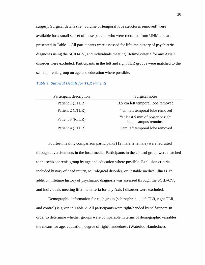
surgery. Surgical details (i.e., volume of temporal lobe structures removed) were
available for a small subset of these patients who were recruited from UNM and are
presented in Table 1. All participants were assessed for lifetime history of psychiatric
diagnoses using the SCID-CV, and individuals meeting lifetime criteria for any Axis I
disorder were excluded. Participants in the left and right TLR groups were matched to the
schizophrenia group on age and education where possible.
Table 1. Surgical Details for TLR Patients
Participant description Surgical notes
Patient 1 (LTLR) 3.5 cm left temporal lobe removed
Patient 2 (LTLR) 4 cm left temporal lobe removed
Patient 3 (RTLR) “at least 5 mm of posterior right
hippocampus remains”
Patient 4 (LTLR) 5 cm left temporal lobe removed
Fourteen healthy comparison participants (12 male, 2 female) were recruited
through advertisements in the local media. Participants in the control group were matched
to the schizophrenia group by age and education where possible. Exclusion criteria
included history of head injury, neurological disorder, or unstable medical illness. In
addition, lifetime history of psychiatric diagnosis was assessed through the SCID-CV,
and individuals meeting lifetime criteria for any Axis I disorder were excluded.
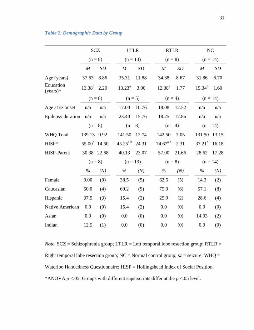
Demographic information for each group (schizophrenia, left TLR, right TLR,
and control) is given in Table 2. All participants were right-handed by self-report. In
order to determine whether groups were comparable in terms of demographic variables,
the means for age, education, degree of right-handedness (Waterloo Handedness
31
Table 2. Demographic Data by Group
SCZ LTLR RTLR NC
(n = 8) (n = 13) (n = 8) (n = 14)
M SD M SD M SD M SD
Age (years) 37.63 8.86 35.31 11.88 34.38 8.67 31.86 6.70
Education
(years)* 13.38
b 2.20
13.23
a 3.00
12.38
a 1.77
15.34
b 1.60
(n = 8) (n = 5) (n = 4) (n = 14)
Age at sz onset n/a n/a 17.00 10.76 18.08 12.52 n/a n/a
Epilepsy duration n/a n/a 23.40 15.76 18.25 17.86 n/a n/a
(n = 8) (n = 8) (n = 4) (n = 14)
WHQ Total 139.13 9.92 141.50 12.74 142.50 7.05 131.50 13.15
HISP* 55.00a 14.60 45.25
a,b 24.31 74.67
a,d 2.31 37.21
b 16.18
HISP-Parent 30.38 22.68 40.13 23.07 57.00 21.66 28.62 17.28
(n = 8) (n = 13) (n = 8) (n = 14)
% (N) % (N) % (N) % (N)
Female 0.00 (0) 38.5 (5) 62.5 (5) 14.3 (2)
Caucasian 50.0 (4) 69.2 (9) 75.0 (6) 57.1 (8)
Hispanic 37.5 (3) 15.4 (2) 25.0 (2) 28.6 (4)
Native American 0.0 (0) 15.4 (2) 0.0 (0) 0.0 (0)
Asian 0.0 (0) 0.0 (0) 0.0 (0) 14.03 (2)
Indian 12.5 (1) 0.0 (0) 0.0 (0) 0.0 (0)
Note. SCZ = Schizophrenia group; LTLR = Left temporal lobe resection group; RTLR =
Right temporal lobe resection group; NC = Normal control group; sz = seizure; WHQ =
Waterloo Handedness Questionnaire; HISP = Hollingshead Index of Social Position.
*ANOVA p <.05. Groups with different superscripts differ at the p <.05 level.
32
Questionnaire [WHQ] total score), participant socioeconomic status (SES), and parental
SES (measured by Hollingshead Index of Social Position Scale [HISP], Hollingshead,
1957; see Appendix A) were tested as dependent variables in a series of univariate
ANOVAS with group membership as the independent variable. Differences between
groups were significant for years of education [F(3,39) = 3.08, p <.05] and participant
SES [F(3,29) = 4.46, p <.05]. Planned contrasts were conducted to determine the
directionality of these results. Regarding education, the control group had significantly
more years of education than the left TLR [t(39) = 2.20, p <.05] and right TLR [t(39) =
2.77, p <.01] groups. There were no significant differences in educational level between
the schizophrenia and either left or right TLR groups. In terms of SES, the control group
had significantly lower scores, and therefore higher SES, than the schizophrenia [t(29) = -
2.27, p <.05] and right TLR [t(29) = -3.33, p <.01] groups, while the left TLR group had
significantly lower scores/higher SES than the right TLR group [t(29) = -2.46, p <.05].
There were no significant differences in mean age or overall parent SES.
Demographic Instruments
Demographic Questionnaire (Appendix A). A short questionnaire established
participants‟ age, years of education, ethnicity, and primary language. For the TLR
groups, date of resection surgery was also established.
Hollingshead Index of Social Position Scale (HISP; Hollingshead, 1957;
Appendix B) Socioeconomic status (SES) was assessed with the Hollingshead ISP
(Hollingshead, 1957), a brief assessment of social class position obtained from self-
reported occupational and educational status. Occupation is ranked on a 1 (executive) to 7
(unskilled employee) scale, and education is ranked on a 1 (professional degree) to 7 (less
33
than 7 years of school) scale. To calculate ISP, the ranking on the occupation scale is
multiplied by 7, and the ranking on the education scale is multiplied by 4. These numbers
are then added to determine the final ISP score, such that higher numbers represent lower
SES. For the current study, information was obtained for the participant and both of his
or her parents. For purposes of data analysis, parent SES was reported as the lower of the
two possible parent ISP scores.
Diagnostic and Symptom Inventory Instruments
Structured Clinical Interview for DSM-IV – Clinician Version (SCID-CV; First,
Spitzer, Gibbon, & Williams, 1996). The SCID is a semi-structured interview with a
format that corresponds directly to the diagnostic criteria in the DSM-IV, thus providing
the necessary information to make appropriate diagnoses. The SCID is broken down into
a number of modules; for this study, the overview and modules A (Mood episodes), B
(Psychotic and associated symptom), C (Differential diagnosis of psychotic disorders), D
(Mood disorders), E (Alcohol and other substance use disorders), and F (Anxiety and
other disorders) were administered.
Beck Depression Inventory, Second Edition (BDI-II; Beck, Steer, & Brown, 1996;
Appendix C). Studies have shown that comorbid depression affects a variety of
neuropsychological test scores, often more so than would be seen with a single diagnosis
(e.g., Moritz et al., 2001; Purcell, Maruff, Kyrios, & Pantelis, 1998). It is therefore
important to assess depressive symptoms in neuropsychological studies. The BDI-II is a
widely used self-report scale consisting of 21 categories of depressive symptoms (e.g.,
“Loss of Pleasure”). Each category is followed by four statements, arranged in a Likert-
type scale from 0 to 3, with higher scores indicating more depressive symptomatology.
34
For the example noted above, the extreme responses are: “I get as much pleasure as I ever
did from the things I enjoy,” (score of 0); and “I can‟t get any pleasure from the things I
used to enjoy,” (score of 3). The BDI-II has demonstrated acceptable validity and
reliability in clinical (Chemerinski, Bowie, Anderson, & Harvey, 2008) and nonclinical
(Storch, Roberti, & Roth, 2004) samples. Measures of the internal consistency of the
BDI-II range from .91 to .93 in college students, and the BDI-II correlates .93 with the
BDI, providing evidence for its convergent validity (Beck, Steer, & Brown, 1996;
Dozois, Dobson, & Anhberg, 1998).
Magical Ideation Scale (MIS; Eckblad & Chapman, 1983; Appendix D). The
Magical Ideation scale is a 30-item true/false questionnaire that includes items such as “I
have felt that I might cause something to happen just by thinking too much about it,”
(keyed “true”), and “I have never had the feeling that certain thoughts of mine really
belonged to someone else,” (keyed “false”). Scores on the MIS range from 0 to 30, with
higher scores indicating more pronounced magical thinking.
Revised Social Anhedonia Scale (RSAS; Eckblad, Chapman, Chapman, &
Mishlove., 1982; Appendix E). The RSAS is a self-report true/false questionnaire
consisting of 40 items concerning asociality and indifference to others, assessed by
measuring interpersonal pleasures. The RSAS includes items such as, “Having close
friends is not as important as many people say,” (keyed true), and “I feel pleased and
gratified as I learn more and more about the emotional life of my friends,” (keyed false).
Higher total scores on these scales indicate a lower capacity to feel pleasure, and thus
greater levels of anhedonia.
35
Neuropsychological Measures
Wechsler Adult Intelligence Scale, Third Edition (WAIS-III; Wechsler, 1997). The
WAIS-III is a widely used measure of overall intellectual ability. It is broken down into
14 subtests: Vocabulary, Similarities, Information, Comprehension, Arithmetic, Digit
Span, Letter-Number Sequencing, Picture Completion, Block Design, Matrix Reasoning,
Picture Arrangement, Digit-Symbol Coding, Symbol Search, and Object Assembly.
Different combinations of subtests contribute to the Full Scale Intelligence Quotient
(FSIQ), Verbal IQ (VIQ), Performance IQ (PIQ), and to indices of Verbal
Comprehension (VCI), Perceptual Organization (POI), Working Memory (WMI), and
Processing Speed (PSI). Raw scores on each subtest are converted to age-corrected scaled
scores (mean = 10, standard deviation = 3). Overall indices are computed from the sum of
the scaled scores of the appropriate subtests, and are expressed in standard scores (mean
= 100, standard deviation = 15). Vocabulary (FSIQ, VIQ, and VCI) is a measure of
expressive language skills in which the participant is asked to define single words.
Similarities (FSIQ, VIQ, and VCI) is a measure of verbal abstract reasoning in which
participants are asked to describe how two seemingly dissimilar items (e.g., eye and ear)
are alike. Information (FSIQ, VIQ, and VCI) is a measure of general fund of knowledge
(e.g., “In what country did the Olympic games originate?”). Arithmetic (FSIQ, VIQ, and
WMI) is a time-limited measure of mental calculation in which participants are read word
problems aloud and asked to provide the correct answer. Digit Span (FSIQ, VIQ, and
WMI) is a measure of auditory attention and is comprised of two parts: Digit Span
Forward, in which participants are asked to repeat a series of numbers, and Digit Span
Backward, in which participants are asked to repeat a series of numbers in reverse.
36
Letter-Number Sequencing (FSIQ, VIQ, and WMI) is a measure of sequencing ability in
which participants are asked to recall and sequence numbers and letters presented in an
unordered format. Picture Completion (FSIQ, PIQ, and POI) is a timed measure of
attention to essential visual detail in which participants are asked to identify what is
missing from pictures. Block Design (FSIQ, PIQ and POI) is a measure of speeded
constructional skills in which participants are asked to reconstruct 2x2 and 3x3 pictured
designs using colored blocks. Matrix Reasoning (FSIQ, PIQ, and POI) is a measure of
pattern analysis and non-verbal abstract reasoning in which participants are asked to
extrapolate from a visual pattern or design and choose the design which completes the
pattern. Digit-Symbol Coding (FSIQ, PIQ, and PSI) is a measure of efficiency in
matching and reproduction of symbols associated with numbers. Symbol Search (FSIQ,
PIQ, and PSI) is a measure of speeded symbolic information identification (Kaufman &
Lichtenberger, 1999). In the current study, the subtests Comprehension, Picture
Arrangement, and Object Assembly were not administered.
Wechsler Memory Scale, Third Edition (WMS-III; Wechsler, 1997). The WMS-III
is a widely used measure of auditory and visual memory skills. In the current study, the
following subtests were administered: Logical Memory I & II, Faces I & II, Verbal
Paired Associates I & II, Mental Control, and Visual Reproduction I & II. Raw scores for
each subtest are converted to age-adjusted scaled scores. Logical Memory is a measure of
memory for contextual verbal information (paragraph-length short stories). Two stories
are read to participants; the first story is read and participants are asked to repeat the story
details immediately after its presentation. The second story is then read, with immediate
recall, followed by a second presentation and recall trial, providing a measure of verbal
37
learning (learning slope). Approximately 30 minutes later, a delayed recall trial is
administered, followed by a yes/no recognition trial for story elements (e.g., “Was the
woman‟s name Anna Thompson?”). Faces is a measure of visual memory in which
participants are shown a series of human faces and asked to identify them from a larger
group of faces presented immediately afterward. A delayed recognition trial is
administered 30 minutes later. Verbal Paired Associates is a measure of auditory
associative memory in which participants are asked to learn eight word-pairs (e.g.,
“insect-acorn”) over four trials. Participants are given corrective feedback after each trial
in order to facilitate learning. A delayed recall trial (without corrective feedback) is
administered 30 minutes later, followed by a yes/no recognition trial in which the eight
target word-pairs are presented intermixed with novel word-pairs. Visual Reproduction is
a measure of visual memory for a series of geometric designs. Participants are presented
with five designs, one at a time, for ten seconds, then immediately asked to draw the
figures. A delayed recall trial is administered 30 minutes later, followed by a yes/no
recognition trial. Mental Control is a measure of attention, processing speed, working
memory, and simple set-shifting in which participants are asked to recite sequences of
numbers, letters, and months as quickly as they can, both forward and in reverse. The last
item assesses set-shifting, as participants are asked to count by 6, starting with 0, and
after every number, say a day of the week starting with Sunday (i.e., 0/Sunday,
6/Monday, etc.).
California Verbal Learning Test, Second Edition (CVLT-II; Delis, Kramer,
Kaplan, & Ober, 2000). The CVLT-II is a verbal memory test in which participants are
asked to recall a 16-item word list over five learning trials. The words are presented in an
38
intermixed order, but can be mentally arranged into four semantic categories (furniture,
vegetables, ways of traveling, and animals). Short-delay free and semantically-cued recall
trials are administered following presentation and immediate free recall of a 16-item
distracter list (List B). Long-delay free and semantically-cued trials are administered 20
minutes later, followed by a yes/no recognition trial consisting of target words, List B
words, semantically-related distracter words, and unrelated distracter words. Outcome
measures include total correct responses for learning trials 1-5, number of correct words
recalled from List B, short- and long-delay free and cued recall number correct, and
number of “hits” (true positives) and false positive errors on recognition. All raw scores
are converted to z-scores (mean = 0.0, standard deviation = 1), except Trials 1-5 Total,
which is expressed as a T-score.
Connors’s Continuous Performance Test, Second Edition (CPT-II; Connors &
Multi-Health Systems, 2000). The CPT-II is a widely used computerized measure of
sustained visual attention. Participants are seated in front of a computer and told that they
will see capital letters flash one at a time in the middle of the screen. They are instructed
to press the space bar as quickly as they can whenever they see a letter, with the
exception of the letter “X.” They are instructed not to respond when they see the letter
“X.” The letters flash at interstimulus intervals of 1, 2, or 4 seconds with a display time of
250 ms. Multiple output variables are obtained from the CPT-II, including number of
omission and commission errors, hit reaction time, variability, perseverations,
detectability, and response style. Raw scores for each variable are converted to age-
corrected T-scores.
39
Controlled Oral Word Association Test (COWA; Benton & Hamsher, 1989;
Strauss, Sherman, & Spreen, 2006). This easily administered test of verbal fluency
requires the participant to name, during a 60-second time limit, as many words as
possible that begin with certain letters. Proper nouns, numbers, and words that only differ
in suffix (e.g., “eat” and “eating”) must be excluded. The most commonly used letters are
F, A, and S. Following the phonemic fluency test, participants were asked to name as
many animals as they could in 60 seconds to assess semantic fluency. Outcome measures
included total correct responses, intrusive errors, and perseverative errors (repetitions).
The raw score for total correct is compared to demographically-matched norms (based on
gender, ethnicity [Caucasian or African-American], age, and education) to give a T-score
(Heaton, Miller, Taylor, & Grant, 2004).
Ruff Figural Fluency Test (RFFT; Ruff, Light & Evans, 1987). The RFFT consists
of five sheets of paper on which are arranged 40 squares containing a pattern of dots.
Sheets 2 and 3 contain interference patterns as well as dots (in the same positions as on
sheet 1), and sheets 4 and 5 have dot patterns that differ from sheet 1 and from each
other. Each sheet also has a three-square practice page. Participants were instructed that
they would have one minute (per sheet) to connect any two or more dots in each square to
produce as many different patterns as they could. The test is scored for number of unique
patterns and perseverations. Raw scores for total unique designs and the error score are
converted to T-scores, and raw scores for total perseverative errors are converted to z-
scores.
Trail Making Test, Parts A & B (TMT; Reitan & Wolfson, 1985). The TMT is an
easily administered test of psychomotor speed, attention, and set-shifting. In Part A,
40
participants must connect 25 numbered circles in consecutive order as quickly as possible
(giving measures of attention and psychomotor speed). In Part B, participants must
connect 25 consecutively numbered and lettered circles by alternating numbers with
letters as quickly as possible (adding a measure of set-shifting ability). Raw scores
(number of seconds to completion) are expressed as demographically-corrected T-scores
(Heaton, Miller, Taylor, & Grant, 2004).
Halstead Finger Tapping Test (FTT; Halstead, 1947). The Finger Tapping Test is
a measure of simple bilateral finger motor speed in which participants were asked to
depress a lever using their index finger, keeping the rest of their fingers and palm flat on
the board, as quickly as they could for ten-second intervals. Five consecutive trials with
total number of taps within five points of each other were required for each hand.
Participants began with their dominant hand, and were given short breaks between each
trial, with a full one-minute break after every three trials. The score is the average of the
five trials for each hand, and is expressed as a demographically-corrected T-score
(Heaton, Miller, Taylor, & Grant, 2004).
Grooved Pegboard (GP; Lafayette Instruments, 1989). The Grooved Pegboard
test provides a measure of bilateral fine motor coordination and manual dexterity.
Participants were asked to place 25 asymmetrically-shaped pegs from a large receptacle
into 25 similarly-shaped holes arranged in five rows of five on the pegboard. They were
told to pick up one peg at a time, and for the right hand, to work left to right, and for the
left hand, to work right to left to fill in all the holes as quickly as possible. The outcome
measure is the number of seconds to complete 25 holes for each hand. Raw scores are
41
converted to demographically-corrected T-scores (Heaton, Miller, Taylor, & Grant,
2004).
Auditory Consonant Trigrams (ACT; Brown, 1958; Peterson & Peterson, 1959).
ACT is a measure of working memory in which participants are asked to recall a series of
letters after an interference delay task. A screening section was administered prior to the
working memory task: a consonant trigram (e.g., QLX) was presented orally to
participants and they were asked to repeat the trigram immediately. Following five
screening trials, participants were told that they would next hear three letters followed by
a number. When they heard the number, they were instructed to begin counting
backward, out loud, by threes, from this number, for interval delays of 3, 9, or 18 seconds
(the examiner would say “stop” at the appropriate time), and then recite the three letters.
Five trials each of 3-, 9-, and 18-second delays were administered. The outcome variables
were total number of correctly recalled letters (regardless of order recalled) for each
delay interval, plus total number of correctly recalled letters. Raw scores for each interval
delay were converted to z-scores (Strauss, Sherman, & Spreen, 2006).
Boston Naming Test, Second Edition (BNT-2; Kaplan, Goodglass, & Weintraub,
1983). The BNT-2 is a widely used measure of one-word expressive vocabulary skills.
Participants were shown line drawings of 60 items and asked to identify each word (e.g.,
hammock, compass). If the picture was obviously misperceived, a semantic cue was
given (e.g., for item #45 “unicorn,” the semantic cue is “a mythical animal”). If the
participant was unable to provide the correct response either after 20 seconds with no cue
or after presentation of the semantic cue, a phonemic cue was provided (e.g., “it starts
with un-” for “unicorn”). The score is comprised of total number of correct responses
42
spontaneously given (i.e., with no cue) plus number of correct responses given following
a semantic cue. Raw scores were converted into demographically-corrected T-scores
(Heaton, Miller, Taylor, & Grant, 2004).
Waterloo Handedness Questionnaire (WHQ; Bryden, 1977; Appendix F). The
WHQ is a 32-item self-report questionnaire which measures degree of right-handedness.
Participants were asked to indicate which hand they would use to perform a series of
unimanual activities (such as using a hammer or writing). Some of the items on the
questionnaire reflect skilled performance (i.e., writing), whereas other items reflect
relatively unskilled activities (i.e., opening a drawer). Five possible responses were
offered for each question, allowing the participant to rate the frequency with which they
would use a particular hand for each activity using a 5-point scale (i.e., 1 = “left always,”
2 = “left usually,” 3 = “left and right equally,” 4 = “right usually,” and 5 = “right
always”). The outcome handedness measure was calculated as the total composite score
of these individual responses. Higher scores correspond to a higher degree of right-
handedness (i.e., scores below 96 would indicate increasing degrees of left-handedness, a
score of 96 would indicate ambidextrousness, and scores above 96 would indicate
increasing degrees of right-handedness).
Procedure
For the participants recruited from the NMVAMC and UNMH/UNMHSC and
tested by this author, pilot data (MRI, MEG, and neuropsychological measures) were
collected from each participant at three separate times. In the screening phase,
participants were administered the SCID-CV to determine eligibility and diagnoses after
providing written informed consent and agreeing to Health Insurance Portability and
43
Accountability Act policies. Participants were compensated upon completion of the
screening process.
Once participants had been enrolled in the study and assigned group membership,
neuroimaging and neuropsychological data were collected in separate sessions within two
weeks of the initial interview. Neuroimaging results are reported elsewhere; only the
results of the neuropsychological testing are reported here. Participants were
compensated upon completion of each session (see Appendix K).
The neuropsychological test battery administered by the author included the
Wechsler Adult Intelligence Scale, Third Edition (WAIS-III); Wechsler Memory Scale,
Third Edition (WMS-III; Logical Memory, Faces, Verbal Paired-Associates, and Visual
Reproduction subtests); California Verbal Learning Test, Second Edition (CVLT-II);
Grooved Pegboard (GP); Finger Tapping Test (FTT); Controlled Oral Word Association
Test (COWAT; FAS and Animals); Auditory Consonant Trigrams (ACT); Trail Making
Test, Parts A & B (TMT); Boston Naming Test, Second Edition (BNT-2); Ruff Figural
Fluency Test (RFFT); and Connor‟s Continuous Performance Test, Second Edition
(CPT-2). The Waterloo Handedness Questionnaire (WHQ) was administered to
determine handedness. The neuropsychological measures were administered in random
order in an effort to minimize order effects. In addition to neuropsychological measures,
several self-report inventories of emotional functioning and current symptomatology
were administered. These included the Beck Depression Inventory, Second Edition (BDI-
II); the Revised Social Anhedonia Scale (RSAS); the Magical Ideation Scale (MIS).
Upon completion of the neuropsychological battery, all participants were provided
compensation in the form of payment of $50. The neuropsychological battery
44
administered to the Emory University participants was identical, except the following
measures were omitted: CPT-2, ACT, BDI-II, MIS, and RSAS. In addition, different
versions of two neuropsychological tests were given. The Delis-Kaplan Executive
Function System (DKEFS) Trail Making Test and Design Fluency tests were given in
place of the Trail Making Test and RFFT. For the DKEFS Trail Making Test, Condition
2 (Number Sequencing) is similar to Trails A and Condition 4 (Number-Letter
Sequencing) is similar to Trails B. Although different normative data is used, standard
scores are calculated which can be reliably compared across the different measures, thus
data from the DKEFS TMT is included in the current study. The DKEFS Design Fluency
test is administered differently than the RFFT, thus data from the DKEFS version is not
included in the results presented here.
Testing by this author took place at the UNM Department of Psychiatry‟s Center
for Neuropsychological Services over a three- to four-hour period in one day. One
participant required a second session to complete the testing due to time constraints on
the initially scheduled day.
45
Results
Descriptive Statistics
Information about general psychological functioning and overall intellectual
ability for each group is given in Table 3. In order to determine whether groups were
comparable in terms of general psychological and emotional functioning, as well as
overall intelligence, the means for BDI-II, MIS, RSAS, and WAIS-III FSIQ were tested
as dependent variables in a series of univariate analyses of variance (ANOVA) with
group membership as the independent variable. No significant differences were found for
FSIQ, indicating that all groups were comparable on overall intelligence. Importantly,
FSIQ for all groups fell within the average range. On emotional measures, differences
between groups were significant for RSAS only [F(3,28) = 3.88, p <.05]. Planned
contrasts were conducted to determine the directionality of these results. There were no
differences in scores among the experimental groups, but the schizophrenia [t(28) = -
2.32, p <.05] and left TLR [t(28) = -3.20, p <.01] groups both had higher scores,
indicating greater levels of social anhedonia, than the control group.
Neuropsychological Test Battery
To simplify analyses given the large number of individual outcome variables,
standardized scores (mean = 0, standard deviation = 1) were calculated for each test, and
combined to create neuropsychological composite scores (see Table 4). The Verbal
Memory (VM) composite was created by averaging the WMS-III Logical Memory (LM),
WMS-III Verbal Paired Associates (VPA), and CVLT-II subscale scores. The Nonverbal
Memory (NVM) composite was created by averaging the WMS-III Faces and WMS-III
Visual Reproduction (VR) subscale scores. The Executive Attention (EA) composite was
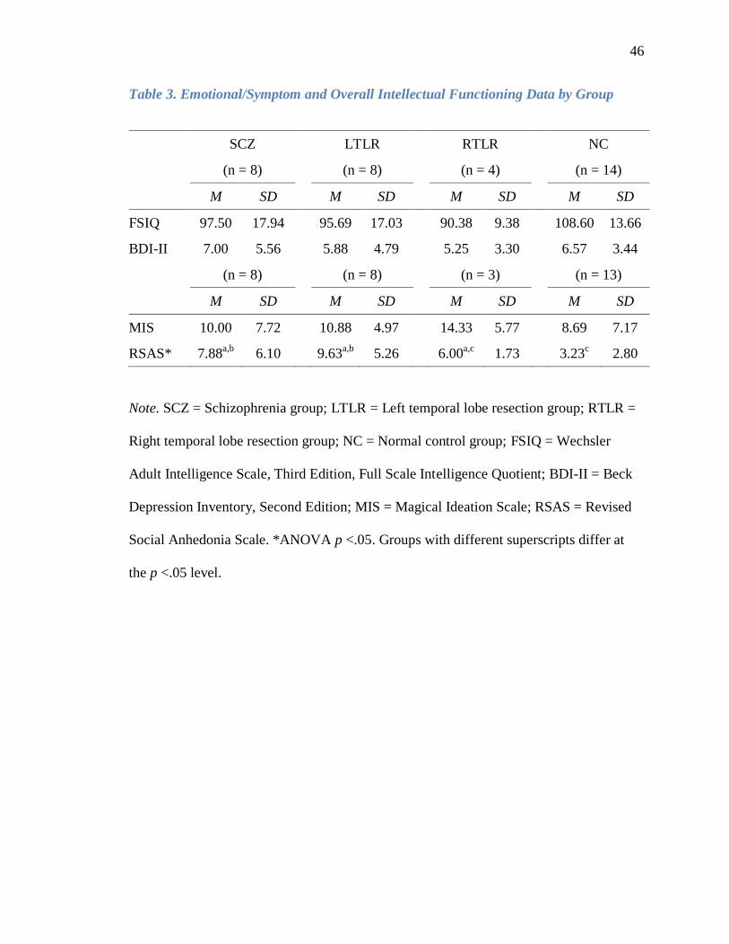
46
Table 3. Emotional/Symptom and Overall Intellectual Functioning Data by Group
SCZ LTLR RTLR NC
(n = 8) (n = 8) (n = 4) (n = 14)
M SD M SD M SD M SD
FSIQ 97.50 17.94 95.69 17.03 90.38 9.38 108.60 13.66
BDI-II 7.00 5.56 5.88 4.79 5.25 3.30 6.57 3.44
(n = 8) (n = 8) (n = 3) (n = 13)
M SD M SD M SD M SD
MIS 10.00 7.72 10.88 4.97 14.33 5.77 8.69 7.17
RSAS* 7.88a,b
6.10 9.63a,b
5.26 6.00a,c
1.73 3.23c 2.80
Note. SCZ = Schizophrenia group; LTLR = Left temporal lobe resection group; RTLR =
Right temporal lobe resection group; NC = Normal control group; FSIQ = Wechsler
Adult Intelligence Scale, Third Edition, Full Scale Intelligence Quotient; BDI-II = Beck
Depression Inventory, Second Edition; MIS = Magical Ideation Scale; RSAS = Revised
Social Anhedonia Scale. *ANOVA p <.05. Groups with different superscripts differ at
the p <.05 level.
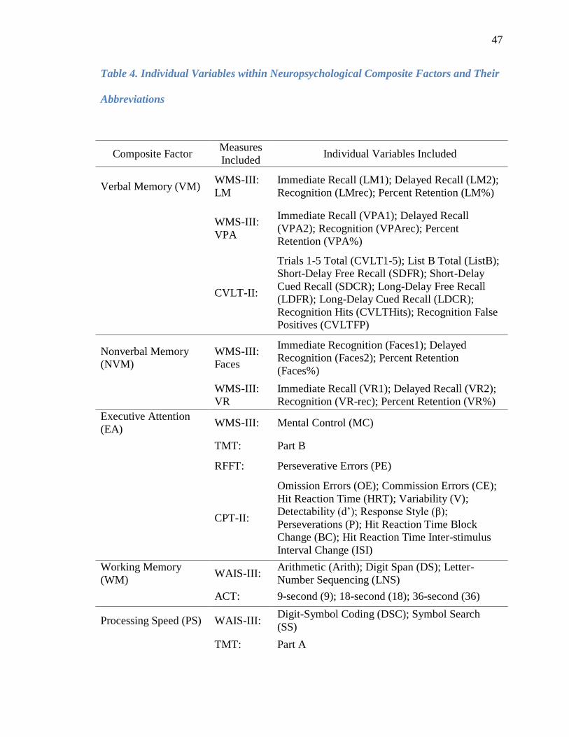
47
Table 4. Individual Variables within Neuropsychological Composite Factors and Their
Abbreviations
Composite Factor Measures
Included Individual Variables Included
Verbal Memory (VM) WMS-III:
LM
Immediate Recall (LM1); Delayed Recall (LM2);
Recognition (LMrec); Percent Retention (LM%)
WMS-III:
VPA
Immediate Recall (VPA1); Delayed Recall
(VPA2); Recognition (VPArec); Percent
Retention (VPA%)
CVLT-II:
Trials 1-5 Total (CVLT1-5); List B Total (ListB);
Short-Delay Free Recall (SDFR); Short-Delay
Cued Recall (SDCR); Long-Delay Free Recall
(LDFR); Long-Delay Cued Recall (LDCR);
Recognition Hits (CVLTHits); Recognition False
Positives (CVLTFP)
Nonverbal Memory
(NVM)
WMS-III:
Faces
Immediate Recognition (Faces1); Delayed
Recognition (Faces2); Percent Retention
(Faces%)
WMS-III:
VR
Immediate Recall (VR1); Delayed Recall (VR2);
Recognition (VR-rec); Percent Retention (VR%)
Executive Attention
(EA) WMS-III: Mental Control (MC)
TMT: Part B
RFFT: Perseverative Errors (PE)
CPT-II:
Omission Errors (OE); Commission Errors (CE);
Hit Reaction Time (HRT); Variability (V);
Detectability (d‟); Response Style (β);
Perseverations (P); Hit Reaction Time Block
Change (BC); Hit Reaction Time Inter-stimulus
Interval Change (ISI)
Working Memory
(WM) WAIS-III:
Arithmetic (Arith); Digit Span (DS); Letter-
Number Sequencing (LNS)
ACT: 9-second (9); 18-second (18); 36-second (36)
Processing Speed (PS) WAIS-III: Digit-Symbol Coding (DSC); Symbol Search
(SS)
TMT: Part A
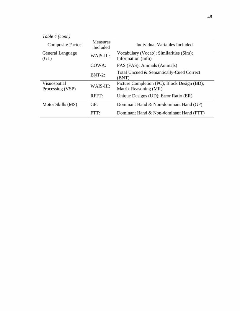
48
Table 4 (cont.)
Composite Factor Measures
Included Individual Variables Included
General Language
(GL) WAIS-III:
Vocabulary (Vocab); Similarities (Sim);
Information (Info)
COWA: FAS (FAS); Animals (Animals)
BNT-2: Total Uncued & Semantically-Cued Correct
(BNT)
Visuospatial
Processing (VSP) WAIS-III:
Picture Completion (PC); Block Design (BD);
Matrix Reasoning (MR)
RFFT: Unique Designs (UD); Error Ratio (ER)
Motor Skills (MS) GP: Dominant Hand & Non-dominant Hand (GP)
FTT: Dominant Hand & Non-dominant Hand (FTT)
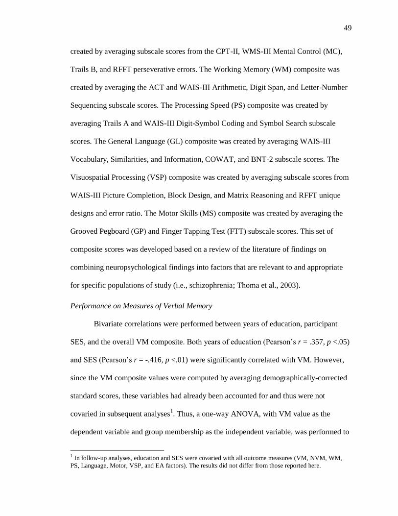
49
created by averaging subscale scores from the CPT-II, WMS-III Mental Control (MC),
Trails B, and RFFT perseverative errors. The Working Memory (WM) composite was
created by averaging the ACT and WAIS-III Arithmetic, Digit Span, and Letter-Number
Sequencing subscale scores. The Processing Speed (PS) composite was created by
averaging Trails A and WAIS-III Digit-Symbol Coding and Symbol Search subscale
scores. The General Language (GL) composite was created by averaging WAIS-III
Vocabulary, Similarities, and Information, COWAT, and BNT-2 subscale scores. The
Visuospatial Processing (VSP) composite was created by averaging subscale scores from
WAIS-III Picture Completion, Block Design, and Matrix Reasoning and RFFT unique
designs and error ratio. The Motor Skills (MS) composite was created by averaging the
Grooved Pegboard (GP) and Finger Tapping Test (FTT) subscale scores. This set of
composite scores was developed based on a review of the literature of findings on
combining neuropsychological findings into factors that are relevant to and appropriate
for specific populations of study (i.e., schizophrenia; Thoma et al., 2003).
Performance on Measures of Verbal Memory
Bivariate correlations were performed between years of education, participant
SES, and the overall VM composite. Both years of education (Pearson‟s r = .357, p <.05)
and SES (Pearson‟s r = -.416, p <.01) were significantly correlated with VM. However,
since the VM composite values were computed by averaging demographically-corrected
standard scores, these variables had already been accounted for and thus were not
covaried in subsequent analyses1. Thus, a one-way ANOVA, with VM value as the
dependent variable and group membership as the independent variable, was performed to
1 In follow-up analyses, education and SES were covaried with all outcome measures (VM, NVM, WM,
PS, Language, Motor, VSP, and EA factors). The results did not differ from those reported here.
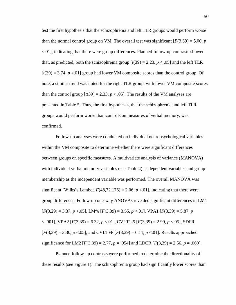
50
test the first hypothesis that the schizophrenia and left TLR groups would perform worse
than the normal control group on VM. The overall test was significant [F(3,39) = 5.00, p
<.01], indicating that there were group differences. Planned follow-up contrasts showed
that, as predicted, both the schizophrenia group [t(39) = 2.23, p < .05] and the left TLR
[t(39) = 3.74, p <.01] group had lower VM composite scores than the control group. Of
note, a similar trend was noted for the right TLR group, with lower VM composite scores
than the control group [t(39) = 2.33, p < .05]. The results of the VM analyses are
presented in Table 5. Thus, the first hypothesis, that the schizophrenia and left TLR
groups would perform worse than controls on measures of verbal memory, was
confirmed.
Follow-up analyses were conducted on individual neuropsychological variables
within the VM composite to determine whether there were significant differences
between groups on specific measures. A multivariate analysis of variance (MANOVA)
with individual verbal memory variables (see Table 4) as dependent variables and group
membership as the independent variable was performed. The overall MANOVA was
significant [Wilks‟s Lambda F(48,72.176) = 2.06, p <.01], indicating that there were
group differences. Follow-up one-way ANOVAs revealed significant differences in LM1
[F(3,29) = 3.37, p <.05], LM% [F(3,39) = 3.55, p <.01], VPA1 [F(3,39) = 5.87, p
<..001], VPA2 [F(3,39) = 6.32, p <.01], CVLT1-5 [F(3,39) = 2.99, p <.05], SDFR
[F(3,39) = 3.30, p <.05], and CVLTFP [F(3,39) = 6.11, p <.01]. Results approached
significance for LM2 [F(3,39) = 2.77, p = .054] and LDCR [F(3,39) = 2.56, p = .069].
Planned follow-up contrasts were performed to determine the directionality of
these results (see Figure 1). The schizophrenia group had significantly lower scores than
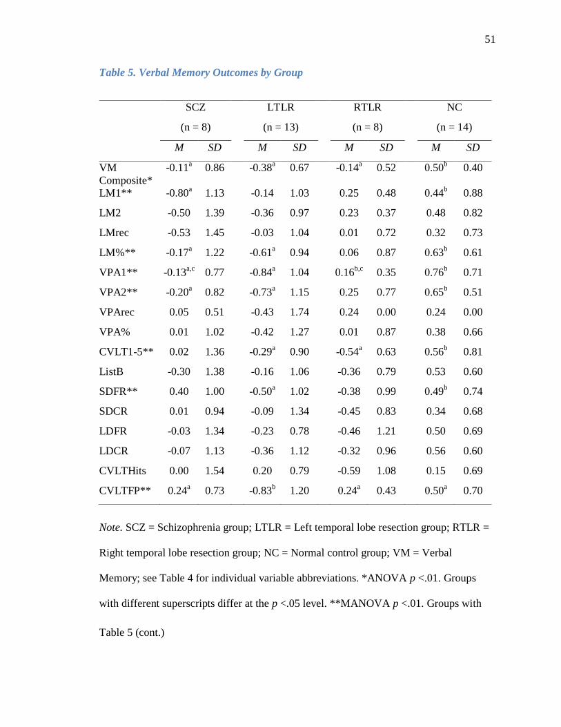
51
Table 5. Verbal Memory Outcomes by Group
SCZ LTLR RTLR NC
(n = 8) (n = 13) (n = 8) (n = 14)
M SD M SD M SD M SD
VM
Composite*
-0.11a 0.86 -0.38
a 0.67 -0.14
a 0.52 0.50
b 0.40
LM1** -0.80a 1.13 -0.14 1.03 0.25 0.48 0.44
b 0.88
LM2 -0.50 1.39 -0.36 0.97 0.23 0.37 0.48 0.82
LMrec -0.53 1.45 -0.03 1.04 0.01 0.72 0.32 0.73
LM%** -0.17a 1.22 -0.61
a 0.94 0.06 0.87 0.63
b 0.61
VPA1** -0.13a,c
0.77 -0.84a 1.04 0.16
b,c 0.35 0.76
b 0.71
VPA2** -0.20a 0.82 -0.73
a 1.15 0.25 0.77 0.65
b 0.51
VPArec 0.05 0.51 -0.43 1.74 0.24 0.00 0.24 0.00
VPA% 0.01 1.02 -0.42 1.27 0.01 0.87 0.38 0.66
CVLT1-5** 0.02 1.36 -0.29a 0.90 -0.54
a 0.63 0.56
b 0.81
ListB -0.30 1.38 -0.16 1.06 -0.36 0.79 0.53 0.60
SDFR** 0.40 1.00 -0.50a 1.02 -0.38 0.99 0.49
b 0.74
SDCR 0.01 0.94 -0.09 1.34 -0.45 0.83 0.34 0.68
LDFR -0.03 1.34 -0.23 0.78 -0.46 1.21 0.50 0.69
LDCR -0.07 1.13 -0.36 1.12 -0.32 0.96 0.56 0.60
CVLTHits 0.00 1.54 0.20 0.79 -0.59 1.08 0.15 0.69
CVLTFP** 0.24a 0.73 -0.83
b 1.20 0.24
a 0.43 0.50
a 0.70
Note. SCZ = Schizophrenia group; LTLR = Left temporal lobe resection group; RTLR =
Right temporal lobe resection group; NC = Normal control group; VM = Verbal
Memory; see Table 4 for individual variable abbreviations. *ANOVA p <.01. Groups
with different superscripts differ at the p <.05 level. **MANOVA p <.01. Groups with
Table 5 (cont.)

52
different superscripts differ at the p <.05 level (planned comparisons) or Bonferroni-
corrected p <.0125 level (post-hoc comparisons).
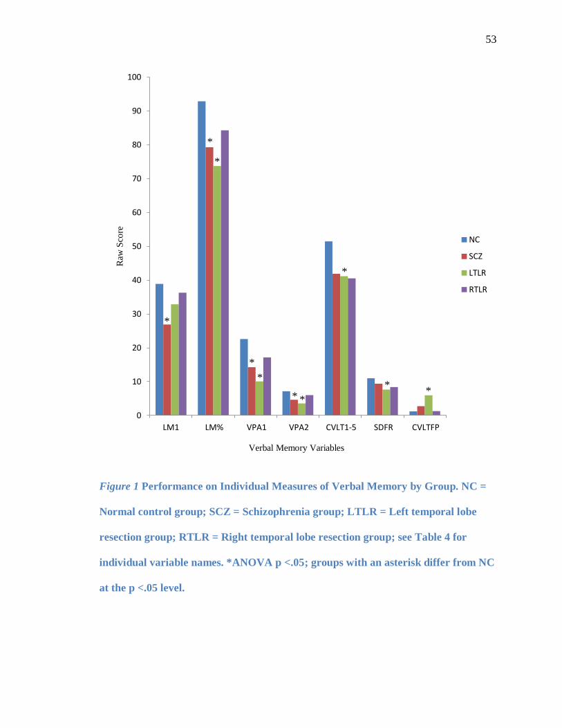
53
Figure 1 Performance on Individual Measures of Verbal Memory by Group. NC =
Normal control group; SCZ = Schizophrenia group; LTLR = Left temporal lobe
resection group; RTLR = Right temporal lobe resection group; see Table 4 for
individual variable names. *ANOVA p <.05; groups with an asterisk differ from NC
at the p <.05 level.
0
10
20
30
40
50
60
70
80
90
100
LM1 LM% VPA1 VPA2 CVLT1-5 SDFR CVLTFP
Raw
Sco
re
Verbal Memory Variables
NC
SCZ
LTLR
RTLR
*
*
*
*
*
* *
*
* *
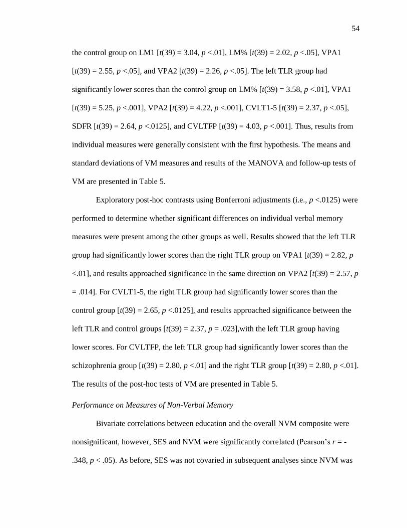
54
the control group on LM1 [t(39) = 3.04, p <.01], LM% [t(39) = 2.02, p <.05], VPA1
[t(39) = 2.55, p <.05], and VPA2 [t(39) = 2.26, p <.05]. The left TLR group had
significantly lower scores than the control group on LM% [t(39) = 3.58, p <.01], VPA1
[t(39) = 5.25, p <.001], VPA2 [t(39) = 4.22, p <.001], CVLT1-5 [t(39) = 2.37, p <.05],
SDFR [t(39) = 2.64, p <.0125], and CVLTFP [t(39) = 4.03, p <.001]. Thus, results from
individual measures were generally consistent with the first hypothesis. The means and
standard deviations of VM measures and results of the MANOVA and follow-up tests of
VM are presented in Table 5.
Exploratory post-hoc contrasts using Bonferroni adjustments (i.e., p <.0125) were
performed to determine whether significant differences on individual verbal memory
measures were present among the other groups as well. Results showed that the left TLR
group had significantly lower scores than the right TLR group on VPA1 [t(39) = 2.82, p
<.01], and results approached significance in the same direction on VPA2 [t(39) = 2.57, p
= .014]. For CVLT1-5, the right TLR group had significantly lower scores than the
control group [t(39) = 2.65, p <.0125], and results approached significance between the
left TLR and control groups [t(39) = 2.37, p = .023],with the left TLR group having
lower scores. For CVLTFP, the left TLR group had significantly lower scores than the
schizophrenia group [t(39) = 2.80, p <.01] and the right TLR group [t(39) = 2.80, p <.01].
The results of the post-hoc tests of VM are presented in Table 5.
Performance on Measures of Non-Verbal Memory
Bivariate correlations between education and the overall NVM composite were
nonsignificant, however, SES and NVM were significantly correlated (Pearson‟s r = -
.348, p < .05). As before, SES was not covaried in subsequent analyses since NVM was
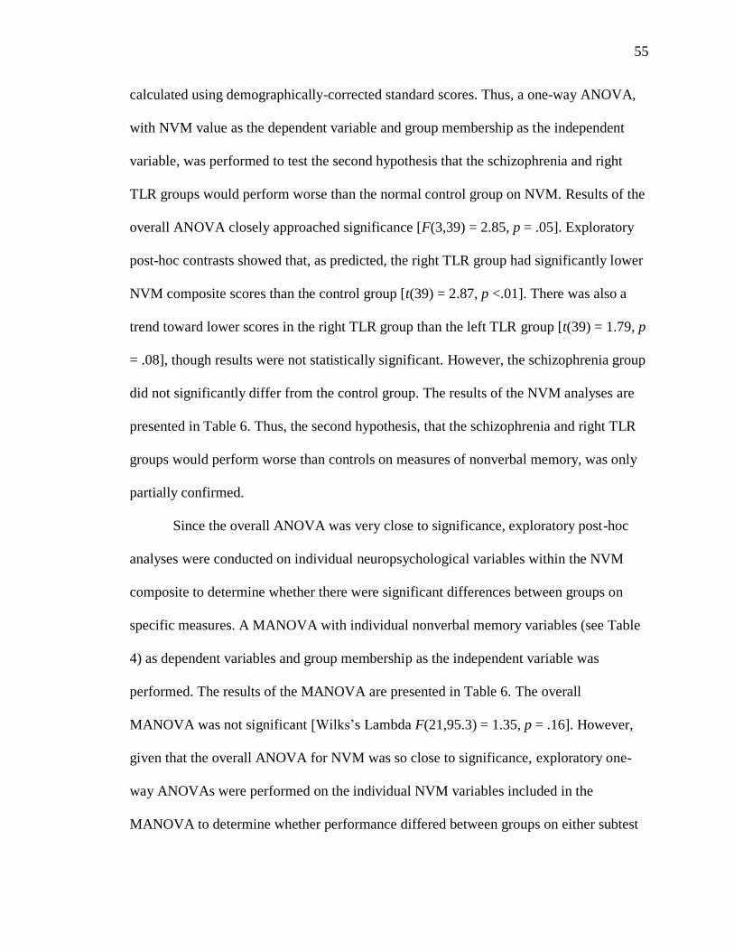
55
calculated using demographically-corrected standard scores. Thus, a one-way ANOVA,
with NVM value as the dependent variable and group membership as the independent
variable, was performed to test the second hypothesis that the schizophrenia and right
TLR groups would perform worse than the normal control group on NVM. Results of the
overall ANOVA closely approached significance [F(3,39) = 2.85, p = .05]. Exploratory
post-hoc contrasts showed that, as predicted, the right TLR group had significantly lower
NVM composite scores than the control group [t(39) = 2.87, p <.01]. There was also a
trend toward lower scores in the right TLR group than the left TLR group [t(39) = 1.79, p
= .08], though results were not statistically significant. However, the schizophrenia group
did not significantly differ from the control group. The results of the NVM analyses are
presented in Table 6. Thus, the second hypothesis, that the schizophrenia and right TLR
groups would perform worse than controls on measures of nonverbal memory, was only
partially confirmed.
Since the overall ANOVA was very close to significance, exploratory post-hoc
analyses were conducted on individual neuropsychological variables within the NVM
composite to determine whether there were significant differences between groups on
specific measures. A MANOVA with individual nonverbal memory variables (see Table
4) as dependent variables and group membership as the independent variable was
performed. The results of the MANOVA are presented in Table 6. The overall
MANOVA was not significant [Wilks‟s Lambda F(21,95.3) = 1.35, p = .16]. However,
given that the overall ANOVA for NVM was so close to significance, exploratory one-
way ANOVAs were performed on the individual NVM variables included in the
MANOVA to determine whether performance differed between groups on either subtest
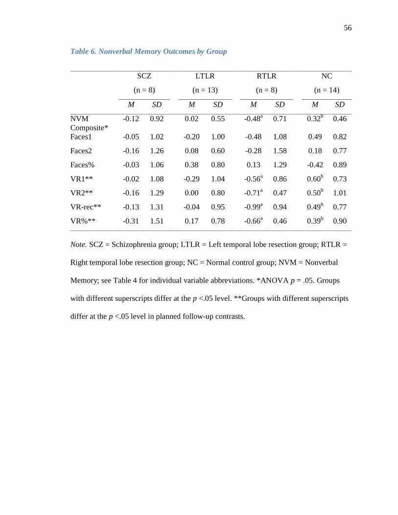
56
Table 6. Nonverbal Memory Outcomes by Group
SCZ LTLR RTLR NC
(n = 8) (n = 13) (n = 8) (n = 14)
M SD M SD M SD M SD
NVM
Composite*
-0.12 0.92 0.02 0.55 -0.48a 0.71 0.32
b 0.46
Faces1 -0.05 1.02 -0.20 1.00 -0.48 1.08 0.49 0.82
Faces2 -0.16 1.26 0.08 0.60 -0.28 1.58 0.18 0.77
Faces% -0.03 1.06 0.38 0.80 0.13 1.29 -0.42 0.89
VR1** -0.02 1.08 -0.29 1.04 -0.56a 0.86 0.60
b 0.73
VR2** -0.16 1.29 0.00 0.80 -0.71a 0.47 0.50
b 1.01
VR-rec** -0.13 1.31 -0.04 0.95 -0.99a 0.94 0.49
b 0.77
VR%** -0.31 1.51 0.17 0.78 -0.66a 0.46 0.39
b 0.90
Note. SCZ = Schizophrenia group; LTLR = Left temporal lobe resection group; RTLR =
Right temporal lobe resection group; NC = Normal control group; NVM = Nonverbal
Memory; see Table 4 for individual variable abbreviations. *ANOVA p = .05. Groups
with different superscripts differ at the p <.05 level. **Groups with different superscripts
differ at the p <.05 level in planned follow-up contrasts.

57
(i.e., Faces or VR). Significant group differences were found for the VR subtest only:
VR1 [F (3,39) = 3.34, p <.05], VR2 [F(3,39) = 2.92, p <.05], and VR-rec [F(3,39) = 3.96,
p <.05]. Results approached significance for VR% [F(3,39) = 2.49, p = .07].
Planned contrasts were performed to determine the directionality of these
differences (see Figure 2). The right TLR group had significantly lower scores than the
control group on VR1 [t(39) = 2.82, p <.01], VR2 [t(39) = 2.90, p <.01], VR% [t(39) =
2.49, p <.05], and VR-rec [t(39) = 3.44, p <.01].
Exploratory post-hoc contrasts using Bonferroni adjustments (i.e., p <.0125) were
performed to determine whether significant differences on individual non-verbal memory
measures were present among the other groups as well. Results of all comparisons were
non-significant.
Performance on Measures of Other Neuropsychological Domains
Several bivariate correlations between education, SES, and the overall
neuropsychological factors (EA, MS, WM, PS, VSP, and GL) were significant (see Table
7). However, as before, education and SES were not covaried in subsequent analyses
since all neuropsychological composite scores were calculated using demographically-
corrected standard scores. Thus, a series of one-way ANOVAs, with composite value as
the dependent variable and group membership as the independent variable, were
performed to test the third hypothesis that the schizophrenia group would perform worse
than the normal control group on measures of executive attention, motor skills, working
memory, and processing speed. Overall ANOVAs were significant for motor skills
[F(3,39) = 3.66, p <.05], general language [F(3,39) = 4.87, p <.01], working memory
[F(3,39) = 3.37, p <.05], and processing speed [F(3,39) = 6.63, p <.01]. There were no
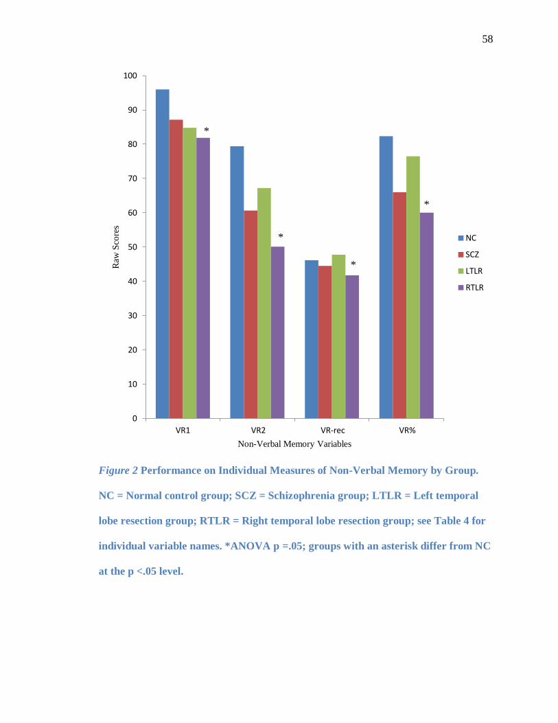
58
Figure 2 Performance on Individual Measures of Non-Verbal Memory by Group.
NC = Normal control group; SCZ = Schizophrenia group; LTLR = Left temporal
lobe resection group; RTLR = Right temporal lobe resection group; see Table 4 for
individual variable names. *ANOVA p =.05; groups with an asterisk differ from NC
at the p <.05 level.
0
10
20
30
40
50
60
70
80
90
100
VR1 VR2 VR-rec VR%
Raw
Sco
res
Non-Verbal Memory Variables
NC
SCZ
LTLR
RTLR
*
*
*
*
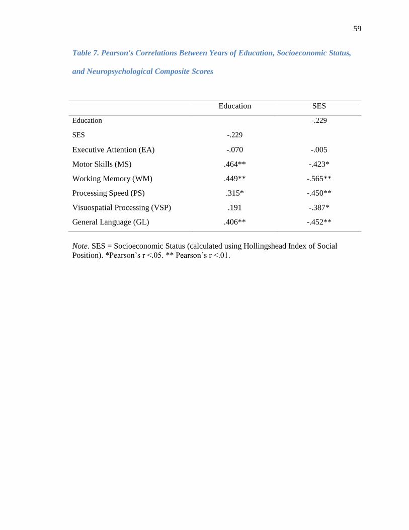
59
Table 7. Pearson's Correlations Between Years of Education, Socioeconomic Status,
and Neuropsychological Composite Scores
Education SES
Education -.229
SES -.229
Executive Attention (EA) -.070 -.005
Motor Skills (MS) .464** -.423*
Working Memory (WM) .449** -.565**
Processing Speed (PS) .315* -.450**
Visuospatial Processing (VSP) .191 -.387*
General Language (GL) .406** -.452**
Note. SES = Socioeconomic Status (calculated using Hollingshead Index of Social
Position). *Pearson‟s r <.05. ** Pearson‟s r <.01.
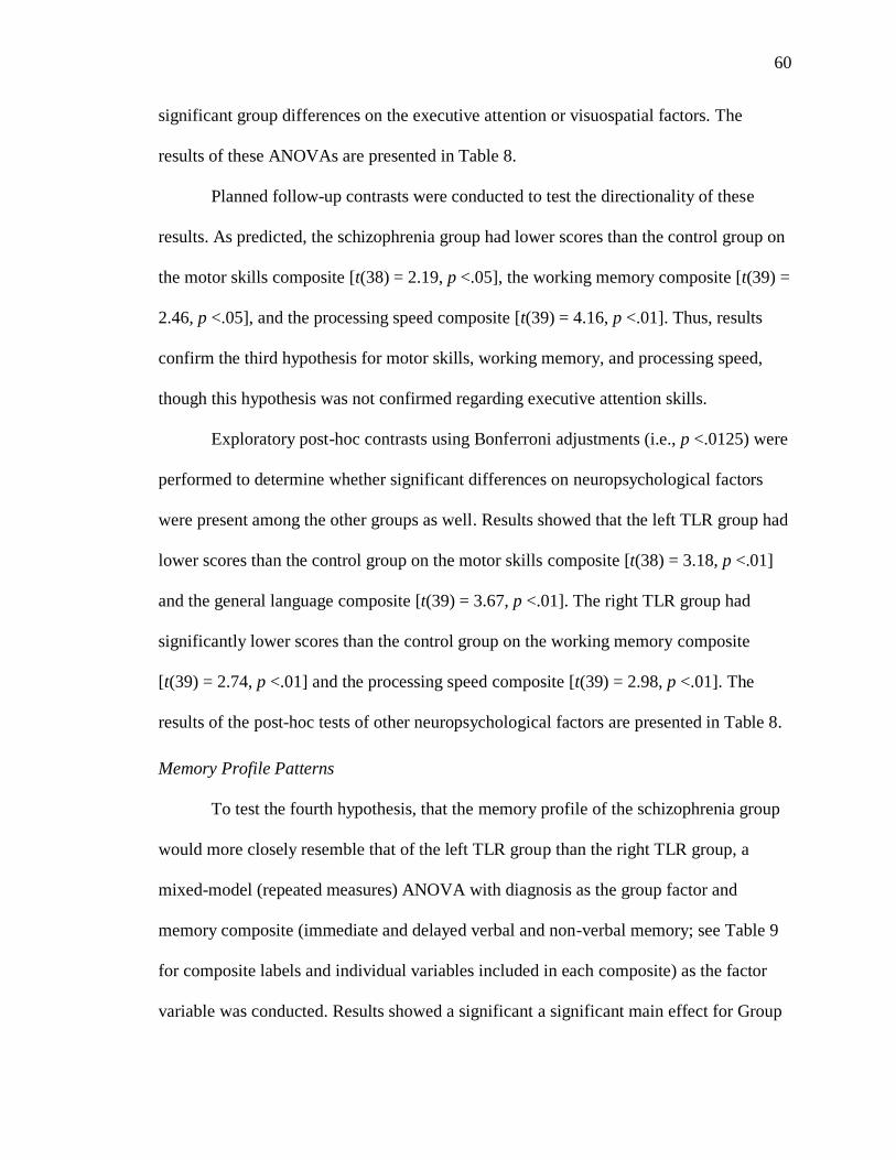
60
significant group differences on the executive attention or visuospatial factors. The
results of these ANOVAs are presented in Table 8.
Planned follow-up contrasts were conducted to test the directionality of these
results. As predicted, the schizophrenia group had lower scores than the control group on
the motor skills composite [t(38) = 2.19, p <.05], the working memory composite [t(39) =
2.46, p <.05], and the processing speed composite [t(39) = 4.16, p <.01]. Thus, results
confirm the third hypothesis for motor skills, working memory, and processing speed,
though this hypothesis was not confirmed regarding executive attention skills.
Exploratory post-hoc contrasts using Bonferroni adjustments (i.e., p <.0125) were
performed to determine whether significant differences on neuropsychological factors
were present among the other groups as well. Results showed that the left TLR group had
lower scores than the control group on the motor skills composite [t(38) = 3.18, p <.01]
and the general language composite [t(39) = 3.67, p <.01]. The right TLR group had
significantly lower scores than the control group on the working memory composite
[t(39) = 2.74, p <.01] and the processing speed composite [t(39) = 2.98, p <.01]. The
results of the post-hoc tests of other neuropsychological factors are presented in Table 8.
Memory Profile Patterns
To test the fourth hypothesis, that the memory profile of the schizophrenia group
would more closely resemble that of the left TLR group than the right TLR group, a
mixed-model (repeated measures) ANOVA with diagnosis as the group factor and
memory composite (immediate and delayed verbal and non-verbal memory; see Table 9
for composite labels and individual variables included in each composite) as the factor
variable was conducted. Results showed a significant a significant main effect for Group
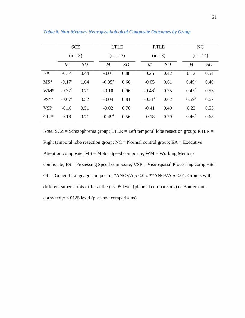
61
Table 8. Non-Memory Neuropsychological Composite Outcomes by Group
SCZ LTLE RTLE NC
(n = 8) (n = 13) (n = 8) (n = 14)
M SD M SD M SD M SD
EA -0.14 0.44 -0.01 0.88 0.26 0.42 0.12 0.54
MS* -0.17a 1.04 -0.35
a 0.66 -0.05 0.61 0.49
b 0.40
WM* -0.37a 0.71 -0.10 0.96 -0.46
a 0.75 0.45
b 0.53
PS** -0.67a 0.52 -0.04 0.81 -0.31
a 0.62 0.59
b 0.67
VSP -0.10 0.51 -0.02 0.76 -0.41 0.40 0.23 0.55
GL** 0.18 0.71 -0.49a 0.56 -0.18 0.79 0.46
b 0.68
Note. SCZ = Schizophrenia group; LTLR = Left temporal lobe resection group; RTLR =
Right temporal lobe resection group; NC = Normal control group; EA = Executive
Attention composite; MS = Motor Speed composite; WM = Working Memory
composite; PS = Processing Speed composite; VSP = Visuospatial Processing composite;
GL = General Language composite. *ANOVA p <.05. **ANOVA p <.01. Groups with
different superscripts differ at the p <.05 level (planned comparisons) or Bonferroni-
corrected p <.0125 level (post-hoc comparisons).
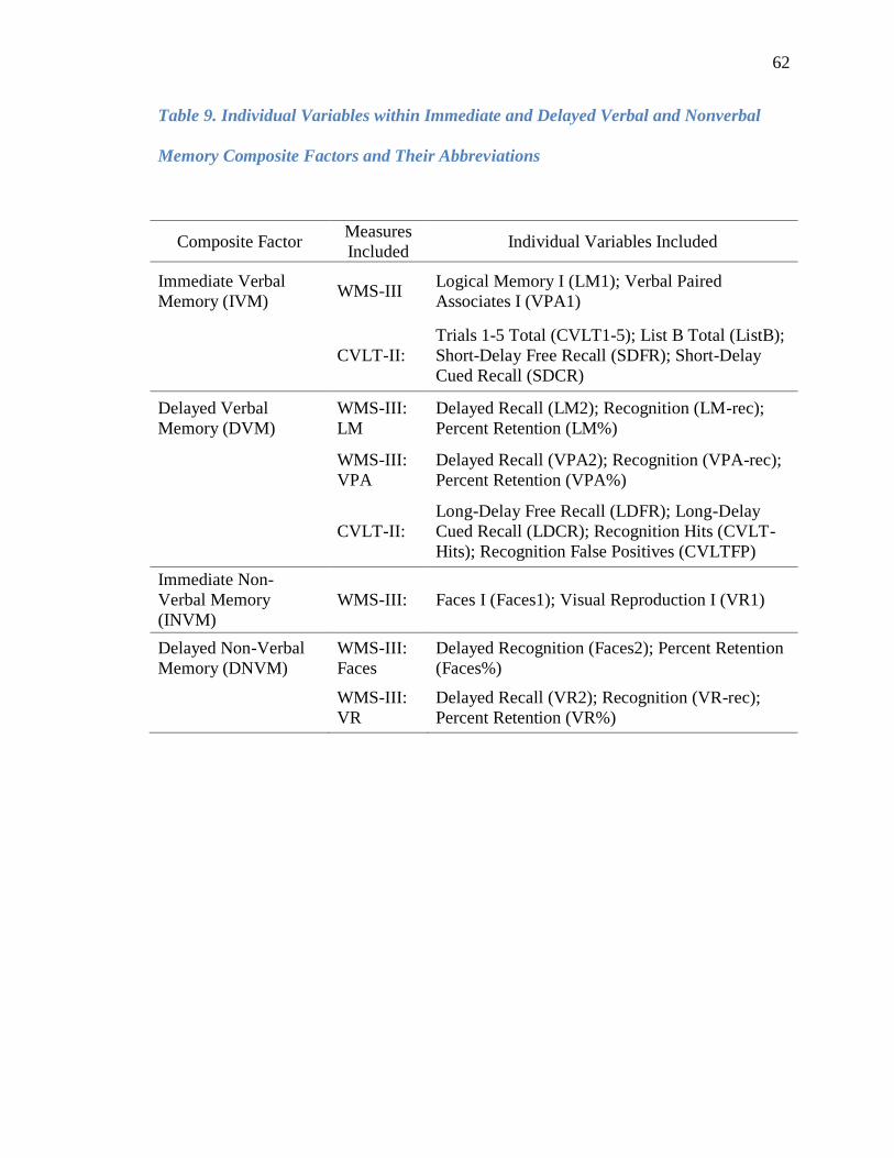
62
Table 9. Individual Variables within Immediate and Delayed Verbal and Nonverbal
Memory Composite Factors and Their Abbreviations
Composite Factor Measures
Included Individual Variables Included
Immediate Verbal
Memory (IVM) WMS-III
Logical Memory I (LM1); Verbal Paired
Associates I (VPA1)
CVLT-II:
Trials 1-5 Total (CVLT1-5); List B Total (ListB);
Short-Delay Free Recall (SDFR); Short-Delay
Cued Recall (SDCR)
Delayed Verbal
Memory (DVM)
WMS-III:
LM
Delayed Recall (LM2); Recognition (LM-rec);
Percent Retention (LM%)
WMS-III:
VPA
Delayed Recall (VPA2); Recognition (VPA-rec);
Percent Retention (VPA%)
CVLT-II:
Long-Delay Free Recall (LDFR); Long-Delay
Cued Recall (LDCR); Recognition Hits (CVLT-
Hits); Recognition False Positives (CVLTFP)
Immediate Non-
Verbal Memory
(INVM)
WMS-III: Faces I (Faces1); Visual Reproduction I (VR1)
Delayed Non-Verbal
Memory (DNVM)
WMS-III:
Faces
Delayed Recognition (Faces2); Percent Retention
(Faces%)
WMS-III:
VR
Delayed Recall (VR2); Recognition (VR-rec);
Percent Retention (VR%)
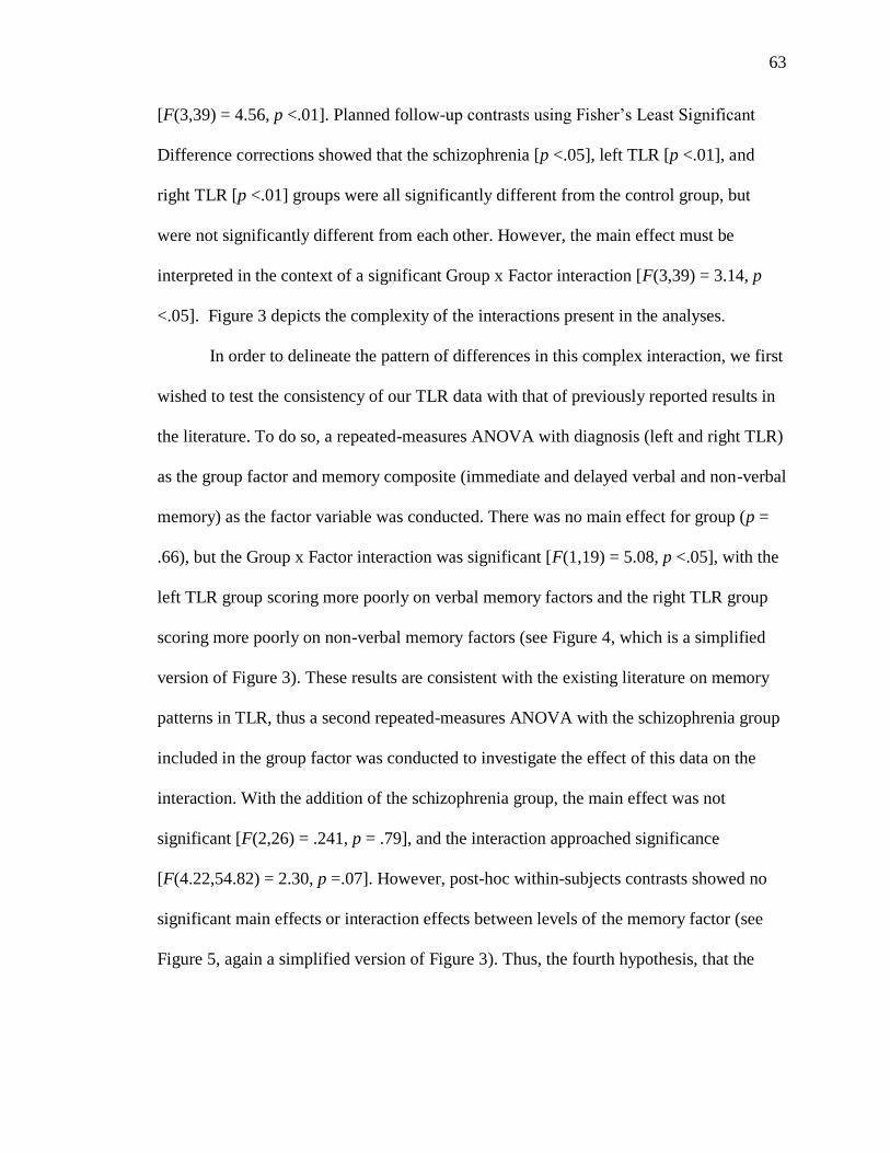
63
[F(3,39) = 4.56, p <.01]. Planned follow-up contrasts using Fisher‟s Least Significant
Difference corrections showed that the schizophrenia [p <.05], left TLR [p <.01], and
right TLR [p <.01] groups were all significantly different from the control group, but
were not significantly different from each other. However, the main effect must be
interpreted in the context of a significant Group x Factor interaction [F(3,39) = 3.14, p
<.05]. Figure 3 depicts the complexity of the interactions present in the analyses.
In order to delineate the pattern of differences in this complex interaction, we first
wished to test the consistency of our TLR data with that of previously reported results in
the literature. To do so, a repeated-measures ANOVA with diagnosis (left and right TLR)
as the group factor and memory composite (immediate and delayed verbal and non-verbal
memory) as the factor variable was conducted. There was no main effect for group (p =
.66), but the Group x Factor interaction was significant [F(1,19) = 5.08, p <.05], with the
left TLR group scoring more poorly on verbal memory factors and the right TLR group
scoring more poorly on non-verbal memory factors (see Figure 4, which is a simplified
version of Figure 3). These results are consistent with the existing literature on memory
patterns in TLR, thus a second repeated-measures ANOVA with the schizophrenia group
included in the group factor was conducted to investigate the effect of this data on the
interaction. With the addition of the schizophrenia group, the main effect was not
significant [F(2,26) = .241, p = .79], and the interaction approached significance
[F(4.22,54.82) = 2.30, p =.07]. However, post-hoc within-subjects contrasts showed no
significant main effects or interaction effects between levels of the memory factor (see
Figure 5, again a simplified version of Figure 3). Thus, the fourth hypothesis, that the
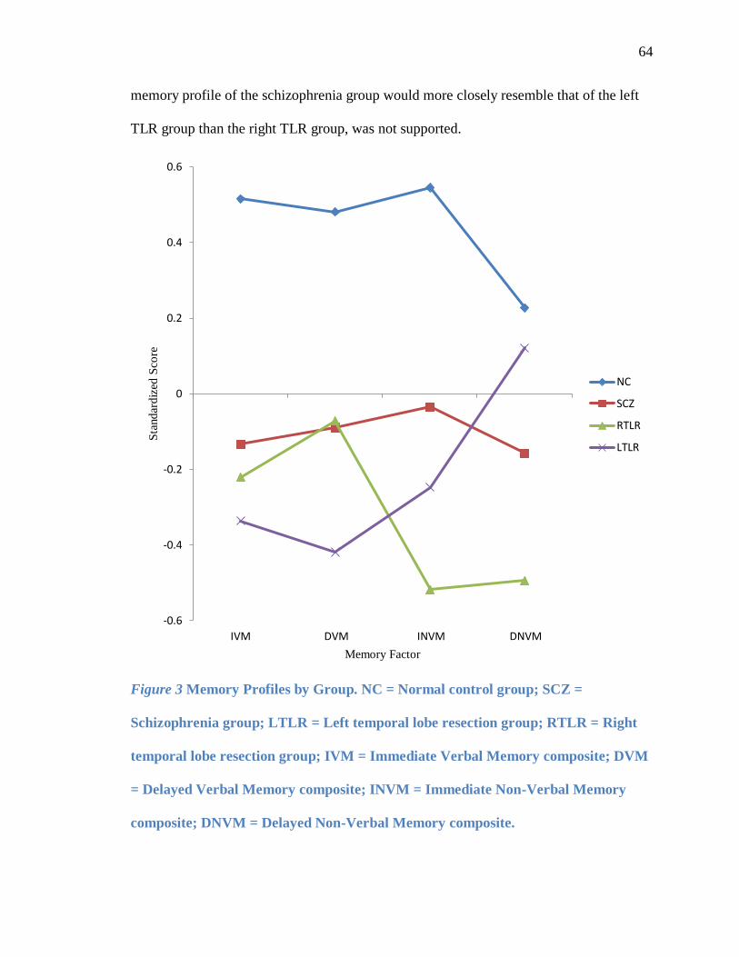
64
memory profile of the schizophrenia group would more closely resemble that of the left
TLR group than the right TLR group, was not supported.
Figure 3 Memory Profiles by Group. NC = Normal control group; SCZ =
Schizophrenia group; LTLR = Left temporal lobe resection group; RTLR = Right
temporal lobe resection group; IVM = Immediate Verbal Memory composite; DVM
= Delayed Verbal Memory composite; INVM = Immediate Non-Verbal Memory
composite; DNVM = Delayed Non-Verbal Memory composite.
-0.6
-0.4
-0.2
0
0.2
0.4
0.6
IVM DVM INVM DNVM
Sta
ndar
diz
ed S
core
Memory Factor
NC
SCZ
RTLR
LTLR
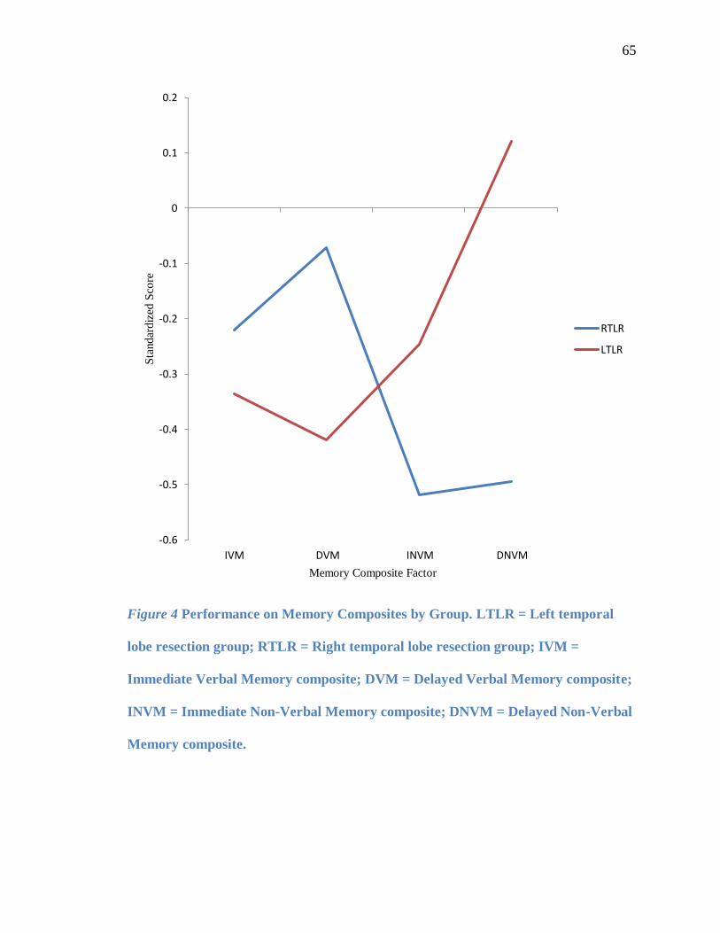
65
Figure 4 Performance on Memory Composites by Group. LTLR = Left temporal
lobe resection group; RTLR = Right temporal lobe resection group; IVM =
Immediate Verbal Memory composite; DVM = Delayed Verbal Memory composite;
INVM = Immediate Non-Verbal Memory composite; DNVM = Delayed Non-Verbal
Memory composite.
-0.6
-0.5
-0.4
-0.3
-0.2
-0.1
0
0.1
0.2
IVM DVM INVM DNVM
Sta
ndar
diz
ed S
core
Memory Composite Factor
RTLR
LTLR
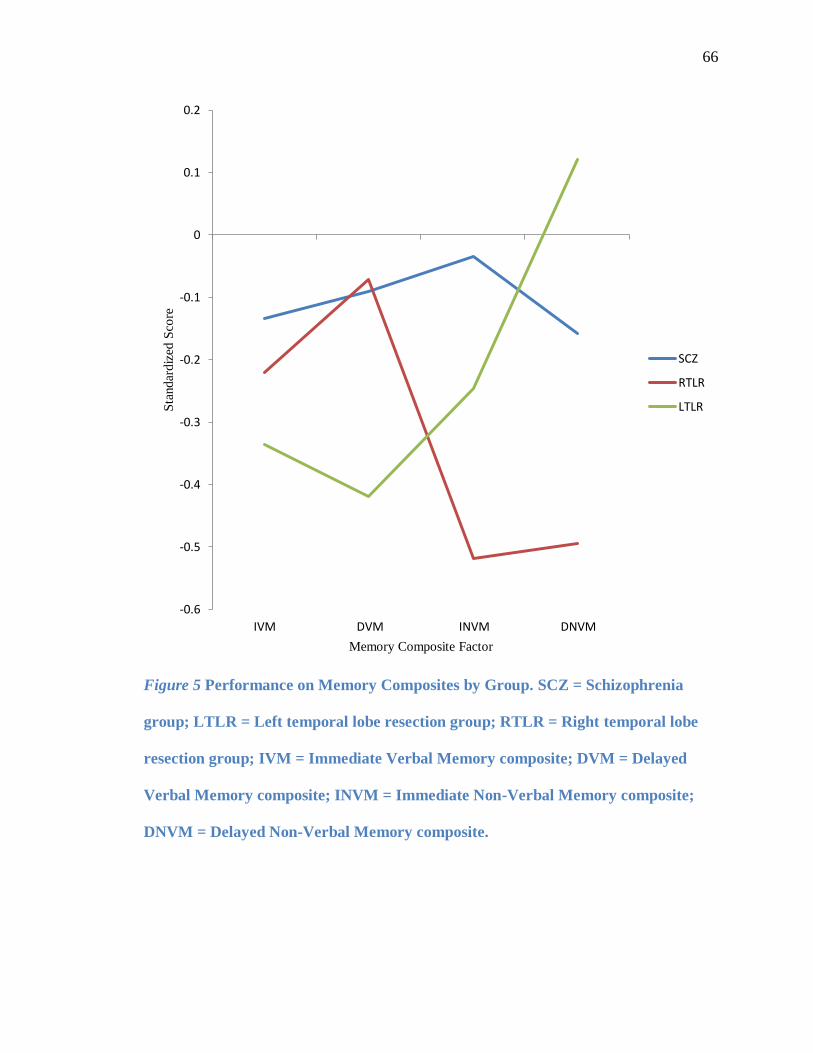
66
Figure 5 Performance on Memory Composites by Group. SCZ = Schizophrenia
group; LTLR = Left temporal lobe resection group; RTLR = Right temporal lobe
resection group; IVM = Immediate Verbal Memory composite; DVM = Delayed
Verbal Memory composite; INVM = Immediate Non-Verbal Memory composite;
DNVM = Delayed Non-Verbal Memory composite.
-0.6
-0.5
-0.4
-0.3
-0.2
-0.1
0
0.1
0.2
IVM DVM INVM DNVM
Sta
ndar
diz
ed S
core
Memory Composite Factor
SCZ
RTLR
LTLR
67
Post-hoc Exploratory Analyses of Neuropsychological Factors
To extend the scope of hypothesis four and investigate whether specific
neuropsychological performance profiles across multiple domains may better characterize
any pattern similarities between the schizophrenia and TLR groups, an exploratory
discriminant function analysis utilizing the entire set of neuropsychological variables
available was conducted to determine whether the eight factors (verbal memory, non-
verbal memory, motor skills, general language, visuospatial processing, working
memory, processing speed, and executive attention) could predict group membership.
The overall Wilks‟s lambda was significant, [Λ = .25, χ2(24, N = 43) = 49.15, p <.01],
indicating that overall the predictors differentiated among the four diagnostic groups. In
addition, the residual Wilks‟s lambda was significant, [Λ = .48, χ2(14, N = 43) = 25.34, p
<.05]. This test indicated that the neuropsychological factors differentiated significantly
among the four diagnostic groups after partialling out the effects of the first discriminant
function. Because these tests were significant, both discriminant functions were
interpreted.
In Table 10, the within-groups correlations between the predictors (i.e.,
neuropsychological composite factors) and both discriminant functions, as well as the
standardized weights, are presented. Based on these coefficients, the Processing Speed
composite demonstrates the strongest relationship with the first discriminant function,
with Working Memory and Nonverbal Memory showing slightly weaker, but still
significant, relationships. The General Language factor demonstrated the strongest
relationship with the second discriminant function, with Verbal Memory and Motor Skills
showing weaker yet significant relationships. On the basis of the results presented in
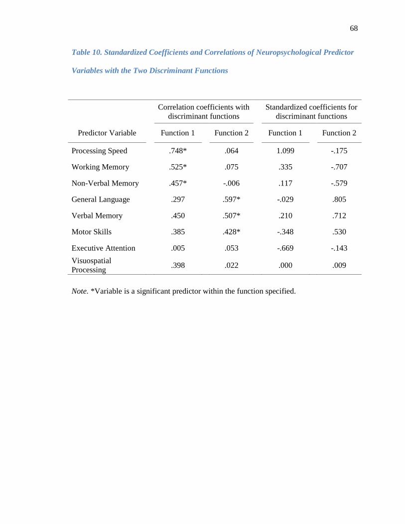
68
Table 10. Standardized Coefficients and Correlations of Neuropsychological Predictor
Variables with the Two Discriminant Functions
Correlation coefficients with
discriminant functions
Standardized coefficients for
discriminant functions
Predictor Variable Function 1 Function 2 Function 1 Function 2
Processing Speed .748* .064 1.099 -.175
Working Memory .525* .075 .335 -.707
Non-Verbal Memory .457* -.006 .117 -.579
General Language .297 .597* -.029 .805
Verbal Memory .450 .507* .210 .712
Motor Skills .385 .428* -.348 .530
Executive Attention .005 .053 -.669 -.143
Visuospatial
Processing .398 .022 .000 .009
Note. *Variable is a significant predictor within the function specified.
69
Table 10, we labeled the first discriminant function Attention-NVM and the second
discriminant function Language-VM.
Although the outcomes of the discriminant function analysis do not indicate
which groups are being discriminated by the functions, the means on the discriminant
functions are consistent with the interpretation of the first function as loading heavily on
attentional processing and nonverbal memory, and with the interpretation of the second
function as loading heavily on language skills, including verbal memory. The normal
control group had the highest mean on the Attention-NVM function (M = 1.24) and the
left TLR group had the next highest mean (M = .022), while the schizophrenia (M = -
1.02) and right TLR group (M = -1.03) had the lowest mean scores. On the other hand,
the schizophrenia (M = .600) and normal control (M = .569) groups had the highest mean
scores on the Language-VM function, followed closely by the right TLR group (M =
.475), while the left TLR group had the lowest mean score (M = -1.23).
Utilizing the discriminant function analysis to predict diagnostic group
membership (i.e., control, schizophrenia, left TLR, or right TLR), 73.8% of the
individuals in our sample were able to be correctly classified. In order to take into
account chance agreement, a kappa coefficient was computed; the value of kappa was
.645, indicating moderate to high accuracy in prediction. Finally, to assess how well the
classification procedure would predict in a different sample, the leave-one-out technique
was applied. The percent of individuals correctly classified based on this technique was
estimated at 45.2%.
70
Discussion
Summary
Previous research has been suggestive of significant left-hemisphere
frontotemporal dysconnectivity and resulting dysfunction in schizophrenia (e.g., Baaré et
al., 1999; Bogerts et al., 1990; Breier et al., 1992; Bryant et al., 1999; Buchanan, Vladar,
Barta, & Pearlson, 1998; DeLisi et al., 1991; Gur et al., 1998; Hirayasu et al., 1998; Hoff
et al., 1999; Honea et al., 2005; Pearlson, Petty, Ross, & Tien, 1996; Shenton et al., 1992,
2001; Turetsky et al., 1995; Velakoulis et al., 2006; Weinberger, Berman, Suddath, &
Torrey, 1992; Wible et al., 1995). In particular, hippocampal dysfunction has been
implicated as a core feature of schizophrenia resulting in impaired verbal, and to a lesser
extent, visual memory (Harrison, 2004; Thoma et al., 2008). In the current study, it was
hypothesized that, overall, participants with schizophrenia would demonstrate a pattern of
performance on multiple memory tests similar to that of individuals who had undergone
temporal lobe, including hippocampus, resection for intractable epilepsy. Specifically,
given the significant evidence in the literature for left-hemisphere temporal lobe
involvement in schizophrenia, it was reasonable to hypothesize that while multiple
neuropsychological domains may be affected in schizophrenia, verbal memory deficits
may be especially apparent and similar to those reliably described in individuals with
temporal lobe epilepsy localized to the left hemisphere (e.g., Aleman, Hijman, de Haan,
& Kahn, 1999; Clare, McKenna, Mortimer, & Baddeley, 1993; Delaney, Rosen, Mattson,
& Novelly, 1980; Giovagnoli, Casazza, & Avanzini, 1995; Hanlon et al., 2006; Hoff et
al., 1999; Johnson, Klinger, & Williams, 1977; Ladavas, Umilta, & Provincialo, 1979;
Majdan, Sziklas, & Jones-Gotman, 1996; Nacmani & Cohen, 1969; Ojemann & Dodrill,
71
1985; Toulopoulou, Morris, Rabe-Hesketh, & Murray, 2003). Namely, in addition to
impairments in attention (e.g., working memory and processing speed), executive
functioning, and motor skills, individuals with schizophrenia were expected to
demonstrate impaired verbal memory, similar to a left temporal lobe resection (TLR)
group, and impaired nonverbal memory, similar to a right TLR group, with an overall
memory profile more similar to the left TLR group, highlighting the importance of left-
hemispheric temporal lobe dysfunction in schizophrenia.
Verbal Memory
The first hypothesis, that the schizophrenia and left TLR groups would both
perform more poorly on verbal memory tasks than a healthy control group, was
supported. The schizophrenia group performed worse than controls on measures of
contextual verbal memory (WMS-III Logical Memory) and paired-associative verbal
learning (WMS-III Verbal Paired Associates), and the left TLR group performed worse
than controls on measures of contextual verbal memory, paired-associative verbal
learning, and rote verbal learning and recall (CVLT-II). Of note, there were no clinically
significant differences between the schizophrenia group and the left TLR group on any of
these variables, suggesting that the verbal memory performance of individuals with
schizophrenia in this sample was similar to that of those individuals who had undergone
left TLR. Consistent with previous research (e.g., Aleman, Hijman, de Haan, & Kahn,
1999; Clare, McKenna, Mortimer, & Baddeley, 1993; Hanlon et al., 2006), immediate
and delayed recall for verbal information appears to have been more affected than
recognition memory, as the only significant difference on recognition variables which
emerged was a higher number of false positive errors on the CVLT-II committed by the
72
left TLR group compared to controls. That is, the number of correctly recognized items
on this test was not significantly different across groups. This pattern is consistent with
previous theories which suggest that the difficulties with verbal memory seen in
schizophrenia may be the result of poor encoding and retrieval of information rather than
deficient storage of material (e.g., Aleman, Hijman, de Haan, & Kahn, 1999; Heaton,
Miller, Taylor, & Grant, 1994; McClain, 1983; Paulsen et al., 1995).
The pattern of performance in the left TLR group is also consistent with previous
research which has demonstrated significant impairment in recall of verbal information in
left TLE/TLR patients (e.g., Delaney, Rosen, Mattson, & Novelly, 1980; Ladavas,
Umilta, & Provinciali, 1979; Majdan, Sziklas, & Jones-Gotman, 1996; Ojemann &
Dodrill, 1985). Prior studies have demonstrated that verbal learning on the CVLT-II (i.e.,
total items recalled on trials 1-5) is highly correlated with hippocampal neuropathology
and is a strong indicator of seizure lateralization in patients with TLE, with the strongest
association emerging between verbal learning and left hippocampal neuron loss (Baños et
al., 2004; Sass et al., 1995). Interestingly, in the current study, the left TLR group, but not
the schizophrenia group, demonstrated poorer performance than controls on rote verbal
learning and recall. It is possible that the extent of temporal lobe injury and resection in
the TLR group limits the ability to learn and recall verbal information regardless of type
of presentation, versus a structurally intact yet functionally impaired temporal lobe in
schizophrenia which impacts complex verbal learning and memory requiring more
developed general language skills. However, previous research has demonstrated
impairments in learning, retrieval, and to a lesser extent, recognition, on the CVLT-II in a
wide variety of patients with schizophrenia (e.g, Altshuler et al., 2004; Hill, Beers,

73
Kmiec, Keshavan, & Sweeny, 2004; Paulsen et al., 1995), typically in the context of a
generalized neurocognitive deficit including impaired attention and executive
functioning. The schizophrenia group in the current study did not demonstrate
generalized neuropsychological impairment, which may explain why there were no
differences in their rote verbal learning compared to controls. Regardless, the similar
patterns of verbal memory performance overall in the left TLR group and the
schizophrenia group provide some empirical support for the hypothesis that left-
hemisphere temporal lobe dysfunction may be a core feature of schizophrenia.
Non-Verbal Memory
The second hypothesis was that the schizophrenia and right TLR groups would
perform more poorly than the healthy control group on measures of non-verbal memory.
Although this hypothesis was not confirmed, the results were in the predicted direction
and very closely approached significance for the right TLR, but not for the schizophrenia,
group. The right TLR group performed worse than controls on all aspects of the WMS-III
Visual Reproduction subtest (i.e., immediate and delayed free recall, retention, and
recognition); however, the results of the schizophrenia group were not significantly
different from those of the control group on any measure of nonverbal memory. The
current results are consistent with previous research demonstrating a visual memory
deficit in patients with right-hemisphere TLE/TLR (e.g., Delaney, Rosen, Mattson, &
Novelly, 1980; Giovagnoli, Casazza, & Avanzini, 1995; Gleiβner, Helmstaedter, &
Elger, 1998; Ladavas, Umilta, & Povinciali, 1979). In contrast, prior studies of visual
memory in schizophrenia have been somewhat inconsistent, with some demonstrating
impairments (e.g., Calev, Korin, Kugelmass, & Lerer, 1987; Gold et al., 1992b; Kolb &
74
Whishaw, 1983; Rushe, Woodruff, Murray, & Morris, 1999; Sullivan, Shear, Zipursky,
Sagar, & Pfefferbaum, 1994) and others showing relatively minor impairment compared
to verbal memory impairment or an impairment only in nonverbal encoding (e.g., Saykin
et al., 1991; Tracy et al., 2001). A complete lack of nonverbal memory impairment in
patients with schizophrenia in the current study is surprising, however, given the overall
cognitive profile in which statistically, but not necessarily clinically, significant
differences between performance on neuropsychological measures in this group
compared to controls suggests that the schizophrenia group in the current study may be
particularly high-functioning. Thus, the results must be interpreted with some caution, as
they are in contradiction to the results of the predominance of previous studies. On the
other hand, in conjunction with previous research which demonstrates significant left-
hemisphere neuroanatomical and neurocognitive abnormalities (e.g., Gur et al., 1998;
Shenton et al., 1992; Siedman et al., 2002; Turetsky et al., 1995), the results of the
current study suggest that left temporal and fronto-temporal dysfunction may be more
prominent than bilateral temporal or fronto-temporal dysfunction in schizophrenia and
lend support to the left-hemisphere theory of schizophrenia.
Other Neuropsychological Domains
The third hypothesis, that the schizophrenia group would perform more poorly
than controls on measures of executive function/attention, motor skills, working memory,
and processing speed, was partially confirmed. Consistent with previous research (Braff
& Saccuzzo, 1985; Buchanan, Summerfelt, Tek, & Gold, 2002; Carter et al., 1998;
Egeland et al., 2003; Goldman-Rakic, 1994; Heinrichs & Zakzanis, 1998), results showed
that the schizophrenia group had lower composite scores than the control group on
75
indices of working memory, motor skills, and processing speed. The executive attention
factor in the current study was comprised of measures which assess mental set-shifting
(i.e., WMS-III Mental Control and Trail Making Test, Part B), perseveration/cognitive
inflexibility (RFFT perseverative errors), and sustained visual attention (CPT-2). It is
somewhat surprising that the schizophrenia group did not demonstrate impaired
performance on the executive attention composite, as executive functioning impairment
has been fairly reliably demonstrated in previous studies (e.g., Bilder et al., 2000; Braff et
al., 1991; Goldberg & Gold, 1995; Hutton et al., 1998; Martínez-Arán et al., 2002;
Velligan & Bow-Thomas, 1999). There are several possibilities which may explain the
lack of a significant difference between patients with schizophrenia and healthy controls
in the current study. First, many previous studies included the Wisconsin Card Sorting
Test (WCST) in their protocols; this is a widely used neuropsychological measure of
executive function which also requires intact working memory and visual attention. It is
possible that had this measure been included in the present study, it may have captured an
executive function deficit if indeed one was present. Patients with schizophrenia have
fairly consistently demonstrated impaired performances on the WCST (Heinrichs &
Zakzanis, 1998), though research relating to correlation of WCST results with positive
and negative symptoms of schizophrenia have been less consistent (e.g., Braff et al.,
1991; McGrath et al., 1997; Morice, 1990; Nieuwenstein, Aleman, & de Haan, 2001;
Seidman et al., 1991). As schizophrenia subtype and positive/negative symptomatology
were not evaluated in the present study, including the WCST in the current protocol may
not have altered the results regarding executive attention.

76
A second possibility is that by analyzing only the composite score for executive
attention, differences on individual variables within this composite may have been
overlooked. To explore this possibility, post-hoc analyses were conducted on the
individual variables within the executive attention factor: significant differences were
found only for WMS-III Mental Control (schizophrenia and right TLR groups worse than
controls) and CPT-2 commission errors (right TLR group worse than control and
schizophrenia groups). Thus it seems that the schizophrenia group in the present study
performed more poorly than healthy controls only on a measure of mental set-shifting.
These results are consistent with the possibility that the patients with schizophrenia in
this study may represent a relatively high-cognitive-functioning group, as suggested by
their overall standardized scores within one standard deviation of average, indicating a
lack of a generalized neurocognitive deficit. In general, however, current results are in
keeping with those of previous studies regarding poor performance on measures of
working memory, processing speed, and motor skills.
Overall Memory and Neuropsychological Profiles
The final hypothesis was that, overall, the memory pattern of the schizophrenia
group would more closely resemble that of the left than the right TLR group. This
hypothesis was not supported, as the schizophrenia group‟s scores on composites of
immediate and delayed verbal and non-verbal memory were intermediate between those
of the left and right TLR groups, and were not significantly different from either TLR
group. That is, while the left and right TLR groups demonstrated the predicted interactive
pattern of poorer verbal memory (left TLR) versus poorer non-verbal memory (right
TLR), the schizophrenia group demonstrated a relatively flat pattern in which neither
77
verbal nor non-verbal memory scores were significantly different from each other. In
fact, plotting the memory composite scores for the schizophrenia group resulted in a
profile which most closely resembled that of the control group, although the scores were
significantly lower than those of the control group.
The results of the exploratory discriminant function analysis which utilized all
neuropsychological factors are intriguing. Two functions emerged which significantly
predicted group membership: Attention-NVM and Language-VM. Regarding the
Attention-NVM function, attentional skills, including working memory, processing
speed, and sustained visual and auditory attention are known to be impaired in
schizophrenia (Goldstein, Rosenbaum, & Taylor, 1997; Nieuwenstein, Aleman, & de
Haan, 2001; Nuechterlein et al., 1994), thus the importance of this factor in
discriminating between schizophrenia and control groups is quite obvious. Its role in
differentiating between schizophrenia and TLR groups is less clear. Previous research has
demonstrated that within left-hemisphere TLE/TLR groups, verbal memory is impaired
while simple and sustained auditory attention are relatively spared (Fleck et al., 1999;
Fleck, Shear, & Strakowski, 2002; Gold et al., 1994; Mirsky, Primac, Marsan, Rosvold,
& Stevens, 1960). This may suggest that the functional neuroanatomy which is
characteristic of left TLE (i.e., medial and lateral temporal lobe structural abnormalities)
is closely associated with a specific verbal memory impairment, while in schizophrenia,
impaired verbal and non-verbal memory coincide with impaired attentional functions,
providing support for the involvement of the frontal lobe and its connections as well as
temporal lobe dysfunction. Prior research has implicated abnormalities in fronto-temporal
networks and the cognitive functions subserved by these connections (e.g., executive
78
function, attentional processing) as key features of schizophrenia (Green, 1998;
Randolph, Goldberg, & Weinberger, 1993), and the results of the current study are
generally consistent with this theory: the normal control and left TLR groups, in which
no attentional or non-verbal memory deficits would be expected, had the highest means
on the Attention-NVM factor, while the schizophrenia group, in which attentional and
non-verbal memory impairment would be expected, as well as the right TLR group with
expected non-verbal memory impairment, demonstrated the lowest means. The results of
this discriminant function highlight the importance of assessing attention as a precursor to
memory impairment in schizophrenia, and lend support to the existing literature
describing impairments in fronto-temporal systems in this population.
On the second discriminant function, general language processing skills (such as
expressive vocabulary, verbal abstract reasoning, and verbal fluency) and verbal memory,
as well as motor skills, emerged as significant predictors of group membership.
Interestingly, this function seemed to discriminate only the left TLR group from the other
groups, as the control, schizophrenia, and right TLR groups all had relatively high means
on this function whereas the left TLR group had a much lower mean score. While the
contribution of language and verbal memory as discriminating between the left TLR
group versus healthy controls and the right TLR group is consistent with previous
research demonstrating deficits in these domains within left TLE/TLR groups (e.g.,
Hermann, Seidenberg, Haltiner, & Wyler, 1992; Ojeman & Dodrill, 1885; Richardson et
al., 2004), it is somewhat surprising that the schizophrenia group in the current study
displayed such a high mean on this function, given their overall lower performance on
verbal memory measures compared to controls. It is possible that language and verbal
79
memory develop along different trajectories in schizophrenia versus in TLE, so that
relatively poorer overall language development in TLE in conjunction with difficulties in
verbal memory present as a more significant overall deficit than generally intact overall
language processing skills combined with impaired verbal memory in schizophrenia. In a
study by Gold and colleagues (1994), results showed that patients with schizophrenia
performed better than patients with TLE on a measure of sight reading thought to predict
premorbid potential, and demonstrated better semantic knowledge than patients with left-
hemisphere TLE. The authors concluded that schizophrenia and TLE follow different
developmental pathways, with TLE affecting the acquisition of academic skills such as
vocabulary and semantic knowledge as well as cognitive functions, while in
schizophrenia, language development is relatively spared, though cognitive functions
such as verbal memory are significantly affected. The results of the Language-VM
discriminant function in the current study are consistent with this theory, as the group
means would seem to indicate that the schizophrenia group could be distinguished from
the left TLR group on the basis of performance on measures of general language
processing, given that these two groups performed similarly on measures of verbal
memory.
Overall Conclusions
This is one of few studies to directly compare verbal and non-verbal memory
functions, as well as other neuropsychological domains, in schizophrenia and temporal
lobe epilepsy groups. As such, complex neuropsychological profiles which could
demonstrate similarities and differences between these groups were able to be
constructed, in order to further clarify the memory deficits typically reported in the
80
schizophrenia literature and determine whether the results were consistent with the theory
that the symptoms and cognitive impairment associated with schizophrenia are largely
the result of left-hemispheric dysfunction. In addition, the battery of tests administered in
the current study included measures of multiple neuropsychological domains, which
allowed an examination of a general versus domain-specific deficit in order to
supplement the existing literature. The results of this study, which included significantly
worse performances in verbal memory, processing speed, working memory, and motor
skills, are consistent with previous research which has demonstrated several domain-
specific impairments, (Bilder, 1996; Bilder et al., 2000; Braff et al., 1991; Goldberg &
Gold, 1995; Pantelis, Nelson, & Barnes, 1996; Saykin et al., 1994); however, although
significant differences were observed, the schizophrenia group‟s overall performance on
all measures remained within the average to low average range, which would not be
expected if a generalized neurocognitive deficit were present. In addition, the results of a
discriminant function analysis did not yield a single factor which accounted for a
substantial amount of variance within the schizophrenia group, further supporting the
theory that several domain-specific deficits (or, in our sample, significantly worse
performances) are present within a neurocognitive profile in schizophrenia.
Though comparisons of memory profiles between patients with schizophrenia and
those who had undergone left- or right-TLR did not show the predicted pattern, the
results of a discriminant function analysis do provide support for the presence of fronto-
temporal dysfunction which may be mediated by cognitive functions such as language
processing that functionally dissociate to the left hemisphere. Given the proximity of
language processing areas to structures of the left temporal lobe associated with memory
81
(i.e., hippocampus), the results of the current study, which implicate abnormal
hippocampal function (verbal memory) in the absence of impaired language functioning
in the schizophrenia group, but not in the left TLR group, would seem to warrant further
exploration of the hypothesis that structural and functional abnormalities in the
hippocampus may be a core feature of schizophrenia, which have downstream effects on
frontal and prefrontal lobe functioning. For example, previous research with rats has
demonstrated that hippocampal insult results in prefrontal changes: neonatal damage to
the ventral hippocampus alters the response of medial prefrontal cortical pyramidal
neurons to dopaminergic/GABA-ergic projections from the midbrain, an effect which has
been postulated to explain the impact of damage to human hippocampus in schizophrenia
(O‟Donnell, Lewis, Weinberger, & Lipska, 2002). Lipska (2004) described a series of
studies which suggest that neonatal excitotoxic disconnection of the ventral hippocampus
in rats result in a model of neurobiological and behavioral sequelae similar to those seen
in humans with schizophrenia, including deficits in social functioning and working
memory and changes in the development of frontal and prefrontal cortical regions.
Further investigation of the hippocampus as a site of primary deficit in schizophrenia
may help delineate the complex frontal-temporal disconnects seen in neuroimaging and
neuropsychological studies.
Additional factors may also have contributed to the results of this study regarding
demonstration of group differences. First, test characteristics such as familiarity of items
(e.g., word lists versus abstract geometric figures) and type of stimulus presentation (e.g.,
free recall of word lists versus recognition of faces) may vary in their relative cognitive
demands. Recall for stimulus items to which participants have had exposure prior to
82
testing (such as words from the CVLT-II) may result in differential neuronal activation
patterns than recall for non-familiar stimulus items (such as the abstract designs from
WMS-III VR), such that additional neuronal networks are involved in the encoding and
retrieval of these items. Likewise, verbal material may be more easily „chunked‟ in
learning trials and related to an individual‟s personal experience with language (e.g.,
utilizing mnemonic strategies to learn rote verbal material), whereas no previous template
or learning strategy may exist for abstract visual material.
A second factor which may have affected the results of this study in terms of
group differences is the purported laterality of the TLR groups. Neuronal processes such
as extent of regenesis, dendritic branching, and contralateral compensation (i.e., the non-
affected hemisphere assuming some of the functions of the affected hemisphere)
following cerebral insult may all play a role in the cognitive functioning of post-surgical
patients as well as in patients with schizophrenia. However, the TLR patients in the
current study all underwent intracarotid sodium amobarbital (Wada) testing prior to
resection, and all participants demonstrated the predicted pattern of lateralization, so to
the extent possible, our comparison groups were chosen to represent typical left- and
right-hippocampal functions.
Bilingualism is another factor which may affect neuropsychological outcomes.
Although research on the relationship between bilingualism and cognitive output is rather
limited, some studies have demonstrated that variables such as age at acquisition of the
second language (e.g., Hernandez & Li, 2007) and attained proficiency of the second
language (e.g., Perani et al., 1998) may result in differences in neuropsychological
performance. Other research has shown that bilingualism selectively affects semantic
83
verbal fluency (e.g, animal naming) but not phonemic fluency, free-form fluency, or
sentence repetition tasks, and that these effects are more pronounced in groups with
earlier age of acquisition of the second language (Rosselli et al., 2000). In the current
study, participants were asked whether they were bilingual, and if so, at what age they
learned English proficiently, in order to provide some measure of control for bilingualism
when analyzing data, particularly for verbal measures. However, it may have been more
informative to have conceptualized bilingualism as a continuous rather than dichotomous
variable, so that potential moderating or mediating variables such as age of acquisition
and attained proficiency could have been investigated.
Other factors which may have had an effect on the results of the current study
include psychometric properties of the test instruments themselves, such as
characteristics of the normative sample, kurtosis and skew of the distribution of the
normative scores, measurement error, and the presence of ceiling or floor effects.
Neuropsychological assessment utilizes a comparison model in which an individual‟s
performance is compared to a normative sample; however, every neuropsychological test
is normed on a different sample of the general population, making generalization to the
entire population difficult. In addition, there are differences in the number and type of
dynamic demographic variables included in normative information for individual
comparison (i.e., whereas one test may include a set of norms based on age, gender, and
education, others may only include age and specify that the normative sample included
men and women with a certain educational level), so that an individual may be compared
to quite variable normative samples to derive his or her standardized scores. This
complicates the characterization of overall performance, or in the case of the present
84
study, may complicate the creation of composite scores based on data derived from
differing normative samples. Characteristics of the distribution of the normative sample
are also important to consider: whether the distribution is truly normal, or whether it has a
positive or negative skew or kurtosis, affects the mean and standard deviation of the
sample. As these are the most commonly used summary statistics when deriving
normative data and level of individual performance, normality in the distribution is ideal.
Floor and ceiling effects are often the source of positive and negative skew (e.g., in a
sample in which most participants performed at perfect or near-perfect levels, there
would be few low scores and an asymmetrical distribution of high scores, resulting in a
negatively skewed distribution), and the decrease in variance of scores in the normative
sample can result in somewhat biased standardized scores when applied in
neuropsychological assessment. In addition, it is important to consider the reliability and
measurement error of a test when deriving normative data. Rather than representing
actual performance, test scores may best be conceptualized as representing an estimate of
function with a specified degree of variance, or error: the lower the degree of
measurement error, the more reliable the measurement in the general population and the
more confidence in the validity of individual scores (Brooks, Strauss, Sherman, Iverson,
& Slick, 2009).
In the current study, the psychometric characteristics of the measures have been
well documented, and tests were chosen as much for their reported reliability as for the
functions which they measure. In addition, by utilizing a multiple-methods design in
which composite factor scores could be created, we can consider these values as trends
rather than absolute values and make reasonable conclusions regarding the data.
85
Nevertheless, examination of the characteristics of the distributions of the composite
factors in this study revealed several deviations from normality. The Verbal Memory,
Non-Verbal Memory, Motor, Working Memory, and Executive Attention composites
were negatively skewed, while the Language composite was positively skewed. The
Visuospatial and Processing Speed composites were quite close to normal (with skewness
statistics of 0.025 and 0.081, respectively). Regarding kurtosis, the Executive Attention
composite was highly leptokurtic, while all other factors were platykurtic (no composite
had a kurtosis statistic which approached 3.0, indicating normal peakedness). Thus, it
may have been useful to apply a different set of statistical analyses to these composite
scores, or to transform the variables statistically so that they more closely approximated a
normal distribution.
One limitation of the present study was its small sample size, particularly in the
schizophrenia and right TLR groups. Statistical analyses based on small sample sizes and
unequal cell sizes can be problematic and it is likely that given a larger sample size and
more evenly distributed group numbers, more reliable and significant results would have
emerged, particularly given the high number of measures with trends in the expected
direction (i.e., p-values between 0.05 and 0.1 or which did not meet post-hoc Bonferroni-
corrected levels). In addition, data was missing for a small subset of participants,
particularly in the right TLR group (e.g., SES, CPT-2), such that some analyses were
based on even smaller sample sizes. Furthermore, small sample size results in reduced
power to determine whether a difference exists between groups. For example, a large
effect size (d = .64) would be necessary to detect a difference with 80% power between
the schizophrenia and left TLR groups in the current study, based on sample sizes of 8
86
schizophrenia and 13 left TLR participants. In all likelihood, the actual effect size for the
variables examined in the current study may be lower (particularly between
schizophrenia and TLR groups, but perhaps less so between patient and control groups),
thus concluding that no significant differences exist between schizophrenia and left TLR
groups on memory and other neuropsychological domains may be premature. Likewise,
an even larger effect size (d = .75) would be necessary to conclude with 80% power that a
significant difference exists between the schizophrenia and right TLR groups, based on
our sample sizes of 8 participants per group. Nevertheless, despite limited statistical
power, significant differences on multiple variables did emerge between groups in the
present study, suggesting that these variables may be useful in future research to further
delineate the patterns of neuropsychological deficits in schizophrenia and TLR groups.
A second limitation, related to small sample size, is that we were unable to
evaluate the influence of schizophrenia subtype (e.g., paranoid, disorganized, etc.) on
cognitive outcome measures. Previous research has shown that, for example, although
global indices of verbal memory and executive functioning in patients with paranoid
schizophrenia are impaired in comparison to a normal control group, these deficits are not
as pronounced as they are in patients with disorganized or undifferentiated schizophrenia
(Bornstein, Nasrallah, Olson, Coffman, & Schwarzkopf, 1990). It is possible that
performance on measures of verbal and nonverbal memory for different types of
information, for example, rote memorization of semantically-unrelated words versus
recall of contextual verbal information, may be differentially affected in schizophrenia
subtypes as well, and would likely be more reliably examined in more homogenous
subject populations. In addition, it should be noted that while significant differences
87
between the schizophrenia group and the healthy control group in our sample did emerge,
the average standardized scores on most variables were not within what is typically
considered a clinically significant range of impairment. It is possible that with a larger
sample comprised of individuals with different subtypes of schizophrenia, patterns of
memory performance specific to each subtype, with greater degrees of impairment in
those groups with disorganized or undifferentiated subtypes, might emerge which would
further supplement the existing literature on verbal memory in schizophrenia.
Furthermore, it should be noted that the age of onset of seizure activity in the left
and right TLR groups in the current study was quite variable, ranging from infancy to 29
years of age. Previous research has demonstrated that age of seizure onset is correlated
with hippocampal injury (i.e., younger age of onset results in greater hippocampal
sclerosis), which is in turn related to memory functioning post-TLR (i.e., pre-TLR
sclerosis is associated with equal or improved memory performance post-TLR, versus
decreased memory performance in patients without medial temporal sclerosis; e.g., Bell
& Davies, 1998; Davies et al. 1996; Hermann et al., 1995). The wide range of seizure
onset age in the present study may have resulted in a mixed-outcome group of TLR
patients, thus possibly obscuring potential differences.
In conclusion, the present study demonstrated that cognitive profiles in
schizophrenia, involving memory alone and in conjunction with other neuropsychological
domains, are highly complex and may best be represented by an interactive model rather
than a simple model of hemispheric impairment such as that theorized to exist in
temporal lobe epilepsy. These results seem to warrant future research to further explore
the contributory role of overall attentional processing skills and general language ability

88
to impairments in verbal and non-verbal memory in schizophrenia, as well as the
complicated relationship of frontal and temporal lobe structures and their
interconnections to neurocognitive functioning in schizophrenia.
89
Appendices
Appendix A. Demographic Questionnaire
Appendix B. Hollingshead Index of Social Position Scale (HISP)
Appendix C. Beck Depression Inventory, Second Edition (BDI-II)
Appendix D. Magical Ideation Scale (MIS)
Appendix E. Revised Social Anhedonia Scale (RSAS)
Appendix F. Waterloo Handedness Questionnaire (WHQ)
Appendix G. Participant Compensation Form
90
Appendix A. Demographic Questionnaire
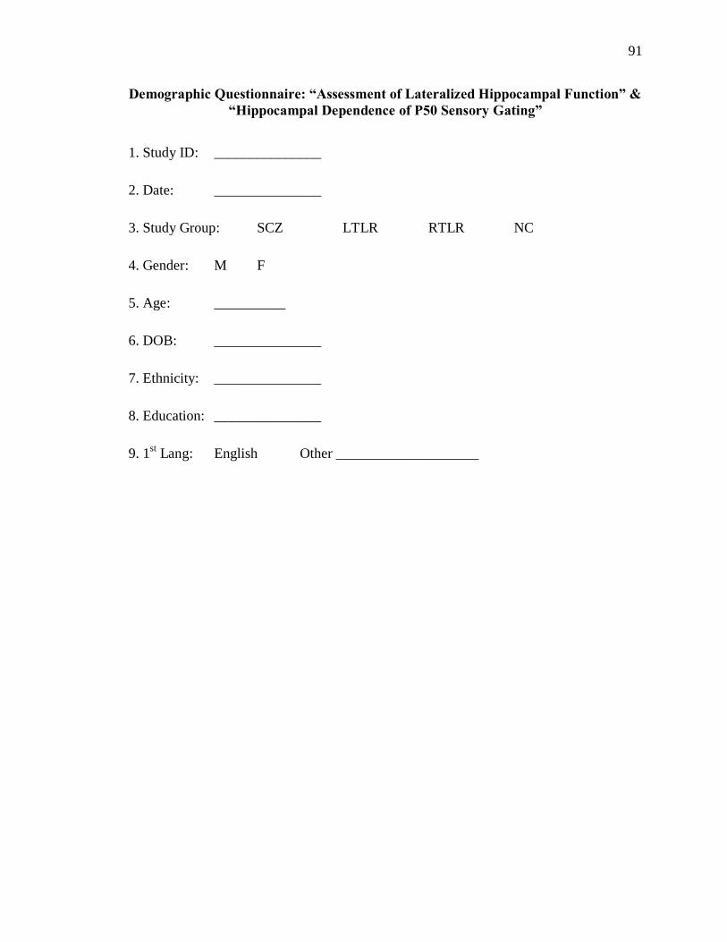
91
Demographic Questionnaire: “Assessment of Lateralized Hippocampal Function” &
“Hippocampal Dependence of P50 Sensory Gating”
1. Study ID: _______________
2. Date: _______________
3. Study Group: SCZ LTLR RTLR NC
4. Gender: M F
5. Age: __________
6. DOB: _______________
7. Ethnicity: _______________
8. Education: _______________
9. 1st Lang: English Other ____________________
92
Appendix B. Hollingshead Index of Social Position Scale (HISP)
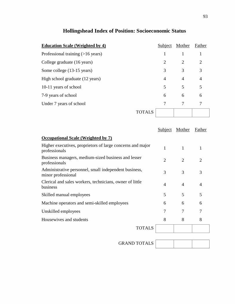
93
Hollingshead Index of Position: Socioeconomic Status
Education Scale (Weighted by 4) Subject Mother Father
Professional training (>16 years) 1 1 1
College graduate (16 years) 2 2 2
Some college (13-15 years) 3 3 3
High school graduate (12 years) 4 4 4
10-11 years of school 5 5 5
7-9 years of school 6 6 6
Under 7 years of school 7 7 7
TOTALS
Subject Mother Father
Occupational Scale (Weighted by 7)
Higher executives, proprietors of large concerns and major
professionals 1 1 1
Business managers, medium-sized business and lesser
professionals 2 2 2
Administrative personnel, small independent business,
minor professional 3 3 3
Clerical and sales workers, technicians, owner of little
business 4 4 4
Skilled manual employees 5 5 5
Machine operators and semi-skilled employees 6 6 6
Unskilled employees 7 7 7
Housewives and students 8 8 8
TOTALS
GRAND TOTALS
94
Appendix C. Beck Depression Inventory, Second Edition (BDI-II)
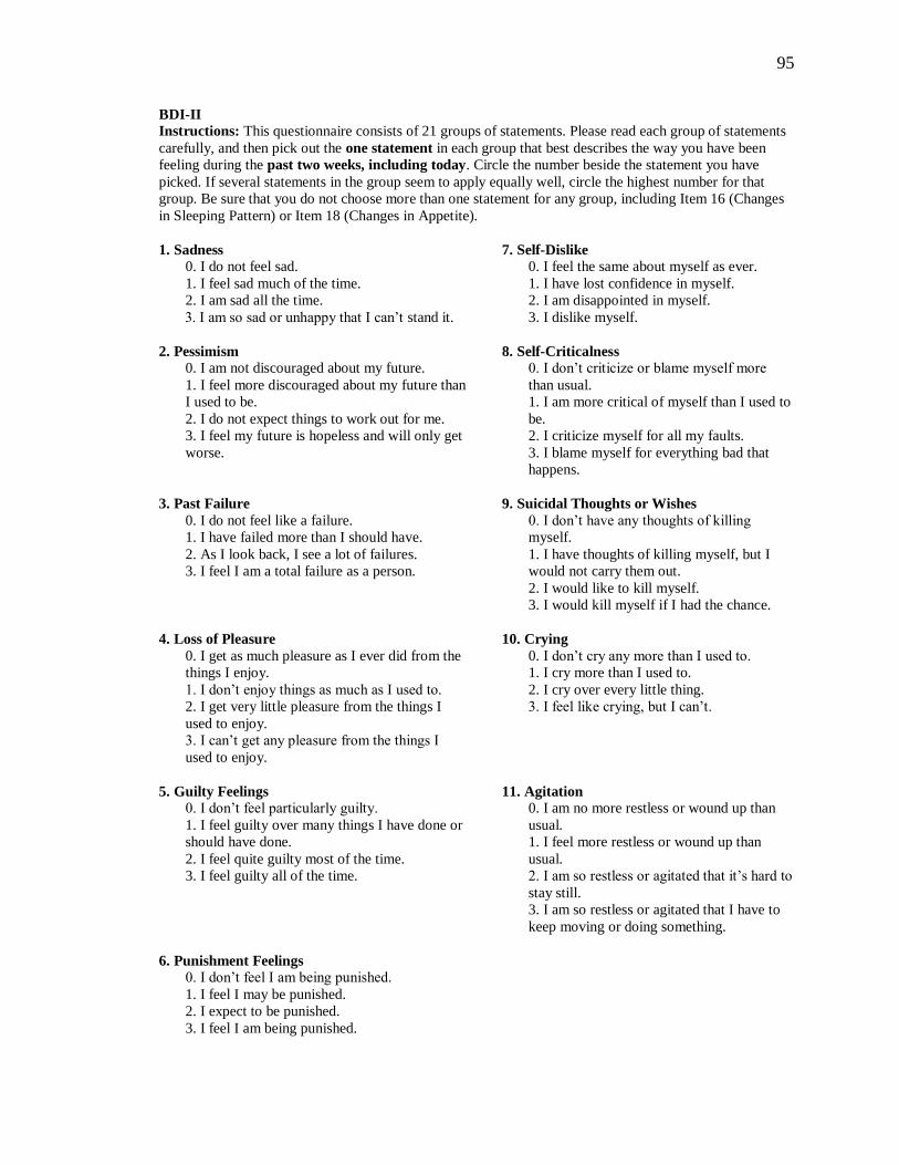
95
BDI-II
Instructions: This questionnaire consists of 21 groups of statements. Please read each group of statements
carefully, and then pick out the one statement in each group that best describes the way you have been
feeling during the past two weeks, including today. Circle the number beside the statement you have
picked. If several statements in the group seem to apply equally well, circle the highest number for that
group. Be sure that you do not choose more than one statement for any group, including Item 16 (Changes
in Sleeping Pattern) or Item 18 (Changes in Appetite).
1. Sadness
0. I do not feel sad.
1. I feel sad much of the time.
2. I am sad all the time.
3. I am so sad or unhappy that I can‟t stand it.
7. Self-Dislike
0. I feel the same about myself as ever.
1. I have lost confidence in myself.
2. I am disappointed in myself.
3. I dislike myself.
2. Pessimism
0. I am not discouraged about my future.
1. I feel more discouraged about my future than
I used to be.
2. I do not expect things to work out for me.
3. I feel my future is hopeless and will only get
worse.
8. Self-Criticalness
0. I don‟t criticize or blame myself more
than usual.
1. I am more critical of myself than I used to
be.
2. I criticize myself for all my faults.
3. I blame myself for everything bad that
happens.
3. Past Failure
0. I do not feel like a failure.
1. I have failed more than I should have.
2. As I look back, I see a lot of failures.
3. I feel I am a total failure as a person.
9. Suicidal Thoughts or Wishes
0. I don‟t have any thoughts of killing
myself.
1. I have thoughts of killing myself, but I
would not carry them out.
2. I would like to kill myself.
3. I would kill myself if I had the chance.
4. Loss of Pleasure
0. I get as much pleasure as I ever did from the
things I enjoy.
1. I don‟t enjoy things as much as I used to.
2. I get very little pleasure from the things I
used to enjoy.
3. I can‟t get any pleasure from the things I
used to enjoy.
10. Crying
0. I don‟t cry any more than I used to.
1. I cry more than I used to.
2. I cry over every little thing.
3. I feel like crying, but I can‟t.
5. Guilty Feelings
0. I don‟t feel particularly guilty.
1. I feel guilty over many things I have done or
should have done.
2. I feel quite guilty most of the time.
3. I feel guilty all of the time.
11. Agitation
0. I am no more restless or wound up than
usual.
1. I feel more restless or wound up than
usual.
2. I am so restless or agitated that it‟s hard to
stay still.
3. I am so restless or agitated that I have to
keep moving or doing something.
6. Punishment Feelings
0. I don‟t feel I am being punished.
1. I feel I may be punished.
2. I expect to be punished.
3. I feel I am being punished.
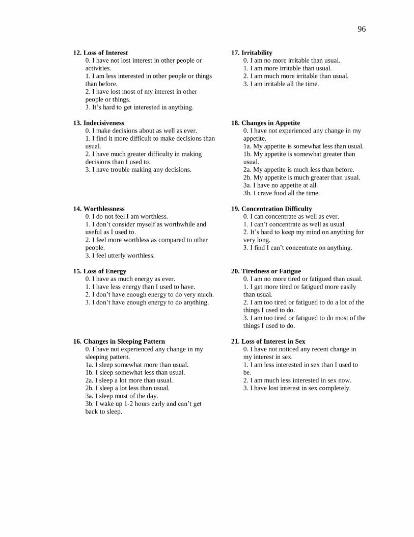
96
12. Loss of Interest
0. I have not lost interest in other people or
activities.
1. I am less interested in other people or things
than before.
2. I have lost most of my interest in other
people or things.
3. It‟s hard to get interested in anything.
17. Irritability
0. I am no more irritable than usual.
1. I am more irritable than usual.
2. I am much more irritable than usual.
3. I am irritable all the time.
13. Indecisiveness
0. I make decisions about as well as ever.
1. I find it more difficult to make decisions than
usual.
2. I have much greater difficulty in making
decisions than I used to.
3. I have trouble making any decisions.
18. Changes in Appetite
0. I have not experienced any change in my
appetite.
1a. My appetite is somewhat less than usual.
1b. My appetite is somewhat greater than
usual.
2a. My appetite is much less than before.
2b. My appetite is much greater than usual.
3a. I have no appetite at all.
3b. I crave food all the time.
14. Worthlessness
0. I do not feel I am worthless.
1. I don‟t consider myself as worthwhile and
useful as I used to.
2. I feel more worthless as compared to other
people.
3. I feel utterly worthless.
19. Concentration Difficulty
0. I can concentrate as well as ever.
1. I can‟t concentrate as well as usual.
2. It‟s hard to keep my mind on anything for
very long.
3. I find I can‟t concentrate on anything.
15. Loss of Energy
0. I have as much energy as ever.
1. I have less energy than I used to have.
2. I don‟t have enough energy to do very much.
3. I don‟t have enough energy to do anything.
20. Tiredness or Fatigue
0. I am no more tired or fatigued than usual.
1. I get more tired or fatigued more easily
than usual.
2. I am too tired or fatigued to do a lot of the
things I used to do.
3. I am too tired or fatigued to do most of the
things I used to do.
16. Changes in Sleeping Pattern
0. I have not experienced any change in my
sleeping pattern.
1a. I sleep somewhat more than usual.
1b. I sleep somewhat less than usual.
2a. I sleep a lot more than usual.
2b. I sleep a lot less than usual.
3a. I sleep most of the day.
3b. I wake up 1-2 hours early and can‟t get
back to sleep.
21. Loss of Interest in Sex
0. I have not noticed any recent change in
my interest in sex.
1. I am less interested in sex than I used to
be.
2. I am much less interested in sex now.
3. I have lost interest in sex completely.
97
Appendix D. Magical Ideation Scale (MIS)
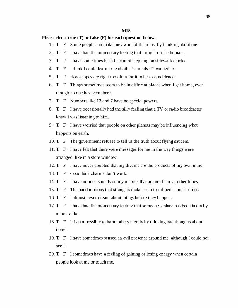
98
MIS
Please circle true (T) or false (F) for each question below.
1. T F Some people can make me aware of them just by thinking about me.
2. T F I have had the momentary feeling that I might not be human.
3. T F I have sometimes been fearful of stepping on sidewalk cracks.
4. T F I think I could learn to read other‟s minds if I wanted to.
5. T F Horoscopes are right too often for it to be a coincidence.
6. T F Things sometimes seem to be in different places when I get home, even
though no one has been there.
7. T F Numbers like 13 and 7 have no special powers.
8. T F I have occasionally had the silly feeling that a TV or radio broadcaster
knew I was listening to him.
9. T F I have worried that people on other planets may be influencing what
happens on earth.
10. T F The government refuses to tell us the truth about flying saucers.
11. T F I have felt that there were messages for me in the way things were
arranged, like in a store window.
12. T F I have never doubted that my dreams are the products of my own mind.
13. T F Good luck charms don‟t work.
14. T F I have noticed sounds on my records that are not there at other times.
15. T F The hand motions that strangers make seem to influence me at times.
16. T F I almost never dream about things before they happen.
17. T F I have had the momentary feeling that someone‟s place has been taken by
a look-alike.
18. T F It is not possible to harm others merely by thinking bad thoughts about
them.
19. T F I have sometimes sensed an evil presence around me, although I could not
see it.
20. T F I sometimes have a feeling of gaining or losing energy when certain
people look at me or touch me.
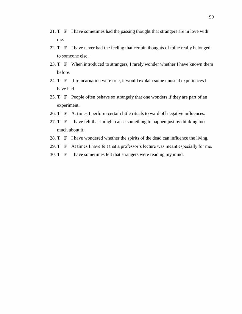
99
21. T F I have sometimes had the passing thought that strangers are in love with
me.
22. T F I have never had the feeling that certain thoughts of mine really belonged
to someone else.
23. T F When introduced to strangers, I rarely wonder whether I have known them
before.
24. T F If reincarnation were true, it would explain some unusual experiences I
have had.
25. T F People often behave so strangely that one wonders if they are part of an
experiment.
26. T F At times I perform certain little rituals to ward off negative influences.
27. T F I have felt that I might cause something to happen just by thinking too
much about it.
28. T F I have wondered whether the spirits of the dead can influence the living.
29. T F At times I have felt that a professor‟s lecture was meant especially for me.
30. T F I have sometimes felt that strangers were reading my mind.
100
Appendix E. Revised Social Anhedonia Scale (RSAS)
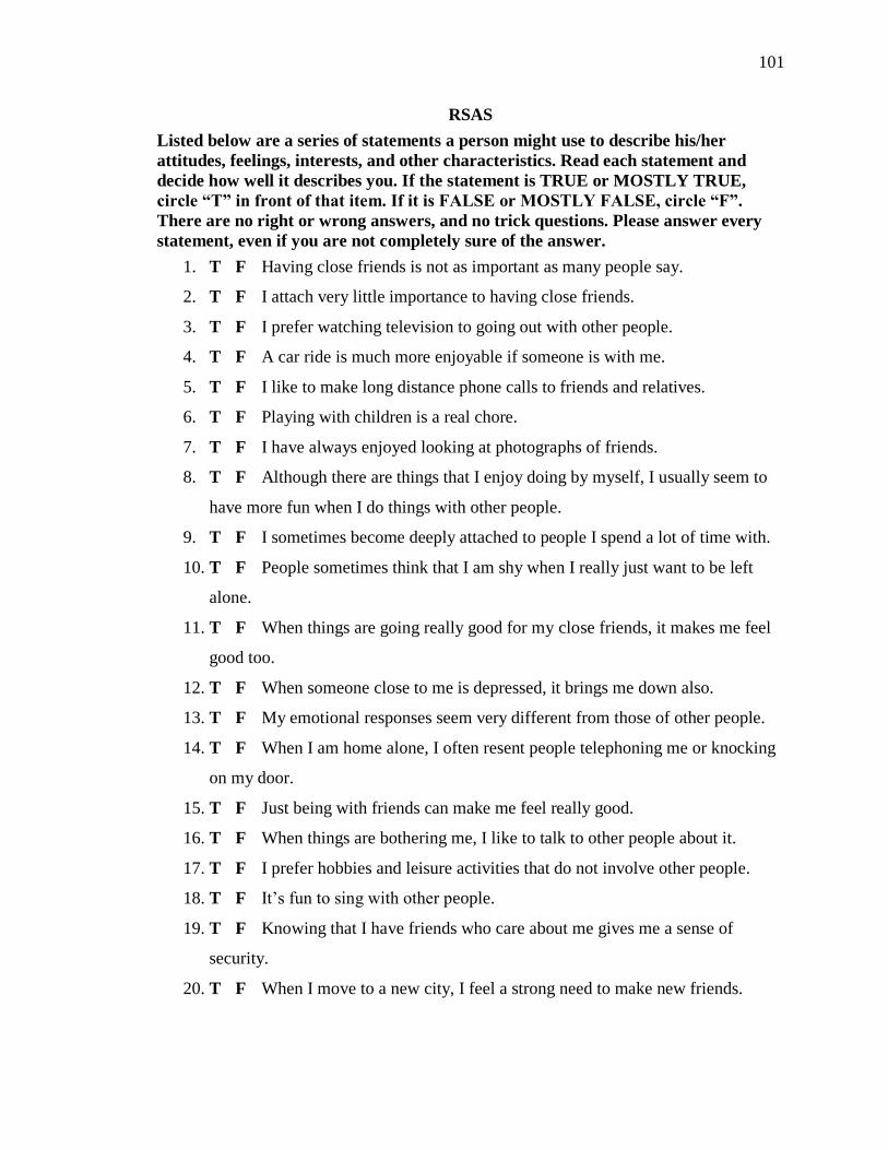
101
RSAS
Listed below are a series of statements a person might use to describe his/her
attitudes, feelings, interests, and other characteristics. Read each statement and
decide how well it describes you. If the statement is TRUE or MOSTLY TRUE,
circle “T” in front of that item. If it is FALSE or MOSTLY FALSE, circle “F”.
There are no right or wrong answers, and no trick questions. Please answer every
statement, even if you are not completely sure of the answer.
1. T F Having close friends is not as important as many people say.
2. T F I attach very little importance to having close friends.
3. T F I prefer watching television to going out with other people.
4. T F A car ride is much more enjoyable if someone is with me.
5. T F I like to make long distance phone calls to friends and relatives.
6. T F Playing with children is a real chore.
7. T F I have always enjoyed looking at photographs of friends.
8. T F Although there are things that I enjoy doing by myself, I usually seem to
have more fun when I do things with other people.
9. T F I sometimes become deeply attached to people I spend a lot of time with.
10. T F People sometimes think that I am shy when I really just want to be left
alone.
11. T F When things are going really good for my close friends, it makes me feel
good too.
12. T F When someone close to me is depressed, it brings me down also.
13. T F My emotional responses seem very different from those of other people.
14. T F When I am home alone, I often resent people telephoning me or knocking
on my door.
15. T F Just being with friends can make me feel really good.
16. T F When things are bothering me, I like to talk to other people about it.
17. T F I prefer hobbies and leisure activities that do not involve other people.
18. T F It‟s fun to sing with other people.
19. T F Knowing that I have friends who care about me gives me a sense of
security.
20. T F When I move to a new city, I feel a strong need to make new friends.

102
21. T F People are usually better off if they stay aloof from emotional
involvements with most others.
22. T F Although I know I should have affection for certain people, I don‟t really
feel it.
23. T F People often expect me to spend more talking with them than I would like.
24. T F I feel pleased and gratified as I learn more and more about the emotional
life of my friends.
25. T F When others try to tell me about their problems and hang-ups, I usually
listen with interest and attention.
26. T F I never had really close friends in high school.
27. T F I am usually content to just sit alone, thinking and daydreaming.
28. T F I‟m much too independent to really get involved with other people.
29. T F There are few things more tiring than to have a long, personal discussion
with someone.
30. T F It made me sad to see all my high school friends go their separate ways
when high school was over.
31. T F I have often found it hard to resist talking to a good friend, even when I
have other things to do.
32. T F Making new friends isn‟t worth the energy it takes.
33. T F There are things that are more important to me than privacy.
34. T F People who try to get to know me better usually give up after awhile.
35. T F I could be happy living all alone in a cabin in the woods or mountains.
36. T F If given the choice, I would much rather be with others than alone.
37. T F I find that people too often assume that their daily activities and opinions
will be interesting.
38. T F I don‟t really feel very close to my friends.
39. T F My relationships with other people never get very intense.
40. T F In many ways, I prefer the company of pets to the company of people.
103
Appendix F. Waterloo Handedness Questionnaire (WHQ)
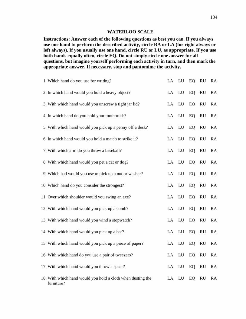
104
WATERLOO SCALE
Instructions: Answer each of the following questions as best you can. If you always
use one hand to perform the described activity, circle RA or LA (for right always or
left always). If you usually use one hand, circle RU or LU, as appropriate. If you use
both hands equally often, circle EQ. Do not simply circle one answer for all
questions, but imagine yourself performing each activity in turn, and then mark the
appropriate answer. If necessary, stop and pantomime the activity.
1. Which hand do you use for writing? LA LU EQ RU RA
2. In which hand would you hold a heavy object? LA LU EQ RU RA
3. With which hand would you unscrew a tight jar lid? LA LU EQ RU RA
4. In which hand do you hold your toothbrush? LA LU EQ RU RA
5. With which hand would you pick up a penny off a desk? LA LU EQ RU RA
6. In which hand would you hold a match to strike it? LA LU EQ RU RA
7. With which arm do you throw a baseball? LA LU EQ RU RA
8. With which hand would you pet a cat or dog? LA LU EQ RU RA
9. Which had would you use to pick up a nut or washer? LA LU EQ RU RA
10. Which hand do you consider the strongest? LA LU EQ RU RA
11. Over which shoulder would you swing an axe? LA LU EQ RU RA
12. With which hand would you pick up a comb? LA LU EQ RU RA
13. With which hand would you wind a stopwatch? LA LU EQ RU RA
14. With which hand would you pick up a bar? LA LU EQ RU RA
15. With which hand would you pick up a piece of paper? LA LU EQ RU RA
16. With which hand do you use a pair of tweezers? LA LU EQ RU RA
17. With which hand would you throw a spear? LA LU EQ RU RA
18. With which hand would you hold a cloth when dusting the
furniture?
LA LU EQ RU RA
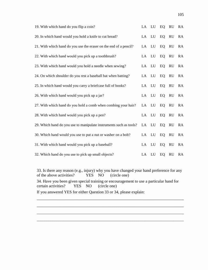
105
19. With which hand do you flip a coin? LA LU EQ RU RA
20. In which hand would you hold a knife to cut bread? LA LU EQ RU RA
21. With which hand do you use the eraser on the end of a pencil? LA LU EQ RU RA
22. With which hand would you pick up a toothbrush? LA LU EQ RU RA
23. With which hand would you hold a needle when sewing? LA LU EQ RU RA
24. On which shoulder do you rest a baseball bat when batting? LA LU EQ RU RA
25. In which hand would you carry a briefcase full of books? LA LU EQ RU RA
26. With which hand would you pick up a jar? LA LU EQ RU RA
27. With which hand do you hold a comb when combing your hair? LA LU EQ RU RA
28. With which hand would you pick up a pen? LA LU EQ RU RA
29. Which hand do you use to manipulate instruments such as tools? LA LU EQ RU RA
30. Which hand would you use to put a nut or washer on a bolt? LA LU EQ RU RA
31. With which hand would you pick up a baseball? LA LU EQ RU RA
32. Which hand do you use to pick up small objects? LA LU EQ RU RA
33. Is there any reason (e.g., injury) why you have changed your hand preference for any
of the above activities? YES NO (circle one)
34. Have you been given special training or encouragement to use a particular hand for
certain activities? YES NO (circle one)
If you answered YES for either Question 33 or 34, please explain:
________________________________________________________________________
________________________________________________________________________
________________________________________________________________________
________________________________________________________________________
106
Appendix G. Participant Compensation Form
107
PARTICIPANT COMPENSATION SIGNATURE FORM
Project Title: Assessment of Lateralized Hippocampal Function
Principal Investigator: Faith M. Hanlon, Ph.D.
Additional Investigators: Michael Weisend, Ph.D., Robert J. Thoma, Ph.D., Rex Jung,
Ph.D., & S. Laura Lundy, M.S.
I, the undersigned, certify that I have been paid $50 for my participation in the
neuropsychological testing session for this study.
Print Name
Signature Date
SIGNATURE OF INVESTIGATOR
I certify that the participant has been paid in full.
Signature Date
108
References
Aleman, A., Hijman, R., de Haan, E. H. F., & Kahn, R. S. (1999). Memory impairment in
schizophrenia: A meta-analysis. American Journal of Psychiatry, 156, 1358-1366.
Altshuler, L. L., Ventura, J., van Gorp, W. G., Green, M. F., Theberge, D. C., & Mintz, J.
(2004). Neurocognitive function in clinically stable men with bipolar I disorder or
schizophrenia and normal control subjects. Biological Psychiatry, 56, 560-569.
American Psychiatric Association. (2000). Diagnostic and Statistical Manual for Mental
Disorders (4th
ed. Text Revision). Washington DC: Author.
Andreasen, N. C., Ehrhardt, J. C., Swayze II, V. W., Alliger, R. J., Yuh, W. T. C., Cohen,
G., et al. (1990). Magnetic resonance imaging of the brain in schizophrenia: The
pathophysiologic significance of structural abnormalities. Archives of General
Psychiatry, 47, 35-44.
Apostolova, L. G., Beyer, M., Green, A. E., Hwang, K. S., Morra, J. H., Chou, Y. Y., et
al. (2010). Hippocampal, caudate, and ventricular changes in Parkinson‟s disease
with and without dementia. Movement Disorders, 25, 687-688.
Astur, R. S., Taylor, L. B., Mamelak, A. N., Philpott, L., & Sutherland, R. J. (2002).
Humans with hippocampus damage display severe spatial memory impairments in
a virtual Morris water task. Behavioural Brain Research, 132, 77-84.
Baaré, W. F., Hulshoff Pol, H. E., Hijman, R., Mali, W. P. Th., Viergever, M. A., &
Kahn, R. S. (1999). Volumetric analysis of frontal lobe regions in schizophrenia:
Relation to cognitive function and symptomatology. Biological Psychiatry, 45,
1597-1605.
109
Baños, J. H., Roth, D. L., Palmer, C., Morawetz, R., Knowlton, R., Faught, E., et al.
(2004). Confirmatory factor analysis of the California Verbal Learning Test in
patients with epilepsy: Relationship to clinical and neuropathological markers of
temporal lobe epilepsy. Neuropsychology, 18, 60-68.
Barta, P. E., Pearlson, G. D., Brill II, L. B., Royall, R., McGilchrist, I. K., Pulver, A. E.,
et al. (1997a). Planum temporale asymmetry reversal in schizophrenia:
Replication and relationship to gray matter abnormalities. American Journal of
Psychiatry, 154, 661-667.
Barta, P. E., Powers, R. E., Aylward, E. H., Chase, G. A., Harris, G. J., Rabins, P. V., et
al. (1997b). Quantitative MRI volume changes in late onset schizophrenia and
Alzheimer‟s disease compared to normal controls. Psychiatry Research:
Neuroimaging Section, 68, 65-75.
Barta, P. E., Pearlson, G. D., Powers, R. E., Richards, S. S., & Tune, L. E. (1990).
Auditory hallucinations and smaller superior temporal gyral volume in
schizophrenia. American Journal of Psychiatry, 147, 1457-1462.
Beck, A. T., Steer, R. A., & Brown, B. K. (1996). Beck Depression Inventory manual (2nd
edition). San Antonio, TX: Psychological Corporation.
Bell, B. D. & Davies, K. G. (1998). Anterior temporal lobectomy, hippocampal sclerosis,
and memory: Recent neuropsychological findings. Neuropsychology Review, 8,
25-40.
Benton, A. L. & Hamsher, K. D. S. (1994). Multilingual aphasia examination. Iowa City,
IA: AJA Associates Inc.
110
Bilder, R. M. (1996). Neuropsychology and neurophysiology in schizophrenia. Current
Opinion in Psychiatry, 9, 57–62.
Bilder, R. M., Bogerts, B., Ashtari, M., Wu, H., Alvir, J. M., Jody, D., et al. (1995).
Anterior hippocampal volume reductions predict frontal lobe dysfunction in first
episode schizophrenia. Schizophrenia Research, 17, 47-58.
Bilder, R. M., Goldman, R. S., Robinson, D., Reiter, G., Bell, L., Bates, J. A., et al.
(2000). Neuropsychology of first-episode schizophrenia: Initial characterization
and clinical correlates. American Journal of Psychiatry, 157, 549-559.
Blanchard, J. J. & Neale, J. M. (1994). The neuropsychological signature of
schizophrenia: Generalized or differential deficit? American Journal of
Psychiatry, 151, 40-48.
Bogerts, B., Ashtari, M., Degreef, G., Alvir, J. M. J., Bilder, R. M., & Lieberman, J. A.
(1990). Reduced temporal limbic structure volumes on magnetic resonance
images in first episode schizophrenia. Psychiatry Research: Neuroimaging, 35, 1–
13 .
Bogerts, B., Lieberman, J. A., Ashtari, M., Bilder, R. M., Degreef, G., Lerner, G., et al.
(1993). Hippocampus-amygdala volumes and psychopathology in chronic
schizophrenia. Biological Psychiatry, 33, 236–246.
Bornstein, R. A., Nasrallah, H. A., Olson, S. C., Coffman, M. T., & Schwarzkopf, S. B.
(1990). Neuropsychological deficit in schizophrenic subtypes: Paranoid,
Nonparanoid, and schizoaffective subgroups. Psychiatry Research, 31, 15-24.
Braff, D. L., Heaton, R., Kuck, J., Cullum, M., Moranville, J., Grant, I., et al. (1991). The
generalized pattern of neuropsychological deficits in outpatients with chronic

111
schizophrenia with heterogeneous Wisconsin Card Sorting Test results. Archives
of General Psychiatry, 48, 891-898.
Braff, D. L. & Saccuzzo, D. P. (1985). The time course of information-processing deficits
in schizophrenia. American Journal of Psychiatry, 142, 170-174.
Breier, A., Buchanan, R. W., Elkashef, A., Munson, R. C., Kirkpatrick, B., & Gellad, F.
(1992). Brain morphology and schizophrenia: A magnetic resonance imaging
study of limbic, prefrontal cortex, and caudate structures. Archives of General
Psychiatry, 49, 921-926.
Brooks, B. L., Strauss, E., Sherman, E. M. S., Iverson, G. L., & Slick, D. J. (2009).
Developments in neuropsychological assessment: Refining psychometric and
clinical interpretive methods. Canadian Psychology, 50, 196-209.
Brown, J. (1958). Some tests of the decay theory of immediate memory. Quarterly
Journal of Experimental Psychology, 10, 12-21.
Bryant, N. L., Buchanan, R. W., Vladar, K., Breier, A., & Rothman, M. (1999). Gender
differences in temporal lobe structures of patients with schizophrenia: A
volumetric MRI study. American Journal of Psychiatry, 156, 603-609.
Bryden, M. P. (1977). Measuring handedness with questionnaires. Neuropsychologia, 15,
617-624.
Buchanan, R. W., Summerfelt, A., Tek, C., & Gold, J. (2002). An open-labeled trial of
adjunctive donepezil for cognitive impairments in patients with schizophrenia.
Schizophrenia Research, 59, 29-33.
112
Buchanan, R. W., Vladar, K., Barta, P. E., & Pearlson, G. D. (1998). Structural
evaluation of the prefrontal cortex in schizophrenia. American Journal of
Psychiatry, 155, 1049-1055.
Bustillo, J. R., Chen, H., Gasparovic, C., Mullins, P., Caprihan, A., Qualls, C., et al.
(2011). Glutamate as a marker of cognitive function in schizophrenia: A proton
spectroscopic imaging study at 4 Tesla. Biological Psychiatry, 69, 19-27.
Calev, A., Korin, Y., Kugelmass, S., & Lerer, B. (1987). Performance of chronic
schizophrenics on matched word and design recall tasks. Biological Psychiatry,
22, 699-709.
Carter, C. S., Perlstein, W., Ganguli, R., Brar, J., Mintun, M., & Cohen, J. D. (1998).
Functional hypofrontality and working memory dysfunction in schizophrenia.
American Journal of Psychiatry, 15, 1285-1287.
Cecil, K. M., Lenkinski, R. E., Gur, R. E., Gur, R. C. (1999). Proton magnetic resonance
spectroscopy in the frontal and temporal lobes of neuroleptic naive patients with
schizophrenia. Neuropsychopharmacology 20, 131-140.
Censits, D. M., Ragaland, J. D., Gur, R. C., & Gur, R. E. (1997). Neuropsychological
evidence supporting a neurodevelopmental model of schizophrenia: A
longitudinal study. Schizophrenia Research, 24, 289-298.
Chance, S. A., Esiri, M. M., & Crow, T. J. (2002). Amygdala volume in schizophrenia:
post-mortem study and review of magnetic resonance imaging findings. British
Journal of Psychiatry, 180, 331-338.
113
Chemerinski, E., Bowie, C., Anderson, H., & Harvey, P. D. (2008). Depression in
schizophrenia: Methodological artifact or distinct feature of the illness? Journal of
Neuropsychiatry and Clinical Neurosciences, 20, 431-440.
Cirillo, M. A., & Seidman, L. J. (2003). Verbal declarative memory dysfunction in
schizophrenia: From clinical assessment to genetics and brain mechanisms.
Neuropsychology Review, 13, 43-77.
Clare, L., McKenna, P. J., Mortimer, A. M., & Baddeley, A. D. (1993). Memory in
schizophrenia: What is impaired and what is preserved? Neuropsychologia, 31,
1225-1241.
Clusmann, H., Schramm, J., Kral, T., Helmstaedter, C., Ostertun, B., Fimmers, R., et al.
(2002). Prognostic factors and outcome after different types of resection for
temporal lobe epilepsy. Journal of Neurosurgery, 97, 1131-1141.
Cohen, J. D., Barch, D. M., Carter, C., & Servan-Schreiber, D. (1999). Context-
processing deficits in schizophrenia: Converging evidence from three
theoretically motivated cognitive tasks. Journal of Abnormal Psychology, 108,
120-133.
Connors, C. K., & Multi-Health Systems. (2000). Connors’ Continuous Performance
Test (CPT II): Computer Program for Windows Technical Guide and Software
Manual. Toronto, ON: Multi-Health Systems, Inc.
Constantine-Paton, M. (1994). Effects of NMDA receptor antagonists on the developing
brain. Psychopharmacological Bulletin, 30, 561–565.
114
Csernansky, J. G., Wang, L., Jones, D., Rastogi-Cruz, D., Posener, J. A., Heydebrand, G.,
et al. (2002). Hippocampal deformities in schizophrenia characterized by high
dimensional brain mapping. American Journal of Psychiatry, 159, 2000-2006.
Davies, K. G., Hermann, B. P., Dohan Jr., F. C., Foley, K. T., Bush, A. J., & Wyler, A. R.
(1996). Relationship of hippocampal sclerosis to duration and age of onset of
epilepsy, and childhood febrile seizures in temporal lobectomy patients. Epilepsy
Research, 24, 119-126.
Deakin, J. F. W., Slater, P., Simpson, M. D. C., Gilchrist, A. C., Skan, W. J., Royston, M.
C., et al. (1989). Frontal cortical and left temporal glutamatergic dysfunction in
schizophrenia. Journal of Neurochemistry, 52, 1781-1786.
Delaney, R. C., Rosen, A. J., Mattson, R. H., & Novelly, R. A. (1980). Memory function
in focal epilepsy: a comparison of non-surgical, unilateral temporal lobe and
frontal lobe samples. Cortex, 16, 103-117.
Delis, D. C., Kaplan, E., Kramer, J. H., & Ober, B. A. (2000). California Verbal
Learning Test – Second Edition (CVLT-II) Manual. San Antonio, TX:
Psychological Corporation.
DeLisi, L. E., Hoff, A. L., Schwartz, J. E., Shields, G. W., Halthore, S. N., Gupta, S. M.,
et al. (1991). Brain morphology in first-episode schizophrenic-like psychosis: A
quantitative magnetic resonance imaging study . Biological Psychiatry, 29, 159–
175.
Dickinson, D. & Harver, P. D. (2009). Systemic hypotheses for generalized cognitive
deficits in schizophrenia: A new take on an old problem. Schizophrenia Bulletin,
35, 403-414.
115
Dickinson, D., Iannone, V. N., Wilk, C. M., & Gold, J. M. (2004). General and specific
cognitive deficits in schizophrenia. Biological Psychiatry, 55, 826-833.
Dickinson, D., Ragland, J. D., Gold, J. M., & Gur, R. C. (2008). General and specific
cognitive deficits in schizophrenia: Goliath defeats David? Biological Psychiatry,
64, 823-827.
Dozois, D. J. A., Dobson, K. S., & Anhberg, J. L. (1998). A psychometric evaluation of
the Beck Depression Inventory – II. Psychological Assessment, 10, 83-89.
Eckblad, M. L. & Chapman, L. J. (1983). Magical ideation as an indicator of schizotypy.
Journal of Consulting and Clinical Psychology, 51, 215-225.
Eckblad, M. L., Chapman, L. J., Chapman, J. P., & Mishlove, M. (1982). The revised
social anhedonia scale. Unpublished test, University of Wisconsin, Madison, WI.
Egeland, J., Rund, B. R., Sundet, K., Landrø, N. I., Asbjørnsen, A., Lund, A., et al.
(2003). Attention profile in schizophrenia compared with depression: differential
effects of processing speed, selective attention and vigilance. Acta Psychiatrica
Scandinavica, 108, 276-284.
Eichenbaum, H., Otto, T., & Cohen, N. J. (1994). Two functional components of the
hippocampal memory system. Behavioral and Brain Sciences, 17, 449-472.
Eichenbaum, H., Schoenbaum, G., Young, B., & Bunsey, M. (1996). Functional
organization of the hippocampal memory system. Proceedings of the National
Academy of Sciences USA, 93, 13500-13507.
First, M. B., Spitzer, R. L., Gibbon, M., & Williams, J. B. W. (1996). Structured Clinical
Interview for DSM-IV Axis I Disorders (SCID), Clinician Version. Washington,
DC, American Psychiatric Press.
116
Fleck, D. E., Berch, D. B., Shear, P. K., Schefft, B. K., Privitera, M. D., & Strakowski, S.
M. (1999). Directed forgetting deficits in patients with temporal lobe epilepsy: an
information processing perspective. Journal of the International
Neuropsychological Society, 5, 543–549.
Fleck, D. E., Shear, P. K., & Strakowski, S. M. (2002). A reevaluation of sustained
attention performance in temporal lobe epilepsy. Archives of Clinical
Neuropsychology, 17, 399-405.
Frederikse, M., Lu, A., Aylward, E., Barta, P., Sharma, T., & Pearlson, G. (2000). Sex
differences in inferior parietal lobule volume in schizophrenia. American Journal
of Psychiatry, 157, 422-427.
Giovagnoli, A. R., Casazza, M., & Avanzini, G. (1995). Visual learning on a selective
reminding procedure and delayed recall in patients with temporal lobe epilepsy.
Epilepsia, 36, 704–11.
Gleiβner, U., Helmstaedter, C., & Elger, C. E. (1998). Right hippocampal contribution to
visual memory: a presurgical and postsurgical study in patients with temporal
lobe epilepsy. Journal of Neurology, Neurosurgery, and Psychiatry, 65, 665-669.
Gold, J. M., Hermann, B. P., Randolph, C., Wyler, A. R., Goldberg, T. E., & Weinberger,
D. R. (1994). Schizophrenia and temporal lobe epilepsy: A neuropsychological
analysis. Archives of General Psychiatry, 51, 265-272.
Gold, J. M., Randolph, C., Carpenter, C. J., Goldberg, T. E., & Weinberger, D. R.
(1992b). The performance of patients with schizophrenia on the Wechsler
Memory Scale-Revised. The Clinical Neuropsychologist, 6, 367-373.
117
Goldberg, T. E. & Gold, J. M. (1995). Neurocognitive functioning in patients with
schizophrenia. In Bloom, F. E. & Kupfer D. J. (Eds). Psychopharmacology: The
Fourth Generation of Progress. New York: Raven Press, pp 1245–1257.
Goldberg, T. E., Torrey, E. F., Gold, J. M., Ragland, J. D., Bigelow, L. B., &
Weinberger, D. R. (1993). Learning and memory in monozygotic twins discordant
for schizophrenia. Psychological Medicine, 23, 71–85.
Goldman-Rakic, P. S. (1994). Working memory dysfunction in schizophrenia. Journal of
Neuropsychiatry and Clinical Neurosciences, 6, 348-357.
Goldstein, J. M., Goodman, J. M., Seidman, L. J., Kennedy, D. M., Makris, N., Lee, H.,
et al. (1999). Cortical abnormalities in schizophrenia identified by structural
magnetic resonance imaging. Archives of General Psychiatry, 56, 537-547.
Goldstein, L. H. & Polkey, C. E. (1993). Short-term cognitive changes after unilateral
temporal lobectomy or unilateral amygdalo-hippocampectomy for the relief of
temporal lobe epilepsy. Journal of Neurology, Neurosurgery, and Psychiatry, 56,
135-140.
Goldstein, P. C., Rosenbaum, G. & Taylor, M. J. (1997). Assessment of differential
attention mechanisms in seizure disorders and schizophrenia. Neuropsychology,
11, 309-317.
Gras-Vincendon, A., Danion, J. M., Grange, D., Bilik, M., Willard-Schroeder, D., Sichel,
J.P., et al. (1994). Explicit memory, repetition priming and cognitive skill learning
in schizophrenia. Schizophrenia Research, 13, 117–126.
Green, M. F. (1998). Schizophrenia from a neurocognitive perspective: probing the
impenetrable darkness. Needham Heights, MA: Allyn & Bacon.
118
Gruzelier, J., Seymour, K., Wilson, L., Jolley, A., & Hirsch, S. (1988). Impairments on
neuropsychologic tests of temporohippocampal and frontohippocampal functions
and word fluency in remitting schizophrenia and affective disorders. Archives of
General Psychiatry, 45, 623–629.
Guiliani, N. R., Calhoun, V. D., Pearlson, G. D., Francis, A., & Buchanan, R. W. (2005).
Voxel-based morphometry versus region of interest: A comparison of two
methods for analyzing gray matter differences in schizophrenia. Schizophrenia
Research, 74, 135-147.
Gur, R. E., Cowell, P. E., Latshaw, A., Turetsky, B. I., Grossman, R. I., Arnold, S. E., et
al. (2000a). Reduced dorsal and orbital prefrontal gray matter volumes in
schizophrenia. Archives of General Psychiatry, 57, 761-768.
Gur, R. E., Cowell, P. E., Turetsky, B. I., Gallacher, F., Cannon, T., Bilker, W., et al.
(1998). A follow-up magnetic resonance imaging study of schizophrenia.
Archives of General Psychiatry, 55, 145-152.
Gur, R. E., Turetsky, B. I., Cowell, P. E., Finkelman, C., Maany, V., Grossman, R. I., et
al. (2000b). Temporolimbic volume reductions in schizophrenia. Archives of
General Psychiatry, 57, 769–775.
Halstead, W. C. (1947). Brain and Intelligence: A Quantitative Study of the Frontal
Lobes. Chicago: University of Chicago Press.
Hanlon, F. M., Weisend, M. P., Hamilton, D. A., Jones, A. P., Thoma, R. J., Huang, M.,
et al. (2006). Impairment on the hippocampal-dependent virtual Morris water task
in schizophrenia. Schizophrenia Research, 87, 67-80.
119
Hanlon, F. M., Weisend, M. P., Yeo, R. A., Huang, M., Lee, R. R., Thoma, R. J., Moses,
S. N., et al. (2005). A specific test of hippocampal deficit in schizophrenia.
Behavioral Neuroscience, 119, 863–875.
Harrison, P. J. (2004). The hippocampus in schizophrenia: A review of the
neuropathological evidence and its pathophysiological implications.
Psychopharmacology, 174, 151-162.
Harrison, P. J. & McLaughlin, D. (1991). Decreased hippocampal expression of a
glutamate receptor gene in schizophrenia. Lancet, 337, 450-452.
Heaton, R. K., Miller, S. W., Taylor, M. J., & Grant, I. (2004). Revised Comprehensive
Norms for an Expanded Halstead-Reitan Battery: Demographically Adjusted
Neuropsychological Norms for African-American and Caucasian Adults. Lutz,
FL: Psychological Assessment Resources, Inc.
Heckers, S. (2001). Neuroimaging studies of the hippocampus in schizophrenia.
Hippocampus, 11, 520-528.
Heckers, S., Rauch, S. L., Goff, D., Savage, C. R., Schacter, D. L., Fischman, A. J., et al.
(1998). Impaired recruitment of the hippocampus during conscious recollection in
schizophrenia. Nature Neuroscience, 1, 318-323.
Heinrichs, R. W. (2005). The primacy of cognition in schizophrenia. American
Psychologist, 60, 229-242.
Heinrichs, R. W. & Zakzanis, K. K. (1998). Neurocognitive deficit in schizophrenia: A
quantitative review of the evidence. Neuropsychology, 12, 426-445.
120
Hermann, B. P., Seidenberg, M., Haltiner, A., & Wyler, A. R. (1992). Adequacy of
language function and verbal memory performance in unilateral temporal lobe
epilepsy. Cortex, 28, 423-433.
Hermann, B. P., Seidenberg, M., Haltiner, A., & Wyler, A. R. (1995). Relationship of age
at onset, chronologic age, and adequacy of preoperative performance to verbal
memory change after anterior temporal lobectomy. Epilepsia, 36, 137-145.
Hernandez, A. E. & Li, P. (2007). Age of acquisition: Its neural and computational
mechanisms. Psychological Bulletin, 133, 638-650.
Hill, S. K., Beers, S. R., Kmiec, J. A., Keshavan, M. S., & Sweeny, J. A. (2004).
Impairment of verbal memory and learning in antipsychotic-naïve patients with
first-episode schizophrenia. Schizophrenia Research, 68, 12-136.
Hirayasu, Y., Shenton, M. E., Salisbury, D. F., Dickey, C. C., Fischer, I. A., Mazzoni, P.
M., et al. (1998). Lower left temporal lobe MRI volumes in patients with first-
episode schizophrenia compared with psychotic patients with first-episode
affective disorder and normal subjects. American Journal of Psychiatry, 155,
1384–1391.
Hoff, A. L., Sakuma, M., Wieneke, M., Horon, R., Kushner, M., & DeLisi, L. E. (1999).
Longitudinal neuropsychological follow-up study of patients with first-episode
schizophrenia. American Journal of Psychiatry, 156, 1336–1341.
Holinger, D. P., Shenton, M. E., Wible, C. G., Donnino, R., Kikinis, R., Jolesz, F. A., et
al. (1999). Superior temporal gyrus volume abnormalities and thought disorder in
left-handed schizophrenic men. American Journal of Psychiatry, 156, 1730-1735.
121
Hollingshead, A. B. (1957) Two Factor Index of Social Position. New Haven, CT:
Author.
Honea, R., Crow, T. J., Passingham, D., & Mackay, C. E. (2005). Regional deficits in
brain volume in schizophrenia: A meta-analysis of voxel-based morphometry
studies. American Journal of Psychiatry, 162, 2233–2245.
Honey, G. D., Honey, R. A., O‟Loughlin, C., Sharar, S. R., Kumaran, D., Suckling, J., et
al. (2005). Ketamine disrupts frontal and hippocampal contribution to encoding
and retrieval of episodic memory: an fMRI study. Cerebral Cortex, 15, 749–759.
Honey, R.A., Honey, G. D., O‟Loughlin, C., Sharar, S. R., Kumaran, D., Bullmore, E.T.,
et al. (2004). Acute ketamine administration alters the brain responses to
executive demands in a verbal working memory task: an FMRI study.
Neuropsychopharmacology, 29, 1203–1214.
Hutton, S. B., Puri, B. K., Duncan, L-J., Robbins, T. W., Barnes, T. R. E., & Joyce, E. M.
(1998). Executive function in first-episode schizophrenia. Psychological
Medicine, 28, 463-473.
Jacobsen, L. K., Giedd, J. N., & Vaituzis, A. C. (1996). Temporal lobe morphology in
childhood-onset schizophrenia. American Journal of Psychiatry, 153, 35-361.
Jentsch, J. D. & Roth, R. H. (1999). The neuropsychopharmacology of phencyclidine:
From NMDA receptor hypofunction to the dopamine hypothesis of schizophrenia.
Neuropsychopharmacology, 20, 201-225.
Jernigan, T. L., Zisook, S., Heaton, R. K., Moranville, J. T., Hesselink, J. R., & Braff, D.
L. (1991). Magnetic resonance imaging abnormalities in lenticular nuclei and
cerebral cortex in schizophrenia. Archives of General Psychiatry, 48, 881-890.
122
Jessen, F., Scheef, L., Germeshausen, L., Tawo, Y., Kockler, M., Kuhn, K-U., et al.
(2003). Reduced hippocampal activation during encoding and recognition of
words in schizophrenia patients. American Journal of Psychiatry, 160, 1305-
1312.
Johnson, J. H., Klinger, D. E., & Williams, T. A. (1977). Recognition in episodic long-
term memory in schizophrenia. Journal of Clinical Psychology, 33, 643-647.
Kaplan, E., Goodglass, H., & Weintraub, S. (1983). The Boston Naming Test.
Philadelphia: Lea and Febiger.
Katz, A., Awad, I. A., Kongy, A. K., Chelune, G. J., Naugle, R. I., Wyllie, E., et al.
(1989). Extent of resection in temporal lobectomy for epilepsy II: Memory
changes and neurologica complications. Epilepsia, 30, 763-771.
Kaufman, A. S. & Lichtenberger, E. O. (1999). Essentials of WAIS-III Assessment. New
York: John Wiley & Sons, Inc.
Kazes, M., Danion, J. M., Robert, P., Berthet, L., Amado, I., Willard, D., et al. (1999).
Impairment of consciously controlled use of memory in schizophrenia.
Neuropsychology, 13, 54–61.
Kegeles, L. S., Shungu, D. C., Anjilvel, S., Chan, S., Ellis, S. P., Xanthopoulos, E., et al.
(2000). Hippocampal pathology in schizophrenia: Magnetic resonance imaging
and spectroscopy studies. Psychiatry Research: Neuroimaging Section, 98, 163-
175.
Kelsoe Jr., J. R., Cadet, J. L., Pickar, D., & Weinberger, D. R. (1988). Quantitative
neuroanatomy in schizophrenia: A controlled magnetic resonance imaging study.
Archives of General Psychiatry, 45, 533-541.
123
Kerwin, R. W., Patel, S., Meldrum, B. S., Czudek, C., & Reynolds, G. P. (1988).
Asymmetrical loss of glutamate receptor subtype in left hippocampus in
schizophrenia. The Lancet, 583-584.
Kolb, B. & Whishaw, I. Q. (1983). Performance of schizophrenic patients on tests
sensitive to left or right frontal, temporal, or parietal function in neurological
patients. Journal of Nervous and Mental Disease, 17, 435-443.
Kolb, B. & Whishaw, I. Q. (2009). Fundamentals of Human Neuropsychology, Sixth
Edition. New York: Worth Publishers.
Kraepelin, E. (1979). Dementia praecox and paraphrenia (pp 282-329). Translated by R.
M. Barclay (1919). Huntington, NY: Robert E. Krieger Publishing Co.
Kremen, W. S., Seldman, L. J., Pepple, J. R., Lyons, M. J., Tsuang, M. T., & Faraone, S.
V. (1994). Neuropsychological risk indicators for schizophrenia: A review of
family studies. Schizophrenia Bulletin, 20, 103-119.
Kubicki, M., Shenton, M. E., Salisbury, D. F., Hirayasu, Y., Kasai, K., Kikinis, R., et al.
(2002). Voxel-based morphometric analysis of gray matter in first episode
schizophrenia. Neuroimage, 17, 1711-1719.
Kuroki, N., Kubicki, M., Nestor, P. G., Salisbury, D. F., Park, H-J., Levitt, J. J., et al.
(2006). Fornix integrity and hippocampal volume in male schizophrenic patients.
Biological Psychiatry, 60, 22-31.
Ladavas, E., Umilta, C., & Povinciali, L. (1979). Hemisphere-dependent cognitive
performances in epileptic patients. Epilepsia, 20, 493–502.
Lafayette Instruments. (1989). Instruction manual for the 32025 Grooved Pegboard Test.
Lafayette, IN: Lafayette Instruments.
124
Lahti, A. C., Weiler, M. A., Michaelidis, T., Parwani, A., & Tamminga, C. A. (2001).
Effects of ketamine in normal and schizophrenic volunteers.
Neuropsychopharmacology, 25, 455-467.
Lauriello, J., Hoff, A., Wineke, M. H., Blankfield, H., Faustman, W. O., Rosenbloom,
M., et al. (1997). Similar extent of brain dysmorphology in severely ill women
and men with schizophrenia. American Journal of Psychiatry, 154, 819-825.
Lee, T. M. C., Yip, J. T. H., & Jones-Gotman, M. (2002). Memory deficits after resection
from left or right anterior temporal lobe in humans: A meta-analytic review.
Epilepsia, 43, 283-291.
Lencz, T., Smith, C. W., McLaughlin, D., Auther, A., Nakayama, E., Hovey, L., et al.
(2006). Generalized and specific neurocognitive deficits in prodromal
schizophrenia. Biological Psychiatry, 59, 863-871.
Lieberman, J., Chakos, M., Wu, H., Alvir, J., Hoffman, E., Robinson, D., et al. (2001).
Longitudinal study of brain morphology in first episode schizophrenia. Biological
Psychiatry, 49, 487-499.
Lipska, B. L. (2004). Using animal models to test a neurodevelopmental hypothesis of
schizophrenia. Journal of Psychiatry and Neurosciences, 29, 282-286.
Maguire, E. A., Burke, T., Phillips, J., & Staunton, H. (1996). Topographical
disorientation following unilateral temporal lobe lesions in humans.
Neuropsychologia, 34, 993-1001.
Majdan, A., Sziklas, V., & Jones-Gotman, M. J. (1996). Performance of healthy subjects
and patients with resection from the anterior temporal lobe on matched tests of
125
verbal and visuoperceptual learning. Journal of Clinical and Experimental
Neuropsychology, 18, 416–30.
Marsh, L., Harris, D., Lim, K. O., Beal, M., Hoff, A. L., Minn, K., et al. (1997).
Structural magnetic resonance imaging abnormalities in men with severe chronic
schizophrenia and an early age at clinical onset. Archives of General Psychiatry,
54, 1104–1112.
Marsh, L., Suddath, R. L., Higgins, N., & Weinberger, D. R. (1994). Medial temporal
lobe structures in schizophrenia: relationship of size to duration of illness.
Schizophrenia Research, 11, 225-238.
Martínez-Arán, A., Penadés, R., Vieta, E., Colom, F., Reinares, M., Benabarre, A., et al.
(2002). Executive function in patients with remitted bipolar disorder and
schizophrenia and its relationship with functional outcomes. Psychotherapy and
Psychosomatics, 71, 39-46.
Mathern, G. W., Pretorius, J. K., & Babb, T. L. (1995). Influence of the type of initial
precipitating injury and at what age it occurs on course and outcome in patients
with temporal lobe seizures. Journal of Neurosurgery, 82, 220-227.
McCarley, R. W., Wible, C. G., Frumin, M., Hirayasu, Y., Levitt, J. J., Fischer, I. A., et
al. (1999). MRI anatomy of schizophrenia. Biological Psychiatry, 45, 1099–1119.
McClain, L. (1983). Encoding and retrieval in schizophrenics‟ free recall. Journal of
Nervous and Mental Disorders, 171, 471-479.
McGrath, J., Scheldt, S., Welham, J., & Clair, A. (1997). Performance on tests sensitive
to impaired executive ability in schizophrenia, mania, and well controls: acute and
subacute phases. Schizophrenia Research, 26, 127-137.
126
McKenna, P. J., Tamlyn, D., Lund, C. E., Mortimer, A. M., Hammond, S., & Baddeley,
A.D. (1990). Amnesic syndrome in schizophrenia. Psychological Medicine, 20,
967–972.
Mirsky, A. F. Primac, D. W., Marsan, C. A., Rosvold, H. E., & Stevens, J. R. (1960). A
comparison of the psychological test performance of patients with focal and
nonfocal epilepsy. Experimental Neurology, 2, 75-89.
Mohamed, S., Paulsen, J. S., O‟Leary, D., Arndt, S., & Andreasen, N. (1999).
Generalized cognitive deficits in schizophrenia. Archives of General Psychiatry,
56, 749-754.
Molina, V., Reig, S., Desco, M., Gispert, J. D., Sanz, J., Sarramea, F., et al. (2002).
Multimodal neuroimaging studies and neurodevelopment and neurodegeneration
hypotheses of schizophrenia. Neurotoxicity Research, 4, 437-451.
Morice, R. (1990). Cognitive inflexibility and pre-frontal dysfunction in schizophrenia
and mania. British Journal of Psychiatry, 157, 50-54.
Moritz, S., Birkner, C., Kloss, M., Jacobsen, D., Fricke, S., Bothern, A., et al. (2001).
Impact of comorbid depressive symptoms on neuropsychological performance in
obsessive-compulsive disorder. Journal of Abnormal Psychology, 110, 653-657.
Nacmani, G. & Cohen, B. D. (1969). Recall and recognition free learning in
schizophrenics. Journal of Abnormal Psychiatry, 74, 511–516.
Narr, K. L., Thompson, P. M., Sharma, T., Moussai, J., Blanton, R., Anvar, B., et al.
(2001). Three-dimensional mapping of temporo-limbic regions and the lateral
ventricles in schizophrenia: Gender effects. Biological Psychiatry, 50, 84-97.
127
Narr, K. L., Thompson, P. M., Szeszko, P., Robinson, D., Jang, S., Woods, R. P., et al.
(2004). Regional specificity of hippocampal volume reductions in first-episode
schizophrenia. Neuroimage, 21, 1563-1575.
Nelson, M. D., Saykin, A. J., Flashman, L. A., & Riordan, H. J. (1998). Hippocampal
volume reduction in schizophrenia as assessed by magnetic resonance imaging.
Archives of General Psychiatry, 55, 433-440.
Niemann, K., Hammers, A., Coenen, V. A., Thron, A., & Klosterkӧtter, J. (2000).
Evidence of a smaller left hippocampus and left temporal horn in both patients
with first episode schizophrenia and normal control subjects. Psychiatry
Research: Neuroimaging Section, 99, 93-110.
Nieuwenstein, M. R., Aleman, A., & de Haan, E. H. F. (2001). Relationship between
symptom dimensions and neurocognitive functioning in schizophrenia: a meta-
analysis of WCST and CPT studies. Journal of Psychiatric Research, 35, 119-
125.
Niznikiewicz, M., Donnino, R., McCarley, R. W., Nestor, P. G., Iosifescu, D. V.,
O‟Donnell, B., et al. (2000). Abnormal angular gyrus asymmetry in
schizophrenia. American Journal of Psychiatry, 157, 428-437.
Nuechterlein, K. H., Barch, D. M., Gold, J. M. Goldberg, T. E., Green, M. F., & Heaton,
R. K. (2004). Identification of separable cognitive factors in schizophrenia.
Schizophrenia Research, 72, 29-39.
O‟Donnell, P., Lewis, B. L., Weinberger, D. R., & Lipska, B. K. (2002). Neonatal
hippocampal damage alters electrophysiological properties of prefrontal cortical
neurons in adult rats. Cerebral Cortex, 12, 975-982.
128
O‟Driscoll, G. A., Florencio, P. S., Gagnon, D., Wolff, A-L. V., Benkelfat, C., Mikula,
L., et al. (2001). Amygdala-hippocampal volume and verbal memory in first-
degree relatives of schizophrenic patients. Psychiatry Research Neuroimaging,
107, 75-85.
Ojemann, G. A. & Dodrill, C. B. (1985). Verbal memory deficits after left temporal
lobectomy for epilepsy. Journal of Neurosurgery, 62, 101-107.
Öngür, D., Cullen, T. J., Wolf, D. H., Rohan, M., Barreira, P., Zalesak, M., et al. (2006).
The neural basis of relational memory deficits in schizophrenia. Archives of
General Psychiatry, 63, 356–365.
Pantelis, C., Nelson, H. E., Barnes, T. R. E., (Eds). (1996). Schizophrenia: A
Neuropsychological Perspective. Chichester, UK: John Wiley & Sons.
Pantelis, C., Yücel, M., Wood, S. J., McGorry, P. D., & Velakoulis, D. (2003). Early and
late neurodevelopmental disturbances in schizophrenia and their functional
consequences. Australian and New Zealand Journal of Psychiatry, 37, 399-406.
Paulsen, J. S., Heaton, R. K., Sadek, J. R., Perry, W., Delis, D. C., Braff, D., et al. (1995).
The nature of learning and memory impairments in schizophrenia. Journal of the
International Neuropsychological Society, 1, 88-99.
Pearlson, G. D., Petty, R. G., Ross, C. A., & Tien, A. Y. (1996). Schizophrenia: A disease
of heteromodal association cortex? Neuropsychopharmacology, 14, 1-17.
Pegues, M. P., Rogers, L. J., Amend, D., Vinogradov, S., & Deicken, R. F. (2003).
Anterior hippocampal volume reduction in male patients with schizophrenia.
Schizophrenia Research, 60, 105-115.
129
Penfield, W. & Perot, P. (1963). The brain‟s record of auditory and visual experience: A
final summary and discussion. Brain, 86, 595-696.
Perani, D., Paulesu, E., Galles, N. S., Dupoux, E., Dehaene, S., Bettinardi, V., et al.
(1998). The bilingual brain: Proficiency and age of acquisition of the second
language. Brain, 121, 1841-1852.
Peterson, L. R. & Peterson, M. J. (1959). Short-term retention of individual verbal items.
Journal of Experimental Psychology, 58, 193-198.
Petty, R. G., Barta, P. E., Pearlson, G. D., McGilchrist, I. K., Lewis, R. W., Tien, A. Y.,
et al. (1995). Reversal of asymmetry of the planum temporale in schizophrenia.
American Journal of Psychiatry, 152: 715-721.
Purcell, R., Maruff, P., Kyrios, M., & Pantelis, C. (1998). Neuropsychological deficits in
obsessive-compulsive disorder: A comparison with unipolar depression, panic
disorder, and normal controls. Archives of General Psychiatry, 55, 415-423.
Rametti, G., Segarra, N., Junqué, C., Bargalló, N., Caldú, X., Ibarretxe, N., et al. (2007).
Left posterior hippocampal density reduction using VBM and stereological MRI
procedures in schizophrenia. Schizophrenia Research, 96, 62-71.
Randolph, C., Goldberg, T. E., & Weinberger, D. R. (1993). The neuropsychology of
schizophrenia. In K. M. Hielman & E. Valenstein (Eds.), Clinical
neuropsychology (3rd ed.). (pp. 499-522). New York: Oxford University Press.
Rapoport, J. L., Addington, A. M., Frangou, S., & Psych, M. R. C. (2005). The
neurodevelopmental model of schizophrenia: update 2005. Molecular Psychiatry,
10, 434-449.
130
Reitan, R. M., & Wolfson, D. (1985). The Halstead–Reitan Neuropsychological Test
Battery: Therapy and clinical interpretation. Tucson, AZ: Neuropsychological
Press.
Rezvani, A. H. (2006). Involvement of the NMDA system in learning and memory. In E.
D. Levin & J. J. Buccafusco (Eds.), Animal models of cognitive impairment.
Boca Raton, FL: CRC Press.
Richardson, M. P., Strange, B. A., Thompson, P. J., Baxendale, S. A., Duncan, J. S., &
Dolan, R. J. (2004). Pre-operative verbal memory fMRI predicts post-operative
memory decline after left temporal lobe resection. Brain, 127, 2419-2426.
Riley, E. M., McGovern, D., Mockler, D., Doku, V. C. K., ÓCeallaigh, S., Fannon, D. G.,
et al. (2000). Neuropsychological functioning in first-episode psychosis –
evidence of specific deficits. Schizophrenia Research, 43, 47-55.
Rosselli, M., Ardila, A., Araujo, K., Weekes, V. A., Caracciolo, V., Padilla, M., et al.
(2000). Verbal fluency and repetition skills in healthy older Spanish-English
bilinguals. Applied Neuropsychology, 7, 17-24.
Ruff, R. M., Light, R. M., & Evans, R. W. (1987). Ruff Figural Fluency Test: A
normative study with adults. Developmental Neuropsychology, 3, 37-51.
Rushe, T. M., Woodruff, P. W. R., Murray, R. M., & Morris, R. G. (1999). Episodic
memory and learning in patients with chronic schizophrenia. Schizophrenia
Research, 35, 85-96.
Ryan, L., Nadel, L., Keil, K., Putnam, K., Schnyer, D., Trouard, T., et al. (2001).
Hippocampal complex and retrieval of recent and very remote autobiographical
131
memories: Evidence from functional magnetic resonance imaging in
neurologically intact people. Hippocampus, 11, 707-714.
Sachdev, P., Brodaty, H., Cheang, D.,& Cathcart, S. (2000). Hippocampus and amygdala
volumes in elderly schizophrenic patients as assessed by magnetic resonance
imaging. Psychiatry and Clinical Neurosciences, 54, 105-112.
Sanfilipo, M., Lafargue, T., Rusinek, H., Arena, L., Loneragan, C., Lautin, A., et al.
(2002). Cognitive performance in schizophrenia: Relationship to regional brain
volumes and psychiatric symptoms. Psychiatry Research Neuroimaging, 116, 1-
23.
Sass, K. J., Buchanan, C. P., Kraemer, S., Westerveld, M., Kim, J. H., & Spencer, D. D.
(1995). Verbal memory impairment resulting from hippocampal neuron loss
among epileptic patients with structural lesions. Neurology, 45, 2154–2158.
Saykin, A. J., Gur, R. C., Gur, R. E., Mozley, P. D., Mosley, L. H., Resnick, S. M., et al.
(1991). Neuropsychological function in schizophrenia: Selective impairment in
memory and learning. Archives of General Psychiatry, 48, 618–624.
Saykin, A. J., Shtasel, D. L., Gur, R. E., Kester, D. B., Mozley, L. H., Stafiniak, P., et al.
(1994). Neuropsychological deficits in neuroleptic naive patients with first-
episode schizophrenia. Archives of General Psychiatry, 51, 124-131.
Scoville, W. B. & Milner, B. (1957) Loss of recent memory after bilateral hippocampal
lesions. Journal of Neurology and Neurosurgical Psychiatry, 20, 11-21.
Segall, J. M., Turner, J. A., van Erp, T. G. M., White, T., Bockholt, H. J., Gollub, R. L.,
et al. (2009). Voxel-based morphometric multisite collaborative study on
schizophrenia. Schizophrenia Bulletin, 3, 82-95.
132
Seidman, L. J., Faraone, S. V., Goldstein, J. M., Goodman, J. M., Kremen, W. S.,
Toomey, R., et al. (1999). Thalamic and amygdala-hippocampal volume
reductions in first-degree relatives of patients with schizophrenia: An MRI-based
morphometric analysis. Biological Psychiatry, 46, 941-954.
Seidman, L. J., Faraone, S. V., Goldstein, J. M., Kremen, W. S., Horton, N. J., Makris,
N., et al. (2002). Left hippocampal volume as a vulnerability indicator for
schizophrenia: a magnetic resonance imaging morphometric study of
nonpsychotic first-degree relatives. Archives of General Psychiatry, 59, 839-849.
Seidman, L. J., Pepple, J. R., Faraone, S. V., Kremen, W. S., Cassens, G., McCarley, R.
W., et al. (1991). Wisconsin Card Sorting Test performance over time in
schizophrenia. Schizophrenia Research, 5, 233-242.
Seidman, L. J., Stone, W. S., Jones, R., Harrison, R. H., & Mirsky, A. F. (1998).
Comparative effects of schizophrenia and temporal lobe epilepsy on memory.
Journal of the International Neuropsychological Society, 4, 342-352.
Shapleske, J., Rossell, S. L., Woodruff, P. W. R., & David, A. S. (1999). The planum
temporale: A systematic, quantitative review of its structural, functional, and
clinical significance. Brain Research Reviews, 29, 26-49.
Shenton, M. E., Dickey, C. C., Frumin, M. & McCarley, R. W. (2001). A review of MRI
findings in schizophrenia. Schizophrenia Research, 49, 1-52.
Shenton, M. E., Kikinis, R., Jolesz, F. A., Pollak, S. D., LeMay, M., Wible, C. G., et al.
(1992). Abnormalities of the left temporal lobe and thought disorder in
schizophrenia: A quantitative magnetic resonance imaging study. New England
Journal of Medicine, 327, 604–612.
133
Sitskoorn, M. M., Aleman, A., Ebisch, S. J. H., Appels, M. C. M, & Kahn, R. S. (2004).
Cognitive deficits in relatives of patients with schizophrenia: A meta-analysis.
Schizophrenia Research, 71, 285– 295.
Snitz, B. E., MacDonald III, A. W., & Carter, C. S. (2006). Cognitive deficits in
unaffected first-degree relatives of schizophrenia patients: A meta-analytic review
of putative endophenotypes. Schizophrenia Bulletin, 32, 179-194.
Southard, E. B. (1915). On the topographical distribution of cortex lesions and anomalies
in dementia praecox, with some account of their functional significance.
American Journal of Insanity (Psychiatry), 71, 603-671.
Spiers, H. J., Burgess, N., Maguire, E. A., Baxendale, S. A., Hartley, T., Thompson, P. J.
et al. (2001). Unilateral temporal lobectomy patients show lateralized
topographical and episodic memory deficits in a virtual town. Brain, 124, 2476-
2489.
Squire, L. R. & Zola-Morgan, S. (1991). The medial temporal lobe memory system.
Science, 253, 1380-1386.
Staal, W. G., Hulshoff, H. E., Schnack, H. G., Hoogendoorn, L. C., Jellema, K, & Kahn,
R. S. (2000). Structural brain abnormalities in patients with schizophrenia and
their healthy siblings. American Journal of Psychiatry, 157, 416-421.
Staubli, U., Rogers, G., & Lynch, G. (1994). Facilitation of glutamate receptors enhances
memory. Proceedings of the National Academy of Sciences of the United States of
America, 91, 777-781.
134
Steen, R. G., Mull, C., McClure, R., Hamer, R. M., & Lieberman, J. A. (2006). Brain
volume in first-episode schizophrenia. British Journal of Psychiatry, 188, 510–
518.
Storch, E. A., Roberti, J. W., & Roth, D. A. (2004). Factor structure, concurrent validity,
and internal consistency of the Beck Depression Inventory – Second Edition in a
sample of college students. Depression and Anxiety, 19, 187-189.
Strauss, E., Sherman, E. M. S., & Spreen, O. (2006). A Compendium of
Neuropsychological Tests: Administration, Norms, and Commentary (3rd
Edition). New York: Oxford University Press.
Sullivan, E. V., Shear, P. K., Zipursky, R. B., Sagar, H. J., & Pfefferbaum, A. (1994). A
deficit profile of executive, memory, and motor functions in schizophrenia.
Biological Psychiatry, 36, 641-653.
Swayze, V. W., Andreasen, N. C., Alliger, R. J., Yuh, W. T. C., & Ehrhardt, J. C. (1992).
Subcortical and temporal structures in affective disorder and schizophrenia: A
magnetic resonance imaging study. Biological Psychiatry, 31, 221-240.
Szeszko, P. R., Goldberg, E., Gunduz-Bruce, H., Ashtari, M., Robinson, D., Malhotra, A.
K., et al. (2003). Smaller anterior hippocampal formation volume in
antipsychotic-naïve patients with first-episode schizophrenia. American Journal
of Psychiatry, 160, 2190-2197.
Szeszko, P. R., Strous, R. D., Goldman, R. S., Ashtari, M., Knuth, K. H., Lieberman, J.
A., et al. (2002). Neuropsychological correlates of hippocampal volumes in
patients experiencing a first episode of schizophrenia. American Journal of
Psychiatry, 159, 217-226.
135
Tamlyn, D., McKenna, P. J., Mortimer, A. M., Lund, C. E., Hammond, S., & Baddely, A.
D. (1992). Memory impairment in schizophrenia: Its extent, affiliations and
neuropsychological character. Psychological Medicine, 22, 101-115.
Thoma, R. J., Hanlon, F. M., Moses, S. N., Edgar, J. C., Huang, M., Weisend, M. P., et
al. (2003). Lateralization of auditory sensory gating and neuropsychological
dysfunction in schizophrenia. American Journal of Psychiatry, 160, 1595-1605.
Thoma, R. J., Hanlon, F. M., Petropoulos, H., Miller, G. A., Moses, S. N., Smith, A., et
al. (2008). Schizophrenia diagnosis and anterior hippocampal volume make
separate contributions to sensory gating. Psychophysiology, 45, 926-935.
Thoma, R. J., Monnig, M., Hanlon, F. M., Miller, G. A., Petropoulous, H., Mayer, A. M.,
et al. (2009). Hippocampus volume and episodic memory in schizophrenia.
Journal of the International Neuropsychological Society, 15, 1-14.
Torres, I. J., Flashman, L. A., O‟Leary, D. S., Swayze II, V., & Andreasen, N. C. (1997).
Lack of an association between delayed memory and hippocampal and temporal
lobe size in patients with schizophrenia and healthy controls. Biological
Psychiatry, 42, 1087-1096.
Toulopoulou, T., Grech, A., Morris, R. G., Schulze, K., McDonald, C., Chapple, B., et al.
(2004). The relationship between volumetric brain changes and cognitive
function: A family study on schizophrenia. Biological Psychiatry, 56, 447-453.
Toulopoulou, T., Morris, R. G., Rabe-Hesketh, S., & Murray, R. M. (2003). Selectivity of
verbal memory deficit in schizophrenic patients and their relatives. American
Journal of Medical Genetics Part B (Neuropsychiatric Genetics), 116B, 1-7.
136
Tracy, J. I., Mattson, R., King, C., Bundick, T., Celenza, M. A., & Glosser, G. (2001). A
comparison of memory for verbal and non-verbal material in schizophrenia.
Schizophrenia Research, 50, 199-211.
Tsai, G., Passani, L. A., Slusher, B. S., Carter, R., Baer, L., Kleinman, J. E., et al. (1995).
Abnormal excitatory neurotransmitter metabolism in schizophrenic brains.
Archives of General Psychiatry, 52, 829-836.
Turetsky, B., Cowell, P. E., Gur, R. C., Grossman, R. I., Shtasel, D. L., & Gur, R. E.
(1995). Frontal and temporal lobe brain volumes in schizophrenia: Relationship to
symptoms and clinical subtype. Archives of General Psychiatry, 52, 1061-1070.
van Elst, L. T., Valerius, G., Büchert, M., Thiel, T., Rüsch, N., Bubl, E., et al. (2005).
Increased prefrontal and hippocampal glutamate concentration in schizophrenia:
Evidence from a magnetic resonance spectroscopy study. Biological Psychiatry,
58, 724-730.
Varga-Khadem, F., Gadian, D. G., Watkins, K. E., Connelly, A., van Paesschen, W., &
Mishkin, M. (1997). Differential effects of early hippocampal pathology on
episodic and semantic memory. Science, 277, 376-380.
Velakoulis, D., Wood, S. J., Wong, M. T. H., McGorry, P. D., Yung, A., Phillips, L., et
al. (2006). Hippocampal and amygdala volumes according to psychosis stage and
diagnosis. Archives of General Psychiatry, 63, 139–149.
Velligan, D. I. & Bow-Thomas, C. C. (1999). Executive function in schizophrenia.
Seminars in Clinical Neuropsychiatry, 4, 24-33.
137
Viskontas, I. V., McAndrews, M. P., & Moscovitch, M. (2000). Remote episodic
memory deficits in patients with unilateral temporal lobe epilepsy and excisions.
The Journal of Neuroscience, 20, 5853-5857.
Weinberger, D. R., Berman, K. F., Suddath, R., & Torrey, E. F. (1992). Evidence of
dysfunction of a prefrontal-limbic network in schizophrenia: A magnetic
resonance imaging and regional cerebral blood flow study of discordant
monozygotic twins. American Journal of Psychiatry, 149, 890-897.
Weiss, A. P., DeWitt, I., Goff, D., Ditman, T., & Heckers, S. (2005). Anterior and
posterior hippocampal volumes in schizophrenia. Schizophrenia Research, 73,
103-112.
Weiss, A. P. & Heckers, S. (2001). Neuroimaging of declarative memory in
schizophrenia. Scandinavian Journal of Psychology, 42, 239–250.
Wechsler, D. (1997). Wechsler Adult Intelligence Scale – Third Edition, (WAIS-III)
Administration and Scoring Manual. San Antonio, TX: The Psychological
Corporation.
Wechsler, D. (1997). Wechsler Memory Scale: Third Edition Manual. San Antonio, TX:
The Psychological Corporation.
White, T., Cullen, K., Rohrer, L. M., Karatekin, C., Luciana, M., Schmidt, M., et al.
(2008). Limbic structures and networks in children and adolescents with
schizophrenia. Schizophrenia Bulletin, 34, 18–29.
Wible, C. G., Shenton, M. E., Hokama, H., Kikinis, R., Jolesz, F. A., Metcalf, D., et al.
(1995). Prefrontal cortex and schizophrenia: A quantitative magnetic resonance
imaging study. Archives of General Psychiatry, 52, 279-288.
138
Wittenberg, G. M. & Tsien, J. Z. (2002). An emerging molecular and cellular framework
for memory processing by the hippocampus. Trends in Neurosciences, 25, 501-
505.
Wright, I. C., Rabe-Hesketh, S., Woodruff, P. W. R., David, A. S., Murray, R. M., &
Bullmore, E. T. (2000). Meta-analysis of regional brain volumes in schizophrenia.
American Journal of Psychiatry, 157, 16–25.
Wright, I. C., Sharma, T., Ellison, Z. R., McGuire, P. K., Friston, K. J., Brammer, M. J.,
et al. (1999). Supra-regional brain systems and the neuropathology of
schizophrenia. Cerebral Cortex, 9, 366-378.
Zipursky, R. B., Marsh, L., Lim, K. O., DeMent, S., Shear, P. K., Sullivan, E. V., et al.
(1994). Volumetric MRI assessment of temporal lobe structures in schizophrenia.
Biological Psychiatry, 35, 501-516.