Embed Size (px)
Citation preview
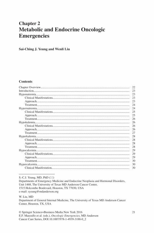
21© Springer Science+Business Media New York 2016 E.F. Manzullo et al. (eds.), Oncologic Emergencies, MD Anderson Cancer Care Series, DOI 10.1007/978-1-4939-3188-0_2
Chapter 2Metabolic and Endocrine Oncologic Emergencies
Sai-Ching J. Yeung and Wenli Liu
S.-C.J. Yeung, MD, PhD (*) Departments of Emergency Medicine and Endocrine Neoplasia and Hormonal Disorders, Unit 1468, The University of Texas MD Anderson Cancer Center, 1515 Holcombe Boulevard, Houston, TX 77030, USAe-mail: [email protected]
W. Liu, MD Department of General Internal Medicine, The University of Texas MD Anderson Cancer Center, Houston, TX, USA
Contents
Chapter Overview 22 Introduction 23 Hyponatremia 23
Clinical Manifestations 23 Approach 23 Treatment 24
Hypernatremia 24 Clinical Manifestations 25 Approach 25 Treatment 26
Hypokalemia 26 Clinical Manifestations 26 Approach 26 Treatment 27
Hyperkalemia 28 Clinical Manifestations 28 Approach 28 Treatment 28
Hypocalcemia 29 Clinical Manifestations 29 Approach 29 Treatment 30
Hypercalcemia 30 Clinical Manifestation 30
22
Approach 31 Treatment 31
Hypomagnesemia 32 Clinical Manifestations 32 Approach 32 Treatment 33
Hypermagnesemia 33 Clinical Manifestations 33 Approach 33 Treatment 34
Hypophosphatemia 34 Clinical Manifestations 34 Approach 35 Treatment 36
Hyperphosphatemia 36 Clinical Manifestations 37 Approach 37 Treatment 37
Hyperglycemia 38 Clinical Manifestations 38 Approach 38 Treatment 39
Hypoglycemia 41 Clinical Manifestations 41 Approach 41 Treatment 42
Adrenal Crisis 43 Clinical Manifestations 43 Approach 43 Treatment 45
Hypothyroidism 45 Clinical Manifestations 45 Approach 46 Treatment 47
Thyrotoxicosis 47 Clinical Manifestations 47 Approach 48 Treatment 49
Carcinoid Crisis 50 Clinical Manifestations 51 Approach 51 Treatment 51
Key Practice Points 52 Suggested Readings 54
Chapter Overview
Homeostatic regulation of key metabolites in cancer patients is often dysfunctional or perturbed by the malignancy or its treatment. Cancer and its treatment can also perturb the endocrine systems that regulate organ functions and metabolism. Deficiencies and excesses in electrolytes, metabolites, and hormones are discussed
S.-C.J. Yeung and W. Liu
23
from the practical standpoint of acute clinical management in this chapter. Common etiologies and treatment approaches are also presented.
Introduction
Cancer and its treatment can lead to endocrine and metabolic dysfunction. Oncologists and emergency physicians should be vigilant in checking for these endocrine and metabolic sequelae so that prompt, appropriate treatment can be given to improve the patient’s quality of life and avoid serious morbidity or mortality. Syndrome of inappropriate antidiuretic hormone secretion and tumor lysis syndrome are covered elsewhere in the Nephro-Urologic Emergencies in Patients with Cancer chapter and thus are not discussed in this chapter.
Hyponatremia
The human body contains about 60 % water, and the sodium/water balance (i.e., intake and loss of sodium relative to intake and loss of water) is regulated by the renin-angiotensin system, atrial natriuretic peptides, and the osmoregulation centers in the brain and antidiuretic hormone. Hyponatremia (sodium level less than 135 mEq/dL) is a common abnormality in cancer patients that may indicate serious underlying disease. It is associated with adverse prognosis for cancer.
Clinical Manifestations
Hyponatremia has a nonspecific clinical presentation that ranges from no symptoms to multiple neurologic symptoms of headache, behavioral changes, lethargy, confu-sion, seizure, stupor, and even coma. The severity of symptoms depends on the rate of decline and degree of hypo-osmolality. Severe hyponatremia can cause seizures, permanent brain damage, brain stem herniation, respiratory failure, and death.
Approach
Hyponatremia is often recognized via laboratory measurement of plasma electro-lytes. Hypotonicity must be confirmed by measuring osmolality. Hyponatremia with normal osmolality (pseudohyponatremia) can be a laboratory artifact caused by hyperlipidemia (corrected sodium level = sodium level + 0.2 × triglyceride level [mg/L]) or hyperproteinemia (corrected sodium level = sodium level + 0.025 × protein
2 Metabolic and Endocrine Oncologic Emergencies
24
[if protein level is greater than 8 g/dL]). Extreme hyperglycemia (corrected sodium level = sodium level + [glucose level − 5]/3.5) and administration of hypertonic man-nitol result in hypertonic hyponatremia by shifting intracellular water to the extracel-lular fluid, diluting the plasma sodium concentration. Identifying the causes of hyponatremia requires additional laboratory evaluations, including urinary sodium measurement, thyroid and adrenal function tests, and correlation with clinical history. Hypovolemic hyponatremia owing to gastrointestinal and renal salt loss is common in cancer patients. Hypervolemic hyponatremia also is often seen in patients with severe liver cirrhosis, fluid third-spacing, or congestive heart failure.
Treatment
Treatment of hyponatremia involves rebalancing the total body water and sodium levels using the following means (usually in combination):
• Decreased free water intake
– Fluid restriction to 500–800 mL of free water per day if not hypovolemic.
• Increased free water excretion
– Treatment with demeclocycline at the usual dose range of 600–1200 mg a day produces a reversible form of nephrogenic diabetes insipidus (DI), inhibiting antidiuretic hormone-induced cyclic adenosine monophosphate formation.
– Treatment with loop diuretics such as furosemide may be added in nonhypo-volemic patients to enhance free water clearance.
– Vaptans can be used to block V2 receptors and promote free water excretion (aquaresis). Their action peaks within a few hours and generally subsides after 12–24 h, and they are efficacious for hypervolemic hyponatremia.
• Increased sodium intake
– Oral salt intake: sodium chloride tablets. – Parenteral salt intake: normal saline (0.9 % NaCl) or hypertonic saline (3 %
NaCl) at a rate of 1 mL/kg/h.
• Decreased sodium loss
– Fludrocortisone: 0.1–0.6 mg a day orally.
• Treatment of the underlying etiology of hyponatremia
Hypernatremia
Hypernatremia (sodium level greater than 145 mEq/L) is always accompanied by a hyperosmolar state and cellular dehydration. Hypernatremia results from excess sodium intake, excess renal reabsorption of sodium, reduced water intake, or
S.-C.J. Yeung and W. Liu
25
increased water loss. Hypernatremia is seen in about 1 % of hospitalized patients, and young, old, and chronically ill patients are vulnerable to it.
Clinical Manifestations
The clinical manifestations of hypernatremia are primarily related to cellular dehy-dration leading to central nervous system dysfunction and are more pronounced with a high level or acute increase in the level of sodium. Thirst is the first symptom unless the patient has hypodipsia owing to hypothalamic dysfunction. Other symp-toms include restlessness, weakness, and lethargy that may progress to coma. Muscle weakness and central nervous system changes are usually not manifested until the sodium level is greater than 160 mEq/L. DI is characterized by polyuria, urine hypo-osmolality, and compensatory polydipsia. If water loss exceeds water intake, intravascular volume depletion and hypernatremia will ensue.
Approach
Central DI is most frequently caused by events that affect the anterior pituitary or related hypothalamic nuclei (e.g., surgery, destruction by tumors, hemorrhage, head injury, infarction, infection).
Most cases of familial/congenital nephrogenic DI are caused by V2 receptor mutations and aquaporin-2 water channel mutations. However, these causes are rare in cancer patients. Acquired nephrogenic DI can result from the nephrotoxicity of drugs. Common nephrogenic DI-inducing drugs are lithium, foscarnet, and clozap-ine. Although distal tubular defects develop in about half of patients given ifos-famide, nephrogenic DI leading to hypernatremia is uncommon in them.
In cancer patients, inadequate water intake can have many causes, including obstruction of the gastrointestinal tract, chemotherapy-induced nausea and vomit-ing, and chemotherapy- or radiotherapy-induced mucositis. Primary hypodipsia can result from dysfunction of the thirst center in the supraoptic nucleus of the hypo-thalamus owing to a primary or metastatic malignancy (e.g., breast cancer, lung cancer) or treatment of a central nervous system tumor using surgical resection and/or radiation. Reasons for increased water loss include diuretic use, high fever, burn, or diarrhea. Iatrogenic causes of hypernatremia include inappropriate intravenous fluid administration, total parenteral nutrition, and hemodialysis. Drugs that decrease the effect of antidiuretic hormone include demeclocycline, lithium, amphotericin, vinblastine, glyburide, propoxyphene, colchicine, acetohexamide, tolazamide, and methoxyflurane.
A water deprivation test may differentiate between central and nephrogenic DI. A serum uric acid level greater than 5 mg/dL in a polyuric polydipsic patient is highly suggestive of central DI.
2 Metabolic and Endocrine Oncologic Emergencies
26
Treatment
Administration of free water
• Give water enterally or intravenously with solutions low in electrolytes (i.e., dextrose 5 % in water, 0.2 % NaCl). Total body water deficit can be estimated by 0.6 weight (kg) × ([serum sodium level/140] − 1).
• In patients with acute hypernatremia, free water can be replaced rapidly.• In patients with chronic hypernatremia, the serum sodium level should be
decreased by less than 2 mEq/L/h until the symptoms resolve. The remaining water deficit can be corrected in 48 h.
• Patients with hypodipsia should receive a prescribed amount of water per day on a regular basis.
• Central DI usually is treated with desmopressin (DDAVP) at a typical dose of 5–20 μg intranasally every 12 h, 1–2 μg subcutaneously once a day, and 0.1–0.2 mg orally twice a day.
• A low-salt diet along with use of thiazide diuretics that induce natriuresis is the treatment of choice for nephrogenic DI. Indomethacin has been used to treat drug-induced nephrogenic DI.
• Discontinue treatment with any drugs that may contribute to nephrogenic DI (e.g., lithium) if clinically appropriate.
Hypokalemia
Hypokalemia (potassium level less than 3.5 mEq/L) is perhaps the most common electrolyte abnormality in cancer patients.
Clinical Manifestations
Patients with mild hypokalemia (3.0–3.5 mEq/L) usually are asymptomatic. In those with severe hypokalemia (less than 3 mEq/L), symptoms may range from mild to severe and are potentially fatal. Cardiac manifestations may range from flat T waves, T-wave depression, and prominent U waves to serious arrhythmias. Neurologic manifestations include muscle weakness, paresthesia, and paralysis.
Approach
Potassium intake in cancer patients may decrease for various reasons, such as nausea, vomiting, anorexia, and gastrointestinal obstruction. Potassium may be lost from the gastrointestinal tract via vomiting or diarrhea, from the skin during profuse sweating
S.-C.J. Yeung and W. Liu
27
or owing to severe burns, and from the kidneys as a result of intrinsic tubular defects, type 1 renal tubular acidosis, or drug-related effects. Common examples of potas-sium-wasting drugs are loop diuretics, aminoglycosides, cyclophosphamide, ifos-famide, carboplatin, cisplatin, and amphotericin B. Hypokalemia owing to excess mineralocorticoid activity may result from pharmacologic administration of cortico-steroids or ectopic Cushing syndrome, which is associated with some cancers. Alkalosis, either respiratory or, on a larger scale, metabolic, may precipitate hypoka-lemia via a transcellular potassium shift. Drugs that cause potassium redistribution include insulin, vitamin B12, β-adrenergic agonists, theophylline, and chloroquine.
Hypokalemia is diagnosed via potassium measurement. Medications used and dietary history are helpful in determining the cause of hypokalemia. Physical examination will give clues regarding Cushing syndrome. Measurement of serum electrolytes, including magnesium, blood urea nitrogen, and creatinine; urinalysis; and urine electrolyte measurement will help diagnose renal potassium loss.
Treatment
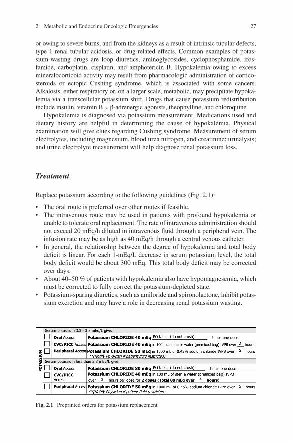
Replace potassium according to the following guidelines (Fig. 2.1):
• The oral route is preferred over other routes if feasible. • The intravenous route may be used in patients with profound hypokalemia or
unable to tolerate oral replacement. The rate of intravenous administration should not exceed 20 mEq/h diluted in intravenous fluid through a peripheral vein. The infusion rate may be as high as 40 mEq/h through a central venous catheter.
• In general, the relationship between the degree of hypokalemia and total body deficit is linear. For each 1-mEq/L decrease in serum potassium level, the total body deficit would be about 300 mEq. This total body deficit may be corrected over days.
• About 40–50 % of patients with hypokalemia also have hypomagnesemia, which must be corrected to fully correct the potassium-depleted state.
• Potassium-sparing diuretics, such as amiloride and spironolactone, inhibit potas-sium excretion and may have a role in decreasing renal potassium wasting.
Fig. 2.1 Preprinted orders for potassium replacement
2 Metabolic and Endocrine Oncologic Emergencies
28
Hyperkalemia
Hyperkalemia is also a common electrolyte disorder in cancer patients.
Clinical Manifestations
Severe clinical manifestations of hyperkalemia usually are absent until the serum potassium level is greater than 7.5 mEq/L. Some patients (e.g., those with chronic renal failure) can tolerate high serum potassium levels without having any clinical signs or symptoms. At greater than 7.5 mEq/L, nonspecific symptoms such as mus-cle weakness, cramping, and paralysis of different muscle groups may occur.
Hyperkalemia causes depolarization of excitable membranes. This membrane depolarization leads to the excitability of nerves and muscles, causing cramps, muscle weakness, and paralysis. The most vital organ with excitable membranes is the heart. Specific electrocardiogram (EKG) changes and potentially fatal arrhythmias may be present, but the serum potassium level is not correlated directly with a par-ticular EKG pattern. An early EKG abnormality associated with hyperkalemia is peak T waves followed by a progressive QRS widening to a “sinusoidal” wave. Ventricular tachycardia, fibrillation, and asystole may occur.
Approach
Inappropriate potassium content in intravenous fluid and total parenteral nutrition are common iatrogenic causes of hyperkalemia. A significant release of intracellular potassium will cause hyperkalemia, as in the case of tumor lysis syndrome. Insulin deficiency, β-blocker therapy, and acidemia can elevate serum potassium levels.
Drug-induced hyperkalemia most often occurs in patients with impaired renal excre-tion of potassium. The drugs commonly used by cancer patients that may cause hyper-kalemia include cyclosporin A, tacrolimus, heparin, mitomycin C, and pentamidine.
Diminished renal excretion of potassium occurs in patients with acute or chronic renal failure, renal hypoperfusion, or type 4 renal tubular acidosis. Drugs that can lead to decreased potassium excretion include potassium-sparing diuretics and angiotensin-converting enzyme inhibitors.
Treatment
Treatment of hyperkalemia depends on its severity and rate of development.
• If possible, discontinue medications that may contribute to hyperkalemia, such as β-adrenergic blockers, nonsteroidal anti-inflammatory drugs,
S.-C.J. Yeung and W. Liu
29
angiotensin- converting enzyme inhibitors, potassium supplements, and others described above.
• Monitor EKG continuously if the potassium level is greater than 6 mEq/L.• For EKG changes, infuse intravenously (usually for less than 60 min):
– Calcium (1–2 g of calcium gluconate or 0.5–1.0 g of chloride) – Sodium bicarbonate – Glucose (usually 25 g) plus 6–8 U of regular insulin – β-adrenergic agonists, which promote potassium entry into cells
• Increasing the renal excretion of potassium can be attempted with the use of loop diuretics.
• Removal of potassium from the body should be attempted with the use of ion exchange resins, such as sodium polystyrene sulfonate (Kayexalate), which can be administered orally (15–30 g/dose) or rectally (30–60 g/dose) as a retention enema.
• Emergent hemodialysis may be used in refractory cases.
Hypocalcemia
In hospitalized cancer patients, the hypocalcemia rate is about 13.4 %. Hypocalcemia may affect the proper functioning of many intracellular and extracellular processes, such as muscle contraction, nerve conduction, and blood coagulation.
Clinical Manifestations
Hypocalcemia can be asymptomatic if it is mild. Life-threatening problems such as seizures, cardiac dysrhythmias, and laryngospasm can occur if hypocalcemia is severe. Acute hypocalcemia is characterized by neuromuscular irritability. Acute symptoms are muscle weakness, paresthesia, spasm, tetany, hyperreflexia, Chvostek sign, Trousseau sign, seizure, bronchospasm, laryngeal spasm, and respiratory failure. Cardiovascular presentations are bradycardia, hypotension, QT-interval prolongation, congestive heart failure, and cardiac arrest. Chronic hypocalcemia with hypoparathy-roidism causes extrapyramidal disorders, cataracts, and skin and hair changes. Vitamin D deficiency causes rickets and osteomalacia in patients with hypocalcemia.
Approach
In most cancer patients, the etiology of hypocalcemia is obvious. Excluding a decreased serum calcium level owing to low albumin and serum protein levels, the major causes of hypocalcemia are hypoparathyroidism and hypomagnesemia.
2 Metabolic and Endocrine Oncologic Emergencies
30
Hypocalcemia may be a feature of tumor lysis syndrome. Severe osteoblastic bone metastases (especially from prostate carcinoma) are often associated with hypocal-cemia. The toxicity of certain chemotherapeutic agents (e.g., platinum compounds) also may lead to hypocalcemia.
Evaluation of hypocalcemia involves confirmation of it by measuring the ionized calcium level. If the cause of hypocalcemia is not clear, laboratory analysis of intact parathyroid hormone (PTH), magnesium, phosphate, 25-hydroxy vitamin D3, 1,25-dihydroxy vitamin D3, creatinine, and 24-hour urinary calcium levels is helpful.
Treatment
Treatment of hypocalcemia depends on its severity and cause.
• Severe hypocalcemia is treated parenterally with intravenous calcium chloride (0.5–1.0 g) or gluconate (1–2 g) over 5–10 min. Calcium gluconate is preferred over other agents because it is less likely to cause tissue necrosis if extravasated.
• Hypomagnesemia is a common cause of hypocalcemia. Concurrent hypomagne-semia should be treated with intravenous magnesium sulfate followed by oral replacement.
• Chronic hypocalcemia is treated with oral calcium preparations (e.g., gluconate, carbonate) containing 1–2 g of elemental calcium per day. Patients with hypoparathyroidism often must receive lifelong supplementation of calcium and vitamin D. Vitamin D supplements can be given in 1-hydroxylated form or as calcitriol. Calcitriol is preferred for patients with renal insufficiency or failure because of decreased 1-α-hydroxylase levels in the kidneys.
• Attention should be paid to phosphate binding.
Hypercalcemia
Hypercalcemia of malignancy is observed in 10–15 % of cancer patients. It is a poor prognostic sign that is associated with short survival durations.
Clinical Manifestation
Patients with mild hypercalcemia (calcium level less than 12 mg/dL) usually have no symptoms, whereas those with moderate or severe hypercalcemia are frequently symptomatic. Central nervous system symptoms are lethargy, ataxia, stupor, coma, mental status changes, and psychosis. Gastrointestinal tract symptoms are anorexia, nausea, constipation, ileus, dyspepsia, and pancreatitis. Renal signs are polyuria,
S.-C.J. Yeung and W. Liu
31
nephrolithiasis, and nephrocalcinosis. Cardiovascular manifestations can be a short QT interval, ST segment depression, sinus arrest, and atrioventricular block. Musculoskeletal symptoms are myalgia, arthralgia, and weakness.
Approach
Hypercalcemia may result from increased bone resorption, renal tubular reabsorp-tion, and gastrointestinal absorption of calcium. In cancer patients, hypercalcemia of malignancy accounts for more than 90 % of hypercalcemia cases. Hypercalcemia in cancer patients may have different pathophysiologic mechanisms. The most common humoral factor secreted by tumors causing hypercalcemia is PTH-related peptide. In general, patients with PTH-related peptide-induced hypercalcemia have advanced malignant disease and poor prognoses. Other humoral factors, such as interleukin-1 and -6, prostaglandins, and tumor necrosis factor, can mediate hyper-calcemia in cancer patients. Extensive lytic bone metastasis, particularly in patients with breast cancer or multiple myeloma, may lead to hypercalcemia. Increased lev-els of 1,25-dihydroxy vitamin D3 may mediate hypercalcemia in patients with Hodgkin disease or non-Hodgkin lymphoma.
Serum calcium levels should be interpreted in the context of protein binding (corrected calcium level = [0.8 × (normal albumin level − patient’s albumin level)] + serum calcium level). However, accurate measurement of the ionized calcium level confirms hypercalcemia. Laboratory studies of the following help diagnose the etiology of hypercalcemia: intact PTH, PTH-related protein, 25-hydroxy vitamin D3, and 1,25-dihydroxy vitamin D3.
Treatment
Treatment of hypercalcemia should be aimed at lowering serum calcium levels and correcting its underlying causes, if possible.
• Primary hyperparathyroidism can be cured via parathyroidectomy.• Use of medications (e.g., calcium-containing medications, thiazide diuretics)
that contribute to hypercalcemia should be discontinued.• The initial and first-line treatment of hypercalcemia is hydration with crystalloid
intravenous fluid. In patients with overall fluid overload, use of a loop diuretic would be helpful.
• Bisphosphonates (etidronate, clodronate, pamidronate, zoledronate, and iban-dronate) inhibit bone resorption by osteoclasts. Zoledronate (4–6 mg intrave-nously over 30 min) is more widely used than pamidronate (60–90 mg intravenously over 4–24 h).
2 Metabolic and Endocrine Oncologic Emergencies
32
• Second-line agents include calcitonin (salmon calcitonin 4 IU/kg subcutaneously every 12 h). Calcitonin has a rapid onset of action, although its effectiveness may decrease within 2–3 days.
• Other less widely used agents include glucocorticoids, plicamycin (25 μg/kg intravenously), and gallium nitrate (200 mg/m2 intravenously).
Hypomagnesemia
The incidence rate of the common electrolyte deficiency hypomagnesemia in hospi-talized cancer patients has been as high as 17.1 %. Hypomagnesemia is defined as a plasma serum concentration of magnesium less than 1.5 mg/dL. However, magne-sium levels that are persistently less than 1.8 mg/dL would indicate depletion of total body magnesium.
Clinical Manifestations
Magnesium is a major cation in the body, and only 1–2 % of total body magnesium is present in the extracellular space. It is needed for a wide variety of enzymatic reactions, including those involving ATP and nucleic acid metabolism. Magnesium is also directly involved in the regulation of calcium and potassium metabolism. The clinical manifestations of hypomagnesemia may be nonspecific and include anorexia, nausea, vomiting, lethargy, dizziness, muscle weakness, tremor, muscle fasciculation, tetany, and tonic-clonic seizures.
Approach
In cancer patients, hypomagnesemia is a very common abnormality that is related to low intake and impairment of renal reabsorption or intestinal absorption of mag-nesium. It is also related to prolonged intravenous feeding, nasogastric suction, chronic alcoholism, intestinal malabsorption, and diarrhea. The renal toxicity of chemotherapy (e.g., platinum-based drugs, cyclophosphamide, ifosfamide) and anti- infective medications (e.g., amphotericin, aminoglycosides) also influences hypomagnesemia.
Hypomagnesemia is often associated with other electrolyte abnormalities, such as hypokalemia and hypocalcemia. Concurrent measurement of other electrolytes, such as calcium, phosphate, and potassium, should be considered.
S.-C.J. Yeung and W. Liu
33
Treatment
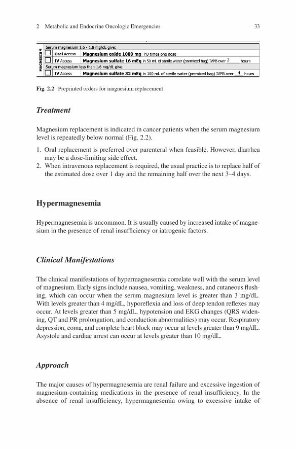
Magnesium replacement is indicated in cancer patients when the serum magnesium level is repeatedly below normal (Fig. 2.2).
1. Oral replacement is preferred over parenteral when feasible. However, diarrhea may be a dose-limiting side effect.
2. When intravenous replacement is required, the usual practice is to replace half of the estimated dose over 1 day and the remaining half over the next 3–4 days.
Hypermagnesemia
Hypermagnesemia is uncommon. It is usually caused by increased intake of magne-sium in the presence of renal insufficiency or iatrogenic factors.
Clinical Manifestations
The clinical manifestations of hypermagnesemia correlate well with the serum level of magnesium. Early signs include nausea, vomiting, weakness, and cutaneous flush-ing, which can occur when the serum magnesium level is greater than 3 mg/dL. With levels greater than 4 mg/dL, hyporeflexia and loss of deep tendon reflexes may occur. At levels greater than 5 mg/dL, hypotension and EKG changes (QRS widen-ing, QT and PR prolongation, and conduction abnormalities) may occur. Respiratory depression, coma, and complete heart block may occur at levels greater than 9 mg/dL. Asystole and cardiac arrest can occur at levels greater than 10 mg/dL.
Approach
The major causes of hypermagnesemia are renal failure and excessive ingestion of magnesium-containing medications in the presence of renal insufficiency. In the absence of renal insufficiency, hypermagnesemia owing to excessive intake of
Fig. 2.2 Preprinted orders for magnesium replacement
2 Metabolic and Endocrine Oncologic Emergencies
34
magnesium is very rare, as excess magnesium in the gastrointestinal tract leads to diarrhea. Overreplacement of magnesium in intravenous fluid or with hyperalimen-tation also can cause hypermagnesemia. A less common cause in cancer patients is tumor lysis syndrome.
Excessive magnesium intake usually is evident in the patient’s dietary and medi-cation history. Hypermagnesemia is diagnosed via direct measurement of serum magnesium levels. Renal function should be assessed by measuring blood urea nitrogen and creatinine levels.
Treatment
• Discontinuation of magnesium intake is the first step.• Patients with mild symptoms and normal renal function can simply be observed
to ensure that the magnesium level returns to normal.• Magnesium excretion can be accelerated by hydration with crystalloid fluid and
a loop diuretic given intravenously.• In cases of severe hypermagnesemia (particularly with hypotension and/or cardiac
arrhythmia), calcium should be administered intravenously to reverse respiratory depression, hypotension, and cardiac arrhythmia.
• Emergent dialysis should be considered for patients with life-threatening hypermagnesemia.
Hypophosphatemia
Hypophosphatemia is quite prevalent, as it is found in about 2–3 % of all hospital-ized patients and about 30 % of cancer patients.
Clinical Manifestations
Acute severe hypophosphatemia may lead to generalized neurologic findings such as lethargy, confusion, disorientation, and hallucinations and focal neurologic find-ings such as dysarthria, dysphagia, oculomotor palsy, anisocoria, nystagmus, ataxia, cerebellar tremor, ballismus, hyporeflexia, distal sensory deficits, paresthesia, and hyperesthesia. Severe neurologic symptoms, such as muscle paralysis, seizure, and coma, are observed only when the serum phosphate level is less than 0.8 mg/dL. Cardiac muscle also can be affected by severe hypophosphatemia, and reversible left ventricular dysfunction can occur.
S.-C.J. Yeung and W. Liu
35
Muscle weakness is the most common complaint. Bone pain is another promi-nent complaint of phosphate-depleted patients. Prolonged hypophosphatemia leads to rickets. Hypophosphatemic rickets can result from ifosfamide nephrotoxicity. Osteomalacia, waddling gait, bone tenderness, pseudofractures, and fractures can occur in patients with chronic hypophosphatemia.
Approach
Acute hypophosphatemia occurs primarily in hospitalized patients with serious illnesses and pre-existing phosphate depletion. Acute severe hypophosphatemia usually results from translocation of phosphate into cells. Respiratory alkalosis, intravenous glucose administration (including hyperalimentation), gram-negative sepsis, and insulin therapy can induce transcellular shift of phosphate.
Chronic hypophosphatemia results from an elevated PTH or PTH-related protein level, consumption of oral phosphate binders, accelerated bone formation, increased humoral factors suppressing renal reabsorption of phosphate, or intrinsic renal tubu-lar defect in phosphate reabsorption.
Tumor-induced (oncogenic) osteomalacia is a rare syndrome characterized by hypophosphatemia, excessive urinary phosphate loss, reduced 1,25-dihydroxy vitamin D concentrations, and osteomalacia. Tumor secretion of fibroblast growth factor 23 may be responsible for renal phosphate wasting.
Rapid cancer or normal cell proliferation in ill patients with nutritional depriva-tion or catabolism may cause hypophosphatemia. Chronic hypophosphatemia together with hypocalcemia occasionally is associated with extensive osteoblastic metastasis of prostate, breast, lung, and other malignancies. Patients with rapidly progressing leukemia or lymphoma (e.g., Burkitt lymphoma) may have hypophos-phatemia. As with the use of granulocyte colony-stimulating factors, hematopoietic reconstitution after stem cell transplantation or stem cell harvesting in preparation for transplantation also cause hypophosphatemia.
The liver plays a significant role in phosphate homeostasis. In a retrospective study, postoperative serum phosphate levels dropped in all 44 patients who under-went right or extended right hepatic lobectomy. Authors have reported hypophos-phatemia in a patient with hepatocellular carcinoma complicating liver cirrhosis. Hypophosphatemia in malnourished patients (especially alcoholics) results from a combination of magnesium deficiency, vitamin D deficiency, and malabsorption. Refeeding of high-calorie diets in severely malnourished patients can lead to refeed-ing syndrome with hypophosphatemia.
Intrinsic renal tubular defects in phosphate reabsorption may occur in patients with Fanconi syndrome, myeloma, or amyloidosis. Hypophosphatemia also may be associated with the use of chemotherapeutic drugs such as platinum compounds and alkylating agents (e.g., ifosfamide).
2 Metabolic and Endocrine Oncologic Emergencies
36
Hypophosphatemia is demonstrated via measurement of the serum phosphate level. Measurement of renal function and potassium, magnesium, calcium, vitamin D metabolite, and PTH levels is helpful in determining the cause of hypophosphate-mia. If urinary loss of phosphate is suspected, urine should be collected to mea-sure the renal phosphate threshold/glomerular filtration rate to confirm phosphaturia.
Treatment
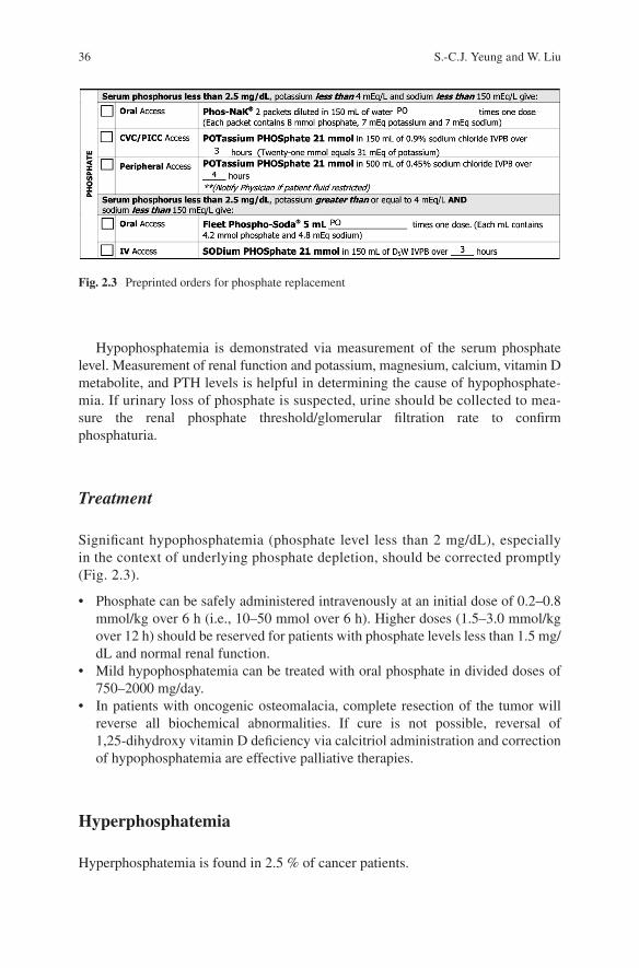
Significant hypophosphatemia (phosphate level less than 2 mg/dL), especially in the context of underlying phosphate depletion, should be corrected promptly (Fig. 2.3).
• Phosphate can be safely administered intravenously at an initial dose of 0.2–0.8 mmol/kg over 6 h (i.e., 10–50 mmol over 6 h). Higher doses (1.5–3.0 mmol/kg over 12 h) should be reserved for patients with phosphate levels less than 1.5 mg/dL and normal renal function.
• Mild hypophosphatemia can be treated with oral phosphate in divided doses of 750–2000 mg/day.
• In patients with oncogenic osteomalacia, complete resection of the tumor will reverse all biochemical abnormalities. If cure is not possible, reversal of 1,25-dihydroxy vitamin D deficiency via calcitriol administration and correction of hypophosphatemia are effective palliative therapies.
Hyperphosphatemia
Hyperphosphatemia is found in 2.5 % of cancer patients.
Fig. 2.3 Preprinted orders for phosphate replacement
S.-C.J. Yeung and W. Liu
37
Clinical Manifestations
The clinical manifestations of acute hyperphosphatemia are similar to those of associated hypocalcemia. Paresthesia, muscle cramps, tetany, and QT-interval pro-longation may be induced directly by severe hyperphosphatemia. Chronic hyper-phosphatemia, especially associated with hypercalcemia, may lead to diffuse visceral deposition of calcium phosphate. Deposition of calcium phosphate in the kidneys may lead to renal failure.
Approach
In the absence of renal failure, the fasting serum phosphate level is determined primarily according to the renal tubular reabsorption rate. A massive amount of phosphate can be released into the extracellular fluid via extensive cellular break-down. Extensive rhabdomyolysis and hemolysis may cause hyperphosphatemia in the same way.
Translocation of phosphate from cells in response to metabolic or respiratory alkalosis can lead to acute hyperphosphatemia. Chronic hyperphosphatemia is present in patients with hypoparathyroidism. Excess phosphate intake (including use of phosphate-containing laxatives) is another potential cause of hyperphosphatemia.
In patients with hyperglobulinemia, pseudohyperphosphatemia must be excluded with a specimen that is free of protein (removed via precipitation with sulfosalicylic acid). In those with hyperphosphatemia, renal function must be assessed. In addi-tion, measurement of lactic dehydrogenase, uric acid, potassium, and calcium levels is necessary in the detection and management of hyperphosphatemia owing to cel-lular breakdown.
Treatment
The emergency treatment of hyperphosphatemia involves supportive care and treatment of symptomatic hypocalcemia.
• In patients with normal renal function, infusion of isotonic saline increases phos-phate excretion.
• Administration of dextrose and insulin drives phosphate into cells, temporarily lowering the serum phosphate level.
• When hyperphosphatemia is life-threatening, hemodialysis or peritoneal analysis should be considered.
• Dietary restriction of phosphorus, although an important factor in the control of the serum phosphorus level in the chronic setting, poses practical problems that limit its success in most patients.
2 Metabolic and Endocrine Oncologic Emergencies
38
• Aluminum-containing antacids are used to inhibit phosphorus absorption in the gastrointestinal tract, but accumulation of aluminum has serious long-term toxic effects in patients with impaired renal function. Calcium-based phosphate bind-ers have largely replaced aluminum compounds. However, excessive amounts of absorbed calcium present a different problem. Use of nonabsorbable phosphate binders that are aluminum- and calcium-free (800–1600 mg of sevelamer with each meal) can prevent these issues.
Hyperglycemia
Diabetes mellitus is a common disease, and a large number of cancer patients have co-existing diabetes. Glucocorticoids are used frequently in cancer patients for vari-ous conditions, and steroid-induced diabetes mellitus is common. Because diabetes mellitus is an extensive subject, this section focuses on the acute complications of it in cancer patients.
Clinical Manifestations
Most patients with significant hyperglycemia have symptoms of polydipsia, poly-uria, and polyphagia. Dehydration of the lenses owing to hyperglycemia leads to blurry vision. Patients with hyperosmolar nonketotic coma experience mental status changes, hypotension, and severe dehydration. Nausea, vomiting, and abdominal pain are present in almost half of patients with diabetic ketoacidosis. Tachypnea with Kussmaul respiration, tachycardia, hypotension, orthostatic blood pressure changes, acetone breaths, and severe signs of dehydration can be present in patients with diabetic ketoacidosis.
Approach
The serum glucose level is regulated by absorption, cellular uptake, gluconeogene-sis, and glycogenolysis, which are regulated by the pancreas, intestines, liver, kid-neys, and muscle. Hyperglycemia can result from perturbation of the hormones involved in glucose regulation, such as insulin and glucagon, and from dysfunction of the organs involved in glucose homeostasis.
Diabetic ketoacidosis is decompensated catabolism triggered by a relative or absolute deficiency in insulin secretion. A deficiency in insulin relative to the gluca-gon level inhibits glycolysis and increases glycogenolysis and gluconeogenesis in the liver. Malonyl coenzyme A levels decrease because of inhibited acetyl coenzyme A carboxylase and glycolysis. As a result, fatty acid oxidation and ketone body for-mation increase. The pathophysiology of hyperosmolar hyperglycemic nonketotic
S.-C.J. Yeung and W. Liu
39
coma is similar to that of diabetic ketoacidosis except that ketone bodies are not formed and extremely high glucose levels result from diminished urine output.
Glucocorticoid administration (in combination therapy regimens and for edema in patients with brain metastasis, prevention of transplant rejection, graft-versus- host disease, and nausea with vomiting) is the most common cause of diabetes mel-litus. Treatment with interleukin-2, interferon-α, interferon-γ, streptozocin, homoharringtonine, or l-asparaginase may result in diabetes. Patients who receive allogeneic stem cell transplants are likely to receive both glucocorticoids and tacro-limus and are particularly at risk for hyperglycemia.
Drugs such as ifosfamide and mercaptopurine can damage the renal tubules and cause glycosuria and Fanconi syndrome. A false-positive reaction with the testing agent for urinary ketones can be caused by treatment with mesna (2-mercaptoethane sulfonate sodium).
A random glucose level greater than 200 mg/dL or a fasting plasma glucose level greater than 126 mg/dL on more than one occasion can indicate diabetes mellitus. A glucose tolerance test (2-hour oral glucose tolerance test: glucose level of at least 200 mg/dL) usually is not necessary except in borderline cases. Glycosylated hemo-globin (hemoglobin A1C) reflects the level of glucose in the preceding 1.5 months.
Diabetic ketoacidosis is diagnosed according to the triad of metabolic acidosis, hyperglycemia, and the presence of ketone bodies in the urine or blood. Arterial blood gas testing will demonstrate acidemia and respiratory compensation for meta-bolic acidosis by hyperventilation. Also, the anion gap will be elevated, and serum ketone testing will be positive. A urine dipstick test for ketones can provide timely information for a quick bedside diagnosis. Absence of ketones from the urine prac-tically excludes diabetic ketoacidosis. Leukocytosis may be associated with ketosis, but an infection must be considered as a precipitating factor for diabetic ketoacido-sis. The serum creatinine level can be falsely elevated because of ketosis. Potassium, phosphate, and magnesium abnormalities result from transcellular shifts caused by acidosis.
In patients with hyperosmolar hyperglycemic nonketotic coma, the plasma glu-cose level may be well over 800 mg/dL, and the serum osmolality may be more than 100 mOsm higher than normal. Mild ketosis may be present because of starvation, but ketoacidosis will not be present. In severe cases, when volume depletion causes circulatory collapse, lactic acidosis will develop.
In immunocompromised cancer patients in particular, sepsis must be ruled out as the precipitating event for diabetic ketoacidosis or hyperosmolar hyperglycemic coma (Table 2.1).
Treatment
Management of the blood glucose level depends on the severity of the blood glucose abnormality and on the underlying pathophysiologic mechanism of the increase in the level. In general, oral agents are less likely to be effective than other types of agents in patients who are deficient in insulin.
2 Metabolic and Endocrine Oncologic Emergencies
40
Treatment of diabetic ketoacidosis or hyperosmolar hyperglycemic coma focuses on supplemental insulin, rehydration, correction of electrolyte abnormalities and severe acidosis, and identification of the precipitating factors (particularly important to rule out sepsis) (Table 2.2).
• Hydrate with intravenous crystalloid fluid.• Regular insulin usually is given as an intravenous bolus of 0.1 U/kg followed by
a maintenance intravenous infusion of 0.1 U/kg/h. The amount of insulin required for treatment of hyperosmolar hyperglycemic coma may be less than that required for diabetic ketoacidosis.
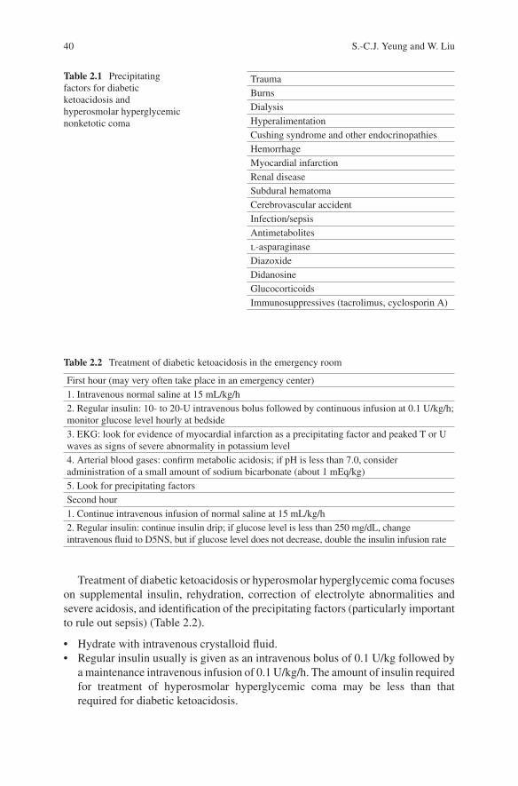
Table 2.1 Precipitating factors for diabetic ketoacidosis and hyperosmolar hyperglycemic nonketotic coma
TraumaBurnsDialysisHyperalimentationCushing syndrome and other endocrinopathiesHemorrhageMyocardial infarctionRenal diseaseSubdural hematomaCerebrovascular accidentInfection/sepsisAntimetabolitesl-asparaginaseDiazoxideDidanosineGlucocorticoidsImmunosuppressives (tacrolimus, cyclosporin A)
Table 2.2 Treatment of diabetic ketoacidosis in the emergency room
First hour (may very often take place in an emergency center)1. Intravenous normal saline at 15 mL/kg/h2. Regular insulin: 10- to 20-U intravenous bolus followed by continuous infusion at 0.1 U/kg/h; monitor glucose level hourly at bedside3. EKG: look for evidence of myocardial infarction as a precipitating factor and peaked T or U waves as signs of severe abnormality in potassium level4. Arterial blood gases: confirm metabolic acidosis; if pH is less than 7.0, consider administration of a small amount of sodium bicarbonate (about 1 mEq/kg)5. Look for precipitating factorsSecond hour1. Continue intravenous infusion of normal saline at 15 mL/kg/h2. Regular insulin: continue insulin drip; if glucose level is less than 250 mg/dL, change intravenous fluid to D5NS, but if glucose level does not decrease, double the insulin infusion rate
S.-C.J. Yeung and W. Liu
41
Hypoglycemia
Hypoglycemia is defined as a blood glucose level less than 50 mg/dL. The timing of symptoms relative to a fasting or postprandial state can distinguish among various etiologies.
Clinical Manifestations
A progressive pattern of responses to hypoglycemia is determined by the availability of glucose to the brain. At a plasma glucose level of about 70 mg/dL, brain glucose uptake can be reduced, and counterregulatory hormone responses are triggered. At 60 mg/dL, autonomic symptoms, such as hunger, anxiety, palpitations, sweating, and nausea, are prevalent. When the glucose level is less than 50 mg/dL, neurogly-copenic symptoms of blurry vision, slurred speech, confusion, and difficulty with mental concentration appear. When the glucose level is less than 40 mg/dL, the patient may become drowsy, confused, or combative. A further prolonged decrease below 30 mg/dL can cause seizures, permanent neurologic deficits, and death.
Approach
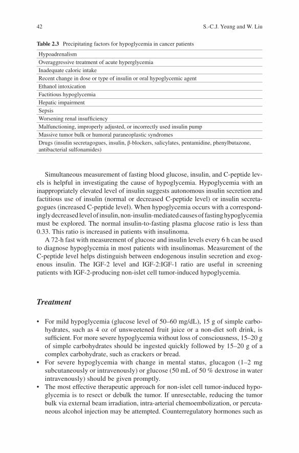
Glucagon and epinephrine are the two major counterregulatory hormones. Other hormones that respond to hypoglycemia are norepinephrine, cortisol, and growth hormone, but their effects are delayed. Glucagon and epinephrine immediately stimulate hepatic glycogenolysis followed by gluconeogenesis. Primary adrenal insufficiency and primary hypothyroidism and hypopituitarism are associated with hypoglycemia (Table 2.3). The kidneys contribute to overall gluconeogenesis dur-ing hypoglycemia stress in about one third of cases and are important to extrahe-patic degradation of insulin. Moreover, a number of oral hypoglycemic drugs are excreted by the kidneys. Therefore, decline in renal function often leads to hypogly-cemic episodes in diabetic patients.
In many cancer patients, hypoglycemia is associated with cancer-related malnu-trition and fat and muscle wasting, which impair gluconeogenesis. Non-islet cell tumors can secrete hormones such as insulin-like growth factor (IGF)-2, which, by binding to insulin receptors, causes hypoglycemia. Excessive glucose consumption by large tumors also may cause hypoglycemia.
For diabetic cancer patients receiving sulfonylurea or insulin, the most common cause of hypoglycemia may be delayed or decreased food intake. Cancer patients who receive irradiation of the head and neck area, have metastatic or primary tumors, or undergo treatment affecting the hypothalamic-pituitary area are at risk for hypopituitarism.
2 Metabolic and Endocrine Oncologic Emergencies
42
Simultaneous measurement of fasting blood glucose, insulin, and C-peptide lev-els is helpful in investigating the cause of hypoglycemia. Hypoglycemia with an inappropriately elevated level of insulin suggests autonomous insulin secretion and factitious use of insulin (normal or decreased C-peptide level) or insulin secreta-gogues (increased C-peptide level). When hypoglycemia occurs with a correspond-ingly decreased level of insulin, non-insulin-mediated causes of fasting hypoglycemia must be explored. The normal insulin-to-fasting plasma glucose ratio is less than 0.33. This ratio is increased in patients with insulinoma.
A 72-h fast with measurement of glucose and insulin levels every 6 h can be used to diagnose hypoglycemia in most patients with insulinomas. Measurement of the C-peptide level helps distinguish between endogenous insulin secretion and exog-enous insulin. The IGF-2 level and IGF-2:IGF-1 ratio are useful in screening patients with IGF-2-producing non-islet cell tumor-induced hypoglycemia.
Treatment
• For mild hypoglycemia (glucose level of 50–60 mg/dL), 15 g of simple carbo-hydrates, such as 4 oz of unsweetened fruit juice or a non-diet soft drink, is sufficient. For more severe hypoglycemia without loss of consciousness, 15–20 g of simple carbohydrates should be ingested quickly followed by 15–20 g of a complex carbohydrate, such as crackers or bread.
• For severe hypoglycemia with change in mental status, glucagon (1–2 mg subcutaneously or intravenously) or glucose (50 mL of 50 % dextrose in water intravenously) should be given promptly.
• The most effective therapeutic approach for non-islet cell tumor-induced hypo-glycemia is to resect or debulk the tumor. If unresectable, reducing the tumor bulk via external beam irradiation, intra-arterial chemoembolization, or percuta-neous alcohol injection may be attempted. Counterregulatory hormones such as
Table 2.3 Precipitating factors for hypoglycemia in cancer patients
HypoadrenalismOveraggressive treatment of acute hyperglycemiaInadequate caloric intakeRecent change in dose or type of insulin or oral hypoglycemic agentEthanol intoxicationFactitious hypoglycemiaHepatic impairmentSepsisWorsening renal insufficiencyMalfunctioning, improperly adjusted, or incorrectly used insulin pumpMassive tumor bulk or humoral paraneoplastic syndromesDrugs (insulin secretagogues, insulin, β-blockers, salicylates, pentamidine, phenylbutazone, antibacterial sulfonamides)
S.-C.J. Yeung and W. Liu

43
glucocorticoids and glucagon may be administered to raise the blood glucose level.
• Treatment of postprandial hypoglycemia is primarily dietary. The diet should have a low carbohydrate content. Use of α-glucosidase inhibitors (acarbose or miglitol) may be helpful.
Adrenal Crisis
The adrenal gland is a site of hematogenous metastasis exceeded in frequency by the lungs, the liver, and bone. Despite the high prevalence of adrenal metastasis, clinically evident primary adrenal insufficiency is seen infrequently. The hypothalamic- pituitary area may be damaged by a tumor or its treatment (irradia-tion or surgery), leading to secondary adrenal insufficiency. However, the most fre-quent cause of adrenal insufficiency in cancer patients is suppression of the hypothalamic-adrenocortical axis by chronic/repeated exposure to corticosteroids.
Clinical Manifestations
The symptoms of adrenal insufficiency include weakness, fatigue, nausea, vomiting, and weight loss. In patients with chronic primary adrenal failure, hyperpigmenta-tion may occur. Acute adrenal crisis involves hypoglycemia and hypotension.
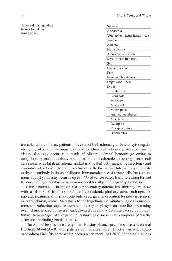
Cachexia, weakness, and electrolyte abnormalities can be easily explained by poor intake, malnutrition, chemotherapy side effects, or paraneoplastic syndromes. Adrenal insufficiency may develop gradually and have a variety of causes not often observed in cancer patients (Table 2.4). Inadequate production of glucocorticoids to meet the metabolic requirements of the body leads to potentially life-threatening adrenal crisis.
Approach
Cancer patients at increased risk for primary adrenal insufficiency are those with loss of adrenal function owing to use of medications that inhibit glucocorticoid synthesis or bilateral adrenal resection, metastasis, infection, or hemorrhage. Etomidate, a commonly used intravenous anesthetic, may inhibit cortisol synthesis, but short-term use of it as in rapid-sequence intubation does not cause any prob-lems. At high doses, imidazoles, ketoconazole, fluconazole, and itraconazole inhibit cytochrome P450-dependent enzymes in glucocorticoid synthesis. Other drugs used in cancer patients that may inhibit glucocorticoid synthesis include aminoglutethi-mide, megestrol, and mitotane. Many cancer patients are immunocompromised, particularly those with leukemia or lymphoma or who have undergone stem cell
2 Metabolic and Endocrine Oncologic Emergencies
44
transplantation. In these patients, infection of both adrenal glands with cytomegalo-virus, mycobacteria, or fungi may lead to adrenal insufficiency. Adrenal insuffi-ciency also may occur as a result of bilateral adrenal hemorrhage owing to coagulopathy and thrombocytopenia or bilateral adrenalectomy (e.g., renal cell carcinoma with bilateral adrenal metastasis treated with radical nephrectomy and contralateral adrenalectomy). Treatment with the anti-cytotoxic T-lymphocyte antigen-4 antibody ipilimumab disrupts immunotolerance of cancer cells, but autoim-mune hypophysitis may occur in up to 17 % of cancer cases. Early screening for and treatment of hypopituitarism is recommended for all patients given ipilimumab.
Cancer patients at increased risk for secondary adrenal insufficiency are those with a history of irradiation of the hypothalamic-pituitary area, prolonged or repeated treatment with glucocorticoids, or surgical intervention for pituitary tumors or craniopharyngiomas. Metastasis to the hypothalamic-pituitary region is uncom-mon, and endocrine sequelae are rare. Pituitary apoplexy is an acute life-threatening event characterized by severe headache and circulatory collapse caused by intrapi-tuitary hemorrhage. An expanding hemorrhagic mass may compress parasellar structures, including cranial nerves.
The cortisol level is measured primarily using plasma specimens to assess adrenal function. About 20–30 % of patients with bilateral adrenal metastasis will experi-ence adrenal insufficiency, which occurs when more than 80 % of adrenal tissue is
Table 2.4 Precipitating factors for adrenal insufficiency
SurgeryAnesthesiaVolume loss, acute hemorrhageTraumaAsthmaHypothermiaAlcohol intoxicationMyocardial infarctionSepsisHypoglycemiaPainPsychotic breakdownDepressive illnessDrugs Imidazoles Etomidate Mitotane Megestrol Metyrapone Aminoglutethimide Morphine Reserpine Chlorpromazine Barbiturates
S.-C.J. Yeung and W. Liu
45
lost. Screening tests include basal 8:00 a.m. serum cortisol measurement, dynamic testing with 1 μg of cosyntropin or metyrapone (30 mg/kg given orally overnight), and insulin tolerance testing (insulin-induced hypoglycemia).
Treatment
If a cancer patient presents to an emergency center in a state of hemodynamic instabil-ity, physicians may have insufficient time to wait for the results of serum cortisol mea-surement or other tests to evaluate adrenal insufficiency. Under such circumstances, empiric treatment with a stress dose of hydrocortisone should be considered, especially if the patient has an increased risk of adrenal insufficiency as described above.
• In the event of severe stress or illness (circulatory instability, sepsis, emergency surgery, or other major complications), hydrocortisone at 300 mg a day or other glucocorticoids at equipotent doses may be administered intravenously in divided doses.
• Fludrocortisone (9-α-fluor-hydrocortisone, 0.05–0.20 mg/day) may replace mineralocorticoids.
• Correction of hypovolemia with an intravenous bolus of normal saline or other crystalloid fluid such as lactated Ringer’s solution may require use of up to 3 L in the first 8 h.
• Treatment of hypoglycemia should be immediate if the patient is symptomatic. Dextrose 50 % in water (50–100 mL) may be given via intravenous push and should be followed by D5W administration. If intravenous access is not quickly available, glucagon (2 mg) may be given subcutaneously or intramuscularly, but the effect may be delayed by about 10–20 min.
Hypothyroidism
The prevalence of hypothyroidism is 2–3 % in the general population, particularly in women (female:male ratio, 10:1). Therefore, female cancer patients with pre- existing or co-existing hypothyroidism are common. Moreover, hypothyroidism may arise as a complication of cancer or its treatment.
Clinical Manifestations
Hypothyroid symptoms are nonspecific and include fatigue, general weakness, cold intolerance, depression, weight gain, joint aches, constipation, dry skin, and menstrual irregularities. Signs of moderate to severe hypothyroidism include hypertension, bradycardia, coarse hair, periorbital edema, carpal tunnel syndrome, and delayed
2 Metabolic and Endocrine Oncologic Emergencies
46
relaxation of the tendon reflexes. Unusual signs of severe hypothyroidism include megacolon, cardiomegaly, and congestive heart failure.
Myxedema coma may occur in patients with hypothyroidism and be life- threatening as the severity of hypothermia, bradycardia, and hypoventilation increases. Pericardial, pleural, and peritoneal effusions are often present. An ileus is present in about two thirds of cases. Central nervous system changes in these patients include seizures, stupor, and coma.
Approach
In cancer patients, irradiation is an important cause of hypothyroidism (primary, secondary, or tertiary). In these patients and long-term cancer survivors, a history of radiotherapy should raise suspicion for hypothyroidism. The radiation exposure threshold for the development of hypothyroidism is about 10 Gy. Neck irradiation, which is administered for a variety of head and neck tumors and lymphoma, is asso-ciated with a high incidence of primary hypothyroidism.
Thyroid dysfunction resulting from use of cytotoxic chemotherapeutic agents is uncommon except for l-asparaginase. In addition to blocking thyroid-binding globulin synthesis, l-asparaginase may reversibly inhibit thyroid-stimulating hormone (TSH) synthesis and lead to temporary hypothyroidism. Treatment with bexarotene (Targretin), a retinoid X receptor-selective ligand, causes secondary hypothyroidism in a dose-dependent manner. Cytokine therapy with interferons and interleukins is also associated with hypothyroidism and transient thyroiditis with eventual hypothyroidism. Hypothyroidism secondary to metastatic infiltration and replacement of the thyroid by cancer is extremely rare.
The diagnosis of hypothyroidism is confirmed using thyroid function tests. In most cases, TSH and free T4 testing is adequate for initial evaluation.
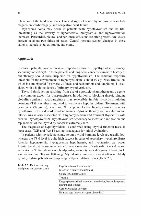
In patients with myxedema coma, serum thyroid hormone levels are usually low, whereas the TSH level is quite high (except in cases of secondary hypothyroidism). Anemia, hyponatremia, hypoglycemia, hypothermia, and hypotension can occur. Arterial blood gas measurement usually reveals retention of carbon dioxide and hypox-emia. An EKG often shows sinus bradycardia, various types and degrees of heart block, low voltage, and T-wave flattening. Myxedema coma occurs most often in elderly hypothyroidism patients with superimposed precipitating events (Table 2.5).
Table 2.5 Factors that may precipitate myxedema coma
Exposure to cold temperatureInfection (usually pneumonia)Congestive heart failureTraumaDrugs (phenobarbital, narcotics, anesthetics, benzodiazepines, lithium, and iodides)Cerebrovascular accidentHemorrhage (especially gastrointestinal)
S.-C.J. Yeung and W. Liu
47
Recognition of hypothyroidism may be difficult in the emergency care setting. Thyroid function test results typically are not available within 24 h. The emergency physician’s responsibility is to consider the diagnosis of hypothyroidism, provide acute care, and order the appropriate thyroid function tests to expedite diagnosis.
Treatment
Once hypothyroidism (frank or subclinical) is diagnosed, the patient should receive thyroid hormone replacement therapy.
Management of myxedema coma in the critical care setting has been reviewed. Rapid clinical diagnosis with early therapy may be life-saving. Treatment may be emergent and is usually given prior to laboratory confirmation. In critically ill patients, if myxedema coma is highly suspected, 0.5 mg of levothyroxine should be given intravenously followed by 0.025–0.100 mg a day. Other supportive measures, such as correction of hypothermia using slow rewarming and ventilatory and circu-latory support, are critical.
Thyrotoxicosis
Although less common than hypothyroidism, thyrotoxicosis is still a common disease, with a prevalence of 20–25 per 100,000 in the general population. Like hypothyroidism, more female than male patients have thyrotoxicosis, with a female:male ratio of 5:1. Therefore, female cancer patients commonly have pre-existing or co- existing hyperthyroidism. Moreover, thyrotoxicosis may arise as a complication of cancer or its treatment.
Clinical Manifestations
Thyrotoxicosis is characterized by a hyperadrenergic state. Sinus tachycardia, systolic flow murmur, and water-hammer pulse are common. Atrial dysrhythmias (atrial fibrillation, atrial flutter, and premature atrial contractions) and congestive heart failure are often observed. Eye signs include Graves ophthalmopathy, exophthal-mos, extraocular muscle palsies, lid lag, and upper lid retraction. Neuropsychiatric symptoms of agitation, anxiety, restlessness, fear, paranoia, and mood swings are observed as well as depressed mental function, which may range from a placid demeanor to frank confusion. Neuromuscular symptoms include fine tremor in the hands, proximal myopathy (common in the elderly), thyrotoxic hypokalemic paral-ysis (mostly in Asians), and acute thyrotoxic polyneuropathy. Gastrointestinal symptoms include hyperphagia, diarrhea, nausea, vomiting, and abdominal pain.
2 Metabolic and Endocrine Oncologic Emergencies
48
Dermatologic symptoms include flushed skin, moist arms, fine and straight hair, alopecia, and pretibial myxedema. Apathetic hyperthyroidism is seen primarily in the elderly, and congestive heart failure, atrial fibrillation, and weight loss are prom-inent features.
Approach
Thyrotoxicosis can result from unregulated release of thyroid hormones and thyroglobulins. This may be caused by direct injury to the thyroid gland, destruc-tive infiltrative processes, or autoimmune-mediated destruction of thyroid follicu-lar cells.
Hyperthyroidism can result from unregulated or stimulated synthesis, release of thyroid hormones, and growth of thyroid tissues. Toxic goiters and adenomas and thyroid carcinomas are examples of unregulated autonomous thyroid tissue. Inappropriate stimuli for hyperfunction of the thyroid may be TSH, human chori-onic gonadotropin, thyroid-stimulating immunoglobulins, and TSH receptor muta-tions, or it may arise from faulty intracellular signal transduction mechanisms.
Large quantities of iodide are present in many drugs (e.g., approximately 9 mg of iodine in a 300-mg dose of amiodarone), antiseptics (e.g., povidone-iodine), and contrast media used in radiology. Iodine-induced hyperthyroidism usually occurs in patients with underlying thyroid diseases.
Thyrotoxicosis can result from autoimmune thyroiditis precipitated by bioimmu-notherapy for cancer with cytokines. In addition to the mechanism of excess iodide described above, amiodarone induces thyroiditis. Radiation-induced painless thyro-toxic thyroiditis occurs infrequently after external beam irradiation of the neck.
Graves disease, toxic multinodular goiters, and solitary toxic nodules are the three forms of primary hyperthyroidism that account for most cases of hyperthy-roidism in the general population. The risk of Graves disease after radiotherapy for Hodgkin disease is estimated to be at least 7.2 times that in the general population.
Thyroid metastasis occurs in 1.25–24.00 % of patients with metastatic carcinoma. However, thyrotoxicosis owing to follicular destruction by metastasis is rare.
Structural homology in the human chorionic gonadotropin and TSH molecules as well as receptors provides the biochemical basis for the ability of human chori-onic gonadotropin to stimulate the TSH receptor. Trophoblastic tumors, hydatidi-form moles, and choriocarcinomas secrete human chorionic gonadotropin in large amounts, often causing hyperthyroidism. When the serum human chorionic gonad-otropin level rises above 200 IU/mL, hyperthyroidism is likely.
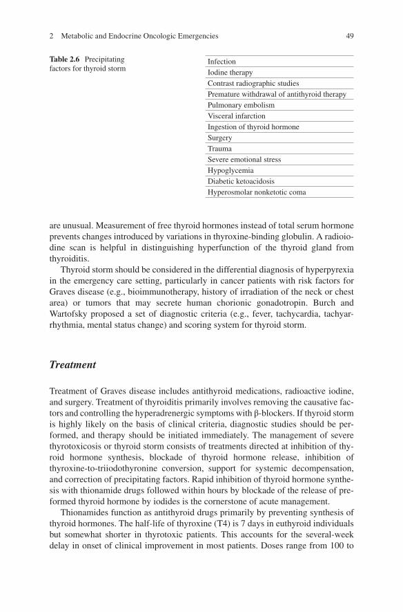
Thyroid storm, an acute decompensation of severe or untreated thyrotoxicosis, is a life-threatening complication with a high mortality rate. Precipitating factors for thyroid storm are listed in Table 2.6.
Thyrotoxicosis is diagnosed by measuring thyroid hormone (thyroxin and triio-dothyronine) and TSH levels. Pituitary and hypothalamic causes of thyrotoxicosis
S.-C.J. Yeung and W. Liu
49
are unusual. Measurement of free thyroid hormones instead of total serum hormone prevents changes introduced by variations in thyroxine-binding globulin. A radioio-dine scan is helpful in distinguishing hyperfunction of the thyroid gland from thyroiditis.
Thyroid storm should be considered in the differential diagnosis of hyperpyrexia in the emergency care setting, particularly in cancer patients with risk factors for Graves disease (e.g., bioimmunotherapy, history of irradiation of the neck or chest area) or tumors that may secrete human chorionic gonadotropin. Burch and Wartofsky proposed a set of diagnostic criteria (e.g., fever, tachycardia, tachyar-rhythmia, mental status change) and scoring system for thyroid storm.
Treatment
Treatment of Graves disease includes antithyroid medications, radioactive iodine, and surgery. Treatment of thyroiditis primarily involves removing the causative fac-tors and controlling the hyperadrenergic symptoms with β-blockers. If thyroid storm is highly likely on the basis of clinical criteria, diagnostic studies should be per-formed, and therapy should be initiated immediately. The management of severe thyrotoxicosis or thyroid storm consists of treatments directed at inhibition of thy-roid hormone synthesis, blockade of thyroid hormone release, inhibition of thyroxine- to-triiodothyronine conversion, support for systemic decompensation, and correction of precipitating factors. Rapid inhibition of thyroid hormone synthe-sis with thionamide drugs followed within hours by blockade of the release of pre-formed thyroid hormone by iodides is the cornerstone of acute management.
Thionamides function as antithyroid drugs primarily by preventing synthesis of thyroid hormones. The half-life of thyroxine (T4) is 7 days in euthyroid individuals but somewhat shorter in thyrotoxic patients. This accounts for the several-week delay in onset of clinical improvement in most patients. Doses range from 100 to
Table 2.6 Precipitating factors for thyroid storm
InfectionIodine therapyContrast radiographic studiesPremature withdrawal of antithyroid therapyPulmonary embolismVisceral infarctionIngestion of thyroid hormoneSurgeryTraumaSevere emotional stressHypoglycemiaDiabetic ketoacidosisHyperosmolar nonketotic coma
2 Metabolic and Endocrine Oncologic Emergencies
50
600 mg/day for propylthiouracil and from 10 to 60 mg/day for methimazole. Gastrostomy or jejunostomy tubes and rectal administration of propylthiouracil or methimazole can be used in patients who cannot receive medications orally or nasogastrically.
β-blockers, both cardioselective and noncardioselective, are important adjuncts in treating hyperthyroidism. β-blockade provides rapid relief of hyperadrenergic symptoms and signs of thyrotoxicosis, such as palpitations, tremors, anxiety, heat intolerance, and various eyelid signs, before any decrease in thyroid hormone level. β-blockers are useful in preventing hypokalemic periodic paralysis in susceptible individuals, and they are the drugs of choice for thyroiditis, which is self-limiting. High doses of propranolol (greater then 160 mg/day) also can inhibit peripheral conversion of T4 to T3.
Saturated solution potassium iodide (3–5 drops) is administered orally every 8 h to block release of thyroid hormones in patients with thyrotoxicosis. At pharmaco-logic concentrations (100 times the normal plasma level), iodides decrease thyroid gland activity. This action involves decreasing thyroid iodide uptake, iodide oxida-tion, and organification and blocking the release of thyroid hormones (Wolff-Chaikoff effect). Iodide has substantial benefits in treating thyroid storm. However, administration of iodide may be problematic in thyrotoxic patients with severe dysfunction of the upper gastrointestinal tract. Rectal delivery of potassium iodide is an effective alternative to parenteral sodium iodide in severely thyrotoxic patients with small bowel obstructions.
The oral contrast agents ipodate and iopanoic acid also are potent inhibitors of T4-to-T3 conversion, making them ideal for treatment of severe or decompensated thyrotoxicosis. They are generally given after starting treatment with thioamide. Although physicians have used intravenous iodinated radiographic contrast medium to treat a case of thyroid storm, this approach is highly nephrotoxic, and its efficacy has yet to be firmly established.
The enterohepatic circulation of thyroid hormones is higher in patients with thy-rotoxicosis than in individuals without it. Bile-salt sequestrants bind thyroid hor-mones and thereby increase their fecal excretion. Colestipol has been an effective, well-tolerated adjunctive agent in the treatment of hyperthyroidism.
Other treatment options include corticosteroids (e.g., dexamethasone, which inhibits peripheral thyroxine conversion), lithium, amiodarone, and potassium per-chlorate. Plasmapheresis and hemoperfusion are effective ways to remove excess thyroid hormone. Emergent thyroidectomy is hazardous in the presence of severe thy-rotoxicosis, and radioactive iodine does not offer rapid control of thyroid function.
Carcinoid Crisis
Carcinoid tumors secrete a variety of polypeptides, biogenic amines, and prosta-glandins, which cause a constellation of symptoms collectively known as carcinoid syndrome. Severe, life-threatening manifestations are known as carcinoid crisis.
S.-C.J. Yeung and W. Liu
51
Clinical Manifestations
Carcinoid syndrome includes the following symptoms: skin flushing, telangiectasia, cyanosis, diarrhea, intestinal cramping, bronchoconstriction, and valvular heart disease. In many patients, the primary complaints are severe flushing, nausea, and faintness. In a crisis situation, seizure, hypotension, severe bronchoconstriction, and cardiopulmonary arrest can occur.
Approach
Provocation of 5-hydroxytryptamine and release of other humoral mediators in patients with carcinoid crisis may be mediated by the release of catecholamines from the adrenals, which activates adrenergic receptors on tumor cells. Somatostatin receptors on the tumor cells primarily have an inhibitory effect.
Typically, cancer patients presenting with symptoms of carcinoid crisis already have diagnoses of carcinoid tumors. Typical carcinoid syndrome is associated most often with midgut carcinoid tumors. Ninety percent of patients with carcinoid syn-drome have metastatic disease. Carcinoid crisis can be precipitated by chemotherapy and invasive procedures such as fine-needle biopsy and laser bronchoscopy.
Treatment
Symptomatic treatments of carcinoid crisis usually target bronchoconstriction, flushing, and diarrhea.
• Octreotide acetate, a somatostatin analog, is effective in controlling and markedly reducing the symptoms of carcinoid crisis. Dose escalation up to 5950 μg a day has been reported.
• Both hypertensive and hypotensive carcinoid crises respond to treatment with octreotide, and octreotide and lanreotide should be considered for prophylactic and emergency use in all patients with carcinoid syndrome prior to and during anesthesia, surgery, biopsy, and chemoembolization of liver lesions.
• Supportive measures include oxygen, intubation, and ventilator support (if necessary) and intravenous crystalloid fluid administration.
• Octreotide, dexamethasone, and H1 and H2 blockers should be administered quickly.
• Mild bronchoconstriction may respond to inhaled anticholinergic and/or β-adrenergic agonists and theophylline.
• Cyproheptadine can block 5-hydroxytryptamine receptors and may be helpful in controlling symptoms caused by 5-hydroxytryptamine.
• Catecholamine administration should be avoided.
2 Metabolic and Endocrine Oncologic Emergencies
52
Key Practice Points
• Treatment of hyponatremia is best tailored after identifying the etiology.• Use of vaptans for hyponatremia increases aquaresis by binding to V2 receptors
in the kidney.• Acute severe symptomatic forms of hyponatremia should be treated with hyper-
tonic saline with the caution that fast correction of hyponatremia can lead to osmotic demyelinating syndrome.
• Thirst is the first line of defense against hypernatremia except in patients with hypodipsia.
• Differential diagnosis of hypernatremia: central versus nephrogenic DI, and administer treatment based on etiology.
• Oral replacement of potassium is preferred over other routes of replacement.• The rate of intravenous potassium replacement must be regulated carefully.
Potassium chloride must be appropriately diluted in intravenous fluid. The infu-sion rate may be as high as 40 mEq/h through a central venous catheter.
• Treat or prevent cardiac arrhythmia in hyperkalemic patients.• Pharmacologically induce transmembrane shift of potassium into cells in patients
with hyperkalemia.• Remove potassium from the body of a hyperkalemic patient by enhancing renal
excretion or administering Kayexalate or dialysis.• Treat hypocalcemia with calcium preparations and vitamin D.• Pay attention to magnesium and phosphate levels in patients with hypocalcemia.• Primary hyperparathyroidism and malignancy account for more than 90 % of
hypercalcemia cases.• Hypercalcemia of malignancy is associated with poor prognosis.• First-line treatments of hypercalcemia of malignancy include intravenous hydra-
tion with crystalloid fluids and bisphosphonate infusions.• Calcitonin is a useful second-line therapeutic for hypercalcemia of malignancy.• Magnesium deficiency is very common in cancer patients, who must be screened
and monitored for it.• Diligent correction of hypomagnesemia is recommended.• Hypermagnesemia is usually iatrogenic in the presence of renal insufficiency.• Use pharmacologic preparations containing magnesium with caution in the pres-
ence of renal insufficiency.• Treatment of hypermagnesemia involves removal of magnesium. In severe cases,
calcium is given intravenously to antagonize the effect of magnesium on the neuromuscular and cardiovascular systems.
• Acute severe hypophosphatemia usually results from a transmembrane shift of phosphate into cells in the setting of respiratory alkalosis, intravenous glucose administration (including hyperalimentation), gram-negative sepsis, or high- dose insulin therapy.
• Oncogenic osteomalacia is rare but causes severe hypophosphatemia and phos-phate renal wasting.
S.-C.J. Yeung and W. Liu
53
• In patients with phosphate abnormalities, the calcium level must be monitored in addition to the phosphate level.
• Treatment of hyperphosphatemia with nonabsorbable phosphate binders that are aluminum- and calcium-free (800–1600 mg of sevelamer with each meal) is pre-ferred over other treatments.
• Diabetic ketoacidosis is diagnosed according to the triad of metabolic acidosis, hyperglycemia, and presence of ketone bodies in the urine or blood.
• Sepsis and serious infections must be ruled out as the precipitating events for diabetic ketoacidosis and hyperosmolar hyperglycemic coma, especially in immunocompromised cancer patients.
• Treatment of hyperglycemia primarily involves intravenous administration of fluids and insulin.
• Severe hypoglycemia with change in mental status can be promptly treated with glucagon (1–2 mg subcutaneously or intravenously) or glucose (50 mL of 50 % dextrose in water intravenously).
• Mild hypoglycemia (glucose level of 50–60 mg/dL) can be treated with simple carbohydrate intake.
• Non-islet cell tumor-induced hypoglycemia is treated with glucose infusion, glucocorticoids, or glucagon and tumor debulking.
• A screening test for adrenal insufficiency is basal 8:00 a.m. serum cortisol measurement.
• Dynamic testing using high- and low-dose cosyntropin, metyrapone (30 mg/kg given orally overnight), and insulin tolerance testing (insulin-induced hypoglyce-mia) can be used to diagnose primary and secondary adrenal insufficiency.
• In patients with adrenal insufficiency (suspected or confirmed) and severe stress (circulatory instability, sepsis, emergency surgery, or other major complica-tions), hydrocortisone at 300 mg a day or other glucocorticoids at equipotent doses may be administered intravenously in divided doses.
• Myxedema coma is rare but life-threatening.• Hypothyroidism is common and easily managed with hormone replacement.
The challenge lies in recognition of signs and symptoms of it for diagnosis.• Thyroiditis is usually self-limiting.• Uncontrolled Graves disease and elevated paraneoplastic β-human chorionic
gonadotropin levels may predispose individuals to thyroid storm upon experi-encing precipitating events.
• The management of severe thyrotoxicosis and thyroid storm involves inhibi-tion of thyroid hormone synthesis, blockade of thyroid hormone release, inhi-bition of thyroxine-to-triiodothyronine conversion, support for systemic decompensation, and correction of precipitating factors. Rapid inhibition of thyroid hormone synthesis with thionamides followed by blockade of the release of preformed thyroid hormone by iodides is the cornerstone of acute management.
• Octreotide is the primary agent for both prevention and treatment of carcinoid crisis.
2 Metabolic and Endocrine Oncologic Emergencies

54
Suggested Readings
Behl D, Hendrickson AW, Moynihan TJ. Oncologic emergencies. Crit Care Clin. 2010;26:181–205.Glover DJ, Glick JH. Metabolic oncologic emergencies. CA Cancer J Clin. 1987;37:302–20.Lewis MA, Hendrickson AW, Moynihan TJ. Oncologic emergencies: pathophysiology, presenta-
tion, diagnosis, and treatment. CA Cancer J Clin. 2011;61:287–314.Spinazze S, Schrijvers D. Metabolic emergencies. Crit Rev Oncol Hematol. 2006;58:79–89.Taub YR, Wolford RW. Adrenal insufficiency and other adrenal oncologic emergencies. Emerg
Med Clin North Am. 2009;27:271–82.
S.-C.J. Yeung and W. Liu

http://www.springer.com/978-1-4939-3187-3