Embed Size (px)
Citation preview

REVIEW Open Access
Metal(loid) speciation and transformationby aerobic methanotrophsObulisamy Parthiba Karthikeyan1,2,3*, Thomas J. Smith4*, Shamsudeen Umar Dandare1, Kamaludeen Sara Parwin5,Heetasmin Singh6, Hui Xin Loh1, Mark R Cunningham1, Paul Nicholas Williams1, Tim Nichol4,Avudainayagam Subramanian5, Kumarasamy Ramasamy7 and Deepak Kumaresan1*
Abstract
Manufacturing and resource industries are the key drivers for economic growth with a huge environmental cost (e.g.discharge of industrial effluents and post-mining substrates). Pollutants from waste streams, either organic or inorganic(e.g. heavy metals), are prone to interact with their physical environment that not only affects the ecosystem healthbut also the livelihood of local communities. Unlike organic pollutants, heavy metals or trace metals (e.g. chromium,mercury) are non-biodegradable, bioaccumulate through food-web interactions and are likely to have a long-termimpact on ecosystem health. Microorganisms provide varied ecosystem services including climate regulation,purification of groundwater, rehabilitation of contaminated sites by detoxifying pollutants. Recent studies havehighlighted the potential of methanotrophs, a group of bacteria that can use methane as a sole carbon and energysource, to transform toxic metal (loids) such as chromium, mercury and selenium. In this review, we synthesise recentadvances in the role of essential metals (e.g. copper) for methanotroph activity, uptake mechanisms alongside theirpotential to transform toxic heavy metal (loids). Case studies are presented on chromium, selenium and mercurypollution from the tanneries, coal burning and artisanal gold mining, respectively, which are particular problems in thedeveloping economy that we propose may be suitable for remediation by methanotrophs.
Keywords: Methanotrophs, Metalloenzymes, Methanobactin, Metal transformation and speciation, Bioremediation
IntroductionThe world’s population is predicted to reach 9.7 billion by2050. Increasing demand for food and energy contributesto over-exploitation of natural resources and environmen-tal degradation. Particularly, release of pollutants into theenvironment from manufacturing and resource industriesis a major concern for ecosystem health. Pollutants, eitherorganic (e.g. polycyclic aromatic hydrocarbons) orinorganic (e.g. heavy metals), when they interact with theirphysical environment, not only affect the quality of the
environment but also have cascading effects on the healthand wellbeing of organisms across all domains of life [1, 2].Unlike organic pollutants, heavy metal(loid)s or tracemetals (e.g. chromium-Cr, mercury-Hg, selenium-Se) arenon-biodegradable, persist longer in the environment andbioaccumulate through the food web. Even at low concen-trations, they are likely to have a long-term impact on eco-system health [3, 4]. Naturally occurring metals are integralto the evolution of living organisms and are critical formetabolic activities e.g. co-factors for enzymes [5]. Despitetheir biological importance, large amounts of these metalscan result in cellular and tissue damage, i.e. cytotoxicity inanimals and growth inhibition in microbes; poor growth,low yields and nutrient inbalances in plants and metabolicinterferences and mutangenesis in all types of organisms[6, 7]. Detailed reviews on the occurrence of heavy metals
© The Author(s). 2021 Open Access This article is licensed under a Creative Commons Attribution 4.0 International License,which permits use, sharing, adaptation, distribution and reproduction in any medium or format, as long as you giveappropriate credit to the original author(s) and the source, provide a link to the Creative Commons licence, and indicate ifchanges were made. The images or other third party material in this article are included in the article's Creative Commonslicence, unless indicated otherwise in a credit line to the material. If material is not included in the article's Creative Commonslicence and your intended use is not permitted by statutory regulation or exceeds the permitted use, you will need to obtainpermission directly from the copyright holder. To view a copy of this licence, visit http://creativecommons.org/licenses/by/4.0/.The Creative Commons Public Domain Dedication waiver (http://creativecommons.org/publicdomain/zero/1.0/) applies to thedata made available in this article, unless otherwise stated in a credit line to the data.
* Correspondence: [email protected]; [email protected];[email protected] of Biological Sciences & Institute for Global Food Security, Queen’sUniversity Belfast, 19 Chlorine Gardens, Belfast, UK4Biomolecular Sciences Research Centre, Sheffield Hallam University,Sheffield, UKFull list of author information is available at the end of the article
Karthikeyan et al. Microbiome (2021) 9:156 https://doi.org/10.1186/s40168-021-01112-y

in the environment, industrial production and usage,potential for human exposure, and their molecularmechanisms of toxicity can be found in [8–11]. In thisreview, we also highlight the impact of tannery industries(i.e. Cr pollution – Table 1), selenium-polluting industriesincluding mining (Table 2), and artisinal gold mining (i.e.Hg pollution – Table 3) as case studies.Current remediation strategies of contaminated sites
include chemical extraction (with acids or chelatingagents), immobilisation, encapsulation and electrolysis.Although these strategies have been useful, they comewith several limitations including significant alterationsof the physicochemical properties, low efficiency andhigh cost of operation [38]. Phyto/bioremediation hasbeen suggested as an alternative eco-friendly approachto detoxify metals from contaminated sites [39]. Thisapproach leverages intrinsic biological mechanismsof plants and microorganisms to transform and/or
bioaccumulate metals from the environment [1]. Inparticular, microorganisms possess remarkable abil-ities to bioaccumulate, retain and transform heavymetal ions [40–42] by taking advantage of reduction/oxidation (redox) and other processes e.g. modulat-ing solubility of metals without changing the oxida-tion state of the metal [43, 44].Metal(loids) exhibit different physical and chemical
forms (i.e. differences in speciation) in the environment.Electronic configuration, oxidation state and ionic radiusall define the chemical speciation of a particular metaland its fractionation (i.e. whether it is labile/inert, ligandcomplexed, precipitated or existing as a free ion). Conse-quently, the chemical form of a metal strongly influencesits reactivity, toxicity, mobility and interaction with micro-organisms in the environment [44, 45]. For instance, cop-per (Cu) becomes potentially toxic when it transitionsbetween Cu(II) and Cu(I), soluble and toxic chromium(Cr(VI)) are less toxic when reduced to Cr(III), mercury
Table 1 Chromium pollution from tannery industries—a casestudy
Tannery industries contribute significiantly to the developing economiessuch as India and Bangladesh (~3.5 and 5 billion USD per annum,respectively). Leather production utilises a large amount of water. It hasbeen estimated that about 25–40m3 of fresh/ground water resources isused and subsequently discharged into the environment as effluentduring the processing of one tonne of hides. Tannery effluents generallycontain high levels of organics (measured as biological/chemical oxygendemand), nitrogen, sulphate and heavy metals such as Cr, Ni, As and Co.Tanneries have been the subject of wide public debate, particularly thedownstream pollution by carcinogenic and teratogenic Cr (VI) thatleaches into water bodies and soil and its subsequent impact onecosystem health. For example, the Vellore district in South India is awell-known tannery hub that is famous for its export of leather [12]. Ex-tensive surveys on tannery-associated groundwater contamination haverevealed that toxic Cr (VI) can be detected (even at a depth of 10 m) ata high concentration up to 38 mg L-1 (critical limit 0.05 mg L-1 [13];) inthe Ranipet, Walajapet and Vaniambadi areas of the Vellore district. Thisis extremely high compared to levels reported in other parts of India (4–7 mg L-1) [14].
Chromium pollution from tanneries extends to soil e.g. about 50,000 haof agricultural land has been affected due to salts and chromium fromthe tannery waste streams. Concentrations of exchangeable Cr fractionshave been reported up to 128 μg kg-1. Research in sites dumped withtannery wastes over the past 20 years in Vellore and surroundingregions has indicated that soil alkalinity facilitates the presence of themore toxic and mobile Cr (VI) that subsequently leaches into thegroundwater. Alarming levels of Cr were also found in borewell watersin Palar river basin (>500 μg Cr L-1), 90% of which was Cr(VI) [15]. Inhighly contaminated zones, the total Cr was reported to be as high as102 g Cr kg-1 soil and has been found even at soil depth of 30 cm (1.1mg Cr(VI) kg-1 [16, 17]. While tanneries use Cr(III) salts for leatherprocessing, the presence of Cr(VI) in the contaminated sites is still anintriguing question. Contrary to the general acceptance that thepresence of organic matter and other species contributing to electrontransfer reactions in soil would rapidly convert Cr(VI) to Cr(III), these soilsshowed higher levels of Cr(VI) despite high-soil organic content (15%)[16]. It has also been reported that high concentrations of sodium andphosphates in soil solution can also trigger Cr (VI) mobility in soils withalkaline pH [18]. In addition manganese oxides are reported to reoxidiseCr(III) to Cr(VI) [19]. While tanners are replacing tannins instead of chro-mium, remediation of Cr(VI) in long-term contaminated soils have notbeen successful owing to reoxidation of Cr(III) to Cr(VI) [20] and continueto be an major issue.
Table 2 Selenium an essential element with toxicity problemsin the mining industry and beyond
The Recommended Daily Intake of selenium in the human diet is 55 mgd-1 (dietary reference intakes, 2000; Dietary Reference Intakes (2000)National Research Council. Washington: National Academic Press). TheWorld Health Organization (WHO) has indicated that Se intake in thehuman diet in excess of 400 mg d-1 may be harmful to health, withsigns of Se overexposure being evident at 750–858 mg d-1 [21].Potentially, toxic levels of selenium in the environment may occurnaturally due to the presence of seleniferous rocks and also due tohuman activities, particularly mining. Selenium concentrations inagricultural drainage water in the range 0.14–1.4 mg L-1 were reportedto cause death and deformity in aquatic birds [22]. The WHO has set themaximum permitted Se concentration for drinking water at 40 mg L-1,although specific jurisdictions have set limits as low as 10 mg L-1. Waterquality guidelines for freshwater and water used for agriculturalirrigation water range from 1 to 150 mg L-1 [23]. Selenium is stronglyenriched in coal compared to other rocks and so coal and the ash fromcoal combustion are major sources of toxic amounts of selenium.Selenium species enter the air due to combustion of coal. The seleniumthat remains in coal ash is predominantly in the toxic and water solubleselenite form. It is subject to sorption to various components of ash,though is generally mobile into the aqueous phase at acidic pH [24].Waste water from coal mining operations may contain more than 1 mgL-1 of selenium [23]. Problems with Se (and other pollutants due toprocessing and burning of coal) are a particular concern in China, wherecoal production and use have more than doubled since 2000 and arepredicted to continue to rise, while they have been stable in most otherareas of the world [25].
Other emerging industries may provide new sources of potentiallyharmful selenium exposure. Selenium is a significant element in wasteelectronic and electrical equipment (e-waste). One study in West Africa(Ghana) found a doubling in blood selenium concentration (togetherwith a tripling of mercury levels) in workers involved in incineration ofe-waste [26].
As detailed in the main text, methanotrophs and other environmentalbacteria have the capacity to produce Se (0)-containing nanoparticles. Inaddition to being valuable in detoxifying selenium contamination andin providing novel nanoparticles for use in electronics, suchnanoparticles may find uses as slow-releasing selenium supplements fordiets [27].
Karthikeyan et al. Microbiome (2021) 9:156 Page 2 of 18

(Hg(II)) becomes neurotoxic when its methylated(CH3Hg) and As(III) is more toxic than As(V) [46–48].Recent research has highlighted the ability of aerobic
methanotrophs, a specialised group of bacteria that can
use methane (CH4) as a sole carbon and energy source,to transform metals (and also metalloids) such as Cu,Cr, Se and Hg [41, 49, 50]. Methanotrophs belong to thephyla Proteobacteria (classes Alphaproteobacteria andGammaproteobacteria), Candidate division NC10 andVerrucomicrobia. Till date, more than 29 methanotrophgenera and 8 families (i.e. Methylococcaceae, Methy-lothermaceae, Crenotrichaceae, Methylocystaceae, Beijer-inkiaceae, and Methylacidiphilaceae and two currentlyunclassified families) have been identified within thesephyla. Complete genome sequences for representativesof >23 genera are available in public repositories [51,52]. Proteobacterial methanotrophs are active primarilyin methane-oxygen counter gradients of oxic-anoxic in-terfaces [and in upland soils (high affinity atmosphericmethane oxidisers)], while methanotrophs found inextremely acidic geothermal sites belong to the phylumVerrucomicrobia. Anaerobic microbial methane oxida-tion has been recently discovered that use reverse meth-anogenesis process to convert CH4 into CO2 [53, 54]. Incontrast, the members of the candidate phylum NC10,such as Candidatus Methylomirabilis oxyfera, use allaerobic methane oxidated pathway-specific proteins,while it acquires oxygen through reduction of nitrite tothe oxidation of methane via a unique oxygen-producingpathway [55]. The activity and diversity of methano-trophs and their impact on methane fluxes in differentenvironments (e.g. landfills, rice paddies, natural gasseeps, hypogenic caves, saline lakes) have been studiedextensively using both cultivation-dependent and mo-lecular ecology tools [56–59]. In this review, we focus onrecent developments in the physiology of aerobic metha-notrophs with emphasis on their role in transformationand speciation of metals and metalloids such as Cu, Cr,Se and Hg. Detailed descriptions of the biochemistryand physiology of methanotrophs/methylotrophs arebeyond the scope of this review and the reader is re-ferred to [5, 60].
Aerobic methane oxidation and metalloenzymesIn aerobic methanotrophs, four major steps are involvedin the enzymatic conversion of CH4 into biomass/CO2,in which the availabilities of different metal ions play acritical role (Fig. 1). The biocatalytic oxidation of CH4 tomethanol (CH3OH) is modulated by the enzyme me-thane monooxygenase (MMO). Two forms of MMOexist: membrane-bound particulate MMO (known aspMMO and its divergent form pXMO [61]) and cyto-plasmic soluble MMO (sMMO).
(a) pMMO composed (i) β-subunit, PmoA (26 kDa);(ii) α-subunit, PmoB (45 kDa); and (iii) γ-subunit,PmoC (23 kDa) with an (αβγ)3 structure. Theirgenes are typically arranged in pmo-operons as
Table 3 Mercury pollution from artisanal/small scale goldmining
Mercury emissions from artisanal and small scale gold mining, estimatedat 727 tonnes per annum, account for a large portion of emissions fromanthropogenic sources (37% [28];). Telmer and colleagues also estimatedthe contribution of artisanal and small scale gold mining to mercuryreleases between 640 and 1350 tonnes per year from at least 70countries, with at least 350 tonnes emitted directly into the atmospherewhile the remainder are released into the rivers, lakes, soil and tailings[29]. In small developing countries such as Guyana (Fig. 4), the goldmining industry is economically significant, where it contributed 13.7%to the total GDP of the country and accounted for more than 60% oftotal exports (USD 817.5 million) in 2017 [30]. The artisanal, small andmedium scale operators, who contribute approximately two thirds oftotal gold declarations, rely almost exclusively on the use of mercury forgold extraction and concentration while the large scale companiesutilise higher-recovering technologies with more control over environ-mental and safety risks [31]. Mercury is often added to the collected un-processed gold ore in order to create a mercury-gold amalgam which isthen heated to release the mercury and recapture the gold in concen-trate. Most of the mercury vapour generated during burning of theamalgam may be collected by a retort, thus reducing mercury emissionsby over 93%. However, studies in Guyana and Suriname have shownthat while miners have some knowledge of the negative health and en-vironmental effects of burning amalgams in the open air, they do notregularly use retorts for a variety of reasons, with the most commoncited as the retorts being ‘too time-consuming’ [32, 33].
In addition, mercury is sometimes used in sluice boxes and in panningwhich can also contaminate tailings, creeks and rivers which will leachinto the surrounding environment. In the Minamata Initial Assessmentconducted for Guyana, over 11,000 kg of mercury is estimated to beemitted annually in Guyana by burning of a mercury-gold amalgam,with 39% released in the air, 32% in water and 29% in land [33]. Mercuryemitted to the atmosphere can be deposited into aqueous environ-ments by wet and dry depositions, and some can be re-emitted intothe atmosphere. In surveys carried out by the Guyana Geology andMines Commission (2000 and 2001) in three rivers within two differentmining regions of Guyana, it was found that 57%, 39% and 25% ofpredatory fishes sampled had mercury levels above the maximum WorldHealth Organization guideline concentration (0.5 μg/g). Data fromneighbouring Suriname and French Guiana, where mercury use in min-ing is also abundant, also indicate high levels of mercury contaminationin fish [34]. In a study by Howard and colleagues, sediments taken fromactive and historically mined areas in Guyana had a mean mercury con-centration of 0.229 μg/g, with a range from 0.029 to 1.2 μg/g, which isabove Canadian Environmental quality guidelines (0.19 μg/g) [35]. Thereis also a lack of extensive data on mercury contamination in communi-ties surrounding mining activities in Guyana. A study conducted from2008 to 2010 by Singh and colleagues reported mercury concentrationsof up to 70.8 μg/g (well over the WHO safe limit of 10 μg/g) in the hairof pregnant and nursing women from indigenous populations livingclose to small scale gold mining activities [36].
Mercury has a long history of uncontrolled use in the mining sector ofGuyana resulting in significant environmental pollution of waterwaysand aquatic ecosystems. The Government of Guyana has, however,signed the Minamata Convention and has subsequently aimed to phaseout the use of mercury by 2022, with particular attention to the goldmining sector as part of this commitment. However, it has witnessedresistance by small miners who have not been able to adapt to othertechniques as there is general lack of awareness and understanding ofthese technologies, along with a lack of fiscal incentives and barriers toaccessing finance to transition from this cheaper alternative [31, 33, 37].
Karthikeyan et al. Microbiome (2021) 9:156 Page 3 of 18

pmoCAB. Th general hypothesis is that the enzymepMMO obtains electrons from the cytochrome bc1complex, which contains heme groups and Fe2S2clusters [62, 63]. It has been proposed that Fe isrequired for pMMO activity [63], although a recentstudy shows only the presence of two mono-coppersites [64]. However, the alphaproteobacterialpMMO acquires electrons from ubiquinone poolthrough NADH oxidation [65, 66], while thepMMO activity in gammaproteobacterialmethanotrophs is reported to be coupled to theoxidation of methanol to formaldehyde [67].
(b) sMMO is a well-characterised three-componentenzyme: (i) hydroxylase contains 3 sub-units (α:β:γ– 54: 42: 22 kDa, respectively) with an (αβγ)2
structure; (ii) reductase (38–40 kDa) that supplieselectrons from NADH to the hydroxylase; and (iii)component B as a regulatory protein (15–17 kDa)[68–70]. The sMMO is encoded by mmoXYBZDC.It contains a di-iron active site cluster in thehydroxylase component as well as an Fe2S2 clusterand flavin adenine dinucleotide (FAD) moiety in itsreductase component [52].
The two types of MMO differ completely in theirmetal ion requirements, substrate specificity and CH4
oxidation kinetics [52, 64, 70–72]. Specifically, Cu(II) tobiomass ratio determines the expression and activity ofMMOs in methanotrophs that can express either formof the enzyme and is often referred to as the ‘copper
Fig. 1 Methane oxidation by aerobic methanotrophs and metal co-factors of the enzymes. pMMO = particulate methane monooxygenase. sMMO= soluble methane monooxygenase. Xox-MeDH = XoxF-methanol dehydrogenase. Mxa-MeDH = MxaFI-methanol dehydrogenase. Fae =formaldehyde activating enzyme. FaDH = formate dehydrogenase. CBB = Calvin Benson Bassham Cycle. RuMP = Ribulose MonoPhosphate cycle.TCA – The Citric Acid cycle. PHB – Polyhydroxybutyrate cycle. Enzymes modulating the reaction are represented in red font, metals in blue &yellow fonts. Small vertical light blue arrows next to each metal ion indicate their effect on the expression and/or activity of enzymes
Karthikeyan et al. Microbiome (2021) 9:156 Page 4 of 18
switch’ [52, 61, 72–74]. Most aerobic methanotrophspossess pMMO or pXMO (with exceptions of alphapro-teobacterial members within the genera Methylocella,Methyloceanibacter and Methyloferula), while few pos-sess both pMMO and sMMO (e.g. Methylosinus spp.,Methylocystis spp., Methylobacter marinus, Methylocal-dum marinum, Methylococcus capsulatus, Methylomag-num ishizawai 175, Methylomicrobium buryatense 5G,Methylovulum miyakonense HT12, Methylomonas spp.[52, 75–77];). A very few methanotrophs e.g. Methylo-cella spp., Methyloferulla spp., Methyloceanibacter spp.,(Alphaproteobacteria) and Methyloterricola spp.(Gammaproteobacteria) contain only sMMO (Vekemanet al., 2016; Semrau et al., 2018). The methane turnovernumber per active site cells producing pMMOs (0.5–2.5s-1) are comparatively lower than sMMO expressing cells(>3.5 s-1) [70].In the second step, methanol is converted into formal-
dehyde by the enzyme methanol dehydrogenase(MeDH), which contains a pyrroloquinoline quinone(PQQ) cofactor. There are two distinct types of MeDH,(a) calcium dependent MeDH (i.e. MxaF-MeDH) or (b)the recently discovered XoxF, which is an MeDHdependent on rare earth elements (REEs) e.g. cerium orlanthanum [5, 78–82].
(a) MxaF-MeDH is tetrameric (α2 – 66 kDa; β2 – 8.5kDa), with greatest activity at pH~9 and requiringactivation (e.g. by ammonia [83, 84];). The α sub-unit contains a single Ca2+ ion, the coordinationsphere of which contains one nitrogen and twooxygen ligands from PQQ and a further four oxygenligands from three aminoacyl sidechains (Glu 205,Asn 289 and Asp 331) [85].
(b) XoxF-MeDH is a α2 homodimer, located in theperiplasm and active at neutral pH (optimum ~7).It coordinates the REE at the active site in a 9-coordinate fashion, via the same three ligands fromPQQ and six oxygen ligands derived from fouramino acyl residues (Glu 197, Asn 285, Asp 327and Asp 329) of the protein [86]. It has been re-ported that the activity of purified XoxF was higherwith light REEs (atomic numbers 57–63; La-Eu) incomparison to heavy REEs (atomic numbers 64–71;Gb-Lu) [87].
It should be noted that XoxF-MeDHs are also presentin yeast, moulds, fungi and non-methylotrophic bacteria[88]. In methanotrophs such as Methylomagnum ishiza-wai 175, Methylomicrobium kenyense AMO1, Methylo-terricola oryzae 73a, Methylocystis sp., Mit Z-2018,Methylosinus spp., R-45379, Methyloacidiphilum fumar-iolicum SolV and Verrucomicrobium spp., the only formof MeDH that have been identified is the XoxF-MeDH.
The physiology and genetic basis of the ‘REE-switch’ thatcontrols expression of the alternate forms of MeDH isreviewed extensively in [5, 52]. XoxF-MeDH is reportedto have higher affinity for methanol and faster conver-sion rate [89]. While there was an assumption thatXoxF-MeDH oxidises methanol to formate by dual ac-tivity [90, 91], a recent study by Good and Colleagues[92], using XoxF purified from Methylobacterium extor-quens AM1, confirmed that formaldehyde is the finalproduct. There are 5 families of XoxF-MeDH that mightdisplay different catalytic properties [93], while all needto be validated to conclude whether the dual activity isrelevant in vivo or not.In subsequent steps, the formaldehyde derived from
the oxidation of methanol is assimilated as biomasseither via the ribulose monophosphate pathway (RuMP)in most of Gammaproteobacteria or the serine pathwayin Alphaproteobacteria. There are multiple pathways foroxidation of formaldehyde to CO2 i.e. in conjugationwith tetrahydromethanopterin (H4MPT) or tetrahydro-folate (H4F) [94].
(a) The multi-step H4MPT- and H4F-dependentpathways, which also exist in non-methanotrophicmethylotrophs such as Methylobacteriumextorquens, operate by conjugating the formaldehydeand subsequent intermediates with the respectivecoenzymes. In model methanotrophs such as Mc.capsulatus (Bath) and Ms. trichosporium, OB3bstudies have shown the use of both H4MPT- andH4F-dependent pathways [95–98].
(b) DL-FalDH is homotetramer with a sub-unit massof ~ 49 kDa, while the sub-unit contains a PQQ asred-ox co-factor at 1:1 ratio. It utilisescytochrome b559/569 complex as a electronacceptor [99].
(c) N-FalDH was reported as an NAD(P)+-linkeddehydrogenase from Mc. capsulatus (Bath)where the ability to oxidise formaldehyedepended on a low molecular-mass heat-stablecomponent [100, 101]. Later attempts to repli-cate these results yielded preparations that wereactive but found to be mixtures of otherenzymes and cofactors involved elsewhere in themethane oxidation pathway [102]. Hence,whether an enzyme that directly catalysesNAD(P)+-linked oxidation of formaldhyde is asubstantial contributor to the aerobic methaneoxidation pathway remains unclear.
However, cytochrome bc1 (with Fe) is required forFalDH as an electron acceptor. Also, other enzymes in-volved in the conversion of methanol into formaldehyde,formate and finally into carbon dioxide primarily rely on
Karthikeyan et al. Microbiome (2021) 9:156 Page 5 of 18

Fe, with minor requirements of Cu, Ca, Mo and Zinc(Zn) ions. In particular, the membrane-associatedformate dehydrogenase (FaDH) enzyme, which is 2 αβγδprotomers (Mw ~400 kDa). The holoenzyme containsflavin, iron, inorganic sulphide and molybdenum [103].The FaDH is involved in the conversion of formate toCO2, contains four FexSx clusters and molybdenum(Mo) as cofactors [62]. Recently, the presence of twoclusters encoding NAD+-dependent FaDH that requireeither tungsten (W) or Mo was reported [90]. Moreover,it was also observed that in Methylomicrobium alcaliphi-lum 20ZR formate production was considerably reducedduring the presence of W [90]. Further work is requiredto develop a better understanding of whether a ‘W-Moswitch’ regulates expression of the two types of FaDH.
Metal uptake mechanisms in aerobic methanotrophsMetal ions are integral to metabolic activities (Table 4)in methanotrophs. It has been estimated that about onequarter to one third of any bacterial cell proteinsrequired metal ions to support their functions [104].However, cells can restrict the number of metal ions tobe transported into their cytoplasm, thereby creating acompetition between different proteins requiring thesame metal ions and thus influencing different enzymeactivities in vivo. Specific energised pumps are used tofacilitate metal ion transport either into the cell (im-porters) or out of it (exporters). Such transporters are gen-erally located in the cell membrane and may use selectivemetal binding proteins or small molecules such as sidero-phores [105]. In methanotrophs, metals are transportedinto the cell by (a) passive diffusion e.g. direct transportthrough porins in surface membrane and/or (b) activetransport e.g. through TonB-dependent metal transport orother metal transport proteins [106, 107].
Passive diffusion of metalsPassive transport of metals and organic solutes usuallyoccurs via porins. The pore size and amino-acid composi-tions in the channel of porins determine their specificitytowards different solutes and transport. In bacteria, theperformance of porins (both non-specific and specificdiffusion channels) is usually regulated by the availability
of specific nutrients and downregulated by the presence oftoxins or harmful solutes [108]. Based on their structureand diffusion characteristics, porins are classified as (a)specific monomeric, (b) specific trimeric, (c) non-specificmonomeric and (d) non-specific trimeric diffusion chan-nels [109], while little is known about the porin structuresin methanotrophs. Reseachers have characterised a smallnumber of outer membrane proteins (e.g. MopE andCorA) that can perform the function of copper transportin methanotrophs [106]. While MopE (some methano-trophs from Gammaproteobacteria express it) has beenreported to bind copper with high affinity (< 1020 M-1)and to have a binding site composed of two imidazolesand a kynurenine (modified tyrptophan side-chain) group[110], other outer membrane proteins, as wells as theirarrangements are yet to be characterised in detail. Unche-lated metal ions are also reported to be transported viaporins e.g. Cu and Hg transport in M. album BG8 [50].
Active transport of metalsIt has been reported that about 10−6 M of intracellularion concentration is required for any cellular activityand at lower levels, metal binding proteins or smallmolecules (e.g. siderophores) are synthesised by bacteria(including methanotrophs) to scavenge metals from theenvironment. For example, methanotrophs producemethanobactin under Cu limited conditions [111, 112].Methanobactin, a chalkophore (chalk – copper inGreek), is a ribosomally produced and post-translationally modified peptide [72, 111, 113]. It plays akey role in active transport of metals and perhaps alsoenables ecological succession of different methanotrophsunder external metal toxicities [72]. Specifically, themethanotrophs can export up to 3–50 methanobactinmolecules per cell per second dependent on the Cu con-centrations in the external solute [72]. Only very fewmethanotrophic species from the Alphaproteobacteriafamily are reported to biosynthesise methanobactin andonly 10% of sequenced methanotrophs containmethanobactin biosynthesis genes [72, 74, 114]. Metha-nobactins produced by different methanotrophs arestructurally distinct (i.e. till date only 7 methanobactinsare characterised from methanotrophs), though with
Table 4 Redox states of metals with metallo-enzymes and their specific catalytic functions
Metal Redox state Enzymes Class of catalysis by enzyme
Copper Cu (II), Cu (I) Most copper-containing enzymes (e.g. Cytochromes) Electron transfer, ferrous oxidase, amine oxidase
Iron Fe (II), Fe (III),Fe (IV), Fe (V)
CytochromesPeroxidase, catalase
Electron transfer, Oxidation
Molybdenum Mo (III) to Mo (VI) Nitrogenase, Aldehyde oxidase Oxidation
Cobalt Co (I)?, Co (II), Co (III) B12- requiring enzymes Carbonic anhydrase
Manganese Mn (III) to Mn (IV)? Photosynthetic enzymes Superoxide dismutase, oxidase
Chromium Cr (VI) to Cr (III) Dehydrogenase Oxidoreductases
Karthikeyan et al. Microbiome (2021) 9:156 Page 6 of 18

similar metal (i.e. Cu) binding sites, and are categorisedas Group-I and II (Table 5). Ms. trichosporium OB3bwas reported to produce the highest concentrations ofGroup-I methanobactin (35–60 mg l−1) followed by theMc. capsulatus (Bath) (18–24 mg l−1) [115]. Interest-ingly, Mc. capsulatus (Bath) does not have the mbn clus-ter encoding methanobactin production [116, 117]indicating a different type of copper-binding molecule(including but possibly not restricted to MopE [118]used in high-affinity copper acquisition). Group-IImethanobactin is mainly produced by Methylocystis sp.strain SB2, while recent bioinformatic analysis hasshown that there are few methanotrophs that can makeboth the forms of methanobactins e.g. Methylocystis par-vus OBBP, Methylocystis sp. LW5, Methylosinus sp. LW3,Methylosinus sp. R-45379 and Methylosinus sav2 [112].As discussed earlier, methanobactin is reported to
show high affinity towards Cu (~1011 to 1034 M−1)followed by Ag (~107 M−1) as summarised in Fig. 2.Based on the metal binding properties of methanobactin,metals are grouped under two categories: Group Ametals—Ag(I), Au(III), Hg(II), Cu(II) and Pb(II) andGroup B metals—Cd(II), Co(II), Fe(III), Mn(II), Ni(II)and Zn(II) [118]. Group A metals bind at both oxazo-lone rings and are reduced upon binding, while Group Bmetals bind at just one oxazolone ring and are notreduced upon binding [118, 119]. Based on the metha-nobactic metal selectivity, the metals are grouped as (a)Group-1 (high—100%): Cu (I) and Ag (I); (b) Group 2(moderate—85–96%): Zn (II), Ni (II) and Co (II); and (c)Group 3 (low—<50%): Mn (II), Pb (II) and Fe (II).
Studies have also shown that the methanobactin can alsobind, transform and detoxify Hg [50, 69, 120] and U (VI)[121]. In a recent study, a methanobactin characterisedfrom Methylosinus sporium was found to have one oxa-zolone and one imidazolone ring with disulphide bondbetween two Cys residues as in Group 1 [116]. Thestructural difference may affect the metal binding andaffinity, which is not completely understood yet. So theclassification, metal affinity and grouping of metals de-scribed above may applicable only to the methanobactinsfrom Methylosinus trichosporium OB3b and Methylocys-tis sp. strain SB2, while it may vary for other methano-bactin structures and requires further investigation. Thisknowledge is critical in determining how to exploitmethanotroph strains and/or methanobactins to seques-ter the metal ions from the environment.
Copper accumulation by methanotrophsCopper is central to methanotrophic activity, while 40%of methanotrophs are reported to have Cu storage pro-teins and only 10% reported to make methanobactins forCu uptake, while the remainder may use passive trans-port of metals using other porin related proteins [122].It has been estimated that methanotrophs have at least aten-fold greater Cu requirement compared with otherbacteria [123]. While Cu is an essential metal ion forpMMO activity (i.e. CH4 to methanol conversion), it hasalso been shown to control expression of other enzymes(e.g. formaldehyde dehydrogenases, hemerythrin) andouter membrane proteins involved in Cu assimilation,regulation and transport. Moreover, it can influence the
Table 5 Difference between two well characterised known groups of methanobactins
Particulars Group 1 methanobactin(e.g. Ms. trichosporium OB3b)
Group 2 methanobactin(e.g. Methylocystis strain SB2)
Molecular weight (Da.) 1154.26 851.20
Structural difference Two oxazolone rings (UV-vis spectra: Ring A ~394nm and B ~ 342nm)
One oxazolone ring (UV-vis spectra: Ring B~ 338nm) andImidazolone (UV-vis spectra: Ring A ~ 387nm)
Partial amino-acids associ-ated with Ring-A
Leucine Arginine
Partial amino-acids associ-ated with Ring-B
Proline Threonine
Amino acids Gly1, Ser2, Cys3, Tyr4, Ser5, Cys6 and Met7 (Gly1 –Downfield shift at 9.28ppm)
Ala1, Ser2, Ala3, Ala4 (Ala1 – Downfield shift at 11.7 and 145 ppm)
Copper affinity 1018–1050 M-1 1026 M-1
Copper binding Ring-A 640 S-1 Not available
Copper co-ordination ratewith Ring-B
121 S-1 >2000 S-1
Structure modification Pyramid-like structure Hairpin-like structure
Stability Both rings hydrolysed within 2–5 days underacidic condition
Ring B (i.e. Oxa) is susceptible to hydrolysis (200 min) and Ring-Amore resistance
Disulphide bond Found Not found
Sulphate group Not found Found
Note: Da. daltons, UV ultraviolet, Gly glycine, Ser serine, Cys cystine, Tyr tyrosine, Met methionine, ppm parts per million
Karthikeyan et al. Microbiome (2021) 9:156 Page 7 of 18

membrane structure or formation in methanotrophs e.g.M. capsulatus (Bath), M. album BG8 and Ms. trichospor-ium OB3b [123, 124]. The Cu-containing protein pMMO,which constitutes up to 20% of the total cellular proteins inmethanotrophs [125] shows high-affinity towards CH4. ThepMMO is expressed at high Cu concentrations, and at lowlevels, sMMO is expressed [126]. At high Cu concentra-tions, the Cu will bind the active site of sMMO and inhibitthe electron transfer between the falvin adenine di-nucleotide and MMOH [127]. High copper-to-biomass ra-tio leads to pMMO expression through the canocial ‘copperswitch’, but the exact mechanisms by which Cu activatespMMO expression is not yet clear. However, the rate ofcopper uptake by passive diffusion or active transport maydiffer between methanotrophs, mainly in Alphaproteobac-teria and Gammaproteobacteria. During active transport,divalent Cu is reduced to monovalent Cu, which forms amore stable complex with methanobactin that will not bedisassociated by simple dissolution mechanisms and trans-port into the cell. The methanobactin-Cu (I) complexes (at1:1 stochiometry [119];) are reported to be too large to betransported via porins, and their transport is mainly medi-ated by outer membrane TonB-dependent transporters(TBDTs [106, 107];). However, it should be noted that themechanism of Cu reduction by methanobactin is still un-known [111], while Cu concentrations and pH are knownto influence the metal-methanobactin interactions [115,128]. The methanobactin-Cu complex is also reported toregulate reductase-dependent oxidase activity, dismutationof O2 to H2O2, and the reductant-dependent reduction ofH2O2 to H2O [129]. Recently, researchers have charac-terised the novel Csp-proteins i.e. Csp1 and Csp2 that werereported to be a major reservoir for Cu (up to 13 Cu(I) in
one Csp protein molecule; affinity ~ 1017 M-1) and pro-posed to be exported from the cytosol to supply copper topMMOs [130]. The related protein, Csp3, can hold upto 80Cu(I) ions. Csp3 is speculated to sequester excess copper inthe cytosol and rescue cellular activity from metal toxicity.Csp3, which also occurs in non-methanotrophs includingBacillus subtilis, is the only bacterial system known to storeCu in the cytoplasm [122]. The mechanism of Cu(I) releasefrom the metallo-proteins methanobactin and the Csp isstill not clear. Considering their copper requirements andability to produce Cu-binding proteins and peptides,methanotrophs could be potentially exploited for copperextraction and recovery from Cu ore, minerals and tailingsor remediation of Cu contaminated sites using either wholecell or immobilised protein based approaches [131]. Exist-ing bioleaching methods work effectively under acidic pH(~2.0), whereas methanotroph-based bioleaching couldwork effectively under neutral pH and thus be consideredas more eco-friendly.
Interaction of methanothrophs with chromium (VI)Chromium is found in the environment mainly in itstwo most stable oxidation states, the highly soluble, bio-available and oxidising hexavalent form and the lesstoxic, less soluble and less bioavailable trivalent form[132]. Despite world-wide regulation of the use of hexa-valent chromium in metal plating, to inhibit corrosionand as a wood preservative, among other applications,hexavalent Cr (VI) continues to be a substantial environ-mental problem. Chromium is heavily used in a numberof industries, especially in the production of chromium-iron alloys such as stainless steel and the use of Cr (III)salts in leather manufacture (see Table 1 for case study
Fig. 2 Grouping of metals based on their affinity and selectivity with methanobactin
Karthikeyan et al. Microbiome (2021) 9:156 Page 8 of 18

on the extent of Cr pollution from tannery industries).In 2017, total production of chromium-iron alloys was31 million tons. In which, 70–80% comes from SouthAfrica (15 MT), Kazakhstan (5.4 MT), India (3.2 MT)and Turkey (2.8 MT), while China and the USA are thetop consumers of Cr alloys. Anthropogenic Cr contam-ination contains a significant amount of Cr (VI) whichmay leach into the aqueous environment [12, 133].A wide range of bacteria including methanotrophs
have been found able to bioremediate Cr(VI) by redu-cing it to the less harmful trivalent form [134]. Amongthe well characterised methanotrophs, Methylococcus
capsulatus (Bath) is able to reduce Cr(VI) over a widerange of concentrations (tested across 1.4 to 1000 mgL-1), which offers the attractive possibility of usingcheaply available methane to drive bioremediation ofCr(VI) and to sequester the Cr(III) product in theinsoluble fraction, associated with the bacterial biomass(Fig. 3a). X-ray spectroscopy has confirmed that the Cris in the +3 oxidation state and appears to have oxygenand phosphorous ligation [41]. Cell fractionation, to-gether with in situ analysis of chromium distribution viaX-ray photoelectron spectroscopy (XPS) and transmis-sion electron microscopy coupled to energy-dispersive
Fig. 3 a Pathways of methane-driven metal biotransformation by obligate aerobic methanotrophs. b Genomic distribution of potential biomarkergenes involved in metal transformation in methanotrophs. Presence/absence of biomarker genes are mapped to a phylogenomic tree constructedusing 74 single-copy marker genes specific to Bacteria via the GtoTree (v1.5.22) pipeline (as described in [135]). Protein sequences were retrieved usingHMMER3 tool and multiple alignments were produced using MUSCLE (v.3.8.31, default settings). Conserved alignment blocks were identified usingtrimal (v1.4; -automated1 option) and subsequently used for tree construction using the IQTREE2 (v2.0.3) using default setting and 1000 boostraps
Karthikeyan et al. Microbiome (2021) 9:156 Page 9 of 18

X-ray spectroscopy (TEM-EDX) showed that the chro-mium is predominantly intracellular [136]. Cells of thealphaproteobacterial methanotroph Ms. trichosporiumdid not reduce chromium (VI), although anothergammaproteobacterial methanotroph Methylomonaskoyamae SHU1, has been identified as able to reducechromium (VI) [137]. Bioinformatic analysis of thegenome of Mc. capsulatus (Bath) revealed five candidatereductases homologous to enzymes from other microor-ganisms known to reduce Cr(VI) [41]. Analysis of repre-sentatives of all methanotroph genera for which genomesequences is available indicates the presence of multiplepotential chromium (VI) reductases in all of them. Com-parative genomics of the three species for which the chro-mium (VI) reduction phenotype is known shows that onlyone homologue, a putative Na+-translocating NADH-quinone reductase subunit F (locus tag MCA2384 in Mc.capsulatus (Bath)), correlates with the ability to reducechromium (VI) and so is a candidate for the chromium(VI)-reducing activity. This homologue is not found in anycurrently available alphaproteobacterial genomes and ispresent in 29 out of 44 gammaproteobacterial methano-troph genomes (Fig. 3b).A mixed culture in a membrane biofilm reactor system
was found able to reduce chromium during continuousoperation (feeding at 1–3 mg L-1 of Cr (VI)) with amicrobial consortium that contained microorganisms ofthe genera Meiothermus and Methylosinus. It wasconcluded that the reduction of Cr (VI) was performedprimarily by the Meiothermus utilising multicarbon nu-trients released by the Methylosinus growing on methane[20]. This is consistent with observations that pure cul-tures of the well characterised alphaproteobacterialmethanotroph Ms. trichosporium OB3b did not reduceCr (VI) [41], while a mixed culture of Ms. trichosporiumOB3b and a Cr (VI)-reducing strain of Escherichia coliwas able to reduce Cr (VI) using methane as the onlyexternally supplied carbon source (A. Al Hasin, T.J.
Smith and P.H.E. Gardiner, unpublished observations).Collectively, these data indicate that methanotrophs,whether in pure culture or mixed microbial communi-ties, have the capacity to bioremediate Cr (VI) contamin-ation over a wide range of concentrations using cheaplyavailable methane as the feedstock.
Interaction of methanothrophs with seleniumSelenium is an essential micronutrient across all do-mains of life (including prokaryotes and humans), prin-cipally because of its role in selenocysteine in certainenzymes such as glycine reductase, formate dehydrogen-ase, glutathione peroxidase, iodothyronine deiodinaseand thioredoxin reductase. Selenium is also a substantialenvironmental problem, where the toxic and water-soluble oxyanions selenite (SeO3
2-) and selenate (SeO42-)
may be present from natural and anthropogenic sourcesand are a risk to humans, animals and other forms of life[138] (see Table 2 for case study on problems of selen-ium associated with the mining industry and beyond).Some bacteria are able to respire using selenate (SeO4
2-)as their terminal electron acceptor, while others are ableto reduce selenium species to elemental selenium andperform methylation reactions [40, 139]. The primarymethylated forms of selenium produced by microorgan-isms are dimethyl selenide (CH3-Se-CH3) and dimethyldiselenide (CH3-Se-Se-CH3), although others includingdimethyl selenone [(CH3)2SeO2], dimethyl triselenide(CH3-Se-Se-Se-CH3), methyl selenol (CH3-Se-H) andmixed selenium/sulphur-methylated species such as di-methyl selenyl sulphide (CH3-Se-S-CH3) and dimethylselenyl disulphide (CH3-Se-S-S-CH3) have also been ob-served [40, 49]. Elemental selenium is insoluble and so isgenerally considered the most benign form of the elem-ent with lowest bioavailability. When ingested by mice,Se as nanoparticulate Se(0) had 7-fold lower LD50 com-pared with Se as selenite [140]. In contrast, ingestion ofSe(0) nanoparticles by fish has about 5-fold lower LD50
compared with selenite. A study using the estuarineinvertebrate Potamocorbula amurensis suggested thatSe(0)-rich particles produced by environmental consortiawere more bioavailable than nanoparticulate Se(0) pro-duced chemically or by pure bacterial cultures [141].The diversity of selenium nanoparticles that can beproduced by microorganisms may find applications inelectronics and other industries [142–144].Pure cultures of the Mc. capsulatus (Bath) or Ms. tri-
chosporium OB3b do not detectably transform selenate,although both are able to remove selenite. The principalproduct is elemental selenium in the form of extracellu-lar nanoparticles (Fig. 3a), as well as a small proportionthat is converted to methylated Se species. The removalof selenite occurs more rapidly in Mc. capsulatus (Bath)compared with Ms. trichosporium OB3b (2-fold
Fig. 4 A typical artisanal/small scale gold mining operation in Guyana
Karthikeyan et al. Microbiome (2021) 9:156 Page 10 of 18

difference in rate observed in comparable tests in thelaboratory), at the respective optimum temperatures (45and 30°C) of the two strains. Cultures of Mc. capsulatus(Bath) completely removed selenite from a starting con-centration of 40 mg L-1 within 50 h, with 75% conver-sion to elemental Se and the production of detectablemethylated species (Fig. 3a [49];).While it is not clear whether all volatile selenium spe-
cies are produced via elemental Se reduction, Mc. capsu-latus (Bath) and Ms. trichosporium OB3b are each ableto transform selenium nanoparticles into methylatedspecies. Various mixtures of Se volatiles were detected,depending on the methanotroph strain used and thetype of Se supplied (selenite, biogenic selenium nanopar-ticles produced by the methanotroph, or chemicallyproduced selenium). Mc. capsulatus supplied withselenite produced the largest number of detectable Sevolatiles: dimethyl selenide (CH3-Se-CH3), dimethyldiselenide (CH3-Se-Se-CH3), dimethyl selenyl sulphide(CH3-Se-S-CH3), methyl selenol (CH3-Se-H) and methylselenoacetate (CH3-Se-CH2-COOH [49];).A study by Lai and colleagues indicated that methane-
driven conversion of selenate to elemental Se is possible[142]. A mixed community of microorganisms operatingunder anoxic conditions was able to perform methane-dependent reduction of selenate to elemental selenium.This consortium contained a substantial population ofgenera associated with aerobic methane oxidation,particularly Methylomonas. A more recent study of amethane-driven selenate-reducing community in amembrane biofilm reactor, in which the predominantmethanotrophs were Methylocystis, showed that the rateof selenate reduction peaked at an intermediate rate ofoxygen supply. These results are consistent with a role ofmethane monooxygenase in the methanotrophs inoxygen-dependent conversion of methane to methanol asthe principal carbon and energy source of the community.Cross-feeding from methanotrophs to non-methanotophsthen enables reduction of selenate, via a reaction that issuppressed in the presence of excess oxygen. Consistentwith this explanation, as the oxygen delivery rate wasdecreased to the intermediate level at which the rate ofmethane-driven selenate reduction was maximised, ex-pression of pMMO genes (pmoA) decreased only 5.4-fold.At the same time, expression of nitrate reductase genes(narG, which may be involved in selenate reduction innon-methanotrophs including Variovorax and Arthrobac-ter) increased 50-fold [145].The reduction and methylation of Se species by
methanotrophs or consortia offer the possibility formethane-driven remediation and concentration of Sespecies, as well as producing Se as nanoparticles withnovel properties that may be useful in electronics andother technologies, and for other uses such as
addition as a micronutrient to selenium-poorfoodstuffs.The reduction of selenite to elemental selenium has
been attributed to a range of reductase enzymes and alsoto non-enzymatic reactions of selenite with thiol-containing molecules such as glutathione [40]. In Mc.capsulatus (Bath), analysis of the low molecular weightselenium-containing compounds during transformationof selenite and Se(0) suggested an extracellular mechan-ism of selenite reduction with methyl selenol as an inter-mediate [146]. Glutathione reductase, which has beenimplicated in the reduction of selenite in microorgan-isms including Pseudomonas stutzeri [147], appears tobe absent from the genome of Mc. capsulatus (Bath),though is distributed among alpha- and gamma-proteobacterial methanotroph genomes (Fig. 3b), beingpresent in at least 49 out of 66 such genomes includingthat of Ms. trichosporium OB3b, the alphaproteobacter-ial methanotroph known to reduce selenite to Se (0).
Mercury detoxification by methanotrophsMercury (II) or Hg (II), a priority pollutant is releasedinto environment to the tune of 4500–7500 tonnes peryear. Around 55% (~1500 tonnes per annum) of globalindustrial mercury emissions arise from China, Indiaand the USA [28]. Coal-fired industries, especially powergeneration, have been identified as the major source ofmercury pollution alongside gold mining industries (Fig.4; see Table 3 for case study on the extent of Hg pollu-tion from artisanal/small scale gold mining fromGuyana) [29, 148]. In the environment, elemental mer-cury (Hg(0)) is oxidised to inorganic Hg (II), which canthen react with various organic compounds in water andsoil sediment by biotic reactions facilitated by bacteria,and abiotic reactions mediated by sunlight photolysis,resulting in conversion into organic mercury such asmethylmercury [148]. Methyl mercury (CH3Hg+), whichis a more toxic form than Hg (II) or Hg (0), can easilybe absorbed by organisms from the lower levels of thefood chain and accumulated in higher trophic organismse.g. through fishes to humans. Methyl mercury can affectthe human body negatively, especially the nervous sys-tem and is particularly dangerous for pregnant womenas the foetus can be affected by mercury passing throughthe placenta. Damage done to the brains of babies leadsto symptoms such as deafness, blindness, microcephally,cerebral palsy and problems with swallowing [28].Methanotrophs are able to reduce mercury and detoxifymethyl mercury [149, 150]. Recently Shi and colleagues[150] reported the presence of all genes required for thereduction of Hg (II) in a metagenome-assembled gen-ome of the alphaproteobacterial methanotroph Methylo-cystis. Our analysis of representative genomes (Fig. 3b)detected merA genes in most of the gammaproteobacterial
Karthikeyan et al. Microbiome (2021) 9:156 Page 11 of 18
methanotrophs (13 out of 44), all of verrucomicrobialmethanotrophs (6 out of 6) and in a very few alphaproteo-bacterial methanotrophs (4 out of 22). We also detectedtwo copies of the merA gene in few methanotrophs suchas Methylacidiphilum fumariolicum, Methylacidiphilumkamchatkense and Methylocystis sp. MitZ-2018. However,in other methanotroph genomes, we detected copies ofmerA homologues i.e. dihydrolipoamide dehydrogenase(DLD) and/or NADPH-glutathione reductase (gorA)genes. GorA, DLD and MerA belong to the family offlavin-dependent disulphide oxidoreductases that aremainly involved in redox reactions [151]. It has beenreported that Hg (II) can bind strongly with sulfohy-dryl groups of proteins [152–155]. Previously charac-terised GorA and DLD enzymes are inhibited by Hg(II) upon binding at their sulfohydral binding sites,while MerA is catalytically active in the presence ofHg (II) [156]. This difference in the inhibitory effectsof Hg (II) could be mainly due to the structural andredox cycling differences between the GorA or DLDand MerA [156, 157]. The possible role of DLD andGorA homologous proteins on Hg (II) or CH3Hg+ re-duction and or detoxification in methanotrophs needsto be investigated.It has been reported that there are two different
mechanisms through which the specific enzymes aredeactivated by binding of Hg (II). First mechanism: Hg(II) usually binds with proteins containing thiols (e.g.cysteine) and thioethers (e.g. methionine) causing proteinunfolding, aggregation and precipitations [158]. Specific-ally, the N-termal cysteine residues are critical for Hg(II)binding and transport [156]. Second mechanism: In theabsence of cysteine side chains, histidine binds Hg (II) atthe constant of log Kf-7.4 [159]. Moreover, Stratton andcolleagues [158] reported that the S-ligands offer higherbinding constants with Hg (II) (i.e. log Kf -14.4–52.7) incomparison to N-ligands (i.e. log Kf 3.5–8.8).Methanobactin can bind Hg (II) similar to Cu (II) or Cu
(I) (binding with both oxazolone rings at the rate > 2000s-1) [52]. As discussed above, methanobactins contain cys-thiols groups that readily bind Hg (II). However, thebinding ratios differ for methanobactins from Ms. trichos-poium OB3b and Methylocystis sp. strain SB2, whichmight be linked to the presence of different heterocyclicrings and associated enithiols [119]. It has been reportedthat the binding of Hg:methanobactin ratio were differentfor Ms. trichosporium OB3b-methanobactin (i.e. 0.1) andMethylocystis sp. strain SB2-methanobactin (i.e. 0.5–6),while the Hg replaced the Cu from Methylocystis sp.strain SB2-methanobactin when added together. More-over, the binding ratios of methanobactin with differentHg species (e.g. Hg(II), CH3Hg+ or Hg(CN)2) are sig-nificantly different for the two methanobactins due totheir structural disparity.
Baral and colleagues [119] observed that Hg (II) isreduced (presumably to Hg (0) as inferred from theappearance of a grey colour) by methanobactin from Ms.trichosporium OB3b, but not from Methylocystis sp.strain SB2. Similarly, Hg (0) produced from Hg (II) byMs. trichosporium OB3b producing methanobactin isnot volatile but associated with the biomass at a protein:mercury mass ratio of approximately 2:1 [50]. Methano-bactin has been shown to bind with CH3Hg+, particu-larly with oxazolone rings and not with otherheterocyclic ring [119]. Methanotrophs lacking the abil-ity to produce methanobactin are able to take upCH3Hg+ [42, 149], but their accumulation capacity iscomparatively lower. It is hypothesised that the metha-nobactin facilitated uptake of CH3Hg+ (e.g. 100 nM toup to 500 μM) can result in conversion into inorganicHg by an non-conventional ‘oxidative demethylation’(i.e. not through the conventional organomercurial lyaseencoded by merB) [52, 160]. In contrast to the CH3Hg+
degradation system of methanotrophs, MerB enzymesusually show very poor affinity for CH3Hg+ (Km around500 μM) and are expressed only under certain environ-mental conditions such as concentrations of Hg > 1 μMand pH > 7.0 (optimum pH~ 10.2) [42]. Not all metha-notrophs degrade CH3Hg+; e.g. Ms. trichosporium OB3bcan degrade CH3Hg+, while M. album BG8 cannot. Ithas been speculated that the CH3Hg+ initially binds withmethanobactin and is internalised for MeDH to cleavethe C-Hg+ bonds in Ms. trichosporium OB3b, as shownby the inhibition of demethylation of CH3Hg+ whenmethanol is added. Overall, mechanistic understandingof demethylation characteristics in methanotrophs willallow researchers to develop remediation strategies forcontaminated environments. While there is no evidenceof Hg (II) methylation by methanotrophs, the role ofmethanobactin on Hg (II) methylation in the presence ofGeobacter sulfurreducens PCA and Desulfovibrio desul-furicans ND132 was recently reported [120]. Since thereare no studies reporting the specific binding proteinsfrom methanotrophs other than methanobactin regulat-ing Hg (II) and CH3Hg+ toxicity, there is a need tocharacterise methanotroph cell surface proteins withsulfohydral groups to understand their possible role inHg (II) and CH3Hg+ binding and transport [155].
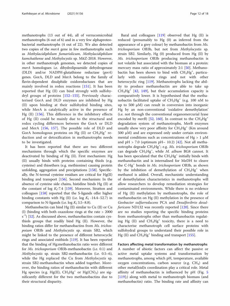
Factors affecting metal transformation by methanotrophsA number of abiotic factors can affect the passive oractive metal uptake systems and transformation bymethanotrophs, among which pH, temperature, availableoxygen concentrations, carbon source (e.g. CH4) andother metal(loid)s coordination play a critical role. Metalaffinity of methanobactin is influenced by pH (Fig. 5[119];) along with metal to methanotroph biomass (andmethanobactin) ratio. The binding rate and affinity can
Karthikeyan et al. Microbiome (2021) 9:156 Page 12 of 18
alter with the molar ratio of metal to methanobactin (i.e.Cu and methanobactin). If the ratio is 0.5 or above,methanobactin binds as a monomer and at lower ratiosas tetramer or oligomer. In the presence of other metals,Cu will be the preferred metal ion that will be readilytaken up by the methanotrophs, while least preferred isiron. In presence of Ag (I) or Au (II), the copper uptakewill be limited. Our understanding of the physiology ofmethanotrophs, particularly the regulation of genes (e.g.TonB, mbnABCM, arsRBC, merABCD, etc.) for metaluptake and/or transformations in relation to concentra-tions of Cu and other metals is limited. Uptake of Zncan be affected by Hg (II) or Cd, which are thiophilic innature and bind to the cysteine in methanobactin.Temperature influences the solubility of methane insolution and thereby influences methanotrophic growth,which in turn may significantly influence metal ionuptake. However, there is no detailed understanding onhow metal affinity for methanobactin and other
methanotroph metal uptake systems varies at differenttemperatures. While methanotrophs with no capacity tomake methanobactin can also uptake and accumulatemetals, studies on these methanotrophs are also verylimited. Pure cultures and mixed methanotrophs cul-tures with or without heterotrophs (that mimic naturalconditions) may also behave differently in metal uptakeand transformations and future research is required tounderstand metal transformations in near in situ condi-tions. Lai and colleagues [161] found that the availablenitrate had significant impact on bromate reduction,while they also correlated it with polyhydroxyalkanoate(PHA) accumulation capacity. However, not all thestrains can make PHA as storage material.A range of biotic interactions (e.g. methanotroph—het-
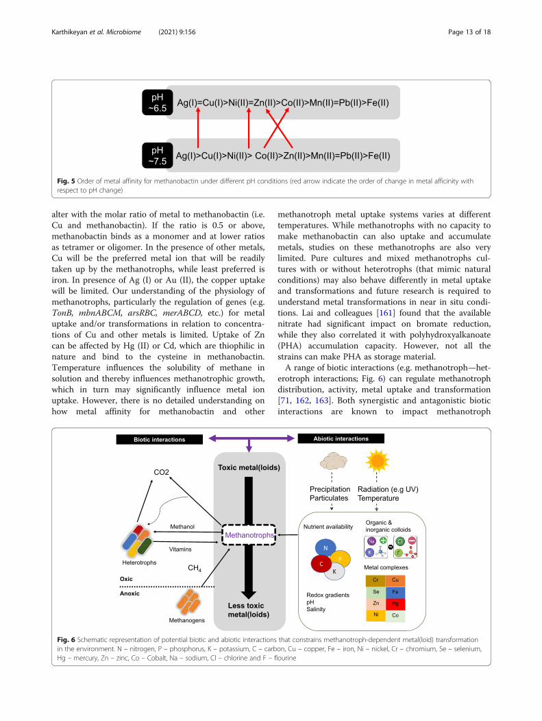
erotroph interactions; Fig. 6) can regulate methanotrophdistribution, activity, metal uptake and transformation[71, 162, 163]. Both synergistic and antagonistic bioticinteractions are known to impact methanotroph
Fig. 6 Schematic representation of potential biotic and abiotic interactions that constrains methanotroph-dependent metal(loid) transformationin the environment. N – nitrogen, P – phosphorus, K – potassium, C – carbon, Cu – copper, Fe – iron, Ni – nickel, Cr – chromium, Se – selenium,Hg – mercury, Zn – zinc, Co – Cobalt, Na – sodium, Cl – chlorine and F – flourine
Fig. 5 Order of metal affinity for methanobactin under different pH conditions (red arrow indicate the order of change in metal afficinity withrespect to pH change)
Karthikeyan et al. Microbiome (2021) 9:156 Page 13 of 18

functional diversity and have been extensivelyreviewed in [162]. It has been well-established thatexchange of metabolites between methanotrophs andheterotrophs improve methanotroph growth and ac-tivity [163, 164]. Methanotroph-heterotroph interac-tions are also constrainted by various abiotic factors.For instance, the ratio of CH4 to O2 altered themethanotroph-heterotroph community structure inthe enrichment. In particular, PHB accumulatingalphaproteobacterial methanotrophs dominanted withincreasing CH4 content [165, 166]. Moreover, differ-ences in Cu to Fe ratio were also found to impactcommunity composition [167]. Our understanding onthe role of biotic interactions on metal transformationby methanotrophs is currently limited. Given thepotential role of methanotrophs in bioremediationstrategies, there is an immediate need to explore howcommunity level biotic interactions impact metaltransformations and uptake.
Conclusions and future considerationsManufacturing and resources industries are key driversfor economic growth, yet this comes at a huge environ-mental cost affecting not only ecosystem services butalso the livelihood of local communities. Aerobic metha-notrophs are metabolically versatile and are able todetoxify toxic heavy metals such as chromium andmercury while growing on a cheap feedstock i.e. me-thane. In order to fully exploit these traits for mitigationof polluted sites, future research is required to betterunderstand (i) physiological and genetic basis of Cr(VI)reduction and demethylation of mercury, (ii) mechanismof Cu reduction by methanobactin, (iii) role of porins inpassive uptake of metals in methanotrophs, and (iv) roleof molybdenum and tungsten in formate dehydrogenaseactivity. More importantly, our knowledge of metaltransformations by methanotrophs is based to a large ex-tent on laboratory strains. Further research is requiredto understand metal uptake/transformation mechanismsin the environment, particularly in polluted sites with el-evated and/or multiple metal concentrations.
AcknowledgementsThe authors thank Mr Khaleel Mohammed (Erode Tannery Limited) for usefuldiscussions and two anonymous reviewers for their insightful comments.
Authors’ contributionsOPK and DK conceived the idea and and all authors contributed to the finaldraft. The author(s) read and approved the final manuscript.
FundingWe acknowledge the funding from the Department for Economy(DfE)—Global Challenges Research (GCRF) grant to DK and GCRF—GlobalImpact Accelerator Award to DK and PNW, BBSRC (BB/C00399X/1 and BB/F01449X/1), UK Science and Technology Facilities Council and EuropeanSynchotron Radiation Facility for TJS.
Availability of data and materialsThe datasets (genome sequences) analysed during the current study areavailable through NCBI genome assembly repository (www.ncbi.nlm.nih.gov)
Declarations
Ethics approval and consent to participateNot applicable
Consent for publicationNot applicable
Competing interestsThe authors declare no competing interests.
Author details1School of Biological Sciences & Institute for Global Food Security, Queen’sUniversity Belfast, 19 Chlorine Gardens, Belfast, UK. 2Civil and EnvironmentalEngineering, University of Michigan, Ann Arbor, MI, USA. 3Department ofEngineering Technology, College of Technology, University of Houston,Houston, TX, USA. 4Biomolecular Sciences Research Centre, Sheffield HallamUniversity, Sheffield, UK. 5Department of Environmental Sciences, Tamil NaduAgricultural University, Coimbatore, India. 6Department of Chemistry,University of Guyana, Georgetown, Guyana. 7Faculty of Science, SRMUniversity, Chennai, India.
Received: 21 January 2021 Accepted: 9 June 2021
References1. Kotrba P, Najmanova J, Macek T, Ruml T, Mackova M. Genetically modified
plants in phytoremediation of heavy metal and metalloid soil and sedimentpollution. Biotechnol Adv. 2009;27(6):799–810. https://doi.org/10.1016/j.biotechadv.2009.06.003.
2. Nabulo G, Young SD, Black CR. Assessing risk to human health from tropicalleafy vegetables grown on contaminated urban soils. Sci Total Environ.2010;408(22):5338–51. https://doi.org/10.1016/j.scitotenv.2010.06.034.
3. Aryal M, Liakopoulou-Kyriakides M. Bioremoval of heavy metals by bacterialbiomass. Environ Monit Assess. 2015;187(1):4173. https://doi.org/10.1007/s10661-014-4173-z.
4. Nagajyoti PC, Lee KD, Sreekanth TVM. Heavy metals, occurrence and toxicityfor plants: a review. Environ Chem Lett. 2010;8(3):199–216. https://doi.org/10.1007/s10311-010-0297-8.
5. Chistoserdova L, Kalyuzhnaya MG. Current trends in methylotrophy. TrendsMicrobiol. 2018;26(8):703–14. https://doi.org/10.1016/j.tim.2018.01.011.
6. Barbosa F Jr. Toxicology of metals and metalloids: promising issues forfuture studies in environmental health and toxicology. J Toxicol EnvironHealth A. 2017;80(3):137–44. https://doi.org/10.1080/15287394.2016.1259475.
7. Dalcorso G, Fasani E, Furini A. Recent advances in the analysis of metalhyperaccumulation and hypertolerance in plants using proteomics. FrontPlant Sci. 2013;4:280.
8. Jaishankar M, Tseten T, Anbalagan N, Mathew BB, Beeregowda KN. Toxicity,mechanism and health effects of some heavy metals. Interdiscip Toxicol.2014;7(2):60–72. https://doi.org/10.2478/intox-2014-0009.
9. Jan AT, Azam M, Siddiqui K, Ali A, Choi I, Haq QMR. Heavy metals andhuman health: mechanistic insight into toxicity and counter defense systemof antioxidants. Int J Mol Sci. 2015;16(12):29592–630. https://doi.org/10.3390/ijms161226183.
10. Meena RAA, Sathishkumar P, Ameen F, Yusoff ARM, Gu FL. Heavy metalpollution in immobile and mobile components of lentic ecosystems-areview. Env Sci Pollut R. 2018;25(5):4134–48. https://doi.org/10.1007/s11356-017-0966-2.
11. Wu X, Cobbina SJ, Mao G, Xu H, Zhang Z, Yang L. A review of toxicity andmechanisms of individual and mixtures of heavy metals in the environment.Environ Sci Pollut R. 2016;23(9):8244–59. https://doi.org/10.1007/s11356-016-6333-x.
12. Ramasamy K, Naidu R. Status of tanning industries in India. In: Towardsbetter management of soils contaminated with tannery waste. Canberra:Australian Centre for International Agricultural Research; 1998. p. 13–21.
Karthikeyan et al. Microbiome (2021) 9:156 Page 14 of 18

13. World Health Organisation & International Programme on Chemical Safety.Guidelines for drinking-water quality, vol. 2. Health Criteria and supportinginformation. 2nd ed. 1996.
14. Sunitha R, Gayathri P, Bharani A, Mahimairajah SJ. Chromium contaminationin soil and groundwater due to tannery waste disposals at Vellore district ofTamil Nadu. Int J Environ Sci. 2015;6:114–24.
15. Naidu R, Kookana RS, Cox J, Mowat D, Smith LH. Fate and chromium attannery waste contaminated sites at Mount Barker. In: Towards bettermanagement of soils contaminated with tannery waste. In: ACIAR(Australian Centre for International Agricultural Research) proceedings.Canberra; 2000. p. 57–70.
16. Avudainayagam S, Naidu R, Kookana R, Angus A, Smith LH. Effect ofelectrolyte composition on chromium desorption in soil contaminated bytannery waste. Aust J Soil Res. 2001;39(5):1077–89. https://doi.org/10.1071/SR00085.
17. Kamaludeen SPB, Banu P, Megharaj M, Juhasz A, Sethunathan N, Naidu R.Chromium-Microorganism interactions in soil: implications to remediation.Rev Environ Contam T. 2003;178:93–164.
18. Sethunathan N, Megharaj M, Smith L, Kamaludeen SPB, Avudainayagam S,Naidu R. Microbial role in the failure of natural attenuation of Cr(VI) in long-term tannery waste contaminated soil. Agric Ecosyst Environ. 2005;105(4):657–61. https://doi.org/10.1016/j.agee.2004.08.008.
19. Hausladen DM, Fendorf S. Hexavalent chromium generation within naturallystructured soils and sediments. Environ Sci Technol. 2017;51(4):2058–67.https://doi.org/10.1021/acs.est.6b04039.
20. Lai C, Wen L, Shi L, Zhao K, Wang Y, Yang X, et al. Bioreduction of chromatein a methane-based membrane biofilm reactor. Environ Sci Technol. 2016;50(18):10179–86. https://doi.org/10.1021/acs.est.6b02807.
21. WHO: Trace elements in human nutrition and health. 1996.22. Ohlendorf HM, Kilness AW, Simmons JL, Stroud RK, Hoffman DJ, Moore JF.
Selenium toxicosis in wild aquatic birds. J Toxicol Environ Health. 1988;24(1):67–92. https://doi.org/10.1080/15287398809531141.
23. Etteieb S, Magdouli S, Zolfaghari M, Brar S. Monitoring and analysis ofselenium as an emerging contaminant in mining industry: a critical review.Sci Total Environ. 2020;698:134339. https://doi.org/10.1016/j.scitotenv.2019.134339.
24. Izquierdo M, Querol X. Leaching behaviour of elements from coalcombustion fly ash: an overview. Int J Coal Geol. 2012;94:54–66. https://doi.org/10.1016/j.coal.2011.10.006.
25. George A, Shen B, Kang D, Yang J, Luo J. Emission control strategiesof hazardous trace elements from coal-fired power plants in China. JEnviron Sci (China). 2020;93:66–90. https://doi.org/10.1016/j.jes.2020.02.025.
26. Srigboh RK, Basu N, Stephens J, Asampong E, Perkins M, Neitzel RL, et al.Multiple elemental exposures amongst workers at the Agbogbloshieelectronic waste (e-waste) site in Ghana. Chemosphere. 2016;164:68–74.https://doi.org/10.1016/j.chemosphere.2016.08.089.
27. Skalickova S, Milosavljevic V, Cihalova K, Horky P, Richtera L, Adam V.Selenium nanoparticles as a nutritional supplement. Nutrition. 2017;33:83–90. https://doi.org/10.1016/j.nut.2016.05.001.
28. United Nations Environmental Programme. Global mercury assessmentsources, emissions, releases and environmental transport. Geneva: UNEP;2013. https://wedocs.unep.org/20.500.11822/7984.
29. Telmer KH, Veiga MM, Mason R, Pirrone N, Boston MA. World emissions ofmercury from artisanal and small scale gold mining. In: Pirrone N, Mason R,editors. Mercury fate and transport in the global atmosphere. Boston:Springer; 2009.
30. Bank of Guyana. Annual Report. Bank of Guyana Georgetown; 2018.31. Pasha S, Wenner MD, Clarke D. Toward the greening of the gold mining
sector of Guyana: transition issues and challenges. Georgetown: CountryDepartment Caribbean Group; 2017.
32. WWF-Guinanas: Gold miner’s knowledge, attitudes and practice withregards to mercury: a study in three small-scale gold mining regions inSuriname. 2014.
33. Romero R, Ohashi C, Williams P, Hourty T, Bynore P, Insanally O. GuyanaMinamata initial assessment report; 2016.
34. Legg ED, Ouboter PE, Wright MAP. Small-scale gold mining relatedmercury contamination in the Guianas: a review. Paramaribo: WorldWildlife Fund; 2015.
35. Howard J, Trotz MA, Thomas K, Omisca E, Chiu HT, Halfhide T, et al. Totalmercury loadings in sediment from gold mining and conservation areas in
Guyana. Environ Monit Assess. 2011;179(1-4):555–73. https://doi.org/10.1007/s10661-010-1762-3.
36. Singh H, Bernard C, Rampersaud P, Laing T, Balraj D, Priester M, et al.Guyana’s extractive industry sector (EIS): a synopsis of issues andrecommendations for the mining sector as a sustainable element ofGuyana’s low carbon development strategy (LCDS): CI-Guyana. Georgetown:Projekt-Consult GmbH and WWF Guianas; 2018.
37. Environmental Protection Agency. State of the Environment ReportGeorgetown Guyana 2016.
38. Bolan N, Kunhikrishnan A, Thangarajan R, Kumpiene J, Park J, Makino T, et al.Remediation of heavy metal(loid)s contaminated soils--to mobilize or toimmobilize? J Hazard Mater. 2014;266:141–66. https://doi.org/10.1016/j.jhazmat.2013.12.018.
39. Ma Y, Oliverira R, Freitas H, Zhang C. Biochemical and molecularmechanisms of plant-microbe-metal interactions: relevance forphytoremediation. Front Plant Sci. 2016;7:918.
40. Eswayah AS, Smith TJ, Gardiner PHE. Microbial transformations of seleniumspecies of relevance to bioremediation. Appl Environ Microbiol. 2016;82(16):4848–59. https://doi.org/10.1128/AEM.00877-16.
41. Hasin A, Gurman SJ, Murphy LM, Perry A, Smith TJ, Gardiner PHE.Remediation of chromium (VI) by a methane-oxidising bacterium. EnvironSci Technol. 2010;44(1):400–5. https://doi.org/10.1021/es901723c.
42. Lu X, Gu W, Zhao L. Farhan Ul Haque M, DiSpirito AA, Semrau JD, Gu B:Methylmercury uptake and degradation by methanotrophs. Sci Adv. 2017;3(5):e1700041. https://doi.org/10.1126/sciadv.1700041.
43. Borch T, Kretzschmar R, Kappler A, Cappellen PV, Ginder-Vogel M, VoegelinA, et al. Biogeochemical redox processes and their impact on contaminantdynamics. Environ Sci Technol. 2010;44(1):15–23. https://doi.org/10.1021/es9026248.
44. Gadd GM. Metals, minerals and microbes: geomicrobiology andbioremediation. Microbiology (Reading). 2010;156(Pt 3):609–43. https://doi.org/10.1099/mic.0.037143-0.
45. Reeder RJ, Schoonen MAA, Lanzirotti A. Metal speciation and its role inbioaccessibility and bioavailability. Rev Mineral Geochem. 2006;64(1):59–113.https://doi.org/10.2138/rmg.2006.64.3.
46. Abbas G, Murtaza B, Bibi I, Shahid M, Niazi NK, Khan MI, et al. Arsenicuptake, toxicity, detoxification, and speciation in plants: physiological,biochemical, and molecular aspects. Int J Environ Res Public Health.2018;15(1):59. https://doi.org/10.3390/ijerph15010059.
47. Wang D, Liang D, Wang S, Hu B, Wei W. Individual and joint toxicity effectsof Cu, Cr(III), and Cr(VI) on pakchoi: a comparison between solution and soilcultures. Biol Trace Elem Res. 2012;146(1):116–23. https://doi.org/10.1007/s12011-011-9219-2.
48. Obrist D, Kirk JL, Zhang L, Sunderland EM, Jiskra M, Selin NE. A review ofglobal environmental mercury processes in response to human and naturalperturbations: Changes of emissions, climate, and land use. Ambio. 2018;47(2):116–40. https://doi.org/10.1007/s13280-017-1004-9.
49. Eswayah AS, Smith TJ, Scheinost AC, Hondow N, Gardiner PHE.Microbial transformations of selenite by methane-oxidizing bacteria.Appl Microbiol Biotechnol. 2017;101(17):6713–24. https://doi.org/10.1007/s00253-017-8380-8.
50. Vorobev A, Jagadevan S, Baral BS, Dispirito AA, Freemeier BC, Bergman BH,et al. Detoxification of mercury by methanobactin from Methylosinustrichosporium OB3b. Appl Environ Microbiol. 2013;79(19):5918–26. https://doi.org/10.1128/AEM.01673-13.
51. Kalyuzhnaya MG, Gomez OA, Murrell JC. The methane-oxidizing bacteria(methanotrophs). In Taxonomy, genomics and ecophysiology ofhydrocarbon-degrading microbes. Edited by McGenity TJ. Cham: SpringerSpringer Nature; 2019. p. 1–34.
52. Semrau JD, DiSpirito AA, Gu W, Yoon S. Metals and methanotrophy. ApplEnviron Microbiol. 2018:84(6):e02289–17.
53. Ettwig KF, Zhu B, Speth D, Keltjens JT, Jetten MSM, Kartal B. Archaeacatalyze iron-dependent anaerobic oxidation of methane. P Natl Acad SciUSA. 2016;113(45):12792–6. https://doi.org/10.1073/pnas.1609534113.
54. Vigneron A, Alsop EB, Cruaud P, Philibert G, King B, Baksmaty L, et al.Contrasting pathways for anaerobic methane oxidation in gulf of Mexicocold seep sediments. mSystems. 2019;4(1):e00091–18.
55. Versantvoort W, Guerrero-Cruz S, Speth DR, Frank J, Gambelli L, Cremers G,et al. Comparative genomics of Candidatus Methylomirabilis species anddescription of Ca. Methylomirabilis Lanthanidiphila. Front Microbiol. 2018;9:1672. https://doi.org/10.3389/fmicb.2018.01672.
Karthikeyan et al. Microbiome (2021) 9:156 Page 15 of 18

56. Antony CP, Kumaresan D, Ferrando L, Boden R, Moussard H, Scavino AF,et al. Active methylotrophs in the sediments of Lonar Lake, a saline andalkaline ecosystem formed by meteor impact. ISME J. 2010;4(11):1470–80.https://doi.org/10.1038/ismej.2010.70.
57. Kumaresan D, Abell GCJ, Bodrossy L, Stralis-Pavese N, Murrell JC. Spatial andtemporal diversity of methanotrophs in a landfill cover soil are differentiallyrelated to soil abiotic factors. Environ Microbiol Rep. 2009;1(5):398–407.https://doi.org/10.1111/j.1758-2229.2009.00059.x.
58. Kumaresan D, Stephenson J, Doxey AC, Bandukwala H, Brooks E, Hillebrand-Voiculescu A, et al. Aerobic proteobacterial methylotrophs in Movile Cave:genomic and metagenomic analyses. Microbiome. 2018;6(1):1. https://doi.org/10.1186/s40168-017-0383-2.
59. Sheng R, Chen A, Zhang M, Whiteley AS, Kumaresan D, Wei W.Transcriptional activities of methanogens and methanotrophs vary withmethane emission flux in rice soils under chronic nutrient constraints ofphosphorus and potassium. Biogeosciences. 2016;13(23):6507–18. https://doi.org/10.5194/bg-13-6507-2016.
60. Trotsenko YA, Murrell JC. Metabolic aspects of aerobic obligatemethanotrophy. Adv Appl Microbiol. 2008;63:183–229. https://doi.org/10.1016/S0065-2164(07)00005-6.
61. Tavormina PL, Orphan VJ, Kalyuzhnaya MG, Jetten MSM, Klotz MG. A novelfamily of functional operons encoding methane/ammonia monooxygenase-related proteins in gammaproteobacterial methanotrophs. EnvironMicrobiol Rep. 2011;3(1):91–100. https://doi.org/10.1111/j.1758-2229.2010.00192.x.
62. Glass JB, Orphan VJ. Trace metal requirements for microbial enzymesinvolved in the production and consumption of methane and nitrous oxide.Front Microbiol. 2012;3:61.
63. Takeguchi M, Ohashi M, Okura I. Role of iron in particulate methanemonooxygenase from Methylosinus trichosporium OB3b. Biometals. 1999;12(2):123–9. https://doi.org/10.1023/A:1009257826998.
64. Ross MO, MacMillan F, Wang J, Nisthal A, Lawton TJ, Olafson BD, et al.Particulate methane monooxygenase contains only mononuclearcopper centers. Science. 2019;364(6440):566–70. https://doi.org/10.1126/science.aav2572.
65. Bordel S, Rodriguez Y, Hakobyan A, Rodriguez E, Lebrero R, Munoz R.Genome scale metabolic modeling reveals the metabolic potential of threetype II methanotrophs of the genus Methylocystis. Metab Eng. 2019;54:191–9. https://doi.org/10.1016/j.ymben.2019.04.001.
66. Hakobyan A, Liesack W. Unexpected metabolic versatility among type IImethanotrophs in the Alphaproteobacteria. Biol Chem. 2020;401(12):1469–77. https://doi.org/10.1515/hsz-2020-0200.
67. Kalyuzhnaya MG, Puri AW, Lidstrom ME. Metabolic engineering inmethanotrophic bacteria. Metab Eng. 2015;29:142–52. https://doi.org/10.1016/j.ymben.2015.03.010.
68. Murrell JC, Smith TJ. Biochemistry and Molecular biology of methanemonooxygenase. In: Timmis KN, editor. Handbook of Hydrocarbon andLipid Microbiology. Berlin, Heidelberg: Springer; 2010.
69. Semrau JD, DiSpirito AA, Yoon S. Methanotrophs and copper. FEMSMicrobiol Rev. 2010;34(4):496–531. https://doi.org/10.1111/j.1574-6976.2010.00212.x.
70. Sirajuddin S, Rosenzweig AC. Enzymatic oxidation of methane. Biochemistry.2015;54(14):2283–94. https://doi.org/10.1021/acs.biochem.5b00198.
71. Chidambarampadmavathy K, Karthikeyan OP, Huerlimann R, Maes GE,Heimann K. Response of mixed methanotrophic consortia to differentmethane to oxygen ratios. Waste Manag. 2017;61:220–8. https://doi.org/10.1016/j.wasman.2016.11.007.
72. DiSpirito AA, Semrau JD, Murrell JC, Gallagher WH, Dennison C, VuilleumierS. Methanobactin and the link between copper and bacterial methaneoxidation. Microbiol Mol Biol Rev. 2016;80(2):387–409. https://doi.org/10.1128/MMBR.00058-15.
73. Choi D-W, Kunz RC, Boyd ES, Semrau JD, Antholine WE, Han JI, et al. Themembrane-associated methane monooxygenase (pMMO) and pMMO-NADH:quinone oxidoreductase complex from Methylococcus capsulatusBath. J Bacteriol. 2003;185(19):5755–64. https://doi.org/10.1128/JB.185.19.5755-5764.2003.
74. Dassama LMK, Kenney GE, Ro SY, Zielazinski EL, Rosenzweig AC.Methanobactin transport machinery. P Natl Acad Sci USA. 2016;113(46):13027–32. https://doi.org/10.1073/pnas.1603578113.
75. Kalyuzhnaya MG, Yang S, Rozova ON, Smalley NE, Clubb J, Lamb A,et al. Highly efficient methane biocatalysis revealed in a
methanotrophic bacterium. Nat Commun. 2013;4(1):2785. https://doi.org/10.1038/ncomms3785.
76. Osborne CD, Haritos VS. Horizontal gene transfer of three co-inheritedmethane monooxygenase systems gave rise to methanotrophy in theProteobacteria. Mol Phylogenet Evol. 2018;129:171–81. https://doi.org/10.1016/j.ympev.2018.08.010.
77. Vekeman B, Kerckhof F-M, Cremers G, de Vos P, Vandamme P, Boon N. Opden Camp HJM, Heylen K: New Methyloceanibacter diversity from NorthSea sediments includes methanotroph containing solely the solublemethane monooxygenase. Environ Microbiol. 2016;18(12):4523–36. https://doi.org/10.1111/1462-2920.13485.
78. Chu F, Lidstrom ME. XoxF acts as the predominant methanoldehydrogenase in the type I methanotroph Methylomicrobium buryatense.J Bacteriol. 2016;198(8):1317–25. https://doi.org/10.1128/JB.00959-15.
79. Pol A, Barends TRM, Dietl A, Khadem AF, Eygensteyn J, Jetten MSM. Op denCamp HJM: Rare earth metals are essential for methanotrophic life involcanic mudpots. Environ Microbiol. 2014;16(1):255–64. https://doi.org/10.1111/1462-2920.12249.
80. Wu ML, Wessels JCT, Pol A. Op den Camp HJM, Jetten MSM, van Niftrik L:XoxF-type methanol dehydrogenase from the anaerobic methanotroph“Candidatus Methylomirabilis oxyfera”. Appl Environ Microbiol. 2015;81(4):1442–51. https://doi.org/10.1128/AEM.03292-14.
81. Yu Z, Chistoserdova L. Communal metabolism of methane and the rareearth element switch. J Bacteriol. 2017;199(22):e00328–17. https://doi.org/10.1128/JB.00328-17.
82. Anthony C, Williams P. The structure and mechanism of methanoldehydrogenase. Biochim Biophys Acta. 2003;1647(1-2):18–23. https://doi.org/10.1016/S1570-9639(03)00042-6.
83. Kalyuzhnaya MG, Hristova KR, Lidstrom ME, Chistoserdova L.Characterization of a novel methanol dehydrogenase in representatives ofBurkholderiales: implications for environmental detection of methylotrophyand evidence for convergent evolution. J Bacteriol. 2008;190(11):3817–23.https://doi.org/10.1128/JB.00180-08.
84. Reddy SY, Bruice TC. Mechanisms of ammonia activation and ammoniumion inhibition of quinoprotein methanol dehydrogenase: a computationalapproach. Proc Natl Acad Sci U S A. 2004;101(45):15887–92. https://doi.org/10.1073/pnas.0407209101.
85. Culpepper MA, Rosenzweig AC. Structure and protein-protein interactionsof methanol dehydrogenase from Methylococcus capsulatus (Bath).Biochemistry. 2014;53(39):6211–9. https://doi.org/10.1021/bi500850j.
86. Deng YW, Ro SY, Rosenzweig AC. Structure and function of the lanthanide-dependent methanol dehydrogenase XoxF from the methanotrophMethylomicrobium buryatense 5GB1C. J Biol Inorg Chem. 2018;23(7):1037–47. https://doi.org/10.1007/s00775-018-1604-2.
87. Picone N. Op den Camp HJ: Role of rare earth elements in methanoloxidation. Curr Opin Chem Biol. 2019;49:39–44. https://doi.org/10.1016/j.cbpa.2018.09.019.
88. Huang J, Yu Z, Chistoserdova L. Lanthanide-dependent methanoldehydrogenases of XoxF4 and XoxF5 clades are differentially distributed amongmethylotrophic bacteria and they reveal different biochemical properties. FrontMicrobiol. 2018;9:1366. https://doi.org/10.3389/fmicb.2018.01366.
89. Bogart JA, Lewis AJ, Schelter EJ. DFT study of the active site of the XoxF-type natural, cerium-dependent methanol dehydrogenase enzyme.Chemistry. 2015;21(4):1743–8. https://doi.org/10.1002/chem.201405159.
90. Akberdin IR, Thompson M, Hamilton R, Desai N, Alexander D, HenardCA, et al. Methane utilization in Methylomicrobium alcaliphilum 20ZR: asystems approach. Sci Rep. 2018;8(1):2512. https://doi.org/10.1038/s41598-018-20574-z.
91. Keltjens JT, Pol A, Reimann J. Op den Camp HJ: PQQ-dependent methanoldehydrogenases: rare-earth elements make a difference. Appl MicrobiolBiotechnol. 2014;98(14):6163–83. https://doi.org/10.1007/s00253-014-5766-8.
92. Good NM, Moore RS, Suriano CJ, Martinez-Gomez NC. Contrasting in vitroand in vivo methanol oxidation activities of lanthanide-dependent alcoholdehydrogenases XoxF1 and ExaF from Methylobacterium extorquens AM1.Sci Rep. 2019;9(1):4248. https://doi.org/10.1038/s41598-019-41043-1.
93. Akberdin IR, Collins DA, Hamilton R, Oshchepkov DY, Shukla AK, Nicora CD,et al. Rare earth elements alter redox balance in Methylomicrobiumalcaliphilum 20Z(R). Front Microbiol. 2018;9:2735. https://doi.org/10.3389/fmicb.2018.02735.
94. Khmelenina VV, Murrell JC, Smith TJ, Trotsenko YA, Rojo F. Physiology andbiochemistry of the aerobic methanotrophs. In: Aerobic utilization of
Karthikeyan et al. Microbiome (2021) 9:156 Page 16 of 18

hydrocarbons oils and lipids handbook of hydrocarbon and lipidmicrobiology. Edited by Rojo F. Cham: Springer Nature; 2018. p. 1–25.
95. Chistoserdova L, Vorholt JA, Lidstrom ME. A genomic view of methaneoxidation by aerobic bacteria and anaerobic archaea. Genome Biol. 2005;6(2):208. https://doi.org/10.1186/gb-2005-6-2-208.
96. Matsen JB, Yang S, Stein LY, Beck D, Kalyuzhnaya MG. Global molecularanalyses of methane metabolism in Methanotrophic Alphaproteobacterium,Methylosinus trichosporium OB3b. Part I: Transcriptomic Study. FrontMicrobiol. 2013;4:40.
97. Stein LY, Yoon S, Semrau JD, Dispirito AA, Crombie A, Murrell JC, et al. Opden Camp HJ, Bringel F et al: Genome sequence of the obligatemethanotroph Methylosinus trichosporium strain OB3b. J Bacteriol. 2010;192(24):6497–8. https://doi.org/10.1128/JB.01144-10.
98. Vorholt JA, Chistoserdova L, Stolyar SM, Thauer RK, Lidstrom ME.Distribution of tetrahydromethanopterin-dependent enzymes inmethylotrophic bacteria and phylogeny of methenyltetrahydromethanopterin cyclohydrolases. J Bacteriol. 1999;181(18):5750–7. https://doi.org/10.1128/JB.181.18.5750-5757.1999.
99. Zahn JA, Bergmann DJ, Boyd JM, Kunz RC, DiSpirito AA. Membrane-associated quinoprotein formaldehyde dehydrogenase from Methylococcuscapsulatus Bath. J Bacteriol. 2001;183(23):6832–40. https://doi.org/10.1128/JB.183.23.6832-6840.2001.
100. Stirling DI, Dalton H. Purification and properties of an NAD(P)+-linkedformaldehyde dehydrogenase from Methylococcus capsulatus (Bath). J GenMicrobiol. 1978;107(1):19–29. https://doi.org/10.1099/00221287-107-1-19.
101. Tate S, Dalton H. A low-molecular-mass protein from Methylococcuscapsulatus (Bath) is responsible for the regulation of formaldehydedehydrogenase activity in vitro. Microbiology (Reading). 1999;145(Pt 1):159–67. https://doi.org/10.1099/13500872-145-1-159.
102. Adeosun EK, Smith TJ, Hoberg AM, Velarde G, Ford R, Dalton H.Formaldehyde dehydrogenase preparations from Methylococcus capsulatus(Bath) comprise methanol dehydrogenase and methylenetetrahydromethanopterin dehydrogenase. Microbiology (Reading). 2004;150(Pt 3):707–13. https://doi.org/10.1099/mic.0.26707-0.
103. Jollie DR, Lipscomb JD. Formate dehydrogenase from Methylosinustrichosporium OB3b. Purification and spectroscopic characterization of thecofactors. J Biol Chem. 1991;266(32):21853–63. https://doi.org/10.1016/S0021-9258(18)54716-5.
104. Waldron KJ, Robinson NJ. How do bacterial cells ensure thatmetalloproteins get the correct metal? Nat Rev Microbiol. 2009;7(1):25–35.https://doi.org/10.1038/nrmicro2057.
105. Koh E-I, Henderson JP. Microbial copper-binding siderophores at the host-pathogen interface. J Biol Chem. 2015;290(31):18967–74. https://doi.org/10.1074/jbc.R115.644328.
106. Balasubramanian R, Kenney GE, Rosenzweig AC. Dual pathways for copperuptake by methanotrophic bacteria. J Biol Chem. 2011;286(43):37313–9.https://doi.org/10.1074/jbc.M111.284984.
107. Gu W, Farhan Ul Haque M, Baral BS, Turpin EA, Bandow NL, Kremmer E,et al. A TonB-Dependent transporter is responsible for methanobactinuptake by Methylosinus trichosporium OB3b. Appl Environ Microbiol. 2016;82(6):1917–23. https://doi.org/10.1128/AEM.03884-15.
108. Vollan HS, Tannæs T, Vriend G, Bukholm G. In silico structure and sequenceanalysis of bacterial porins and specific diffusion channels for hydrophilicmolecules: conservation, multimericity and multifunctionality. Int J Mol Sci.2016;17(4):599.
109. Kim HJ, Graham DW, DiSpirito AA, Alterman MA, Galeva N, Larive CK, et al.Methanobactin, a copper-acquisition compound from methane-oxidizingbacteria. Science. 2004;305(5690):1612–5. https://doi.org/10.1126/science.1098322.
110. Helland R, Fjellbirkeland A, Karlsen OA, Ve T, Lillehaug JR, Jensen HB. Anoxidized tryptophan facilitates copper binding in Methylococcus capsulatus-secreted protein MopE. J Biol Chem. 2008;238:13897–904.
111. Kenney GE, Dassama LMK, Pandelia M-E, Gizzi AS, Martinie RJ, Gao P, et al.The biosynthesis of methanobactin. Science. 2018;359(6382):1411–6. https://doi.org/10.1126/science.aap9437.
112. Semrau JD, DiSpirito AA, Obulisamy PK, Kang-Yun CS. Methanobactin frommethanotrophs: genetics, structure, function and potential applications.FEMS Microbiol Lett. 2020;367(5):fnaa045.
113. Dassama LMK, Kenney GE, Rosenzweig AC. Methanobactins: fromgenome to function. Metallomics. 2017;9(1):7–20. https://doi.org/10.1039/C6MT00208K.
114. Choi DW, Bandow NL, McEllistrem MT, Semrau JD, Antholine WE, Hartsel SC,et al. Spectral and thermodynamic properties of methanobactin from γ-proteobacterial methane oxidizing bacteria: a case for copper competitionon a molecular level. J Inorg Biochem. 2010;104(12):1240–7. https://doi.org/10.1016/j.jinorgbio.2010.08.002.
115. Choi DW, Zea CJ, Do YS, Semrau JD, Antholine WE, Hargrove MS, et al.Spectral, kinetic, and thermodynamic properties of Cu(I) and Cu(II) bindingby methanobactin from Methylosinus trichosporium OB3b. Biochemistry.2006;45(5):1442–53. https://doi.org/10.1021/bi051815t.
116. Baslé A, El Ghazouani A, Lee J, Dennison C. Insight into metal removal frompeptides that sequester copper for methane oxidation. Chemistry. 2018;24(18):4515–8. https://doi.org/10.1002/chem.201706035.
117. Kang CS, Dunfield PF, Semrau JD. The origin of aerobic methanotrophywithin the Proteobacteria. FEMS Microbiol Letts. 2019;366(9):fnz096.
118. McCabe JW, Vangala R, Angel LA. Binding selectivity of methanobactin fromMethylosinus trichosporium OB3b for copper(I), silver(I), zinc(II), nickel(II),cobalt(II), manganese(II), lead(II), and iron(II). J Am Soc Mass Spectr. 2017;28(12):2588–601. https://doi.org/10.1007/s13361-017-1778-9.
119. Baral BS, Bandow NL, Vorobev A, Freemeier BC, Bergman BH, Herdendorf TJ,et al. Mercury binding by methanobactin from Methylocystis strain SB2. J InorgBiochem. 2014;141:161–9. https://doi.org/10.1016/j.jinorgbio.2014.09.004.
120. Yin X, Wang L, Zhang L, Chen H, Liang X, Lu X, et al. Synergistic effects of achalkophore, methanobactin, on microbial methylation of mercury. ApplEnviron Microbiol. 2020;86(11):e00122–20.
121. Choi DW, Do YS, Zea CJ, McEllistrem MT, Lee S-W, Semrau JD, et al. Spectraland thermodynamic properties of Ag(I), Au(III), Cd(II), Co(II), Fe(III), Hg(II),Mn(II), Ni(II), Pb(II), U(IV), and Zn(II) binding by methanobactin fromMethylosinus trichosporium OB3b. J Inorg Biochem. 2006;100(12):2150–61.https://doi.org/10.1016/j.jinorgbio.2006.08.017.
122. Dennison C, David S, Lee J. Bacterial copper storage proteins. J Biol Chem.2018;293(13):4616–27. https://doi.org/10.1074/jbc.TM117.000180.
123. Fjellbirkeland A, Kruger PG, Bemanian V, Høgh BT, Murrell JC, Jensen HB.The C-terminal part of the surface-associated protein MopE of themethanotroph Methylococcus capsulatus (Bath) is secreted into the growthmedium. Arch Microbiol. 2001;176(3):197–203. https://doi.org/10.1007/s002030100307.
124. Martinho M, Choi DW, Dispirito AA, Antholine WE, Semrau JD, Münck E.Mössbauer studies of the membrane-associated methane monooxygenasefrom Methylococcus capsulatus bath: evidence for a Diiron center. J AmChem Soc. 2007;129(51):15783–5. https://doi.org/10.1021/ja077682b.
125. Semrau JD, Jagadevan S, DiSpirito AA, Khalifa A, Scanlan J, Bergman BH,et al. Methanobactin and MmoD work in concert to act as the 'copper-switch' in methanotrophs. Environ Microbiol. 2013;15(11):3077–86. https://doi.org/10.1111/1462-2920.12150.
126. El Ghazouani A, Baslé A, Gray J, Graham DW, Firbank SJ, Dennison C.Variations in methanobactin structure influences copper utilization bymethane-oxidizing bacteria. P Natl Acad Sci USA. 2012;109(22):8400–4.https://doi.org/10.1073/pnas.1112921109.
127. Jahng D, Wood T. Metal ions and chloramphenicol inhibition of solublemethane monooxygenase from Methylosinus trichosporium OB3b. ApplMicrobiol Biotechnol. 1996;45(6):744–9. https://doi.org/10.1007/s002530050757.
128. Pesch M-L, Christl I, Hoffmann M, Kraemer SM, Kretzschmar R. Coppercomplexation of methanobactin isolated from Methylosinus trichosporiumOB3b: pH-dependent speciation and modeling. J Inorg Biochem. 2012;116:55–62. https://doi.org/10.1016/j.jinorgbio.2012.07.008.
129. Vita N, Platsaki S, Baslé A, Allen SJ, Paterson NG, Crombie AT, et al. A four-helix bundle stores copper for methane oxidation. Nature. 2015;525(7567):140–3. https://doi.org/10.1038/nature14854.
130. Vita N, Landolfi G, Baslé A, Platsaki S, Lee J, Waldron KJ, et al. Bacterialcytosolic proteins with a high capacity for Cu(I) that protect against coppertoxicity. Sci Rep. 2016;6(1):39065. https://doi.org/10.1038/srep39065.
131. Cornu JY, Huguenot D, Jezequel K, Lollier M, Lebeau T. Bioremediation ofcopper-contaminated soils by bacteria. World J Microbiol Biotechnol. 2017;33(2):26. https://doi.org/10.1007/s11274-016-2191-4.
132. Cervantes C, Campos-García J, Devars S, Gutiérrez-Corona F, Loza-Tavera H,Torres-Guzmán JC, et al. Interactions of chromium with microorganisms andplants. FEMS Microbiol Rev. 2001;25(3):335–47. https://doi.org/10.1111/j.1574-6976.2001.tb00581.x.
133. Beukes JP, du Preez SP, van Zyl PG, Paktunc D, Fabritius T, Paatalo M, et al.Review of Cr(VI) environmental practices in the chromite mining and
Karthikeyan et al. Microbiome (2021) 9:156 Page 17 of 18

smelting industry – relevance to development of the Ring of Fire, Canada. JClean Prod. 2017;165:874–89. https://doi.org/10.1016/j.jclepro.2017.07.176.
134. Coetzee JJ, Bansal N, Chirwa EMN. Chromium in environment, its toxiceffect from chromite-mining and ferrochrome industries, and its possiblebioremediation. Expo Health. 2020;12:51–62. https://doi.org/10.1007/s12403-018-0284-z.
135. Krober E, Cunningham MR, Peixoto J, Spurgin L, Wischer D, Kruger R, et al.Comparative genomics analyses indicate differential methylated amineutilization trait within members of the genus Gemmobacter. EnvironMicrobiol Rep. 2021;13(2):195–208. https://doi.org/10.1111/1758-2229.12927.
136. Enbaia S, Eswayah A, Hondow N, Gardiner PHE, Smith TJ. Detoxification,active uptake, and intracellular accumulation of chromium species by amethane-oxidizing bacterium. Appl Environ Microbiol. 2021;87(2):e00947–20.
137. Challa S, Smith TJ. Isolation of a methane-oxidizing bacterium thatbioremediates hexavalent chromium from a formerly industrializedSuburban River. Lett Appl Microbiol. 2020;71(3):287–93. https://doi.org/10.1111/lam.13330.
138. Qin H, Zhu J, Liang L, Wang M, Su H. The bioavailability of selenium and riskassessment for human selenium poisoning in high-Se areas, China. EnvironInt. 2013;52:66–74. https://doi.org/10.1016/j.envint.2012.12.003.
139. Dungan RS, Frankenberger WT. Microbial transformations of selenium andthe bioremediation of seleniferous environments. Bioremediat J. 1999;3(3):171–88. https://doi.org/10.1080/10889869991219299.
140. Zhang JS, Gao XY, Zhang LD, Bao YP. Biological effects of a nano redelemental selenium. Biofactors. 2001;15(1):27–38. https://doi.org/10.1002/biof.5520150103.
141. Schlekat CE, Dowdle PR, Lee BG, Luoma SN, Oremland RS. Bioavailability ofparticle-associated se to the bivalve Potamocorbula amurensis. Environ SciTechnol. 2000;34(21):4504–10. https://doi.org/10.1021/es001013f.
142. Lai CY, Wen LL, Shi LD, Zhao KK, Wang YQ, Yang X, et al. Selenate andnitrate bioreductions using methane as the electron donor in a membranebiofilm reactor. Environ Sci Technol. 2016;50(18):10179–86. https://doi.org/10.1021/acs.est.6b02807.
143. Nancharaiah YV, Lens PNL. Selenium biomineralization for biotechnologicalapplications. Trends Biotechnol. 2015;33(6):323–30. https://doi.org/10.1016/j.tibtech.2015.03.004.
144. Oremland RS, Herbel MJ, Blum JS, Langley S, Beveridge TJ, Ajayan PM, et al.Structural and spectral features of selenium nanospheres produced by Se-respiring bacteria. Appl Environ Microbiol. 1994;70:52–60.
145. Wang Y, Lai CY, Wu M, Song Y, Hu S, Yuan Z, et al. Roles of oxygen inmethane-dependent selenate reduction in a membrane biofilm reactor:stimulation or suppression. Water Res. 2021;198:117150. https://doi.org/10.1016/j.watres.2021.117150.
146. Eswayah AS, Hondow N, Scheinost AC, Merroun M, Romero-Gonzalez M,Smith TJ, et al. Methyl selenol as a precursor in selenite reduction toSe/S species by methane-oxidizing bacteria. Appl Environ Microbiol.2019;85(22):e01379–19.
147. Wang D, Xia X, Wu S, Zheng S, Wang G. The essentialness of glutathionereductase GorA for biosynthesis of Se(0)-nanoparticles and GSH for CdSequantum dot formation in Pseudomonas stutzeri TS44. J Hazard Mater.2019;366:301–10. https://doi.org/10.1016/j.jhazmat.2018.11.092.
148. Kim M, Zoh K. Fate and transport of mercury in environmental media andhuman exposure. J Prev Med Public Health. 2012;45(6):335–43. https://doi.org/10.3961/jpmph.2012.45.6.335.
149. Boden R, Murrell JC. Response to mercury (II) ions in Methylococcuscapsulatus (Bath). FEMS Microbiol Letts. 2011;324(2):106–10. https://doi.org/10.1111/j.1574-6968.2011.02395.x.
150. Shi LD, Chen YS, Du JJ, Hu YQ, Shapleigh JP, Zhao HP. Metagenomicevidence for a methylocystis species capable of bioremediation of diverseheavy metals. Front Microbiol. 2018;9:3297.
151. Buey RM, Arellano JB, Lopez-Maury L, Galindo-Trigo S, Velazquez-Campoy A,Revuelta JL, et al. Unprecedented pathway of reducing equivalents in adiflavin-linked disulfide oxidoreductase. Proc Natl Acad Sci U S A. 2017;114(48):12725–30. https://doi.org/10.1073/pnas.1713698114.
152. Ajsuvakova OP, Tinkov AA, Aschner M, Rocha JBT, Michalke B, Skalnaya MG,et al. Sulfhydryl groups as targets of mercury toxicity. Coord Chem Rev.2020;417:213343. https://doi.org/10.1016/j.ccr.2020.213343.
153. Ji M, Barnwell CV, Grunden AM. Characterization of recombinantglutathione reductase from the psychrophilic Antarctic bacterium Colwelliapsychrerythraea. Extremophiles. 2015;19(4):863–74. https://doi.org/10.1007/s00792-015-0762-1.
154. Nell RM, Fein JB. Influence of sulfhydryl sites on metal binding by bacteria.Geochim Cosmochim Acta. 2017;199:210–77. https://doi.org/10.1016/j.gca.2016.11.039.
155. Mishra B, Shoenfelt E, Yu Q, Yee N, Fein JB, Myneni SCB. Stoichiometry ofmercury-thiol complexes on bacterial cell envelopes. Chem Geol. 2017;464:137–46. https://doi.org/10.1016/j.chemgeo.2017.02.015.
156. Moore MJ, Distefano MD, Zydowsky LD, Cummings RT, Walsh CT.Organomercurial lyase and mercuric ion reductase: nature's mercurydetoxification catalysts. Acc Chem Res. 1990;23(9):301–8. https://doi.org/10.1021/ar00177a006.
157. Rennex D, Cummings RT, Pickett M, Walsh CT, Bradley M. Role of tyrosineresidues in mercury(II) detoxification by mercuric reductase from Bacillus sp.strain RC607. Biochemistry. 1993;32(29):7475–8. https://doi.org/10.1021/bi00080a019.
158. Stratton A, Ericksen M, Harris TV, Symmonds N, Silverstein TP. Mercury(II)binds to both of chymotrypsin's histidines, causing inhibition followed byirreversible denaturation/aggregation. Protein Sci. 2017;26(2):292–305.https://doi.org/10.1002/pro.3082.
159. Ngu-Schwemlein M, Merle JK, Healy P, Schwemlein S, Rhodes S.Thermodynamics of the complexation of Hg(II) by cysteinyl peptide ligandsusing isothermal titration calorimetry. Thermochim Acta. 2009;496(1):129–35.https://doi.org/10.1016/j.tca.2009.07.010.
160. Begley TP, Walts AE, Walsh CT. Bacterial organomercurial lyase:overproduction, isolation and characterization. Biochemistry. 1986;25(22):7192–200. https://doi.org/10.1021/bi00370a064.
161. Lai CY, Lv PL, Dong QY, Yeo SL, Rittmann BE, Zhao HP. Bromate and nitratebioreduction coupled with poly-beta-hydroxybutyrate production in amethane-based membrane biofilm reactor. Environ Sci Technol. 2018;52(12):7024–31. https://doi.org/10.1021/acs.est.8b00152.
162. Ho A, Angel R, Veraart AJ, Daebeler A, Jia Z, Kim SY, et al. Biotic interactionsin microbial communities as modulators of biogeochemical processes:methanotrophy as a model system. Front Microbiol. 2016;7:1285.
163. Ho A, de Roy K, Thas O, De Neve J, Hoefman S, Vandamme P, et al. Themore, the merrier: heterotroph richness stimulates methanotrophic activity.ISME J. 2014;8(9):1945–8. https://doi.org/10.1038/ismej.2014.74.
164. Stock M, Hoefman S, Kerckhof FM, Boon N, De Vos P, De Baets B, et al.Exploration and prediction of interactions between methanotrophs andheterotrophs. Res Microbiol. 2013;164(10):1045–54. https://doi.org/10.1016/j.resmic.2013.08.006.
165. Karthikeyan OP, Chidambarampadmavathy K, Nadarajan S, Lee PK, HeimannK. Effect of CH4/O2 ratio on fatty acid profile and polyhydroxybutyratecontent in a heterotrophic-methanotrophic consortium. Chemosphere.2015;141:235–42. https://doi.org/10.1016/j.chemosphere.2015.07.054.
166. Zhu J, Xu X, Yuan M, Wu H, Ma Z, Wu W. Optimum O2:CH4 ratio promotesthe synergy between aerobic methanotrophs and denitrifiers to enhancenitrogen removal. Front Microbiol. 2017;8:1112. https://doi.org/10.3389/fmicb.2017.01112.
167. Chidambarampadmavathy K, Karthikeyan OP, Huerlimann R, Maes GE,Heimann K. Responses of mixed methanotrophic consortia to variableCu(2+)/Fe(2+) ratios. J Environ Manag. 2017;197:159–66. https://doi.org/10.1016/j.jenvman.2017.03.063.
Publisher’s NoteSpringer Nature remains neutral with regard to jurisdictional claims inpublished maps and institutional affiliations.
Karthikeyan et al. Microbiome (2021) 9:156 Page 18 of 18