Embed Size (px)
Citation preview
1
Human pulmonary arterial hypertension (PAH) is a progres-sive disease of the pulmonary vasculature, which often
leads to right heart failure. PAH is characterized by elevated pulmonary pressure (>25 mm Hg at rest), vascular remodeling, and occlusive pulmonary vascular lesions. Although survival rates are improving slowly; there remains an unacceptably poor survival rate.1
Females are more susceptible to PAH than males, as indi-cated in, for example, the REVEAL (Registry to Evaluate Early and Long-Term PAH Disease Management) registry where ≈80% of the patients registered were female.2,3 However, male patients have a poorer survival rate than females, implicating sex hormones in both PAH susceptibility and survival.4
Consequently, the role of estrogens in PAH has been accumulating significant interest.5–8 There is an estrogen paradox however as while exogenous estrogen may be pro-tective against experimental pulmonary hypertension (PH) and right ventricular (RV) hypertrophy in rodent models,9–12 endogenous estrogen is causative in female animal mod-els.13–17 Estrogen is synthesized from testosterone through the enzyme aromatase and inhibition of aromatase attenu-ates experimental PH but only in females.17 Estrogen can be
metabolized through the cytochrome p450 1B1 (CYP1B1) enzyme to form the mitogenic 16α-hydroxyestrone, a metab-olite established to be pathogenic in PH.16 Furthermore, single-nucleotide polymorphisms of CYP1B1 have been recently identified to be associated with PH and indicated to play a key role in sexual dimorphism in RV failure.18 One transcription factor for CYP1B1 is the aryl hydrocarbon receptor (AhR), suppression of which has been shown to be antiproliferative (protective) in PH.19
Metformin is a well-established drug for use in type 2 diabetes mellitus20,21 and has been demonstrated to acti-vate AMP-activated protein kinase (AMPK) in many tis-sues, although AMPK does not underlie the hypoglycemic actions of metformin.22 AMPK is a ubiquitously expressed serine/threonine protein kinase, which plays a critical role in cellular and organ metabolism.23,24 In breast cancer and polycystic ovarian syndrome, the therapeutic effects of met-formin involves the enhancement of the AMPK pathway and inhibition of the nuclear translocation of cyclic AMP-responsive element binding protein–regulated transcription coactivator 2 (CRTC2). CRTC2 (a downstream target of AMPK) can bind to the PII promoter site of the aromatase
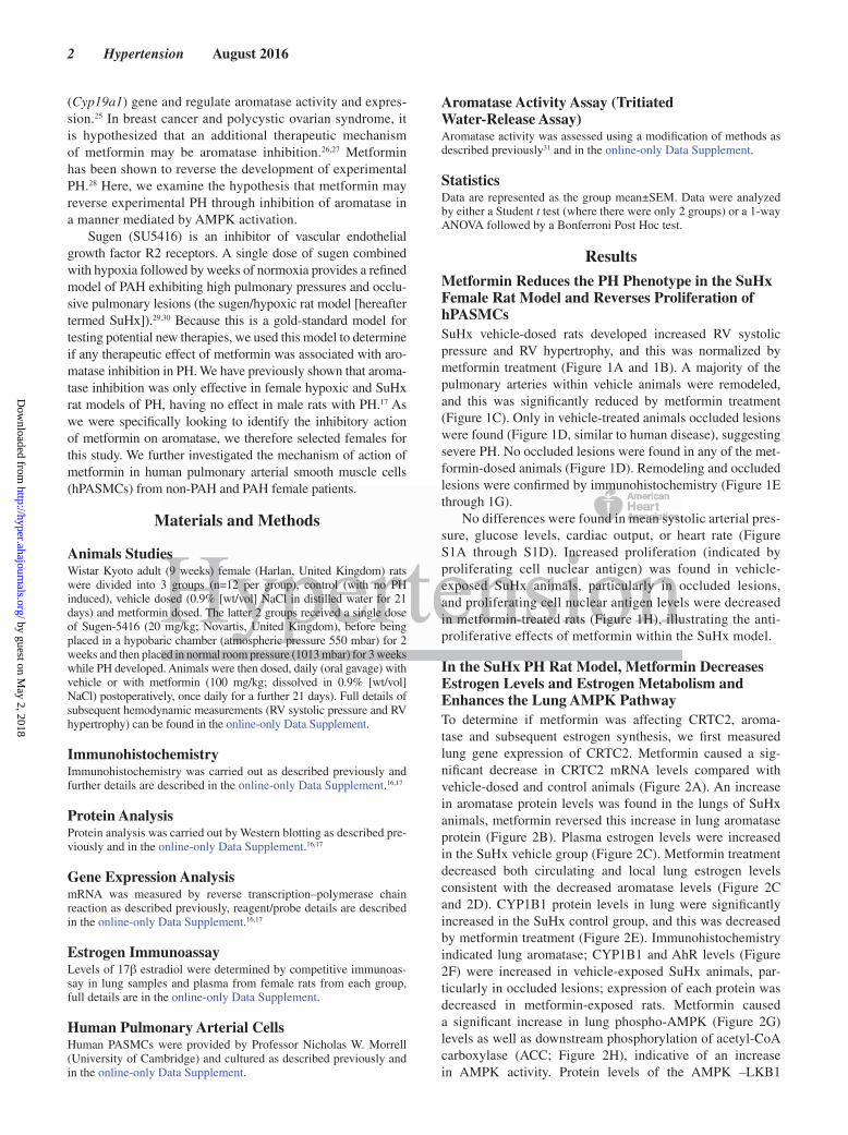
Abstract—Females are more susceptible to pulmonary arterial hypertension than males, although the reasons remain unclear. The hypoglycemic drug, metformin, is reported to have multiple actions, including the inhibition of aromatase and stimulation of AMP-activated protein kinase. Inhibition of aromatase using anastrazole is protective in experimental pulmonary hypertension but whether metformin attenuates pulmonary hypertension through this mechanism remains unknown. We investigated whether metformin affected aromatase activity and if it could reduce the development of pulmonary hypertension in the sugen 5416/hypoxic rat model. We also investigated its influence on proliferation in human pulmonary arterial smooth muscle cells. Metformin reversed right ventricular systolic pressure, right ventricular hypertrophy, and decreased pulmonary vascular remodeling in the rat. Furthermore, metformin increased rat lung AMP-activated protein kinase signaling, decreased lung and circulating estrogen levels, levels of aromatase, the estrogen metabolizing enzyme; cytochrome P450 1B1 and its transcription factor; the aryl hydrocarbon receptor. In human pulmonary arterial smooth muscle cells, metformin decreased proliferation and decreased estrogen synthesis by decreasing aromatase activity through the PII promoter site of Cyp19a1. Thus, we report for the first time that metformin can reverse pulmonary hypertension through inhibition of aromatase and estrogen synthesis in a manner likely to be mediated by AMP-activated protein kinase. (Hypertension. 2016;68:00-00. DOI: 10.1161/HYPERTENSIONAHA.116.07353.) • Online Data Supplement
Key Words: AMP-activated protein kinase ■ aromatase ■ estrogen ■ metformin ■ pulmonary hypertension
Received February 16, 2016; first decision March 21, 2016; revision accepted May 13, 2016.From the Institute of Cardiovascular and Medical Sciences, College of Medical, Veterinary, and Life Sciences, University of Glasgow, Glasgow, United
Kingdom.This article was sent to R. Clinton Webb, Guest Editor, for review by expert referees, editorial decision, and final disposition.The online-only Data Supplement is available with this article at http://hyper.ahajournals.org/lookup/suppl/doi:10.1161/HYPERTENSIONAHA.
116.07353/-/DC1.Correspondence to Margaret R. MacLean, College of Medical, Veterinary and Life Sciences, Research Institute of Cardiovascular and Medical Sciences,
Room 448, West Medical Bldg/Wolfson Link Bldg, University of Glasgow, Glasgow G12 8QQ, United Kingdom. E-mail [email protected]
Metformin Reverses Development of Pulmonary Hypertension via Aromatase Inhibition
Afshan Dean, Margaret Nilsen, Lynn Loughlin, Ian P. Salt, Margaret R. MacLean
© 2016 American Heart Association, Inc.
Hypertension is available at http://hyper.ahajournals.org DOI: 10.1161/HYPERTENSIONAHA.116.07353
Original Article
by guest on May 2, 2018
http://hyper.ahajournals.org/D
ownloaded from
by guest on M
ay 2, 2018http://hyper.ahajournals.org/
Dow
nloaded from
by guest on May 2, 2018
http://hyper.ahajournals.org/D
ownloaded from
by guest on M
ay 2, 2018http://hyper.ahajournals.org/
Dow
nloaded from
by guest on May 2, 2018
http://hyper.ahajournals.org/D
ownloaded from
by guest on M
ay 2, 2018http://hyper.ahajournals.org/
Dow
nloaded from
by guest on May 2, 2018
http://hyper.ahajournals.org/D
ownloaded from
by guest on M
ay 2, 2018http://hyper.ahajournals.org/
Dow
nloaded from
by guest on May 2, 2018
http://hyper.ahajournals.org/D
ownloaded from
by guest on M
ay 2, 2018http://hyper.ahajournals.org/
Dow
nloaded from
by guest on May 2, 2018
http://hyper.ahajournals.org/D
ownloaded from
by guest on M
ay 2, 2018http://hyper.ahajournals.org/
Dow
nloaded from
by guest on May 2, 2018
http://hyper.ahajournals.org/D
ownloaded from
by guest on M
ay 2, 2018http://hyper.ahajournals.org/
Dow
nloaded from
2 Hypertension August 2016
(Cyp19a1) gene and regulate aromatase activity and expres-sion.25 In breast cancer and polycystic ovarian syndrome, it is hypothesized that an additional therapeutic mechanism of metformin may be aromatase inhibition.26,27 Metformin has been shown to reverse the development of experimental PH.28 Here, we examine the hypothesis that metformin may reverse experimental PH through inhibition of aromatase in a manner mediated by AMPK activation.
Sugen (SU5416) is an inhibitor of vascular endothelial growth factor R2 receptors. A single dose of sugen combined with hypoxia followed by weeks of normoxia provides a refined model of PAH exhibiting high pulmonary pressures and occlu-sive pulmonary lesions (the sugen/hypoxic rat model [hereafter termed SuHx]).29,30 Because this is a gold-standard model for testing potential new therapies, we used this model to determine if any therapeutic effect of metformin was associated with aro-matase inhibition in PH. We have previously shown that aroma-tase inhibition was only effective in female hypoxic and SuHx rat models of PH, having no effect in male rats with PH.17 As we were specifically looking to identify the inhibitory action of metformin on aromatase, we therefore selected females for this study. We further investigated the mechanism of action of metformin in human pulmonary arterial smooth muscle cells (hPASMCs) from non-PAH and PAH female patients.
Materials and Methods
Animals StudiesWistar Kyoto adult (9 weeks) female (Harlan, United Kingdom) rats were divided into 3 groups (n=12 per group), control (with no PH induced), vehicle dosed (0.9% [wt/vol] NaCl in distilled water for 21 days) and metformin dosed. The latter 2 groups received a single dose of Sugen-5416 (20 mg/kg; Novartis, United Kingdom), before being placed in a hypobaric chamber (atmospheric pressure 550 mbar) for 2 weeks and then placed in normal room pressure (1013 mbar) for 3 weeks while PH developed. Animals were then dosed, daily (oral gavage) with vehicle or with metformin (100 mg/kg; dissolved in 0.9% [wt/vol] NaCl) postoperatively, once daily for a further 21 days). Full details of subsequent hemodynamic measurements (RV systolic pressure and RV hypertrophy) can be found in the online-only Data Supplement.
ImmunohistochemistryImmunohistochemistry was carried out as described previously and further details are described in the online-only Data Supplement.16,17
Protein AnalysisProtein analysis was carried out by Western blotting as described pre-viously and in the online-only Data Supplement.16,17
Gene Expression AnalysismRNA was measured by reverse transcription–polymerase chain reaction as described previously, reagent/probe details are described in the online-only Data Supplement.16,17
Estrogen ImmunoassayLevels of 17β estradiol were determined by competitive immunoas-say in lung samples and plasma from female rats from each group, full details are in the online-only Data Supplement.
Human Pulmonary Arterial CellsHuman PASMCs were provided by Professor Nicholas W. Morrell (University of Cambridge) and cultured as described previously and in the online-only Data Supplement.
Aromatase Activity Assay (Tritiated Water-Release Assay)Aromatase activity was assessed using a modification of methods as described previously31 and in the online-only Data Supplement.
StatisticsData are represented as the group mean±SEM. Data were analyzed by either a Student t test (where there were only 2 groups) or a 1-way ANOVA followed by a Bonferroni Post Hoc test.
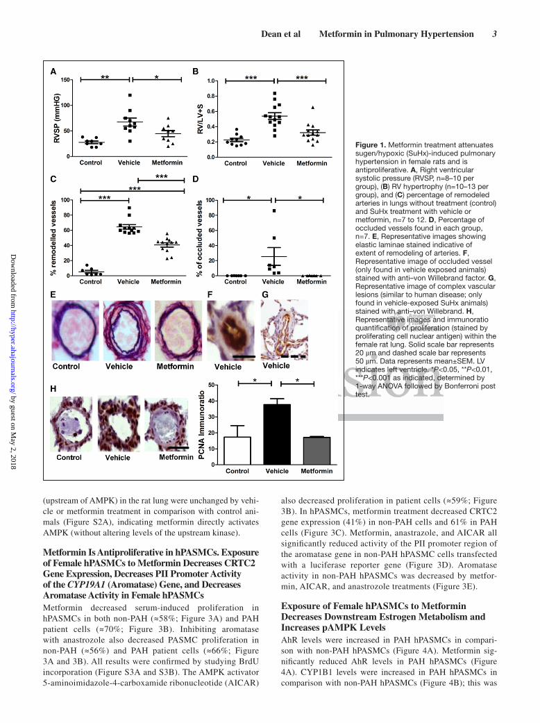
ResultsMetformin Reduces the PH Phenotype in the SuHx Female Rat Model and Reverses Proliferation of hPASMCsSuHx vehicle-dosed rats developed increased RV systolic pressure and RV hypertrophy, and this was normalized by metformin treatment (Figure 1A and 1B). A majority of the pulmonary arteries within vehicle animals were remodeled, and this was significantly reduced by metformin treatment (Figure 1C). Only in vehicle-treated animals occluded lesions were found (Figure 1D, similar to human disease), suggesting severe PH. No occluded lesions were found in any of the met-formin-dosed animals (Figure 1D). Remodeling and occluded lesions were confirmed by immunohistochemistry (Figure 1E through 1G).
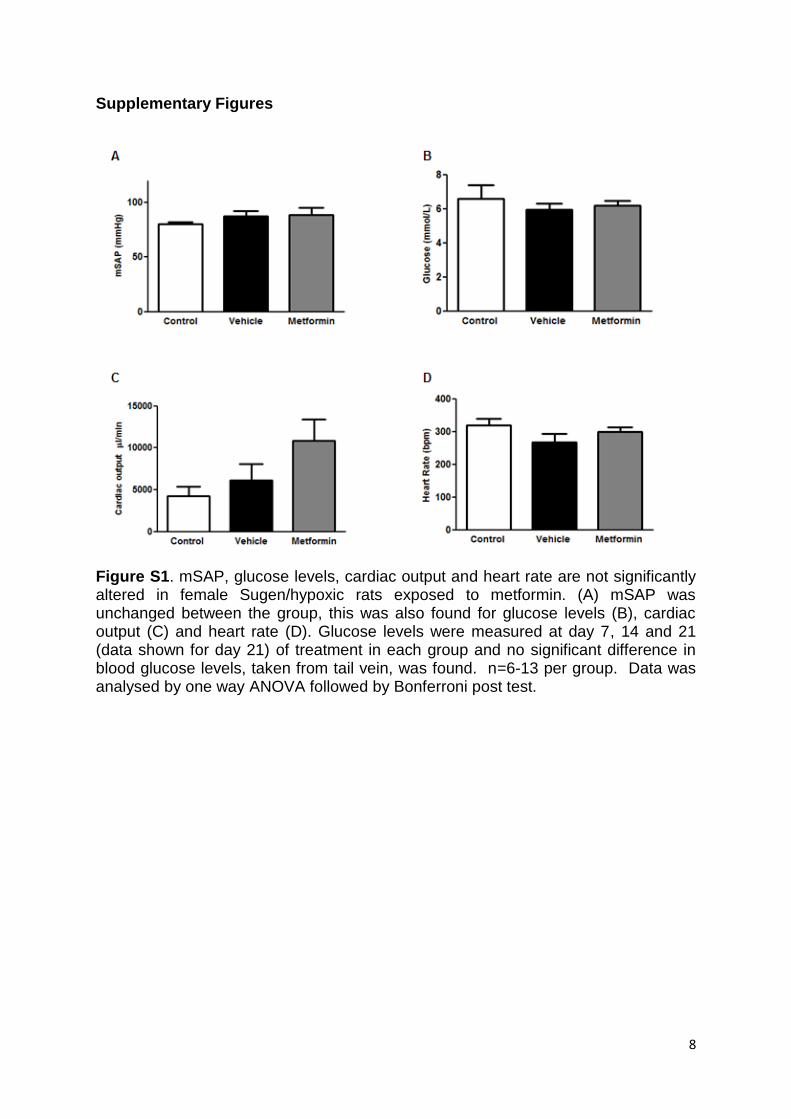
No differences were found in mean systolic arterial pres-sure, glucose levels, cardiac output, or heart rate (Figure S1A through S1D). Increased proliferation (indicated by proliferating cell nuclear antigen) was found in vehicle-exposed SuHx animals, particularly in occluded lesions, and proliferating cell nuclear antigen levels were decreased in metformin-treated rats (Figure 1H), illustrating the anti-proliferative effects of metformin within the SuHx model.
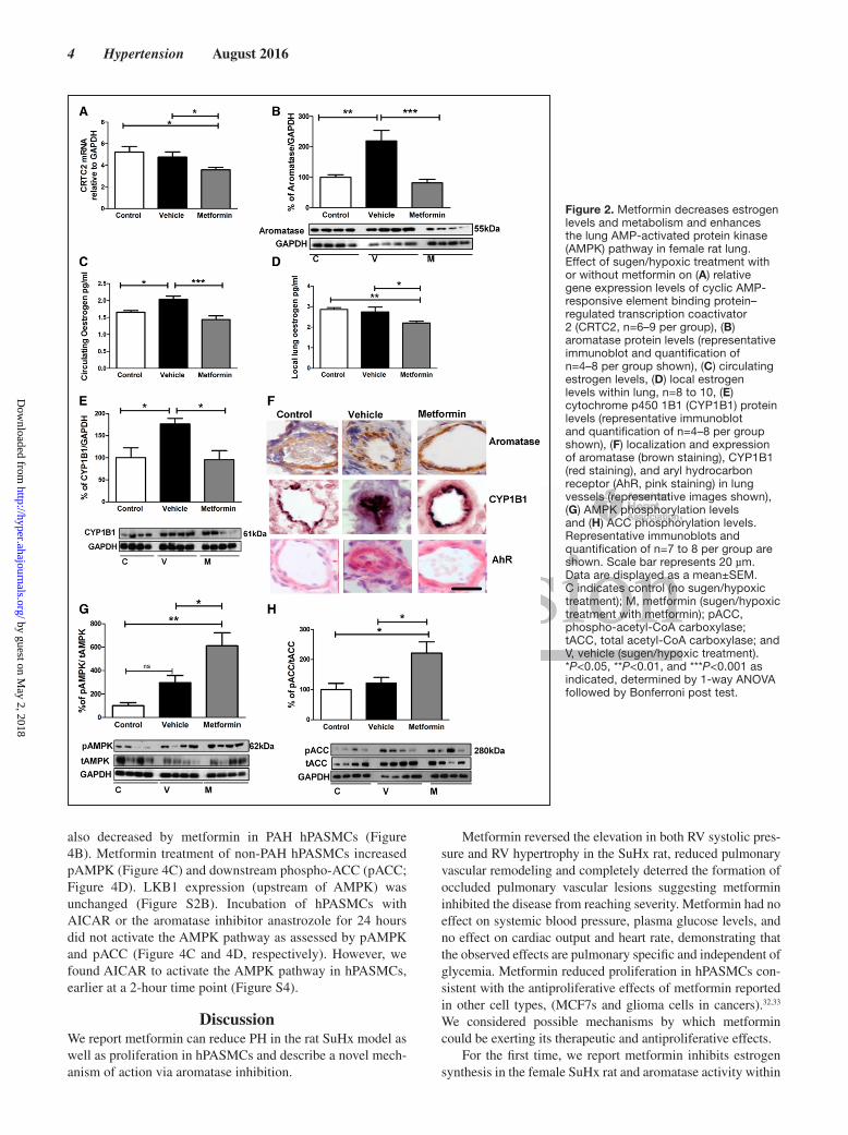
In the SuHx PH Rat Model, Metformin Decreases Estrogen Levels and Estrogen Metabolism and Enhances the Lung AMPK PathwayTo determine if metformin was affecting CRTC2, aroma-tase and subsequent estrogen synthesis, we first measured lung gene expression of CRTC2. Metformin caused a sig-nificant decrease in CRTC2 mRNA levels compared with vehicle-dosed and control animals (Figure 2A). An increase in aromatase protein levels was found in the lungs of SuHx animals, metformin reversed this increase in lung aromatase protein (Figure 2B). Plasma estrogen levels were increased in the SuHx vehicle group (Figure 2C). Metformin treatment decreased both circulating and local lung estrogen levels consistent with the decreased aromatase levels (Figure 2C and 2D). CYP1B1 protein levels in lung were significantly increased in the SuHx control group, and this was decreased by metformin treatment (Figure 2E). Immunohistochemistry indicated lung aromatase; CYP1B1 and AhR levels (Figure 2F) were increased in vehicle-exposed SuHx animals, par-ticularly in occluded lesions; expression of each protein was decreased in metformin-exposed rats. Metformin caused a significant increase in lung phospho-AMPK (Figure 2G) levels as well as downstream phosphorylation of acetyl-CoA carboxylase (ACC; Figure 2H), indicative of an increase in AMPK activity. Protein levels of the AMPK –LKB1
by guest on May 2, 2018
http://hyper.ahajournals.org/D
ownloaded from
Dean et al Metformin in Pulmonary Hypertension 3
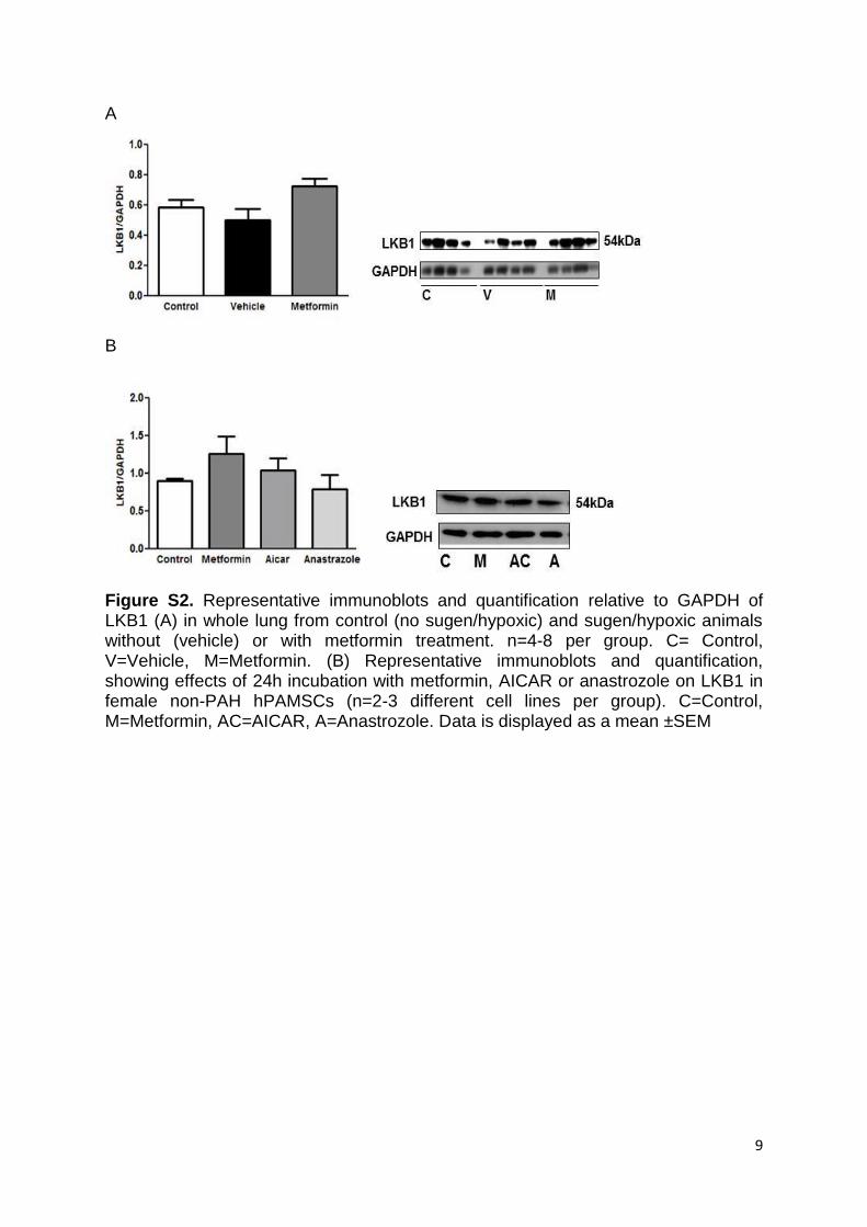
(upstream of AMPK) in the rat lung were unchanged by vehi-cle or metformin treatment in comparison with control ani-mals (Figure S2A), indicating metformin directly activates AMPK (without altering levels of the upstream kinase).
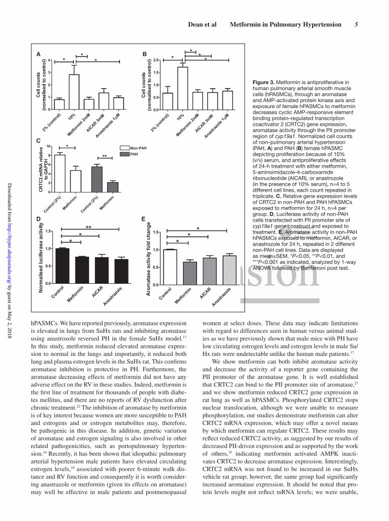
Metformin Is Antiproliferative in hPASMCs. Exposure of Female hPASMCs to Metformin Decreases CRTC2 Gene Expression, Decreases PII Promoter Activity of the CYP19A1 (Aromatase) Gene, and Decreases Aromatase Activity in Female hPASMCsMetformin decreased serum-induced proliferation in hPASMCs in both non-PAH (≈58%; Figure 3A) and PAH patient cells (≈70%; Figure 3B). Inhibiting aromatase with anastrozole also decreased PASMC proliferation in non-PAH (≈56%) and PAH patient cells (≈66%; Figure 3A and 3B). All results were confirmed by studying BrdU incorporation (Figure S3A and S3B). The AMPK activator 5-aminoimidazole-4-carboxamide ribonucleotide (AICAR)
also decreased proliferation in patient cells (≈59%; Figure 3B). In hPASMCs, metformin treatment decreased CRTC2 gene expression (41%) in non-PAH cells and 61% in PAH cells (Figure 3C). Metformin, anastrazole, and AICAR all significantly reduced activity of the PII promoter region of the aromatase gene in non-PAH hPASMC cells transfected with a luciferase reporter gene (Figure 3D). Aromatase activity in non-PAH hPASMCs was decreased by metfor-min, AICAR, and anastrozole treatments (Figure 3E).
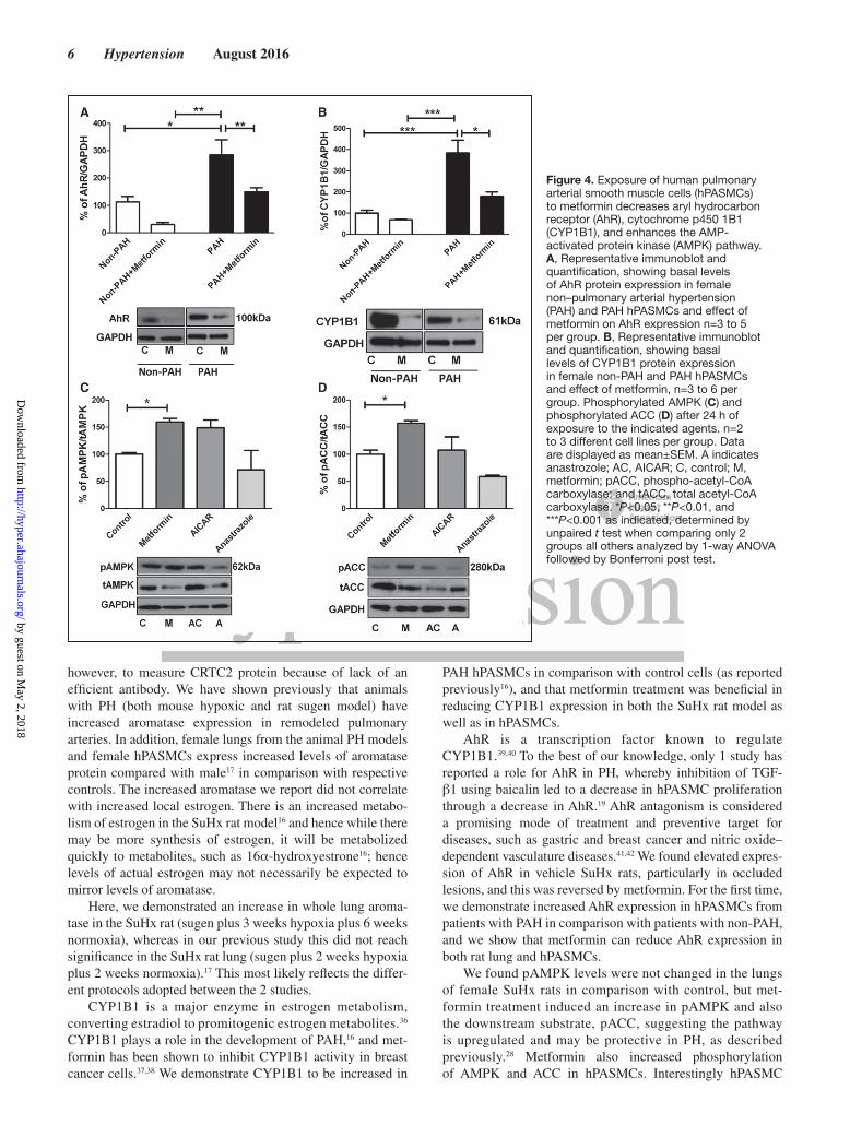
Exposure of Female hPASMCs to Metformin Decreases Downstream Estrogen Metabolism and Increases pAMPK LevelsAhR levels were increased in PAH hPASMCs in compari-son with non-PAH hPASMCs (Figure 4A). Metformin sig-nificantly reduced AhR levels in PAH hPASMCs (Figure 4A). CYP1B1 levels were increased in PAH hPASMCs in comparison with non-PAH hPASMCs (Figure 4B); this was
Figure 1. Metformin treatment attenuates sugen/hypoxic (SuHx)-induced pulmonary hypertension in female rats and is antiproliferative. A, Right ventricular systolic pressure (RVSP, n=8–10 per group), (B) RV hypertrophy (n=10–13 per group), and (C) percentage of remodeled arteries in lungs without treatment (control) and SuHx treatment with vehicle or metformin, n=7 to 12. D, Percentage of occluded vessels found in each group, n=7. E, Representative images showing elastic laminae stained indicative of extent of remodeling of arteries. F, Representative image of occluded vessel (only found in vehicle exposed animals) stained with anti–von Willebrand factor. G, Representative image of complex vascular lesions (similar to human disease; only found in vehicle-exposed SuHx animals) stained with anti–von Willebrand. H, Representative images and immunoratio quantification of proliferation (stained by proliferating cell nuclear antigen) within the female rat lung. Solid scale bar represents 20 µm and dashed scale bar represents 50 µm. Data represents mean±SEM. LV indicates left ventricle. *P<0.05, **P<0.01, ***P<0.001 as indicated, determined by 1-way ANOVA followed by Bonferroni post test. by guest on M
ay 2, 2018http://hyper.ahajournals.org/
Dow
nloaded from
4 Hypertension August 2016
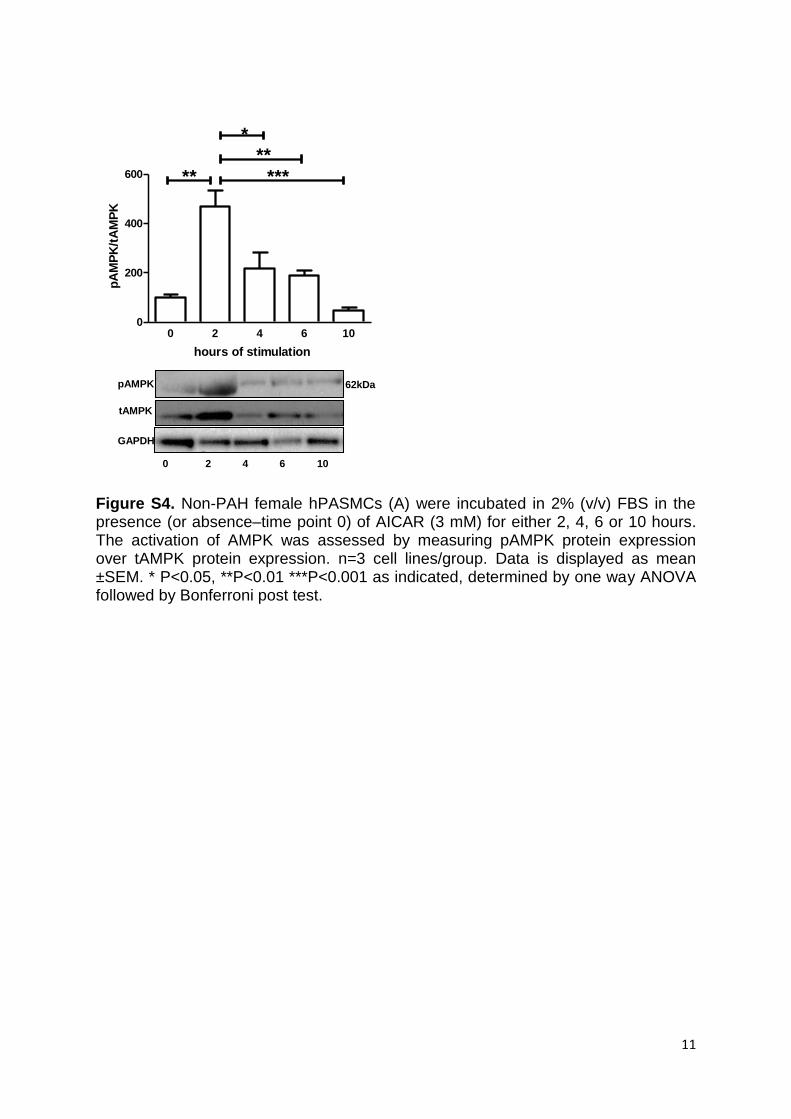
also decreased by metformin in PAH hPASMCs (Figure 4B). Metformin treatment of non-PAH hPASMCs increased pAMPK (Figure 4C) and downstream phospho-ACC (pACC; Figure 4D). LKB1 expression (upstream of AMPK) was unchanged (Figure S2B). Incubation of hPASMCs with AICAR or the aromatase inhibitor anastrozole for 24 hours did not activate the AMPK pathway as assessed by pAMPK and pACC (Figure 4C and 4D, respectively). However, we found AICAR to activate the AMPK pathway in hPASMCs, earlier at a 2-hour time point (Figure S4).
DiscussionWe report metformin can reduce PH in the rat SuHx model as well as proliferation in hPASMCs and describe a novel mech-anism of action via aromatase inhibition.
Metformin reversed the elevation in both RV systolic pres-sure and RV hypertrophy in the SuHx rat, reduced pulmonary vascular remodeling and completely deterred the formation of occluded pulmonary vascular lesions suggesting metformin inhibited the disease from reaching severity. Metformin had no effect on systemic blood pressure, plasma glucose levels, and no effect on cardiac output and heart rate, demonstrating that the observed effects are pulmonary specific and independent of glycemia. Metformin reduced proliferation in hPASMCs con-sistent with the antiproliferative effects of metformin reported in other cell types, (MCF7s and glioma cells in cancers).32,33 We considered possible mechanisms by which metformin could be exerting its therapeutic and antiproliferative effects.
For the first time, we report metformin inhibits estrogen synthesis in the female SuHx rat and aromatase activity within
Figure 2. Metformin decreases estrogen levels and metabolism and enhances the lung AMP-activated protein kinase (AMPK) pathway in female rat lung. Effect of sugen/hypoxic treatment with or without metformin on (A) relative gene expression levels of cyclic AMP-responsive element binding protein–regulated transcription coactivator 2 (CRTC2, n=6–9 per group), (B) aromatase protein levels (representative immunoblot and quantification of n=4–8 per group shown), (C) circulating estrogen levels, (D) local estrogen levels within lung, n=8 to 10, (E) cytochrome p450 1B1 (CYP1B1) protein levels (representative immunoblot and quantification of n=4–8 per group shown), (F) localization and expression of aromatase (brown staining), CYP1B1 (red staining), and aryl hydrocarbon receptor (AhR, pink staining) in lung vessels (representative images shown), (G) AMPK phosphorylation levels and (H) ACC phosphorylation levels. Representative immunoblots and quantification of n=7 to 8 per group are shown. Scale bar represents 20 µm. Data are displayed as a mean±SEM. C indicates control (no sugen/hypoxic treatment); M, metformin (sugen/hypoxic treatment with metformin); pACC, phospho-acetyl-CoA carboxylase; tACC, total acetyl-CoA carboxylase; and V, vehicle (sugen/hypoxic treatment). *P<0.05, **P<0.01, and ***P<0.001 as indicated, determined by 1-way ANOVA followed by Bonferroni post test.
by guest on May 2, 2018
http://hyper.ahajournals.org/D
ownloaded from
Dean et al Metformin in Pulmonary Hypertension 5
hPASMCs. We have reported previously, aromatase expression is elevated in lungs from SuHx rats and inhibiting aromatase using anastrozole reversed PH in the female SuHx model.17 In this study, metformin reduced elevated aromatase expres-sion to normal in the lungs and importantly, it reduced both lung and plasma estrogen levels in the SuHx rat. This confirms aromatase inhibition is protective in PH. Furthermore, the aromatase decreasing effects of metformin did not have any adverse effect on the RV in these studies. Indeed, metformin is the first line of treatment for thousands of people with diabe-tes mellitus, and there are no reports of RV dysfunction after chronic treatment.22 The inhibition of aromatase by metformin is of key interest because women are more susceptible to PAH and estrogens and or estrogen metabolites may, therefore, be pathogenic in this disease. In addition, genetic variation of aromatase and estrogen signaling is also involved in other related pathogenicities, such as portopulmonary hyperten-sion.34 Recently, it has been shown that idiopathic pulmonary arterial hypertension male patients have elevated circulating estrogen levels,18 associated with poorer 6-minute walk dis-tance and RV function and consequently it is worth consider-ing anastrazole or metformin (given its effects on aromatase) may well be effective in male patients and postmenopausal
women at select doses. These data may indicate limitations with regard to differences seen in human versus animal stud-ies as we have previously shown that male mice with PH have low circulating estrogen levels and estrogen levels in male Su/Hx rats were undetectable unlike the human male patients.17
We show metformin can both inhibit aromatase activity and decrease the activity of a reporter gene containing the PII promoter of the aromatase gene. It is well established that CRTC2 can bind to the PII promoter site of aromatase,27 and we show metformin reduced CRTC2 gene expression in rat lung as well as hPASMCs. Phosphorylated CRTC2 stops nuclear translocation, although we were unable to measure phosphorylation, our studies demonstrate metformin can alter CRTC2 mRNA expression, which may offer a novel means by which metformin can regulate CRTC2. These results may reflect reduced CRTC2 activity, as suggested by our results of decreased PII-driven expression and as supported by the work of others,35 indicating metformin activated AMPK inacti-vates CRTC2 to decrease aromatase expression. Interestingly, CRTC2 mRNA was not found to be increased in our SuHx vehicle rat group; however, the same group had significantly increased aromatase expression. It should be noted that pro-tein levels might not reflect mRNA levels; we were unable,
Figure 3. Metformin is antiproliferative in human pulmonary arterial smooth muscle cells (hPASMCs), through an aromatase and AMP-activated protein kinase axis and exposure of female hPASMCs to metformin decreases cyclic AMP-responsive element binding protein–regulated transcription coactivator 2 (CRTC2) gene expression, aromatase activity through the PII promoter region of cyp19a1. Normalized cell counts of non–pulmonary arterial hypertension (PAH, A) and PAH (B) female hPASMC depicting proliferation because of 10% (v/v) serum, and antiproliferative effects of 24-h treatment with either metformin, 5-aminoimidazole-4-carboxamide ribonucleotide (AICAR), or anastrozole (in the presence of 10% serum), n=4 to 5 different cell lines, each count repeated in triplicate. C, Relative gene expression levels of CRTC2 in non-PAH and PAH hPASMCs exposed to metformin for 24 h, n=4 per group. D, Luciferase activity of non-PAH cells transfected with PII promoter site of cyp19a1 gene construct and exposed to treatment. E, Aromatase activity in non-PAH hPASMCs exposed to metformin, AICAR, or anastrozole for 24 h, repeated in 2 different non-PAH cell lines. Data are displayed as mean±SEM. *P<0.05, **P<0.01, and ***P<0.001 as indicated, analyzed by 1-way ANOVA followed by Bonferroni post test.
by guest on May 2, 2018
http://hyper.ahajournals.org/D
ownloaded from
6 Hypertension August 2016
however, to measure CRTC2 protein because of lack of an efficient antibody. We have shown previously that animals with PH (both mouse hypoxic and rat sugen model) have increased aromatase expression in remodeled pulmonary arteries. In addition, female lungs from the animal PH models and female hPASMCs express increased levels of aromatase protein compared with male17 in comparison with respective controls. The increased aromatase we report did not correlate with increased local estrogen. There is an increased metabo-lism of estrogen in the SuHx rat model16 and hence while there may be more synthesis of estrogen, it will be metabolized quickly to metabolites, such as 16α-hydroxyestrone16; hence levels of actual estrogen may not necessarily be expected to mirror levels of aromatase.
Here, we demonstrated an increase in whole lung aroma-tase in the SuHx rat (sugen plus 3 weeks hypoxia plus 6 weeks normoxia), whereas in our previous study this did not reach significance in the SuHx rat lung (sugen plus 2 weeks hypoxia plus 2 weeks normoxia).17 This most likely reflects the differ-ent protocols adopted between the 2 studies.
CYP1B1 is a major enzyme in estrogen metabolism, converting estradiol to promitogenic estrogen metabolites.36 CYP1B1 plays a role in the development of PAH,16 and met-formin has been shown to inhibit CYP1B1 activity in breast cancer cells.37,38 We demonstrate CYP1B1 to be increased in
PAH hPASMCs in comparison with control cells (as reported previously16), and that metformin treatment was beneficial in reducing CYP1B1 expression in both the SuHx rat model as well as in hPASMCs.
AhR is a transcription factor known to regulate CYP1B1.39,40 To the best of our knowledge, only 1 study has reported a role for AhR in PH, whereby inhibition of TGF-β1 using baicalin led to a decrease in hPASMC proliferation through a decrease in AhR.19 AhR antagonism is considered a promising mode of treatment and preventive target for diseases, such as gastric and breast cancer and nitric oxide–dependent vasculature diseases.41,42 We found elevated expres-sion of AhR in vehicle SuHx rats, particularly in occluded lesions, and this was reversed by metformin. For the first time, we demonstrate increased AhR expression in hPASMCs from patients with PAH in comparison with patients with non-PAH, and we show that metformin can reduce AhR expression in both rat lung and hPASMCs.
We found pAMPK levels were not changed in the lungs of female SuHx rats in comparison with control, but met-formin treatment induced an increase in pAMPK and also the downstream substrate, pACC, suggesting the pathway is upregulated and may be protective in PH, as described previously.28 Metformin also increased phosphorylation of AMPK and ACC in hPASMCs. Interestingly hPASMC
Figure 4. Exposure of human pulmonary arterial smooth muscle cells (hPASMCs) to metformin decreases aryl hydrocarbon receptor (AhR), cytochrome p450 1B1 (CYP1B1), and enhances the AMP-activated protein kinase (AMPK) pathway. A, Representative immunoblot and quantification, showing basal levels of AhR protein expression in female non–pulmonary arterial hypertension (PAH) and PAH hPASMCs and effect of metformin on AhR expression n=3 to 5 per group. B, Representative immunoblot and quantification, showing basal levels of CYP1B1 protein expression in female non-PAH and PAH hPASMCs and effect of metformin, n=3 to 6 per group. Phosphorylated AMPK (C) and phosphorylated ACC (D) after 24 h of exposure to the indicated agents. n=2 to 3 different cell lines per group. Data are displayed as mean±SEM. A indicates anastrozole; AC, AICAR; C, control; M, metformin; pACC, phospho-acetyl-CoA carboxylase; and tACC, total acetyl-CoA carboxylase. *P<0.05, **P<0.01, and ***P<0.001 as indicated, determined by unpaired t test when comparing only 2 groups all others analyzed by 1-way ANOVA followed by Bonferroni post test.
by guest on May 2, 2018
http://hyper.ahajournals.org/D
ownloaded from
Dean et al Metformin in Pulmonary Hypertension 7
treatment with the AMPK activator, AICAR did not have the same effect at the 24-hour time point. This may reflect the mechanism by which AICAR activates AMPK, whereby AICAR is transported into the cell and phosphorylated to form 5-aminoimidazole-4-carboxamide ribonucleotide (ZMP).43 The accumulated ZMP binds directly to the γ-subunit of AMPK, mimicking AMP and stimulating AMPK phosphory-lation. Initial activation of AMPK would be sufficient to retard the cell cycle and thereby reduce proliferation, in the same way that stimulating cells with growth factors only transiently increases proximal signaling pathways whereas increased pro-liferation, which relies on the time taken to undergo cytokine-sis remains increased long after those signaling pathways have returned to basal levels. The timescale of our experiments may, therefore, have exceeded the time course of ZMP accumula-tion and AMPK activation. Metformin inhibits mitochondrial ATP synthesis, reducing the cellular energy charge, which acti-vates AMPK.44 This process may well continue for 24 hours, such that the AMPK pathway may still be activated after 24 hours in response to metformin but not AICAR. Subsequently, we demonstrated AICAR activates AMPK in hPASMCs at a much earlier time point of 2 hours (Figure SF4).
Enhancement of the AMPK pathway is involved in regula-tion of several different pathways; therefore, the therapeutic effects of metformin could involve additional mechanisms. For example, AMPK activation can affect the cyclooxygen-ase pathway as well as the mTOR pathway, both known to be dysregulated in PAH.45,46 Moreover, metformin targets the mitochondria of cells, and this is where steroidogenesis takes places. Emerging data indicate metformin can dampen the entire steroidogenesis pathway,43,47 and activated AMPK is reported to inhibit HMG CoA (3-hydroxy-3-methylglutaryl-coenzyme A) reductase (which regulates cholesterol synthe-sis) thus leading to a decrease in steroidogenesis.48
It is possible that all the therapeutic effects of metformin could be driven via its ability to increase AMPK, and this work is supported by other studies, which indicate AMPK to be protective in PH28; however, conflict exists, whereby it has also been shown that a decrease in AMPK may be protective against PH.49
We previously showed that aromatase inhibition was only effective in female hypoxic and SuHx rat models of PH, hav-ing no effect in male rats with PH.17 As we were specifically looking to identify the additional inhibitory action of metfor-min on aromatase, we therefore selected female rats. In addi-tion, metformin has already been shown to reverse PH in male hypoxic and monocrotaline PH rats via other mechanisms of action28; therefore, it would most certainly have had an effect in males in our model via other mechanisms. This would not, however, have determined any additional effect on aromatase activity. We were unable to measure different estrogen metab-olites in the lung tissue from our experimental rats because there are no available assays with sufficient sensitivity. We and others have, however, provided evidence that increased CYP1B1 may contribute to PAH pathology via increased accumulation of mitogenic metabolites, such as 16α-hydroxyestrone.15,16,50
In summary, we have demonstrated metformin to be pro-tective in PH in the rat SuHx model and antiproliferative in
hPASMCs. We show aromatase expression and activity as well as estrogen metabolism (via AhR/CYP1B1 axis inhi-bition) in both rat and human models were downregulated by metformin. We report that metformin increases pAMPK, which can affect CRTC2 gene expression, and aromatase PII promoter activity. This mechanism is summarized in Figure S5. Because metformin is already in widespread use in the clinic and well tolerated by patients, our study denotes this drug to be beneficial in experimental PH and merits further investigation as a therapy for PAH. The study also suggests a role for AhR in PH, which is worthy of fur-ther investigation.
PerspectivesThe precise role of estrogen, its formation and its metabo-lites, in PAH requires to be fully understood to understand why females are more susceptible to the disease. Our studies show metformin can inhibit aromatase expression and activ-ity to decrease estrogen but also its metabolism, and this is beneficial in reversing the disease phenotype in females. In PH, this is a novel mechanism which is highly relevant given the role of estrogen metabolites and estrogen synthesis in the disease. Furthermore, metformin is already in widespread use as an anticancer drug and well tolerated by patients and thus suggests a practical therapy for females with this devastating disease.
AcknowledgmentsWe thank Dr Kristy Brown and Maria Docanto from Monash University, Australia for providing the PII promoter plasmid. We thank Marie Anne Ewart and Benoit Viollet for assistance in inter-pretation of the data. We thank Prof. Nick Morrell for providing all hPASMCs.
Sources of FundingThis work was funded by the British Heart Foundation grants RG/11/7/28916 and PG/13/7/29913.
DisclosuresNone.
References 1. McLaughlin VV, Gaine SP, Howard LS, Leuchte HH, Mathier MA, Mehta
S, Palazzini M, Park MH, Tapson VF, Sitbon O. Treatment goals of pul-monary hypertension. J Am Coll Cardiol. 2013;62(suppl 25):D73–D81. doi: 10.1016/j.jacc.2013.10.034.
2. Mair KM, Johansen AK, Wright AF, Wallace E, MacLean MR. Pulmonary arterial hypertension: basis of sex differences in incidence and treatment response. Br J Pharmacol. 2014;171:567–579. doi: 10.1111/bph.12281.
3. Badesch DB, Raskob GE, Elliott CG, Krichman AM, Farber HW, Frost AE, Barst RJ, Benza RL, Liou TG, Turner M, Giles S, Feldkircher K, Miller DP, McGoon MD. Pulmonary arterial hypertension: baseline char-acteristics from the REVEAL Registry. Chest. 2010;137:376–387. doi: 10.1378/chest.09-1140.
4. Shapiro S, Traiger GL, Turner M, McGoon MD, Wason P, Barst RJ. Sex differences in the diagnosis, treatment, and outcome of patients with pul-monary arterial hypertension enrolled in the registry to evaluate early and long-term pulmonary arterial hypertension disease management. Chest. 2012;141:363–373. doi: 10.1378/chest.10-3114.
5. Tofovic SP. Estrogens and development of pulmonary hyperten-sion: interaction of estradiol metabolism and pulmonary vascular disease. J Cardiovasc Pharmacol. 2010;56:696–708. doi: 10.1097/FJC.0b013e3181f9ea8d.
by guest on May 2, 2018
http://hyper.ahajournals.org/D
ownloaded from
8 Hypertension August 2016
6. Paulin R, Michelakis ED. The estrogen puzzle in pulmonary arte-rial hypertension. Circulation. 2012;126:1016–1019. doi: 10.1161/CIRCULATIONAHA.112.126474.
7. Wright AF, Ewart MA, Mair K, Nilsen M, Dempsie Y, Loughlin L, Maclean MR. Oestrogen receptor alpha in pulmonary hypertension. Cardiovasc Res. 2015;106:206–216. doi: 10.1093/cvr/cvv106.
8. Lahm T. Sex differences in pulmonary hypertension: are we cleaning up the mess? Eur Respir J. 2016;47:390–393. doi: 10.1183/13993003. 01999-2015.
9. Rabinovitch M, Gamble WJ, Miettinen OS, Reid L. Age and sex influence on pulmonary hypertension of chronic hypoxia and on recovery. Am J Physiol. 1981;240:H62–H72.
10. Lahm T, Albrecht M, Fisher AJ, Selej M, Patel NG, Brown JA, Justice MJ, Brown MB, Van Demark M, Trulock KM, Dieudonne D, Reddy JG, Presson RG, Petrache I. 17β-Estradiol attenuates hypoxic pulmonary hypertension via estrogen receptor-mediated effects. Am J Respir Crit Care Med. 2012;185:965–980. doi: 10.1164/rccm.201107-1293OC.
11. Parker TA, Ivy DD, Galan HL, Grover TR, Kinsella JP, Abman SH. Estradiol improves pulmonary hemodynamics and vascular remodeling in perinatal pulmonary hypertension. Am J Physiol Lung Cell Mol Physiol. 2000;278:L374–L381.
12. Frump AL, Goss KN, Vayl A, Albrecht M, Fisher A, Tursunova R, Fierst J, Whitson J, Cucci AR, Brown MB, Lahm T. Estradiol improves right ventricular function in rats with severe angioproliferative pulmonary hypertension: effects of endogenous and exogenous sex hormones. Am J Physiol Lung Cell Mol Physiol. 2015;308:L873–L890. doi: 10.1152/ajplung.00006.2015.
13. White K, Dempsie Y, Nilsen M, Wright AF, Loughlin L, MacLean MR. The serotonin transporter, gender, and 17β oestradiol in the development of pulmonary arterial hypertension. Cardiovasc Res. 2011;90:373–382. doi: 10.1093/cvr/cvq408.
14. Dempsie Y, Nilsen M, White K, Mair KM, Loughlin L, Ambartsumian N, Rabinovitch M, Maclean MR. Development of pulmonary arterial hypertension in mice over-expressing S100A4/Mts1 is specific to females. Respir Res. 2011;12:159. doi: 10.1186/1465-9921-12-159.
15. Dempsie Y, MacRitchie NA, White K, Morecroft I, Wright AF, Nilsen M, Loughlin L, Mair KM, MacLean MR. Dexfenfluramine and the oes-trogen-metabolizing enzyme CYP1B1 in the development of pulmonary arterial hypertension. Cardiovasc Res. 2013;99:24–34. doi: 10.1093/cvr/cvt064.
16. White K, Johansen AK, Nilsen M, Ciuclan L, Wallace E, Paton L, Campbell A, Morecroft I, Loughlin L, McClure JD, Thomas M, Mair KM, MacLean MR. Activity of the estrogen-metabolizing enzyme cytochrome P450 1B1 influences the development of pulmonary arte-rial hypertension. Circulation. 2012;126:1087–1098. doi: 10.1161/CIRCULATIONAHA.111.062927.
17. Mair KM, Wright AF, Duggan N, Rowlands DJ, Hussey MJ, Roberts S, Fullerton J, Nilsen M, Loughlin L, Thomas M, MacLean MR. Sex-dependent influence of endogenous estrogen in pulmonary hyperten-sion. Am J Respir Crit Care Med. 2014;190:456–467. doi: 10.1164/rccm.201403-0483OC.
18. Ventetuolo CE, Mitra N, Wan F, Manichaikul A, Barr RG, Johnson C, Bluemke DA, Lima JA, Tandri H, Ouyang P, Kawut SM. Oestradiol metabolism and androgen receptor genotypes are associated with right ventricular function. Eur Respir J. 2016;47:553–563. doi: 10.1183/13993003.01083-2015.
19. Huang S, Chen P, Shui X, He Y, Wang H, Zheng J, Zhang L, Li J, Xue Y, Chen C, Lei W. Baicalin attenuates transforming growth factor-β1-induced human pulmonary artery smooth muscle cell proliferation and phenotypic switch by inhibiting hypoxia inducible factor-1α and aryl hydrocarbon receptor expression. J Pharm Pharmacol. 2014;66:1469–1477. doi: 10.1111/jphp.12273.
20. Goodarzi MO, Bryer-Ash M. Metformin revisited: re-evaluation of its properties and role in the pharmacopoeia of modern anti-diabetic agents. Diabetes Obes Metab. 2005;7:654–665. doi: 10.1111/j.1463-1326.2004.00448.x.
21. Rojas LB, Gomes MB. Metformin: an old but still the best treat-ment for type 2 diabetes. Diabetol Metab Syndr. 2013;5:6. doi: 10.1186/1758-5996-5-6.
22. Miller RA, Chu Q, Xie J, Foretz M, Viollet B, Birnbaum MJ. Biguanides suppress hepatic glucagon signalling by decreasing production of cyclic AMP. Nature. 2013;494:256–260. doi: 10.1038/nature11808.
23. Hardie DG, Ross FA, Hawley SA. AMPK: a nutrient and energy sensor that maintains energy homeostasis. Nat Rev Mol Cell Biol. 2012;13:251–262. doi: 10.1038/nrm3311.
24. Ewart MA, Kennedy S. Diabetic cardiovascular disease–AMP-activated protein kinase (AMPK) as a therapeutic target. Cardiovasc Hematol Agents Med Chem. 2012;10:190–211.
25. Brown KA, Hunger NI, Docanto M, Simpson ER. Metformin inhibits aro-matase expression in human breast adipose stromal cells via stimulation of AMP-activated protein kinase. Breast Cancer Res Treat. 2010;123:591–596. doi: 10.1007/s10549-010-0834-y.
26. Palomba S, Falbo A, Zullo F, Orio F Jr. Evidence-based and potential ben-efits of metformin in the polycystic ovary syndrome: a comprehensive review. Endocr Rev. 2009;30:1–50. doi: 10.1210/er.2008-0030.
27. Brown KA, McInnes KJ, Hunger NI, Oakhill JS, Steinberg GR, Simpson ER. Subcellular localization of cyclic AMP-responsive element bind-ing protein-regulated transcription coactivator 2 provides a link between obesity and breast cancer in postmenopausal women. Cancer Res. 2009;69:5392–5399. doi: 10.1158/0008-5472.CAN-09-0108.
28. Agard C, Rolli-Derkinderen M, Dumas-de-La-Roque E, Rio M, Sagan C, Savineau JP, Loirand G, Pacaud P. Protective role of the antidiabetic drug metformin against chronic experimental pulmo-nary hypertension. Br J Pharmacol. 2009;158:1285–1294. doi: 10.1111/j.1476-5381.2009.00445.x.
29. Abe K, Toba M, Alzoubi A, Ito M, Fagan KA, Cool CD, Voelkel NF, McMurtry IF, Oka M. Formation of plexiform lesions in experimental severe pulmonary arterial hypertension. Circulation. 2010;121:2747–2754. doi: 10.1161/CIRCULATIONAHA.109.927681.
30. Gomez-Arroyo JG, Farkas L, Alhussaini AA, Farkas D, Kraskauskas D, Voelkel NF, Bogaard HJ. The monocrotaline model of pulmonary hypertension in perspective. Am J Physiol Lung Cell Mol Physiol. 2012;302:L363–L369. doi: 10.1152/ajplung.00212.2011.
31. Lephart ED, Simpson ER. Assay of aromatase activity. Methods Enzymol. 1991;206:477–483. doi: 10.1016/0076-6879(91)06116-K
32. Hadad SM, Hardie DG, Appleyard V, Thompson AM. Effects of metformin on breast cancer cell proliferation, the AMPK pathway and the cell cycle. Clin Transl Oncol. 2014;16:746–752. doi: 10.1007/s12094-013-1144-8.
33. Sesen J, Dahan P, Scotland SJ, Saland E, Dang V-T, Lemarié A, Tyler BM, Brem H, Toulas C, Moyal E, Sarry JE, Skuli N. Metformin inhibits growth of human glioblastoma cells and enhances therapeutic response. PLoS One. 2015;10:1–24. doi: 10.1371/journal.pone.0123721
34. Roberts KE, Fallon MB, Krowka MJ, et al; Pulmonary Vascular Complications of Liver Disease Study Group. Genetic risk factors for portopulmonary hypertension in patients with advanced liver dis-ease. Am J Respir Crit Care Med. 2009;179:835–842. doi: 10.1164/rccm.200809-1472OC.
35. Xu JN, Zeng C, Zhou Y, Peng C, Zhou YF, Xue Q. Metformin inhibits StAR expression in human endometriotic stromal cells via AMPK-mediated disruption of CREB-CRTC2 complex formation. J Clin Endocrinol Metab. 2014;99:2795–2803. doi: 10.1210/jc.2014-1593.
36. Seeger H, Wallwiener D, Kraemer E, Mueck AO. Estradiol metabolites are potent mitogenic substances for human ovarian cancer cells. Eur J Gynaecol Oncol. 2005;26:383–385.
37. Jiao H, Liu C, Guo W, Peng L, Chen Y, Martin FL. Association of CYP1B1 Polymorphisms with Breast Cancer: A Case-Control Study in the Han Population in Ningxia Hui Autonomous Region, P. R. China. Biomark Insights. 2010;5:21–27.
38. Do MT, Kim HG, Tran TT, Khanal T, Choi JH, Chung YC, Jeong TC, Jeong HG. Metformin suppresses CYP1A1 and CYP1B1 expression in breast cancer cells by down-regulating aryl hydrocarbon receptor expression. Toxicol Appl Pharmacol. 2014;280:138–148. doi: 10.1016/j.taap.2014.07.021.
39. Yang X, Solomon S, Fraser LR, Trombino AF, Liu D, Sonenshein GE, Hestermann EV, Sherr DH. Constitutive regulation of CYP1B1 by the aryl hydrocarbon receptor (AhR) in pre-malignant and malignant mammary tissue. J Cell Biochem. 2008;104:402–417. doi: 10.1002/jcb.21630.
40. Jacob A, Hartz AM, Potin S, Coumoul X, Yousif S, Scherrmann JM, Bauer B, Declèves X. Aryl hydrocarbon receptor-dependent upregulation of Cyp1b1 by TCDD and diesel exhaust particles in rat brain microves-sels. Fluids Barriers CNS. 2011;8:23. doi: 10.1186/2045-8118-8-23.
41. Yin XF, Chen J, Mao W, Wang YH, Chen MH. Downregulation of aryl hydrocarbon receptor expression decreases gastric cancer cell growth and invasion. Oncol Rep. 2013;30:364–370. doi: 10.3892/or.2013.2410.
42. Zhang N. The role of endogenous aryl hydrocarbon receptor signaling in cardiovascular physiology. J Cardiovasc Dis Res. 2011;2:91–95. doi: 10.4103/0975-3583.83033.
43. Abdou HS, Bergeron F, Tremblay JJ. A cell-autonomous molecular cas-cade initiated by AMP-activated protein kinase represses steroidogenesis. Mol Cell Biol. 2014;34:4257–4271. doi: 10.1128/MCB.00734-14.
by guest on May 2, 2018
http://hyper.ahajournals.org/D
ownloaded from
Dean et al Metformin in Pulmonary Hypertension 9
44. Zhou G, Myers R, Li Y, Chen Y, Shen X, Fenyk-Melody J, Wu M, Ventre J, Doebber T, Fujii N, Musi N, Hirshman MF, Goodyear LJ, Moller DE. Role of AMP-activated protein kinase in mechanism of metformin action. J Clin Invest. 2001;108:1167–1174. doi: 10.1172/JCI13505.
45. Lee YK, Park SY, Kim YM, Lee WS, Park OJ. AMP kinase/cyclooxy-genase-2 pathway regulates proliferation and apoptosis of cancer cells treated with quercetin. Exp Mol Med. 2009;41:201–207. doi: 10.3858/emm.2009.41.3.023.
46. Kim J, Kundu M, Viollet B, Guan KL. AMPK and mTOR regulate autoph-agy through direct phosphorylation of Ulk1. Nat Cell Biol. 2011;13:132–141. doi: 10.1038/ncb2152.
47. Hirsch A, Hahn D, Kempná P, Hofer G, Mullis PE, Nuoffer JM, Flück CE. Role of AMP-activated protein kinase on steroid hormone biosynthesis
in adrenal NCI-H295R cells. PLoS One. 2012;7:e30956. doi: 10.1371/journal.pone.0030956.
48. Lim CT, Kola B, Korbonits M. AMPK as a mediator of hormonal signal-ling. J Mol Endocrinol. 2010;44:87–97. doi: 10.1677/JME-09-0063.
49. Ibe JC, Zhou Q, Chen T, Tang H, Yuan JX, Raj JU, Zhou G. Adenosine monophosphate-activated protein kinase is required for pulmonary artery smooth muscle cell survival and the development of hypoxic pulmonary hypertension. Am J Respir Cell Mol Biol. 2013;49:609–618. doi: 10.1165/rcmb.2012-0446OC.
50. Austin ED, Cogan JD, West JD, Hedges LK, Hamid R, Dawson EP, Wheeler LA, Parl FF, Loyd JE, Phillips JA III. Alterations in oestrogen metabolism: implications for higher penetrance of familial pulmonary arterial hypertension in females. Eur Respir J. 2009;34:1093–1099. doi: 10.1183/09031936.00010409.
What Is New?•We have identified that metformin can reverse pulmonary hypertension
in females through inhibition of aromatase and decrease of estrogen and its metabolites, this mechanism has not been reported in pulmonary hy-pertension previously.
•We report, for the first time, that aryl hydrocarbon receptor (AhR) is in-creased in female patient’s pulmonary arterial smooth muscle cells in comparison with nonpatient pulmonary arterial smooth muscle cells and metformin can decrease AhR.
What Is Relevant?•The ability of metformin to affect steroidogenesis to reverse pulmonary
hypertension in females is extremely relevant as a gender bias exists in this disease.
•Given the importance of AhR in mediating toxicity, it is relevant to note that metformin can decrease levels of AhR and that AhR may be modu-lated by AMP-activated protein kinase in this disease.
Summary
Our study shows that metformin may provide an alternative therapy for females with pulmonary arterial hypertension through its target of AMP-activated protein kinase and estrogen synthesis and me-tabolism.
Novelty and Significance
by guest on May 2, 2018
http://hyper.ahajournals.org/D
ownloaded from
Afshan Dean, Margaret Nilsen, Lynn Loughlin, Ian P. Salt and Margaret R. MacLeanMetformin Reverses Development of Pulmonary Hypertension via Aromatase Inhibition
Print ISSN: 0194-911X. Online ISSN: 1524-4563 Copyright © 2016 American Heart Association, Inc. All rights reserved.
is published by the American Heart Association, 7272 Greenville Avenue, Dallas, TX 75231Hypertension published online June 13, 2016;Hypertension.
http://hyper.ahajournals.org/content/early/2016/06/13/HYPERTENSIONAHA.116.07353World Wide Web at:
The online version of this article, along with updated information and services, is located on the
http://hyper.ahajournals.org/content/suppl/2016/06/13/HYPERTENSIONAHA.116.07353.DC1Data Supplement (unedited) at:
http://hyper.ahajournals.org//subscriptions/
is online at: Hypertension Information about subscribing to Subscriptions:
http://www.lww.com/reprints Information about reprints can be found online at: Reprints:
document. Permissions and Rights Question and Answer this process is available in the
click Request Permissions in the middle column of the Web page under Services. Further information aboutOffice. Once the online version of the published article for which permission is being requested is located,
can be obtained via RightsLink, a service of the Copyright Clearance Center, not the EditorialHypertensionin Requests for permissions to reproduce figures, tables, or portions of articles originally publishedPermissions:
by guest on May 2, 2018
http://hyper.ahajournals.org/D
ownloaded from

1
Online Supplementary Data METFORMIN REVERSES DEVELOPMENT OF PULMONARY HYPERTENSION VIA AROMATASE INHIBTION Afshan Dean (PhD), Margaret Nilsen, Lynn Loughlin, Ian P Salt (PhD),Margaret R MacLean (PhD) Institute of Cardiovascular and Medical Sciences, College of Medical, Veterinary and Life Sciences, University of Glasgow, Glasgow, G12 8QQ, U.K
Short Title: Metformin in pulmonary hypertension
Address correspondence to: Margaret R MacLean Ph.D. College of Medical, Veterinary and Life Sciences Research Institute of Cardiovascular and Medical Sciences, Room 448 West Medical Building/Wolfson Link Building University of Glasgow, G12 8QQ UK phone +44(0)1413304768 fax +44 (0) 141 330 5481 email: [email protected]
2
Materials and Methods Ethics All experimental procedures were carried out in accordance with the United Kingdom Animal Procedures Act (1986) and with the "Guide for the Care and Use of Laboratory Animals" published by the US National Institutes of Health (NIH publication No. 85-23, revised 1996), and ethical approval was also granted by the University of Glasgow Ethics Committee. Rodents were housed in a 12-hour light dark cycle with access to food and water ad libitum. Experimental procedures using hPASMCs conform to the principles outlined in the Declaration of Helsinki. Animal studies In vivo Hemodynamic Measurements Wistar Kyoto female rats, aged 9 weeks, were purchased from Harlan, UK. The animals were housed in a 12-hour light-dark cycle with access to food and water ad libitum. Animals were housed together to promote synchronisation of their estrous cycles two weeks prior to the study. In order to ensure animals were synchronising together, a blunt, shortened tip of a Pasteur pipette was placed at the vaginal orifice. One drop of PBS was gently expelled into the vagina and aspirated back before being transferred to a microscope slide. Smears were examined microscopically and classified as to the stage of the cycle. In vivo Hemodynamic Measurements Animals were anaesthetically induced in 3% (v/v) isoflurane and then maintained at 1.5-2% (v/v) isoflurane supplemented with a constant flow of 5% (v/v) oxygen. Under anaesthesia, prior to hemodynamic measurements being taken, a small volume (2-3 drops) of blood was removed from the tail vein for glucose measurements, using an Accu-Chek mobile glucose monitor and strips (https://www.accu-chek.co.uk/gb/). There was no requirement for analgesic or tranquilizing drugs in this study. Haemodynamic measurements were done in a blinded fashion. Right ventricular systolic pressure (RVSP) measurements were taken using a Polyimide Mikro-Tip pressure volume catheter (ADI instruments spr-869NR); 12.5cm effective length, with a pressure sensor (2F) and four electrodes, with a pressure sensor centered between E2 and E3. The catheter was used as per the manufacturer’s instructions and attached to corresponding software (LabChart Pro). This catheter was inserted into the jugular vein and guided into the right ventricle of the heart to measure RVSP. After RVSP was determined, the carotid vein was isolated and the same catheter used to determine mean systemic arterial pressures. This pressure- volume (PV) loop system also generated the cardiac output (CO) data. Blood was collected immediately in a heparinised syringe for plasma analysis. Right ventricular hypertrophy and tissue harvest Immediately following hemodynamic assessment, the heart and lungs were flushed with ice-cold PBS at a low pressure to clear peripheral blood cells. The right lung was excised for molecular analysis. The left lung was inflated with 10% (v/v) neutral buffered formalin (NBF) and left in NBF solution for 48 hours before paraffin processing and embedding for immunohistological analysis. Right ventricular hypertrophy (RVH) was assessed by the Fulton Index (dry weight of the right ventricle/(dry weight of the left ventricle + septum).1 Immunohistochemistry Sections (3μm) were cut and stained using Millers elastin/picro Sirius Red for
3
identification of vascular thickening, characterized by an increase in the vessel wall diameter in more than 50% of the arterial wall. The total number of remodelled vessels was expressed over the total number of vessels present in a lung section as assessed by a blinded investigator. In order to determine the presence of occluded vessels, sections were stained with rabbit anti-von wilibrand factor (Dako, A0082 1:1000). The total number of occluded vessels were counted in lung sections of each animal by a blinded investigator. Additional sections were stained using anti-aromatase (Abexxa abx13974 1:200), anti-CYP1B1 (Abcam, Ab32649 1:500), anti-AHR (Abcam Ab153744 1:200) and anti-PCNA (Abcam Ab2426 1:1000) antibodies. An anti-rabbit IgG secondary antibody was used for each primary antibody (Vector Laboratories IMMpress kit) and protein immuno-localisation was visualized with the DAB substrate kit (Vector labs UK (sk-4600). PCNA staining was quantified using ImunoRatio software (jvsmicroscope.uta.fi/immunoratio/). Human Pulmonary Arterial cells Human PASMCs were provided by Professor Nicholas W. Morrell (University of Cambridge). Female hPASMCs were explanted from the distal pulmonary microvasculature from subjects with either no reported presence of PAH, referred to as non-PAH cells or from patients with reported PAH. Patient characteristics are shown in Table 1. Assays were performed between passages 4 and 7. Cells were seeded in 24-well plates (for cell proliferation or luciferase assay) and 6 well plates (for protein, RNA or aromatase activity analysis) at a density of 10,000 cells per cm2. Cells were grown to ~60% confluency and then synchronized by serum-deprivation (0.2% (v/v) charcoal-stripped FBS) in phenol-red free DMEM (Invitrogen, UK) for 24 hours for all experiments apart from the aromatase activity protocol, in which cells were treated without serum deprivation. Proliferation studies were carried out in charcoal-stripped media using cell counting and BrdU incorporation studies. Charcoal-Stripped Fetal Bovine Serum Fetal bovine serum (FBS; Sera Labs, UK) was charcoal-stripped twice to remove estrogens. Dextran-coated charcoal (1% (w/v), Sigma-Aldrich, UK) in FBS was agitated gently overnight at 4ºC. Samples were centrifuged at 1811 g at 4ºC for 30 minutes. The stripped serum was decanted and filtered through a 0.22µm filter. Effect of Metformin on PASMC proliferation Cell counts: PASMCs plated in 24 -well plates were used for these studies. Following serum deprivation, cells were replenished with fresh phenol-red free DMEM (Invitrogen, UK) supplemented with 2% (v/v) or 10% (v/v) FBS and metformin (2 mM), AICAR (3 mM), anastrazole (1 μM), 17β-estradiol, TMS (2,3’,4,5’-tetramethoxystilbene) or AhR antagonist CH223191 ( 1-Methyl-N-[2-methyl-4-[2-(2-methylphenyl)diazenyl]phenyl-1H-pyrazole-5-carboxamide) (10μM). Cells were incubated for 24 hours before cell proliferation was assessed, in a blinded fashion, by cell counting using a haemocytometer. BrdU incorporation assay: Cells were seeded in 96 well plates at a density of 2000 cells per well. Cells were grown to ~60% confluency and then synchronized by serum-deprivation for 24 hours. Cells were then treated for a further 24 hours in medium supplemented with 2% (v/v) or 10% (v/v) FBS and metformin (2 mM), AICAR (3 mM) or anastrazole (1 μM).
4
BrdU incorporation was measured in each well as per the manufacturer’s instructions (Millipore UK). Absorbance in each well was measured using a spectrophotometric plate reader at dual wavelengths of 450-540 nm Aromatase activity Assay (tritiated water-release assay) Female non-PAH hPASMCs were plated in six-well plates and grown to 60% confluency. Cells were cultured in medium supplemented with 2% serum and 2mM metformin, 3mM AICAR or 1µM Anastrazole. Aromatase activity in these cells was measured using the 3H2O release assay in the presence or absence of 30 μM [3H]androst-4-ene-3, 17-dione (NET926001MC, PerkinElmer) for 18 hours. Media was removed and placed in 30% TCA and 100% chloroform and left overnight for steroid extraction. The upper phase was removed, added to 5% (w/v) charcoal dextran and centrifuged at 3000g for 30 minutes at 4°C. The upper phase was removed and radioactivity determined using a liquid scintillation counter. Activity was corrected for by protein concentration. Plasmid hPASMC transfection studies Non-PAH cells were transfected with the CYP19A1 p11-516 reporter construct (generated and provided by Dr Kristy Brown of Monash University) using lipofectamine (as per the manufacturer’s instructions). Eight hours post transfection; cells were stimulated with experimental agents for 24 hours. Luciferase reporter assay was carried out using a Promega Luciferase Assay system E1500 as instructed by the manufacturer. Further details are described in the online supplement. Female non-PAH hPASMCs were plated in six-well plates and grown to 60% confluency. Cells were synchronized by serum-deprivation for 24 hours (0.1% (w/v) charcoal stripped serum). PII promoter plasmid was provided by Dr Kristy Brown of Monash University. Cells were transfected with 400ng of plasmid in lipofectamine (Invitrogen) as per manufacturer’s instruction. Briefly, cells were washed twice with 500µl optimem (Invitrogen) before addition of 500µl of 10% (v/v) fetal bovine serum DMEM containing no antibiotics to each well. Next, 100µl of transfection mix (containing 400 ng of plasmid) was added to each well and cells transferred back to the humidified cell culture chamber. After 8 hours the transfection media was removed and replaced with medium containing 10% (v/v) charcoal-stripped FBS in the presence or absence of metformin (2mM), AICAR (3 mM) or anastrazole (1μM). Cells were transferred back to a humidified cell culture chamber at 37°C and incubated for 24 hours. To measure the luciferase activity, a luciferase reporter assay was carried out using a Promega Luciferase Assay system E1500 according to the manufacturer’s instructions. Briefly, growth media was aspirated and each well washed with PBS. PBS was aspirated, and 100µl of 1x passive lysis buffer (PLB) was added into each well. The 6 well plates were transferred to -80°C for 5 minutes to flash freeze the lysates. Cells were placed on ice, on a shaker for 10 minutes and each well was scraped into a microcentrifuge tube and centrifuged at 12,000rpm for 5 minutes at 4°C. Thereafter 20µl of supernatant was transferred into a single well of a white 96-well plate (in duplicate). 100µl of Luciferase Assay Reagent (LAR) was added per well and light reaction measured using LUMIstar OPTIMA microplate reader (BMG Labtech).

5
Protein Analysis Whole lung rat samples were homogenized and hPASMCs were lysed in ice-cold 0.1% (w/v) lauryl maltoside (Abcam, UK) solution in PBS (v/v). Protein concentrations were determined on a nanodrop (ND-1000 spectrophotometer (Thermo Scientific, UK). 20µg of protein was loaded for hPASMCs and whole lung homogenates, for protein identification by SDS-PAGE and immunoblotting. Protein expression was quantitated in immunoblots probed with rabbit anti-aromatase (Abbexa, abx13974 1:200), anti-CYP1B1 (Abcam ab32649 1:200) anti-phospho-AMPK Thr172 (Cell Signaling, #2535, 1:500), anti-phospho-ACC Ser79 (Cell signaling #3361 1:800) , anti-LKB1 (Cell Signalling #3047 1:1000), anti-AhR (Abcam ab153744 1:100), total-AMPK (Cell signaling 2532 1:1000) and total-ACC (Cell signaling 3676 1:1000). antibodies by overnight incubation at 4°C, subsequently incubated with anti-rabbit secondary antibodies and immunoblots developed using Pierce™ ECL Western Blotting Substrate (Life Technologies)and normalized to GAPDH (abcam, ab8264, UK; 0.2µg/ml). Densitometrical analysis was performed using TotalLab TL100 software Gene expression analysis For quantitative analysis of gene expression by qRT-PCR, total RNA was extracted from rat lung samples from the different treatment groups (control, vehicle and metformin) and hPASMCs treated in the presence or absence of either 2% or 10% charcoal stripped serum supplemented with 2 mM metformin, 3 mM AICAR or 1 µM anastrazole, using an RNeasy Micro Kit with on-column DNase digestion (Qiagen, UK). Random hexamer primed cDNA was prepared using the Applied Biosystems TaqmanTM RT kit (Applied Biosystems, CA). Quantitative real time PCR (qRT-PCR) was performed on the ABI Prism Sequence Detection System (Applied Biosystems). Expression of CRTC2 mRNA was determined using the Life Technologies Universal Probe Library (Roche Applied Sciences, Burgess Hill, UK) using primers listed in Table S2 below. The expression level of each gene was corrected using GAPDH expression as internal control. All samples were performed in duplicate. CT values were determined with Opticon2 software. Estrogen Immunoassay The levels of 17β estradiol were determined by competitive immunoassay in lung samples and plasma from female rats from each group, (control, vehicle and metformin). 400µg of protein was loaded and assayed in duplicate as per the manufacturer’s instructions (Demeditec, USA). The plate was read at a wavelength of 405nm for kinetic and end point measurements (SpectraMax M2 plate reader, Molecular Devices, California, USA). Protein samples from lung tissue were made in 1% LM buffer. We contacted Demeditec to discuss that we had detected estradiol levels in these samples which were within the standard curve range/detection limit. They stated that although the kit was designed for serum and plasma, there is no technical reason as to why estradiol would not be detected in the protein sample from tissue.
6
References 1. Fulton RM, Hutchinson EC, Jones AM. Ventricular weight in cardiac
hypertrophy. British Heart Journal. 1952;14:413-420. 2. Dempsie Y, MacRitchie NA, White K, Morecroft I, Wright AF, Nilsen
M, Loughlin L, Mair KM, MacLean MR. Dexfenfluramine and the estrogen-metabolizing enzyme CYP1B1 in the development of pulmonary arterial hypertension. Cardiovascular Research. 2013;99:24-34.
3. White K, Johansen AK, Nilsen M, Ciuclan L, Wallace E, Paton L, Campbell A, Morecroft I, Loughlin L,McClure JD, Thomas M, Mair KM, MacLean MR. Activity of the estrogen-metabolizing enzyme cytochrome P450 1B1 influences the development of pulmonary arterial hypertension. Circulation. 2012;126:1087-1098.
7
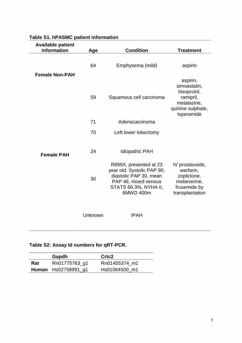
Table S1. hPASMC patient information
Available patient information Age Condition Treatment
Female Non-PAH
64 Emphysema (mild) aspirin
59 Squamous cell carcinoma
aspirin, simvastatin, bisoprolol, ramipril,
metalazine, quinine sulphate,
loperamide
71 Adenocarcinoma
70 Left lower lobectomy
Female PAH 24 Idiopathic PAH
30
R899X, presented at 23 year old. Systolic PAP 90,
diastolic PAP 30, mean PAP 46, mixed venous
STATS 66.3%, NYHA II, 6MWD 400m
IV prostanoids, warfarin,
zopliclone, mebeverine, frusemide by
transplantation
Unknown IPAH
Table S2: Assay Id numbers for qRT-PCR.
Gapdh Crtc2
Rat Rn01775763_g1 Rn01455374_m1
Human Hs02758991_g1 Hs01064500_m1
8
Supplementary Figures
Figure S1. mSAP, glucose levels, cardiac output and heart rate are not significantly altered in female Sugen/hypoxic rats exposed to metformin. (A) mSAP was unchanged between the group, this was also found for glucose levels (B), cardiac output (C) and heart rate (D). Glucose levels were measured at day 7, 14 and 21 (data shown for day 21) of treatment in each group and no significant difference in blood glucose levels, taken from tail vein, was found. n=6-13 per group. Data was analysed by one way ANOVA followed by Bonferroni post test.
9
A
B
Figure S2. Representative immunoblots and quantification relative to GAPDH of LKB1 (A) in whole lung from control (no sugen/hypoxic) and sugen/hypoxic animals without (vehicle) or with metformin treatment. n=4-8 per group. C= Control, V=Vehicle, M=Metformin. (B) Representative immunoblots and quantification, showing effects of 24h incubation with metformin, AICAR or anastrozole on LKB1 in female non-PAH hPAMSCs (n=2-3 different cell lines per group). C=Control, M=Metformin, AC=AICAR, A=Anastrozole. Data is displayed as a mean ±SEM

10
Figure S3. Non PAH female hPASMCs (A) and PAH female hPASMCs (B) were incubated in 2% (v/v) FBS or 10% (v/v) FBS in the presence or absence of the indicated agents for 24h and BrdU incorporation assessed. n=2 cell lines/group, repeated seven times per group. Data is displayed as mean ±SEM. * P<0.05, **P<0.01 ***P<0.001 as indicated, determined by one way ANOVA followed by Bonferroni post test.
11
62kDa
tAMPK
pAMPK
0 2 4 6 10
GAPDH
0 2 4 6 100
200
400
600 **
*
*****
hours of stimulation
pA
MP
K/t
AM
PK
Figure S4. Non-PAH female hPASMCs (A) were incubated in 2% (v/v) FBS in the presence (or absence–time point 0) of AICAR (3 mM) for either 2, 4, 6 or 10 hours. The activation of AMPK was assessed by measuring pAMPK protein expression over tAMPK protein expression. n=3 cell lines/group. Data is displayed as mean ±SEM. * P<0.05, **P<0.01 ***P<0.001 as indicated, determined by one way ANOVA followed by Bonferroni post test.

12
Decreased proliferation/ artery
remodeling in PH
CRTC2 PII promoter Aromatase
Aromatase
Estrogen
AhR
CYP1B1
Metformin
pAMPK
pACC
Decreased accumulation of proliferative estrogenmetabolites.
Figure S5. Schematic illustrating the proposed pathway of mechanism of metformin action in experimental PH. Metformin increases pAMPK and one effect of this is to cause a decrease in CRTC2. CRTC2 binding to the PII promoter of aromatase is decreased, which means less estrogen is synthesised from testosterone by aromatase. Metformin treatment decreased AhR expression. AhR is a transcription factor for the estrogen metabolising enzyme CYP1B1. CYP1B1 expression was decreased by metformin, possibly due to the decrease in its transcription factor. We have previously suggested decreased CYP1B1 activity can protect against PH by decreasing accumulation of estrogen metabolites.2, 3