Embed Size (px)
Citation preview
Lee et al. BMC Developmental Biology 2014 14:781 DOI 10.1186/s12861-014-0048-3
METHODOLOGY ARTICLE Open Access
Improved application of the electrophoretic tissueclearing technology, CLARITY, to intact solidorgans including brain, pancreas, liver, kidney,lung, and intestineHyunsu Lee1 ? , Jae-Hyung Park2? , Incheol Seo3, Sun-Hyun Park2,5* and Shin Kim4*
Abstract
Background: Mapping of tissue structure at the cellular, circuit, and organ-wide scale is important for understandingphysiological and biological functions. A bio-electrochemical technique known as CLARITY used for three-dimensionalanatomical and phenotypical mapping within transparent intact tissues has been recently developed. This methodprovided a major advance in understanding the structure-function relationships in circuits of the nervous system andorgans by using whole-body clearing. Thus, in the present study, we aimed to improve the original CLARITY procedureand developed specific CLARITY protocols for various intact organs.
Results: We determined the optimal conditions for reducing bubble formation, discoloration, and depositing of blackparticles on the surface of tissue, which allowed production of clearer organ images. We also determined theappropriate replacement cycles of clearing solution for each type of organ, and convincingly demonstrated that250? 280 mA is the ideal range of electrical current for tissue clearing. We then acquired each type of cleared organsincluding brain, pancreas, liver, lung, kidney, and intestine. Additionally, we determined the images of axon fibers ofhippocampal region, the Purkinje layer of cerebellum, and vessels and cellular nuclei of pancreas.
Conclusions: CLARITY is an innovative biochemical technology for the structural and molecular analysis of varioustypes of tissue. We developed improved CLARITY methods for clearing of the brain, pancreas, lung, intestine, liver, andkidney, and identified the appropriate experimental conditions for clearing of each specific tissue type. These optimizedmethods will be useful for the application of CLARITY to various types of organs.
Keywords: CLARITY, Brain, Nervous system, Electrophoretic tissue clearing, 3D Reconstruction, Purkinje layer
BackgroundTissue clearing technologies, such as CLARITY [1] andperfusion-assisted agent release in situ (PARS) [2], whichcreate optically transparent and macromolecule-permeableimages, have provided a major advance in the imaging ofbiological systems. These methods improve tissue per-meability by replacing the lipid bilayer of plasma mem-branes with a nanoporous hydrogel. Unlike mechanical
* Correspondence: [email protected]; [email protected]? Equal contributors2Department of Physiology, Keimyung University School of Medicine, 1095Dalgubeoldae-Ro, Dalseo-Gu, Daegu 704-701, South Korea4Department of Immunology, Keimyung University School of Medicine, 1095Dalgubeoldae-Ro, Dalseo-Gu, Daegu 704-701, South KoreaFull list of author information is available at the end of the article
? 2014 Lee et al.; licensee BioMed Central. ThisCommons Attribution License (http://creativecreproduction in any medium, provided the orDedication waiver (http://creativecommons.orunless otherwise stated.
micro-dissection methods, which aggravate deformation oftissue structure alongside of micro-dissection, CLARITYpreserves the intact structure of brain organs, allowing thetracing of neurite? projections, and the three-dimensional(3D) and topological reconstruction of traced neurons [1].PARS can also produce macromolecule permeability andoptical transparency in the brain and other organs [2].Obtaining detailed system-wide informations of organs
using a general optical microscope is formidable. For ex-ample, single-photon microscopy provides a maximum of50 μm imaging depth below the organ surface, while evenwell-optimized two-photon microscopy cannot image dee-per than approximately 800 μm [3-5]. However, in variousintact organs, CLARITY or PARS can overcome these
is an Open Access article distributed under the terms of the Creativeommons.org/licenses/by/4.0), which permits unrestricted use, distribution, andiginal work is properly credited. The Creative Commons Public Domaing/publicdomain/zero/1.0/) applies to the data made available in this article,

Table 1 Optimized ETC. conditions for various tissues
Organ OptimizedETC. time(days)
Constantcurrent (mA)
Varyingvoltage (V)
Replacementinterval ofclearing solution(days)
Brain Whole brain:12-16
280 22 ~ 26 5
3-mm-thick-block: 3
N/A
Pancreas 8-12 250 7
Kidney 18-22 280 7
Liver 18-22 280 7
Intestine 8-12 250 7
Lung 13-17 250 7
n = 30 for optimization of each organ clearing. ETC., electrophoretic tissueclearing; mA, milliampere; V, voltage; N/A, not available.
Lee et al. BMC Developmental Biology 2014 14:781 Page 2 of 7
limitations to permit enhanced viewing of organ structuresat greater depths [1,2], providing access to integratedstructural and molecular information from the brain andother intact biological systems [1,2]. In particular, CLAR-ITY provides an accelerated rate of un-dissected tissueclearing in the brain through applied electric force, andalso avoids tissue damage during clearing [1,2,6].In the present study, we evaluated whether CLARITY
could be effectively applied to other intact organs fromthe adult mouse (12 weeks-old), including the pancreas,liver, lungs, intestines, and kidneys, as well as the wholebrain. Although, this technique has been applied tovarious cerebral regions of the brain [1], herein, we par-ticularly focused on the Purkinje cell layer of the cerebel-lum. Furthermore, we examined the structural integrityof cell-vasculature relationships in the pancreatic tail re-gion using single-photon microscopy (0.7 mm). This ap-proach allowed us to obtain a detailed, objective pictureof the complexity of the islet vascular system. These datasuggest that organ clearing is useful for examining thephysiology and pathophysiology of the vascular system aswell as other organs.
MethodsExperimental animalsAll animal experimental procedures were conducted in ac-cordance with the guidelines of the University Committeeon Animal Resources at Keimyung University (ApprovalNo. KM-2014-20R1).
CLARITY applied to the mouse brainAdult mice (12 weeks old) were anesthetized using a com-bination of tiletamine-zolazepam-xylazine and perfusedtranscardially with 30 mL of ice cold 1X phosphate-buffered saline followed by 30 mL of ice cold hydrogel so-lution with a mixture of 4% PFA, 4% acrylamide, 0.05% bis-acrylamide, 0.25% VA044 in PBS. Organs were extractedand incubated in the same solution at 4?C for 7 days. Thesolution temperature was then increased to 37?C to initiatepolymerization. After 3 h at 37?C, hydrogel-embedded or-gans were placed in an electrophoretic tissue clearing(ETC.) chamber. While sodium borate buffer (200 mM,pH 8.5) containing 4% SDS (the clearing solution) was cir-culated through the chamber, 250? 280 mA was appliedacross the organs at 42?C for 2 ? 4 weeks. The solution cir-culation velocity was 28 L/min, and the volume of clearingsolution was 10 L. The clearing solution replacement cycleis described in Table 1. After clearing, the organs wereincubated in PBS at 37?C for 4 days to remove SDS. 3-mm-thick horizontal blocks of mouse brain were cleared byelectrophoresis for 3 days as described in experimentalconditions. The detailed experiment processes were as pre-viously reported [1].
Immunostaining of CLARITY - processed mouse brain andpancreasTo prepare mouse brain for immunostaining, hydrogel-embedded and clarified brains were cut into 3-mm-thickhorizontal blocks using a mouse brain matrix (Ted pella).For axon staining, the clarified brain was incubated at 37?Cfor 3 weeks in 0.1% triton X-100, and 1 M sodium boratebuffer, pH 8.5, with an anti-tau1 antibody (1:50; Cat#MAB3420, Millipore Corp., Bedford, MA, USA). For pan-creatic blood vessels, the clarified pancreas was incubatedat 37?C for 2 weeks in 0.1% triton X-100 and 1 M sodiumborate buffer, pH 8.5, with anti-α-smooth muscle actin(1:50; Cat# ab5694, Abcam, Cambridge, UK). The brainand pancreas were washed at 37?C for 1 week in 0.1% tritonX-100 and 1 M sodium borate buffer, pH 8.5. The sampleswere then incubated at 37?C for 2 weeks in 0.1% triton X-100 and 1 M sodium borate buffer, pH 8.5, with a goatanti-rabbit-AlexaFluor488 secondary antibody (1:100; Cat#A11029, Invitrogen). For nuclear staining, the clarified pan-creas was incubated at 37?C for 30 min in DAPI (0.1 μg/mL,Cat# D9542, Sigma-Aldrich, St. Louis, MO, USA).
Imaging of CLARITY - processed mouse brain and pancreasFor imaging the structures of brain and pancreas, theclarified brain and pancreas were incubated in FocusClear,a water-based immersion medium, for 4 days. The brainand pancreas were then enclosed between two coverglass-bottom petri dishes. The brain and pancreas were imaged(Z-stack volume, 110? 650 μm) using confocal laser scan-ning microscopy (LSM 5 EXCITER; Carl Zeiss, Jena,Germany) at excitation wavelengths of 488 nm with a C-Apochromat ? 40 objective (1.2 numerical aperture).Three-dimensional reconstruction was generated usingLSM5 EXCITER software (Carl Zeiss). To extract the pan-creatic microvasculature images, we carefully traced theanti-α-smooth muscle actin profiles using the ImageJplug-in Single Neurite Tracer [7].

Lee et al. BMC Developmental Biology 2014 14:781 Page 3 of 7
ResultsElectrical current conditions related to enhancing CLARITYTo clear the hydrogel-embedded tissues after completingthe polymerization, an electric field was applied to thetissues in an ETC. chamber which was built as describedpreviously [1]. Although the previous study recom-mended using high voltage and temperature to clear tis-sue, the high constant voltage caused bubble formation,discoloration and/or the deposition of black particlesin tissue samples (Additional file 1). Specifically, the
Figure 1 CLARITY of intact adult mouse tissues. In adult mouse tissueCLARITY. (A) Brain. (B) Pancreas. (C) Lung. (D) Intestine. (E) Liver. (F) Kidne
constant voltage created a high current thus damagingthe tissue. The previous study also recommended lowvoltage (V) (10 - 40 V) for ETC. [1], but this conditionwas shown to be ineffective in applying to other types oftissue as well as the whole brain. Therefore, we deter-mined the optimal range of electrical current conditionfor clearing tissues.We found that for brain samples, 280 mA was the op-
timal current without causing any damage. When theconstant current of 280 mA was maintained across the
s (12 weeks old), imaging was performed before and/or aftery.
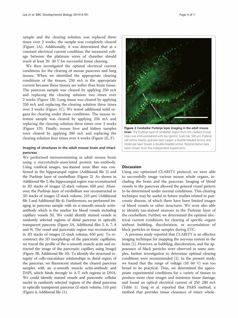
Figure 2 Cerebellar Purkinje layer imaging in the adult mousebrain. The Purkinje layer of cerebellar region form the clarified mousebrain was immunostained with tau (green). Scale bar, 500 μm. Purkinjecell (arrow heads), granular layer (upper, a double-headed arrow), andmolecular layer (lower, a double-headed arrow). Representative datawere chosen from five independent experiments.
Lee et al. BMC Developmental Biology 2014 14:781 Page 4 of 7
sample and the clearing solution was replaced threetimes over 2 weeks, the sample was completely cleared(Figure 1A). Additionally, it was determined that at aconstant electrical current condition, the measured volt-age between the platinum wires of chamber shouldreach at least 20 ? 30 V for successful tissue clearing.We then investigated the optimal electrical current
conditions for the clearing of mouse pancreas and lungtissues. When we identified the appropriate clearingconditions of the tissues, 250 mA is the appropriatecurrent because these tissues are softer than brain tissue.The pancreas sample was cleared by applying 250 mAand replacing the clearing solution two times over2 weeks (Figure 1B). Lung tissue was cleared by applying250 mA and replacing the clearing solution three timesover 3 weeks (Figure 1C). We tested additional solid or-gans for clearing under these conditions. The mouse in-testine sample was cleared by applying 250 mA andreplacing the clearing solution three times over 3 weeks(Figure 1D). Finally, mouse liver and kidney sampleswere cleared by applying 280 mA and replacing theclearing solution four times over 4 weeks (Figure 1E, F).
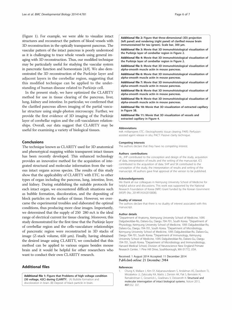
Imaging of structures in the adult mouse brain and intactpancreasWe performed immunostaining in adult mouse brainusing a microtubule-associated protein tau-antibody.Using confocal images, tau-stained axon fiber was con-firmed in the hippocampal region (Additional file 2) andthe Purkinje layer of cerebellum (Figure 2). As shown inAdditional file 2, the hippocampal region was reconstructedin 3D stacks of images (Z-stack volume, 650 μm). More-over, the Purkinje layer of cerebellum was reconstructed in3D stacks of images (Z-stack volume, 210 μm) (Additionalfile 3 and Additional file 4). Furthermore, we performed im-aging in pancreas sample with an α-smooth muscle actin-antibody which is the marker for blood vessels includingcapillary vessels [8]. We could identify stained vessels inrandomly selected regions of distal pancreas in opticallytransparent pancreas (Figure 3A, Additional files 5, 6, 7, 8and 9). The vessel and pancreatic region was reconstructedin 3D stacks of images (Z-stack volume, 650 μm). To re-construct the 3D morphology of the pancreatic capillaries,we traced the profile of the α-smooth muscle actin and ex-tracted the image of the pancreatic capillary using ImageJ(Figure 3B, Additional file 10). To identify the structural in-tegrity of cells-vasculature relationships in distal region ofthe pancreas, we fluorescent stained the cleared pancreassamples with an α-smooth muscle actin-antibody andDAPI, which binds strongly to A-T rich regions in DNA.We could identify stained vessels and pancreatic cellularnuclei in randomly selected regions of the distal pancreasin optically transparent pancreas (Z-stack volume, 110 μm)(Figure 4, Additional file 11).
DiscussionUsing our optimized CLARITY protocol, we were ableto successfully image various mouse whole organs, in-cluding the brain and the pancreas. Imaging of bloodvessels in the pancreas allowed the general vessel patternto be determined under normal conditions. This clearingtechnique may be useful in future studies related to pan-creatic disease, of which there have been limited imagesof blood vessels or other structures. We were also ableto identify tau-stained neurons in the Purkinje layer ofthe cerebellum. Further, we determined the optimal elec-trical current conditions for clearing of specific organswithout bubbling, discoloration, or accumulation ofblack particles in tissue samples during ETC.A previous study reported that CLARITY is an effective
imaging technique for mapping the nervous system in thebrain [1]. However, as bubbling, discoloration, and the ap-pearance of black particles were observed in some sam-ples, further investigation to determine optimal clearingconditions were recommended [1]. In the present study,we found that the range of voltage (10? 60 V) was toobroad to be practical. Thus, we determined the appro-priate experimental conditions for a variety of tissues toproduce more clear images and minimize tissue damage,and found an optical electrical current of 250? 280 mA(Table 1). Yang et al. reported that PARS method, amethod that provides tissue clearance of intact whole-
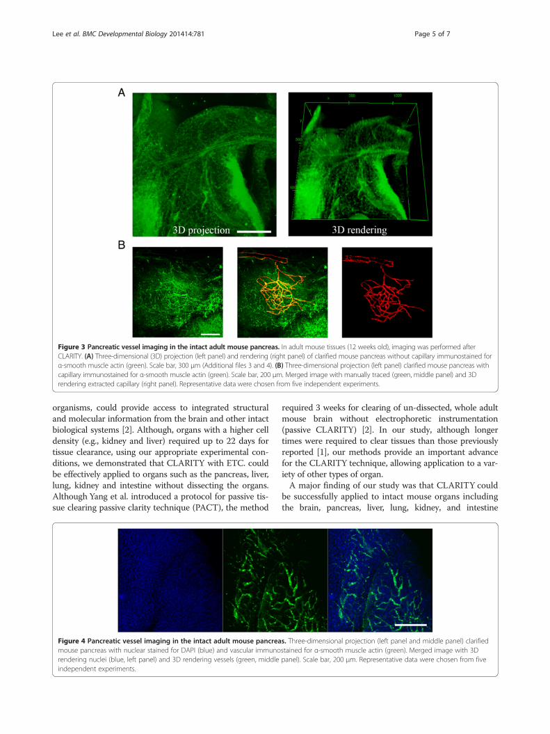
Figure 3 Pancreatic vessel imaging in the intact adult mouse pancreas. In adult mouse tissues (12 weeks old), imaging was performed afterCLARITY. (A) Three-dimensional (3D) projection (left panel) and rendering (right panel) of clarified mouse pancreas without capillary immunostained forα-smooth muscle actin (green). Scale bar, 300 μm (Additional files 3 and 4). (B) Three-dimensional projection (left panel) clarified mouse pancreas withcapillary immunostained for α-smooth muscle actin (green). Scale bar, 200 μm. Merged image with manually traced (green, middle panel) and 3Drendering extracted capillary (right panel). Representative data were chosen from five independent experiments.
Lee et al. BMC Developmental Biology 2014 14:781 Page 5 of 7
organisms, could provide access to integrated structuraland molecular information from the brain and other intactbiological systems [2]. Although, organs with a higher celldensity (e.g., kidney and liver) required up to 22 days fortissue clearance, using our appropriate experimental con-ditions, we demonstrated that CLARITY with ETC. couldbe effectively applied to organs such as the pancreas, liver,lung, kidney and intestine without dissecting the organs.Although Yang et al. introduced a protocol for passive tis-sue clearing passive clarity technique (PACT), the method
Figure 4 Pancreatic vessel imaging in the intact adult mouse pancreamouse pancreas with nuclear stained for DAPI (blue) and vascular immunorendering nuclei (blue, left panel) and 3D rendering vessels (green, middleindependent experiments.
required 3 weeks for clearing of un-dissected, whole adultmouse brain without electrophoretic instrumentation(passive CLARITY) [2]. In our study, although longertimes were required to clear tissues than those previouslyreported [1], our methods provide an important advancefor the CLARITY technique, allowing application to a var-iety of other types of organ.A major finding of our study was that CLARITY could
be successfully applied to intact mouse organs includingthe brain, pancreas, liver, lung, kidney, and intestine
s. Three-dimensional projection (left panel and middle panel) clarifiedstained for α-smooth muscle actin (green). Merged image with 3Dpanel). Scale bar, 200 μm. Representative data were chosen from five
Lee et al. BMC Developmental Biology 2014 14:781 Page 6 of 7
(Figure 1). For example, we were able to visualize intactstructures and reconstruct the pattern of blood vessels with3D reconstruction in the optically transparent pancreas. Thevascular pattern of the intact pancreas is poorly understoodas it is challenging to trace whole vessels using general im-aging with 3D reconstruction. Thus, our modified techniquemay be particularly useful for studying the vascular systemin pancreatic function and homeostasis [4,9]. We also dem-onstrated the 3D reconstruction of the Purkinje layer andadjacent layers in the cerebellar region, suggesting thatthis modified technique can be applied to the under-standing of human disease related to Purkinje cell.In the present study, we have optimized the CLARITY
method for use in tissue clearing of the pancreas, liver,lung, kidney and intestine. In particular, we confirmed thatthe clarified pancreas allows imaging of the partial vascu-lar structure using single-photon microscopy. Further, weprovide the first evidence of 3D imaging of the Purkinjelayer of cerebellar region and the cell-vasculature relation-ships. Overall, our data suggest that CLARITY may beuseful for examining a variety of biological tissues.
ConclusionsThe technique known as CLARITY used for 3D anatomicaland phenotypical mapping within transparent intact tissueshas been recently developed. This enhanced technologyprovides an innovative method for the acquisition of inte-grated structural and molecular information from the vari-ous intact organs across species. The results of this studyshow that the applicability of CLARITY with ETC. to othertypes of organ including the pancreas, lung, intestine, liver,and kidney. During establishing the suitable protocols foreach intact organ, we encountered difficult situations suchas bubble formation, discoloration, and the depositing ofblock particles on the surface of tissue. However, we over-came the experimental troubles and elaborated the optimalconditions, thus producing more clear images. Importantly,we determined that the supply of 250? 280 mA is the idealrange of electrical current for tissue clearing. Moreover, thisstudy demonstrated for the first time that the Purkinje layerof cerebellar region and the cells-vasculature relationshipsof pancreatic region were reconstructed in 3D stacks ofimage (Z-stack volume, 650 μm). Finally, having obtainedthe desired image using CLARITY, we concluded that thismethod can be applied to various organs besides mousebrain and it would be helpful for other researchers whowant to conduct their own CLARITY research.
Additional files
Additional file 1: Figure that Problems of high voltage condition(30 voltage, 42?C) during CLARITY. (A) Bubble formation anddiscoloration in brain. (B) Deposit of black particle in brain.
Additional file 2: Figure that three-dimensional (3D) projection(left panel) and rendering (right panel) of clarified mouse brainimmunostained for tau (green). Scale bar, 300 μm.
Additional file 3: Movie that 3D immunohistological visualization ofthe Purkinje layer of cerebellar region in Figure 2.
Additional file 4: Movie that 3D immunohistological visualization ofthe Purkinje layer of cerebellar region in Figure 2.
Additional file 5: Movie that 3D immunohistological visualization ofalpha-smooth muscle actin in mouse pancreas.
Additional file 6: Movie that 3D immunohistological visualization ofalpha-smooth muscle actin in mouse pancreas.
Additional file 7: Movie that 3D immunohistological visualization ofalpha-smooth muscle actin in mouse pancreas.
Additional file 8: Movie that 3D immunohistological visualization ofalpha-smooth muscle actin in mouse pancreas.
Additional file 9: Movie that 3D immunohistological visualization ofalpha-smooth muscle actin in mouse pancreas.
Additional file 10: Movie that 3D visualization of extracted capillaryin Figure 3B.
Additional file 11: Movie that 3D visualization of vessels andextracted capillary in Figure 4.
AbbreviationsmA: milliampere; ETC.: Electrophoretic tissue clearing; PARS: Perfusion-assisted agent release in situ; PACT: Passive clarity technique.
Competing interestsThe authors declare that they have no competing interest.
Authors ? contributionsHL, JHP contributed to the conception and design of the study, acquisitionof data, interpretation of results and the writing of the manuscript. ICScontributed to the acquisition of data. SHP and SK contributed to theconception of the study, the interpretation of results and writing of themanuscript. All authors gave final approval of the version to be published.
AcknowledgementsWe thank all our colleagues in Keimyung University School of Medicine forhelpful advice and discussions. This work was supported by the NationalResearch Foundation of Korea (NRF) Grant funded by the Korean Govrnment(MSIP) (No. 2014R1A5A2010008).
Duality of interestThe authors declare that there is no duality of interest associated with thismanuscript.
Author details1Department of Anatomy, Keimyung University School of Medicine, 1095Dalgubeoldae-Ro, Dalseo-Gu, Daegu 704-701, South Korea. 2Department ofPhysiology, Keimyung University School of Medicine, 1095 Dalgubeoldae-Ro,Dalseo-Gu, Daegu 704-701, South Korea. 3Department of Microbiology,Keimyung University School of Medicine, 1095 Dalgubeoldae-Ro, Dalseo-Gu,Daegu 704-701, South Korea. 4Department of Immunology, KeimyungUniversity School of Medicine, 1095 Dalgubeoldae-Ro, Dalseo-Gu, Daegu704-701, South Korea. 5Department of Microbiology and Immunobiology,Harvard Medical School, Division of Neuroscience New England PrimateResearch Center, 1 Pine Hill Drive, Southborough, MA 01772, USA.
Received: 1 August 2014 Accepted: 11 December 2014
References1. Chung K, Wallace J, Kim SY, Kalyanasundaram S, Andalman AS, Davidson TJ,
Mirzabekov JJ, Zalocusky KA, Mattis J, Denisin AK, Pak S, Bernstein H,Ramakrishnan C, Grosenick L, Gradinaru V, Deisseroth K: Structural andmolecular interrogation of intact biological systems. Nature 2013,497:332? 337.
Lee et al. BMC Developmental Biology 2014 14:781 Page 7 of 7
2. Yang B, Treweek JB, Kulkarni RP, Deverman BE, Chen CK, Lubeck E, Shah S,Cai L, Gradinaru V: Single-cell phenotyping within transparent intacttissue through whole-body clearing. Cell 2014, 158:945 ? 958.
3. Helmchen F, Denk W: Deep tissue two-photon microscopy. Nat Methods2005, 2:932 ? 940.
4. Rodriguez-Diaz R, Abdulreda MH, Formoso AL, Gans I, Ricordi C, Berggren PO,Caicedo A: Innervation patterns of autonomic axons in the humanendocrine pancreas. Cell Metab 2011, 14:45? 54.
5. Speier S, Nyqvist D, Kohler M, Caicedo A, Leibiger IB, Berggren PO:Noninvasive high-resolution in vivo imaging of cell biology in theanterior chamber of the mouse eye. Nat Protoc 2008, 3:1278 ? 1286.
6. Tomer R, Ye L, Hsueh B, Deisseroth K: Advanced CLARITY for rapid andhigh-resolution imaging of intact tissues. Nat Protoc 2014, 9:1682? 1697.
7. Abr?moff MD, Magalh?es PJ, Ram SJ: Image processing with ImageJ.Biophoton Int 2004, 11:36 ? 43.
8. Skalli O, Pelte MF, Peclet MC, Gabbiani G, Gugliotta P, Bussolati G, Ravazzola M,Orci L: Alpha-smooth muscle actin, a differentiation marker of smoothmuscle cells, is present in microfilamentous bundles of pericytes.J Histochem Cytochem 1989, 37:315? 321.
9. Speier S, Nyqvist D, Cabrera O, Yu J, Molano RD, Pileggi A, Moede T,Kohler M, Wilbertz J, Leibiger B, Ricordi C, Leibiger IB, Caicedo A, BerggrenPO: Noninvasive in vivo imaging of pancreatic islet cell biology. Nat Med2008, 14:574 ? 578.
Submit your next manuscript to BioMed Centraland take full advantage of:
? Convenient online submission
? Thorough peer review
? No space constraints or color ?gure charges
? Immediate publication on acceptance
? Inclusion in PubMed, CAS, Scopus and Google Scholar
? Research which is freely available for redistribution
Submit your manuscript at www.biomedcentral.com/submit