Embed Size (px)
Citation preview
Royal Ontario MuseumLife SciencesMiscellaneous Publication
UJ
< ;
C\J
5 o-i h-z h~~*» mUJ o3 = CO5 "3-
oE T—- CD>-• r~o ^
CO<HOY
MHi—
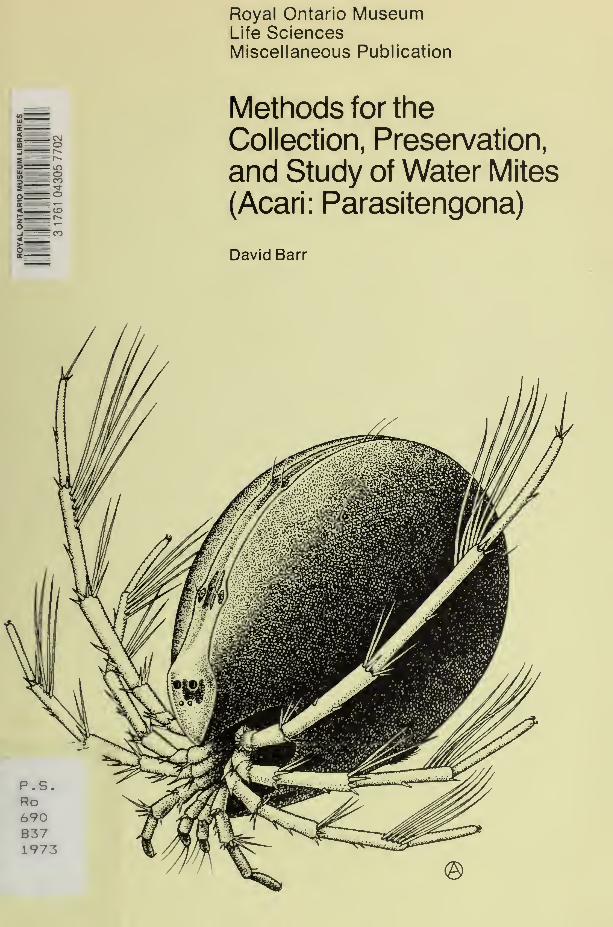
Methods for the
Collection, Preservation,
and Study of Water Mites
(Acari: Parasitengona)
David Barr
Digitized by the Internet Archive
in 2012 with funding from
Royal Ontario Museum
http://archive.org/details/methodsforcollecOObarr
LIFE SCIENCES
MISCELLANEOUS PUBLICATIONS
ROYAL ONTARIO MUSEUM
david barr Methods for the
Collection, Preservation,
and Study of Water Mites
(Acari: Parasitengona)
Publication date: 15 October 1973
ISBN 0-88854- 1 48-1
Suggested citation: LifeSci. Misc. Publ., R. Out. Mus.
ROYAL ONTARIO MUSEUMPUBLICATIONS IN LIFE SCIENCES
The Royal Ontario Museum publishes three series in the Life Sciences:
life sciences contributions, a numbered series of original scientific publi-
cations, including monographic works.
life sciences occasional papers, a numbered series of original scientific
publications, primarily short and usually of taxonomic
significance.
life sciences miscellaneous publications, an unnumbered series of
publications of varied subject matter and format.
All manuscripts considered for publication are subject to the scrutiny and
editorial policies of the Life Sciences Editorial Board, and to review by
persons outside the Museum staff who are authorities in the particular field
involved.
LIFE SCIENCES EDITORIAL BOARD
Chairman: r. l. Peterson
Editor, j. r. tamsitt
Associate Editor, d. barr
Associate Editor, e. j. crossman
david barr is Associate Curator in the Department of Entomology and
Invertebrate Zoology, Royal Ontario Museum and Assistant Professor
in the Department of Zoology. University of Toronto.
price: $1.50
©The Royal Ontario Museum, 1973
100 Queen's Park, Toronto, Canada
PRINTED AT THE UNIVERSITY OF TORONTO PRESSLIBRARY
ROYAL ONTARIO MUSEUM
Contents
Abstract. /
Resume. /
Introduction. 2
Collecting, 2
Lentic Habitats. 2
Lotie Habitats. 3
Water Margin Habitats. 6
Moss Habitats. 6
Phreaticolous Habitats. 7
Hosts, 7
Larvae and Eggs, 7
Preservation of Adults and Nymphs, 8
Larval Mites. 9
Rearing, 9
Harvesting and Preservation, 10
Practical Considerations. 12
Observation and Final Storage of Skeletal Preparations, 12
Exoskeleton of Adults and Nymphs, 12
Sorting and Clearing. 12
Dissection, 13
Classical Slide Mount, 13
Fluid Storage, 14
Temporary Mounts, 14
Semi-permanent Mounts, 15
Permanent Mounts, 16
Ejaculatory Complex, 16
Larvae. 17
Optical Systems, 18
Studies on Internal Anatomy, 19
Scanning Electron Microscopy, 19
Applications, 19
Desiccation, 20
Mounting and Coating, 20
Limitations, 21
Illustrations, 2/
Line Drawings. 21
Photomicrography, 23
Acknowledgments, 24
Appendix: Formulae for Reagents, 25
Literature Cited, 26

Methods for the Collection, Preservation,
and Study of Water Mites
(Acari: Parasitengona)
Abstract
Methods required for the systematic study of water mites
include new techniques that complement or replace some al-
ready in existence. Netting, trapping, and separation pro-
cedures are described for mites collected from lentic, lotic,
moss, water-film, and phreaticolous habitats. The most useful
larval specimens are obtained by collecting gravid females and
harvesting larvae hatched from eggs deposited in the labora-
tory.
Fluid storage is preferable for most specimens. A versatile
method of examining exoskeletal morphology is to observe
temporary slide mounts with light microscopy (bright-field,
interference contrast, or phase contrast). Scanning electron
microscopy of desiccated mites enhances the study of surface
fine-structure but does not replace conventional techniques
of illustration (line drawings, photomicrography).
Resume
Les methodes requises pour l'etude systematique des mites
aquatiques comprennent des techniques nouvelles qui comple-
mentent ou remplacent quelques-unes de celles en existence
maintenant. Des procedes de prendre en filet, d'attraper et de
separer sont decrits pour des mites recueillies des habitats
lentiques, lotiques, phreatiques, de la mousse, et de la filme
d'eau. Les specimens des larves les plus utiles sont obtenus en
receuillant des femelles enceintes et en recoltant des larves
ecloses des oeufs deposes au laboratoire.
Conservation en fluide est preferable pour la plupart des
specimens. Une methode versatile d'examination de la mor-
phologic exosquelettique c'est Tobservation par microscopic
optique (illumination ordinaire, interference contrasted, ou
phase contrasted) des montures temporaires en lamelle. La
microscopie electronique exploratrice des mites dessechees
augmente l'etude de la structure fine de la surface, mais elle
ne remplace pas les techniques conventionelles d'illustration
( de-sins en lignc, photomicrographie).
Introduction
The aquatic families of the suborder Parasitengona are among the most
studied taxa of the Acari. Nevertheless, methods of collecting, preparing, and
studying these creatures are not standardized, and many useful techniques
are either unpublished or scattered throughout acarological literature. I
therefore summarize methods found to be useful for systematic research
with specimens of water mites. The account is directed primarily to those
beginning in the field but also to established workers wishing to explore
unfamiliar techniques. Because new methods are continually developed while
old ones are being modified and improved, only an interim report is possible.
The treatment is not always comparative, for less satisfactory alternatives
are not discussed unless well entrenched in the literature. Nor is the survey
complete, for important areas of research (e.g., karyology, transmission
electron microscopy, chromatography, electrophoresis) with which I have
minimal experience are omitted. But procedures fundamental to systematic,
evolutionary, ecological, life history, and zoogeographic studies are covered.
Viets (1936) first compiled methods for water mite study in his general
treatment of the German species. There was no other comprehensive survey
until those of Cook and Mitchell (Cook and Mitchell, 1952; Mitchell and
Cook, 1952) appeared. Other sources of information have been limited in
scope. Several authors of general works have described techniques of spe-
cific interest to water mite workers (Pennak, 1953; Newell, 1959) or for
other mites (Grandjean, 1935, 1949; Newell, 1947; Baker and Wharton,
1952; Krantz, 1970) that can be used for water mites, either directly or
with modification. Evans et a\. (1961) published perhaps the most com-
plete description of general acarological techniques available in English.
Collecting
Most collecting procedures described here involve gathering a portion of
the habitat (vegetation, gravel, soil, detritus) and separating the mites from
it. Although separation is usually performed in the field, material may be
returned to the laboratory for this operation when convenient.
In collecting adult mites, it is worthwhile to strive for quantity. Series of
50-100 specimens of a single species per locality are not too extensive.
Not only are large numbers valuable in taxonomic research but are manda-
tory if mites are to be used as well for sectioning, rearing, karyology, or
scanning electron microscope (SEM) observation.
LENTIC HABITATS
Many larger species of water mites occur in standing bodies of surface water
- ponds, lakes, and ditches. These mites are easy to collect and have received
attention from students of the group for more than a century. Cook and
Mitchell ( 1952) described the dip-netting technique that is most useful for
collecting in this habitat, a method particularly effective because most of the
mites of standing waters swim well. They readily leave netted vegetation
and detritus floated in a white enamel tray and are easily picked up with a
dropping pipette and placed in the collecting container.
A useful collecting accessory is a polyethylene dropping pipette with a
tapered tip. It progressively larger sections of the soft, plastic tip are sliced
off. the corresponding intake bore of the pipette is increased. With several
of these pipettes, one can efficiently handle mites of widely different sizes.
Periodically the amount of water in the collecting vial is reduced with the
pipette (tip held against the bottom to exclude mites) so that additional
specimens can be added. The collection is then transported to the laboratory
for biological work; or all water may be pipetted off and the preservative
added in the field.
Deep-water (> 2 m). benthic mites (e.g.. Huitjeldtia) are not accessible
with standard techniques and are usually collected using a dredge sampler,
a slow process yielding small numbers of specimens. Pieczynski (1962,
1965) employed underwater traps for sampling mites of standing-water
habitats.
Certain lentic habitats, notably temporary pools and many boggy situa-
tions, contain at the same time large amounts of dead vegetation and detritus
and numerous species of mites that either do not swim readily or that crawl
on the bottom. As Cook and Mitchell (1952) pointed out, crawling species
are difficult to separate by the technique of dip net and enamel tray. Gener-
ally, too much detritus is taken in the net. Most specimens do not come out
but must be sought by turning over every piece of plant material in the tray.
Collection, even of swimming species, is hampered by excess debris. The
suggestion of picking over vegetation in the pond (Cook and Mitchell,
1952) is valuable, and in some situations a more specialized technique,
developed by Ian M. Smith (pers. comm.) to collect species that do not swim
readily, may be helpful. His method involves holding an enamel tray sub-
merged 3-15 cm beneath the surface of the water to form a clear, white
background. Working it gently through and under bottom detritus, one
can see mites swimming or occasionally crawling across the white back-
ground and pick them up, one by one, with a pipette.
LOTIC HABITATS
Numerous mite species also inhabit running water. Perhaps the method
proved to be most useful in a variety of lotic situations is the technique
(Cook and Mitchell, 1952) of washing submerged vegetation (panning).
Masses of aquatic or submerged terrestrial vegetation are collected by hand
and transferred to a white enamel tray filled with 3-5 cm of clear water.
Vegetation is floated in the tray, taking care that not too much is added to
a given tray. Within 5-30 minutes, depending on the initial temperature of
the stream water, mites leave the vegetation to swim in the open water or
crawl across the tray bottom. The method is slow, particularly when the
stream temperature is low (< 15°C) and the appearance of mites delayed.
Thus, efficiency is increased by running two or more trays simultaneously.
The coarse screen mentioned by Cook and Mitchell (1952) is not necessary
and is restrictive when collecting specimens continuously as they appear. It
is necessary, however, to move the clumps of vegetation occasionally to dis-
cover mites on the tray beneath.
With this technique are taken many species occurring in various micro-
habitats in the stream other than the truly phreaticolous species that charac-
teristically inhabit gravel several inches below the stream bed (see p. 00).
The relative numbers of species found in vegetation differ from those at
other locations in the stream, but the species diversity of vegetation collec-
tions is sufficiently great to justify this method as the first choice in a newlocality when time is limited.
The type of vegetation chosen for panning will vary with stream type.
Fine-structured plants are more productive than simple, large plants of
gross structure; i.e., aquatic mosses (Musci) are usually superior to Nastur-
tium spp. In turn, fine-structured mosses are usually more productive than
species with larger leaves. Mites are also found in clumps and mats of algae,
particularly those taken from riffle areas of a stream. If truly aquatic vegeta-
tion is sparse, submerged terrestrial vegetation will often be productive; e.g.,
grasses (Graminae) hanging over the bank, roots of grasses and other angio-
sperms along the bank and occasionally in the centre of the stream, or islets
of grass or other terrestrial vegetation growing on a submerged log or other
stable location in the watercourse. The technique of panning submerged
vegetation will yield at least a few mites from almost any running water.
A useful method to separate crawling or weakly-swimming mites from
vegetation in rheocrene springs and seepage areas has been used extensively
by David R. Cook (described in litt.). When examined directly in a pan,
this material often contains so much mud and suspended organic matter
that organisms are difficult to see. Instead, handfuls of vegetation, including
moss, matted roots, and leaves, are placed in a pail or large, wide-mouth
jar, and only enough water is added to cover the plant material. Pieces of
cheesecloth cut into 10 x 20 cm strips are crisscrossed on top of the material
so that at least two or three layers are placed everywhere. If the pail is then
not disturbed for a few hours, most mites move toward the surface (perhaps
in response to decreasing oxygen levels in the vegetation) into the cheese-
cloth, which can later be removed strip by strip and examined in a tray of
clear water. Mites clinging to the cheesecloth are easily seen and collected.
A stream bed with a depth of 5-30 cm of loosely consolidated gravel often
yields many mites, most of which live in the top 5-10 cm of gravel or under-
neath stones on the stream bed. A method developed by the author has
proved to be valuable in a variety of gravel-bed stream habitats. A cone-
shaped (base: height 1:3) net of 125 fim plankton netting is held by hand
in the stream, with the mouth opening upstream. The opening is covered
with coarse aluminum window screening (seven wires/cm) to exclude
stones and large pieces of debris. The collector stands upstream from the
net and, with his boots, vigorously disturbs the substrate to a depth of 15-20
cm. Still holding the net under water, he slowly collects upstream for a dis-
tance of 3-4 m. The net is then withdrawn from the water and is held up so
that sediment drains to the bottom. Additional water is splashed into the top
of the net to wash down the sediment.
All sediment collected is then distributed evenly over the bottom of a
white enamel tray containing 3-5 cm of clear water. Material collected,
which is composed o( fine gravel, silt, and other detritus, should form a layer
no more than about 3 mm deep when spread over the pan. Mites begin to
leave the sediment as soon as it settles, climbing over the surface and bur-
rowing through the material. Main species that live in this habitat are small
but usually can be seen against the dark substrate because of the dorsal
white or pale spot that marks the site o( the excretory gland. Species lacking
a visible excretory gland are often otherwise conspicuous because of colora-
tion or movement. Main smaller mites (e.g., Aturus spp.) swim actively
above the substrate and may even reach the surface of the water in the col-
lecting tray. Detection of specimens is nonetheless a problem, and collecting
under a heaw overcast or in rain is difficult.
The plankton-net technique is superior to the screening method described
by Cook and Mitchell ( 1952), for many specimens pass through coarse
insect screening without clinging to the wire. In a productive gravel-stream
habitat, a comparative test of the two methods clearly demonstrated the
superioritv of the modified plankton net (Barr and C. A. Lanciani, unpubl.
data). It offers probabl) the best way to obtain the large samples (up to
several hundred specimens per net sample) required for diversified research
needs. Even so, dip-netting should not be neglected in the stream habitat,
especially in areas of marginal vegetation and silt deposits in slow water.
Most lotic species are relatively small and can be readily collected with a
small, glass, dropping pipette (medicine dropper type). Because many stream
mites are adapted to cling to smooth surfaces in a current (often with the
aid of secreted mucilaginous substances), they easily become lodged in the
pipette and may be difficult to extract. But individuals usually detach when
water in the pipette is allowed to rest momentarily, and the mite again floats
or swims. Then water in the pipette is rapidly expelled into the collecting
vial.
Experience has shown that not all suitable-appearing streams have a rich
gravel fauna. As many different collecting techniques should be tried at each
locality as time permits. In many streams and rivers and along rocky, wave-
swept lakeshores, water mites occur on the underside of flattened rocks
lying on the gravel-based substrate. By carefully examining the surface of
the rock after removing it from the water, specimens can be detected and
picked off, either with fine forceps or a sharpened toothpick as described by
Cook and Mitchell (1952). Collecting by this method, however, is tedious.
Crowell (1960) described a method of brushing the underside of the rocks
into a pan of water and collecting the dislodged mites with a pipette. Speci-
mens can also be obtained by merely placing the stone in a pan of still water
and allowing mites to leave of their own accord. Most rheophilic mites begin
to move when placed in still water, more actively as the temperature of the
water increases and oxygen content drops.
Water mites are usually among the fauna captured by drift nets placed
in running waters (e.g., see Clifford, 1972). Drift sampling may represent
a means of obtaining data on the composition of a stream mite fauna but is
unlikely to reveal diversity as well as several of the methods described above.
WATER-MARGIN HABITATS
The water-margin habitat supports several highly-specialized species
(Mitchell, 1960), the most common being Tyrrellia spp. These mites, which
live in a film of water less than the thickness of their own bodies, can be col-
lected slowly and tediously by scanning a prospective habitat visually from
a distance of 20-40 cm. Specimens are removed from the surface one at a
time with a pair of fine forceps. Appropriate situations are beds of moss
kept moist by spray from falls or rapids, moist banks of streams and lakes
(particularly wet bits of wood), stretches of mud-bank, and the stem bases
of semi-terrestrial vegetation near the water-line. The surface must glisten
with a thin film of water to be suitable for mites. Many water-film species
are bright red and easy to see against the background, but Tyrrellia spp. are
occasionally dark and inconspicuous.
MOSS HABITATS
Clumps of moss (Musci) growing on stones or wood submerged in flowing
water may harbour populations of unusual and often primitive genera of
mites (e.g., Trichothyas, Panisus, or Hydrovolzia) . Often the most produc-
tive moss samples grow at the air-water interface along banks or on pro-
jecting objects in the centre of the water where they are always abundantly
saturated but never submerged. The same productive habitat may occur in
wet areas created by springs or in the spray zone of rapids or waterfalls.
Species found in saturated moss are usually sessile and will not leave the
vegetation even if the sample is submerged in still, shallow water indefinitely.
Two methods can be used to extract them from the sample, one simpler and
more straightforward than the other. Although both techniques are equally
effective, both are biased for certain taxa.
The first is a simple field expedient. A clump of moss is held in one hand
and systematically picked apart, strand by strand, with a pair of iris forceps.
Most species found in mosses are bright red or orange, are easily seen near
the base of the stalks of moss, and can be removed with little difficulty using
forceps. Eggs and imagochrysalids attached to leaves and branches of the
moss can be removed only by breaking off a small section of the plant and
placing it in a collecting vial. Adding vegetation also gives living mites in the
vial additional surface area for climbing, thus reducing the chance of en-
counter and consequently of damage by predation. With the hand-picking
technique, large numbers of adult and immature mites, coloured red, orange,
yellow, pink or even brown, are detected. Dark coloured specimens, such as
some Laversia spp., however, could be missed in this type of scanning.
A second technique is more likely to yield cryptically coloured mites
(I. M. Smith, pers. comm.). Moss is rapidly shredded, strand by strand,
into a tray of still water. When the entire sample has been immersed, the
water is stirred and vigorously swirled around the tray to allow all mites to
become detached. Then loose moss is gathered, rinsed slightly as it is re-
moved from the water, and set aside. When the water clears, numerous
mites can be seen lying on or crawling through a layer of brown sediment
that has settled on the bottom of the tray. Specimens can then be collected
rapidly. Although dark-coloured mites will be free and actively crawling.
thev still may be difficult to sec against the dark background of sediment.
The method does not. however, facilitate the collection of wholly sessile life
history stages.
Both techniques were tried on moss samples from the same locality,
subsequently subjecting discarded moss to examination by the alternate
technique. In each instance a number of mites missed by the first method
were taken b\ the second. At present, data are lacking to indicate the relative
efficiency of the two techniques. Hand-picking is simpler in the field, but both
methods require good lighting for effective scanning.
PHREATICOLOUS HABITATS
The diverse mite fauna of interstitial (subterranean) waters can be collected
by the method of Karaman-Chappuis (Motas, 1962). First, a hole deep
enough to penetrate the water table is dug in the bank of a stream. The hole
fills with water, which is scooped out and passed through plankton netting
to concentrate specimens. Water removed from the hole is replaced by flow
from ground water percolating through the sand or gravel interstices and
carrying with it the fragile, colourless mites of the phreatic habitat. Specimens
are best removed from the accumulated debris under a dissecting microscope.
Cook published on North American collections (e.g., 1963, 1968) taken in
this manner and found poorly consolidated gravel bars along the banks or
in the centre of streams to be particularly productive.
HOSTS
Studies of water mite life histories and host-parasite interactions demandconsiderable effort and ingenuity in collecting parasitized host insects. Fromthe host can be obtained larvae, larval sclerites and, often, associated nymphs.
There is, of course, no general collecting technique. The host of the species
of interest must be determined and then standard entomological techniques
(Oldroyd, 1970) applied to collect that host. Although the incidence of
parasitism is occasionally high, especially with the aquatic Hemiptera, it is
often less than 10% (e.g., Coleoptera;Lanciani, 1970).
Aquatic hosts are efficiently collected with the dip net and enamel tray
described for collecting adult water mites. A useful accessory to remove
fast-swimming Coleoptera and Hemiptera from the collecting tray is a small
tea strainer. Once isolated in the strainer, the specimen may be knocked
into a collecting jar or removed with forceps and preserved. Oldroyd
( 1970) described an underwater light trap for capturing aquatic Hemiptera
(also see Pieczynski, 1962). Aerial hosts may be netted on the wing or at
rest by sweeping vegetation close to the banks of the body of water. They
may also be taken with black (ultraviolet) light or in various types of
emergence cages.
Engorged larvae or nymphochrysalids removed from hosts may be satis-
factory for identification but are usually too damaged or deformed to be
valuable for taxonomic descriptions.
LARVAE AND EGGS
Larvae are usually collected in the field only incidentally and may be dis-
covered in collections of adults at the time of initial sorting. Larvae can
occur in stream collections taken with a plankton net, probably having been
mistaken for adults of some small stream species, and collected with the
pipette. Occasionally larvae occur in large numbers on the surface film of
standing waters, where they may be collected with any type of cloth net
swept across the water with the lower edge just beneath the surface. The
net must be everted into alcohol immediately upon removal from the water
to kill the larvae and prevent them from swarming out.
When picking through moss samples, larvae will occasionally be noticed
climbing over the vegetation. Single larvae may be picked up with the tip of
a pair of fine forceps or a fine camel's hair brush dampened in 70% ethanol.
Eggs also are not commonly sought in the field but may be observed in
numbers in pond and moss habitats. Egg masses should be taken with a bit
of vegetation to the laboratory, isolated in a small container of water, and
held until the time of hatching. Field-collected larvae and egg masses are
less useful taxonomic material at present, because in the absence of larval
descriptions, only circumstantial evidence is available for association with
the adult form.
Preservation of Adults and Nymphs
For taxonomic purposes the best preservative for adult and nymphal water
mites is modified Koenike's solution (GAW; see Appendix) as recom-
mended by Mitchell and Cook (1952), for specimens preserved in alcohol
or formalin are more difficult to clear (see p. 12).
GAW-preserved specimens of most water mites are flexible, yet firm, and
are easily cleared in acetic corrosive (Andre's fluid) or potassium hydroxide
solution (Mitchell and Cook, 1952). The resulting preparations are strong
and transparent. So robust are GAW-preserved specimens that they are
easily handled, and breakage of specimens caused by routine transfer or
dissection is rare. Mites with large areas of weak, membranous integument
(e.g., large Eylais spp., Limnochares spp., and Wandesia spp.) become fra-
gile in GAW, but at least the sclerites are easily cleared. In practice, adult
mites are stored indefinitely in the original preservative until they are cleared
for detailed study.
When adult mites are prepared for the study of internal morphology, a
histological fixative is recommended. Several standard reagents, for example
Brasil's fluid (Mitchell, 1964), are satisfactory, especially for soft-bodied
mites. Often, however, in the preparation of serial sections, hard sclerites of
the body wall interfere with the cutting process. Adjacent areas of the section
are often damaged, or the entire section may be destroyed. One solution
involves embedding in a plastic medium (De Giusti and Ezman, 1955;
Woodring and Cook, 1962).
To eliminate these disadvantages more simply, adult mites can be pre-
served with a fixative that hardens soft tissue but does not unduly harden
sclerotizcd structures. Such a fixative (modified Carnoy's), described by
De Giusti and Ezman ( 1955), has proved to be satisfactory in preparing
hard-bodied, sclerotizcd mites for sectioning. The fixative should be prc-
8
pared freshly each time. After one week in the fixative, specimens can be
washed and stored in 70 r; isopropyl alcohol until ready for embedding.
Larval Mites
REARING
A prime requisite in collecting and preserving larval mites is that larvae be
readily associated with adults of the correct species. An unambiguous asso-
ciation makes identification possible and permits further study of the biology
of the species. The single most satisfactory method of obtaining larval
material for study is to rear eggs laid in the laboratory by female mites.
Females, obtained in the field by any of the methods previously described,
are taken to the laboratory as rapidly as possible and isolated in small con-
tainers of water. Two-dram shell vials containing about 1 cm depth (i.e.,
1 ml) of water proved satisfactory for most species reared in this laboratory.
A short ( 1-2 cm) strip of filter paper placed in the vial seems to stimulate
oviposition in some species. Females are then held at room temperature
( which in Toronto may rise to 30°C in summer) and observed every 3 days
to determine if oviposition has occurred or if water must be added to replace
loss through evaporation. Whenever possible, the original water is from
a clean lake or stream near the laboratory, but the level is maintained with
glass-distilled water.
Some mites collected from temporary ponds shortly after the ice melts or
from cold springs and streams may survive better if maintained at 5-1 5 °C.
Cooling is occasionally necessary to keep the female alive, although low
temperatures also severely retard the rate of embryonic development.
Several systems of organizing females and reared larvae have been tried in
this laboratory. The most convenient to date consists of holding isolated
females in rearing vials in clear plastic boxes grouped bv collection. A shal-
low box (approximately 55 x 120 mm) that holds 27 two-dram vials can
be placed easily under a dissecting microscope and scanned to check each
specimen. Scanning should be done every 3 days, watching for either ovipo-
sition or death of the mite in the vial. If a mite dies, it is removed and pre-
served in GAVV. If oviposition occurs, the vial is separated with others con-
taining eggs by a cardboard divider. The female is left with the eggs until
they hatch or until she dies. If the female dies before the eggs hatch, she
should be removed, preserved in GAW, and assigned a rearing number that
is also given to the vial of unhatched eggs. This number allows a female and
the larvae reared from her eggs to be associated subsequently and also
identifies individual rearings within monospecific series from a given habitat.
Although only females have been discussed, an advantage of the system
described is that when space and equipment permit, entire collections of
mites can be set up without preliminary field sorting. They are scanned as
before. Gravid females that oviposit are separated from non-ovipositing
mites, and the rearing process is followed to completion. Non-gravid females,
males, and nymphs, are simply preserved in GAW at the time of death and
stored in a single vial kept for that collection. The only additional processing
required for non-reproductive specimens is the time spent scanning them.
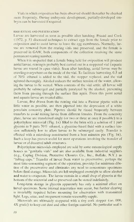
Vials in which oviposition has been observed should thereafter be checked
more frequently. During embryonic development, partially-developed em-
bryos can be harvested if required.
HARVESTING AND PRESERVATION
Larvae are harvested as soon as possible after hatching. Prasad and Cook
(1972, p. 5) discussed techniques to extract eggs from the female prior to
oviposition and to assist larvae to leave the egg membranes. Normally, lar-
vae are removed from the rearing vials and preserved, and the female is
preserved in GAW, both components of the collection receiving the same
permanent rearing number.
When it is suspected that a female being held for oviposition will produce
aerial larvae, rearing is probably best carried out in a stoppered vial (aquatic
larvae are reared in open vials). Soon after hatching, aerial larvae may be
crawling everywhere on the inside of the vial. To facilitate harvesting, 0.5 ml
of 70% ethanol is added to the vial, the stopper replaced, and the vial
agitated thoroughly. Alcohol reduces the surface tension of the water, allow-
ing the larvae to be wetted. When contents of the vial settle, all larvae will
probably be submerged and partially paralyzed by the alcohol, preventing
them from passing through the surface film again. From this point aerial
and aquatic larvae are treated alike.
Larvae, first drawn from the rearing vial into a Pasteur pipette with as
little water as possible, are then pipetted into the depression of a white
porcelain concavity plate. Pipettes should be rinsed frequently between
transfers to avoid mixing larvae from different females. From the concavity
plate, larvae are transferred singly (or two or three at once if possible) to a
polyethylene microvial (Fig. 1a) filled to the brim with a solution of 1 part
glycerin in 9 parts 10% ethanol, a glycerine-based fluid with a surface ten-
sion sufficiently low to allow larvae to be submerged easily. Transfer is
effected with a microloop constructed from a bent minuten pin (Fig. 1b).
Such a loop has proven useful for most operations involving the transfer of
larvae or of dissected adult structures.
Polyethylene microvials employed are sold by some entomological supply
houses as "genitalia vials" and are also available from industrial suppliers
(e.g., Caplug Division, Protective Closures Co., Inc., Buffalo, N.Y.) as
"tubing caps." Transfer of larvae from water to preservative, perhaps the
most time-consuming segment of the operation, provides for minimum dilu-
tion of the preservative and eliminates any need to concentrate specimens
before final storage. Microvials are left unplugged overnight to allow alcohol
and water to evaporate. The larvae remain in a small drop of glycerin at the
bottom of the microvial and so preserved can be stored indefinitely.
Long-term storage in glycerin apparently has only a minimal effect on
larval specimens. Some internal maceration may occur, but further clearing
is invariably required before mounting for observation with transmitted
light. Legs, setae, and integument remain reasonably supple.
Microvials are ultimately stoppered with a tiny cork stopper (no. 000,
4X grade) to keep out dust and other foreign material. No particular seal is
10
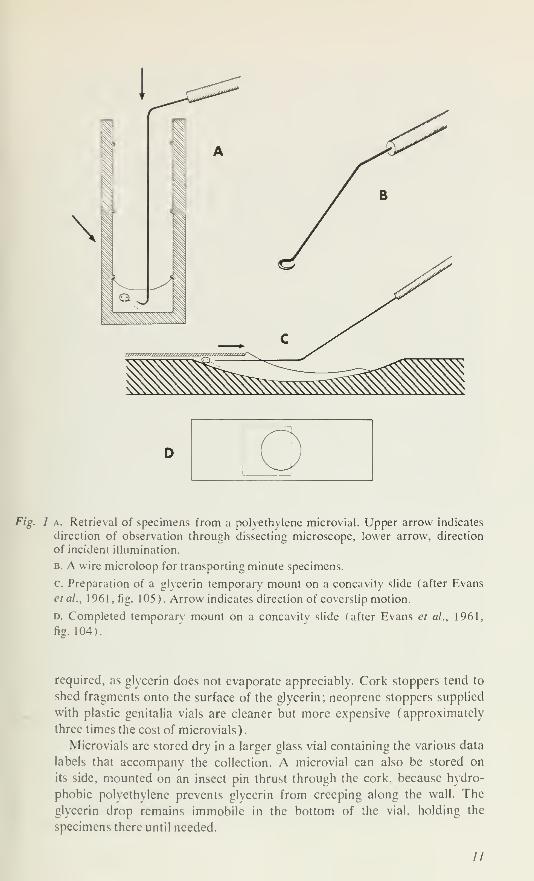
Fig. 1 a. Retrieval of specimens from a polvethvlene microvial. Upper arrow indicates
direction of observation through dissecting microscope, lower arrow, direction
of incident illumination.
B. A wire microloop for transporting minute specimens.
c. Preparation of a glvcerin temporary mount on a concavity slide (after Evans
etal., 1961 , fig. 105). Arrow indicates direction of coverslip motion.
D. Completed temporary mount on a concavity slide (after Evans et al., 1961,
fig. 104).
required, as glycerin does not evaporate appreciably. Cork stoppers tend to
shed fragments onto the surface of the glycerin; neoprene stoppers supplied
with plastic genitalia vials are cleaner but more expensive (approximately
three times the cost of microvials)
.
Microvials are stored dry in a larger glass vial containing the various data
labels that accompany the collection. A microvial can also be stored on
its side, mounted on an insect pin thrust through the cork, because hydro-
phobic polvethvlene prevents glycerin from creeping along the wall. The
glvcerin drop remains immobile in the bottom of the vial, holding the
specimens there until needed.
//
The final step in preservation of the associated female and larvae is the
transfer of the female from GAW to a drop of glycerin in a polyethylene
microvial. The microvial can then be placed dry in the same larger vial con-
taining larvae and labels.
PRACTICAL CONSIDERATIONS
For most rearing operations (sorting field collections, handling adults and
larvae) a low-power, stereoscopic dissecting microscope with continuously
variable (pancratic or zoom) magnification is recommended. The lower
extreme of the magnification range should be as low as 10X, and rarely
(including, for the experienced worker, most generic determinations) will
magnifications greater than 40X be required.
The entire operation of rearing mite larvae is time-consuming, and to
carry out a large-scale program in the field during extended collecting trips
is inadvisable; three days' work on the rearing material is required for each
day to collect adults. Field rearing, however, may be necessary for rare mites
or for species of particular interest. But the most efficient arrangement is to
maintain both a field party collecting adults and a support staff rearing and
harvesting in the laboratory.
Field collections receive an initial sorting (although they need not) and
are mailed to the laboratory (airmail if necessary) in capped vials in a
sturdy, protective, mailing container. Mortality is low when mailing time is
no more than 2 or 3 days, but larvae from females that were as long as 5
days in transit have been successfully reared. It is essential when packing
mites for shipment to provide a high level of dissolved oxygen by filling the
vials no more than half full of water (short, wide vials also improve the
surface: volume ratio) and by drastically restricting the number of mites per
vial. Only 10-15 medium-sized mites (e.g., Limnesia spp., Piona spp., or
Arrenurus spp.) travel successfully together. Even smaller numbers (2-5)
per vial will be necessary with large species, such as some Hydraclma and
Eylais. For cold-water species, it might be advisable to insulate the shipping
container and possibly to provide some means to cool the contents in transit.
Most adult mites, however, tolerate high temperatures for some time, and
we have successfully mailed and reared cold-water mites at ambient summer
temperatures.
If the duration of the collecting trip is no more than a week, collections
may be stockpiled in an ice chest (5°C±) and sorted for laboratory rearing
on return. Some oviposition may occur in the field, and larvae from these
eggs cannot be positively associated with a specific female.
Observation and Final Storage of Skeletal Preparations
EXOSKELETON OF ADULTS AND NYMPHSSorting and Clearing
Adult mites preserved in GAW are easily cleared for microscopic exami-
nation. Initial sorting by an experienced worker may be carried out under
a dissecting microscope without clearing. For critical study, species determi-
nation, and separation of similar or closely related genera, a cleared speci-
12
men mounted on a slide should be examined b\ transmitted light at 100-400X. Acetic corrosive (Andre's fluid), described by Mitchell and Cook(1952), is satisfactory for clearing most adult water mites preserved in
GAW. The other clearing agent also mentioned by those authors (ibid.),
10^ potassium hydroxide (K.OH). should be used with the several families
of heavil) sclerotized mites having characteristic pigmention of the dorsal
sclerite (e.g., Torrenticolidae, Aturidae, Axonopsidae, Mideidae, Mideopsi-dae. Krendowskijidae. and the genus Koeniked). Such patterns arc obliter-
ated b) acetic corrosive but are unaffected by gentle KOH clearing.
The technique of Essig (1948) for clearing alcohol-preserved specimensis satisfactory, but the process is tedious. Possibly the enzymatic macerationdescribed by Newell (1947) could also be valuable in reclaiming adult
mites preserved in improper fluids.
Dissection
Specimens can be dissected with only a few, specialized instruments. Mostappendages are easily removed by inserting a fine needle (e.g., no. 00 insect
pin) at the base; only a small amount of pressure is needed to separate the
appendage from its socket. A difficult dissection is the separation of dorsumfrom venter in species lacking a sclerotized dorsal plate. Large specimens
(^ 2 mm in diameter) can be dissected readily into dorsum and venter with
a pair of fine, surgical scissors. Scissors cannot be used on mites less than
1 mm in diameter, although other workers have had success with sharpenedneedles or glass scalpels (pers. comm.). Separation of dorsum and venter
is important primarily in large or heavily-sclerotized mites where the pres-
ence of one surface seriously impedes the transmission of light through the
surface being examined. Usually, in heavily-sclerotized species, a well-
marked line separates the dorsum and venter, and the insertion of needles at
several points along this membranous zone will separate the two halves of
the body completely. In smaller, lightl) -sclerotized species, such interference
is minimal, and these specimens can be successfully studied in temporarymounts without extensive dissection.
Classical Slide Mounting
The classical method of preparing specimens for observation and perma-nent storage (Mitchell and Cook, 1952) consists of a double-coverslipslide mount containing a dissected specimen embedded in glycerin jelly, a
medium of low refractive index. Although these mounts arc easy to examineinitially and provide a high degree of permanence for museum collections,
they have several disadvantages. After 10-15 years, specimens preservedby the double-coverslip technique tend to accumulate an external deposit
consisting of orange-brown droplets that often obscure structural details. Adisadvantage common to all permanent mounts is that structures cannot befurther manipulated or dissected. A related restriction is that the relative
thickness of many such preparations precludes the free use of oil immersionand some "high-dry" objectives. A final and perhaps most compelling dis-
advantage of the permanent mount is the considerable amount of time re-
quired to prepare each specimen.
/.?
Fluid Storage
Permanent preservation in glycerin eliminates the difficulties described for
permanent, double-coverslip slide mounts and has few serious disadvantages.
Adult mites are cleared and then stored in 96% glycerin, either in groups
in large vials or singly in polyethylene microvials (see p. 10). To examine
the specimen, it is removed from the microvial, placed on a concavity slide
in a drop of glycerin, and treated as a temporary mount. The main disad-
vantages are the handling time required for each observation and the diffi-
culty of maintaining numerous temporary mounts simultaneously during
a taxonomic study. The second restriction may be eased by routine and con-
sistent use of black-and-white photography during an investigation. Muchsorting and gross inspection can be done with photographs, resorting to the
specimens only for critical details.
Storage in fluid allows any of the standard dissections needed to examine
discrete organs (viz. removal of dorsum, capitulum, palp, leg, or ejaculatory
complex). Most parts can be stored together in the same drop of glycerin
in a microvial; each can be retrieved individually with the proper tools. Al-
though the ejaculatory complex can be stored with other structures (see
p. 16), because of its great transparency and small size it may be easier to
recover if stored separately in its own microvial.
To retrieve specimens, the microvial should be uncorked and placed
vertically on the stage of a dissecting microscope. With practice, the con-
tainer can be held in the fingers for this operation or placed in a rack or
stand, holding it firmly under a strong source of light (Fig. 1a). Illumination
is through the side or from below; translucent polyethylene walls disperse
light, illuminating the interior of the vial so that even the smallest parts are
visible. Smaller organs are worked up through the glycerin with a straight
pin and then taken through the surface by simple adhesion to the side of
the pin. Larger structures (legs, capitulum) can be removed with a minuten-
pin microloop similar to that described for transferring mite larvae (Fig.
1b). The largest parts (dorsum, venter, whole mite) are also retrieved with
the loop or with a minuten pin hooked at the tip. Retrieval of specimens
can be facilitated if the upper part of the microvial is sliced off so that total
height is about 1 cm. A shorter vial is still satisfactory for permanent storage.
I advocate glycerin (96%) for long-term storage of larvae and adults.
The low volatility and high density of this inert fluid make it ideal to protect
minute chitinous specimens from chemical and mechanical damage. The
polyethylene microvial is also resistant to damage and should further pro-
tect the material. Thus, the method seems to satisfy the requirements for
indefinite museum storage of important taxonomic materials (types, voucher
specimens). Entomologists, of course, have stored genitalia preparations in
glycerin for sonic time with apparently satisfactory results (Beirnc, 1955,
p. 69).
Temporary Mounts
A specimen stored in fluid can be transferred to a drop of glycerin in a con-
cavity slide and observed as a temporary mount. For examination at low
14
power or with a high-dry objective of 1-2 mm working distance, no cover-
slip is required. If the surface of the glycerin is approximately flat, illumina-
tion and resolution will be sufficient for routine examination.
A temporary mounting technique allowing critical observation (Grand-
jean. 1949) is commonly used by acarologists to study terrestrial mites. The
specimen is placed on a concavity slide near one edge of the depression. Asquare coverglass is laid over that edge of the depression so that glycerin is
drawn under it by capillary action (Fig. lc). The coverslip is slid farther
across the depression, trapping the specimen beneath it, while a minuten pin
inserted beneath the slip keeps the specimen in place near the edge of the
depression. Glycerin is added when needed to prevent formation of an air
bubble. The preparation is complete when the leading edge of the coverslip
is about two-thirds the distance across the depression (Fig. Id), with the
specimen wedged between the coverslip and the sloping bottom near the
edge (Fig. lc). In this position, small structures can be oriented by slight
movements of the coverslip. If the coverslip is later fixed in place on the
slide with three or four drops of clear nail polish or wax, the preparation
can be safely examined with an oil immersion objective.
Semi-Permanent Mounts
For a reference collection of identified material, slide-mounted specimens
provide rapid and easy access and require a minimum of experience for
examination. A collection of slides can be quickly accumulated by mount-
ing adult mites in Hoyer's Medium (Wards Natural Science Establishment,
Inc., Rochester, N.Y.). These slides are not permanent even though cover-
slips are sealed with an impervious ringing compound such as Murrayite
(Fisher Scientific Co. Ltd., Montreal, Que.). But for short term (10-20
years) use, they are satisfactory. Additional coatings of the sealant after
1-5 years probably extend the useful life of the preparation.
A major problem with single-step mounting techniques is that they do not
allow for orientation of dissected parts (Mitchell and Cook, 1952). If the
mount is prepared in two stages, however, perfect placement of structures
is possible. A large drop of Hover's medium (the exact size determined by
experience) is placed on a slide, and in it are placed the dissected structures
of the mite. These structures may be transferred from 70% ethanol, water,
or GAW and should have been cleared before mounting. The parts are then
arranged as described by Mitchell and Cook (1952), excess mountant is
removed by drawing it across the surface of the slide with a needle, and the
preparation is set aside under a dust cover to dry. In dry weather, after a day
or two at room temperature, the mounts will have hardened and the speci-
mens are covered by a thin film of the medium. Occasionally air bubbles
develop under sclcrites and within appendages, and the value of such mounts
is reduced.
Next, a second drop of Hoyer's medium is placed on the dissected parts
(now glued to the slide), and a coverslip is lowered gently onto the prep-
aration at once. Minimal pressure is applied during the final seating of the
coverslip because fresh medium rapidly softens the hardened film, and
15
parts can still be displaced. The slide is then dried (1-2 weeks at room
temperature or 3-5 days at 40°C), and the coverslip is ringed with Murray-
ite. Water-soluble ringing compounds should be avoided, because they do
not protect the slide from further desiccation or excessive hydration.
It is important that the amount of hardened medium covering the speci-
men after the first operation be small; sclerites must appear as structures
raised above the surface of the slide, with the medium only covering them.
Then, when the final drop of medium is applied, the coverslip can settle
down to the surface of the specimen. The thickness of the resulting mount is
comparable or superior to those prepared by the double-coverslip technique.
Permanent Mounts
A good method (Newell, 1947) of preparing permanent mounts of dis-
sected mites utilizes Hyrax (Van Waters and Rogers, Inc., Seattle, Wash-
ington). This medium is permanent, has a resinous base, does not yellow ap-
preciably with age (I. M. Newell, pers. comm.), and has a favourable refrac-
tive index (1.65) for mite exoskeletal parts. Hyrax can be employed in a
two-step mounting process as described for Hoyer's. Dissected parts are
first dehydrated (70% ethanol, 95% ethanol, beechwood creosote) and
transferred to xylene before being placed in a drop of Hyrax on the slide.
Hardening time for each stage is longer than with Hoyer's but can be de-
creased by drying at 40°C.
Hyrax may be replaced in permanent mounts by Canada balsam or other
resinous medium. Balsam, however, becomes yellow with age and has an
unfavourable refractive index for mite chitin (Mitchell and Cook, 1952);
but, with the availability of phase and interference contrast microscopes at
present, balsam mounts can be studied easily.
EJACULATORY COMPLEXOne organ of the adult mite is unsuited to preservation as a fixed mount.
The male ejaculatory complex (EC), because of its small size and intricate,
three-dimensional structure (Barr, 1972), is necessarily stored and studied
in fluid. Skeletal preparations of the EC are readily prepared from mites
preserved in GAW. Soft-bodied species are punctured in the posterior or
dorsal body wall, and the EC is teased free with a hooked minuten pin. With
hard-bodied mites one must first separate the dorsum from the venter (or
remove the caudum for Arrenurus spp.) before the EC skeleton can be
extracted.
The detached EC is transferred in a drop of glycerin to a white porce-
lain, concavity plate (16 holes), and subsequent steps are carried out in
the various depressions of this plate. The preparation is stained in Eosin-Y
in glycerin-alcohol (about 2 minutes) to make it easier to keep in view dur-
ing the next, or clearing, step in Nesbitt's fluid (5-15 minutes). Clearing
must be monitored closely for, if left overlong, the specimen may become
too transparent to detect and be lost. After clearing has removed all soft
tissues, the preparation is washed in distilled water, rcstained (Eosin-Y in
glycerin-alcohol), passed through 75% ethanol, 95% ethanol, beechwood
creosote, and xylene (2 minutes each), and eventually transferred to VH
16
viscosity immersion oil (R. P. Cargille Laboratories. Inc.. Cedar Grove,
N.J.). The specimen is handled each time under the dissecting microscope
with a minuten-pin microloop (Fig. 1b) and must be transferred rapidly to
the next fluid (especially after alcohol steps) to prevent drying. Once in im-
mersion oil. the EC can be transferred with a straight minuten (in holder) to
which it will adhere when brought to the surface. Restaining the specimen
aids somewhat in observation but is suggested primarily to make it easy to
locate after storage in a drop of oil at the bottom of the microvial. Handling
the EC skeleton of minute species is simplified if the entire male mite is pro-
cessed as described above, and the EC is removed only after the specimen
is in immersion oil.
The skeletal preparation is studied in a drop of VH viscosity immersion
oil on a shallow, glass depression-slide. The temporary mount may be
examined without a coverslip. A small mat of cellulose fibers from a facial
tissue submerged in the oil forms a suitable, irregular surface on which to
place the specimen. The cellulose fibers support and hold the EC, and the
high viscosity of the immersion oil ensures that the preparation remains in
any orientation at least for several days, sufficient time for observation,
drawing, or photography. For critical examination a temporary mount with
coverslip (see p. 14) is used, and VH viscosity immersion oil is substituted
for glycerin. The ability to orient the specimen by pressure on the cover-
slip is particularly valuable for EC mounts.
The skeletal preparation may be stored permanently in a drop of im-
mersion oil at the bottom of a polyethylene microvial (see p. 10). The vial
is corked and must be stored upright, as the oil will slowly flow along the
side of a horizontal vial. VH viscosity immersion oil is advertised to be non-
drying by the manufacturer, and I have noted no evaporation in prepara-
tions stored for 5 years.
LARVAE
The small size of larval specimens (100-500 /im in length) creates diffi-
culties of storage and observation. Preservation in 70% ethanol plus glyc-
erin and subsequent storage in a drop of glycerin has been described.
Specimens stored in glycerin appear to remain in good condition indefinitely.
Perhaps the most satisfactory method for examining larvae is in fresh
Hover's mounts (see Semi-permanent Mounts, p. 15) prepared at the time
of harvesting. As many as six siblings can be mounted under the same cover-
slip. Larvae are initially opaque, but after drying in an oven at 40°C for 3
days to a week, they become thoroughly cleared. The coverslip must be
ringed with Murrayite after drying, however.
I have no evidence that Hoyer's mounts continue to shrink with time,
eventually pressing the dorsum and venter of the larva closely together, even
in slides 6 years old. If dorso-ventral compression were to occur in poorly
sealed mounts, a working knowledge of larval anatomy should prevent con-
fusion. Nor have any of the specimens stored for 6 years become over-
cleared by continuing action of the medium.
17
Semi-permanent mounts, however, eannot be manipulated or turned to
observe different aspects of the specimen, nor can specimens readily be
prepared for viewing with the scanning electron microscope. Specimens in
Hover's can be freed by cracking the coverslip and submerging the entire
slide in a small petri dish of water at 40°C for an hour (longer if necessary).
The coverslip can then be floated away and the larvae removed.
Larvae preserved in glycerin can be mounted directly in Hoyer's mediumwith results almost equal to those for a living specimen. Specimens, first
cleared in lactic acid (or acetic corrosive) and preserved in glycerin, can
also be examined as temporary mounts and rotated, turned, and manipu-
lated to view structures from different angles. Dissection by teasing, or by
crushing beneath the coverslip as suggested by Prasad and Cook (1972,
p. 6) , can aid in the observation of individual structures.
Dried larvae may be the only material available because of accidents in
processing, because of drying specimens for the scanning electron micro-
scope, or because larvae were obtained from dried insects in museum or
other collections. These specimens can be reclaimed for transmitted light
microscopy by softening in lactic acid at 40 °C for 24 hours. Larvae can,
in fact, be preserved indefinitely in lactic acid, although I prefer to keep all
material in glycerin to avoid confusion concerning the preservative used
for a given specimen.
Larvae killed by gentle heating and left to macerate in water for several
days expand slightly (Prasad and Cook, 1972, p. 6), with the result that
setal patterns may be seen more clearly. This procedure is probably not
necessary for routine preparations after basic morphology is known.
OPTICAL SYSTEMS
A dissecting microscope and reflected-light illumination suffice to identify
most genera of adult water mites and species of the genus Arrenurus. Study
of most adult mites for detailed morphology requires preparation of the
cleared exoskcleton in a temporary or permanent slide mount and observa-
tion at magnifications of 100-1000X by bright-field microscopy. In an
embedding medium of low (< 1.5) refractive index, most adult structures
are too heavily sclerotized for observation with phase or interference con-
trast systems.
The male EC skeleton, however, requires the highest levels of magnifica-
tion, and observation is enhanced by increased contrast. Phase contrast
microscopy cannot be used successfully on this material as the excessive
multiplication of interference fringes obscures detail. But interference con-
trast (Normarski phase) microscopy improves the image. The thickness
and moderate sclerotization of the specimens do not affect resolution, and
the detailed structure of membranes, lightly-sclcrotized surfaces, and apical
setae are more easily seen than with phase or bright-field. Internal structure
is also somewhat clearer.
Cleared larvae are transparent and nearly invisible with ordinary bright-
field illumination. Phase contrast is generally satisfactory except in hcavily-
sclerotized areas of the gnathosoma and in lightly-sclcrotized structures or
18
membranous areas. The technique is effective for determining size, shape,
and location of setae and of major body sclerites. Interference contrast is
equally as effective for setal size, shape, and position and for major sclerites.
In addition, somewhat greater detail is visible in heavily-sclerotized areas,
in areas where the setae arc dense, as in the palpal tarsus, and in lightly-
sclerotized structures.
Studies of Internal Anatomy
Apparently techniques for investigating the internal morphology of larvae
have not yet been tested. Ultra-thin sections cut from resin-embedded speci-
mens with an ultra-microtome would probably produce informative results.
Adult specimens for gross dissection can be prepared by cellulose nitrate
infiltration and then affixed to a paraffin surface (Mitchell, 1964). Quali-
tative information on shape, size, and location of muscle bundles of the
ejaculatory complex can be obtained by examination of the organ, removed
before clearing from mites preserved in GAW. Muscle bundles are semi-
transparent and only loosely attached to the chitinous framework, so super-
ficial layers can be removed to examine deeper muscle groups.
Serial sections of whole mites require specimens infiltrated with cellulose
nitrate and embedded in Paraplast (Mitchell, 1964). Sections are cut at
5-10 /urn in longitudinal (sagittal), transverse, and frontal planes. Ribbons
are affixed to slides with Mayer's albumin, stained with Mallory's triple
stain (Gray, 1964, pp. 111-112), and mounted in Canada balsam.
Mallory's stain tends to fade with time, although less so in acid balsam
mounts (Gray, 1964), but for initial morphological studies of arthropods
it is invaluable because of the colour distinction given to different tissues.
Nuclei stain red, connective tissues blue, glandular tissues blue or purple,
muscles red, gonadal tissues red or magenta, and secretions are often blue.
The components of the exoskeleton are particularly well differentiated;
unsclerotized chitin stains bright blue (hvpostracum or endocuticle), lightly
sclerotized exoskeleton stains red (ectostracum or exocuticle), and heavily
sclerotized areas remain yellow (i.e.. unstained). Once various organs and
tissues have been identified subsequent preparations should be stained with
the permanent dye, iron hematoxylin.
Scanning Electron Microscopy
APPLICATIONS
The scanning electron microscope (SEM) has proven useful for the study
of external features of water mites. For adults it has provided micrographs
of the entire ventral surface showing sclerites and setae. SEM micrographs
yield new information on setae and on structure of small or difficult-to-
observe surface features of the body such as acetabula (genital suckers),
eupathidia of the papal tarsus, various glandularia, pores, tip of the capitu
lum. and eye capsules. Particularly for larvae, scanning electron microscopy
is invaluable. Much of the larval observation done with a light microscope
is at the limits of magnification and resolution of conventional optical sys-
19
terns; additional clarification of palpal setation, setal types, sense organs,
the anal plate, and other exoskeletal structures is possible only with the SEM.
DESICCATION
To make a satisfactory specimen, the mite must be dried, fixed to an alumi-
num specimen stub, and vacuum coated with a thin layer of metal or other
conductor (e.g., carbon) so that every point on the surface is grounded.
With many adult mites simple air drying suffices. Specimens are removedfrom the preservative (glycerin, GAW, or a histological fixative) and trans-
ferred through several (2-6, as needed) changes of 70% ethanol to wash
out glycerin or other non-volatile reagents. Fragile specimens may be trans-
ferred to absolute ethanol and then to xylene to render exoskeletal struc-
tures more rigid and to reduce collapse upon desiccation. After removal
from xylene, air drying is rapid. Weakly-sclerotized species of certain genera
(e.g., Atractides, Eylais, Unionicola, and Hygrobates) and all nymphs bene-
fit by freeze-drying. Aerial larvae are fragile and can be desiccated satis-
factorily only by freeze-drying. But aquatic larvae are more robust and can
be treated much the same as adults; air-drying directly from xylene is ad-
visable for additional rigidity. Although the air-drying technique for aquatic
larvae is rapid, it usually obscures integumental areas, which collapse in-
ward, leaving the heavily-sclerotized plates outermost.
For freeze-drying, specimens should be washed thoroughly in 70 rr etha-
nol (at 40°C, six changes in 24 hours) to be certain that all glycerin is
removed. The material is then frozen in a gentle, two-stage method. Liquid
freon is prepared by spraying the gas from a pressurized tin into a small
metal vial floating on the surface of liquid nitrogen. The freon freezes at
the bottom of the vial, and a quarter-inch deposit of frozen freon can be
built up quickly. The container is then removed and placed on the surface
of a laboratory bench at room temperature. Within a few minutes the freon
melts but will not evaporate in less than 20-30 minutes. Mite specimens are
isolated in small cups of aluminum foil. Excess alcohol is drawn off so that
specimens are covered by only a thin film. The aluminum cup is then floated
on the surface of the liquid freon, which freezes it almost instantaneously,
and after a minute or two the cup is transferred to the surface of liquid
nitrogen. Frozen specimens may then be stored in liquid nitrogen or in a
freezer, or they may be transferred directly to the pre-coolcd stage of the
freeze-dryer. After freeze-drying. no particular precautions need be taken
with specimens, and they can be stored in the laboratory under atmospheric
conditions.
MOUNTING AND COATING
Affixing dried specimens lo stubs has been one o( the most difficult steps in
the preparation of SEM material. Any gluing agent that dries too rapidly is
useless because the light, small specimens cannot be transferred to the drop
of glue before a thin, dried film forms over the surface of the drop, pre-
venting the specimen from being pushed into it. Consequently, the otherwise
favourable silver dag (a dense suspension o\' silver particles in a quick-
drying carrier fluid ) cannot be used as a gluing agent for mites. The best
20
LIBRARY
ROYAL ONTARIO MUSEUM
alternative has been epoxy resin cement applied to the stub in small quan-
tities, each spin no more than I0 r(' of the total volume o\' the body of the
animal to be affixed. 1 arger quantities tend to migrate up the sides of the
specimen, coating it and obscuring most surface detail. Adult mites can be
transferred to the dab of cement with extreme care by a pair of fine watch-
maker's forceps. But dried larvae are transferred more easily because they
tend to adhere to the tip of a dry minuten pin by electrostatic attraction.
Once on the liquid resin, the specimens should be slightly pushed into it to
ensure firm adhesion.
The hardened resin is totally non-conducting, and so grounding of the
surface of the specimen must be thorough. With larger specimens, ground-
ing can be aeeomplished by painting silver dag (Acheson Colloids Ltd.,
Brantford. Ont.) around the margin of the specimen to ensure a conductive
path from the body surface to the aluminum of the stub. Dag, however, can-
not be used for larvae; even the slow-drying variety forms a surface skin too
rapidly to allow a drop to be touched to the edge of the larva and drawn out
across the stub.
A second method of ensuring good grounding is to vacuum-coat the speci-
men with a particularly heavy metallic layer (e.g., 400-500 A) and, if neces-
sary, to coat a second time before observation. Excessive coating does not
obscure detail on most structures of interest in water mite morphology. For
all mites observed here, pure gold has been the most satisfactory coating
agent.
At high magnifications, debris and dirt particles on specimens, particu-
larly those that have been freeze-dried, occasionally impede observation and
certainly decrease the quality of micrographs. Specimens can be cleaned
and most particulate matter removed by sonication as described by Shear
and Levi (1970).
LIMITATIONS
Scanning electron microscopy is not a completely satisfactory method for
the study of the external morphology of water mites. Many well-marked
sclerites that are easily seen by transmitted light are difficult to observe with
scanning electron microscopy because they are partially sunken beneath
the surface of the integument. This is also true, of course, for apodemes,ridges, and any other internal structure. Legs and other long, thin appen-
dages projecting from the body are difficult to observe because they cannot
be thoroughly grounded; they often charge in the presence of the electron
beam and glow too brightly. When using the scanning electron microscopeto prepare taxonomic illustrations, it is difficult to orient specimens so that
legs and other appendages are well displayed. Moreover, in micrographs of
the entire ventral surface, the smaller coxal setae are difficult to distinguish.
Illustrations
MNE DRAWINGS
Black-and-white line drawings are still the most versatile illustration for all
phases of water mite study, especially for taxonomic description. Methods
21
w/ini/wijinimmmm\\m\\\\\
//////////'puijiih ///limn iinmuj
7////////////////// i/u/iiu iiiiiiui .iiiiiiii///////'fiiiijiih n/uiiiii inn
r/////////////////////lll/llll 1 1 III! Ill //IIIIIIII
/////////////////// //iiiiiii iiiiiiui iiiiiiui'//mi iiiij T'/////1/11n1 1
11 1 1 iwin\\\\\\u\}\\\\\\\\\\\\\\\\\
'//////////////l//tllllltllllllllillll\\l\\\
mmmwmm
uii|iu\m\\\u\\\\\\\\\\\\\\\\\\\\\\\\\\\N
\\\\u\uu\\\u\\\\\\\\\\\\\\\\\\\\\v
i\\\\\\\\\\\\\\\\\A\\\\\\\\\\\\\N
limill^ll\\\\\\\\\\\\\\\\\\\\\\\\\\\\\\\N
iiilim\m\\i\\\\\\\\\\\\\\\\\\\\\\\\\\\\\\\\\\
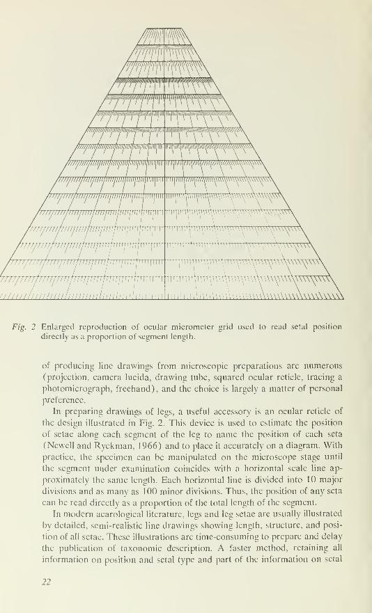
F/g. 2 Enlarged reproduction of ocular micrometer grid used to read setal position
directly as a proportion of segment length.
of producing line drawings from microscopic preparations are numerous
(projection, camera lucida, drawing tube, squared ocular reticle, tracing a
photomicrograph, freehand), and the choice is largely a matter of personal
preference.
In preparing drawings of legs, a useful accessory is an ocular reticle of
the design illustrated in Fig. 2. This device is used to estimate the position
of setae along each segment of the leg to name the position of each seta
(Newell and Ryckman, 1966) and to place it accurately on a diagram. With
practice, the specimen can be manipulated on the microscope stage until
the segment under examination coincides with a horizontal scale line ap-
proximately the same length. Each horizontal line is divided into 10 major
divisions and as many as 100 minor divisions. Thus, the position of any seta
can be read directly as a proportion of the total length of the segment.
In modern acarological literature, legs and leg setae are usually illustrated
by detailed, semi-realistic line drawings showing length, structure, and posi-
tion of all setae. These illustrations arc time-consuming to prepare and delay
the publication of taxonomic description. A faster method, retaining all
information on position and setal type and part of the information on setal
22
size, was first used for water mite larvae hv Ban ( 1968). Each larval leg
is represented b\ a standardized diagram marked with symbols to indicate
the position ^( the seta! bases. Individual setae that are diagnostically im-
portant because of size or structure are illustrated separately. Although the
value of the seta] diagram is only auxiliary in diagnosis, it facilitates inter-
specific comparison of individual seta] positions and of overall setal patterns
as noted b\ Evans ( 1963) for Mesostigmata. Such diagrams also provide
suitable data for quantitative analysis. Because distortion is involved in mak-
ing each segment tit a standard shape and size, setal diagrams should always
be accompanied by an outline drawing of the leg segments marked to indicate
the true position of setal bases.
PHOTOMICROGRAPHY
Photomicrograph) has been important as a method of illustrating water
mites. Many of Lundblad's publications (e.g., 1941. 1962) contain photo-
micrographs of entire ventral and dorsal views as well as isolated views of
palp, genital area, and specific leg segments. His photographs were usually
supplemented by detailed line drawings of structures of particular interest.
Certain water mite structures, e.g., palps, flat dorsal plates, pigmentation
pattern in dorsal plates, and often the external genitalia, are more rapidly
and better illustrated bv photomicrography than by line drawings. By photo-
graphing the specimen at low magnification with increased enlargement of
the negative, one can take advantage of a greater depth of field than is
available at high magnification. A good photograph is a more objective
record of shape and relative sizes than many taxonomic drawings.
In studying a specimen or series of specimens, a record set of photo-
micrographs is valuable. Photographs can be sorted and examined more
easily and efficiently than slides, especially temporary mounts, which need
only be re-examined in the final stages of the investigation rather than
throughout. Photomicrographs can also serve as the basis for line illustra-
tions and for certain quantitative analyses. Moreover, photographs can be
used to illustrate variation of shape in a particular structure more expediently
than a series of line drawings. Possibly improved scanning electron micro-
graphs will eventually eliminate some of the disadvantages (shallow depth
of field, low contrast) of standard photomicrographs.
23
Acknowledgments
Ian M. Smith developed several techniques described here and has been a
continuing source of helpful ideas, practical assistance, and stimulating
discussion. Conversations with Drs. David R. Cook, G. Owen Evans,
Carmine A. Lanciani, Rodger D. Mitchell, and Vikram Prasad provided
many valuable suggestions. T. Gledhill and K. H. Hyatt provided informa-
tion on phreaticolous collecting and storage methods, respectively.
During the 10-year period that these methods have been developed,
financial support came from a number of sources: Cornell University, the
National Research Council (Canada), Allied Chemical Company, Ontario
Department of University Affairs (Grant-in-Aid of Research), and the
Canadian National Sportsmen's Show (Conservation grant). Some collec-
tions were made during rom field trips supported by grants to Dr. G. B.
Wiggins from the National Science Foundation (grant GB4021 ), the Cana-
dian National Sportsmen's Show, the Fisheries Research Board of Canada,
and the National Research Council of Canada (grant A5707).
Acknowledgment is made for use of the scanning electron microscope of
the Laboratory of Analytical Systematics, University of Toronto, in the
Royal Ontario Museum. Mrs. C. Johnston provided valuable technical
assistance in use of the SEM, and A. Troiki, Laboratory of Analytical
Systematics, developed the freeze-drying method described here. Mrs. P. B.
Buckley carried out several thousand larval rearings, and Mrs. J. Allan
typed two drafts of the manuscript. The cover illustration of Frontipoda
americana Marshall was done by A. Odum.
24
Appendix: Formulae tor Reagents
1. Acetic Corrosive| \ndre*s Fluid) (Mitchell and Cook, 1952)
acetic acid ( glacial
)
50 ml
chloral hydrate 50 g
distilled water 50 ml
2. Brasil's Fluid (calculated from Baker. 1958)
80% ethanol 150 ml
picric acid 1 g
formalin (37 70 ml
acetic acid (glacial) 15 ml
3. Eosin-Y in Glycerin-Alcohol ( Barr)
saturated solution of
Eosin-Y in 95 % ethanol 10 ml
glycerin (9b 10 ml
4. GAW ( modified Koenike's fluid
)
acetic acid (glacial) 100 ml
glycerin (96%) 500 ml
distilled water 400 ml
5. Modified Carnov's Fluid ( De Giusti and Ezman, 1 955
)
isopropvl alcohol (abs.) 60 ml
chloroform 30 ml
formic acid (90%) 10 ml
6. Nesbitfs fluid ( Evans et al., 1961
)
chloral hydrate 40 g
distilled water 25 ml
hydrochloric acid (IN) 2.5ml
25
Literature Cited
BAKER, W. AND G. W. WHARTON1952 An introduction to acarology. New York, Macmillan. 465 pp.
BAKER, J. R.
1 958 Principles of biological microtechnique. London, Methuen. 357 pp.
BARR, D.
1968 Limnesia (Limnesiella) marshallae, description of larva. Pilot Regis-
ter of Zoology, Card No. 26.
1972 The ejaculatorv complex in water mites (Acari: Parasitengona)
:
morphology and potential value for systematics. Life Sci. Contr.,
R. Ont. Mus., no. 8 1 , pp. 1-87.
beirne, b. p. (compiler)
1955 Collecting, preparing and preserving insects. Sci. Serv., Entom. Div.,
Can. Dept. Agric, publ. 932, pp. 1-133.
CLIFFORD, H. F.
1972 A year's study of the drifting organisms in a brown-water stream of
Alberta, Canada. Can. J. Zool., vol. 50, no. 7, pp. 975-983.
COOK, D. R.
1963 Studies on the phreaticolous water mites of North America: new or
unreported genera of Axonopsidae. Amer. Midi. Nat., vol. 70,
pp. 110-125.
1968 Water mites of the genus Stygomomonia in North America (Aca-
rina, Momoniidae). Proc. Ent. Soc. Wash., vol. 70, pp. 210-224.
COOK, D. R. AND R. D. MITCHELL
1952 Notes on collecting water-mites. Turtox News, vol. 30, no. 8, 4 pp.
crowell, r. m.
1960 The taxonomy, distribution and developmental stages of Ohio water
mites. Bull. Ohio Biol. Surv., N. Ser., vol. 1, no. 2, pp. 1-77.
DE GIUSTI, D. L. AND L. EZMAN1955 Two methods for serial sectioning of arthropods and insects. Trans.
Amer. Micros. Soc, vol. 74, pp. 197-201.
essig, e. o.
1 948 Mounting aphids and other small insects on microscope slides. Pacific
Ent., vol. 24, pp. 9-22.
EVANS, G. O.
1963 Observations on the chaetotaxy of the legs in the free-living Gama-sina (Acari: Mesostigmata). Bull. Brit. Mus. (Nat. Hist.), Zool.,
vol. 10, no. 5, pp. 277-303.
EVANS, G. O., J. G. SHEALS AND D. MAC FARLANE
1961 Terrestrial Acari of the British Isles. Vol. 1 . London, British Museum(Natural History). 219 pp.
GRANDJEAN, F.
1935 Observations sur les acariens ( lore serie). Bull. Mus. Nat. Hist. Nat.,
Paris, 2'' ser., vol. 7, pp. 1 19-126.
26
GRANDJEAN, F.
1949 Observation et conservation des tres petits Arthropodes. Bull. Mus.
Nat. Hist. Natur., Paris. 2e sen, vol. 21. pp. 363-370.
GRAY. P.
1964 Handbook of basic microtechnique. New York. McGraw-Hill.
302 pp.
KRANTZ, G. \V.
1970 A manual ol acarolog) . Corvallis, O.S.U. Book Stores, Inc. 335 pp.
LANCIANI, C. A.
1970 New species of Eylais ( Acari: Eylaidae) parasitic on aquatic Coleop-
tera. Trans. Amer. Micros. Soc, vol. 89, pp. 169-188.
LU.NDBLAD, O.
1941 Die Hydracarinenfauna Sudbrasiliens und Paraguays. Erster Teil.
Svensk. Vetensk.-akad. Handling., Stockholm, (ser. 3), vol. 19,
no. 7, pp. 1-183.
1962 Die Hydracarinen Schwedens. u. Arkiv For Zoologi: Svenska
Vetensk.-akad., ser. 2, vol. 14, no. 1, pp. 1-635.
MITCHELL, R. D.
1960 The evolution of thermiphilous water mites. Evolution, vol. 14,
pp. 361-377.
1964 The anatomy of an adult chigger mite Blankaartia acuscutellaris
(Walch).J. Morph.,vol. 1 14, pp. 373-391.
MITCHELL, R. D. AND D. R. COOK
1952 The preservation and mounting of water-mites. Turtox News, vol.
30, no. 9, 4 pp.
motas, c.
1962 Procede des ondages phreatiques - Division du domaine souterrain -
Classification ecologique des animaux Souterrains - Le psammon.Acta Mus. Maced Sci. Nat., vol. 8, no. 7, pp. 135-173.
NEWELL, I. M.
1947 A systematic and ecological study of the Halacaridae of eastern
North America. Bull. Bingham Oceanographic Collection, vol. 10,
art. 3, pp. 1-232.
1959 Acari. //; Edmondson, W. T. ed., Fresh-water biology. 2nd ed. NewYork, Wiley, pp. 1080-1116.
NEWELL, I. M. AND R. E. RYCKMAN1966 Species of Pimeliaphilus (Acari, Pterygosomidae) attacking insects,
with particular reference to the species parasitizing Triatominae
(Hemiptera, Reduviidae). Hilgardia, vol. 37, pp. 403—436.
OLDROYD, H.
1970 Collecting, preserving and studying insects. London, Hutchinson.
336 pp.
PENNAK, R. W.
1953 Fresh-water invertebrates of the United States. New York, Ronald
Press. 769 pp.
27
PRASAD, V. AND D. R. COOK
1972 The taxonomy of water mite larvae. Mem. Am. Ent. Inst., no. 18,
pp. 1-326.
PIECZYNSKI, E.
1962 Notes on the use of light traps for water mites (Hvdracarina) . Bull.
Acad. Pol. Sci., CI. n, vol. 10, no. 10, pp. 421-424.
1965 Effect of the size of traps and their exposure time on the catches of
aquatic invertebrates, chiefly water mites (Hydracarina). Bull. Acad.
Pol. Sci., CI. II, vol. 13, no. 10, pp. 577-581.
SHEAR, W. A. AND H. W. LEVI
1970 Cleaning museum specimens of spiders and myriapods with ultra-
sonics. Bull. Brit. Arach. Soc, vol. 1 , p. 117.
VIETS, K.
1936 Wassermilben oder Hydracarina ( Hydrachnellae und Halacaridae).
DieTierwelt Deutschlands, vol. 31-32, pp. 1-574.
WOODRING, J. P. AND E. F. COOK
1962 The internal anatomy, reproductive physiology, and molting process
oiCeratozetes cisalpinus. Ann. Ent. Soc. Amer., vol. 55, pp. 164-181.
28
LIBRARY
ROYAL I 10 MUSEUM
ISBN 0-88854-148-1