Embed Size (px)
Citation preview

OPHT1HALM1c RADIA,riON LAMPI
ibn Ibrahim ibn Bokhtisho, who probably lived about the secondhalf of the tenth century. (3) "The Memorial of the Oculists,"by Ali ibn Isa, a Christian oculist of Baghdad, a contemporary inthe ninth century of the famous physician Ibn Sina (Avicenna).Meyerhof fully agrees with Hirschberg when he says that no betterophthalmological text-book was written either in the Orient orthe Occident until the beginning of the eighteenth century. (4)"Selection in the Treatment of Eve Diseases," by El-Mausili.This is the first Moslem author among the oculists. He lived atCairo in the early part of the eleventh century. The only completecopy of this work exists in the library of Taimur Pasha in Cairo.It contains a unique account of a new method of operating onsoft cataract by sucking out the soft matter through a hollow needlemade of gold or brass.Enough has been said to indicate the important new material
which Meyerhof has brought to light. It is of interest to note thatthe eartier Arabic science was derived from Greek sources andthat its practitioners were Christians.
AN OPHTHALMIC RADIATION LAMP: WITH A NOTEON ITS DIAGNOSTIC AND THERAPEUTIC
APPLICATIONSBY
W. STEWART DUKE-ELDER(FROM THE NATIONAL INSTITUTE OF MEDICAL RESEARCH AND THE
ULTRA-VIOLET CLINIC, ROYAL LONDON OPHTHALMIC HOSPITAL)
THE presentation of this instrument is premature in that itspossibilities both in diagnosis and in therapeutics have not yetbeen thoroughly investigated. Its early notice, however, seemsjustified if merely to amend a technique in local phototherapy ofthe eye which I have already described (1926, a and b) and whichI am led to understand is being presently adopted, but which Ihave now partly abandoned as being inadequate and in somecases potentially dangerous. This technique consisted in localizingthe rays from a mercury vapour lamp into a parallel beam by meansof a screening apparatus learing a curved quartz director, andplaying them by this nmeans upon the eye. Such a technique issuitable for raying the lids and the conjunctiva, especially in thosecases (e.g., trachoma) where the director tip can be inserted intothe upper fornix. Incidentally in these, instead of the rod-shapeddirector originally recommended, an elongated cone-shaped one(K.B.B.) with a broad base tapering to a rounded point at its benttip will be found more effective. All the rays entering at the base
,.N
on 28 July 2019 by guest. Protected by copyright.
http://bjo.bmj.com
/B
r J Ophthalm
ol: first published as 10.1136/bjo.11.2.67 on 1 February 1927. D
ownloaded from

THE BRITrISH JOURNAL OF OPHTHALMOLOGY
are brought to a focus at the tip by internal reflection, and thebeam is therefore concentrated here by an amount depending onthe ratio of the squares of the radii of the director at these twopoints. The quartz must be from a good specimen and the basemust be optically polished if much of the intensity is not to belost.For radiation of the eye, however, such a beam is not susceptible
of sufficient control. I have elsewhere elaborated the thesis(1926, c) that an important factor in the aetiology of cataract isthe incidence of radiant energy of any wave-length upon the lens.This acts by denaturing its proteins and deranging the autoxida-tion system upon which the efficiency of its internal metabolismdepends, the final process of flocculation of the lens proteins beinginduced by changes in the concentration of hydrogen ions andsalts and osmotic variations, to which in some cases may possiblybe added a process of continuous photosensitization. Ultra-violetlight, if used experimentally in intensity enough, can causelenticular opacity. If used in lower concentrations such as do notobviously produce an opacity, it increases the lability of the colloidsystem of the lens and diminishes its glutathione content, both ofwhich reactions, while they still leave the lens optically clear,predispose to the subsequent incidence of cataract. Some writerson the Continent have stated that with the relatively smallintensities of radiation such as are used clinically, applied as theyare intermittently, comparatively uncontrolled applications arewithin the margin of safety. It is to be remembered, however, thatmany of the eyes subjected to treatment are already diseased, andin these the lens is presumablv already predisposed to pathologicalchange; and in any case in the present state of our knowledgethe exposure of the lens to any direct radiation seems an unjusti-fiable risk. With the apparatus figured such a risk can be com-pletely eliminated.The apparatus is used on the standard Gullstrand slit-lamp table
and stand (Zeiss), the slit-lamp and horizontal bracket being liftedoff bodily at the point corresponding to (X) in the diagram. Thelamp used is a quartz mercury vapour lamp (220 volts, 2-3 amps.):this is preferable to any type of carbon arc owing to the absencefrom its sp-ectrum of infra-red, a type of ray peculiarly dangerousto the eye. With this type of lamp at the working distance the heateffect is negligible. The atmospheric type (K.B.B.) is used as it isthe most easily manipulated and works over prolonged periodsunder the most constant conditions. It is enclosed in a casing (C)and is so designed that it occupies the minimum of space, and thatthe vertical arc is situated in front of a wide slit-aperture (A) whichcan be rotated in any meridian. The light is concentrated on adiaphragm (D) and brought to a focus by means of a quartz lens
68
on 28 July 2019 by guest. Protected by copyright.
http://bjo.bmj.com
/B
r J Ophthalm
ol: first published as 10.1136/bjo.11.2.67 on 1 February 1927. D
ownloaded from

OPHTHALMIC RADIATION LAMP 69
Xi .d~~L
OprrIAiTI.N 1c RADIATlION LIxN'I i'I1 S1PECTRU 0O,AT1 USIJsio).
D
IL4A
I x
on 28 July 2019 by guest. Protected by copyright.
http://bjo.bmj.com
/B
r J Ophthalm
ol: first published as 10.1136/bjo.11.2.67 on 1 February 1927. D
ownloaded from
THE BRITISH JOURNAL OF OPHTHALMOLOGY
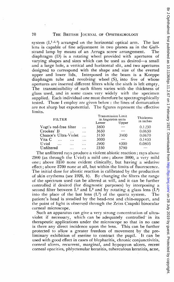
system (L1 3) arranged on the lhorizontal optical arm. The lastlens is capable of fine adjustment in two planes as in the Gull-strand lamp by means of an Arruga screw arrangement. Thediaphragm (D) is a rotating wheel provided with apertures ofvarying shapes and sizes which can be used as desired-a smalland a large hole, a vertical and lhorizontal slit, and two aperturesdesigned to correspond with the shape and size of the evertedupper and lower lids. Interposed in the beam is a Koeppediaphragm tube and revolving wheel (S), into five of whoseapertures are inserted different filters while the sixth is left empty.The transmissibility of such filters varies with the thickness ofglass used, and in some cases verv widely with the specimensupplied. Eaclh individual one must therefore be spectrograpllicallytested. Those I employ are given below: the lines of demarcationatre not shlarp but exponential. Thle figures represent the effectivelimits.
Transmission Limit ThicknessFILTER in Angstrdm units ilces
Lower Upper in inchesVogt's red-free filter ... 3800 0.1230Crookes' B ... ... 3650 - 0.0650Chance's Ultra-Violet ... 3150 3900 0.0670Vita C ... ... ... 3000 0.1455Uviol ... ... ... 2900 4300 0.0805Unfiltered ... ... 2350 5790
T he unfiltered rays produce a violent abiotic reaction; rays above21.900 (as tlhrough tlle tUviol) a mild one; above 3000, a very mildone; above 3150 none evident clinically, but having a sedativeeffect; above 3700 none at all, but within the limits of fluorescence.The initial dose for abiotic reaction is calibrated by the productionof skin erythema (see 1926, b). By changing the filters the rangeof the spectrum used can be altered at will, and it can be furthercontrolled if desired (for diagnostic purposes) by interposing asecond filter between L2 and L3 and by rotating a glass lens (I,')into the place of the last lens (L3) of the quartz system. Thepatient's head is steadied by the lhead-rest and chin-support, andthe point of liglht is observed through tlle Zeiss Czapski binocularcorneal microscope.Such an apparatus can give a very strong concentration of ultra-
violet if necessary, whichi can be adequately controlled in itstherapeutic applications under the microscope so that in no caseis there any direct incidence upon the lens. This can be furtherprotected to allow a greater freedom of movement by the pre-liminary exlhibition of eserine to contract the pupil. It can beused with good effect in cases of blepharitis, chronic conjunctivitis,corneal ulcers, recurrent, marginal, and hypopyon ulcers, recentcorneal opacities, plhlyctenular keratitis, ttuberculous keratitis, acne,
(iO
on 28 July 2019 by guest. Protected by copyright.
http://bjo.bmj.com
/B
r J Ophthalm
ol: first published as 10.1136/bjo.11.2.67 on 1 February 1927. D
ownloaded from

OPHTHALMIC RADIATION LAMP
and some cases of episcleritis. I am still of the opinion that localphototherapv is contra-indicated in diseases of the inner eye, withthe exception of those cases of iritis showing nodules on theanterior surface of the iris. For clhronic cases of iritis, irido-cyclitis,and clhoroiditis, many of which halve otlherwise seemed lhopeless-not only the tuberculous, but in many cases of infective origin-treatment by general light baths is giving results wlhich cancertainly be described as eminently satisfactory.The diagnostic applications of the ultra-violet slit-lamp are at
present more of academic than clinical value. R. Thiel of Berlin(1925) has made some interesting, observations on this subject;he uses a carbon arc as a source of light, wlhich however requiresheavy filtering off of the infra-red and therefore loses much ineffective intensity of liglht. With the slit-lamp described abioticallyactive liglht can be completely filtered off by the Crookes' filter,and a red-free picture is obtained. The interest depends largelyon the plhenomena of fluorescence the effect of whiclh may beintensified, if desired, by giving the patient 2 gmms. of sodiumfluorescine by the moutlh half an h1our before the examination.The skin of the lids fluoresces, and the effect is modified or abolishedby sliglht lesions wlhiclh are almost imperceptible by ordinary light:this fact can be applied to dermatology generally. The cornea islit up by a definite fluorescent glow; all details, vessels, nerves,etc., can b5e sharply intensified in the practically monochromaticliglht wlhiclh can be obtained with a conmbination of filters; patlho-logical lesions, opacities, and folds in Descemet's inembraneshow no fluorescence but appear as black. New vessels in thecornea appear black, the individual corpuscles can often bedistinguished, and the reaction of the capillaries to drugs andotlher stimuli applied locally can be readily followed. A similarpicture is obtained of the iris. The lens fluoresces markedly, andsince the absorption of tlle nucleus and cortex are different, thefluorescence of the various optical layers offers a sharp contrast.Siagreen is well seen, and inflammatory deposits, opacities, andvacuoles and fluid lacunae appear in relief and sharply definedas black or dark blue ag-ainst the pale yellow-green or lavender-grey of the fluorescent light. A lens dislocated into the anteriorchamber or into the vitreous is quite apparent, and can bedefinitely localized without trouble; an indication of the extentof damage after trauma or needling is given; and the state andextent of the remains of the lenticular substance after extractioncan be accurately discerned. The fluorescent light, moreover, canto a large extent be appreciated through a cornea so opaque thatit is for practical purposes impermeable to ordinary light, andsome idea of the state of the lens can thus be deduced in these cases.
It is hoped in the near future to publish a fuller account of the
71
on 28 July 2019 by guest. Protected by copyright.
http://bjo.bmj.com
/B
r J Ophthalm
ol: first published as 10.1136/bjo.11.2.67 on 1 February 1927. D
ownloaded from

THE BRITISH JOURNAI OF OPHTHALMOLOGY
clinical applications of the method and the diagnostic applicationsof ultra-violet biomicroscopy, when a sufficient amount of clinicalmaterial has been gathered to render it of real value. In the mean-time it cannot be over-emphasized that, despite the deplorablyunfortunate attentions of the lay press, ultra-violet light is not auniversal panacea. Further, owing to its equally unfortunateexploitation by practitioners unqualified either in medical orphysical training and having no knowledge of the biological actionof the potent agent with which they are dealing, it is in manycases being judged by the results which it is only natural to expect.While much positive harm may not usually follow its applicationin this manner to the skin, the local phototherapist of the eyeshould certainly be a competent physicist and a competent ophthal-mologist, and he should work only with a lamp which runs underconstant conditions, and whose individual spectrum is known andwhose output of energy intensity as obtained at the point ofapplication has been determined, and on to which are fitted filterseach of which has been spectrographically standardized.The practice of the "treatment" of cataract by ultra-violet light
by local application has been carried out in some quarters: thoseresults which have come under my observation might lead meto suggest it as a method for the maturation of cataract. It hasbeen shown that ultra-violet light causes the coagulation of proteinwhich is recognized clinically as lenticular opacity. It is truethat in some early cases of acute cataract (diabetic, traumatic)where the process has not yet progressed to the actual coagulationof the proteins of the lens (see 1926, c), transparency may berecovered by the control of the exciting causes; but in all cases anorganic opacity once formed is immutable. The coagulation ofprotein is an irreversible chemical change. It seems not un-reasonable to hope that in the future when our knowledge of thischange and the factors causing it has been more fully developedit may be possible to prevent its incidence or to delay or arrestits progress, but to set out to "cure" cataract in the sense usedabove by any means, medical or physical, is quite unjustified.
I am indebted to Messrs. Carl Zeiss Ltd. for their courtesy andto Messrs. Kelvin Bottomley and Baird for the care they havetaken in constructing the lamp.
REFERENCESDuke-Elder, W. S. (1926, a).-Phototherapy in ophthalmology. Trans.
Ophthal. Soc. U.K., Vol. XLVI, p. 213. 1926.(1926, b).-Observations on the therapeutic action of ultra-violet light
upon the eye. Brit. Med. Ji., pp. 891-895, May 29.(1926, c).-The pathological action of light upon the eye. Lancet,
Vol. I; I. Action on the outer eye, pp. 1137-1141; II. Action on the lens:theory of the genesis of cataract, pp. 1188-1192, 1250-1255.
Thiel, R. (1925).-Ein beitrag zur Spaltlampenmikroskopie des Auges in ultra-violetten Licht. Zeitschr. f. Augenheilk., Bd. LVIII, S. 86-91.
72
on 28 July 2019 by guest. Protected by copyright.
http://bjo.bmj.com
/B
r J Ophthalm
ol: first published as 10.1136/bjo.11.2.67 on 1 February 1927. D
ownloaded from