Embed Size (px)
Citation preview

© 2001 S. KargerAG, Basel0006–8977/01/0581–0001$17.50/0
Fax + 41 61 306 12 34E-Mail [email protected] Accessible online at:www.karger.com www.karger.com/journals/bbe
Microanatomy of Facial Vibrissae in theFlorida Manatee: The Basis forSpecialized Sensory Function and OripulationR.L. Reepa M.L. Stoll a C.D. Marshalld B.L. Homerb D.A. Samuelsonc
aDepartment of Physiological Sciences and Brain Institute, bDepartment of Pathobiology, cDepartment of SmallAnimal Clinical Sciences, College of Veterinary Medicine, University of Florida, Gainesville, Fla.,dDepartment of Orthodontics/Burke Museum of Natural History University of Washington, Seattle, Wash., USA
Original Paper
Brain Behav Evol 2001;58:1–14
Dr. R.L. ReepDepartment of Physiological Sciences, Box 100144, HSCUniversity of Florida, Gainesville, FL 32610 (USA)Tel. +1 352 392-4700, ext. 3859, Fax +1 352 392-5145E-Mail [email protected]
Key WordsMammals • Sirenia • Manatee • Vibrissae • Hair •
Bristles • Face
AbstractSirenians, including Florida manatees, possess an arrayof hairs and bristles on the face. These are distributedin a pattern involving nine distinct regions of the face,unlike that of any other mammalian order. Some of thesebristles and hairs are known to be used in tactile explo-ration and in grasping behaviors. In the present study wecharacterized the microanatomical structure of the hairand bristle follicles from the nine regions of the face.All follicles had the attributes of vibrissae, including adense connective tissue capsule, prominent blood sinuscomplex, and substantial innervation. Each of the nineregions of the face exhibited a distinct combination ofthese morphological attributes, congruent with the pre-vious designation of these regions based on location andexternal morphological criteria. The present data sug-gest that perioral bristles in manatees might have atactile sensory role much like that of vibrissae in othermammals, in addition to their documented role in grasp-ing of plants during feeding. Such a combination ofmotor and sensory usages would be unique to sirenians.Finally, we speculate that the facial hairs and bristlesmay play a role in hydrodynamic reception.
Copyright © 2001 S. Karger AG, Basel
Introduction
Sirenians possess a varied array of modified facial hairsthat are used in tactile exploration, interactions with con-specifics, and feeding [Hartman, 1979; Marshall, 1997;Marshall et al., 1998a, 2000; Bachteler and Dehnhardt,1999]. In manatees these include six types of perioral bris-tles and bristle-like hairs on the oral disk [Reep et al., 1998].These are distinguished from other body hairs by theirgreater stiffness, which appears to result from smallerlength/diameter ratios rather than from compositional dif-ferences. Hair having the external appearance of body hairis located on the supradisk portion of the face posterior tothe orofacial ridge and on the chin [Reep et al., 1998].
As illustrated in figure 1, the six fields of perioral bristleson the face of the Florida manatee consist of four (U1–U4)on each side of the upper lips and oral cavity and two(L1–L2) on each side of the lower lip pad along the lip mar-gin [Reep et al., 1998]. Each field has a characteristic loca-tion, number of bristles, and range of external bristle lengthand diameter. Branches of the infraorbital nerve innervatethe bases of the largest bristles (U2 group) on the upper bris-tle pad, and the inferior alveolar nerve supplies the L1–L2bristles of the lower bristle pad. These innervation patternsand the topographical locations of the six bristle fields sug-gest that the U1–U4 bristles are homologous to the mysta-cial vibrissae of other mammals, whereas the L1–L2 bristlescorrespond to the mental vibrissae [Reep et al., 1998]. Dor-sal and ventral buccal branches of the facial nerve innervate
Received: June 21, 2001Returned for revision: July 24, 2001Accepted after revision: August 10, 2001

the superficial facial musculature, which is likely to beinvolved in bristle eversion and other movements that occurduring feeding behavior [Marshall et al., 1998b; Reep et al.,1998].
Sirenians use the modified hairs of the face in a varietyof behaviors. Uniquely among mammals, they use some ofthe perioral bristles (primarily those of the U2 and L1 fields)in a grasping fashion during feeding and oripulation ofobjects [Marshall et al., 1998a, 2000]. (We have coined theterm ‘oripulation’ to refer to use of the mobile lips and asso-ciated perioral bristles in coordinated rhythmic graspingbehaviors. This seems preferable to the term ‘manipulation’,which connotes use by the manus, or hand.) Tactile explo-ration without grasping typically involves the bristle-likehairs of the oral disk [Hartman, 1979; Marshall et al., 1998a;Bachteler and Dehnhardt, 1999]. Most mammals use theirvibrissae in a purely sensory fashion, primarily to detectprey or to orient in low light environments. Vibrissae arecapable of providing detailed textural information aboutsurfaces and objects in the immediate environment [Dykes1975; Brecht et al., 1997]. Psychophysical measures indi-cate that harbor seals may be able to use their vibrissae insensory detection as effectively as monkeys use their handsin touch discrimination [Dehnhardt and Kaminski, 1995]. Inrats the geometry of vibrissal lengths within a row and theangular arrangements of the vibrissae of each row relative tothe body axis and the other rows are important variablesinfluencing the behavioral use of the vibrissae [Brecht et al.,1997].
Among non-sirenians, movement of vibrissae occurs inseveral contexts. Rodents engage in whisking behavior inwhich the mystacial vibrissae are swept in a rhythmic fash-ion during tactile exploration of the external environment[Welker, 1964; Wineski, 1985; Carvell and Simons, 1990].Pinnipeds move their long vibrissae during display behav-iors and tactile exploration [Peterson and Bartholomew,1967; Miller, 1975; Kastelein and Van Gaalen, 1988; Dehn-hardt, 1994; Dehnhardt and Kaminski, 1995; Dehnhardt andDucker, 1996]. When out of water with the snout pointedupward, walruses have been observed to move pieces of fish
in the direction of the mouth by passing them from vibrissato vibrissa in a wave-like manner [Marshall et al., 1998a],but they engage in no grasping. Freshwater river dolphins(Platanistidae) have reduced visual systems and relativelywell developed bristle-like vibrissae along the upper andlower jaws. These non-mobile vibrissae are reportedly usedto detect fish and crustacean prey along river bottoms [Nor-man and Fraser, 1948 as cited in Ling, 1977; Layne andCaldwell, 1964]. Collectively, these findings indicate thatsensory detection in non-sirenians is often accompaniedby movements of the vibrissae, but not by grasping.
One of the defining features of vibrissae in non-sirenianmammals is their organization as sinus hairs, in which ablood-filled sinus encircles the hair shaft and contains alongits wall a substantial number of sensory nerve endings.Vibrissae in rodents, rabbits, and cats are innervated by asingle deep vibrissal nerve containing 100–200 axons thatascend along the inner margin of the sinus wall and ter-minate in receptors located in the outer root sheath andmesenchymal sheath [Rice et al., 1986, 1997]. In monkeysthe deep vibrissal nerve supplies each vibrissae with80–100 axons entering through 2–3 nerve fascicles [Halataand Munger, 1980]. In addition to the deep vibrissal nerve,a superficial vibrissal nerve containing 20–30 unmyelinatedaxons innervates the rete ridge collar and outer conicalbody, components of the follicle located near the skin sur-face [Mosconi et al., 1993].
There has been no comparable study of hair folliclemorphology and innervation in sirenians. Earlier studiesreported morphological attributes of some of the facial bris-tles and hairs, but these were not based on systematic sam-pling methods and did not include nerve counts. Dosch[1915] reported that all body hairs on sirenians were sinushairs, intermediate in form between pelage hairs and vibris-sae. He expanded upon the earlier studies of Kükenthal andothers [referenced in Dosch, 1915], and even used some ofthe same specimens used by Kükenthal. His conclusion hasbeen supported in dugongs by the findings of Bryden et al.[1978], Kamiya and Yamasaki [1981], and Sokolov [1982],and in Antillean manatees by Sokolov [1986]. However, in
2 Brain Behav Evol 2001;58:1–14 Reep/Stoll/Marshall/Homer/Samuelson
Abbreviations
app ring sinus appendages epi epidermis ors outer root sheathbl blood gm glassy membrane rs ring sinuscap follicle capsule hs hair shaft trb trabeculae of cavernous sinusder dermis lcs lower cavernous sinus ucs upper cavernous sinusdp dermal papilla ms mesenchymal sheath

most of these cases a lack of regional specification togetherwith a paucity of figures make it impossible to determine thelocation from which follicle samples were taken. Sokolov[1986] did provide some quantitative morphological charac-terization of the ‘thick vibrissae of the upper and lower lips’(probably corresponding to bristle fields U2 and L1) in theAntillean manatee (T. manatus manatus) but did not sys-tematically explore other subfields of the face.
The nine regions of the manatee face are distinguishablebased on location, distribution of bristles or hairs, externalmorphology of bristles or hairs, and the behavioral roleplayed by the bristles and hairs during feeding and otherbehaviors. Therefore, in the present study we sought to
define internal follicle microanatomy and innervation inthese regions of the face. One goal was to determinewhether all the facial hairs of the Florida manatee possessthe structural characteristics of sinus hairs, as would beexpected if they were homologous to the vibrissae of othermammals. A second goal was to obtain estimates of inner-vation density for the follicles in each of the nine regions ofthe face.
Materials and Methods
A total of 166 follicles were processed and 82 of these were ana-lyzed quantitatively in this study (tables 1 and 2). They were collectedfrom eleven fresh (w/in 24 h) postmortem manatee carcasses in con-junction with the statewide manatee carcass salvage program managedby the Marine Mammal Pathobiology Laboratory in St. Petersburg,Fla., USA (USFWS permit PRT-684532). All but one of the carcasseswere in the subadult-adult range of body length, corresponding to 1.5+years of age, as determined by the USGS Sirenia Project (unpubl.data). However, specimen SWFTM 9515 was much smaller, in therange of a small calf 1–6 months of age. As shown in table 1, follicleswere taken from nine regions of the face which were defined in a pre-vious study [Reep et al., 1998]. Bristle-like hairs were found in twolocations; most were located on the oral disk (BLHod) but occasionallyone was found among the bristles (BLHbr) in the U1 or U2 field.
A #11 scalpel blade was used to extract blocks of tissue (~5 × 5 mmat the surface and ~10–15 mm in length) that included single folliclesand the immediately surrounding connective tissue. Specimens werestored in 4% phosphate buffered formalin (pH 7.4) then cut frozen at40 µm on a Lipshaw sliding microtome. Serial longitudinal or crosssections were stored in dilute fixative in 24-well plastic tissue cultureplates. Sections were mounted on gelatin coated glass slides, using avery dilute (0.5%) gelatin mixture to minimize background staining inthe case of silver staining.
After drying overnight, slides with sections were hydrated andstained using hematoxylin and eosin, Masson’s trichrome stain, or amodified Bodian stain. The longitudinal sections were used for mea-surements of follicle morphology and to identify deep vibrissal nervebundles and their trajectories. The cross sections were used for count-ing the number of axons innervating a follicle.
Approximately 30 follicles were prepared using paraffin embed-ding and stained using Masson’s trichrome or hematoxylin and eosin.Bodian staining of paraffin sections did not prove as reliable as thatperformed on frozen sections. The paraffin embedded sections werenot used for quantitative measurements of follicle geometry, in order toavoid shrinkage artifacts.
In 57 selected longitudinal sections we measured maximum fol-licle length, maximum total sinus length (upper + ring + lower sinus),maximum ring sinus width, maximum follicle capsule thickness, andmaximum hair shaft diameter at the level of the ring sinus. These mea-surements were made on an AIS/C imaging workstation (ImagingResearch, Inc.) interfaced with a Zeiss Axiophot 2 microscope andDage 72 video camera. Sections used for measurement were chosenbased on their orientation as near as possible to the central longitudinalaxis of the hair shaft. The left and right sides of each section were mea-sured, and we retained only the maximum values. Data are presented asranges due to low sample sizes and high variability.
3Microanatomy of Facial Vibrissaein the Manatee
Brain Behav Evol 2001;58:1–14
Fig. 1. Schematic diagram of the perioral region, showing the loca-tion of the upper (U1–4) and lower (L1–2) bristle fields, bristle-likehairs (BLH), supradisk, and chin. Dotted lines indicate cuts made in thecheek muscles to allow a fuller view of the oral cavity.

For making axon counts, we used 33 cross sections in which axonsappeared well stained by the modified Bodian procedure. We alsorequired that these sections be located just apical relative to the regionof nerve entry, in order to estimate the maximum number of axonsper follicle. In such material one observes spaced bundles of axons,and individual axons are separately identifiable by their myelin sheath(fig. 4). However, due to the capricious nature of silver staining, itwas rare that a section exhibited well defined staining throughout thecircumference of the follicle. Rather, some axon bundles appeareddark and others appeared too light to permit accurate counting. In thesecases we capitalized on the fact that just apical to their entry, the 2–6large nerve bundles innervating each follicle disperse into smaller bun-dles of 2–5 axons each. These smaller bundles are uniformly spaced
around the circumference of the follicle as they ascend toward theirsites of termination along the sinus wall. Therefore, in many cases weestimated total axon number by performing direct axon counts on halfof the total 360° and multiplying the result by two. In order to assessrelationships between nerve counts and follicle geometry, we mea-sured the perimeter of the innervated ring sinus wall and estimated thelength of the ring sinus on the same follicles from which nerve countswere obtained. Perimeters were measured on the same sections fromwhich axon counts were obtained, using the imaging system describedabove. Ring sinus length was estimated by counting the number ofcross sections on which the ring sinus appeared as a distinct entitycharacterized by large width and a lack of trabeculae, then multiplyingthis number by section thickness. The beginning and end of the ring
4 Brain Behav Evol 2001;58:1–14 Reep/Stoll/Marshall/Homer/Samuelson
Table 1. Number of follicle samples taken from eleven specimens
Animal Sex Weight Length U1 U2 U3 U4 L1 L2 BLHod BLHbr Chin Supradisk(kg) (cm)
MNW 9425 M 458 301 2 3 5 2 2 2 3 1SWFTM 9515 M 31 119 1 11 1SWFTM 9530 M 248 243 2SWFTM 9534 M 370 267 3 3 3 3 2SWFTM 9607 M 387 278 2 1MEC 9903 M 300 257 3 3 3 5MSE 9210 M NA 258 1MSW 9524 F 330 247 5 9 5 5 4 7 5 6MSE 9807 F 321 250 5 9 6 7 6 6 5 2 5MSTM 9908 F 134 220 3MEC 9952 F 123 171 4
Table 2. Microanatomical attributes of perioral bristles and bristle-like hairs
U1 U2 U3 U4 L1 L2 BLH BLH BLH Supra- Chin(total) od br disk
Follicle length 8.63–9.68 10.66–18.52 8.19–10.05 6.54–9.56 7.33–8.83 3.30–7.40 6.11–9.36 5.41–8.77 5.41–8.77 3.51–5.80 4.62–8.22(mm)
Max total sinus 5.72–5.91 5.90–9.34 4.57–5.62 3.95–6.25 3.82–6.67 2.17–4.21 4.90–7.20 4.15–6.78 5.5–7.2 1.83–5.14 2.65–6.49length (mm)
Max ring sinus 509–698 546–1123 445–534 403–592 469–698 338–600 300–690 277–546 390–690 262–511 462–485width (µm)
Max capsule 601–655 588–866 385–503 394–591 396–761 198–529 225–560 225–548 504–560 144–423 263–586thickness (µm)
Axons per follicle 61–84 210–254 89–116 88–100 184–202 82–108 49–74 49–60 50–74 34–42 40–48Hair diameter (µm) 362–428 720–1800 486–662 435–494 315–798 328–546 131–362 92–218 131–362 62–118 108–362Hair length/dia ratio 6 2–5 3 7 1–7 1 32 32 NA 80 var# samples for 2 14 4 6 4 5 14 10 4 5 3
geometry# samples for nerve 2 3 2 2 2 3 5 3 2 3 2
counts
Data for external hair length/diameter ratios are from Reep et al. [1998]. Top row gives abbreviations for the perioral fields described in thetext.

sinus were defined as those sections on which trabeculae were seenassociated with less than 50% of the sinus area. This procedure neces-sitated the use of serial sections, which were available for 13 of the24 follicles. The area of the ring sinus wall was estimated by multiply-ing the perimeter and length measurements.
Results
Follicles from every region of the face exhibited thestructural characteristics of vibrissae, including a welldefined circumferential blood sinus, dense connective tissuecapsule, and substantial innervation. Therefore, they mayaccurately be referred to as follicle-sinus complexes, orF-SCs [Rice et al., 1986]. Variations in F-SC geometry andinnervation were seen among regions, but F-SCs within agiven region tended to exhibit similar characteristics.
F-SC MicroanatomyPerioral bristles of the U1–4 and L1–2 fields are illus-
trated in figure 2. The basic F-SC morphology common toall bristle fields is illustrated by reference to the largest bris-tles, those of the U2 field (fig. 2B). A cylindrical capsule ofdense connective tissue constitutes the exterior wall of theF-SC, and encloses the dermal papilla basally. The outermargin of the capsule abuts the dermal connective tissue inwhich it is embedded. The apical terminus of the capsule islocated just beneath the epidermis. The interior wall mergeswith the epidermis apically, providing a circular openingthrough which the bristle shaft emerges. The interior wallof the F-SC is composed of three layers, from interior toexterior: outer root sheath (ors), glassy membrane (gm), andmesenchymal sheath (ms) (fig. 2C, 4E). A well defined cir-cumferential blood sinus complex is located between thecapsule and the interior wall and extends most of the lengthof the F-SC. The sinus complex consists of a lower cav-ernous portion spanned by connective tissue trabeculae andan intermediate ring sinus that is broader. A short upper cav-ernous sinus is sometimes present. The ring sinus lacks tra-beculae but possesses multiple appendages that project fromits inner margin. Unlike trabeculae, these appendages do notattach to the capsular side of the sinus (fig. 2C). The uppercavernous sinus is short and tapers apically so that the innerwall of the F-SC joins the capsule near the junction of theinner wall and epidermis. The lower cavernous sinus ismore extensive than the upper, and surrounds the base of thedermal papilla. Several (~5) nerve bundles traveling throughthe dermis penetrate the capsule at a level between the F-SCbase and the lower margin of the ring sinus (fig. 4A, B).Each of these bundles traverses the lower cavernous sinus
along trabeculae and then divides into evenly spacedbundles of 3–5 axons that ascend along the mesenchymalsheath (fig. 3G, 4E) and terminate along the wall of the ringsinus.
Regional Variations in F-SC GeometryThe U2 bristle field contains the largest F-SCs by every
measure (table 2). The U2 F-SCs are substantially longerthan those of the other fields; they exhibit the widest ringsinuses, thickest capsules and thickest hair shafts. Consis-tent with these features, the exposed lengths of the U2bristles are the longest of any of the bristles, 5–10 mm[Reep et al., 1998]. Of the remaining upper bristle fields, theU1 F-SCs tend to possess thinner bristles, fewer axons,wider ring sinuses, and thicker capsules, compared to theU3 and U4 F-SCs. In addition, the U1 F-SCs have an elon-gated lower cavernous sinus and shorter upper cavernoussinus, whereas in the U3 and U4 F-SCs the upper andlower cavernous sinuses appear to be nearer the same length(fig. 2D, E). The U3 and U4 F-SCs are similar to each otherin many respects, but the U3 bristles are thicker. Althoughexternal bristle length is comparable in the U3 and U4 bris-tles, the greater thickness of the U3 bristles confers uponthem a length/diameter ratio of 3 compared to a ratio of 7for the U4 bristles [Reep et al., 1998]. In the lower perioralregion, F-SCs of the L1 bristle field resemble those of theU3 field, but the L2 F-SCs are shorter, as are their bristles.Likewise, the sizes of the L2 sinuses and capsules are at thelow end of the range of the L1 F-SCs.
Bristle-like hairs are found in two locations – on the oraldisk (BLHod) and among the bristles (BLHbr). The latterare not found on every animal; when present they occursingly in the U1 or U2 field. The anatomical characteristicsof bristle-like hair F-SCs are quite similar to those of the U3and U4 F-SCs in most respects except for hair thickness.Even the thickest adult bristle-like hair is barely within therange of the thinnest bristle, and this is consistent with thedistinct designation of the former based on external mor-phology. The BLHod also have fewer axons per F-SC thanthe U3 and U4 F-SCs (table 2).
Supradisk F-SCs are small and their hairs are thin com-pared to the F-SCs of the perioral and oral disk regions.They feature a relatively long lower cavernous sinus, result-ing in a more superficial location of the ring sinus (fig. 2K).Supradisk F-SCs also have a reduced innervation comparedto F-SCs from the other facial fields (table 2). Chin F-SCsare intermediate in size and resemble the U3 and U4 F-SCsin most respects except for having much thinner hairs. Theirinnervation is reduced and is similar to that of the supradiskF-SCs (table 2).
5Microanatomy of Facial Vibrissaein the Manatee
Brain Behav Evol 2001;58:1–14

6 Brain Behav Evol 2001;58:1–14 Reep/Stoll/Marshall/Homer/Samuelson
Fig. 2. Morphology of follicles from nine regions of the face, as seen in longitudinal frozen sections along the centralaxis of the follicle. Masson’s Trichrome stain. Scale bars in all figures = 1 mm. A U1 bristle follicle, specimen 105. B U2follicle, specimen 93. The indicated structural features are present in the other follicles as well. C Ring sinus region onthe right side of a longitudinally sectioned U2 follicle, specimen OH72-5. D U3 follicle, specimen 106. Arrowheaddenotes nerve bundle penetrating the capsule. E U4 follicle, specimen 107. F L1 follicle, specimen 133. G L2 follicle,specimen 108. H Chin follicle, specimen 260. I Oral disk bristle-like hair follicle, specimen 91. J Bristle-like hair folli-cle from the U1 bristle field, specimen 36. K Supradisk follicle, specimen 259.

7Microanatomy of Facial Vibrissaein the Manatee
Brain Behav Evol 2001;58:1–14
2

8 Brain Behav Evol 2001;58:1–14 Reep/Stoll/Marshall/Homer/Samuelson
Fig. 3. Nerves innervating follicles, seen in longitudinal sections. Some are slightly displaced from the central axis ofthe follicle, others are oblique. Modified Bodian stains. A Arrows on right indicate a nerve penetrating the capsule of aL2 follicle, crossing the lower cavernous sinus along a trabecula, then ascending toward the wall of the ring sinus.Another nerve with a similar trajectory is seen on the left. Specimen 103. B Oblique section of a U1 follicle. Arrows indi-cate several nerve bundles in the follicle and external connective tissue. Specimen 94. C Nerves ascending along the wallof the lower cavernous sinus of a U3 follicle. Arrow at lower right denotes penetration of nerve through the follicle cap-

9Microanatomy of Facial Vibrissaein the Manatee
Brain Behav Evol 2001;58:1–14
sule. Specimen 96. D Nerves ascending toward the ring sinus wall in a U3 follicle. Arrow at lower left indicates pene-tration of capsule by nerve. Specimen 97. E Nerves ascending toward the ring sinus wall in a U3 follicle. Specimen 97.F Para-axial section of the same follicle as in E, showing several nerve bundles. G Arrows indicate a nerve bundletraveling in the mesenchymal sheath along the ring sinus wall in a U2 follicle. Specimen 104. H Nerve ascending andtraveling along the mesenchymal sheath of the ring sinus in a U4 follicle. Specimen 98.
3

10 Brain Behav Evol 2001;58:1–14 Reep/Stoll/Marshall/Homer/Samuelson
Fig. 4. A, B Two cross sections of a U2 follicle at the level of the lower cavernous sinus. Nerve bundles (arrows) canbe seen piercing the follicle capsule and then spreading radially. Modified Bodian stain. Specimen 128. C Cross sectionof a U3 follicle at the level of the ring sinus. Visible are numerous evenly spaced groups of 2–5 axons in the mesenchymalsheath. Modified Bodian stain. Specimen 110. D High magnification view of axon bundles in a L1 follicle. Numbersindicate counts of axons in each bundle. Specimen 240, modified Bodian stain. E High magnification view of the ringsinus wall of a U2 follicle. Arrows designate stained axons entering the region of the ring sinus from the right. The basalaspect of the follicle is toward the right. Paraffin section, Bodian stain. Specimen OH72-50.
F-SC InnervationIn all F-SCs in which nerves were observed, nerve bun-
dles enter the capsule, traverse the lower cavernous trabecu-lae, and divided into smaller bundles that ascend the mes-enchymal sheath (fig. 3). The number of axons per F-SCranges from 34–254, as shown in table 2. The U2 F-SCsreceived greater than 200 axons each, and the L1 F-SCs hadcounts in the range of 200. The U3, U4, and L2 F-SCs hadabout half as many axons, about 100 each. Among the peri-oral bristles, the U1 F-SCs had the fewest axons. Bristle-likehairs received 49–74 axons each, a range that overlaps withthe range of the U1 bristles. F-SCs of the supradisk and chinhad far fewer axons, ranging from 34–48 each.
In longitudinal sections the number of stained axonsappears to decrease as one approaches the apical aspect ofthe ring sinus. This was verified in case 196, in whichnerves were counted on three cross sections in the region ofthe ring sinus of a U1 F-SC, and in case 239, where similarcounts were made in an L1 F-SC. In both cases axon num-ber decreased from the most basal to the most apical section(84, 76 and 63 axons in case 196; 184, 176 and 158 axons incase 239), suggesting that, as in other taxa, many axons ter-minate in the region of the ring sinus wall.
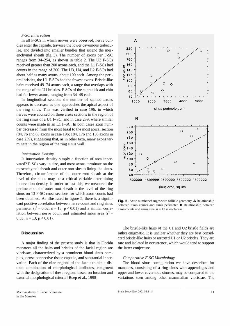
Innervation DensityIs innervation density simply a function of area inner-
vated? F-SCs vary in size, and most axons terminate on themesenchymal sheath and outer root sheath lining the sinus.Therefore, circumference of the outer root sheath at thelevel of the sinus may be a critical variable determininginnervation density. In order to test this, we measured theperimeter of the outer root sheath at the level of the ringsinus on 13 F-SC cross sections for which axon counts hadbeen obtained. As illustrated in figure 5, there is a signifi-cant positive correlation between nerve count and ring sinusperimeter (r2 = 0.62; n = 13, p < 0.01) and a similar corre-lation between nerve count and estimated sinus area (r2 =0.53; n = 13, p < 0.01).
Discussion
A major finding of the present study is that in Floridamanatees all the hairs and bristles of the facial region arevibrissae, characterized by a prominent blood sinus com-plex, dense connective tissue capsule, and substantial inner-vation. Each of the nine regions of the face exhibits a dis-tinct combination of morphological attributes, congruentwith the designation of these regions based on location andexternal morphological criteria [Reep et al., 1998].
The bristle-like hairs of the U1 and U2 bristle fields arerather enigmatic. It is unclear whether they are best consid-ered bristle-like hairs or arrested U1 or U2 bristles. They arerare and isolated in occurrence, which would tend to supportthe latter conjecture.
Comparative F-SC MorphologyThe blood sinus configuration we have described for
manatees, consisting of a ring sinus with appendages andupper and lower cavernous sinuses, may be compared to thevariations seen among other mammalian vibrissae. The
11Microanatomy of Facial Vibrissaein the Manatee
Brain Behav Evol 2001;58:1–14
Fig. 5. Axon number changes with follicle geometry. A Relationshipbetween axon counts and sinus perimeter. B Relationship betweenaxon counts and sinus area. n = 13 in each case.
mystacial vibrissae of rodents exhibit a circumferential ringsinus with a ringwulst (ring body), as well as a ventrallyadjacent trabeculated cavernous sinus [Wineski, 1985; Riceet al., 1986, 1997; Dehnhardt et al., 1999]. Ringed seals[Hyvarinen and Katajisto, 1984], California sea lions[Stephens et al., 1973] and southern elephant seals [Ling,1966] possess elongated vibrissae F-SCs having trabecu-lated cavernous sinuses superficial and deep to the ringsinus, which has a ringwulst. The ringwulst in southern ele-phant seals is relatively small compared to the ringwulst inrats [Ling, 1966]. Ling [1977], in a review of vibrissal struc-ture in marine mammals, noted a variety of sinus configura-tions among the cetaceans. The vibrissae of rhesus monkeys[Van Horn, 1970], tammar wallabies [Marotte et al., 1992]and brush-tailed possums [Hollis and Lyne, 1974] lack aring sinus and ringwulst and have only cavernous tissue.Hippopotamus facial vibrissae appear to have a similarconfiguration as these three species, with umbrella-likeappendages containing Merkel cells [Bachteler et al., 1998].
The ring sinus appendages we observed in manateeF-SCs are quite unlike the ringwulst seen in other species.Whereas the latter are club shaped in longitudinal sectionand constitute a single bulbous ring around the inner wall ofthe sinus, the appendages in manatees are multiple in num-ber and appear flat rather than bulbous on their ends. Thefunction of the ring sinus appendages is unclear. We did notobserve axons entering the appendages, and the ringwulstseen in most other species is also not highly innervated[Rice et al., 1997]. Stephens et al. [1973] suggested that theringwulst may function to transmit vibratory energy fromthe hair shaft to the ring sinus, and this conjecture may beapplied to the appendages in manatees as well.
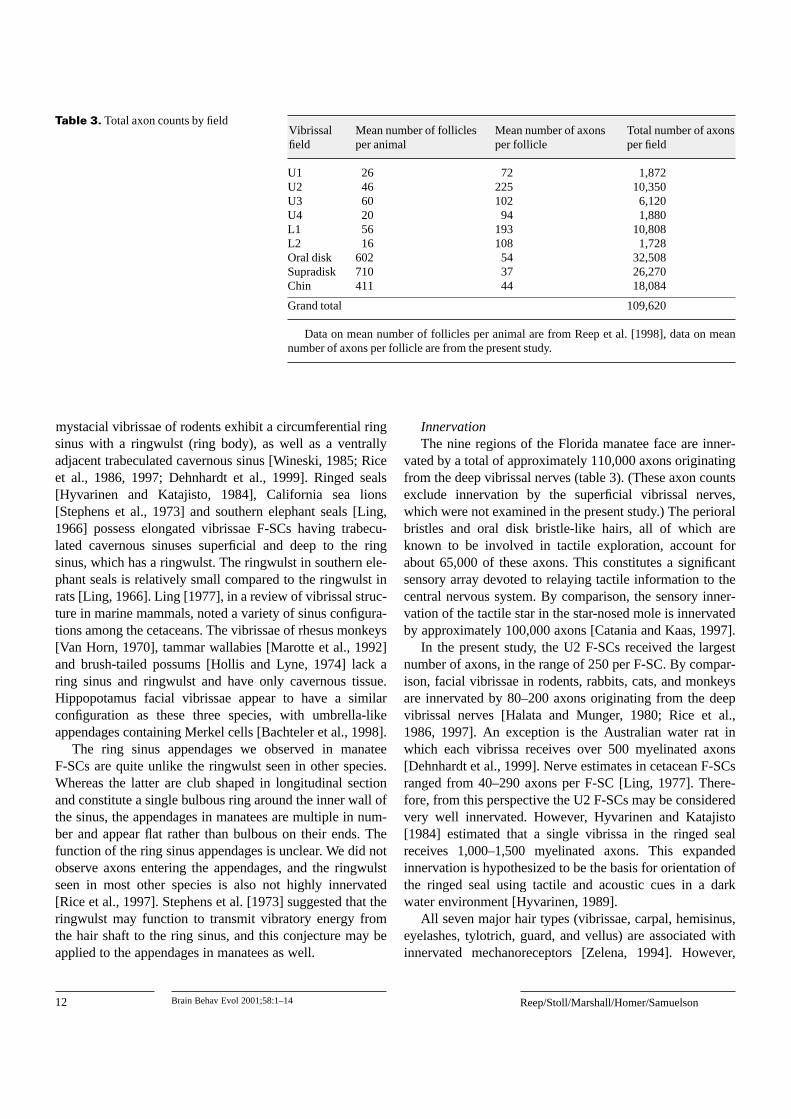
InnervationThe nine regions of the Florida manatee face are inner-
vated by a total of approximately 110,000 axons originatingfrom the deep vibrissal nerves (table 3). (These axon countsexclude innervation by the superficial vibrissal nerves,which were not examined in the present study.) The perioralbristles and oral disk bristle-like hairs, all of which areknown to be involved in tactile exploration, account forabout 65,000 of these axons. This constitutes a significantsensory array devoted to relaying tactile information to thecentral nervous system. By comparison, the sensory inner-vation of the tactile star in the star-nosed mole is innervatedby approximately 100,000 axons [Catania and Kaas, 1997].
In the present study, the U2 F-SCs received the largestnumber of axons, in the range of 250 per F-SC. By compar-ison, facial vibrissae in rodents, rabbits, cats, and monkeysare innervated by 80–200 axons originating from the deepvibrissal nerves [Halata and Munger, 1980; Rice et al.,1986, 1997]. An exception is the Australian water rat inwhich each vibrissa receives over 500 myelinated axons[Dehnhardt et al., 1999]. Nerve estimates in cetacean F-SCsranged from 40–290 axons per F-SC [Ling, 1977]. There-fore, from this perspective the U2 F-SCs may be consideredvery well innervated. However, Hyvarinen and Katajisto[1984] estimated that a single vibrissa in the ringed sealreceives 1,000–1,500 myelinated axons. This expandedinnervation is hypothesized to be the basis for orientation ofthe ringed seal using tactile and acoustic cues in a darkwater environment [Hyvarinen, 1989].
All seven major hair types (vibrissae, carpal, hemisinus,eyelashes, tylotrich, guard, and vellus) are associated withinnervated mechanoreceptors [Zelena, 1994]. However,
12 Brain Behav Evol 2001;58:1–14 Reep/Stoll/Marshall/Homer/Samuelson
Table 3. Total axon counts by fieldVibrissal Mean number of follicles Mean number of axons Total number of axonsfield per animal per follicle per field
U1 26 72 1,872U2 46 225 10,350U3 60 102 6,120U4 20 94 1,880L1 56 193 10,808L2 16 108 1,728Oral disk 602 54 32,508Supradisk 710 37 26,270Chin 411 44 18,084
Grand total 109,620
Data on mean number of follicles per animal are from Reep et al. [1998], data on meannumber of axons per follicle are from the present study.

sinus hairs are the most extensively innervated and themost profusely supplied with mechanoreceptors, includingMerkel cells, lanceolate endings, and free (‘blebbed’) nerveendings. Receptor types and innervation patterns have beenstudied in a variety of mammals including rodents [Mungerand Rice, 1986; Rice et al., 1986, 1997; Mosconi et al.,1993], rabbits and cats [Rice et al., 1986], primates [Halataand Munger, 1980], California sea lions [Stephens et al.,1973], ringed seals [Hyvarinen, 1995], hippopotamuses[Bachteler et al., 1998], and tammar wallabies [Marotte etal., 1992]. The densest accumulation of receptors is alongthe mesenchymal sheath and outer root sheath that line thering sinus [Stephens et al., 1973; Hyvarinen, 1995; Rice etal., 1997]. Similarly, we found that axon counts decreasedas one ascended the mesenchymal sheath in the region ofthe ring sinus, suggesting that manatee receptors are likelyto be similarly distributed. Because some axons innervatesingle receptors and other axons branch to supply several,there is no simple relationship between axon number andreceptor number. Thus the axon counts made in the presentstudy represent only a first order approximation of innerva-tion pattern, in the absence of data on receptor types andnumbers in manatee F-SCs. Our finding of a correlationbetween number of axons and area innervated suggests thatto some extent high axon counts, as in the U2 bristles, maysimply be related to the large size of these F-SCs.
Functional ConsiderationsIt is not known exactly how sinus hairs work. The
integrity of the sinus capsule is necessary for normal func-tion; discharge patterns of afferents innervating the vibrissacease upon opening the sinus capsule [Gottschaldt et al.,1973]. Blood pressure may set the mechanical tension (andthus response sensitivity) of the tissue in which lanceolateendings and Merkel cells are located [Woolsey et al., 1981;Rice et al., 1986]. Thus the neural response properties ofindividual F-SCs would depend on at least the followingfactors at the microanatomical level: (1) hair length, diam-eter, and stiffness, both external to the skin surface andwithin the F-SC; (2) F-SC geometry, especially F-SC lengthand diameter, ring sinus wall thickness, and ring sinus sizeand shape; (3) innervation density; (4) quantity, types, anddistribution of receptor endings. A more complete apprecia-tion of the functional capacities of manatee vibrissae nowawaits the characterization of receptor types and their distri-bution patterns.
One implication of the present findings is that perioralbristles in manatees may have a tactile sensory role muchlike that of vibrissae in other mammals, in addition to theirdocumented role in grasping of plants during feeding. This
combination of motor and sensory usages would be uniqueto sirenians. In support of this conjecture, the U2 and L1bristles, which are the major fields involved in graspingplants during feeding [Marshall et al., 1998a], also have themost densely innervated F-SCs. It seems likely that a majorfunction of the sensory innervation of the U2 and L1 F-SCsis to provide tactile feedback concerning contact withgrasped objects. In contrast, tactile exploration is done pre-dominately by the bristle-like hairs of the oral disk, with theperioral bristles playing a secondary role [Bachteler andDehnhardt, 1999]. The oral disk contains approximately600 bristle-like hairs [Reep et al., 1998], each innervatedby approximately 50 axons (present study), for a total ofapproximately 30,000 axons. This represents an impressivesensory array, congruent with the role of the oral disk in tac-tile scanning and exploration [Hartman, 1979; Marshall etal., 1998a; Bachteler and Dehnhardt, 1999].
The elaboration of vibrissae in manatees may suggest theexistence of hydrodynamic receptive capacities not avail-able to terrestrial animals. Pressure waves, including com-pression waves (sound), travel 4.5 faster in water thanair (depending on temperature, depth, and salinity), andundergo less rapid energy dispersal than in air [Urick, 1983;Kalmijn, 1989]. Therefore they are potentially useful stim-uli in the denser aquatic environment in which visibility isoften reduced. Dehnhardt and colleagues have demonstratedthat harbor seals can use their facial vibrissae to detect watermovements of small amplitude [Dehnhardt et al., 1998,2001]. Poulter and co-workers discussed a similar possibil-ity with regard to acoustic reception in California sea lions[Stephens et al., 1973]. Thus vibrissae in manatees mayfunction in hydrodynamic distance reception in addition totheir known roles in tactile exploration and oripulation.
Acknowledgements
We are grateful for the excellent technical help of Betty Hall, PatLewis and Mae Chisholm. We also thank Tom Pitchford, ButchRommell, Donna Szemer and Scott Wright at the Marine MammalPathobiology Laboratory in St. Petersburg, Florida for their enduringcooperation in providing us with specimens for study. We appreciatethe artistic contributions of Laura Line and Gabe Trinity on figure 1,and the work of Sean Kearns in producing the color figures. College ofVeterinary Medicine Journal Series No. 599.
13Microanatomy of Facial Vibrissaein the Manatee
Brain Behav Evol 2001;58:1–14

14 Brain Behav Evol 2001;58:1–14 Reep/Stoll/Marshall/Homer/Samuelson
Bachteler, D., and G. Dehnhardt (1999) Activetouch performance in the Antillean manatee:evidence for a functional differentiation offacial tactile hairs. Zoology, 102: 61–69.
Bachteler, D., G.J. Klauer, and G. Dehnhardt(1998) The morphology of the vibrissae ofHippopotamus amphibious. Euro-AmericanMammal Congress Abst., 669.
Brecht, M., B. Preilowski, and M.M. Merzenich(1997) Functional architecture of the mystacialvibrissae. Behav. Brain Res., 84: 81–97.
Bryden, M.M., H. Marsh, and B.W. MacDonald(1978) The skin and hair of the dugong,Dugong dugon. J. Anat., 126: 637–638.
Carvell, G.E., and D.J. Simons (1990) Biometricanalyses of vibrissal tactile discrimination inthe rat. J. Neurosci., 10: 2638–2648.
Catania, K.C., and J.H. Kaas (1997) Somatosensoryfovea in the star-nosed mole: behavioral use ofthe star in relation to innervation patterns andcortical representation. J. Comp. Neurol., 387:215–233.
Dehnhardt, G. (1994) Tactile size discrimination bya California sea lion (Zalophus californianus)using its mystacial vibrissae. J. Comp. Physiol.A, 175: 791–800.
Dehnhardt, G., and G. Ducker (1996) Tactual dis-crimination of size and shape by a Californiasea lion (Zalophus californianus). AnimalLearning Behav., 24: 366–374.
Dehnhardt, G., and A. Kaminski (1995) Sensitivityof the mystacial vibrissae of harbour seals(Phoca vitulina) for size differences of activelytouched objects. J. Exp. Biol., 198: 2317–2323.
Dehnhardt, G., H. Hyvarinen, A. Palviainen, andG. Klauer (1999) Structure and innervationof the vibrissal follicle-sinus complex in theAustralian water rat, Hydromys chrysogaster.J. Comp. Neurol., 411: 550–562.
Dehnhardt, G., B. Mauck, and H. Bleckmann(1998) Seal whiskers detect water movements.Nature, 394: 235–236.
Dehnhardt, G., B. Mauck, W. Hanke, and H. Bleck-mann (2001) Hydrodynamic trail-followingin harbor seals (Phoca vitulina). Science, 293:102–104.
Dosch, F. (1915) (translated by D.A. Sinclair)Structure and development of the integument ofSirenia. Tech. trans. No. 1626 NationalResearch Council of Canada, Ottawa 1973.(Bau und Entwicklung des Integuments derSirenen. Janaische Zeitschr., 53: 805–854,1914–1915.)
Dykes, R.W. (1975) Afferent fibers from mystacialvibrissae of cats and seals. J. Neurophysiol., 38:650–662.
Gottschaldt, K.-M., A. Iggo, and D.W. Young(1973) Functional characteristics of mechano-receptors in sinus hair follicles of the cat. J.Physiol., 235: 287–315.
Halata, Z., and B.L. Munger (1980) Sensory nerveendings in rhesus monkey sinus hairs. J. Comp.Neurol., 192: 645–663.
Hartman, D.S. (1979) Ecology and behavior of themanatee (Trichechus manatus) in Florida. Am.Soc. Mammal. Spec. Pub., 5: 1–153.
Hollis, D.E., and A.G. Lyne (1974) Innervation ofvibrissa follicles in the marsupial Trichosurusvulpecula. Aust. J. Zool., 22: 263–276.
Hyvarinen, H. (1989) Diving in darkness: whiskersas sense organs of the ringed seal (Phocahispida saimensis). J. Zool. Lond., 218: 663–678.
Hyvarinen, H. (1995) Structure and function of thevibrissae of the ringed seal (Phoca hispida L.).In Sensory Systems of Aquatic Mammals(ed. by R.A. Kastelein, J.A. Thomas, and P.E.Nachtigall), De Spil Publishers, Woerden, TheNetherlands, pp. 429–445.
Hyvarinen, H., and H. Katajisto (1984) Functionalarchitecture of the vibrissae of the ringedseal (Phoca hispida Schr.). Acta Zool. Fennica,171: 27–30.
Kalmijn, A.J. (1989) Functional evolution of lateralline and inner ear sensory systems. In TheMechanosensory Lateral Line, Neurobiologyand Evolution (ed. by S. Coombs, P. Gorner,and H. Munz), Springer-Verlag, New York,pp. 188–215.
Kamiya, T., and F. Yamasaki (1981) A morphologi-cal note on the sinus hair of the dugong. In TheDugong (ed. by H. Marsh), Dept. of Zoology,James Cook University of North Queensland,Australia, pp. 111–113.
Kastelein, R.A., and M.A. Van Gaalen (1988) Thetactile sensitivity of the mystacial vibrissae ofa Pacific Walrus (Odobenus rosmarus diver-gens). Part 1. Aqu. Mamm., 14: 123–133.
Layne, J.N., and D.K. Caldwell (1964) Behaviorof the Amazon dolphin, Inia geoffrensis (Blain-ville), in captivity. Zoologica, 49: 81–108.
Ling, J.K. (1966) The skin and hair of the southernelephant seal, Mirounga leonina (Linn.). I. Thefacial vibrissae. Aust. J. Zool., 14: 855–866.
Ling, J.K. (1977) Vibrissae of marine mammals.In Functional Anatomy of Marine Mammals,Vol. 3 (ed. by J.B. Harrison), Academic Press,London, pp. 387–415.
Marotte, L.R., F.L. Rice, and P.M.E. Waite (1992)The morphology and innervation of facialvibrissae in the tammar wallaby, Macropuseugenii. J. Anat., 180: 401–417.
Marshall, C.D. (1997) Characterization of thedugong oral disk, perioral bristles and bristle-like hairs. Am. Zool., 37: 56A.
Marshall, C.D., L.A. Clark, and R.L. Reep (1998b)The muscular hydrostat of the Florida manatee(Trichechus manatus latirostris) and its role inthe use of perioral bristles. Marine Mamm. Sci.,14: 290–303.
Marshall, C.D., G.D. Huth, V.M. Edmonds, D.L.Halin, and R.L. Reep (1998a) Prehensile use ofperioral bristles during feeding and associatedbehaviors of the Florida manatee (Trichechusmanatus latirostris). Marine Mamm. Sci., 14:274–289.
Marshall C.D., P.S. Kubilis, G.D. Huth, V.M.Edmonds, D.L. Halin, and R.L. Reep (2000)Prehensile use of perioral bristles by Floridamanatees during feeding upon several speciesof aquatic vegetation. J. Mammal., 81: 649–658.
Miller, E.H. (1975) A comparative study of facialexpressions of two species of pinnipeds.Behaviour, 53: 268–284.
Mosconi, T.M., F.L. Rice, and M.J. Song (1993)Sensory innervation in the inner conical bodyof the vibrissal follicle-sinus complex of the rat.J. Comp. Neurol., 328: 232–251.
Munger, B.L., and F.L. Rice (1986) Sequential mat-uration of cutaneous receptors in the rat mysta-cial pad. J. Comp. Neurol., 252: 404–411.
Norman, J.R., and F.C. Fraser (1948) Giant Fishes,Whales and Dolphins. Putnam, London.
Peterson, R.S., and G.A. Bartholomew (1967) Thenatural history and behavior of the Californiasea lion. Am. Soc. Mammal. Spec. Pub., 1:1–79.
Reep, R.L., C.D. Marshall, M.L. Stoll, and D.M.Whitaker (1998) Distribution and innervationof facial bristles and hairs in the Florida mana-tee (Trichechus manatus latirostris). MarineMamm. Sci., 14: 257–273.
Rice, F.L., B.T. Fundin, J. Arviddson, H. Aldsko-gius, and O. Johansson (1997) Comprehensiveimmunofluorescence and lectin binding analy-sis of vibrissal follicle sinus complex innerva-tion in the mystacial pad of the rat. J. Comp.Neurol., 385: 149–184.
Rice, F.L., A. Mance, and B.L. Munger (1986) Acomparative light microscopic analysis of theinnervation of the mystacial pad. I. Vibrissalfollicles. J. Comp. Neurol., 252: 154–174.
Sokolov, V.E. (1982) Mammal Skin. University ofCalifornia Press, Berkeley, Calif.
Sokolov, V.E. (1986) Manatee – MorphologicalDescription. Nauka Press, Moscow. (In Rus-sian.)
Stephens, R.J., I.J. Beebe, and T.C. Poulter (1973)Innervation of the vibrissae of the Californiasea lion, Zalophus californianus. Anat. Rec.,176: 421–442.
Urick, R.J. (1983) Principles of Underwater Sound,3rd Edition. McGraw Hill Book Co., NewYork.
Van Horn, R.N. (1970) Vibrissae structure in therhesus monkey. Folia Primatologica, 13: 241–285.
Welker, W.I. (1964) Analysis of sniffing of thealbino rat. Behavior, 22: 223–244.
Wineski, L.E. (1985) Facial morphology andvibrissal movements in the golden hamster.J. Morph., 183: 199–217.
Zelena, J. (1994) Nerves and Mechanoreceptors.Chapman and Hall, London.
Woolsey, T.A., D. Durham, R.M. Harris, D.J.Simons, and K.L. Valentino (1981) Somato-sensory development. Dev. Percept., 1: 259–292.
References