Embed Size (px)
Citation preview
Microemulsion as drug delivery system for Peptides and Proteins
Landge Anil, Krishnamoorthy Kannan*
Department of Pharmacy, Annamalai University, Annamalainagar,Tamilnadu India.
*Corresponding Author: E‐mail: [email protected]
Abstract Microemulsions (MEs) are isotropic mixtures with or without a cosurfactant along with combination of oil, water and surfactant and most stable as per view of thermodynamics. These systems of drug delivery are currently of prior interest to the pharmacists because of their embryonic potential to act as therapeutic enzymes and peptide based drug delivery vehicles with incorporation of a wide range of active therapeutic protein and peptide molecules. These therapeutic macromolecules in microemulsion drug delivery form is not solely based on compositions of the vehicle but also on the internal structure or composition of the phases which may nurture protein drug distribution in the vehicles for enhanced drug solubilisation capacity, ease of preparation, enhancement of bioavailability and maximum shelf life. In order to appreciate the potential of protein based microemulsions as delivery vehicles for enhanced drug permeation via skin and tolerability of these systems, this review offers an overlook on phase behaviour studies formulation of microemulsion with entrapment and various approaches to incorporate proteins and enzymes into microemulsion, protein engineering methods for stable delivery and enhanced bioavailability, various protein drug compatibility study methods including characterization of microemulsions.
Keywords: Dermal, Enzyme, Microemulsion, Peptide, Protein.
INTRODUCTION Protein and peptide delivery methods have evolved
remarkably over the past decades, focusing the major research efforts on the delivery of varieties of essential proteins and peptides with high molecular weight. These macromolecules are distinguished by a poor incorporation into the blood stream when administered orally and also with short half-life, which ascertains the need of a frequent administration in optimum doses to achieve desired therapeutic efficacy. Even more, these biomolecules are very sensitive to physiological conditions (e.g., acidic pH of GI tract) and further may lead to adverse effects after systemic administration in high doses. [1]
Generally, protein drugs expose lipophilic, hydrophilic and also amphipathic nature, its macromolecular structure and associated substantial physio-chemical hindrance are the basic features strongly interferes with the pharmacokinetic and pharmacodynamic behaviour of the drug in vivo [2]. They also limit the rate of reactions, selected solvent systems and unstable nature in variable environmental conditions which are prime considerable factors in formulation of therapeutic protein-peptide based pharmaceuticals.
Protein and peptide macromolecules are administered systemically by intradermal, subcutaneous, transdermal intravenous, intramuscular and intraperitoneal injections and these formulations often encompass additives (e.g., cosolvents, buffers, preservatives), for improving the stability of the biomolecule in vivo [2, 3]. In this respect, albumin and surfactants including amphiphiles like lecithin exhibits a major role in reducing aggregation and the adsorption processes, thus limiting the probability of protein unfolding, its precipitation and deactivation [4]. High molecular weight, fragile nature and complexity and unreliable structure are the main hurdles in application of protein drugs [5, 6]. Moreover such macromolecules may easily get denatured, degraded and ultimately inactivated during their formulation, storage, and delivery by varieties of physiochemical, and enzymatic processes. Their biopharmaceutical properties also blamed for stability [7].
Bioavailability of protein and poor mucosal permeability is majorly blamed to the varieties of proteolytic enzymes situated in the gut, lungs, and skin [8]. Therapeutic bioavailability is also inadequate due to the rapid clearance from the body due to instantaneous phagocytosis, endocytosis,
glomerular filtration, proteolysis due to certain enzymatic processes, and various immunological factors of these active protein molecules [9]. Xeno-proteins like therapeutic antibodies and antiseras are antigenic and intrinsically immunogenic. Therapeutic proteins with small molecular weight are generally expelled by the kidneys, whereas larger molecular weight proteins mostly undergo proteolytic degradation. Lipoproteins as well as different glycosylated proteins are targeted majorly by the process called endocytosis and phagocytosis [10] while interleukins, cytokines and hormones are most frequently expelled via systemic circulation by intracellular processing and receptor-mediated endocytosis [11]. Moreover numbers of physiological type of proteins are denatured by proteolysis at particular local sites without reaching appropriate therapeutic levels. Thus far, various studies to enhance the protein bioavailability and focus on altering the physicochemical properties of these therapeutic macromolecules are widely being studies for incorporation of functional additives into innovated drug delivery systems adapted specially for these purpose.
In view of these all facts, colloidal drug delivery systems majorly microemulsions (MEs) targeting intradermal and transdermal sites have become the major focus of this research in improving the therapeutic indices of protein biomolecules (effectiveness and safety) by means of a localized and extended release at the target site, without resulting in undesirable side effects. Transport of Protein and Peptide Macromolecules Via Skin
Human skin is considered as largest organ of the body. It functions as a barrier against depletion of water and essential compounds from the body, penetration of toxic agents. Moreover it also serves as a medium for absorption of drugs locally and systemically. Due to the structure, physiology and barrier properties of the skin, there are a number of options and complications for drug delivery across the skin. The skin is fabricated of four distinct layers, namely the stratum corneum, the epidermis, the dermis and the subcutaneous tissue. The stratum corneum is about 10 to 15 cell layers thick, is fabrication of corneocytes or dead cells and constitute as the primary barrier to the delivery of most drugs [12]. The intercellular void spaces between such corneocytes are generally filled with sheets of lipid bilayer membranes that are water impermeable; lipid lamellae within the stratum corneum, functions as permeability barrier for
Landge Anil et al /J. Pharm. Sci. & Res. Vol. 10(1), 2018, 16-25
16

epidermis to water and other penetrants. All intradermal, subcutaneous and transdermal drug delivery requires overcoming this epidermal barrier without interfering the skin functions [13]. A major difference between dermal and transdermal drug delivery, in view of their therapeutic need and efficacy. Dermal delivery is majorly targeted towards various skin disorders such as skin cancer, psoriasis, eczema, acne and other fungal or microbial infections. In the case of dermal delivery, systemic absorption is not important, instead remittance of drugs to the pathological sites, is of major concern [14]. While transdermal type of drug delivery is focussed towards achievement of systemic levels of drugs. The drug, passes through the different layers of the skin, and reaches the systemic circulation, to produce its therapeutic effect. Transdermal Drug Delivery (TDD) is advantageous for specific drugs those have a higher first pass metabolism and for those drugs which indicate adverse effects such as ulcerations and colitis conditions in the gastrointestinal tract [15].
Different kinds of mechanisms are involved in penetration of therapeutic protein drugs such as simple transcellular and paracellular diffusion, carrier-mediated transport, active transport and pinocytosis or endocytosis [16]. Protein drugs those having lack of lipophilic nature, contributing for zero passive absorption and is ingested across an epithelial membrane by migration through the intercellular margin between the cells [17]. Normally intercellular space exists between 10 to 50A˚; therefore such route is not appreciable for large macromolecules. While in case of insulin, it is adsorbed on the microvilli bearing portion of the epithelial cell membrane (apical membrane) and is engulfed by specific types of endocytosis processes [18]. Some of protein and peptides only moves with active transport by binding to the cell surface receptor or binding channels in the epithelial lining of the small intestine(membrane bound vesicles) [19]. However the routine transport mechanism that exist is passive diffusion with reversible way transport: first, paracellular (delivery of drug molecule through the intercellular space between the cells) and another, transcellular (involves migration of drug molecule into or across the cells). Transportation of drugs is majorly blamed for its molecular geometry, lipophilicity and charge of the transport pathway across the mucous membrane [20]. Certain extent of lipophilicity is required in protein biomolecules to get disperse into the epithelial membrane and absorb through transcellular passive diffusion [21]. Oral transport of these biomolecules are contributed by gastrointestinal tract into the systemic circulation is through the muscular mucosa then via the areolar layer or a loose connective tissue layer. Areolar or submucosal are other two intestinal layers join together the mucus and muscular layers [22]. Muscular and mucus layers are most strong layers of the intestine which consists of the loose filamentous connective tissue layer i.e. areolar tissue containing lymph gland, nerves and blood vessels [23]. Though there is success in increased transcellular permeation which earlier disclosed on human Caco-2 monolayerd epithelial cell at highest concentration in vitro, even though the binding any ligand on molecules that opens the tight junctions is the essential one which is targeted [24]. Microemulsion Science
Microemulsion was first introduced in the 1940s by Hoar and Schulman who formulated a clear, single-phase system by titration of a milky emulsion with hexanol [25]. Since then microemulsions have been known and abundant studies undertaken in terms of delivery systems, cause of their multiple advantages. Briefly, microemulsions are transparent, optically isotropic and stable systems generally constitution of an oil, water and surfactant(s) [26]. Microemulsions systems are different system from emulsions with considering number of factors. Microemulsions are clear transparent and composed of globule size (generally up to 150 nm) [27], while emulsions are milky,
coarse dispersions with globule sizes generally in the range of micrometer or slightly below. A large number of small droplets are produced, when microemulsions form. Due to the small size of the droplets in a microemulsion, they possess a large interfacial surface area, from which transport of the drug can occur [28]. Microemulsions generate spontaneously, with or without energy necessity. Most often some energy input (viz. gentle mixing, stirring or heating) enhances microemulsion formation but certain barriers like kinetic energy must be conquered [29]. Different Theories of Microemulsion Formation
As far as formulation part concern ME formations is based on three different theories. Those are - mixed or interfacial film theory [30], solubilisation theory [31], and thermodynamic theory [32]. According to thermodynamical theory of stabilization, ME generates spontaneously due to the low interfacial free energy level in consideration with diffusion of particular surfactant in the interfacial layer and also the contribution of major entropy that resolute the mixing of single phase in the other one in the form of abundant small droplets. While in the mixed film theory, the interfacial film is understood in demonstrating dissimilar behaviour towards the aqueous and oily segment of the interface. While the solubilisation theory is considered as swollen micelles, in which oil or water is solubilised the micelle or reverse micelle structures to form single-phase system. However, despite of all the ME theories of formation, the depletion in interfacial tension to a very moderate value is of considered as ultimate importance in the ME formation. Pharmaceutical Formulation of Microemulsions
Pharmaceutically microemulsion systems are designed and formulated by taking into consideration GRAS -(generally regarded as safe) and preferably pharmaceutical - grade ingredients, that is, ones earlier used in pharmaceutical formulation and devoid of serious adverse effects and toxicity in humans [33]. Nonionic and zwitterionic surfactants are among the most commonly used ingredients to formulate pharmaceutical MEs while vegetable oils, medium - and long - chain triglycerides, and fatty acids ester are the most generally used oils [34].
It is a general concept that, low HLB (about 3-6) surfactants are most preferential for the formulation of w/o microemulsion, while surfactants with higher value of HLB (about 8-18) are recommended for the o/w type microemulsion formulation. Surfactants with HLB value greater than 20 are mostly used along with the co-surfactants to minimise their overall effective HLB value within the accepted range for microemulsion formation [35]. This can be regarded as thumb rule while selecting oil phase, surfactants and cosurfactants with prime concern of stability of ME. Aqueous Phase
An essence of the aqueous phase is a paramount factor for formulation of peptide based microemulsions. In consideration of parenteral microemulsions, the aqueous phase must be isotonic and isosmotic to the blood which can be attained by using additives such as electrolytes (sodium chloride), sorbitol, dextrose, and glycerol. These additives can resolute the microemulsion area of existence [36]. Phase inversion temperature (PIT) of the non-ionic type surfactants can be minimised by electrolytes like sodium chloride [37]. Oil Phase
Oil phase also having its most requisite role in the formulation not only because one can solubilise the required dose of the lipophilic drug, but it boosts the lipophilic drug transportation via the lymphatic system in intestine and thereby enhancing absorption in the gastrointestinal tract based on the molecular nature of particular triglyceride [38].
Landge Anil et al /J. Pharm. Sci. & Res. Vol. 10(1), 2018, 16-25
17

Table 1: List of surfactants commonly used in protein and peptide based microemulsion.
Class HLB status
Examples References
Cationic surfactants -- Cetyldimethylethylammonium bromide, Cetylpyridinium chloride and other salts [42]
Anionic surfactants --
Deoxycholate, and its salts, ursodeoxycholic acid, and taurocholic acid; C5 to C29 monoesters of lactic acid; C8-20sulfonates, including alkyl-, olefin-, and alkylaryl derivatives; C5 to C33 diesters of tartaric acid, tridecyl- and dodecylbenzene sulfonic acids; and C5 to C33 sarcosine and betaine derivatives. phospholipids such as phosphatidic acid and phosphatidyl serine.
[43]
Zwitterionics -- phospholipids as lecithin, phosphatidylethanolamine, and sphingomyelins [44]
Non-ionic surfactants
Low HLB C9 to C13 monoglycerides (HLB 4-7), C19 to C25 diglycerides of mono and poly unsaturated fatty acids (HLB 3-5), C15-C23 diglycerides (HLB 4-6), and C35 to C47 diglycerides of mono and poly unsaturated fatty acids (HLB 2.5-4.5);
[45]
High HLB
C8-96 ethoxylated fatty esters; C14-130 sucrose fatty esters; and C20-130 sorbitol and sorbitan monoesters, diesters, and triesters, polyoxyethylene sorbitan monooleate, sorbitol hexaoleate POE (50). Ethoxylated castor oil (HLB 10-16); and the sorbitan surfactants with HLB from 10-18.
The oil component alters curvature by its capability for penetration and expansion of the tail region of the monolayer of surfactant. Short chain oils perforate the tail group region to a larger extent than long chain alkanes, and hence expand this region to a larger extent, developing in increased negative curvature (and hence reduced effective HLB). Saturated (for example, lauric, myristic and capric acid) and unsaturated fatty acids (for example, oleic acid, linoleic acid and linolenic acid) have penetration enhancing property of their own and those have been studied since a long time. Fatty acid esters such as ethyl or methyl esters of lauric, myristic and oleic acid have also been employed as the oil phase [39]. Lipophilic drugs are most precisely solubilised in o/w microemulsions. The intension while choosing the oil phase is that the drug should be highly solubilised in it, which minimises the volume of the formulation to deliver the required dose of the therapeutic drug in an encapsulated form [40]. Surfactants:
The selection of suitable surfactant system is one of the most crucial step in the designing a ME system. In ME, oil system solubilisation carries most important factor than other micellar solutions. It is feasible for one surfactant molecule, to solubilise 10 to 20 oil molecules (o/w ME) or 10 to 200 water molecules (w/o ME). The surfactant(s) need to dissolve and lower the interfacial free energy to very low level (<10−3 mN/m) between the oil and aqueous phases [41].
Generally all anionics surfactants such as S.L.S. are extreme soluble in water & very little solubile in oil/fats and even almost all cationics and amphotrics shows higher solubility in water, while nonionics’ water solubility can be predicted by their HLB values. Various ionic and non-ionic types of surfactants in consideration of protein based microemulsion system are reviewed here in table 1. Cosurfactants/ cosolvents
Cosurfactants are molecules with weak amphiphilic properties that are mixed with the surfactant(s) to enhance their ability to reduce the interfacial tension of a system and promote the formation of a ME [46]. Most single - chain surfactants do not sufficiently lower the oil – water interfacial tension to form MEs, nor are they of the right molecular structure to act as cosolvents. Such barrier can be conquered as cosurfactant /cosolvent molecules are considered to minimise the interfacial free energy in between oil and water, hydrocarbon boundary of the interfacial film can be fluidised which ultimately determine impact the curvature of the film [44]. Choice of cosurfactants for therapeutic protein candidates are alcohols preferred from the group
comprising of ethanol, isopropanol, n-butanol, Lecithin etc. and isobutanol, propylene glycol, polyethylene glycol and isopropyl myristate [47]. Buffers/ Stabilisers
Protein stabilization is improved by certain buffers like acetate, citrate, histidine, glycine, methionine, tartarate, lactate, succinate either alone or in combination thereof are also included [44]. Phase Behaviour Studies
In order to understand the phase behaviour of any microemulsion, pseudo-ternary phase diagrams of water, oil and co-surfactant/surfactants mixtures at particular point of ternary system or phase triangle is drawn with appropriate cosurfactant or surfactant weight ratios. Phase diagrams are obtained by mixing of the ingredients, which earlier pre-weighed into glass vials and titrated with water and stirred well at room temperature. A typical pseudo-ternary phase diagram is indicated for formulation of various colloids systems with consideration of different phases in figure 1. Development of either monophasic or biphasic system is identified by visual inspection. If there is formation of turbidity followed with a phase separation, the systems are called biphasic while in earlier case monophasic which indicates fine, clear-cut and transparent mixtures that can be visualized after homogenisation; these samples are identified at particular points in the ternary phase diagram. The area covered by these different points is regarded as the microemulsion boundary region of existence. [48]
Figure 1: Pseudo-ternary phase diagram representing the
different regions of various types of colloids.
Landge Anil et al /J. Pharm. Sci. & Res. Vol. 10(1), 2018, 16-25
18
Entrapment of Proteins and Peptides into Microemulsions There are basically three methods employed to
introduce protein and enzymes in reverse micelles. In the first method known as the “injection” method the enzyme solution is introduced to a solution of surfactant in a nonpolar organic solvent. The resultant mixture is shaken vigorously until an optically clear-cut, fine and transparent solution is obtained. The second method consists of the addition of dry lyophilized protein to a surfactant solution in an organic solvent comprising an aqueous phase. The third procedure is dependent on the event of interfacial transfer of the protein spontaneously in a two-phase system consisting of normally equal volumes of the aqueous protein solution and surfactant containing organic solvent. Gentle shaking results the incorporation of enzyme into the reverse micelles of the organic phase. This method is very useful for the separation, extraction, and purification of biomolecules (including enzymes and DNA) [49, 50].
A new technology where protein enzymes like model oxidoreductases, tyrosinase (Tyr) and glucose oxidase (GOx), were processed to an octane compound based ink by entrapping in a system of reverse micelles (RM) of surfactant AOT in octane to separate and stabilize the enzyme molecules in nonpolar organic media [51].
The injection method is by far the most used method to microencapsulate enzymes, due to the simple procedure. One of the major drawbacks of the other two methods is the prolonged contact in between the enzyme molecule and the organic solvent system that attributes to the enzyme deactivation.
Several variables are having their own role during the solubilisation of proteins in microemulsions, including the pH and the ionic strength of the aqueous phase, the molecular and structural size of the protein, the size of the reverse micelles, and the nature of the surfactant [52, 53]. The protein uptake into the aqueous microphase is a complicated process. It was proposed that the lipophilicity of the protein molecule plays major role in its localization among the various microenvironments of the system. In fact, a lipophobic protein can avoid direct contact with the continuous organic phase and remain localized in the water layer; certain surface active enzyme (such as some lipases) produces an interfacial interaction with the micellar interface, while a typical membrane protein could be in conjunction with the hydrophobic boundary line of the micelles and also with the organic solvent [54, 55].
In biological system many proteins and enzymes wield at interface of hydrophilic and hydrophobic domains and these interfaces are usually under stabilisation of polar lipids and certain natural amphiphiles. Lipid particles also can be viewed as reverse micelles units as in sandwich form between polar lipid monolayers. It is also noticed that many enzymes and proteins influence these bilayer types of structures upon inclusion into both model and biological membranes. Hence delivery of proteins and enzyme therapeutics in w/o type microemulsions are in sense to biology with wider pertinent merely than biocatalyst action [56]. The microemulsion formulation meant for dermal or transdermal type is of relevance for skin absorption. The water in continuous vehicle structure allows faster transport of hydrophilic drugs [57].
There are a wide range of reported studies using micro emulsions for dermal peptide delivery in human skin. Water-in-oil microemulsions were used as they are particularly suitable to entrap protein/peptides in the aqueous droplets and deliver the peptides effectively into the dermal layer [57, 58]. Different studies using animal models demonstrated that the topical administration of the high molecular weight proteins, anti-TNF monoclonal antibodies Remicade™ and Humira™, in water-in-oil microemulsions reduced inflammation in the feet of mice [59]. According to bio distribution studies, the molecule rapidly penetrated into the skin and also perforated laterally into the distal
regions of the skin with approximately 70% of the protein found in the skin. In one research article in vivo studies on mice reported on reduction in inflammatory footpad condition of carrageenan induced mice followed with the topical administration of anti-TNF molecules that was formulated via microemulsion system [60].
It is well known that protein based biological are not appropriate dosage forms for oral route administration due to macro size, polarity and unequal charge distribution on protein therapeutics also it may undergo enzymatic breakdown through proteolysis in the gastrointestinal tract and ultimately poor therapeutic index. Most biologicals are available as aqueous injectable components that require repeated dose regimen and frequent visits to the health service providers. The development of a self administrable delivery system would enable patients to avoid discomfort and enhancing patient compliance [61]. Different Approaches To Incorporate Proteins, Peptides and Enzymes In Microemulsion (a) Self-Emulsifying Drug Delivery Systems (SEDDS)
Self-emulsifying drug delivery systems (SEDDS) are one of most novel and comprehensive approach for incorporating of protein in microemulsion. It is under extensive research after the market success of HIV protease inhibitors, ritonavir (Norvirs) and saquinavir (Fortovases), and cyclosporin (Neorals or Sandimmunes) formulations. SEDDS do possess lipidic excipients to improve solubility and permeability of drug substances. These lipid based excipients get emulsified when exposed to gastrointestinal fluids to form oil-in-water emulsions or micro-emulsions [62, 63]. By considering the globule size, SEDDS can be classified either as self-microemulsifying drug delivery systems (SMEDDS) and another one as self-nano-emulsifying drug delivery systems (SNEDDS). SMEDDS are clear and transparent microemulsions with globule size range between 100 to 250 nm, while the globule size of SNEDDS is less than 100 nm [64]. (b) Self-Nano-Emulsifying Drug Delivery Systems (SNEDDS)
Self-Nanoemulsifying Drug Delivery System (SNEDDS) has majorly developed for protein drug delivery via oral route as it devoid of water, hence long term preserving the stability of protein, protecting protein from proteolytic degradation, and augmenting the permeability of therapeutic protein-drug in the gastrointestinal tract. However, due to low solubility of protein in oil, which is almost towards zero, protein-based SNEDDS formulations are difficult and challenging. SNEDDS system was found most compatible for proteins using HLB approach by Lina Winarti et. al. in 2016 [65]. It is investigated that SNEDDS with single hydrophilic surfactant is one of the best content in formula for stability testing of protein template as recipe showed that precipitation or phase separation did not appear by employing model protein such as bovine serum albumin (BSA) during study [65]. (c) Solid-Lipid Nanoparticles (SLN’s)
Oral absorption of drugs can be enhanced by lipids [66]. An excellent model drug such as Cyclosporin A in the form of microemulsion reduces the bioavailability variation of protein molecule. In the early 1960s, the first parenteral administration (Intralipid) began the administration of lipophilic drugs. In the early 1990s, various groups focused attention on solid lipid nanoparticles. As the name implies, these nanocarriers contain solid lipids. They have the advantages of physical stability, controlled release, and low toxicity; they also help in protecting sensitive drugs from degradation from the external environment. They are generally prepared with physiological lipids or molecules that have a history of safe use and are better tolerated than the polymeric carriers. Additionally, organic solvents are not
Landge Anil et al /J. Pharm. Sci. & Res. Vol. 10(1), 2018, 16-25
19
being utilised which makes them better candidates compared to many of the polymeric systems.
There are three important variations of lipid nanoparticles tested in the pharmaceutical literature: solid lipid nanoparticles (SLNs), nanostructured lipid carriers (NLCs), and lipid_drug conjugates (LDCs). Varieties of research works have also constantly targeted on improving stability of SLN’s in body fluids with hydrophilic coating molecules viz. poly(ethylene)glycol (PEG) derivatives. Hydrophilic molecule’s coating to SLN not only enhances plasma stability but also bio-distribution resulting subsequent bioavailability of entrapped drugs [67]. (d) Convertible/ Phase Reversible Microemulsion
Stable w/o microemulsions are formed when HLB value of the microemulsion is between 9 and 12. It is achieved by using high HLB value surfactants are used in combination with low HLB surfactants in the patent invention by Albert J. Owen et. al [44].
Any water soluble biologically active entity in the aqueous phase is liberated for body absorption reason as water in oil (w/o) microemulsion which freely converts phase to an oil in water (o/w) emulsion by the incorporation of aqueous fluid to the said w/o microemulsion. Short chain monoglyceride surfactant that is widely employed as storage depot for proteins and remain stable for longer duration at room temperature and above until they are ready for use in w/o microemulsion.
While at particular period of time the addition of aqueous fluid samples which converts the microemulsion into an o/w emulsion and subsequently releases the therapeutic protein. The precisely stored w/o type convertible microemulsion can be delivered to the body where it is does convert into an o/w emulsion by the interacting with different body fluids. Advantage of such delivery system is by this manner, peptide microemulsion storage problems also minimised. [44].
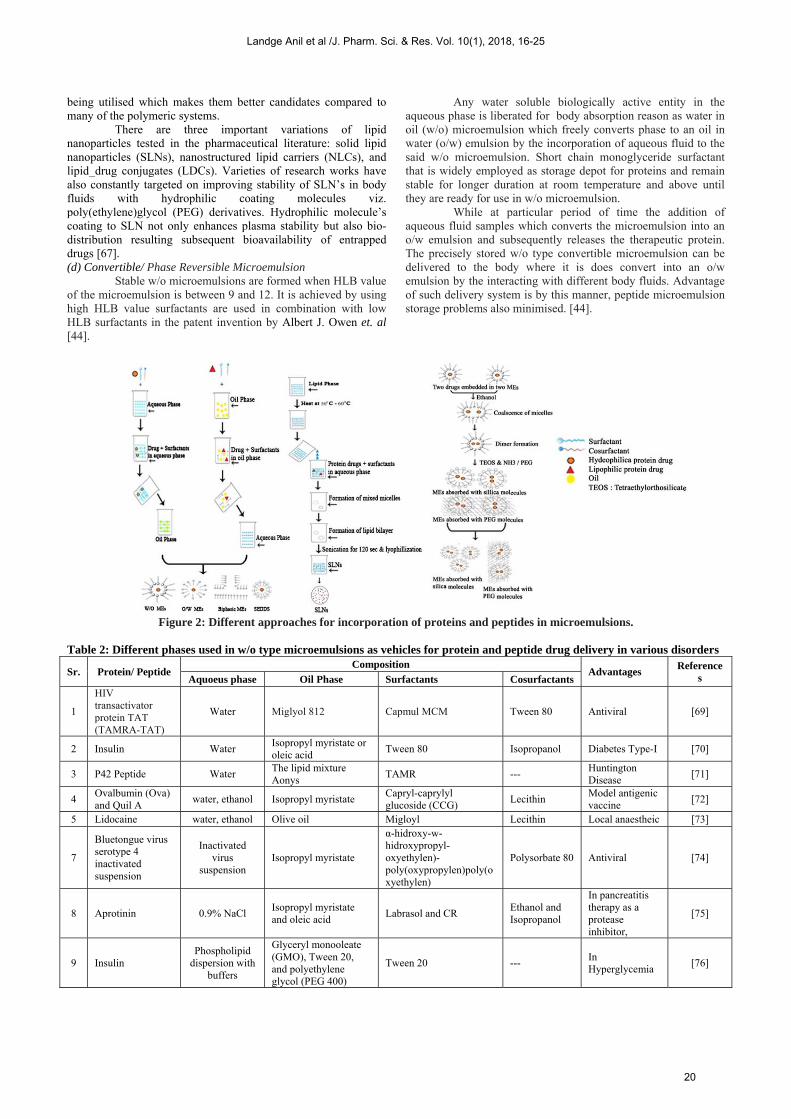
Figure 2: Different approaches for incorporation of proteins and peptides in microemulsions.
Table 2: Different phases used in w/o type microemulsions as vehicles for protein and peptide drug delivery in various disorders
Sr. Protein/ Peptide Composition
Advantages Reference
s Aquoeus phase Oil Phase Surfactants Cosurfactants
1
HIV transactivator protein TAT (TAMRA-TAT)
Water Miglyol 812 Capmul MCM Tween 80 Antiviral [69]
2 Insulin Water Isopropyl myristate or oleic acid
Tween 80 Isopropanol Diabetes Type-I [70]
3 P42 Peptide Water The lipid mixture Aonys
TAMR --- Huntington Disease
[71]
4 Ovalbumin (Ova) and Quil A
water, ethanol Isopropyl myristate Capryl-caprylyl glucoside (CCG)
Lecithin Model antigenic vaccine
[72]
5 Lidocaine water, ethanol Olive oil Migloyl Lecithin Local anaestheic [73]
7
Bluetongue virus serotype 4 inactivated suspension
Inactivated virus
suspension Isopropyl myristate
α-hidroxy-w-hidroxypropyl-oxyethylen)-poly(oxypropylen)poly(oxyethylen)
Polysorbate 80 Antiviral [74]
8 Aprotinin 0.9% NaCl Isopropyl myristate and oleic acid
Labrasol and CR Ethanol and Isopropanol
In pancreatitis therapy as a protease inhibitor,
[75]
9 Insulin Phospholipid
dispersion with buffers
Glyceryl monooleate (GMO), Tween 20, and polyethylene glycol (PEG 400)
Tween 20 --- In Hyperglycemia
[76]
Landge Anil et al /J. Pharm. Sci. & Res. Vol. 10(1), 2018, 16-25
20
Table 3: Different methods for preparation of various forms of microemulsion
Technique Process Approach Type Protein Incorporation method Example References
Phase Titration for W/O and O/W
Homogenisation
General Approach
W/O Solubility by HLB and phase titration
Aprotinin [89]
O/W Ultra High Pressure Homogenisation
Whey Protein [90]
Nanoparticle Approach
SLN
Adsorption onto SLN BSA, HSA [91, 92]
Solvent evaporation (w/o/w) Interferon-alpha (IFN-alpha)
[93]
HPH hot dispersion Cyclosporin [94]
HPH cold dispersion Cyclosporin [95]
Warm microemulsion (o/w) Cyclosporin [96]
Solvent displacement Gonadorelin [97]
Encapsulation HIV-1 gp120 antigen
[98]
SEDDS
Solid carriers Monoclonal Antibodies
[99]
Spray Drying Insulin [100]
Melt Extrusion Insulin [101]
Dry Emulsion Milk Proteins [102]
SNEDDS Solubility by HLB and phase titration
Bovine Serum Albumin
[65]
Processing using Supercritical CO2
carbon dioxide insertion in liquid/ gas form
W/O or O/W Solubility by HLB and phase titration
Bovine Serum Albumin
[103, 104, 105]
Phase Inversion /Bicontinous
Autoemulsification Factors viz. temperature, viscosity, Refractive Index etc.
Convertible type O/W to W/O
and vice versa
Solubility by HLB and phase titration
Growth Hormone Releasing Peptide
[45]
SEDDS Solubility by HLB and phase titration
Nattokinase enzyme
[106]
(e) Microemulsions with Adsorbed Macromolecules and Microparticles
Various types of biocompatible and biodegradable polymers, like polycaprolactone, polyorthoester, polyanhydride including poly(α-hydroxy acid), polyhydroxy butyric acid, poly(lactide-co-glycolides) i.e. PLG and so on along with routine synthetic polymer polyethylene glycol (PEG), are synthesised using different ionic surfactants. Biologically active macromolecules such as nucleic acids, different polypeptides, antigenic proteins, and adjuvant, along with compositions of metaboliable oil and an emulsifying agent are efficiently adsorbed by the surface area of such microparticle polymers. Most immunogenic components composed of an antigenic substance which provokes immunogenic response. Wide studies by researchers resulted into methods of generating microparticles with presence of adsorbent surfaces with use of tetraethylorthosilicate (TEOS) for variety of macromolecules. These invented microparticles shall get adsorbed over such macromolecules in more coherent and efficient manner than any other microparticles available currently [68].
These various approaches of incorporation of proteins and peptides are summarised with diagrammatic representation as below in figure 2. Various newly innovated or in pipeline staged water in oil (w/o) type of protein /peptide microemulsion vehicles for delivery in different disorders are discussed in table 2. Protein Engineering Approaches for Stable Delivery of Protein Therapeutics with Enhanced Bioavailability.
(a) Prodrug strategy The rationale behind use of prodrugs approach is to
optimize the pharmacokinetics (ADMET properties) and pharmacodynamic (to minimise unwanted toxicity and to increase therapeutic index) limitations of the parent drugs [77]. Prodrug concept was invented first in 1958 by Adrien Albert [78], which is known as biological inert compound derivatives of a drug entity to liberate the active parent drug entity which has to undergo
metabolic and/or chemical conversion in vivo. The active drug component would be liberated from its native inactive form, during or after absorption of the prodrug. Certain prodrugs are released into their active form after reaching at particular target site for their pharmacological actions [79, 80]. In accordance of Testa [81], there are primarily three basic intentions in prodrug research: 1. Pharmaceutical objective is to enhance solubility, stability, and organoleptic properties and same time to lessen irritation and pain upon local administration. 2. Pharmacokinetic objective is to enhance absorption to diminish presystemic metabolism and same time to enhance time profile for increased organ/ tissue-specific transportation of the active therapeutic macromolecule. 3. Pharmacodynamic objective is to reduce toxicity and to improve therapeutic index, to design single drug entities combining two active moieties (co-drugs strategy).
(b) Penetration Enhancers Though some proteins and peptides themselves can act
as penetration enhancers to enhance dermal delivery of other proteins, various chemical used for penetration through skin are also called as sorption promoters or accelerants) have been used for the augmentation of skin penetration since long [82]. The mechanism of action is complex specific with most interacting with the lipid domain of the stratum corneum, disrupting these, and causing fluidization. Other mechanisms include breaking of the motif packing, domains present, metabolic processes or altering thermodynamic activity [82]. When coadministered with a peptide/protein their action on the skin is to improve the protein penetration. For supporting this statement, Magnusson and Runn [83] reported a remarkable increase in the flux of thyrotrophic releasing hormone, with the use of ethanol and cineole across human epidermis in vitro. While there have been a number of successful permeation enhancers employed for peptide delivery to the skin, their use is hampered at high concentrations by irritation [84]. Examples of such chemical enhancing agents include the use of dimethylsulphoxide,azone pyrrolidones and fatty acids or fatty alcohols amongst others.
Landge Anil et al /J. Pharm. Sci. & Res. Vol. 10(1), 2018, 16-25
21
(c) Encapsulation Two approaches are commonly used to stabilize protein
entities encapsulated in injectable PLGAs: formulation screening and mechanistic paradigms. Mechanistic paradigms may involve one or more of the following: (i) examination of the denatured state of the protein in the polymer; (ii) characterization of the deleterious stress(es) responsible for the instability mechanism; (iii) simulating the instability of the protein in the polymer matrix; (iv) use of a model protein to isolate one deleterious stress or mechanism of interest, (v) use of a simpler polymer matrix; (vi) use of anhydrous protein encapsulation (to eliminate significant protein instability during encapsulation); (vii) use of combinations of points (i) to (vi) to elucidate the deleterious cause and mechanism of protein instability; and (viii) use of the elucidated instability pathway to develop rational approaches to protein stabilization. Adding adsorption competitors (e.g., albumin) and/or substances to promote preferential hydration of the protein (e.g., trehalose) are two approaches shown useful to minimize instability during preparation by the w/o/w emulsion-solvent evaporation method [85].
(d) PEGylation One of the most extensively studied approaches for
administration of various proteins and peptides via parenteral routes is PEGylation which involves covalent conjugation of activated polyethylene glycol (PEG) to the therapeutic proteins or peptides of interest. PEGylation is beneficial not only in enhancing stability of protein drugs but pharmacokinetics, and therapeutic activity of therapeutic peptide drugs could be enhanced by altering molecular mass, size, shape, solubility criteria and steric hindrance of native protein drugs [10]. Minimal cellular adsorption among all known polymers used for drug delivery, nontoxic properties, nonimmunogenicity, water solubility and FDA approval for injection with biotechnology based recombinant drug products are basic unique characteristics of PEG which make them choice of polymers for parenteral route administration of therapeutic proteins and enzymes [86]. PEGylation process inhibits serum-opsonin reaction with therapeutic peptide drugs and enzymes, hence minimal clearance at cellular level by the reticuloendothelial system. PEGylation is also known in inhibition of protein degradation caused by receptors interaction and proteolytic enzymes and even specific certain cell-protein interactions. Because of such classical features, clearance of therapeutic proteins and peptides is significantly lowered before its onset of action at site and ultimately frequency of administration can be reduced with optimal therapeutic efficacy [86]. In one study, PEGylation has been caused to enhance in vivo performance of recombinant human growth hormone (hGH) [87] Similarly, it also proved advantageous in enhancing half-life of recombinant human thyroid stimulating hormone [88].
(e) Glycosylation Glycosylation has been one of the highly studied
subjects for protein and peptide delivery. It is process involving conjugation of proteins, lipids and varities of organic molecules with polysaccharides to generate “glycoconjugate”. The nature of the carbohydrate conjugated to the protein determines and regu-lates its structure, function, activity, immunogenicity and pharmacokinetic profile. Various attempts have been carried out on hyperglycosylation of therapeutic proteins drugs to enhance its pharmacokinetic properties. Pharmacokinetics of enzymes such as catalase and asparaginase are improved by colominic acid by glycosylation process. It is also showed that hyperglycosylation of recombinant Factor IX, a blood coagulation factor used to treat Hemophilia B, improved and prolonged its systemic circulation duration relative to its native protein [89].
(f) Mannosylation Mannosylation is a process of mannose moiety
conjugation with protein receptor. Macrophages,Kupffer cells, monocyte-derived dendritic, cells alveolar and different subsets of lymphatic and vascular endothelial cells are represetative of mannose receptors. Different mannosylated proteins (Man17-superoxide dismutase [SOD], Man21-SOD, Man12-bovine serum albumin [BSA], Man16-BSA, Man25-BSA, Man35-BSA, Man46-BSA and Man32-IgG and Man42-IgG showed success in mannosylation of which proteins were taken up mainly by liver and uptake saturated with increasing doses [90].
(g) Enzyme Inhibitors In addition to direct protein modification methods, one
is to co-administer with enzyme inhibitors to increase peptide bioavailability. These enzyme inhibitors are usually more effective in the oral and intestinal delivery of protein therapeutics due to variety of proteases present over there. A well-known enzyme inhibitor is soybean trypsin inhibitor, a potent and specific inhibitor of chymotrypsin. Trasylol™, an original brand of aprotinin used to control bleeding during complicated surgeries, is an enzyme inhibitor used in combination with insulin [89].
Various formulation strategies by employing these approaches and different processes are discussed briefly in table 3. Drug–Excipient Compatibility Studies for Protein/ Peptide Drugs
During pre-formulation studies varieties of thermal and non-thermal analytical techniques are employed for earlier prediction of suitable excipients for specific dosage forms for minimising untoward incompatibility reactions and stability issues which may arise in final formulation [107]. There is no any specific or universally accepted procedure is available for prior evaluation the compatibility of protein drug with other excipients till date. However, few reports have published in the last decade that accentuate the use of various analytical tools used in the compatibility screening of therapeutic active drug candidate in search of suitable additives or excipients. The most routinely used techniques for drug compatibility screening studies include thermal methods such as differential thermal analysis, differential scanning calorimetry, thermo gravimetric analysis, isothermal micro calorimetry and hot stage microscopy.
Table 4: Different analytical tools for compatibility assessment of protein-excipients compatibility studies.
Technique Used Analytical method Reference
Spectral Techniques
X-ray diffraction (XRD). [109]
UV-Vis Spectroscopy [110]
Nuclear magnetic resonance spectroscopy (NMR)
[111]
Circular Dichroism [112]
Microscopic Technique
Scanning electron microscopy (SEM)
[113]
Chromatographic Technique
High performance liquid chromatography
[114]
Mass Spectroscopy [115]
Others
Peptide Map [116]
SDS-PAGE [117]
Western Blot [118]
Iso-Electric Focusing [119]
Protein Assays [120]
In-Silico Prediction Predictive software [121]
Landge Anil et al /J. Pharm. Sci. & Res. Vol. 10(1), 2018, 16-25
22

Since these techniques are being utilised widely now days, hence are avoided here for discussion. These techniques differ in their working principles, mechanical stress that is applied to the sample, time of analysis and amount of sample required, sensitivity of the technique to minute changes, and the necessity of internal or external standards [108]. Certain reported techniques for the evaluation of drug-excipient compatibility generated lower predictive value while others are time consuming processes in the pharmaceutical product development. Therefore utilisation of thermal and non-thermal methods is advocated during determination of incompatibility. Techniques Used To Characterize Microemulsion
In the pharmaceutical field, MEs are the group related to colloidal drug delivery systems and can therefore be processed for several physicochemical and analytical techniques in order to characterize conventional colloids. There are varieties of techniques used in the characterization of ME and related systems along with relevant examples will be described below. General physical appearance microemulsion can be inspected visually for homogeneity, fluidity and optical clarity. (a) Limpidity Test (Percent Transmittance Test)
The limpidity test on microemulsions can be carried out spectrophotometrically using UV-Visible spectrophotometer [122].
(b) Conductivity & pH Drug loaded microemulsions can be checked for electrical conductivity (σ) using a (Elico CM 180)conductivity meter. This help us to ascertain if the system is oil-continuous, bi-continuous or water continuous, moreover pH of microemulsions can be recorded at 25±1ºC using (Mettler Toledo, pH compact 220) pH meter. [122].
(c) Viscosity Viscosity measurement of the microemulsions may be done in triplicates using Brookfield viscometer [123].
(d) Drug Solubility Drug solubility is assessed by continuous stirring the formulation under observation for 24 h at room temperature, further samples shall be withdrawn and centrifuged at about 6000 rpm for 10 min. The quantitative estimation of soluble drug in the optimized formulation and also in each individual formulation can be retrieved by difference between the drugs present in the sediment and the total amount of drug added. Dissolved drug’s solubility in microemulsion can be compared with that of individual ingredients [123].
(e) Globule Size and Zeta Potential Measurements Microemulsion can be characterised for the globule size and zeta potential by techniques called dynamic light scattering, using a Zetasizer. The measurement of average globule size in microemulsion at nano scale level can be determined to characterise either the said emulsion is micro or nanoemulsion [124].
(f) In vitro Release Kinetics Franz diffusion apparatus is employed to determine drug permeability in microemulsion through cell membrane. Franz diffusion cell consists of cell where premeasured slaughtered animal skin like pig ear or bovine skin is pre-hydrated in distilled water at 25ºC for 24 hours. The receptor compartment shall be filled with 6.8 pH phosphate buffer and the donor compartment shall be charged with 10 mg of pure drug or 5 mL of the microemulsions. The diffusion medium is stirred constant for 100 rpm throughout the process using a suitable magnetic stirrer. At specific time interval of 1 hour, about 2 ml samples are withdrawn from the receptor compartment continued for 8-10 hour and immediate replaced with equal volume of fresh buffer. These samples will be diluted with diffusion medium and the absorbance
can be measured spectrophotometrically at particular wavelength or chromatographically obtained area is compared to that of standard one [125] thus penetration and efficacy of formulated microemulsion shall be characterised.
CONCLUSION
To date therapeutic protein, enzyme and peptide form of microemulsions have been shown their efficiency in protection of labile drug, control drug release phenomena, enhancement of drug solubility, extending bioavailability, reduction patient variability and so on. Incorporation of particular protein in microemulsion by considering ability to get deliver within the body by appropriate choosing of phases and techniques in therapeutics of acute and chronic disorders including cancer, infections and viral or autoimmune pathologies is paramount importance. This paper summarises not only the different phases but also the variant methods employed in order to formulate microemulsion with consideration of maximum therapeutic efficacy. Furthermore, it has been proved that formulated preparations are suitable one in specific routes of administration of drugs. As far as compatibility studies, varieties of techniques are available for determination of compatibility of protein drugs with excipients but there applications are limited due to denaturing property of proteins and peptides. However with implementation of these techniques in determination of compatibility and characterisation methods the effort could be made in transformation of these ‘macro’ molecules into the substantial ‘micro’ emulsion drug delivery vehicles.
ACKNOWLEDGEMENT
The authors highly appreciate University Grants Commission, New Delhi for UGC-BSR fellowship for their funding and support during the tenure.
REFERENCES 1. M.A. Holgado, M.J. Cozar-Bernal, S. Salas, Protein-loaded PLGA
microparticles engineered by flow focusing: physicochemical characterization and protein detection by reversed-phase hplc, Int. J. of pharm. 380(1-2), 2009, 147-154.
2. R Solaro, F. Chiellini, A. Battisti, Targeted delivery of protein drugs by nanocarriers, Materials 3(3), 2010, 1928-1980.
3. D.J. Crommelin, Formulation of Biotech Products, Including Biopharmaceutical Considerations, Pharmaceutical Biotechnology, Taylor and Francis, London, UK, 1997.
4. A. Bak, D. Leung, S.E. Barrett, Physicochemical and formulation developability assessment for therapeutic peptide delivery- A primer, AAPS J 17(1), 2015, 144-155.
5. S. Salmaso, S. Bersani, A. Semenzato, Nanotechnologies in protein delivery. J. Nanosci. Nanotech. 6(1), 2006, 1–18.
6. Lu, Yingjuan, Jun Yang, and Emanuela Sega, Issues Related to Targeted Delivery of Proteins and Peptides, AAPS J 8.3 (2006): E466–E478.
7. B.N. Violand, N.R. Siegel, Protein and Peptide Chemical and Physical Stability, Peptide and Protein Drug Analysis, Marcel Dekker, New York, USA, 2000.
8. V.H.L. Lee, Peptide and Protein Drug Delivery, Marcel Dekker, New York, USA, 1991.
9. R. Rabkin, D.C. Dahl, Renal Uptake and Disposal of Proteins and Peptides. In Biological Barriers in Protein Delivery, Plenum Press, New York, USA, 1993.
10. M. Hashida, M. Nishikawa, F. Yamashita, Cell-specific delivery of genes with glycosylated carriers, Adv. Drug Deliv. Rev., 52 2001, 187-196.
11. A.C. Kotto-Kome, S.E. Fox, L. Wenge, Evidence that the granulocyte colony-stimulating factor (G-CSF) receptor plays a role in the pharmacokinetics of G-CSF and Pegg-CSF using A G-CSF-R KO model. Pharmacol. Res., 50, 2004, 55–58.
12. Jitendra, P K. Sharma, Sumedha Bansal, Arunabha Banik, Noninvasive routes of proteins and peptides drug delivery, Ind. J. Pharm. Sci., 73(4), 2011, 367–375.
13. Mark R Prausnitz, Skin barrier and transdermal drug delivery, Medical Therapy Section. 19, 2065-2073.
14. H.A. Benson, Transdermal drug delivery: penetration enhancement techniques. Curr. Drug Deliv. 2(1), 2005, 23-33.
15. David D. N’Da. Prodrug strategies for enhancing the percutaneous absorption of drugs, Molecules. 19, 2014, 20780-20807.
16. N. Salamat-Miller, M. Chittchang, T.P. Johnston, The use of mucoadhesive polymers in buccal drug delivery, Adv. Drug Deliv. Rev., 57(11), 2005, 1666–1691.
Landge Anil et al /J. Pharm. Sci. & Res. Vol. 10(1), 2018, 16-25
23

17. Yogeshkumar Nanasaheb Gavhane, Adhikrao Vyankatrao Yadav, A review on the strategies for oral delivery of proteins and peptides and their clinical perspectives, Saudi Pharm J. 20(4), 2012, 331–344.
18. V. Agarwal, M.A. Khan, Current status of the oral delivery of insulin, Pharm. Tech. 25(10), 2001, 76–90.
19. B. Alberts, A. Johnson, J. Lewis, Carrier Proteins and Active Membrane Transport, Molecular Biology of the Cell, 4th ed. New York, Garland Science, 2002.
20. D.J. Brayden, R.J. Mrsny, Oral peptide delivery: prioritizing the leading technologies. Ther. Delivery 2(12), 2011, 1567–1573.
21. J. Renukuntla, A.D. Vadlapudi, A. Patel, Approaches for enhancing oral bioavailability of peptides and proteins, Int. J. of Pharm., 447, 2013, 75–93.
22. J. Blanchette, N. Kavimandan, N.A. Peppas, Principles of transmucosal delivery of therapeutic agents, Biomed. Pharmacol. 58(3), 2004, 142–151.
23. M.R. Rekha, C.P. Sharma, Oral delivery of therapeutic protein/peptide for diabetes – future perspectives, Int. J. Pharm. 440(1), 2013, 48–62.
24. B. J. Aungst, Intestinal Permeation Enhancers, J. Pharm. Sci. 89(4), 2000, 429-442.
25. E. Moghimipour, A. Salimi, F. Leis, Preparation and evaluation of tretinoin microemulsion based on pseudo-ternary phase diagram, Adv. Pharm. Bull. 2(2), 2012, 141-147.
26. R.R. Hegde, A. Verma, A. Ghosh, Microemulsion: new insights into the ocular drug delivery, ISRN Pharm. Article ID 826798, 2013, 1-11.
27. H.N. Prajapati, D.M. Dalrymple, A.T.M. Serajuddin, A comparative evaluation of mono-, di- and triglyceride of medium chain fatty acids by lipid/surfactant/water phase diagram, solubility determination and dispersion testing for application in pharmaceutical dosage form. Dev. Pharm. Res. 29(1), 2012, 285-305.
28. Navneet Sharma, Parshotam Madan, Senshang Lin. Effect of process and formulation variables on the preparation of parenteral paclitaxel-loaded biodegradable polymeric nanoparticles: a co-surfactant study. Asian J. of Pharm. Sci. 11, 2016, 404-416.
29. L.B. Lopes, Overcoming the cutaneous barrier with microemulsions. Pharmaceutics. 6(1), 2014, 52-77.
30. L. M. Prince, A theory of aqueous emulsions. Negative interfacial tension at the oil/water interface, J. of Coll. and Interf. Sci. 23(2), 1967, 165–173.
31. K. Shinoda, S. Friberg, Microemulsions: colloidal aspects, Adv. Colloid and Interface Sci. 4(4), 1975, 281– 300.
32. E Ruckenstein, R Krishnan. Effect of electrolytes and mixtures of surfactants on the oil-water, interface tension and their role in formation of microemulsions, J. Colloid Interface Sci. 76(1), 1980, 201-211.
33. R. Henry, Costantino, S. Mary Kleppe, Annemarie Stoudt Cohen. Formulations for enhanced mucosal delivery of pyy .Patent number WO2007008778 A2.2007
34. Monzer Fanun. Microemulsions: Properties and Applications, Surfactant science, volume 144, CRC Press, 2008.
35. Suryakanta Swain, Chinam Niranjan Patra, M. E. Bhanoji Rao, Pharmaceutical Drug Delivery Systems and Vehicles, CRC Press, 2016.
36. Abhijit A. Date, M.S. Nagarsenker. Parenteral microemulsions: an overview, Int. J. Pharm. 355, 2008, 19-30.
37. Mehrdad Sharif, Amir, Astaraki, Ali, Aberoomand azar Parviz, The effect of NaCl and Na2SO4 concentration in aqueous phase on the phase inversion temperature o/w nanoemulsions, Arabian J. Chem. 5(1), 2012, 41-44.
38. S. Gupta, R. Kesarla, A. Omri, Formulation strategies to improve the bioavailability of poorly absorbed drugs with special emphasis on self-emulsifying systems, ISRN Pharm. Article ID 848043, 2013, 1-16.
39. Hiroyuki Ohshima, Encyclopedia of Biocolloid and Biointerface Science, 2 Volume Set, John Wiley & Sons. 2016.
40. Munmaya Mishra. Handbook of encapsulation and controlled release, CRC Press. 2015.
41. Chegenizadeh Negin, Saeedi Ali, Quan Xie. Most common surfactants employed in chemical enhanced oil recovery, Petroleum 3, 2017, 197-211.
42. Satya Priya Moulik, Md. Emdadul Haque, Pijush Kanti Jana, Micellar properties of cationic surfactants in pure and mixed states, J. Phys. Chem. 100 (2), 1996, 701–708.
43. R.J.A. Gomez del, D.G. Hayes, Protein extraction by Winsor-III microemulsion systems. Biotechnol. Prog. 27(4), 2011, 1091-1100.
44. Albert J. Owen, Seang H. Yiv, Ani B. Sarkahian, Convertible microemulsion formulations Patent number WO1994008604 A1, 1994.
45. Madhavan Vasudevan, John M. Wiencek. Role of the interface in protein extractions using nonionic microemulsions. J of Colloid Interface Sci., 186(1), 1997, 185-192.
46. I. Som, K. Bhatia, M. Yasir Status of surfactants as penetration enhancers in transdermal drug delivery. J Pharm Bioallied Sci. 4(1), 2012, 2–9.
47. Mark T. Bilodeau, Sudhakar Kadiyala, Rajesh Shinde. Targeted conjugates encapsulated in particles and formulations thereof. Patent number US 20140187501 A1, 2013.
48. H.K. Syed, K.K. Peh, Identification of phases of various oil, surfactant/ co-surfactants and water system by ternary phase diagram, Acta. Pol. Pharm. 71(2), 2014, 301-309.
49. P.L. Luisi, L. Magid, Solubilization of enzymes and nucleic acids in hydrocarbon micellar solutions. CRC Crit. Rev. Biochem. 20(4), 1986, 409-474.
50. N.L. Klyachko, A.V. Levashov, Bioorganic synthesis in reverse micelles and related systems. Curr. Opin. Coll. Interf. Sci., 8, 2003, 179-186.
51. Stepan Shipovskov, Daria Trofimova, Eduard Saprykin. Spraying enzymes in microemulsions of AOT in nonpolar organic solvents for fabrication of enzyme electrodes. Anal. Chem., 77 (21), 2005, 7074–7079.
52. H. Stamatis, A. Xenakis, F.N. Kolisis, Bioorganic Reactions in Microemulsions: The Case of Lipases. Biotechnol Adv., 17, 1999 293–318.
53. S.F. Matzke, A.L. Creagh, C.A. Haynes, H.W. Blanch, Mechanisms of Protein Solubilization in Reverse Micelles, Biotechnol. Bioeng. 40, 1992, 91–102.
54. K. Martinek, A.V. Levashov, N.L. Klyachko, Micellar enzymology. Eur. J. Biochem. 155, 1986, 453-468.
55. K. Martinek, N.L. Klyachko, A.V. Kabanov, Micellar enzymology: it’s relation to membranology. Biochim. Biophys. Acta. 981, 1989, 161–172.
56. Promod Kumar, K.L. Mittal, Handbook of Microemulsion Science and Technology, CRC Press, 1, 1999, 713-714.
57. Wafa Naoui, Microemulsion microstructure influences the skin delivery of a hydrophilic drug. Pharm. Res., 28(7), 2011, 1683-1695.
58. A Goebel, R.H. Neubert, Dermal peptide delivery using colloidal carrier systems. Skin Pharmacol. Physiol., 21, 2008, 3-9.
59. R. Himes, S. Lee, K. McMenigall, G.J. Russell Jones, Reduction in inflammation in the footpad of carrageenan treated mice following the topical administration of anti-TNF molecules formulated in a micro-emulsion. J. Control Release 145, 2010, 210-213.
60. I. Androulakis, C. Zavos, P. Christopoulos, Safety of anti-tumor necrosis factor therapy during pregnancy in patients with inflammatory bowel disease, World J. Gastro. 21(47), 2015, 13205-13211.
61. Rustom Sorab Mody, Ajay Kumar Gupta, Sumita Paul. Microemulsion formulation for biologicals. Patent no.WO2011004395 A1, 2011.
62. C.W. Pouton, Formulation of poorly water-soluble drugs for oral administration: physicochemical and physiological issues and the lipid formulation classification system, Eur. J. Pharm. Sci. 29, 2006, 278-287.
63. P.P. Constantinides, Lipid microemulsions for improving drug dissolution and oral absorption: physical and biopharmaceutical aspects, Pharm. Res. 12, 1995, 1561-1572.
64. S. Chakraborty, D. Shukla, B. Mishra, Lipid-an emerging platform for oral delivery of drugs with poor bioavailability, Eur. J. Pharm. Biopharm. 73 (2009) 1-15.
65. Lina winarti, Suwaldi, Ronny Martien. Formulation of self- nanoemulsifying drug delivery system of bovine serum albumin using HLB (hydrophilic-lypophilic balance) approach. Indonesian J. Pharm. 27, 2016, 117-127.
66. W.N. Charman, Lipids, lipophilic drugs and oral drug delivery-some emerging concepts, J. Pharm. Sci. 89, 2000, 967-978.
67. M. Uner, G. Yener, Importance of solid lipid nanoparticles (SLN) in various administration routes and future perspectives, Int J Nanomed. 2(3), 2007, 289-300.
68. Derek O'Hagan, Gary Ott, John Donnelly, Jina Kazzaz, Mildred Ugozzoli. Microemulsions with Adsorbed Macromolecules and Microparticles. Patent no. US 20070116709 A1, 2007.
69. Liu, Dongyun, Kobayashi, Taku, Russo, In vitro and in vivo evaluation of a water-in-oil microemulsion system for enhanced peptide intestinal delivery, AAPS J. 15(1), 2013, 288-298.
70. Jadupati Malakar, Suma Oomen Sen, Amit Kumar Nayak, Development and evaluation of microemulsions for transdermal delivery of insulin, ISRN Pharma., Article ID 780150, 2011, 1-7.
71. Y. Arribat, Y. Talmat-Amar, A. Systemic delivery of P42 peptide: a new weapon to fight huntington’s disease, Acta. Neuropath. Comm. 2(86), 2014, 1-17.
72. Wei Wang, Manmohan Singh, Selection of adjuvants for enhanced vaccine potency, World J. Vacc., 1, 2011, 33-78.
73. Ahmet Dogrul, Seyda Akkus Arslan, and Figen Tirnaksiz. Water/oil type microemulsion systems containing lidocaine hydrochloride: in vitro and in vivo evaluation, J. Microencap. 2014, 1–13.
74. Macedo, Ludmila Branco, Evaluation of different adjuvants formulations for bluetongue vaccine, Braz. arch. biol. technol. 56(6), 2013, 932-941.
75. Derya Ilem-Ozdemir, Neslihan Ustundag-Okur. Effect of microemulsion formulation on biodistribution of 99mTC-aprotinin in acute pancreatitis models induced rats, Drug Deliv., 23(8), 2016, 3055-3062.
76. H. Rachmawati, B.M. Haryadi, K. Anggadiredja, Intraoral film containing insulin-phospholipid microemulsion: formulation and in vivo hypoglycemic activity study, AAPS PharmSciTech., 16(3), 2015, 692-703.
77. K.M. Huttunen, H. Raunio, J. Rautio, Prodrugs – from serendipity to rational design, Pharmacol. Rev., 63, 2011, 750-771.
78. A. Albert, Chemical aspects of selective toxicity, Nature, 182, 1958, 421-422. 79. J. Rautio, H. Kumpulainen, T. Heimbach, Prodrugs: design and clinical
applications, Nat. Rev. Drug Discov., 7, 2008, 255–270. 80. V.J. Stella, Prodrugs: some thoughts and current issues, J. Pharm. Sci., 99,
2010, 4755–65. 81. B. Testa, Prodrugs - bridging pharmacodynamic/pharmacokinetic gaps, Curr.
Opin. Chem. Biol., 13, 2009, 338–44. 82. A.C. Williams, B.W. Barry, Penetration enhancers, Adv. Drug Deliv. Rev. 64
2012, 128-137. 83. B.M. Magnusson, P. Runn, Effect of penetration enhancers on the permeation
of the thyrotropin releasing hormone analogue Pglu-3-Methyl-His-Pro Amide through Human Epidermis, Int. J. Pharm. 178, 1999, 149-159.
84. N. Kanikkannan, M. Singh, Skin permeation enhancement effect and skin irritation of saturated fatty alcohols, Int. J. Pharm., 248, 2002, 219-228.
Landge Anil et al /J. Pharm. Sci. & Res. Vol. 10(1), 2018, 16-25
24
85. Steven P. Schwendeman. Recent advances in the stabilization of proteinsencapsulated in injectable PLGA delivery systems, Criti. Rev. in Thera. DrugCarr. Sys. 19(1), 2002, 73-98.
86. A. Patel, K. Cholkar, A.K. Mitra, Recent developments in protein and peptideparenteral delivery approaches, Ther. Deliv., 5(3), 2014, 337-365.
87. D. Da Silva Freitas, A. Mero, G. Pasut, Chemical and enzymatic site specificPEGylation of hgH, Bioconjug. Chem. 24(3), 2013, 456–463.
88. H. Qiu, E. Boudanova, A. Park, Site-specific PEGylation of human thyroidstimulating hormone to prolong duration of action, Bioconjug. Chem. 24(3), 2013, 408-418.
89. S. Bjerregaard, L. Wulf-Andersen, R.W. Stephens, Sustained elevated plasmaaprotinin concentration in mice following intraperitoneal injections of w/oemulsions incorporating aprotinin, J Contr. Rel. 71(1), 2001, 87-98.
90. G. Ahlen, L. Strindelius, T. Johansson, Mannosylated Mucin-TypeImmunoglobulin Fusion Proteins Enhance Antigen-Specific Antibody and T-Lymphocyte Responses, PLoS ONE., 7(10), 2012, 1-15.
91. J. Gualbert, P. Shahgaldian, A.W. Coleman, Interactions of amphiphilic calyx[4] arene-based solid lipid nanoparticles with bovine serum albumin, Int. J.Pharm., 257, 2003, 69–73.
92. F. Chiellini, C. Bartoli, D. Dinucci, Bioeliminable polymeric nanoparticles forprotein drug delivery, Int. J. Pharm. 343, 2007, 90–97.
93. S. Li, B. Zhao, F. Wang, M. Wang, Yak interferon-alpha loaded solid lipidnanoparticles for controlled release, Res. Vet. Sci. 88, 2010, 148–153.
94. R.H. Muller, S.A. Runge, V. Ravelli, Cyclosporine-loaded solid lipidnanoparticles (SLN): Drug-lipid physicochemical interactions andcharacterization of drug incorporation, Eur. J. Pharm. Biopharm. 68, 2008, 535–544.
95. L Penkler, RH Müller, S Runge, V Ravelli, Pharmaceutical cyclosporinformulation with improved biopharmaceutical properties, improved physicalquality and greater stability, and method for producing said formulation,European Union Patent PCT application PCT/ EP99/028921999.
96. E. Ugazio, R. Cavalli, M.R. Gasco, Incorporation of cyclosporine A in solidlipid nanoparticles (SLN), Int. J. Pharm. 24, 2002, 341–344.
97. F.Q. Hu, Y. Hong, H. Yuan, Preparation and characterization of solid lipidnanoparticles containing peptide, Int. J. Pharm., 273, 2004, 29–35.
98. T. Akagi, X. Wang, T. Uto, M. Baba, Protein direct delivery to dendritic cellsusing nanoparticles based on amphiphilic poly (amino acid)derivatives. Biomaterials 28, 2007, 3427-3436.
99. D. Wu, J. Yang, W.M. Pardridge, Drug targeting of a peptideradiopharmaceutical through the primate blood- brain barrier in vivo with a monoclonal antibody to the human insulin receptor. J. Clin. Invest, 100, 1997,1804–1812.
100. R. Bi, W. Shao, Q. Wang, N. Zhang, Spray-freeze-dried dry powder inhalation of insulin-loaded liposomes for enhanced pulmonary delivery, J. DrugTarget, 16, 2008, 639–648.
101. S. Salmaso, N. Elvassore, A. Bertucco, P. Caliceti, Production of solid lipidsubmicron particles for protein delivery using a novel supercritical gas-assisted melting atomization process, J. Pharm. Sci., 98, 2009, 640–650.
102. A. Millqvist-Fureby, U. Eloffson, B. Bergenstal, Surface composition ofspray-dried milk protein-stabilised emulsions in relation to pre-heat treatmentof proteins, Colloids Surf. B., 21(1-3), 2001, 47-58.
103. M.J. Clarke, K.L. Harrison, K.P. Johnston, S.M. Howdle, Water Insupercritical carbon dioxide microemulsion: spectroscopic investigation of anew environment for aqueous inorganic chemistry, J. Am. Chem. Soc., 119,1997, 6399–6406
104. Y. Tozuka, Y. Miyazaki, H.A.Takeuchi, Combinational supercriticalCO2 system for nanoparticle preparation of indomethacin. Int. J. Pharm., 386, 2010, 243–248.
105. A.I. Cooper, Polymer synthesis and processing using supercritical carbondioxide. J. Mater. Chem., 10, 2000, 207–234.
106. Xiaona Wang, Sifan Jiang, Xinyue Wang, Jie Liao, Zongning Yin. Preparationand evaluation of nattokinaseloaded drug delivery system. Asian j. pharm. sci. 10, 2015, 386–95.
107. Y. Wu, M. Dali, A. Gupta, K. Raghavan, Understanding drug-excipientcompatibility: oxidation of compound A in a solid dosage form, Pharm Dev Technol., 14(5), 2009, 556-564.
108. L. Harding, S. Qi, G. Hill, D.Q.M. Craig, The development of microthermalanalysis and photothermal microspectroscopy as novel approaches to drug-excipient compatibility studies, Int j pharm 354 (1-2), 2007, 149-157.
109. C. Okoli, M. Sanchez-Dominguez, M. Boutonne, Comparison andfunctionalization study of microemulsion-prepared magnetic iron oxide nanoparticles, Langmuir. 28(22), 2012, 8479-8485.
110. F. Rasoulzadeh, D. Asgari, A. Naseri, M.R. Rashidi, Spectroscopic studies onthe interaction between erlotinib hydrochloride and bovine serum albumin, DARU J. Pharm. Sci., 18(3), 2010,179-184.
111. M. Pellecchia, Solution nuclear magnetic resonance spectroscopy techniquesfor probing intermolecular interactions, Chem. and Biol. 12(9), 2005, 961-971.
112. A. Rodger, R. Marrington, D. Roper, S. Windsor, Circular dichroismspectroscopy for the study of protein-ligand interactions, Methods Mol Biol., 305, 2005, 343-364.
113. Y. V. Pathak, V.D. Labhashetwar, Evaluation of drug delivery systems byelectron microscopy techniques, Cells and Materials. 3(1), 1993, 51-66.
114. D.S. Hage, J.A. Anguizola, A.J. Jackson, Chromatographic analysis of druginteractions in the serum proteome, Anal. Methods. 3, 2011, 1449–1460.
115. Graham M. Westa , Chandra L. Tuckerb , Tao Xuc. Quantitative proteomicsapproach for identifying protein–drug interactions in complex mixtures using protein stability measurements. PNAS 107(20), 2010, 9078-9082.
116. A.B. Chakraborty, S.J. Berger, Optimization of reversed-phase peptide liquidchromatography ultraviolet mass spectrometry analyses using an automated blending methodology, J. Biomol. Tech. 16(4), 2005 327-335.
117. A.K. Chakrabarty, M.A. Maire, P.H. Lambert, SDS-PAGE analysis of M.leprae protein antigens reacting with antibodies from sera from lepromatous patients and infected armadillos, Clin. and Exp. Imm. 49(3), 1982, 523-531.
118. M.W. McLane, U. McCann, G. Ricaurte, Identifying the serotonin transportersignal in Western blot studies of the neurotoxic potential of MDMA and related drugs, Synapse, 65(12), 2011, 1368-1372.
119. M. R. Pergande, S. M. Cologna, Isoelectric point separations of peptides andproteins, Proteomes, 5(4), 2017, 1-14.
120. J.P. Aucamp, A.M. Cosme, G.J. Lye, P.A. Dalby, High-throughputmeasurement of protein stability in microtiter plates, Biotechnol. & Bioeng. 89(5), 2005, 599-607.
121. T. S. Barata, C. Zhang, P. A. Dalby, S. Brocchini, Identification of protein–excipient interaction hotspots using computational approaches, Int. J. Mol. Sci. 17(6), 2016, 1-20.
122. HL Rosano, JL Cavallo, G.B. Lyons, Microemulsion Systems, Marcel Dekker, Inc., NY and Basel, Copyright 1987.
123. V. Patel, H. Kukadiya, R. Mashru, Development of microemulsion forsolubility enhancement of clopidogrel, Iran. J. Pharm. Res. 9(4), 2010, 327-334.
124. C.H. Lin, J.H. Chang, Y.Q. Yeh, Formation of hollow silica nanospheres byreverse microemulsion, Nanoscale. 7 (21), 2015, 9614-9626.
125. V. Sabale S. Vora, Formulation and evaluation of microemulsion-basedhydrogel for topical delivery, Int. J. Pharm. Investig. 2(3), 2012, 140–149.
Landge Anil et al /J. Pharm. Sci. & Res. Vol. 10(1), 2018, 16-25
25