Embed Size (px)
Citation preview
Received 03/26/2017 Review began 04/04/2017 Review ended 04/10/2017 Published 04/13/2017
© Copyright 2017Shazadeh Safavi et al. This is an openaccess article distributed under the termsof the Creative Commons AttributionLicense CC-BY 3.0., which permitsunrestricted use, distribution, andreproduction in any medium, provided theoriginal author and source are credited.
Microsurgical Removal of Microcatheter in theMiddle Cerebral Artery During Resection of anArteriovenous Malformation ResectionPejma Shazadeh Safavi , Sohum Desai , Daniel Branch , Juan R. Ortega-Barnett
1. University of Texas Medical Branch at Galveston 2. Surgery, The University of Texas Medical Branch 3. Division ofNeurosurgery, University of Texas Medical Branch at Galveston
Corresponding author: Pejma Shazadeh Safavi, [email protected]
AbstractSurgical resection is the current standard of therapy for the treatment of arteriovenous malformation (AVM).Endovascular embolization is commonly used as an adjunct prior to surgical resection because it is believedto reduce the risk of intraoperative bleeding. Embolization has been associated with other complicationsincluding visual deficits, vessel perforation, and catheter adhesion. Catheter adhesion in which retainedsegments are contained within the embolization cast are not necessarily cause for concern, but retention oflarger portions may confer an increased risk of thrombus formation. Such cases warrant the removal of theretained segments or the patient may suffer serious complications including death related to cerebrovascularevents. In this case report, we describe a unique case of catheter adhesion in which the extension of thefeeding catheter and the length of the introducer was left in its entirety down to the entry portion at thelevel of the groin and later retrieved in its entirety by craniotomy.
Categories: NeurosurgeryKeywords: arteriovenous malformation, microcatheter, adhesion, catheter, removable
IntroductionEndovascular embolization is often employed as a stand-alone procedure or performed preoperativelyfollowed by surgical therapy in the treatment of intracranial arteriovenous malformation (AVM).Embolization is often used to gradually reduce nidus flow to an AVM, rather than abrupt cessation that maybe observed by other operative means [1-3]. Although embolization is a frequently employed procedure usedto treat AVM, much debate exists in the current literature as to when it should be utilized [1-5]. Completesurgical resection is the current standard of therapy for AVM, but adjunctive embolization has beenrecognized in reducing the risk of bleed during surgical resection [1-6]. Adjunctive embolization has alsobeen employed with other therapies for AVM such as stereotactic radiosurgery (SRS) and gamma kniferadiosurgery, but some debate exists as to how embolization may affect complication rates in theseprocedures [2, 4].
Complications relating to embolization procedures are associated with the location and size of the AVM.Complications following endovascular embolization reported in the current literature include visual deficits,vessel perforation, and catheter adhesion [3-4]. Catheter adhesion is of particular concern when it cannot beremoved prior to surgical intervention as thrombus formation can occur anywhere along the length of theretained catheter [7]. In this case report, we describe a unique case of catheter adhesion in which theextension of the feeding catheter and the length of the introducer was left in its entirety down to the entryportion at the level of the groin and later retrieved in its entirety by open osteotomy.
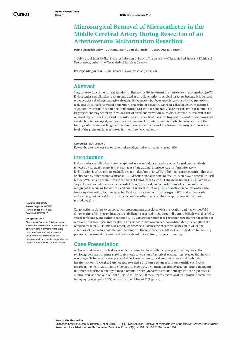
Case PresentationA 38-year-old male with a history of epilepsy presented to us with increasing seizure frequency. Hissemiology consisted of generalized tonic clonic convulsions. A physical examination revealed that he wasneurologically intact with new postictal right lower extremity weakness, which resolved during thehospitalization. T2 weighted MR imaging revealed a 24.2 mm x 16 mm x 13.3 mm roughly ovoid AVMlocated in the right sylvian fissure. Cerebral angiography demonstrated primary arterial feeders arising fromthe anterior division of the right middle cerebral artery (MCA) with venous drainage into the right middlecerebral vein and the vein of Labbe (Figure 1). Figure 2 shows a three dimensional (3D) dynamic computedtomography angiogram (CTA) reconstruction of the AVM (Figure 2).
1 2 3 3
Open Access CaseReport DOI: 10.7759/cureus.1164
How to cite this articleShazadeh Safavi P, Desai S, Branch D, et al. (April 13, 2017) Microsurgical Removal of Microcatheter in the Middle Cerebral Artery DuringResection of an Arteriovenous Malformation Resection. Cureus 9(4): e1164. DOI 10.7759/cureus.1164
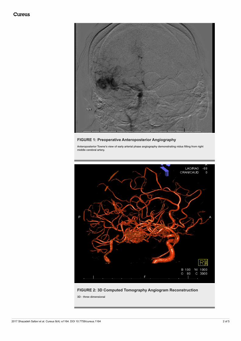
FIGURE 1: Preoperative Anteroposterior AngiographyAnteroposterior Towne's view of early arterial phase angiography demonstrating nidus filling from rightmiddle cerebral artery.
FIGURE 2: 3D Computed Tomography Angiogram Reconstruction3D - three dimensional
2017 Shazadeh Safavi et al. Cureus 9(4): e1164. DOI 10.7759/cureus.1164 2 of 5

The patient then underwent endovascular embolization using Onyx 18 (Covidien, Irvine, CA) where near-complete embolization of the AVM was achieved with minimal contrast filling. Towards the end of theprocedure and during the attempt to remove the microcatheter, its tip was noted to be adhered to the Onyxcast at the perinidal location. Unsuccessful attempts were made to withdraw the microcatheter by reducingthe slack in the system, and traction was then applied over a period of 25 minutes. Given the tortuosity ofthe vessels and tension noted on the MCA branch, the decision to transect the catheter at the groin site wasmade to prevent acute intracranial vascular injury. After transection, the distal catheter tip was noted to bepositioned along the descending thoracic aorta (Figure 3). Since the patient was scheduled for neurosurgicalresection of the AVM the next day, the catheter was removed at that time. The patient was started on theweight-based heparin drip to prevent clot formation along the retained catheter and admitted to theintensive care unit (ICU).
FIGURE 3: Posterior-Anterior Radiograph Showing Catheter in theThoracic Aorta (Red Arrows)Coaxial 4.3 French Detachable Apollo Catheter and Marksman Microcatheter in the thoracic aorta.
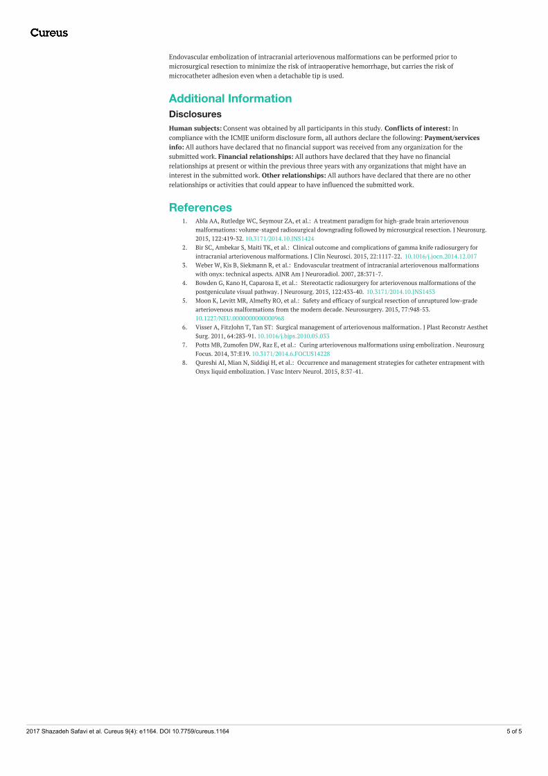
The following morning, the patient was taken to the operating room where a pterional craniotomy wasperformed. The sylvian fissure was then split medial to lateral exposing the proximal internal carotid. Thiswas followed distally until visualizing the bifurcation and M1 was readily identified. We could visualize theretained intraluminal catheter at this point. We then turned our attention to the cortical surface where avenous varix was seen. We circumferentially dissected the AVM maintaining venous outflow throughout.Hemosiderin staining around the resection bed suggested prior hemorrhage. We then encountered thetwo major feeding vessels, which were then bipolared and divided, one of which had the adherent catheter.The entire catheter was then removed (Video 1). Intraoperative angiography was not available. The entiretyof the 50 cm catheter was removed during open craniotomy (Figure 4). A follow-up at 12 months revealedthat the patient was well with no neurologic deficits.
2017 Shazadeh Safavi et al. Cureus 9(4): e1164. DOI 10.7759/cureus.1164 3 of 5
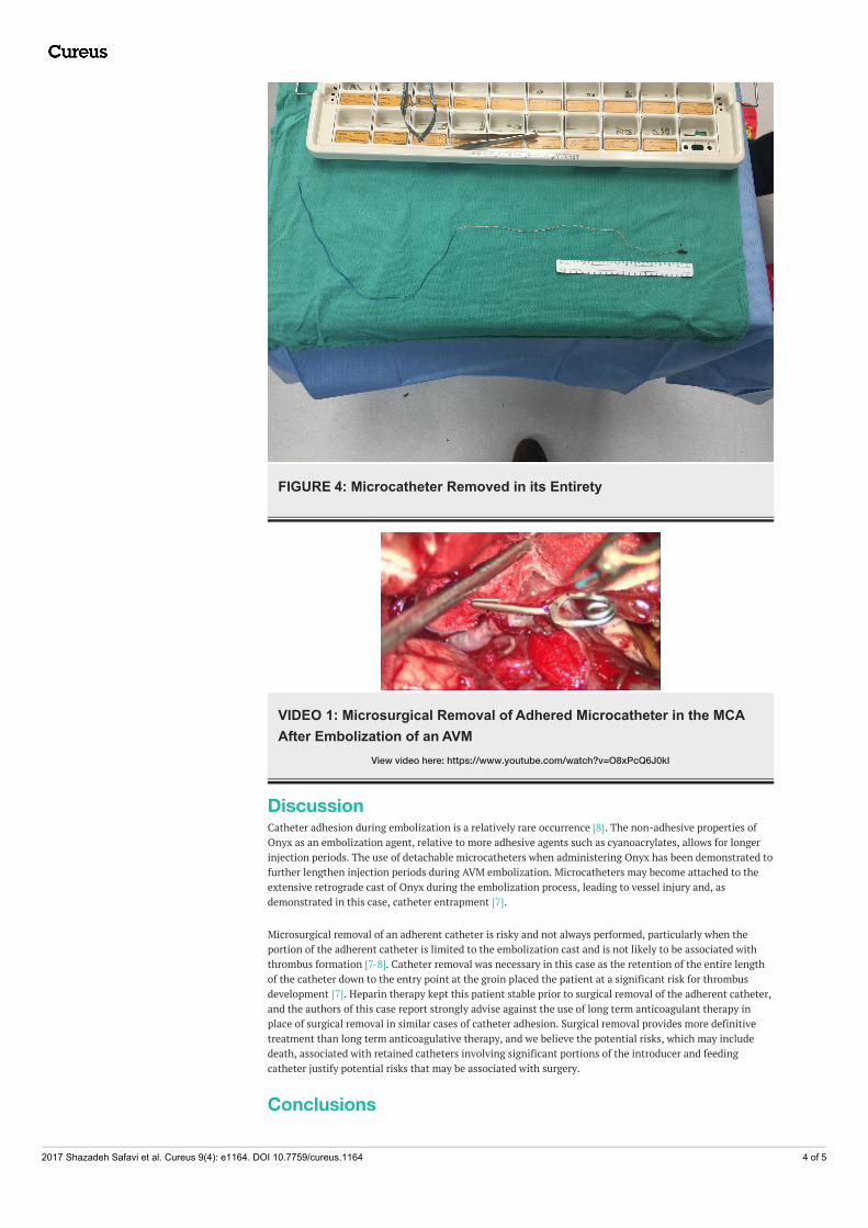
FIGURE 4: Microcatheter Removed in its Entirety
VIDEO 1: Microsurgical Removal of Adhered Microcatheter in the MCAAfter Embolization of an AVM
View video here: https://www.youtube.com/watch?v=O8xPcQ6J0kI
DiscussionCatheter adhesion during embolization is a relatively rare occurrence [8]. The non-adhesive properties ofOnyx as an embolization agent, relative to more adhesive agents such as cyanoacrylates, allows for longerinjection periods. The use of detachable microcatheters when administering Onyx has been demonstrated tofurther lengthen injection periods during AVM embolization. Microcatheters may become attached to theextensive retrograde cast of Onyx during the embolization process, leading to vessel injury and, asdemonstrated in this case, catheter entrapment [7].
Microsurgical removal of an adherent catheter is risky and not always performed, particularly when theportion of the adherent catheter is limited to the embolization cast and is not likely to be associated withthrombus formation [7-8]. Catheter removal was necessary in this case as the retention of the entire lengthof the catheter down to the entry point at the groin placed the patient at a significant risk for thrombusdevelopment [7]. Heparin therapy kept this patient stable prior to surgical removal of the adherent catheter,and the authors of this case report strongly advise against the use of long term anticoagulant therapy inplace of surgical removal in similar cases of catheter adhesion. Surgical removal provides more definitivetreatment than long term anticoagulative therapy, and we believe the potential risks, which may includedeath, associated with retained catheters involving significant portions of the introducer and feedingcatheter justify potential risks that may be associated with surgery.
Conclusions
2017 Shazadeh Safavi et al. Cureus 9(4): e1164. DOI 10.7759/cureus.1164 4 of 5
Endovascular embolization of intracranial arteriovenous malformations can be performed prior tomicrosurgical resection to minimize the risk of intraoperative hemorrhage, but carries the risk ofmicrocatheter adhesion even when a detachable tip is used.
Additional InformationDisclosuresHuman subjects: Consent was obtained by all participants in this study. Conflicts of interest: Incompliance with the ICMJE uniform disclosure form, all authors declare the following: Payment/servicesinfo: All authors have declared that no financial support was received from any organization for thesubmitted work. Financial relationships: All authors have declared that they have no financialrelationships at present or within the previous three years with any organizations that might have aninterest in the submitted work. Other relationships: All authors have declared that there are no otherrelationships or activities that could appear to have influenced the submitted work.
References1. Abla AA, Rutledge WC, Seymour ZA, et al.: A treatment paradigm for high-grade brain arteriovenous
malformations: volume-staged radiosurgical downgrading followed by microsurgical resection. J Neurosurg.2015, 122:419-32. 10.3171/2014.10.JNS1424
2. Bir SC, Ambekar S, Maiti TK, et al.: Clinical outcome and complications of gamma knife radiosurgery forintracranial arteriovenous malformations. J Clin Neurosci. 2015, 22:1117-22. 10.1016/j.jocn.2014.12.017
3. Weber W, Kis B, Siekmann R, et al.: Endovascular treatment of intracranial arteriovenous malformationswith onyx: technical aspects. AJNR Am J Neuroradiol. 2007, 28:371-7.
4. Bowden G, Kano H, Caparosa E, et al.: Stereotactic radiosurgery for arteriovenous malformations of thepostgeniculate visual pathway. J Neurosurg. 2015, 122:433-40. 10.3171/2014.10.JNS1453
5. Moon K, Levitt MR, Almefty RO, et al.: Safety and efficacy of surgical resection of unruptured low-gradearteriovenous malformations from the modern decade. Neurosurgery. 2015, 77:948-53.10.1227/NEU.0000000000000968
6. Visser A, FitzJohn T, Tan ST: Surgical management of arteriovenous malformation. J Plast Reconstr AesthetSurg. 2011, 64:283-91. 10.1016/j.bjps.2010.05.033
7. Potts MB, Zumofen DW, Raz E, et al.: Curing arteriovenous malformations using embolization . NeurosurgFocus. 2014, 37:E19. 10.3171/2014.6.FOCUS14228
8. Qureshi AI, Mian N, Siddiqi H, et al.: Occurrence and management strategies for catheter entrapment withOnyx liquid embolization. J Vasc Interv Neurol. 2015, 8:37-41.
2017 Shazadeh Safavi et al. Cureus 9(4): e1164. DOI 10.7759/cureus.1164 5 of 5


![Research Article Radiofrequency Transoral Microsurgical ...a vallecular cyst [ ], and Kumar et al. in the removal of a laryngeal cyst [ ]. In adductor spasmodic dysphonic microlaryngealRF](https://img.pdfslide.net/doc/110x75/60c722fd93fe8857a20a37ec/research-article-radiofrequency-transoral-microsurgical-a-vallecular-cyst-.jpg)