Embed Size (px)
Citation preview
Development 111, 337-356 (1991)Printed in Great Britain © The Company of Biologists Limited 1991
337
Microsurgically generated discontinuities provoke heritable changes in
cellular handedness of a ciliate, Stylonychia mytilus
XINBAI SHI1*, LI LU\ ZIJIAN QIU1, WEI HE1 and JOSEPH FRANKEL2 t1 Department of Biology, Harbin Normal University, Harbin 150080, China2Department of Biology, University of Iowa, Iowa City, IA 52242, USA
* To whom reprint requests from China should be sentt To whom reprint requests from other countries should be sent
Summary
Stylonychia mytilus is a dorsoventrally flattened ciliatewith compound ciliary structures arranged in a specificmanner on the cell surface. In mirror-image (MI)doublets of this ciliate, two nearly complete sets of ciliarystructures are arrayed side-by-side, one in a normal or'right-handed' (RH) arrangement, the other in areversed or 'left-handed' (LEI) arrangement. MI-doublets exist in two forms, one with the RH componenton the right, the LH component on the left, and feedingstructures near the center ('buccal-adjoining MI-doublet'); the other with the RH component on the left,the LH component on the right, and feeding structureson the lateral edges ('buccal-opposing Mi-doublet').
We describe an operation that can generate eithertype of Mi-doublet. This operation interchanges largeanterior and posterior regions of the cell, transposingthe original posterior region anteriorly (P—>A) and theoriginal anterior region posteriorly (A—>P), whileretaining the original anteroposterior polarity of eachregion. Two sets of new ciliary structures then areformed in mirror-image arrangement, with the set in theP—»A region oriented normally and the set in the A—>Pregion undergoing a reversal of polarity along its
anteroposterior axis. This sometimes creates end-to-endMI forms, but more commonly produces side-by-sideMi-doublets through a folding together of the P—> A andA—*P regions. This folding occurs because one lateraledge of the cell had been removed during the operation;if the left edge was removed, the complex folds to the leftand forms a buccal-adjoining Mi-doublet, whereas if theright edge was removed, the complex folds to the rightand forms a buccal-opposing Mi-doublet. Both types canreorganize and later divide true-to-type, although the'buccal-opposing' type is by far the more stable of the two.
The generation of mirror-image forms is dependent onthe prior abnormal juxtaposition of regions fromopposite ends of the cell, and involves a coordinated^specification of large-scale organization. We interpretthis response to be a consequence of intercalation ofmissing intervening positional values in the zone ofposterior-anterior abutment.
Key words: Stylonychia mytilus, mirror-image, doublets,pattern formation, positional information.
Introduction
Ciliates are distinctive among organisms not only in thecomplexity of the structural order that is expressedwithin the limits of a single cell, but also in the pervasiveasymmetry of that order. The asymmetry is apparentnot only in the internal organization of the basicbuilding block of ciliate organization, the ciliary unit,but also in the arrangement of these units over theciliate surface. One therefore can imagine reversals ofstructural handedness at each of several levels ofintracellular organization.
Detection of pattern reversals in ciliates is facilitatedby a geometry that allows amplification of pre-existingstructural order. This unique ciliate geometry, visual-ized as a 'clonal cylinder' by Tartar (1962), is one inwhich growth is longitudinal and fission transverse; thispermits any structure or arrangement that can growlongitudinally without spreading laterally to propagate
its pre-existing order across cell generations. The classicdemonstration of such propagation of pre-existingorder is the perpetuation of inversions of one or morelongitudinal rows of closely spaced ciliary units (Beis-son and Sonneborn, 1965; Ng and Frankel, 1977). Inthis case, however, the units have undergone a 180°rotational permutation without any change in theirintrinsic coordinates. The failure to observe individualciliary units with reversed asymmetry even though sucha reversal could be perpetuated suggests that reversal atthis level is difficult to achieve.
Reversals of asymmetry of global arrangements ofciliary units have, however, been observed repeatedlyin ciliates, beginning with the discovery by Faure"-Fremiet (1945) of a self-propagating back-to-backmirror-image doublet of the hypotrich Urostyla tricho-gaster. Although one example of such a phenotype,janus in Tetrahymena thermophila (Jerka-Dziadosz andFrankel, 1979; Frankel et al. 1984), is dependent on
338 X. Shi and others
mutant-gene expression (Frankel and Jenkins, 1979;Frankel et al. 1987), most or all of the other knowncases probably do not involve genetic changes. Anoutstanding example is the vegetatively inheritedmirror-image doublet configuration of the hypotrichciliate Stylonychia mytilus, generated from completelynormal cells by regulation following a microsurgicaloperation (Tchang et al. 1964; Tchang and Pang, 1965).
How can a form with normal asymmetry switch to itsmirror-image? In principle, all that is required is thereversal of a single morphological axis. It was proposedearher that nongenic pattern reversals in Tetrahymenacome about through reverse intercalation along thecircumferential (transverse) axis, stimulated by anabnormal distribution of positional values in Siamese-twin doublet cells (Nelsen and Frankel, 1986; Frankeland Nelsen, 1986; Frankel, 1989 chapter 11). However,this pathway of regulation was inferred from obser-vations on populations of cells. What is required for amore direct demonstration is a ciliate that is largeenough to be readily operated upon and that has amorphological pattern sufficiently complex so thatintermediate stages in pattern-reversal can be followedin individual cells examined over a short time period.These requirements are fulfilled by hypotrich ciliates,notably Stylonychia mytilus.
We describe here the morphogenetic regulation ofmicrosurgically rearranged Stylonychia cells, emphasiz-ing the outcome of one specific type of operation thatfrequently generated mirror-image (MI) doublets. Thisoutcome has shown that a clonally heritable change oflarge-scale intracellular handedness can be obtainedfrom rearranged normal components. It also has shownthat this change in handedness is a consequence of areversal of the anteroposterior axis that is dependent ona prior abnormal juxtaposition of anterior and posteriorcellular positions.
Materials and methods
Four stocks (1,6,7, and 10) of Stylonychia mytilus collected in1980 from ponds located near a suburb of Harbin were used inthis work. The photographs shown in this paper were all ofcells of stock 7. Cells were cultured at room temperature(18-20°C) with Pringsheim's solution and wheat infusion,using the flagellate Chilomonas paramecium as food.
Operations were performed with two fine steel micro-needles on cells that were immobilized in a drop of methylcellulose on a glass slide. The two principal types of operationare illustrated in Figs 1 and 2, using schematic diagrams of theventral surface in which only the membranelle band (MB),left-marginal cirri (LM), and right-marginal cirri (RM) areillustrated (see Results for a more detailed description of thiscell). These operations were performed on nondividing cellsof unknown cell-cycle stage.
In one variant of the operation, the cytoplasm of the cell'sleft border, including most or all of the left-marginal cirri, wasremoved (Fig. 1A, cut 1). Immediately afterwards, theremainder of the cell was cut transversely into two equal parts(Fig. 1A, cut 2), and the posterior half was folded up againstthe anterior half along the wounded left edge (Fig. IB). Thenthe front edge of this complex was removed (Fig. IB, cut 3),after which the original posterior end was folded over onto the
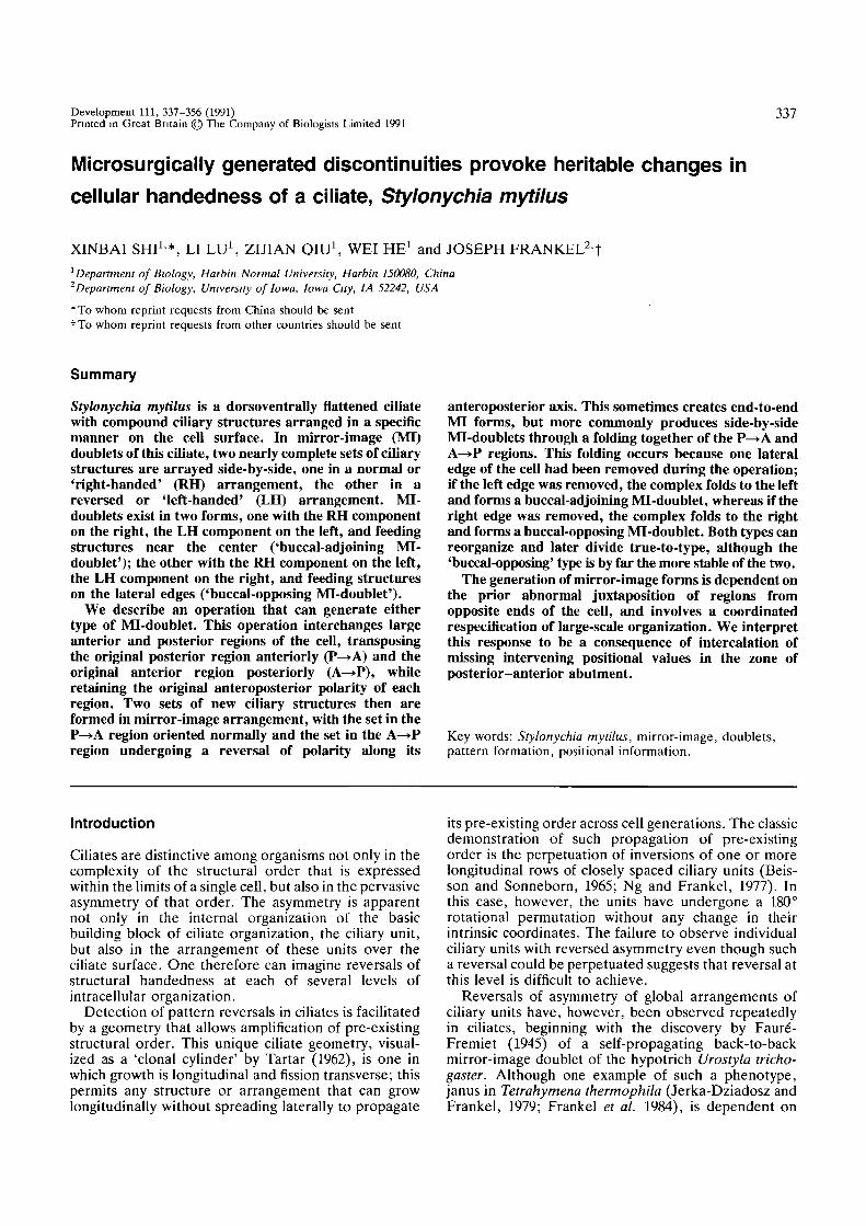
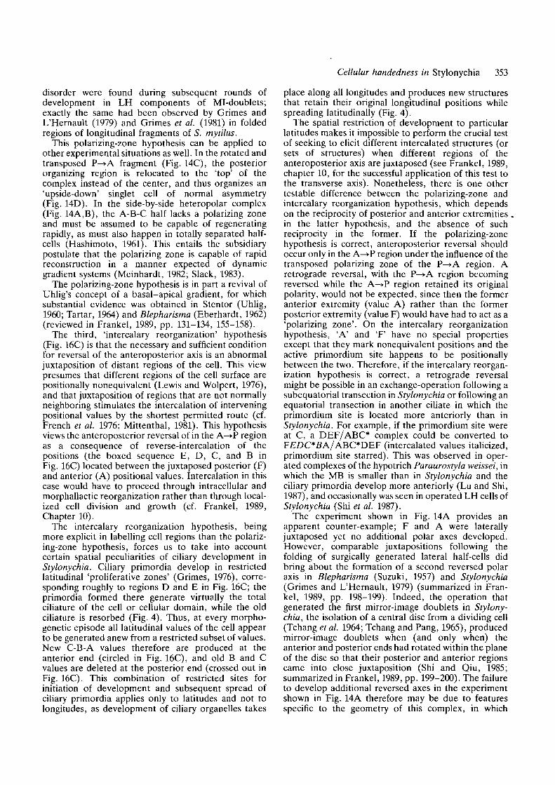
Fig. 1. Microsurgical generation of a 'minus-left' (—L)tandem complex. The cells are viewed from the ventralsurface, with the cell's right (R) at the observer's left, thecell's left (L) at the observer's right. The numbers indicatethe sequence of cuts, and the open-headed arrows showthe direction of folding of parts by the operator. (A) Thefirst cut (1) removes the left edge and a second cut (2)separates the posterior (P) and anterior (A) halves. (B)The P half then is folded 180° counterclockwise to abutside-by-side against the A half. Thereafter, the anteriorand posterior extremities are removed by cut 3, and (C) asecond 180° fold places the P region anterior to the Aregion. After wound-healing, (D) the border (dashed line)between the anteriorly transposed posterior (P—>A) regionand posteriorly transposed anterior (A—>P) region nolonger is visible. The posterior portion (pMB) of theoriginal membranelle-band (MB) ends up at the anteriorend of the complex, and a more anterior portion of themembranelle band (aMB) usually is present near theposterior end of the complex [sometimes it is missing,when cut 2 is more anterior than shown in A]. The right-marginal cirral row (RM) remains at the intact right edge,whereas the left-marginal cirral row (LM) was removed bycut 1, leaving the wounded left edge bare of ciliature.
anterior end. The end result was a tandem complex in whichmost of the former posterior half of the cell was in front of theformer anterior half, with an intact cell border on its rightmargin and wounded edges on the anterior, posterior and leftmargins (Fig. 1C). We call this a minus-left (—L) complex.
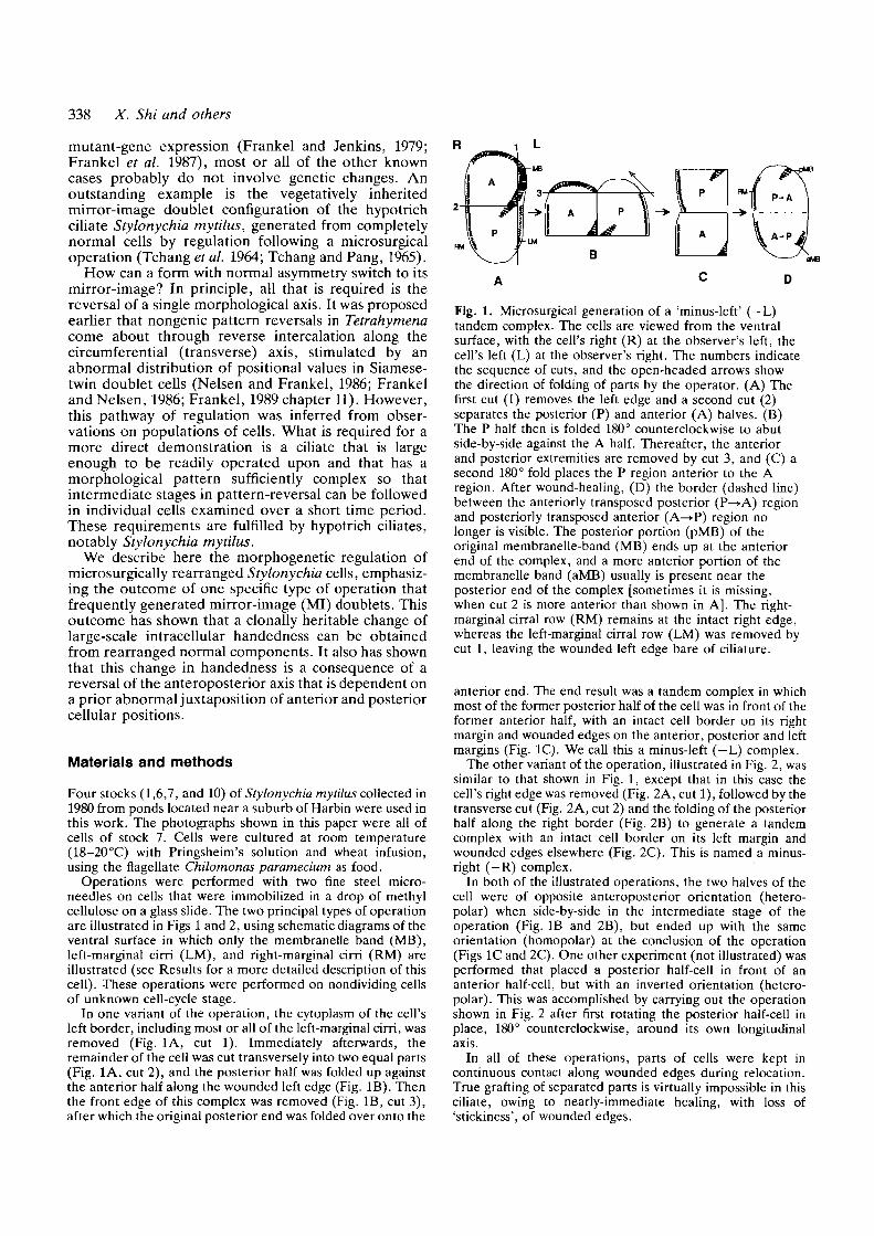
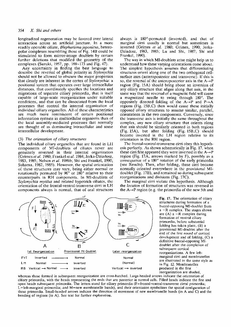
The other variant of the operation, illustrated in Fig. 2, wassimilar to that shown in Fig. 1, except that in this case thecell's right edge was removed (Fig. 2A, cut 1), followed by thetransverse cut (Fig. 2A, cut 2) and the folding of the posteriorhalf along the right border (Fig. 2B) to generate a tandemcomplex with an intact cell border on its left margin andwounded edges elsewhere (Fig. 2C). This is named a minus-right (—R) complex.
In both of the illustrated operations, the two halves of thecell were of opposite anteroposterior orientation (hetero-polar) when side-by-side in the intermediate stage of theoperation (Fig. IB and 2B), but ended up with the sameorientation (homopolar) at the conclusion of the operation(Figs 1C and 2C). One other experiment (not illustrated) wasperformed that placed a posterior half-cell in front of ananterior half-cell, but with an inverted orientation (hetero-polar). This was accomplished by carrying out the operationshown in Fig. 2 after first rotating the posterior half-cell inplace, 180° counterclockwise, around its own longitudinalaxis.
In all of these operations, parts of cells were kept incontinuous contact along wounded edges during relocation.True grafting of separated parts is virtually impossible in thisciliate, owing to nearly-immediate healing, with loss of'stickiness', of wounded edges.
Cellular handedness in Stylonychia 339
MB
Fig. 2. Microsurgical generation of a 'minus-right' (—R)tandem complex. The operation is similar to that shown inFig. 1, except that (A) cut 1 removes the right edge, (B,C)the folding of the P region is in a clockwise direction bothtimes, and (D) the left edge remains intact, with the LMcirral row and a large aMB fragment remaining, whereasthe wounded right edge lacks ciliary structures. Allabbreviations are the same as in Fig. 1.
The operated cells were washed thoroughly and allowed toregenerate in culture medium. Soon after the operation, thecells became ellipsoidal (Figs ID and 2D), and initiatedregenerative development. These cells were fixed at varioustimes after the operation for staining using a modifiedprotargol technique, described previously (Shi and Frankel,1990).
Results
(A) Normal cortical anatomy and development(1) Cortical anatomy
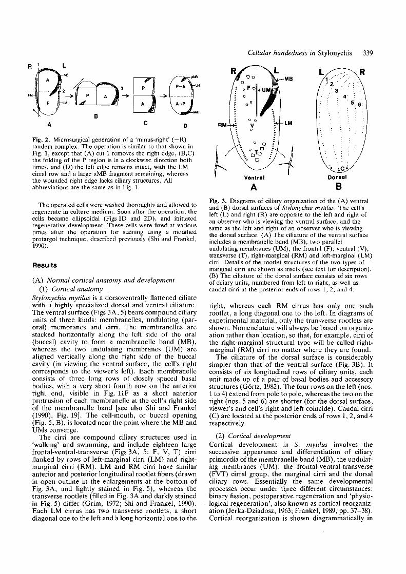
Stylonychia mytilus is a dorsoventrally flattened ciliatewith a highly specialized dorsal and ventral ciliature.The ventral surface (Figs 3A, 5) bears compound ciliaryunits of three kinds: membranelles, undulating (par-oral) membranes and cirri. The membranelles arestacked horizontally along the left side of the oral(buccal) cavity to form a membranelle band (MB),whereas the two undulating membranes (UM) arealigned vertically along the right side of the buccalcavity (in viewing the ventral surface, the cell's rightcorresponds to the viewer's left). Each membranelleconsists of three long rows of closely spaced basalbodies, with a very short fourth row on the anteriorright end, visible in Fig. 11F as a short anteriorprotrusion of each membranelle at the cell's right sideof the membranelle band [see also Shi and Frankel(1990), Fig. 19]. The cell-mouth, or buccal opening(Fig. 5, B), is located near the point where the MB andUMs converge.
The cirri are compound ciliary structures used in'walking' and swimming, and include eighteen largefrontal-ventral-transverse (Figs 3A, 5: F, V, T) cirriflanked by rows of left-marginal cirri (LM) and right-marginal cirri (RM). LM and RM cirri have similaranterior and posterior longitudinal rootlet fibers (drawnin open outline in the enlargements at the bottom ofFig. 3A, and lightly stained in Fig. 5), whereas thetransverse rootlets (filled in Fig. 3A and darkly stainedin Fig. 5) differ (Grim, 1972; Shi and Frankel, 1990).Each LM cirrus has two transverse rootlets, a shortdiagonal one to the left and a long horizontal one to the
Fig. 3. Diagrams of ciliary organization of the (A) ventraland (B) dorsal surfaces of Stylonychia mytilus. The cell'sleft (L) and right (R) are opposite to the left and right ofan observer who is viewing the ventral surface, and thesame as the left and right of an observer who is viewingthe dorsal surface. (A) The ciliature of the ventral surfaceincludes a membranelle band (MB), two parallelundulating membranes (UM), the frontal (F), ventral (V),transverse (T), right-marginal (RM) and left-marginal (LM)cirri. Details of the rootlet structures of the two types ofmarginal cirri are shown as insets (see text for description).(B) The ciliature of the dorsal surface consists of six rowsof ciliary units, numbered from left to right, as well ascaudal cirri at the posterior ends of rows 1,2, and 4.
right, whereas each RM cirrus has only one suchrootlet, a long diagonal one to the left. In diagrams ofexperimental material, only the transverse rootlets areshown. Nomenclature will always be based on organiz-ation rather than location, so that, for example, cirri ofthe right-marginal structural type will be called right-marginal (RM) cirri no matter where they are found.
The ciliature of the dorsal surface is considerablysimpler than that of the ventral surface (Fig. 3B). Itconsists of six longitudinal rows of ciliary units, eachunit made up of a pair of basal bodies and accessorystructures (Gortz, 1982). The four rows on the left (nos.1 to 4) extend from pole to pole, whereas the two on theright (nos. 5 and 6) are shorter (for the dorsal surface,viewer's and cell's right and left coincide). Caudal cirri(C) are located at the posterior ends of rows 1, 2, and 4respectively.
(2) Cortical developmentCortical development in S. mytilus involves thesuccessive appearance and differentiation of ciliaryprimordia of the membranelle band (MB), the undulat-ing membranes (UM), the frontal-ventral-transverse(FVT) cirral group, the marginal cirri and the dorsalciliary rows. Essentially the same developmentalprocesses occur under three different circumstances:binary fission, postoperative regeneration and 'physio-logical regeneration', also known as cortical reorganiz-ation (Jerka-Dziadosz, 1963; Frankel, 1989, pp. 37-38).Cortical reorganization is shown diagrammatically in
340 X. Shi and others
RM
MBRMP
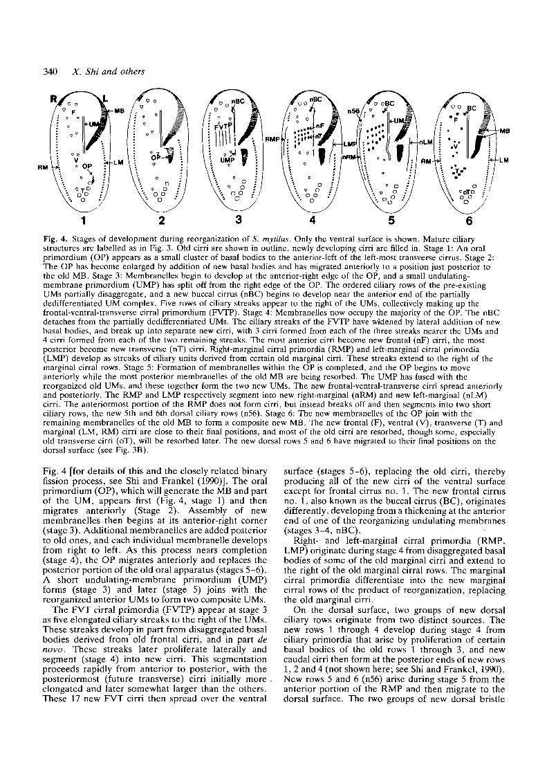
Fig. 4. Stages of development during reorganization of S. mytilus. Only the ventral surface is shown. Mature ciliarystructures are labelled as in Fig. 3. Old cirri are shown in outline, newly developing cirri are filled in. Stage 1: An oralprimordium (OP) appears as a small cluster of basal bodies to the anterior-left of the left-most transverse cirrus. Stage 2:The OP has become enlarged by addition of new basal bodies and has migrated anteriorly to a position just posterior tothe old MB. Stage 3: Membranelles begin to develop at the anterior-right edge of the OP, and a small undulating-membrane primordium (UMP) has split off from the right edge of the OP. The ordered ciliary rows of the pre-existingUMs partially disaggregate, and a new buccal cirrus (nBC) begins to develop near the anterior end of the partiallydedifferentiated UM complex. Five rows of ciliary streaks appear to the right of the UMs, collectively making up thefrontal-ventral-transverse cirral primordium (FVTP). Stage 4: Membranelles now occupy the majority of the OP. The nBCdetaches from the partially dedifferentiated UMs. The ciliary streaks of the FVTP have widened by lateral addition of newbasal bodies, and break up into separate new cirri, with 3 cirri formed from each of the three streaks nearer the UMs and4 cirri formed from each of the two remaining streaks. The most anterior cirri become new frontal (nF) cirri, the mostposterior become new transverse (nT) cirri. Right-marginal cirral primordia (RMP) and left-marginal cirral primordia(LMP) develop as streaks of ciliary units derived from certain old marginal cirri. These streaks extend to the right of themarginal cirral rows. Stage 5: Formation of membranelles within the OP is completed, and the OP begins to moveanteriorly while the most posterior membranelles of the old MB are being resorbed. The UMP has fused with thereorganized old UMs, and these together form the two new UMs. The new frontal-ventral-transverse cirri spread anteriorlyand posteriorly. The RMP and LMP respectively segment into new right-marginal (nRM) and new left-marginal (nLM)cirri. The anteriormost portion of the RMP does not form cirri, but instead breaks off and then segments into two shortciliary rows, the new 5th and 6th dorsal ciliary rows (n56). Stage 6: The new membranelles of the OP join with theremaining membranelles of the old MB to form a composite new MB. The new frontal (F), ventral (V), transverse (T) andmarginal (LM, RM) cirri are close to their final positions, and most of the old cirri are resorbed, though some, especiallyold transverse cirri (oT), will be resorbed later. The new dorsal rows 5 and 6 have migrated to their final positions on thedorsal surface (see Fig. 3B).
Fig. 4 [for details of this and the closely related binaryfission process, see Shi and Frankel (1990)]. The oralprimordium (OP), which will generate the MB and partof the UM, appears first (Fig. 4, stage 1) and thenmigrates anteriorly (Stage 2). Assembly of newmembranelles then begins at its anterior-right corner(stage 3). Additional membranelles are added posteriorto old ones, and each individual membranelle developsfrom right to left. As this process nears completion(stage 4), the OP migrates anteriorly and replaces theposterior portion of the old oral apparatus (stages 5-6).A short undulating-membrane primordium (UMP)forms (stage 3) and later (stage 5) joins with thereorganized anterior UMs to form two composite UMs.
The FVT cirral primordia (FVTP) appear at stage 3as five elongated ciliary streaks to the right of the UMs.These streaks develop in part from disaggregated basalbodies derived from old frontal cirri, and in part denovo. These streaks later proliferate laterally andsegment (stage 4) into new cirri. This segmentationproceeds rapidly from anterior to posterior, with theposteriormost (future transverse) cirri initially more .elongated and later somewhat larger than the others.These 17 new FVT cirri then spread over the ventral
surface (stages 5-6), replacing the old cirri, therebyproducing all of the new cirri of the ventral surfaceexcept for frontal cirrus no. 1. The new frontal cirrusno. 1, also known as the buccal cirrus (BC), originatesdifferently, developing from a thickening at the anteriorend of one of the reorganizing undulating membranes(stages 3-4, nBC).
Right- and left-marginal cirral primordia (RMP,LMP) originate during stage 4 from disaggregated basalbodies of some of the old marginal cirri and extend tothe right of the old marginal cirral rows. The marginalcirral primordia differentiate into the new marginalcirral rows of the product of reorganization, replacingthe old marginal cirri.
On the dorsal surface, two groups of new dorsalciliary rows originate from two distinct sources. Thenew rows 1 through 4 develop during stage 4 fromciliary primordia that arise by proliferation of certainbasal bodies of the old rows 1 through 3, and newcaudal cirri then form at the posterior ends of new rows1, 2 and 4 (not shown here; see Shi and Frankel, 1990).New rows 5 and 6 (n56) arise during stage 5 from theanterior portion of the RMP and then migrate to thedorsal surface. The two groups of new dorsal bristle
Cellular handedness in Stylonychia 341
rows and caudal cirri replace their old counterpartsduring reorganization.
The formation of ciliary primordia typically is highlycoordinated; if one ciliary primordium forms, so domost or all of the others, in a stereotyped temporalsequence. The organism, however, is flexible in thenumber of sets of ciliary primordia produced and tosome degree in their location. During division, forexample, one large OP generates the oral apparatus ofthe posterior division product (the anterior OA remainsintact and is passed on to the anterior division product),whereas two tandem sets of all other ciliary primordiagenerate the remainder of the ciliature of the twodivision products (Grimes, 1972; Wirnsberger et al.1986; Shi and Frankel, 1990). The cortical developmentin operated cells (section C) manifests aspects of bothreorganization and division plus certain unique fea-tures.
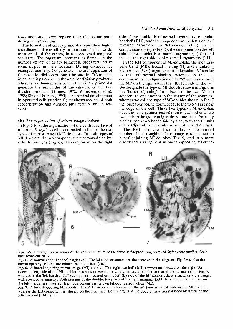
(B) The organization of mirror-image doubletsIn Figs 5 to 7, the organization of the ventral surface ofa normal S. mytilus cell is contrasted to that of the twotypes of mirror-image (MI) doublets. In both types ofMi-doublets, the two components are arranged side-by-side. In one type (Fig. 6), the component on the right
side of the doublet is of normal asymmetry, or 'right-handed' (RH), and the component on the left side is ofreversed asymmetry, or 'left-handed' (LH). In thecomplementary type (Fig. 7), the component on the leftside of the doublet is of normal asymmetry (RH) andthat on the right side is of reversed asymmetry (LH).
In the RH component of Mi-doublets, the membra-nelle band (MB), buccal opening (B) and undulatingmembranes (UM) together form a lopsided 'V similarto that of normal singlets, whereas in the LHcomponent the configuration of the 'V is reversed, withthe MB on the right rather than the left side of the "V".We designate the type of Mi-doublet shown in Fig. 6 asthe 'buccal-adjoining' form because the two Vs areadjacent to one another in the center of the complex,whereas we call the type of Mi-doublet shown in Fig. 7the 'buccal-opposing' form, because the two Vs are nearthe edge of the cell. These two types of Mi-doubletshave the same geometrical relation to each other as thetwo mirror-image configurations one can form byplacing one's two hands side-by-side, with the thumbseither adjacent in the center or opposite at the edges.
The FVT cirri are close to double the normalnumber, in a roughly mirror-image arrangement inbuccal-adjoining Mi-doublets (Fig. 6) and in a moredisordered arrangement in buccal-opposing Ml-doub-
Figs 5-7. Protargol preparations of the ventral ciliature of the three self-reproducing forms of Stylonychia mytilus. Scalebars represent 50 ^M.Fig. 5. A normal (right-handed) singlet cell. The labelled structures are the same as in the diagram (Fig. 3A), plus thebuccal opening (B) and the bilobed macronucleus (Ma).Fig. 6. A buccal-adjoining mirror-image (MI) doublet. The 'right-handed' (RH) component, located on the right (R)(viewer's left) side of the Mi-doublet, has an arrangement of ciliary structures similar to that of the normal cell in Fig. 5,whereas in the 'left-handed' (LH) component, located on the left (L) side of the Mi-doublet, these structures are arrangedwith reversed asymmetry. Both margins of the doublet have cirri of the right-marginal (RM) type, although the ones onthe left margin are inverted. Each component has its own bilobed macronucleus (Ma).Fig. 7. A buccal-opposing Mi-doublet. The RH component is located on the left (viewer's right) side of the Mi-doublet,whereas the LH component is situated on the right side. Both margins of the doublet have normally-oriented cirri of theleft-marginal (LM) type.
342 X. Shi and others
lets (Fig. 7). Cirri of the right-marginal type are presentat both margins of the buccal-adjoining Mi-doublets(Fig. 6 - in this case inverted on the LH side), whereascirri of the left-marginal type are present at bothmargins of buccal-opposing Mi-doublets (Fig. 7). Dor-sally, 8 to 12 rows of cilia are present in a near mirror-image arrangement in both types of Mi-doublets (notshown; see Shi and Frankel, 1990 for the buccal-opposing type).
The LH and RH components of Mi-doublets aremirror-images only with regard to the arrangement ofciliary structures. The internal organization of eachindividual ciliary unit is always the same in bothcomponents, although certain structures, notably mem-branelles but sometimes also marginal cirri, areinverted in the LH component (cf. Grimes et al. 1980;Jerka-Dziadosz, 1983, 1985; Shi and Frankel, 1990).
Whereas the buccal-opposing Mi-doublets (Fig. 7)are stable and self-propagating, and have been studiedfor nearly 30 years (references in Shi and Frankel,1990), the buccal-adjoining Mi-doublets (Fig. 6) can bepropagated for several fissions at most, and are new tothis study, except for the comparable but incomplete(and nondividing) buccal-adjoining Mi-doublets de-scribed by Grimes and L'Hernault (1979).
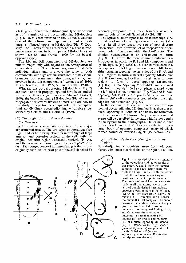
(C) The origin of mirror-image doublets(1) Overview
Fig. 8 provides a schematic overview of the majorexperimental results. The two types of operations (seeFigs 1 and 2) both bring about an interchange of largeanterior and posterior regions of the cell, with theoriginal posterior region displaced anteriorly (P—>A),and the original anterior region displaced posteriorly(A—>P); a consequence of this interchange is that a zoneoriginally near the posterior pole of the cell (labelled F)
becomes juxtaposed to a zone formerly near theanterior pole of the cell (labelled A) (Fig. 8B).
The typical cellular response to this interchange is theformation of one of three types of mirror-image (MI)forms. In all three types, two sets of new ciliaturedifferentiate, with a reversal of anteroposterior orien-tation (polarity) in the set within the A—»P region. Thesimplest consequence is an end-to-end MI form(Fig. 8F). However, the more common outcome is aMi-doublet, in which the RH and LH components endup side-by side (Fig. 8E,G). This can be visualized as aconsequence of folding of an end-to-end MI form,either bringing together the left sides of the P-^A andA—»P regions to form a buccal-adjoining Mi-doublet(Fig. 8E) or bringing together the right sides of theseregions to form a buccal-opposing Mi-doublet(Fig. 8G). Buccal-adjoining Mi-doublets are producedonly from 'minus-left' (—L) complexes created whenthe left edge has been removed (Fig. 8C), and buccal-opposing Mi-doublets are produced only from the'minus-right' (—R) complexes created when the rightedge has been removed (Fig. 8D).
In the sections to follow, we describe the develop-ment of buccal-adjoining Mi-doublets first, then that ofbuccal-opposing Mi-doublets, and finally the formationof the end-to-end MI forms. Only the most essentialevents will be described in the text, with further detailsin the legends to the photographs. The examples thatinvolve development of MI forms are selected from alarger body of operated complexes, many of whichformed normal or reversed singlets (see section C5).
(2) Formation of buccal-adjoining mirror-imagedoublets
Buccal-adjoining Mi-doublets arose from - L com-plexes, with intact marginal cirri at the right but not the
Fig. 8. A simplified schematic summaryof the operations and major results ofthis study. A and B show the featurescommon to the two major operativeprotocols (Figs 1 and 2), with the lettersinside the cell regions marking cellpositions in an anteroposterior order.The horizontal solid lines indicate cutsmade in all operations, whereas thevertical double-dashed lines indicatealternative cuts, removing the left edge(L) or the right edge (R). C shows theminus-L (—L) complex, and D showsthe minus-R (—R) complex. The curvedarrows at the ends of vertical cut edgesgive the direction of the ensuingunilateral shortening and folding. E, F,and G indicate the alternativeoutcomes, a buccal-adjoining MI-doublet (E), an end-to-end Mi-form(F), or a buccal-opposing MI- doublet(G). RH stands for the 'right-handed'(normal asymmetry) component, LHfor the 'left-handed' (reversedasymmetry) component. For furtherdescription, see the text.
Cellular handedness in Stylonychia 343
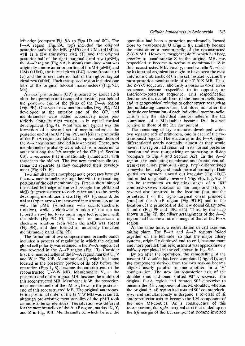
left edge (compare Fig. 9A to Figs ID and 8C). TheP—»A region (Fig. 9A, top) included the originalposterior ends of the MB (pMB) and UMs (pUM) aswell as a few transverse cirri (T) and the originalposterior half of the right-marginal cirral row (pRM);the A-»P region (Fig. 9A, bottom) contained what wasoriginally a more anterior portion of the MB (aMB) andUMs (aUM), the buccal cirrus (BC), some frontal cirri(F) and the former anterior half of the right-marginalcirral row (aRM). Each transposed region included onelobe of the original bilobed macronucleus (Fig. 9D,Ma).
An oral primordium (OP) appeared by about 1.5 hafter the operation and occupied a position just behindthe posterior end of the pMB of the P—»A region(Fig. 9B). One set of new membranelles (Fig. 9C, nM)developed at the anterior end of the OP, andmembranelles were added successively more pos-teriorly along its right margin, as in typical corticaldevelopment (Fig. 4). What was not typical was theformation of a second set of membranelles at theposterior end of the OP (Fig. 9C, nm) [ciliary primordiaof the P—>A region are labelled in upper case; those ofthe A—>P region are labelled in lower-case]. There, newmembranelles probably were added from posterior toanterior along the left margin of the OP (cf. SectionC3), a sequence that is rotationaUy symmetrical withrespect to the nM set. The two new membranelle setsremained separate as they completed their develop-ment (Fig. 9D-F).
Two simultaneous morphogenetic processes broughtthe new membranelle sets together with the remainingportions of the old membranelles. First, a shortening ofthe naked left edge of the cell brought the pMB andaMB fragments closer to each other and to the newlydeveloping membranelle sets (Fig. 9D-F); second, thenM set (open arrow) maneuvered into a seamless unionwith the pMB (sometimes with counterclockwiserotation), while a clockwise rotation of the nm set(closed arrow) led to its more imperfect juncture withthe aMB (Fig. 9D-F). The nm set underwent aclockwise rotation even when the aMB was absent(Fig. 9E), and then formed an anteriorly truncatedmembranelle band (Fig. 91).
The formation of two composite membranelle bandsincluded a process of regulation in which the originalglobal cell polarity was retained in the P—>A region, butwas reversed in the A—»P region (Fig. 10). Considerfirst the membranelles of the P—»A region marked U, Vand W in Fig. 10B. Membranelle U, which had beenlocated in the posterior portion of its MB before theoperation (Figs 1, 8), became the anterior end of thereconstructed U-V-W MB. Membranelle V, at theposterior end of the original MB, became the middle ofthis reconstructed MB. Membranelle W, the posterior-most membranelle of the nM set, became the posteriorend of this reconstructed MB. The original anteropos-terior positional order of the U-V-W MB was retained,although pre-existing membranelles of the pMB tookon more anterior identities. The situation was differentfor the membranelles of the A—>P region, marked X, Y,and Z in Fig. 10B. Membranelle Z, which before the
operation had been a posterior membranelle locatedclose to membranelle U (Figs 1,8), similarly becamethe most anterior membranelle of the reconstructedZ-Y-X MB. However, membranelle Y, which had beenanterior to membranelle Z in the original MB, wasrespecified to become posterior to membranelle Z inthis reconstructed MB. Finally, membranelle X, whichby its internal organization ought to have been the mostanterior membranelle of the nm set, instead became themost posterior membranelle of the Z-Y-X MB. Thus,the Z-Y-X sequence, inherently a posterior-to-anteriorsequence, became respecified to its opposite, ananterior-to-posterior sequence. This respecificationdetermines the overall form of the membranelle bandand its geographical relation to other structures such asthe undulating membranes, but does not alter theintrinsic conformation of each individual membranelle.This is why the individual membranelles of the LHcomponent of a Mi-doublet became 180° invertedrelative to those of the RH component.
The remaining ciliary structures developed withintwo separate sets of primordia, one in each of the twotransposed regions. The structures in the P ^ A regiondifferentiated nearly normally, almost as they wouldhave if the region had remained in its normal posteriorlocation and were reorganizing or preparing to divide(compare to Fig. 4 and Section A2). In the A-»Pregion, the undulating-membrane and frontal-ventral-transverse ciliary primordia (ump, fvtp) differentiatedsomewhat belatedly and much more abnormally. Theirspatial arrangement started out irregular (Fig. 9D,E)and ended up globally reversed (Fig. 9F). Fig. 9D-Fcan be interpreted as depicting stages of a 180°counterclockwise rotation of the ump and fvtp. Areversal also occurred in the location (but not theorientation) of the right-marginal cirral primordia(rmp) of the A—»P region (Fig. 9D,E) and in thelocation of the primordia of the new dorsal ciliary rows5 and 6 (Figs 9F and 13B: n56). Thus, by the stageshown in Fig. 9F, the ciliary arrangement of the A—»Pregion had become a mirror-image of that of the P—»Aregion.
At the same time, a reorientation of cell axes wastaking place. The P—»A and A—»P regions foldedtogether on the left side, so that the major ciliarysystems, originally deployed end-to-end, became moreand more parallel; this readjustment was approximatelyhalfway completed in the cell shown in Fig. 9F.
By 6h after the operation, the remodelling of thenascent Mi-doublet has been completed (Fig. 9G), andthe components derived from the two regions becamealigned nearly parallel to one another, in a 'Vconfiguration. The new anteroposterior axis of thedoublet thus had been shifted 90° clockwise. Theoriginal P—>A region had rotated 90° clockwise tobecome the RH component of the Ml-doublet, whereasthe original A—>P region had rotated 90° counterclock-wise and simultaneously undergone a reversal of itsanteroposterior axis to become the LH component ofthe new Mi-doublet. As a consequence of thisreorientation, the rig/zMnarginal cirri that ended up onthe left margin of the LH component became inverted
344 X. Shi and others
pUM
pMB
aMB
V
B
Ant
Cellular handedness in Stylonychia 345
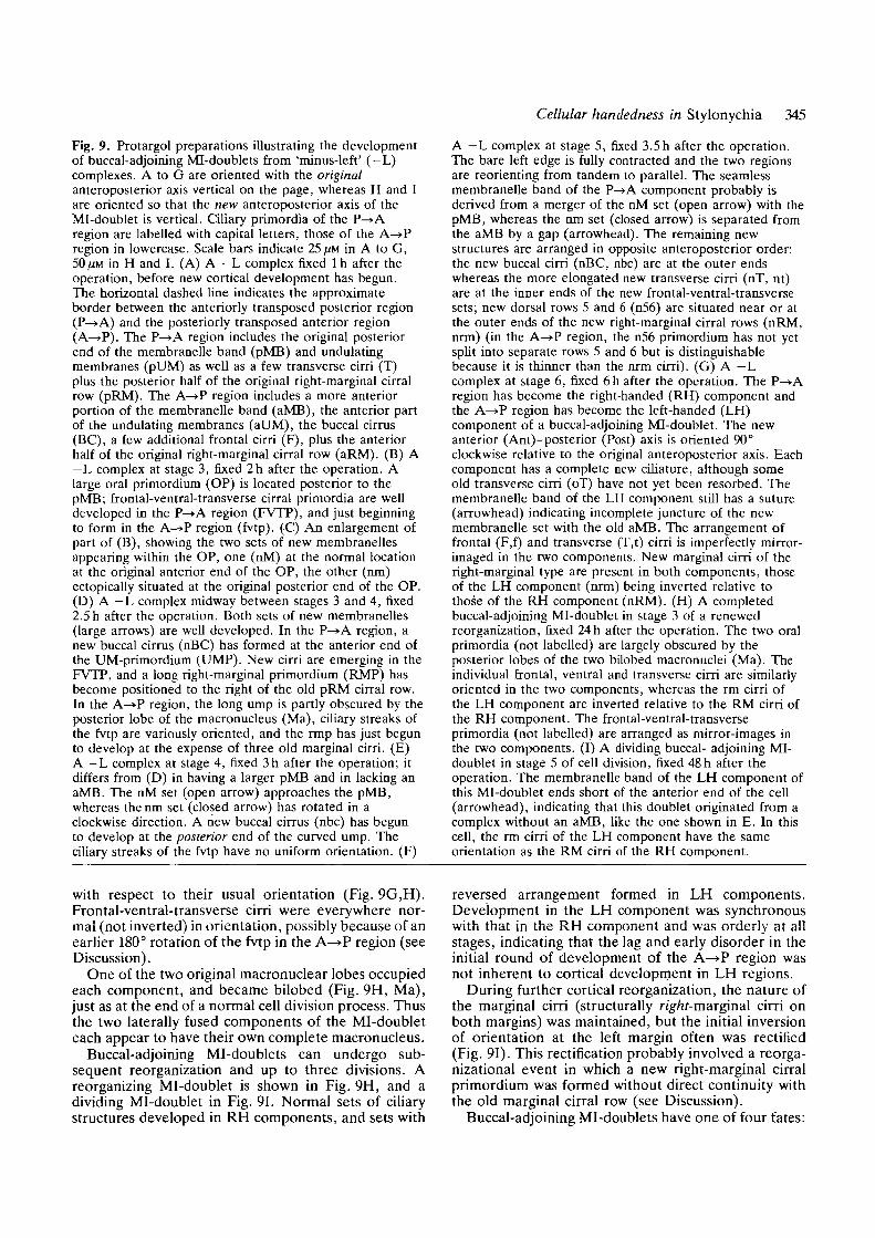
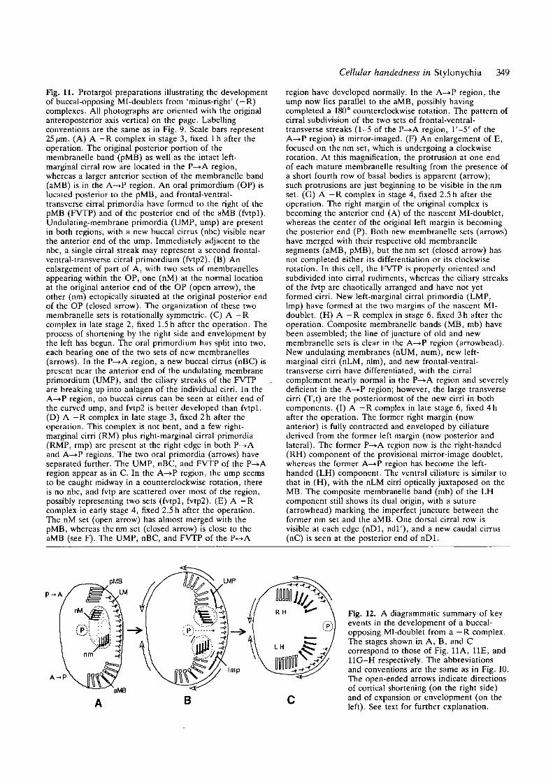
Fig. 9. Protargol preparations illustrating the developmentof buccal-adjoining Mi-doublets from 'minus-left' (—L)complexes. A to G are oriented with the originalanteroposterior axis vertical on the page, whereas H and Iare oriented so that the new anteroposterior axis of theMi-doublet is vertical. Ciliary primordia of the P—>Aregion are labelled with capital letters, those of the A—»Pregion in lowercase. Scale bars indicate 25 ^M in A to G,50^M in H and I. (A) A — L complex fixed 1 h after theoperation, before new cortical development has begun.The horizontal dashed line indicates the approximateborder between the anteriorly transposed posterior region(P—>A) and the posteriorly transposed anterior region(A—>P). The P^>A region includes the original posteriorend of the membranelle band (pMB) and undulatingmembranes (pUM) as well as a few transverse cirri (T)plus the posterior hah0 of the original right-marginal cirralrow (pRM). The A—>P region includes a more anteriorportion of the membranelle band (aMB), the anterior partof the undulating membranes (aUM), the buccal cirrus(BC), a few additional frontal cirri (F), plus the anteriorhalf of the original right-marginal cirral row (aRM). (B) A—L complex at stage 3, fixed 2h after the operation. Alarge oral primordium (OP) is located posterior to thepMB; frontal-ventral-transverse cirral primordia are welldeveloped in the P—>A region (FVTP), and just beginningto form in the A—>P region (fvtp). (C) An enlargement ofpart of (B), showing the two sets of new membranellesappearing within the OP, one (nM) at the normal locationat the original anterior end of the OP, the other (nm)ectopically situated at the original posterior end of the OP.(D) A —L complex midway between stages 3 and 4, fixed2.5 h after the operation. Both sets of new membranelles(large arrows) are well developed. In the P—>A region, anew buccal cirrus (nBC) has formed at the anterior end ofthe UM-primordium (UMP). New cirri are emerging in theFVTP, and a long right-marginal primordium (RMP) hasbecome positioned to the right of the old pRM cirral row.In the A^»P region, the long ump is partly obscured by theposterior lobe of the macronucleus (Ma), ciliary streaks ofthe fvtp are variously oriented, and the rmp has just begunto develop at the expense of three old marginal cirri. (E)A —L complex at stage 4, fixed 3h after the operation; itdiffers from (D) in having a larger pMB and in lacking anaMB. The nM set (open arrow) approaches the pMB,whereas the nm set (closed arrow) has rotated in aclockwise direction. A new buccal cirrus (nbc) has begunto develop at the posterior end of the curved ump. Theciliary streaks of the fvtp have no uniform orientation. (F)
A - L complex at stage 5, fixed 3.5 h after the operation.The bare left edge is fully contracted and the two regionsare reorienting from tandem to parallel. The seamlessmembranelle band of the P—>A component probably isderived from a merger of the nM set (open arrow) with thepMB, whereas the nm set (closed arrow) is separated fromthe aMB by a gap (arrowhead). The remaining newstructures are arranged in opposite anteroposterior order:the new buccal cirri (nBC, nbc) are at the outer endswhereas the more elongated new transverse cirri (nT, nt)are at the inner ends of the new frontal-ventral-transversesets; new dorsal rows 5 and 6 (n56) are situated near or atthe outer ends of the new right-marginal cirral rows (nRM,nrm) (in the A^>P region, the n56 primordium has not yetsplit into separate rows 5 and 6 but is distinguishablebecause it is thinner than the nrm cirri). (G) A — Lcomplex at stage 6, fixed 6h after the operation. The P^»Aregion has become the right-handed (RH) component andthe A—>P region has become the left-handed (LH)component of a buccal-adjoining Mi-doublet. The newanterior (Ant)-posterior (Post) axis is oriented 90°clockwise relative to the original anteroposterior axis. Eachcomponent has a complete new ciliature, although someold transverse cirri (oT) have not yet been resorbed. Themembranelle band of the LH component still has a suture(arrowhead) indicating incomplete juncture of the newmembranelle set with the old aMB. The arrangement offrontal (F,f) and transverse (T,t) cirri is imperfectly mirror-imaged in the two components. New marginal cirri of theright-marginal type are present in both components, thoseof the LH component (nrm) being inverted relative tothose of the RH component (nRM). (H) A completedbuccal-adjoining Mi-doublet in stage 3 of a renewedreorganization, fixed 24 h after the operation. The two oralprimordia (not labelled) are largely obscured by theposterior lobes of the two bilobed macronuclei (Ma). Theindividual frontal, ventral and transverse cirri are similarlyoriented in the two components, whereas the rm cirri ofthe LH component are inverted relative to the RM cirri ofthe RH component. The frontal-ventral-transverseprimordia (not labelled) are arranged as mirror-images inthe two components. (I) A dividing buccal- adjoining MI-doublet in stage 5 of cell division, fixed 48 h after theoperation. The membranelle band of the LH component ofthis Mi-doublet ends short of the anterior end of the cell(arrowhead), indicating that this doublet originated from acomplex without an aMB, like the one shown in E. In thiscell, the rm cirri of the LH component have the sameorientation as the RM cirri of the RH component.
with respect to their usual orientation (Fig. 9G,H).Frontal-ventral-transverse cirri were everywhere nor-mal (not inverted) in orientation, possibly because of anearlier 180° rotation of the fvtp in the A—>P region (seeDiscussion).
One of the two original macronuclear lobes occupiedeach component, and became bilobed (Fig. 9H, Ma),just as at the end of a normal cell division process. Thusthe two laterally fused components of the Mi-doubleteach appear to have their own complete macronucleus.
Buccal-adjoining Mi-doublets can undergo sub-sequent reorganization and up to three divisions. Areorganizing Mi-doublet is shown in Fig. 9H, and adividing Mi-doublet in Fig. 91. Normal sets of ciliarystructures developed in RH components, and sets with
reversed arrangement formed in LH components.Development in the LH component was synchronouswith that in the RH component and was orderly at allstages, indicating that the lag and early disorder in theinitial round of development of the A—>P region wasnot inherent to cortical development in LH regions.
During further cortical reorganization, the nature ofthe marginal cirri (structurally rig/tf-marginal cirri onboth margins) was maintained, but the initial inversionof orientation at the left margin often was rectified(Fig. 91). This rectification probably involved a reorga-nizational event in which a new right-marginal cirralprimordium was formed without direct continuity withthe old marginal cirral row (see Discussion).
Buccal-adjoining Ml-doublets have one of four fates:
346 X. Shi and others
P - A
A - P
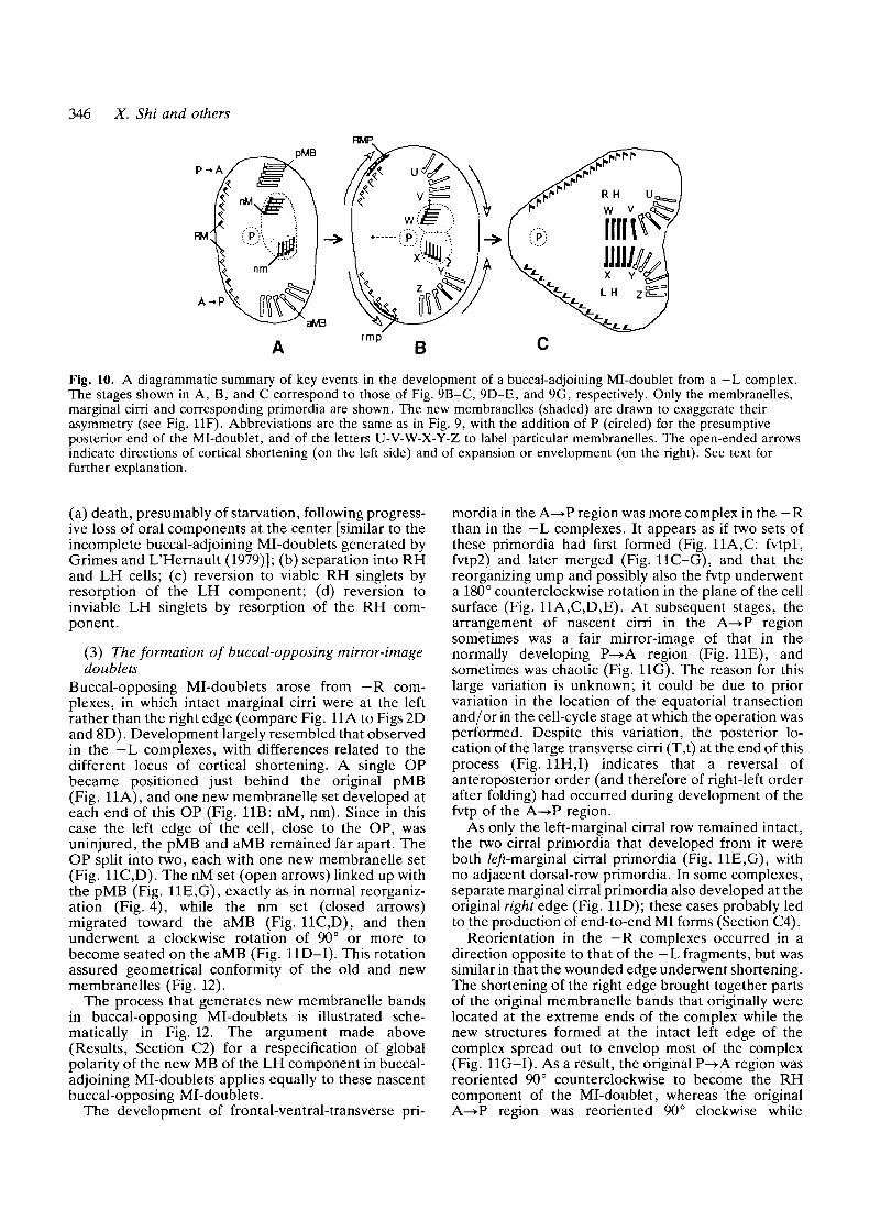
Fig. 10. A diagrammatic summary of key events in the development of a buccal-adjoining Mi-doublet from a — L complex.The stages shown in A, B, and C correspond to those of Fig. 9B-C, 9D-E, and 9G, respectively. Only the membranelles,marginal cirri and corresponding primordia are shown. The new membranelles (shaded) are drawn to exaggerate theirasymmetry (see Fig. 11F). Abbreviations are the same as in Fig. 9, with the addition of P (circled) for the presumptiveposterior end of the Mi-doublet, and of the letters U-V-W-X-Y-Z to label particular membranelles. The open-ended arrowsindicate directions of cortical shortening (on the left side) and of expansion or envelopment (on the right). See text forfurther explanation.
(a) death, presumably of starvation, following progress-ive loss of oral components at the center [similar to theincomplete buccal-adjoining Mi-doublets generated byGrimes and L'Hernault (1979)]; (b) separation into RHand LH cells; (c) reversion to viable RH singlets byresorption of the LH component; (d) reversion toinviable LH singlets by resorption of the RH com-ponent.
(3) The formation of buccal-opposing mirror-imagedoublets
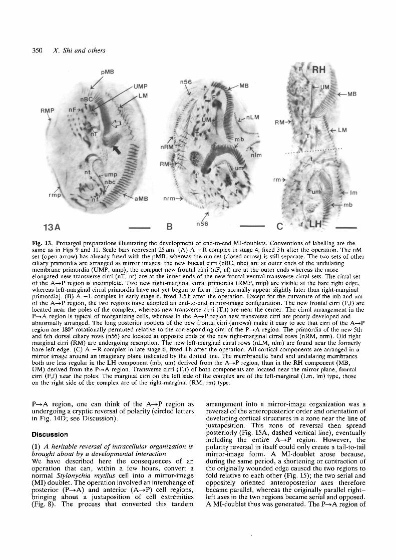
Buccal-opposing Mi-doublets arose from — R com-plexes, in which intact marginal cirri were at the leftrather than the right edge (compare Fig. 11A to Figs 2Dand 8D). Development largely resembled that observedin the — L complexes, with differences related to thedifferent locus of cortical shortening. A single OPbecame positioned just behind the original pMB(Fig. 11A), and one new membranelle set developed ateach end of this OP (Fig. 11B: nM, nm). Since in thiscase the left edge of the cell, close to the OP, wasuninjured, the pMB and aMB remained far apart. TheOP split into two, each with one new membranelle set(Fig. 11C,D). The nM set (open arrows) linked up withthe pMB (Fig. 11E,G), exactly as in normal reorganiz-ation (Fig. 4), while the nm set (closed arrows)migrated toward the aMB (Fig. 11C,D), and thenunderwent a clockwise rotation of 90° or more tobecome seated on the aMB (Fig. 11D-I). This rotationassured geometrical conformity of the old and newmembranelles (Fig. 12).
The process that generates new membranelle bandsin buccal-opposing Mi-doublets is illustrated sche-matically in Fig. 12. The argument made above(Results, Section C2) for a respecification of globalpolarity of the new MB of the LH component in buccal-adjoining Mi-doublets applies equally to these nascentbuccal-opposing Mi-doublets.
The development of frontal-ventral-transverse pri-
mordia in the A—>P region was more complex in the - Rthan in the — L complexes. It appears as if two sets ofthese primordia had first formed (Fig. 11A,C: fvtpl,fvtp2) and later merged (Fig. 11C-G), and that thereorganizing ump and possibly also the fvtp underwenta 180° counterclockwise rotation in the plane of the cellsurface (Fig. 11A,C,D,E). At subsequent stages, thearrangement of nascent cirri in the A—»P regionsometimes was a fair mirror-image of that in thenormally developing P—>A region (Fig. HE), andsometimes was chaotic (Fig. 11G). The reason for thislarge variation is unknown; it could be due to priorvariation in the location of the equatorial transectionand/or in the cell-cycle stage at which the operation wasperformed. Despite this variation, the posterior lo-cation of the large transverse cirri (T,t) at the end of thisprocess (Fig. 11H,I) indicates that a reversal ofanteroposterior order (and therefore of right-left orderafter folding) had occurred during development of thefvtp of the A—*P region.
As only the left-marginal cirral row remained intact,the two cirral primordia that developed from it wereboth /e/r-marginal cirral primordia (Fig. 11E,G), withno adjacent dorsal-row primordia. In some complexes,separate marginal cirral primordia also developed at theoriginal right edge (Fig. 11D); these cases probably ledto the production of end-to-end MI forms (Section C4).
Reorientation in the — R complexes occurred in adirection opposite to that of the - L fragments, but wassimilar in that the wounded edge underwent shortening.The shortening of the right edge brought together partsof the original membranelle bands that originally werelocated at the extreme ends of the complex while thenew structures formed at the intact left edge of thecomplex spread out to envelop most of the complex(Fig. 11G-I). As a result, the original P—>A region wasreoriented 90° counterclockwise to become the RHcomponent of the Mi-doublet, whereas the originalA—»P region was reoriented 90° clockwise while
Cellular handedness in Stylonychia 347
undergoing reversal of its anteroposterior axis, tobecome the LH component. The new /e/r-marginal cirrithat ended up on the right margin of the LH componenttherefore became inverted (photographs not shown; cf.Fig. 12).
The provisional Mi-doublet formed at the conclusionof these events (Fig. Ill) lacked the final contour ofbuccal-opposing Mi-doublets (Fig. 7), but possessed allof the basic elements of the 'body-plan' of suchdoublets. Two or more successive rounds of furthercortical reorganization converted this provisional MI-doublet into a definitive buccal-opposing Mi-doubletsimilar to the one shown in Fig. 7. These reorganiz-ations modified cell shape but did not alter ciliaryorganization, except for the replacement of the invertedleft-marginal cirri located on the right margin of the LHcomponent by normally oriented left-marginal cirri.The membranelle band of the LH component main-tained its inverted configuration through the indefinitenumber of fissions that this stable type of Mi-doubletcan undergo (Shi and Frankel, 1990).
(4) The formation of end-to-end mirror-imageforms
End-to-end MI forms developed from complexes ofboth - L and - R types that failed to fold (Fig. 8). Insuch complexes, sets of new marginal cirral rowsformed at both edges of the cell, including the edgefrom which most or all old marginal cirri had beenremoved (Fig. 11D, 13A,B). In other respects, devel-opment of ciliary primordia was similar to that in thefolding - L complexes described previously. Both thearrangement and the orientation of the structuresformed from the frontal-ventral-transverse and undu-lating-membrane primordia of the A—»P region werereversed: frontal and buccal cirri developed near thepoles and transverse cirri near the center, andindividual cirri of this group were inverted(Fig. 13A-C). On the dorsal surface, new caudal cirrideveloped near the equator of the complex (notshown).
The end-to-end MI forms eventually pulled apart intotwo macronucleated cells: one was a normal RH singletcapable of propagating itself, whereas the other was anLH singlet that ultimately died, almost certainlybecause it could not feed (cf. Shi et al. 1990). Tovisualize the geometry of such an LH cell, one mustrotate Fig. 13C through 180° and then examine the LHcomponent, imagining that it had detached itself at thedotted line. Immediately after separation, such a LHsinglet had normally oriented frontal (f) and transversecirri (t), an inverted right-marginal cirral row on its leftside (Fig. 13C, rm), and an inverted left-marginal cirralrow on its right side (Fig. 13C, lm). Before such a LHsinglet died, it corrected the inverted orientation ofthese marginal rows during reorganization, formingtypical rows of right-marginal cirri on its left margin andof left-marginal cirri on its right margin (cf. Shi et al.1990).
(5) Variations in outcome of operationsThe 'standard' operations shown in Figs 1 and 2 did not
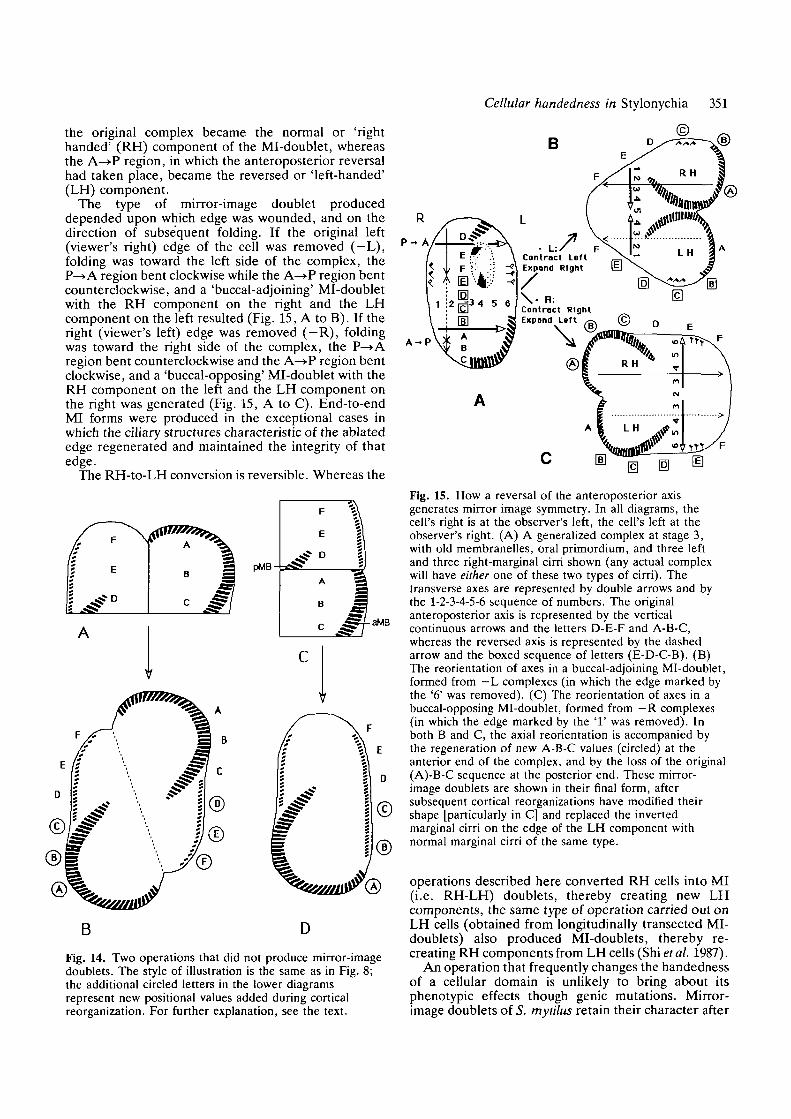
yield mirror-image doublets every time that they wereattempted. Operations normally were .performed inbatches of ten cells, and the proportions of the typesdescribed above varied from zero to ten. In the bestbatch, operations of the type illustrated in Fig. 2yielded six buccal-opposing Mi-doublets and four end-to-end MI forms from ten complexes; the most commonyield of both types of operations was one or two MI-doublets out of ten attempts. The average yield of MI-doublets roughly conformed to the proportion of linearcomplexes of equal-sized components, correspondingto the idealized results depicted in Figs 1C and 2C. Inthe majority of actual operations, either cut no. 2 wasnot truly equatorial, or the two regions becamemisaligned as a consequence of uneven fusion andshortening during the course of the operation. In thesecases, either fewer or more than two sets of ciliaryprimordia were produced, and the complex eventuallyregulated either to an RH- or an LH-singlet. Forexample, when cut 2 was too far posterior, the P—*Aregion was smaller than the A—>P region, and ciliaryprimordia developed successfully only in the latterregion, leading to production of an inviable LH singlet.These 'unsuccessful' cases probably underwent thesame intracellular interactions and developmentalprocesses as the 'canonical' examples considered inSections C2 and C3, but failed to form Mi-doubletsbecause of an imbalance in formation of oral primordiaand/or in the deformations at wounded edges.
(D) Operations that failed to generate mirror-imageformsIf the two cell-halves were allowed to remain side-by-side while still in a heteropolar orientation (Fig. 14A;compare to Fig. 2B), each half retained its originalanteroposterior axis during the production of ciliaryprimordia, and then restored structures characteristic ofthe missing region (circled letters in Fig. 14), as if thehalves were physically separate. A heteropolar doublet(two RH cells fused together in head-to-tail orien-tation) resulted (Fig. 14B). Mirror-image forms or LHsinglets never emerged from this type of operation.
Mirror-image complexes also were not producedfrom an operation that was similar to the type shown inFig. 2 except that the P—>A region was inverted beforebeing transposed to its anterior position, giving it apolarity opposite to that of the A—»P region (Fig. 14C).In these cases, one set of ciliary primordia always wasproduced at the appropriate locations in the P—»Aregion (e.g. an oral primordium appeared near the 'E'in Fig. 14C), and another set sometimes appeared nearthe posterior end of the A—>P region (posterior to C).The P—*A region completed its development success-fully, whereas the new ciliary structures, as well as theaMB, of the A—>P region eventually were resorbed.The result was a normal singlet whose ciliary organiz-ation was derived entirely from the original P—>Aregion (Fig. 14D). Although this was accomplishedwithout any reversal of the anteroposterior order ofretained ciliary structures, it did involve extensiveregulation. Since the A—>P region became occupied bystructures of opposite orientation derived from the
348 X. Shi and others
\ » : : •&
Cellular handedness in Stylonychia 349
Fig. 11. Protargol preparations illustrating the developmentof buccal-opposing MI-doublets from 'minus-right' (—R)complexes. All photographs are oriented with the originalanteroposterior axis vertical on the page. Labellingconventions are the same as in Fig. 9. Scale bars represent25 jjm. (A) A - R complex in stage 3, fixed lh after theoperation. The original posterior portion of themembranelle band (pMB) as well as the intact left-marginal cirral row are located in the P—>A region,whereas a larger anterior section of the membranelle band(aMB) is in the A—>P region. An oral primordium (OP) islocated posterior to the pMB, and frontal-ventral-transverse cirral primordia have formed to the right of thepMB (FVTP) and of the posterior end of the aMB (fvtpl).Undulating-membrane primordia (UMP, ump) are presentin both regions, with a new buccal cirrus (nbc) visible nearthe anterior end of the ump. Immediately adjacent to thenbc, a single cirral streak may represent a second frontal-ventral-transverse cirral primordium (fvtp2). (B) Anenlargement of part of A, with two sets of membranellesappearing within the OP, one (nM) at the normal locationat the original anterior end of the OP (open arrow), theother (nm) ectopically situated at the original posterior endof the OP (closed arrow). The organization of these twomembranelle sets is rotationally symmetric. (C) A —Rcomplex in late stage 2, fixed 1.5 h after the operation. Theprocess of shortening by the right side and envelopment bythe left has begun. The oral primordium has split into two,each bearing one of the two sets of new membranelles(arrows). In the P—>A region, a new buccal cirrus (nBC) ispresent near the anterior end of the undulating membraneprimordium (UMP), and the ciliary streaks of the FVTPare breaking up into anlagen of the individual cirri. In theA—>P region, no buccal cirrus can be seen at either end ofthe curved ump, and fvtp2 is better developed than fvtpl.(D) A - R complex in late stage 3, fixed 2h after theoperation. This complex is not bent, and a few right-marginal cirri (RM) plus right-marginal cirral primordia(RMP, rmp) are present at the right edge in both P—>Aand A ^ P regions. The two oral primordia (arrows) haveseparated further. The UMP, nBC, and FVTP of the P—Aregion appear as in C. In the A—>P region, the ump seemsto be caught midway in a counterclockwise rotation, thereis no nbc, and fvtp are scattered over most of the region,possibly representing two sets (fvtpl, fvtp2). (E) A — Rcomplex in early stage 4, fixed 2.5 h after the operation.The nM set (open arrow) has almost merged with thepMB, whereas thenm set (closed arrow) is close to theaMB (see F). The UMP, nBC, and FVTP of the P-*A
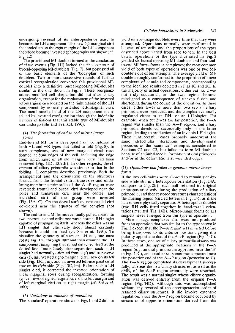
region have developed normally. In the A—>P region, theump now lies parallel to the aMB, possibly havingcompleted a 180° counterclockwise rotation. The pattern ofcirral subdivision of the two sets of frontal-ventral-transverse streaks (1-5 of the P—>A region, l ' -5 ' of theA—>P region) is mirror-imaged. (F) An enlargement of E,focused on thenm set, which is undergoing a clockwiserotation. At this magnification, the protrusion at one endof each mature membranelle resulting from the presence ofa short fourth row of basal bodies is apparent (arrow);such protrusions are just beginning to be visible in the nmset. (G) A —R complex in stage 4, fixed 2.5 h after theoperation. The right margin of the original complex isbecoming the anterior end (A) of the nascent Mi-doublet,whereas the center of the original left margin is becomingthe posterior end (P). Both new membranelle sets (arrows)have merged with their respective old membranellesegments (aMB, pMB), but thenm set (closed arrow) hasnot completed either its differentiation or its clockwiserotation. In this cell, the FVTP is properly oriented andsubdivided into cirral rudiments, whereas the ciliary streaksof the fvtp are chaotically arranged and have not yetformed cirri. New left-marginal cirral primordia (LMP,Imp) have formed at the two margins of the nascent MI-doublet. (H) A - R complex in stage 6, fixed 3h after theoperation. Composite membranelle bands (MB, mb) havebeen assembled; the line of juncture of old and newmembranelle sets is clear in the A—>P region (arrowhead).New undulating membranes (nUM, num), new left-marginal cirri (nLM, nlm), and new frontal-ventral-transverse cirri have differentiated, with the cirralcomplement nearly normal in the P^>A region and severelydeficient in the A—>P region; however, the large transversecirri (T,t) are the posteriormost of the new cirri in bothcomponents. (I) A —R complex in late stage 6, fixed 4hafter the operation. The former right margin (nowanterior) is fully contracted and enveloped by ciliaturederived from the former left margin (now posterior andlateral). The former P^>A region now is the right-handed(RH) component of the provisional mirror-image doublet,whereas the former A—>P region has become the left-handed (LH) component. The ventral ciliature is similar tothat in (H), with the nLM cirri optically juxtaposed on theMB. The composite membranelle band (mb) of the LHcomponent still shows its dual origin, with a suture(arrowhead) marking the imperfect juncture between theformer nm set and the aMB. One dorsal cirral row isvisible at each edge (nDl, ndl'), and a new caudal cirrus(nC) is seen at the posterior end of nDl.
P-A
A - P
Fig. 12. A diagrammatic summary of keyevents in the development of a buccal-opposing Mi-doublet from a — R complex.The stages shown in A, B, and Ccorrespond to those of Fig. HA, HE, and11G-H respectively. The abbreviationsand conventions are the same as in Fig. 10.The open-ended arrows indicate directionsof cortical shortening (on the right side)and of expansion or envelopment (on theleft). See text for further explanation.
350 X. Shi and others
pMB
n56
13AFig. 13. Protargol preparations illustrating the development of end-to-end Mi-doublets. Conventions of labelling are thesame as in Figs 9 and 11. Scale bars represent 25 fan. (A) A —R complex in stage 4, fixed 3h after the operation. The nMset (open arrow) has already fused with the pMB, whereas the nm set (closed arrow) is still separate. The two sets of otherciliary primordia are arranged as mirror images: the new buccal cirri (nBC, nbc) are at outer ends of the undulatingmembrane primordia (UMP, ump); the compact new frontal cirri (nF, nf) are at the outer ends whereas the moreelongated new transverse cirri (nT, nt) are at the inner ends of the new frontal-ventral-transverse cirral sets. The cirral setof the A^>P region is incomplete. Two new right-marginal cirral primordia (RMP, rmp) are visible at the bare right edge,whereas left-marginal cirral primordia have not yet begun to form [they normally appear slightly later than right-marginalprimordia]. (B) A —L complex in early stage 6, fixed 3.5 h after the operation. Except for the curvature of the mb and umof the A—>P region, the two regions have adopted an end-to-end mirror-image configuration. The new frontal cirri (F,f) arelocated near the poles of the complex, whereas new transverse cirri (T,t) are near the center. The cirral arrangement in theP—>A region is typical of reorganizing cells, whereas in the A—»P region new transverse cirri are poorly developed andabnormally arranged. The long posterior rootlets of the new frontal cirri (arrows) make it easy to see that cirri of the A—»Pregion are 180° rotationally permuted relative to the corresponding cirri of the P—>A region. The primordia of the new 5thand 6th dorsal ciliary rows (n56) are located at opposite ends of the new right-marginal cirral rows (nRM, nrm). Old rightmarginal cirri (RM) are undergoing resorption. The new left-marginal cirral rows (nLM, nlm) are found near the formerlybare left edge. (C) A —R complex in late stage 6, fixed 4h after the operation. All cortical components are arranged in amirror image around an imaginary plane indicated by the dotted line. The membranelle band and undulating membranesboth are less regular in the LH component (mb, um) derived from the A—>P region, than in the RH component (MB,UM) derived from the P—>A region. Transverse cirri (T,t) of both components are located near the mirror plane, frontalcirri (F,f) near the poles. The marginal cirri on the left side of the complex are of the left-marginal (Lm, lm) type, thoseon the right side of the complex are of the right-marginal (RM, rm) type.
P—»A region, one can think of the A—>P region asundergoing a cryptic reversal of polarity (circled lettersin Fig. 14D; see Discussion).
Discussion
(1) A heritable reversal of intracellular organization isbrought about by a developmental interactionWe have described here the consequences of anoperation that can, within a few hours, convert anormal Stylonychia mytilus cell into a mirror-image(MI) doublet. The operation involved an interchange ofposterior (P—>A) and anterior (A—»P) cell regions,bringing about a juxtaposition of cell extremities(Fig. 8). The process that converted this tandem
arrangement into a mirror-image organization was areversal of the anteroposterior order and orientation ofdeveloping cortical structures in a zone near the line ofjuxtaposition. This zone of reversal then spreadposteriorly (Fig. 15A, dashed vertical line), eventuallyincluding the entire A—»P region. However, thepolarity reversal in itself could only create a tail-to-tailmirror-image form. A Mi-doublet arose because,during the same period, a shortening or contraction ofthe originally wounded edge caused the two regions tofold relative to each other (Fig. 15); the two serial andoppositely oriented anteroposterior axes thereforebecame parallel, whereas the originally parallel right-left axes in the two regions became serial and opposed.A Mi-doublet thus was generated. The P—»A region of
Cellular handedness in Stylonychia 351
the original complex became the normal or 'righthanded' (RH) component of the Mi-doublet, whereasthe A—>P region, in which the anteroposterior reversalhad taken place, became the reversed or 'left-handed'(LH) component.
The type of mirror-image doublet produceddepended upon which edge was wounded, and on thedirection of subsequent folding. If the original left(viewer's right) edge of the cell was removed (-L),folding was toward the left side of the complex, theP—»A region bent clockwise while the A—»P region bentcounterclockwise, and a 'buccal-adjoining' Mi-doubletwith the RH component on the right and the LHcomponent on the left resulted (Fig. 15, A to B). If theright (viewer's left) edge was removed (—R), foldingwas toward the right side of the complex, the P—>Aregion bent counterclockwise and the A—»P region bentclockwise, and a 'buccal-opposing' Mi-doublet with theRH component on the left and the LH component onthe right was generated (Fig. 15, A to C). End-to-endMI forms were produced in the exceptional cases inwhich the ciliary structures characteristic of the ablatededge regenerated and maintained the integrity of thatedge.
The RH-to-LH conversion is reversible. Whereas the
B
aWB
B D
Fig. 14. Two operations that did not produce mirror-imagedoublets. The style of illustration is the same as in Fig. 8;the additional circled letters in the lower diagramsrepresent new positional values added during corticalreorganization. For further explanation, see the text.
A - P
m m
Fig. 15. How a reversal of the anteroposterior axisgenerates mirror image symmetry. In all diagrams, thecell's right is at the observer's left, the cell's left at theobserver's right. (A) A generalized complex at stage 3,with old membranelles, oral primordium, and three leftand three right-marginal cirri shown (any actual complexwill have either one of these two types of cirri). Thetransverse axes are represented by double arrows and bythe 1-2-3-4-5-6 sequence of numbers. The originalanteroposterior axis is represented by the verticalcontinuous arrows and the letters D-E-F and A-B-C,whereas the reversed axis is represented by the dashedarrow and the boxed sequence of letters (E-D-C-B). (B)The reorientation of axes in a buccal-adjoining Mi-doublet,formed from — L complexes (in which the edge marked bythe '6' was removed). (C) The reorientation of axes in abuccal-opposing Mi-doublet, formed from —R complexes(in which the edge marked by the '1 ' was removed). Inboth B and C, the axial reorientation is accompanied bythe regeneration of new A-B-C values (circled) at theanterior end of the complex, and by the loss of the original(A)-B-C sequence at the posterior end. These mirror-image doublets are shown in their final form, aftersubsequent cortical reorganizations have modified theirshape [particularly in C] and replaced the invertedmarginal cirri on the edge of the LH component withnormal marginal cirri of the same type.
operations described here converted RH cells into MI(i.e. RH-LH) doublets, thereby creating new LHcomponents, the same type of operation carried out onLH cells (obtained from longitudinally transected MI-doublets) also produced Mi-doublets, thereby re-creating RH components from LH cells (Shi et al. 1987).
An operation that frequently changes the handednessof a cellular domain is unlikely to bring about itsphenotypic effects though genie mutations. Mirror-image doublets of S. mytilus retain their character after
352 X. Shi and others
conjugation with normal singlets (Shi and Qiu, 1989),but no detailed genetic analysis was carried out. Inanother ciliate, Tetrahymena thermophila, the differ-ence between heritable LH and RH singlet cells wasshown by genetic analysis to be not based on differencesin nuclear genes (Nelsen et al. 1989a). The pathway ofthe transformation could not be ascertained forTetrahymena in the precise manner accomplished forStylonychia, although an unstable Mi-doublet inter-mediate is strongly suspected (Nelsen and Frankel,1989). In Stylonychia, unlike T. thermophila and therelated Glaucoma scintillans (Suhama, 1985), an LHcomponent must be supported nutritionally by alaterally attached RH partner (Grimes, 1990; Shi et al.1990). Nonetheless, the organization of an LH com-ponent of a Mi-doublet resembles that of an LH singletcell; thus a fundamental generalization that applies toall three of these ciliates is that in an appropriatecellular setting a vegetatively generated LH cell-domaincan propagate its organization indefinitely.
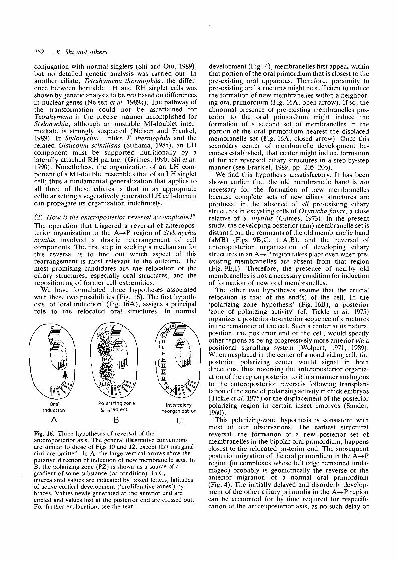
(2) How is the anteroposterior reversal accomplished?The operation that triggered a reversal of anteropos-terior organization in the A—»P region of Stylonychiamytilus involved a drastic rearrangement of cellcomponents. The first step in seeking a mechanism forthis reversal is to find out which aspect of thisrearrangement is most relevant to the outcome. Themost promising candidates are the relocation of theciliary structures, especially oral structures, and therepositioning of former cell extremities.
We have formulated three hypotheses associatedwith these two possibilities (Fig. 16). The first hypoth-esis, of 'oral induction' (Fig. 16A), assigns a principalrole to the relocated oral structures. In normal
Oralinduction
Polarizing zone4, gradient
Intercalaryreorganization
BFig. 16. Three hypotheses of reversal of theanteroposterior axis. The general illustrative conventionsare similar to those of Figs 10 and 12, except that marginalcirri are omitted. In A, the large vertical arrows show theputative direction of induction of new membranelle sets. InB, the polarizing zone (PZ) is shown as a source of agradient of some substance (or condition). In C,intercalated values are indicated by boxed letters, latitudesof active cortical development ('proliferative zones') bybraces. Values newly generated at the anterior end arecircled and values lost at the posterior end are crossed out.For further explanation, see the text.
development (Fig. 4), membranelles first appear withinthat portion of the oral primordium that is closest to thepre-existing oral apparatus. Therefore, proximity topre-existing oral structures might be sufficient to inducethe formation of new membranelles within a neighbor-ing oral primordium (Fig. 16A, open arrow). If so, theabnormal presence of pre-existing membranelles pos-terior to the oral primordium might induce theformation of a second set of membranelles in theportion of the oral primordium nearest the displacedmembranelle set (Fig. 16A, closed arrow). Once thissecondary center of membranelle development be-comes established, that center might induce formationof further reversed ciliary structures in a step-by-stepmanner (see Frankel, 1989, pp. 205-206).
We find this hypothesis unsatisfactory. It has beenshown earlier that the old membranelle band is notnecessary for the formation of new membranellesbecause complete sets of new ciliary structures areproduced in the absence of all pre-existing ciliarystructures in excysting cells of Oxytricha fallax, a closerelative of S. mytilus (Grimes, 1973). In the presentstudy, the developing posterior (nm) membranelle set isdistant from the remnants of the old membranelle band(aMB) (Figs 9B,C; 11A,B), and the reversal ofanteroposterior organization of developing ciliarystructures in an A—>P region takes place even when pre-existing membranelles are absent from that region(Fig. 9E,I). Therefore, the presence of nearby oldmembranelles is not a necessary condition for inductionof formation of new oral membranelles.
The other two hypotheses assume that the crucialrelocation is that of the end(s) of the cell. In the'polarizing zone hypothesis' (Fig. 16B), a posterior'zone of polarizing activity' (cf. Tickle et al. 1975)organizes a posterior-to-anterior sequence of structuresin the remainder of the cell. Such a center at its naturalposition, the posterior end of the cell, would specifyother regions as being progressively more anterior via apositional signalling system (Wolpert, 1971, 1989).When misplaced in the center of a nondividing cell, theposterior polarizing center would signal in bothdirections, thus reversing the anteroposterior organiz-ation of the region posterior to it in a manner analogousto the anteroposterior reversals following transplan-tation of the zone of polarizing activity in chick embryos(Tickle et al. 1975) or the displacement of the posteriorpolarizing region in certain insect embryos (Sander,1960).
This polarizing-zone hypothesis is consistent withmost of our observations. The earliest structuralreversal, the formation of a new posterior set ofmembranelles in the bipolar oral primordium, happensclosest to the relocated posterior end. The subsequentposterior migration of the oral primordium in the A—»Pregion (in complexes whose left edge remained unda-maged) probably is geometrically the reverse of theanterior migration of a normal oral primordium(Fig. 4). The initially delayed and disorderly develop-ment of the other ciliary primordia in the A—>P regioncan be accounted for by time required for respecifi-cation of the anteroposterior axis, as no such delay or
Cellular handedness in Stylonychia 353
disorder were found during subsequent rounds ofdevelopment in LH components of Mi-doublets;exactly the same had been observed by Grimes andL'Hernault (1979) and Grimes et al. (1981) in foldedregions of longitudinal fragments of 5. mytilus.
This polarizing-zone hypothesis can be applied toother experimental situations as well. In the rotated andtransposed P—»A fragment (Fig. 14C), the posteriororganizing region is relocated to the 'top' of thecomplex instead of the center, and thus organizes an'upside-down' singlet cell of normal asymmetry(Fig. 14D). In the side-by-side heteropolar complex(Fig. 14A,B), the A-B-C half lacks a polarizing zoneand must be assumed to be capable of regeneratingrapidly, as must also happen in totally separated half-cells (Hashimoto, 1961). This entails the subsidiarypostulate that the polarizing zone is capable of rapidreconstruction in a manner expected of dynamicgradient systems (Meinhardt, 1982; Slack, 1983).
The polarizing-zone hypothesis is in part a revival ofUhlig's concept of a basal-apical gradient, for whichsubstantial evidence was obtained in Stentor (Uhlig,1960; Tartar, 1964) and Blepharisma (Eberhardt, 1962)(reviewed in Frankel, 1989, pp. 131-134, 155-158).
The third, 'intercalary reorganization' hypothesis(Fig. 16C) is that the necessary and sufficient conditionfor reversal of the anteroposterior axis is an abnormaljuxtaposition of distant regions of the cell. This viewpresumes that different regions of the cell surface arepositionally nonequivalent (Lewis and Wolpert, 1976),and that juxtaposition of regions that are not normallyneighboring stimulates the intercalation of interveningpositional values by the shortest permitted route (cf.French et al. 1976; Mittenthal, 1981). This hypothesisviews the anteroposterior reversal of in the A—>P regionas a consequence of reverse-intercalation of thepositions (the boxed sequence E, D, C, and B inFig. 16C) located between the juxtaposed posterior (F)and anterior (A) positional values. Intercalation in thiscase would have to proceed through intracellular andmorphallactic reorganization rather than through local-ized cell division and growth (cf. Frankel, 1989,Chapter 10).
The intercalary reorganization hypothesis, beingmore explicit in labelling cell regions than the polariz-ing-zone hypothesis, forces us to take into accountcertain spatial peculiarities of ciliary development inStylonychia. Ciliary primordia develop in restrictedlatitudinal 'proliferative zones' (Grimes, 1976), corre-sponding roughly to regions D and E in Fig. 16C; theprimordia formed there generate virtually the totalciliature of the cell or cellular domain, while the oldciliature is resorbed (Fig. 4). Thus, at every morpho-genetic episode all latitudinal values of the cell appearto be generated anew from a restricted subset of values.New C-B-A values therefore are produced at theanterior end (circled in Fig. 16C), and old B and Cvalues are deleted at the posterior end (crossed out inFig. 16C). This combination of restricted sites forinitiation of development and subsequent spread ofciliary primordia applies only to latitudes and not tolongitudes, as development of ciliary organelles takes
place along all longitudes and produces new structuresthat retain their original longitudinal positions whilespreading latitudinally (Fig. 4).
The spatial restriction of development to particularlatitudes makes it impossible to perform the crucial testof seeking to elicit different intercalated structures (orsets of structures) when different regions of theanteroposterior axis are juxtaposed (see Frankel, 1989,chapter 10, for the successful application of this test tothe transverse axis). Nonetheless, there is one othertestable difference between the polarizing-zone andintercalary reorganization hypothesis, which dependson the reciprocity of posterior and anterior extremities .in the latter hypothesis, and the absence of suchreciprocity in the former. If the polarizing-zonehypothesis is correct, anteroposterior reversal shouldoccur only in the A ^ P region under the influence of thetransposed polarizing zone of the P—>A region. Aretrograde reversal, with the P^>A region becomingreversed while the A^»P region retained its originalpolarity, would not be expected, since then the formeranterior extremity (value A) rather than the formerposterior extremity (value F) would have had to act as a'polarizing zone'. On the intercalary reorganizationhypothesis, 'A' and 'F' have no special propertiesexcept that they mark nonequivalent positions and theactive primordium site happens to be positionallybetween the two. Therefore, if the intercalary reorgan-ization hypothesis is correct, a retrograde reversalmight be possible in an exchange-operation following asubequatorial transection in Stylonychia or following anequatorial transection in another ciliate in which theprimordium site is located more anteriorly than inStylonychia. For example, if the primordium site wereat C, a DEF/ABC* complex could be converted toF£DC*5A/ABC*DEF (intercalated values italicized,primordium site starred). This was observed in oper-ated complexes of the hypotrich Paraurostyla weissei, inwhich the MB is smaller than in Stylonychia and theciliary primordia develop more anteriorly (Lu and Shi,1987), and occasionally was seen in operated LH cells ofStylonychia (Shi et al. 1987).
The experiment shown in Fig. 14A provides anapparent counter-example; F and A were laterallyjuxtaposed yet no additional polar axes developed.However, comparable juxtapositions following thefolding of surgically generated lateral half-cells didbring about the formation of a second reversed polaraxis in Blepharisma (Suzuki, 1957) and Stylonychia(Grimes and L'Hernault, 1979) (summarized in Fran-kel, 1989, pp. 198-199). Indeed, the operation thatgenerated the first mirror-image doublets in Stylony-chia, the isolation of a central disc from a dividing cell(Tchang et al. 1964; Tchang and Pang, 1965), producedmirror-image doublets when (and only when) theanterior and posterior ends had rotated within the planeof the disc so that their posterior and anterior regionscame into close juxtaposition (Shi and Qiu, 1985;summarized in Frankel, 1989, pp. 199-200). The failureto develop additional reversed axes in the experimentshown in Fig. 14A therefore may be due to featuresspecific to the geometry of this complex, in which
354 X. Shi and others
always is 180"-permuted (inverted), and that ofmarginal cirri usually is normal but sometimes isinverted (Grimes et al. 1980; Grimes, 1990; Jerka-Dziadosz, 1983, 1985; Lu and Shi, 1987; Shi andFrankel, 1990).
The way in which Mi-doublets arise might help us tounderstand how these varying orientations come about.The simplest hypothesis assumes that differentiatingstructures orient along one of the two orthogonal cell-surface axes (anteroposterior and transverse). If this isso, the reversal of the anteroposterior axis in the A—»Pregion (Fig. 15A) should bring about an inversion ofany ciliary structure that aligns along that axis, in thesame way that the reversal of a magnetic field will causea magnetized needle to swing through 180°. Theoppositely directed folding of the A—>P and P—»Aregions (Fig. 15B,C) then would cause these initiallyopposed ciliary structures to assume similar, parallel,orientations in the two components. Conversely, sincethe transverse axis is initially the same throughout thecomplex, any new ciliary structure that orients alongthat axis should be similarly oriented in both regions(Fig. 15A), but after folding (Fig. 15B,C) shouldbecome inverted in the LH region relative to itsorientation in the RH region.
The frontal-ventral-transverse cirri obey this hypoth-esis perfectly. As shown schematically in Fig. 17, whenthese cirri first appeared they were inverted in the A—>Pregion (Fig. 17A, arrows marked by F), possibly as aconsequence of a 180° rotation of the early primordia(see Results). Then, after folding, these cirri becamenormally oriented everywhere in the provisional MI-doublet (Fig. 17B), and remained so during subsequentreorganizations and divisions (Fig. 17C).
The marginal cirri violate this hypothesis. Althoughthe location of formation of structures was reversed inthe A—»P region (e.g. the primordia of the new 5th and
Fig. 17. The orientation of ciliarystructures during formation of abuccal-opposing Mi-doublet froma —R complex. The stages shownare (A) a — R complex duringformation of ventral ciliaryprimordia, before substantialfolding has taken place, (B) aprovisional Mi-doublet after theend of the first round of corticaldevelopment and of folding, (C) adefinitive buccal-opposing MI-doublet after the completion ofsubsequent corticalreorganizations. A few left-marginal cirri and membraneUesare illustrated in the same style asin Fig. 12. Membranellesproduced in the firstreorganization are shaded,
whereas those formed in subsequent reorganization are cross-hatched. Large-headed arrows indicate the orientation ofciliary primordia, with the heads representing the ends that are posterior in normal cells. Filled heads indicate the first andopen heads subsequent primordia. The letters stand for ciliary primordia (F=frontal-ventral-transverse cirral primordia;L= left-marginal primordia; and M=new membranelle bands), and their orientation symbolizes the spatial configuration ofthese primordia. Small-headed arrows indicate the direction of movement of new membranelle bands (in A and C) and thebending of regions (in A). See text for further explanation.
longitudinal regeneration may be favored over lateralinteraction across an artificial juncture. In a morereadily operable ciliate, Blepharisma japonicus, hetero-polar complexes resembling those of Fig. 14B could bestimulated to form mirror-image doublets by certainfurther deletions that modified the geometry of thecomplexes (Suzuki, 1957, pp. 169-173 and Fig. 47).
Any uncertainty in finding the best language todescribe the reversal of global polarity in Stylonychiashould not be allowed to obscure the major propertiesthat clearly are inherent in the cortex of Stylonychia: apositional system that operates over large intracellulardistances, that coordinately specifies the locations andmigrations of separate ciliary primordia, that is itselfcapable of large-scale reorganization under suitableconditions, and that can be dissociated from the localprocesses that control the internal organization ofindividual ciliary organelles. All of these characteristicsare much more reminiscent of certain positionalinformation systems in multicellular organisms than ofthe local assembly-mediated processes that normallyare thought of as dominating intracellular and someintercellular development.
(3) The orientation of ciliary structuresThe individual ciliary organelles that are found in LHcomponents of Mi-doublets of ciliates never aregenuinely reversed in their internal organization(Grimes et al. 1980; Frankel et al. 1984; Jerka-Dziadosz,1983, 1985; Nelsen et al. 19896; Shi and Frankel, 1990;Suhama, 1982, 1985). However, the spatial orientationof these structures may vary, being either normal orrotationally permuted by 90° or 180° relative to theircounterparts in RH components. In Mi-doublets ofStylonychia mytilus and related hypotrich ciliates, theorientation of the frontal-ventral-transverse cirri in LHcomponents always is normal, that of oral structures
A1st Reorganization Provisional Mi-Doublet
FVT
LM
Inverted
Normal
MB Vertical Normal
Normal
Inverted
Inverted
Later reorganization
Normal
(Normal)
Vertical Inverted
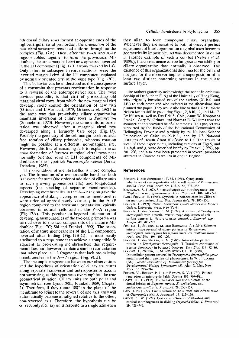
Cellular handedness in Stylonychia 355
6th dorsal ciliary rows formed at opposite ends of theright-marginal cirral primordia), the orientation of thenew cirral structures remained uniform throughout thecomplex (Fig. 17A). Then, after the P—»A and A—»Pregions folded together to form the provisional MI-doublet, the same marginal cirri now appeared invertedin the LH component (Fig. 17B, arrows marked by Ls).Only later, in subsequent reorganizations, were theinverted marginal cirri of the LH component replacedby normally oriented cirri of the same type (Fig. 17C).
This behavior can be understood as the consequenceof a constraint that prevents reorientation in responseto a reversal of the anteroposterior axis. The mostobvious possibility is that cirri of pre-existing oldmarginal cirral rows, from which the new marginal cirridevelop, could control the orientation of new cirri(Grimes and L'Hernault, 1979; Grimes et al. 1981) inthe same way that pre-existing ciliary organizationmaintains inversions of ciliary rows in Paramecium(Sonneborn, 1970). However, the same uniform orien-tation was observed after a marginal cirral rowdeveloped along a formerly bare edge (Fig. 13).Possibly the geometry of the cell margin itself restrictsfree rotation of ciliary primordia, whereas rotationmight be possible at a different, non-marginal site.However, this line of reasoning fails to explain the denovo formation of inverted marginal cirral rows nearnormally oriented rows in LH components of MI-doublets of the hypotrich Paraurostyla weissei (Jerka-Dziadosz, 1985).
The orientation of membranelles is more complexyet. The formation of a membranelle band has bothtransverse features (the order of addition of ciliary unitsto each growing promembranelle) and longitudinalaspects (the stacking of separate membranelles).Developing membranelles in the A—»P region gave theappearance of striking a compromise, as membranelleswere oriented approximately vertically in the A—»Pregion compared to the horizontal orientation typicallyobserved in normal cells and in the P—>A region(Fig. 17A). This peculiar orthogonal orientation ofdeveloping membranelles of the two oral primordia wascarried over to the two components of a mature MI-doublet (Fig. 17C; Shi and Frankel, 1990). The orien-tation of mature membranelles of the LH component,inverted after folding (Fig. 17B,C), is most easilyattributed to a requirement to achieve a compatible fitadjacent to pre-existing membranelles; this require-ment does not, however, explain a similar reorientationthat takes place in —L fragments that lack pre-existingmembranelles in the A—>P region (Fig. 9E,I).
The incomplete agreement between our observationsand the hypothesis of orientation of ciliary structuresalong separate transverse and anteroposterior axes isnot surprising, as this hypothesis oversimplifies the truegeometrical situation. Ciliary units are both polar andasymmetrical (see Lynn, 1981; Frankel, 1989; Chapter2). Therefore, if they rotate 180° in the plane of themembrane to adjust to the reversal of one axis, they willautomatically become misaligned relative to the other,non-reversed axis. Therefore, the hypothesis can becorrect only if ciliary units respond to a single axis when
they align to form compound ciliary organelles.Whenever they are sensitive to both at once, a perfectadjustment of local organization to global axes becomesgeometrically impossible. As was documented in detailin another example of such a conflict (Nelsen et al.1989b), the consequence can be far greater variability inciliary organization than normally is observed. Theexistence of this organizational dilemma for the cell andnot just for the observer implies a superposition of atleast two distinct patterning systems in the ciliatesurface layer.
The authors gratefully acknowledge the scientific ambassa-dorship of Dr Stephen F. Ng of the University of Hong Kong,who originally introduced two of the authors (X.-b.S. andJ.F.) to each other and who assisted in the discussions thatplanned this paper. They would also like to thank Dr E. MarioNelsen for his skill in producing Figs 1, 2, 8 10,12, and 14-17.Dr Nelsen as well as Drs Eric S. Cole, Anne W. KoopmansFrankel, Gary W. Grimes, and Norman E. Williams read themanuscript and provided helpful comments. This project wassupported by the funds of the Educational Commission ofHeilongjang Province and partially by the National ScienceFoundation of China to X.-b.S., and by US NationalInstitutes of Health Grant HD-08485 to J.F. The results ofsome of these experiments, including versions of Figs 5, andlla,b,d, and g, were described briefly by Frankel (1989), pp.201-206, and also have been presented in several publishedabstracts in Chinese as well as in one in English.
References
BEISSON, J. AND SONNEBORN, T. M. (1965). Cytoplasmicinheritance of the organization of the cell cortex of Parameciumaureha. Proc. nam. Acad. Sci. U.S.A. 53, 275-282.
EBERHARDT, R. (1962). Untersuchungen zur morphogenese vonBlepharisma und Spirostomum. Arch. Protistenk. 106, 241-341.
FAURE-FREMIET, E. (1945). Syme'trie et polarite' chez les Cilia's bi-ou multicomposites. Bull. Biol. France Belg. 79, 106-150.
FRANKEL, J. (1989). Pattern Formation: Cihate Studies and Models.Oxford University Press, New York.
FRANKEL, J. AND JENKINS, L. M. (1979). A mutant of Tetrahymenathermophila with a partial mirror-image duplication of cellsurface pattern. II. Nature of genie control. J. Embryo/, exp.Morph. 49, 203-227.
FRANKEL, J., JENKJNS, L. M. AND BAKOWSKA, J. (1984). Selectivemirror-image reversal of ciliary patterns in Tetrahymenathermophila homozygous for a janus mutation. Wilhelm Roux'sArch, devl Biol. 194, 107-120.
FRANKEL, J. AND NELSEN, E. M. (1986). Intracellular patternreversal in Tetrahymena thermophila. II. Transient expression ofa janus phenocopy in balanced doublets. Devi Biol. 114, 72-86.
FRAJJKEL, J., NELSEN, E. M. AND JENKINS. L. M. (1987).Intracellular pattern reversal in Tetrahymena thermophila: janusmutants and their geometrical phenocopies. In W. F. Loomis(ed.), Genetic Regulation of Development (Society forDevelopmental Biology Symposium 45), Alan R. Liss, NewYork, pp. 219-244.
FRENCH, V., BRYANT, P. J. AND BRYANT, S. V. (1976). Patternregulation in epimorphic fields. Science 193, 969-981.
GORTZ, H.-D. (1982). The behavior and fine structure of thedorsal bristles of Euplotes minuta, E. aediculatus, andStylonychia mytilus. J. Protozool. 29, 353-359.
GRIM, J. N. (1972). Fine structure of the surface and infraciliatureof Gastrostyla steini. J. Protozool. 19, 113-126.
GRIMES, G. W. (1972). Cortical structure in nondividing andcortical morphogenesis in dividing Oxytricha fallax. J. Protozool.19, 428-445.
356 X. Shi and others
GRIMES, G. W. (1973). Morphological discontinuity of kinetosomesduring the life cycle of Oxytricha fallax. J. Cell Biol. 57,229-232.
GRIMES, G. W. (1976). Laser microbeam induction of incompletedoublets of Oxytricha fallax. Genet. Res., Camb. 27, 213-226.
GRIMES, G. W. (1990). Inheritance of cortical patterns in ciliatedprotozoa. In G. M. Malacinski (ed.), Cytoplasmic InformationSystems, Primers in Developmental Biology, VolumeIV. Macmillan, New York, (in press).
GRIMES, G. W., KNAUPP-WALDVOGEL, E. A. AND GOLDSMITH-SPOEGLER, C. M. (1981). Cytogeometrical determination ofciliary pattern formation in the hypotrich ciliate Stylonychiamytilus. II. Stability and field regulation. Devi Biol. 84, 477-480.
GRIMES, G. W. AND L'HERNAULT, S. W. (1979). Cytogeometricaldetermination of ciliary pattern formation in the hypotrichciliate Stylonychia mytilus. Devi Biol. 70, 372-395.
GRIMES, G. W., MCKENNA, M. E., GOLDSMITH-SPOEGLER, C. M.AND KNAUPP, E. A. (1980). Patterning and assembly of ciliatureare independent processes in hypotrich ciliates. Science 209,281-283.
HASHIMOTO, K. (1961). Stomatogenesis and formation of cirri infragments of Oxytricha fallax Stein. J. Protozool. 8, 433-442.
JERKA-DZIADOSZ, M. (1963). Morphogenesis in division andregeneration of Urostyla grandis Ehrbg. Acta Protozoologica 1,43-54.
JERKA-DZIADOSZ, M. (1983). The origin of mirror-image symmetrydoublet cells in the hypotrich ciliate Paraurostyla weissei.Wilhelm Roux's Arch, devl Biol. 192, 179-188.
JERKA-DZIADOSZ, M. (1985). Mirror-image configuration in thecortical pattern causes modifications in propagation ofmicrotubular structures in the hypotrich ciliate Paraurostylaweissei. Roux's Arch, devl Biol. 194, 311-324.
JERKA-DZIADOSZ, M. AND FRANKEL, J. (1979). A mutant ofTetrahymena thermophila with a partial mirror-image duplicationof cell surface pattern. I. Analysis of the phenotype.J. Embryol. exp. Morph. 49, 167-202.
LEWIS, J. H. AND WOLPERT, L. (1976). The principle of non-equivalence in development. /. theor. Biol. 62, 479-490.
Lu, L. AND SHI, X. B. (1987). The grafting method applied to fivespecies of hypotrich ciliates for creating mirror-imaged doublets.Symp. Chinese Protozool. Soc. 4, 43-44 (Abstract, in Chinese).
LYNN, D. H. (1981). The organization and evolution ofmicrotubular organelles in ciliated protozoa. Biol. Rev. 56,243-292.
MEINHARDT, H. (1982). Models of Biological Pattern Formation.Academic Press, London.
MITTENTHAL, J. E. (1981). The rule of normal neighbors: Ahypothesis for morphogenetic pattern regulation. Devl Biol. 88,15-26.
NELSEN, E. M. AND FRANKEL, J. (1986). Intraccllular patternreversal in Tetrahymena thermophila. I. Evidence for reverseintercalation in unbalanced doublets. Devl Biol. 114, 53-71.
NELSEN, E. M. AND FRANKEL, J. (1989). Maintenance andregulation of cellular handedness in Tetrahymena. Development105, 457-471.
NELSEN, E. M., FRANKEL, J. AND JENKINS, L. M. (1989a). Non-genic inheritance of cellular handedness. Development 105,447-456.
NELSEN, E. M., FRANKEL, J. AND WILLIAMS, N. E. (19896). Oralassembly in left-handed Tetrahymena thermophila. J. Protozool36, 582-596.
NG, S. F. AND FRANKEL, J. (1977). 180° rotation of ciliary rowsand its morphogenetic implications in Tetrahymena pyriformis.Proc. natn. Acad. Sci. U.S.A. 74, 1115-1119.
SANDER, K. (1960). Analyse des ooplasmatischen Reaktionssystemsvon Euscelis plejibus Fall (Cicadina) durch Isolieren und
Kombinieren von Keimteilen. II. Mitt.: DieDifferenzierungsleistungen nach verlagerung vonHinterpolmaterial. Wilhelm Roux's Arch. EntwMech. Org. 151,660-707.
SHI, X.-B. AND FRANKEL, J. (1990). Morphology and developmentof minor-image doublets of Stylonychia mytilus. J. Protozool.37, 1-13.
SHI, X.-B., LU, L., QIU, Z. AND FRANKEL, J. (1990). Morphologyand development of left-handed singlets derived from mirror-image doublets of Stylonychia mytilus. J. Protozool. 37, 14-19.
SHI, X.-B. AND QIU, Z. (1985). A tracing observation of the courseof jumelle formation of Stylonychia mytilus. Symp. ChineseProtozool. Soc. 3, 29-30 (Abstract, in Chinese).
SHI, X.-B. AND QIU, Z. (1989). Induction of amicronucleatemirror-imaged doublet in the ciliate Stylonychia mytilus andobservation of its reproductive behavior. Acta Zool. Sinica 35,364-369 (In Chinese, with English summary).
SHI, X.-B., QIU, Z. AND LU, L. (1987). The correction of thereversed half in mirror-imaged doublet of Stylonychia mytilusand its genetic implications. Symp. Chinese Protozool. Soc. 4,38-39 (Abstract, in Chinese).
SLACK, J. M. W. (1983). From Egg to Embryo: DeterminativeEvents in Early Development. Cambridge University Press,Cambridge.
SONNEBORN, T. M. (1970). Gene action in development. Proc. R.Soc. Land. B 176, 347-366.
SUHAMA, M. (1982). Homopolar doublets of the ciliate Glaucomascintillans with a reversed oral apparatus. I. Development of theoral primordium. J. Sci. Hiroshima Univ., Ser. B, Div. 1 30,51-65.
SUHAMA, M. (1985). Reproducing singlets with an inverted oralapparatus in Glaucoma scintillans (Ciliophora,Hymenostomatida). /. Protozool. 32, 454—459.
SUZUKI, S. (1957). Morphogenesis in the regeneration ofBlepharisma undulans japonicus Suzuki. Bull Yamagata Univ.Nat. Sci. 4, 85-192.
TARTAR, V. (1962). Morphogenesis in Stentor. Adv. Morphogen. 2,1-26.
TARTAR, V. (1964). Morphogenesis in homopolar tandem graftedStentor coeruleus. J. exp. Zool. 156, 243-252.
TCHANG, T.-R. AND PANG, Y.-B. (1965). Conditions for the artificialinduction of monster jumelles of Stylonychia mytilus which arecapable of reproduction. Scientia Sinica 14, 1331-1338.
TCHANG, T.-R., SHI, X.-B. AND PANG, Y.-B. (1964). An inducedmonster ciliate transmitted through three hundred and moregenerations. Scientia Sinica 13, 850-853.
TICKLE, C, SUMMERBELL, D. AND WOLPERT, L. (1975). Positionalsignalling and specification of digits in chick limbmorphogenesis. Nature 254, 199-202.
UHUG, G. (1960). Entwicklungsphysiologische Untersuchungen zurMorphogenese von Stentor coeruleus Ehrbg. Arch. Protistenk.105, 1-109.
WIRNSBEROER, E., FOISSNER, W. AND ADAM, H. (1986). Biometricand morphogenetic comparison of the sibling species Stylonychiamytilus and S. lemnae, including a phylogenetic system for theOxytrichids. Arch. Protistenk. 133, 167-185.
WOLPERT, L. (1971). Positional information and pattern formation.Curr. Top. devl Biol. 6, 183-224.
WOLPERT, L. (1989). Positional information revisited. In TheMolecular Basis of Positional Signalling (ed. R. Kay and J.Smith), pp. 3-12.
(Accepted 29 October 1990)