Embed Size (px)
Citation preview

Title of the Presentation
Ada-Ioana Bunea 1,*, Einstom Engay 1, Alexandre Wetzel 1
and Rafael Taboryski 1
1 National Centre for Nano Fabrication and Characterization (DTU Nanolab), Technical University of Denmark, Ørsted Plads 347, 2800 Lyngby, Denmark;
* Corresponding author: [email protected]
1
Microswimmers for biomedical applications:
Focus on light

Microswimmers for biomedical applications: Focus on light
2

Abstract: Microswimmers are attracting enormous attention because of their potential applications with high societal value. A particularly promising target field is that of biomedical applications, where many interesting examples have already been reported for e.g., cargo transport and drug delivery, artificial insemination, sensing, indirect manipulation of cells, imaging, and microsurgery. Pioneered only two decades ago, microswimmers are currently progressing at an incredibly fast pace. Different propulsion and control modalities are employed, including biohybrid, optical, magnetic, chemical, thermal or acoustic propulsion. Among these, we focus on the use of light. Light is a flexible actuator which can be tailored to the desired application and can also enable biocompatible actuation. However, because of the limited tissue penetration depth, light is only suited for applications in superficial tissues of the human body. When it comes to light-controlled microswimmers, one option is to use focused near-infrared laser beams for optical trapping. This enables extremely precise manipulation of the microswimmers with six degrees of freedom. Another option is to use visible light to induce shape changes in light-responsive polymers in a controlled manner. Here, we describe these two approaches to light-actuated microswimmers for biomedical applications, and their characteristic advantages and challenges.
Keywords: microswimmers; microrobots; lasers; light; 3D printing;3

Introduction
4
In this presentation, we use the term microswimmers to refer to submillimeter untethered objects that perform specific microscale tasks in liquid environments, autonomously or under external control.
As it is the case for micro- and nanotechnology in general, the history of microswimmers arguably starts with Richard Feynman’s famous speech “There’s plenty of room at the bottom” 1. In the visionary speech, among other topics, Feynman addressed the idea of microscopic surgeons, saying:
“A friend of mine (Albert R. Hibbs) suggests a very interesting possibility for relatively small machines. He says that, although it is a very wild idea, it would be interesting in surgery if you could swallow the surgeon. You put the mechanical surgeon inside the blood vessel and it goes into the heart and <<looks>> around (of course the information has to be fed out). It finds out which valve is the faulty one and takes a little knife and slices it out. Other small machines might be permanently incorporated in the body to assist some inadequately-functioning organ.”
1 Feynman, R.P. There’s plenty of room at the bottom. Eng. Sci. Mag. 1960, 23, 22–36
Microswimmers in literature
5
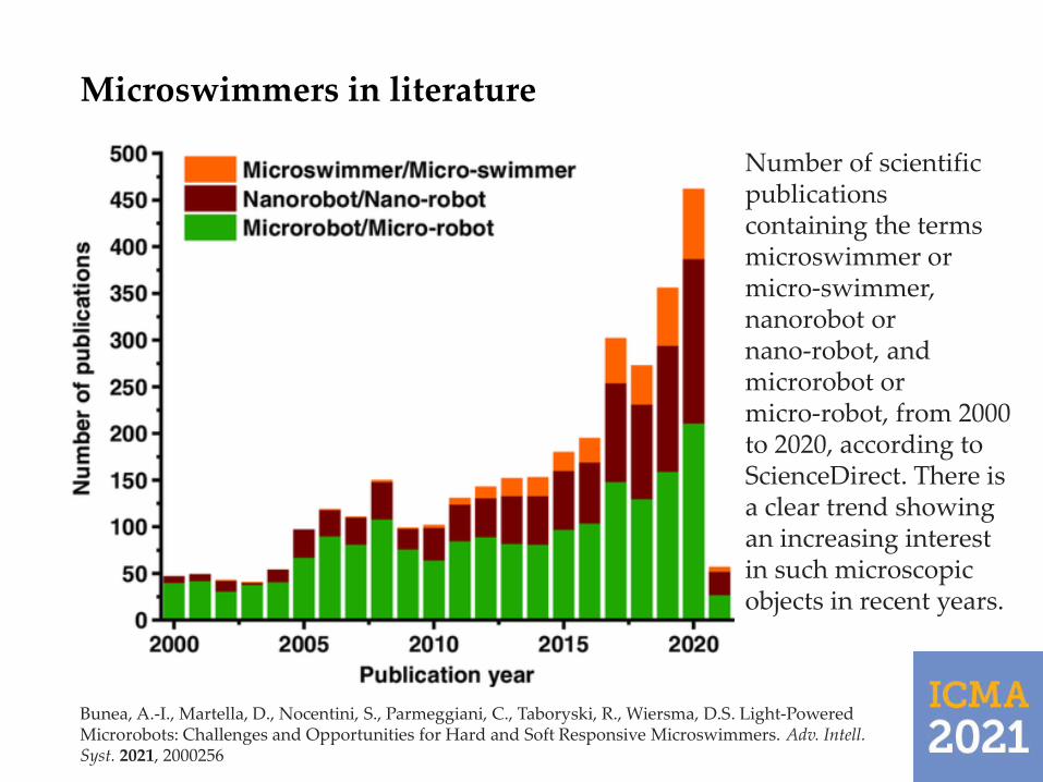
Number of scientific publications containing the terms microswimmer or micro‐swimmer, nanorobot or nano‐robot, and microrobot or micro‐robot, from 2000 to 2020, according to ScienceDirect. There is a clear trend showing an increasing interest in such microscopic objects in recent years.
Bunea, A.-I., Martella, D., Nocentini, S., Parmeggiani, C., Taboryski, R., Wiersma, D.S. Light‐Powered Microrobots: Challenges and Opportunities for Hard and Soft Responsive Microswimmers. Adv. Intell. Syst. 2021, 2000256
Microswimmers for biomedical applications –conceptual drawings on the cover of scientific journals
6
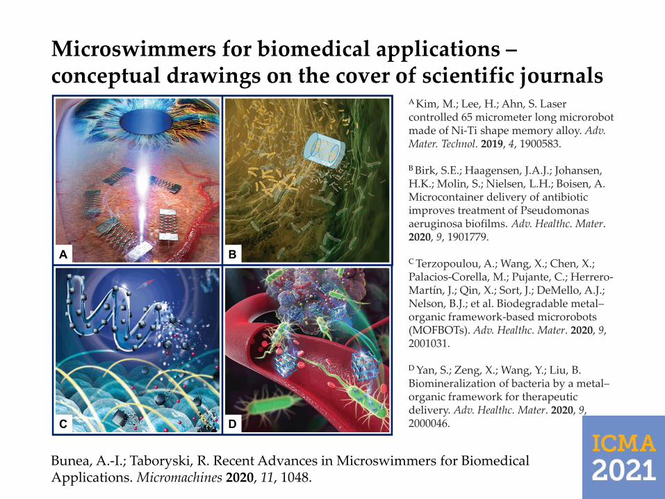
Bunea, A.-I.; Taboryski, R. Recent Advances in Microswimmers for Biomedical Applications. Micromachines 2020, 11, 1048.
A Kim, M.; Lee, H.; Ahn, S. Laser controlled 65 micrometer long microrobot made of Ni-Ti shape memory alloy. Adv. Mater. Technol. 2019, 4, 1900583.
B Birk, S.E.; Haagensen, J.A.J.; Johansen, H.K.; Molin, S.; Nielsen, L.H.; Boisen, A. Microcontainer delivery of antibiotic improves treatment of Pseudomonas aeruginosa biofilms. Adv. Healthc. Mater. 2020, 9, 1901779.
C Terzopoulou, A.; Wang, X.; Chen, X.; Palacios-Corella, M.; Pujante, C.; Herrero-Martín, J.; Qin, X.; Sort, J.; DeMello, A.J.; Nelson, B.J.; et al. Biodegradable metal–organic framework-based microrobots (MOFBOTs). Adv. Healthc. Mater. 2020, 9, 2001031.
D Yan, S.; Zeng, X.; Wang, Y.; Liu, B. Biomineralization of bacteria by a metal–organic framework for therapeutic delivery. Adv. Healthc. Mater. 2020, 9, 2000046.

Microswimmer propulsion and control: Focus on light
7
Among the many different microswimmer propulsion and control modalities, shown on the right, light can be used as a directactuator – in optical trapping and manipulation – or indirect actuator, for inducing e.g. material deformation in light-responsive materials, or thermoplasmonic effects on gold-covered surfaces.
Bunea, A.-I., Martella, D., Nocentini, S., Parmeggiani, C., Taboryski, R., Wiersma, D.S. Light‐Powered Microrobots: Challenges and Opportunities for Hard and Soft Responsive Microswimmers. Adv. Intell. Syst. 2021, 2000256

8
Basic principles of optical trapping for simple gradient force optical tweezers. A) The trapping laser beam is focused with the aid of a high numerical aperture objective into the sample plane and a particle can then be trapped in the focal point of the beam due to the large intensity gradients created. B, C) Trapping in the Mie regime for transparent particles with xy offset (B) or with z‐offset (C) from the beam focus. The refraction of light through the particle (black lines) results in gradient forces (shown as green arrow) attracting the particle towards the beam's focal point, where the light intensity is the highest.
Bunea, A.-I., Glückstad, J. Strategies for Optical Trapping in Biological Samples: Aiming at Microrobotic Surgeons. Laser Photon. Rev. 2019, 13, 1800227
Optical trapping

Optical trapping (2)
9
Optical setup and trapping principle schematic representation. The near-infrared light path used for trapping is shown in red, whereas the visible light source and light path are omitted to simplify the schematics. M—mirror; DM—dichroic mirror; L—lens; SLM—spatial light modulator; A—aperture; Cam—camera; Obj—objective; Cond—condenser; S—sample; BS—beamsplitter. A) A generic HOTs setup. B) The Biophotonics Workstation (BWS) developed at the Technical University of Denmark. A 1080 nm laser beam is shaped and relayed onto the sample plane as a top and bottom counter‐propagating beams. The objects are trapped in between the foci of the top and bottom counter‐propagating beams.
Bunea, A.-I., Martella, D., Nocentini, S., Parmeggiani, C., Taboryski, R., Wiersma, D.S. Light‐Powered Microrobots: Challenges and Opportunities for Hard and Soft Responsive Microswimmers. Adv. Intell. Syst. 2021, 2000256

Optical catapulting in mucus models
10
Light beams shaped by Generalized Phase Contrast (GPC) are excellent for optical catapulting, as the beam intensity is localized around and extends along the catapulting axis.
Bunea, A.-I., Chouliara, M., Harloff-Helleberg, S., Engay, E., Bañas, A., Glückstad, J. Optical catapulting of microspheres in mucus models – toward overcoming the mucus biobarrier. J. Biomed. Opt. 2019, 24, 035001
Catapulting velocities in mucus increase with the laser power, but decrease with the concentration of mucin. However, the velocity trend does not correspond to the viscosity increase trend, which suggests that other factors are involved – we believe that surface interactions between the catapulted objects and mucin play an important role.

3D printing of drug delivery systems
11
3D designs (top) and scanning electron micrographs (bottom) of spherical microswimmers for drug delivery studies involving the mucus biobarrier. The microswimmers were fabricated in the IP-L 780 polyacrylate resin by two-photon polymerization on a Nanoscribe GT. The various features are meant to increase the surface area, allow loading with drugs or excipients, as well as increase penetration through mucus.
Bunea, A.-I., Jakobsen, M.H., Engay, E., Bañas, A., Glückstad, J. Optimization of 3D-printed microstructures for investigating the properties of the mucus biobarrier. Micro Nano Eng. 2019, 2, 41-47

Functionalization of 3D printed polyacrylate structures
12
After fabrication by two-photon polymerization 3D printing, the surface chemistry of polyacrylate structures can be modified in order to confer them the desired properties. For example, an antraquinone amine photolinker can be easily grafted on polymers in the presence of ultraviolet light, and the amine groups enable further functionalization (left). Functionalization with poly-L-lysine followed by the addition of liposomes doped with a fluorescent dye allows for surface modification with a polymer cushioned lipid bilayer, which represents a well established type of model cell membrane 1 (right). Other functionalization options include e.g. plasma treatment or gold coating. Gold coating can be done selectively by sputtering through a mask.
Bunea, A.-I., Jakobsen, M.H., Engay, E., Bañas, A., Glückstad, J. Optimization of 3D-printed microstructures for investigating the properties of the mucus biobarrier. Micro Nano Eng. 2019, 2, 41-47
Sabaté Rovira, D., Nielsen H.M., Taboryski, R., Bunea, A.-I. Additive manufacturing of polymeric scaffolds for biomimetic cell membrane engineering. Mat. Design. 2021, 201, 109486
1 Bunea, A.-I., Harloff-Helleberg, S., Taboryski, R., Nielsen, H.M. Membrane interactions in drug delivery: Model cell membranes and orthogonal techniques. Adv. Colloid Interf. Sci. 2020, 281, 102177

Thermoplasmonic disk microswimmers
13
Disk microswimmers for thermo-plasmonic mixing 3D printed without (left) and with (right) a mask allowing for subsequent selective gold coating of the disk.
Engay, E., Bunea, A.-I., Chouliara, M., Bañas, A., Glückstad, J. Natural convection induced by an optically fabricated and actuated microtool with a thermoplasmonic disk. Opt. Lett. 2018, 43, 3870-3873
Schematic of the flow effect induced by microswimmers without (left) and with (right) gold coating of the disk area.
Natural flow convection observed in a microfluidic channel upon illuminating the gold-coated disk of a single microswimmer. Such localized micromixing can be highly beneficial for lab-on-a-chip biosensing applications.
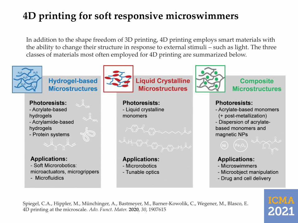
4D printing for soft responsive microswimmers
14
Spiegel, C.A., Hippler, M., Münchinger, A., Bastmeyer, M., Barner-Kowolik, C., Wegener, M., Blasco, E. 4D printing at the microscale. Adv. Funct. Mater. 2020, 30, 1907615
Schematic of the flow effect induced by microswimmers without (left) and with (right) gold coating of the disk area.
In addition to the shape freedom of 3D printing, 4D printing employs smart materials with the ability to change their structure in response to external stimuli – such as light. The three classes of materials most often employed for 4D printing are summarized below.

Soft microswimmers powered by plasmonic heating
15
Mourran, A., Zhang, H., Vinokur, R., Möller, M. Soft Microrobots Employing Nonequilibrium Actuation via Plasmonic Heating. Adv. Mater. 2016, 29, 1604825
A soft microrobot composed of a temperature responsive thin poly(N‐isopropylacrylamide) microgel body loaded with palsmonic gold nanorods is driven by the light‐controlled nonequilibrium dynamics of volume changes. The photothermal response of the microgelenables fast heating/cooling dynamics, while mastering the nonequilibrium response provides control of the complex motion. (Left) Illustration of the locomotion generated by non-reciprocal deformations of the helix. (Right) The helix direction is controlled by the principle curvatures 1/R1, 1/R2 and the mismatch of ϕ toward the length axis of the ribbon. The optical microscopy image shows the helical microgel at 32 °C.

Conclusions
16
Microswimmers have gained significant momentum for biomedical applications.
Two-photon polymerization 3D printing enables the fabrication of microswimmers with complex shapes and sub-micrometer critical dimensions.
Using stimuli-responsive materials for 3D printing facilitates “4D printing”, which further enables adaptable microswimmers.
Surface functionalization can e.g. help confer microswimmers selected properties, facilitate subsequent functionalization, or enable thermoplasmonic effects.
Different propulsion and control modalities are available for microswimmers, including using light, which is a very flexible actuator.
Light can be employed as direct actuator for optical trapping and manipulation, or as indirect actuator for inducing e.g. thermoplasmonic effects or shape changes in light-responsive polymers.

Outlook - essential considerations for developing biomedical microswimmers for in vivo applications
17
Ceylan, H.; Yasa, I.C.; Kilic, U.; Hu, W.; Sitti, M. Translational prospects of untethered medical microrobots. Prog. Biomed. Eng.
2019, 1, 012002.
Before micro-swimmers become truly useful biomedical tools, the puzzle must be solved…

Acknowledgments
VILLUM FONDEN, grant numbers 34424 and 00022918.
Novo Nordisk Foundation, grant number NNF16OC0021948.
18