Embed Size (px)
Citation preview
materials
Article
Microtensile Bond Strength of Fiber-Reinforced and ParticulateFiller Composite to Coronal and Pulp Chamber Floor Dentin
Anja Baraba 1,*, Samir Cimic 2, Matteo Basso 3, Andrei C. Ionescu 4 , Eugenio Brambilla 4 and Ivana Miletic 1
�����������������
Citation: Baraba, A.; Cimic, S.; Basso,
M.; Ionescu, A.C.; Brambilla, E.;
Miletic, I. Microtensile Bond Strength
of Fiber-Reinforced and Particulate
Filler Composite to Coronal and Pulp
Chamber Floor Dentin. Materials 2021,
14, 2400. https://doi.org/10.3390/
ma14092400
Academic Editors: Nikolaos Silikas
and Javier Gil
Received: 1 March 2021
Accepted: 30 April 2021
Published: 5 May 2021
Publisher’s Note: MDPI stays neutral
with regard to jurisdictional claims in
published maps and institutional affil-
iations.
Copyright: © 2021 by the authors.
Licensee MDPI, Basel, Switzerland.
This article is an open access article
distributed under the terms and
conditions of the Creative Commons
Attribution (CC BY) license (https://
creativecommons.org/licenses/by/
4.0/).
1 Department of Endodontics and Restorative Dentistry, School of Dental Medicine, Gunduliceva 5,10 000 Zagreb, Croatia; [email protected]
2 Department of Removable Prosthodontics, School of Dental Medicine, Gunduliceva 5, 10 000 Zagreb, Croatia;[email protected]
3 Department of Dentistry, Galeazzi Institute, University of Milan, via R. Galeazzi 4, 20161 Milan, Italy;[email protected]
4 Department of Biomedical, Surgical and Dental Sciences, Oral Microbiology and Biomaterials Laboratory,University of Milan, via Pascal, 36, 20133 Milan, Italy; [email protected] (A.C.I.);[email protected] (E.B.)
* Correspondence: [email protected]
Abstract: This ex vivo study aimed to compare the microtensile bond strength of fiber-reinforced andparticulate filler composite to coronal and pulp chamber floor dentin using a self-etching adhesivesystem. Coronal dentin of 40 human molar teeth was exposed by cutting occlusal enamel witha low-speed saw. Teeth were then randomly divided into two groups (n = 20). The first groupwas left as is, while in the second group, pulp chamber floor dentin was exposed by trepanation.After placement of a self-etching adhesive system (G-aenial Bond, GC, Tokyo, Japan), groups werefurther divided into two sub-groups (n = 10) according to the type of composite: fiber-reinforcedcomposite (EP, everX Posterior, GC, Tokyo, Japan) and particulate filler composite (GP, G-aenialPosterior, GC, Tokyo, Japan). Then, composite blocks were built up. Sticks (1.0 × 1.0 mm2) were ob-tained from each specimen by sectioning, then microtensile bond strength (µTBS) test was performed.Statistical analysis included one-way ANOVA test and Student’s t-test (p < 0.05). µTBS values were22.91 ± 14.66 and 24.44 ± 13.72 MPa on coronal dentin, 14.00 ± 5.83 and 12.10 ± 8.89 MPa on pulpchamber floor dentin for EP and GP, respectively. Coronal dentin yielded significantly higher µTBSthan pulp chamber floor dentin (p < 0.05), independently from the tested composites.
Keywords: microtensile bond strength test; fiber-reinforced composite; particulate filler composite
1. Introduction
Reduced coronal and radicular residual tissues due to caries, previous restorations,and tissue removal during access cavity and root canal preparation make the restorationof endodontically treated teeth challenging [1–3]. Pulp chamber floor features account foradditional potential difficulty while restoring a tooth following the root canal treatment.According to SEM analysis by Kijsamanmith et al. [4], pulpal floor dentin showed irregulardome-shaped calcospherites of varying size and open dentinal tubules without smear layersince chamber floor is usually not contacted by cutting instruments during access cavitypreparation. Tubule density averaged 24,500 tubules per mm2 [4] meaning that the tubuledensity of pulp chamber floor is lower than coronal dentin [5]. In addition to specificmorphological characteristics, adhesion to pulp chamber dentin may also be affected bythe use of irrigants, most often sodium hypochlorite and EDTA. Such treatments alterthe dentin’s organic and mineral content and influence the interaction with the adhesivematerials used for coronal sealing. These factors contribute to weakening the structureand composition of endodontically treated teeth [6], making restorations more prone tofailure if placed on endodontically treated teeth than on vital teeth [7]. Resin composite
Materials 2021, 14, 2400. https://doi.org/10.3390/ma14092400 https://www.mdpi.com/journal/materials
Materials 2021, 14, 2400 2 of 11
materials are the first choice for direct restorations after endodontic treatment, includingcomposites with short fibers as fillers which have improved mechanical properties com-pared to conventional composites based on particulate fillers only [8] and might thus helpreduce restoration failures. The short fibers, when randomly oriented, provide isotropicreinforcing effect, meaning that the strength of the material is the same in all directions [9].Bijelic-Donova et al. [10] showed that fiber-reinforced composite had significantly higherfracture resistance and higher compressive fatigue limits than particulate filler composite.Furthermore, glass fiber-reinforced composite substructure may aid in eliminating crackpropagation and root fractures [11]. Another two studies showed that fiber-reinforcedcomposite provided endodontically treated teeth with superior fracture resistance [12,13].Among the fiber-reinforced materials that showed this positive behavior, EverX Poste-rior (GC, Tokyo, Japan) is a bulk-fill material placed in increment depths up to 5 mm,which simplifies and speeds up the placement of composite restorations, reducing tech-nique sensitivity. The placement of larger increments of composite material and shortingthe clinical procedure is tempting alternative especially when restoring endodonticallytreated teeth and according to scientific data the effectiveness of bulk composite materialsis comparable to conventional resin composites [14,15].
However, to the best of the authors’ knowledge, there is a lack of data regarding themicrotensile bond strength of short fiber bulk-fill composite to the pulp chamber floorcompared to coronal dentin.
This study, therefore, aimed to compare microtensile bond strength of fiber-reinforcedand particulate filler composite to coronal and pulp chamber floor dentin using a self-etching adhesive system.
2. Materials and Methods2.1. Specimen Preparation
Forty sound human molar teeth, extracted for periodontal or orthodontic reasons,were obtained for the experiment under the approval of the Ethical Committee of theSchool of Dental Medicine, University of Zagreb, Croatia. After extraction, the teeth werethoroughly cleaned using brushes and curettes and stored in 1% chloramine solution at4 ◦C until use. All teeth were randomly divided into two experimental groups (n = 20per group, Figure 1). In the first group, coronal dentin was exposed by cutting occlusalenamel with a diamond blade mounted on a low-speed water-cooled saw (Isomet 1000,Buehler, Dusseldorf, Germany, running at 200 rpm) to obtain a flat dentin surface (Figure 2).The dentin surface was polished with sandpapers of increasing grit (400, 600, 1000) to forma smear layer on dentin’s bonding surface. In the second group, pulp chamber floor dentinwas exposed by trepanation, after which chemo-mechanical instrumentation and root canalfilling were performed. The root canals were instrumented using Reciproc instrumentssize R25 (VDW, Munich, Germany) according to the manufacturer’s instructions. Dur-ing instrumentation, canals were irrigated with 2.5% NaOCl using a 27-gauge needle and a2 mL syringe. Root canals were rinsed with 2 mL of 17% EDTA (pH = 7.7) for one minuteto remove the smear layer, and final irrigation was conducted with saline. Root canalswere dried using Reciproc paper points size R25 (VDW) and obturated with a Reciprocgutta-percha cone size R25 (VDW) and AH Plus sealer (DeTrey Dentsply, Konstanz, Ger-many). After obturation, excess gutta-percha was removed with hot pluggers 1 mm fromthe cementoenamel junction (Figure 2). All roots were stored at 37 ◦C for one week to allowfor the sealer to set.
Materials 2021, 14, 2400 3 of 11
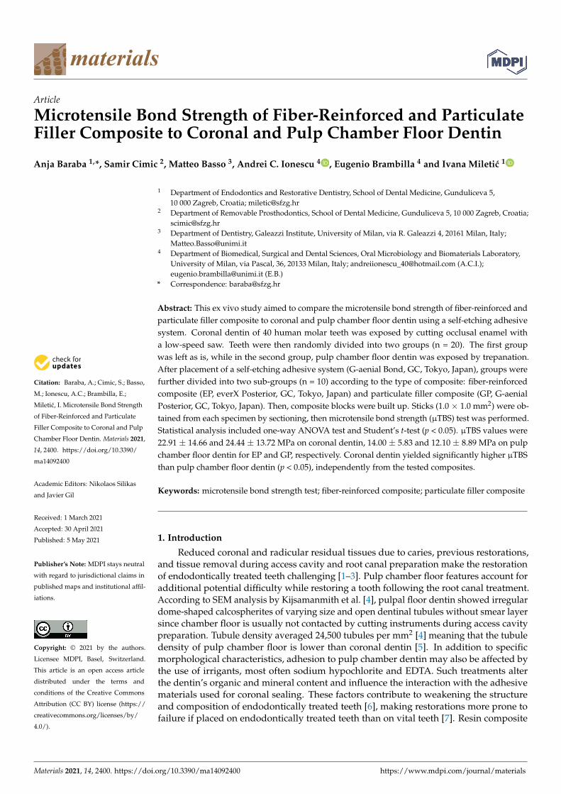
Figure 1. Experimental groups: 40 sound human molar teeth were divided into two groups according to the exposureof coronal dentin (n = 20) or pulp chamber floor dentin (n = 20). Each group was further divided into two subgroupsdepending on the material used for composite block build-up.
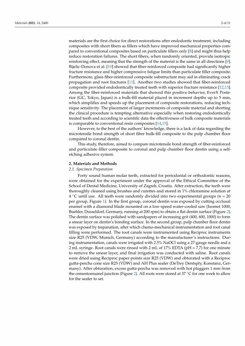
Figure 2. Experimental setup: (1.a) In the first group (n = 20), molar teeth had coronal dentin exposed by cutting of occlusalenamel using diamond blade of Isomet saw to obtain a flat dentin surface for adhesive procedure. (1.b) In the second group(n = 20), root canal treatment of molar teeth was performed. (2.a) and (2.b) In both experimental groups (n = 40), compositebuild-ups were made with the test (fiber-reinforced) or control (particulate filler) composite. (3.a) and (3.b) After compositebuild-ups, all teeth were embedded into acrylic resin in order to prepare them for sectioning. (4.a) and (4.b) Teeth weresectioned longitudinally using a diamond blade of Isomet saw to obtain 1 × 1 mm2 beam-shaped sticks. (5.) Each stickwas glued to metal plates and placed in universal testing machine and a tensile load was applied at a crosshead speed of0.5 mm/min until (6.) the stick fractured and maximum load at failure was recorded.
Both experimental groups were randomly divided into two subgroups (n = 10) accord-ing to the type of composite used for the restoration: fiber-reinforced composite (test, everXPosterior, GC, Tokyo, Japan) and particulate filler composite (control, G-aenial Posterior,GC, Tokyo, Japan) (Figure 1, Table 1).

Materials 2021, 14, 2400 4 of 11
Table 1. Chemical composition, weight percentage of the organic matrix, and volume percentage of fibers and fillers of thetested composites.
Material Manufacturer Composition
EverX Posterior (test) GC, Tokyo, Japan Bis-GMA, PMMA, TEGDMA,74.2 wt%, 53.6 vol% short E-glass fibers, barium glass
G-aenial Posterior (control) GC, Tokyo, JapanUDMA, dimethacrylate-comonomers,77 wt%, 65 vol% pre-polymerized silica/lanthanoid fluridefluoraluminosilicate/silica
The bonding surface in all four groups was washed with distilled water and gentlydried with a dental unit air syringe (Kavo Primus, 1058 S/TM/C/G, Biberach/Riss, Ger-many) before performing the adhesive procedure. The bonding surface was prepared andbonded according to the manufacturers’ instructions using G-Bond (GC, Tokyo, Japan).After bonding, a composite resin block was built-up on the bonding surface, with the appli-cation of layers of the material not thicker than 2 mm for the particulate composite andnot thicker than 5 mm for the bulk fiber-reinforced composite (Figure 2). Each layer wascured with a LED light (Bluephase, Ivoclar Vivadent, Schaan, Liechtenstein, 1200 mW/cm2,soft start) for 20 s, keeping the light tip perpendicular to the substrate and the tip 5 mmaway from the dentin surface. In groups with the pulp chamber dentin exposed, resinblock was built-up to the occlusal surface level keeping the light tip in contact with theocclusal surface. Where coronal dentin was exposed, a 5 mm high composite resin blockwas built-up. All specimens were then stored in distilled water at 37 ◦C for 24 h, andthen they were embedded into acrylic resin (Orthocryl, Dentaurum, Ispringen, Germany)(Figure 2). Afterward, the teeth were longitudinally sectioned (Isomet 1000, same pa-rameters as previously specified) to obtain 1 mm × 1 mm beam-shaped sticks (Figure 2).Before further testing, each beam was checked under a stereomicroscope (Olympus SZX-12,Optical Co, Europe, GMBH, Hamburg, Germany) to verify that the adhesive interfacewas perpendicular its long axis. Only beams with the latter characteristic were used inthis experiment.
2.2. Microtensile Bond Strength Test
The microtensile bond strength was tested with a universal testing machine (TriaxDigital 50, Controls, Milan, Italy). Each beam’s ends were glued with cyanoacrylateadhesive (Loctite gel, Henkel, Dusseldorf, Germany) to specifically designed metal plates.A tensile load was applied to each beam at a crosshead speed of 0.5 mm/min until itfractured (Figures 2 and 3). The maximum load at failure was recorded in newtons (N).Beams were then observed under a stereomicroscope to assess the failure mode (adhesive,cohesive, or mixed). A failure at the dentin/adhesive interface was considered as anadhesive failure; if it occurred in the composite resin material or dentin, the failure wasconsidered cohesive, and failure was considered mixed if it involved the dentin/adhesiveinterface and the composite resin material or dentin at the same time. The cross-sectionalarea at the fracture site was measured to the nearest 0.01 mm with a digital caliper (RocInternational Industry Co., Ltd., Guangdong, China) so the bond strength at failure couldbe calculated and expressed in MPa.
Materials 2021, 14, 2400 5 of 11
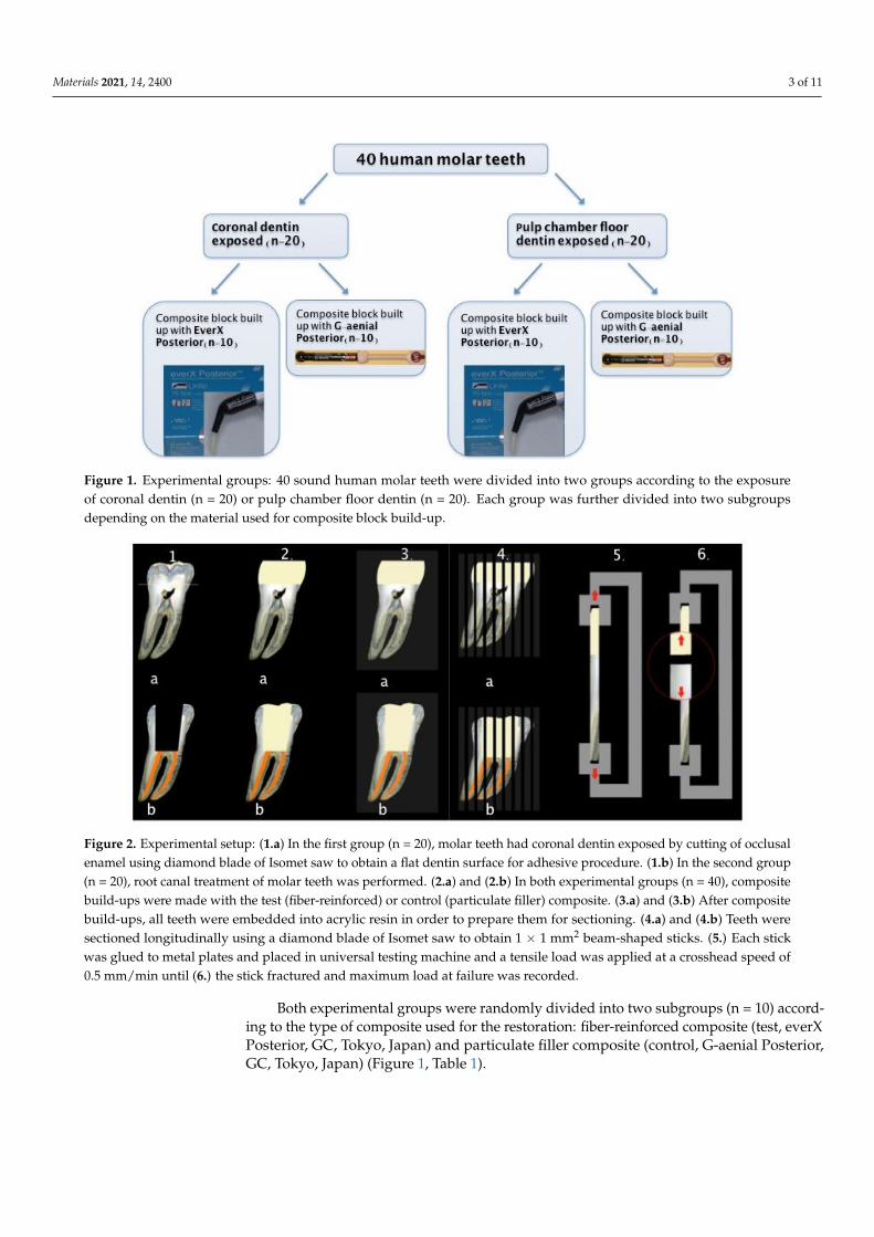
Figure 3. After obtaining 1 mm × 1 mm beam-shaped sticks by longitudinally sectioning teethusing Isomet saw, each stick was glued with cyanoacrylate adhesive to specifically designed metalplates and placed in universal testing machine Afterward, tensile load was applied to each beam at acrosshead speed of 0.5 mm/min until it fractured.
2.3. Statistical Analysis
Statistical analyses were performed using JMP 10.0 software (SAS Institute, Cary, NC,USA). Normal distribution of data was checked using Shapiro-Wilk’s test, and homogeneityof variances was verified using Levene’s test. A two-way ANOVA model was usedconsidering the dentin type and the resin composite as fixed factors. Subsequently, posthoc Student’s t-test was used to highlight significant differences between groups. In alltests, the level of significance was set at 0.05.
3. Results
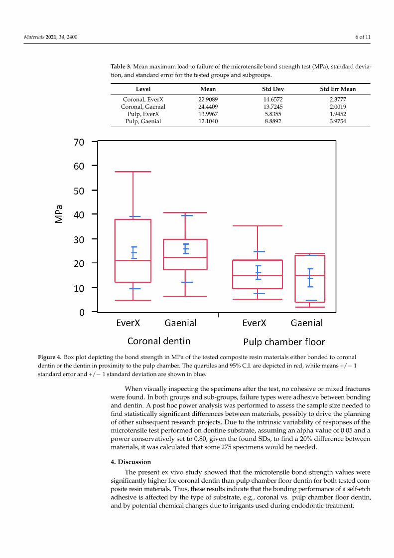
Data were log-transformed to approach a normal distribution. Two-way ANOVA didnot show a significant interaction between factors (Table 2), meaning that the tested typesof composites had the same behavior on both tested dentin substrates. Coronal dentinshowed significantly higher microtensile bond strength values than pulp chamber floordentin (Table 3, Figure 4) irrespective of the tested composite type (p = 0.0079). There wasno statistically significant difference in microtensile bond strength between test and controlresin composites (p = 0.4617).
Table 2. Results of two-way ANOVA analyzing the effect of the two factors and their interaction. Star in superscriptindicates a significant effect (p < 0.05).
Source Nparm Sum of Squares F Ratio Prob > F
Dentin location 1 1258.7711 6.9517 0.0098 *Composite 1 0.3628 0.0020 0.9644
Dentin location*Composite 1 32.6946 0.1806 0.6719
Materials 2021, 14, 2400 6 of 11
Table 3. Mean maximum load to failure of the microtensile bond strength test (MPa), standard devia-tion, and standard error for the tested groups and subgroups.
Level Mean Std Dev Std Err Mean
Coronal, EverX 22.9089 14.6572 2.3777Coronal, Gaenial 24.4409 13.7245 2.0019
Pulp, EverX 13.9967 5.8355 1.9452Pulp, Gaenial 12.1040 8.8892 3.9754
Figure 4. Box plot depicting the bond strength in MPa of the tested composite resin materials either bonded to coronaldentin or the dentin in proximity to the pulp chamber. The quartiles and 95% C.I. are depicted in red, while means +/− 1standard error and +/− 1 standard deviation are shown in blue.
When visually inspecting the specimens after the test, no cohesive or mixed fractureswere found. In both groups and sub-groups, failure types were adhesive between bondingand dentin. A post hoc power analysis was performed to assess the sample size needed tofind statistically significant differences between materials, possibly to drive the planningof other subsequent research projects. Due to the intrinsic variability of responses of themicrotensile test performed on dentine substrate, assuming an alpha value of 0.05 and apower conservatively set to 0.80, given the found SDs, to find a 20% difference betweenmaterials, it was calculated that some 275 specimens would be needed.
4. Discussion
The present ex vivo study showed that the microtensile bond strength values weresignificantly higher for coronal dentin than pulp chamber floor dentin for both tested com-posite resin materials. Thus, these results indicate that the bonding performance of a self-etchadhesive is affected by the type of substrate, e.g., coronal vs. pulp chamber floor dentin,and by potential chemical changes due to irrigants used during endodontic treatment.
Materials 2021, 14, 2400 7 of 11
In agreement with the current study results, previous studies have found bondstrength to pulp chamber floor dentin to be lower than coronal dentin [4,16,17]. Ac-cording to the literature, bond strength to dentine tissue is influenced by structural char-acteristics such as the diameter and number of dentin tubules as well as the relativeamount of peritubular and intertubular dentin [18,19]. Pulpal dentin contains predentin,irregular secondary dentin, and a high tubule density with large diameters. All thesevariations contribute to making pulp chamber floor dentin a relatively challenging bondingsurface [20]. Overall, bond strength of resin composites to dentin can be regarded as asummation of the individual bond strengths provided by surface adhesion, resin tags,and hybrid layer [21]. Taking into consideration the tubules density and diameter andthe amount of peri- and intratubular dentin, hybrid layer formation is expected to be thecrucial factor providing bond strength to coronal dentin, with little contribution from resintags. However, in pulp floor dentin, resin tags would contribute most of the bond strength,while a reduced contribution by hybrid layer formation is due to the limited amountof intertubular dentin available [22]. Interestingly, Lohbauer et al. [23] showed that theformation of resin tags does not influence the bonding strength of a one-step self-etchingadhesive such as the one used in the present study. This observation and the reducedhybrid layer formation when a self-etching adhesive is used for bonding to pulp chamberfloor dentin may explain the low bond strength obtained on pulp chamber floor dentin inthis study.
It is acknowledged that, due to the complex anatomy of the endodontic space, the me-chanical instrumentation of root canals alone is not effective in removing microorganismsand debris from root canals [24,25]. As large areas of root canal dentin remain untouchedby the endodontic instruments, using irrigant solutions is essential for lubrication, de-bridement, dissolution of microbial structures and biofilms, and removal of the smearlayer prior to root canal obturation [26]. Sodium hypochlorite is routinely used duringendodontic treatment. Such a principle was seen to induce the oxidation of some compo-nents in the dentin matrix, forming protein radicals that will compete with the increasingvinyl-free radicals created by the light-activation of resin adhesives to premature chaintermination and incomplete polymerization [27]. The hypochlorite anion can also infiltratemineralized collagen and destroy collagen fibrils [28]. Moreover, decreases in calcium andphosphorus levels and changes in dentin’s mechanical properties, such as elastic modulus,flexural strength, and microhardness, were reported after irrigation of root canals withsodium hypochlorite [29]. Therefore, it is likely that sodium hypochlorite solution, asused in the present study being considered gold standard for root canal irrigation, maynegatively influence the bond strength to pulp chamber floor dentin, possibly explainingthe lower bond strength results for the pulp chamber floor dentin. The experimental setupof this study was not designed to allow us to ascertain to what point the contribution ofdentin characteristics rather than irrigant solution could have negatively influenced thebond strength to the pulp chamber floor. The latter question may be the primary aim offuture studies. EDTA was used as a chelating amino acid in the current study to removethe smear layer before root canal obturation. Several studies have evaluated the effect ofEDTA preconditioning on the bond strength of self-etch adhesives to dentin and reportedthat EDTA was effective in improving dentin bonding for all-in-one adhesives [30–34].This was not the case in the present study. However, it must be noted that EDTA was incontact with the pulp chamber floor dentin for only one minute, while the substrate wasexposed to sodium hypochlorite for a prolonged time during the entire process of rootcanals instrumentation. Therefore, the detrimental effect of sodium hypochlorite on bondstrength might have been more pronounced.
The current study also aimed to test the microtensile bond strength of two differ-ent types of composite materials, e.g., fiber-reinforced and particulate filler composite.Particulate filler composites have been present on the market for decades, but a morerecent restorative dentistry approach uses fiber-reinforced bulk-fill composite as a base inextended restorations. One of the clinical advantages of using such a material is placing
Materials 2021, 14, 2400 8 of 11
it in increments up to 4–5 mm, while achieving, at the same time, the mechanical perfor-mances needed to ensure extended restorations longevity. Bulk-fill materials can avoidseveral drawbacks of classical techniques, usually used for particulate filler compositematerial, including placing the material in 2-mm thickness increments. The latter exposesthe restoration to the incorporation of voids between layers, failure of bonding, and ex-tended treatment time [35]. Furthermore, short E-glass fibers added to the organic matrixof everX Posterior can stop crack initiation and propagation, increase fracture toughnessand decrease the modulus of elasticity to values more similar to dentin tissues [36–40].All these features may be especially beneficial when restoring endodontically treated teeth.
An earlier study showed that the bond strength to dentin was dependent on thecomposition of the restorative material [41] due to mechanical properties [42] or surface freeenergy characteristics of composites [43]. Steiner et al. [44] ascertained that the strongestinfluence on bond strength was exerted by the resin composite type, followed by theadhesive system, while the choice of the curing intensity was not significant. Therefore,it is essential to collect data about the bond strength of all composite materials becomingavailable on the market. Combined with other scientific data collected through in vitro,ex vivo, and in vivo studies, these results may help when choosing a restorative material.Indeed, according to Cekic et al. [45], the presence of fibers in the material and theirorientation may also influence bond strength values. However, in the present study, nodifference in microtensile bond strength between the tested materials, fiber-reinforcedand particulate filler composites, was found, which is in agreement with the results ofTsujimoto et al. [46].
One of the indications for using fiber-reinforced composite is endodontically treatedteeth, and this study found bond strength to pulpal floor dentin significantly lower thancoronal dentin. Minimum bond strength of 17 MPa to dentin and enamel was identifiedas necessary to ensure successful adhesion [47]. In the present study, bond strength topulpal floor dentin was lower than the 17 MPa threshold in both tested materials. For theparticulate composite resin, it means that the polymerization shrinkage is greater thanthe bond strength to tooth structures, possibly resulting in marginal gap formation andcomposite failure [48]. However, the tested fiber-reinforced composite has a very lowpolymerization shrinkage strain (0.17%) primarily due to the random orientation of itsfibers, which minimizes shrinkage during and after curing [45]. Jung and Park showed thatresin-based bulk fill composites showed good marginal adaptation, even better than that offlowable bulk-filled materials. They assumed that a lower level of polymerization shrinkageand polymerization shrinkage stress was mainly responsible for this finding because itcould induce less polymerization shrinkage force at the margin [49]. These considerationssuggest that the tested fiber-reinforced composite might be a better choice than a particulatefiller composite to restore endodontically treated teeth.
The microtensile test was used in this study to test the bond strength to coronal andpulp chamber floor dentin due to several advantages, such as multiple specimens beingobtained from a single tooth and the stress more uniformly distributed during loadingacross the interface, especially for untrimmed specimens as the ones prepared for thepresent study [50,51]. It must be noted that the measured bond strength values may beconsidered reliable in the case of adhesive failures only. For both tested materials, only theadhesive type of failure was observed, indicating that the actual interfacial bond strengthto dentin was determined.
The current study results must be interpreted with caution due to their possible limi-tations. The bond between hard dental tissues and restoration and its resistance to fractureis complex and cannot be correlated with the microtensile bond strength test results in asimple way. In vitro tests such as the used microtensile bond strength test cannot directlypredict the clinical behavior of materials. Nevertheless, it may provide particular insightinto the adhesion performances of restorative materials. Further research is necessary toinvestigate the influence of different types of adhesive systems on bond strength to sucha peculiar substrate as pulp chamber floor dentin. Furthermore, thermal and mechanical
Materials 2021, 14, 2400 9 of 11
cycling of specimens before bond strength tests can be considered when designing clinicallyrelevant in vitro or ex vivo models of accelerated aging.
5. Conclusions
The present study showed that microtensile bond strength to pulp chamber floordentin is significantly lower than coronal dentin when a one-step, self-etch adhesive is used.The microtensile bond strength values were not influenced by the type of composite usedfor the build-up. In a translational sense, fiber-reinforced and particulate filler compositeare equally good alternatives for clinicians to restore vital and nonvital teeth.
Author Contributions: Conceptualization, A.B. and I.M.; methodology, A.B. and S.C.; software,A.C.I.; validation, A.B. and I.M.; formal analysis, M.B.; investigation, A.B., A.C.I. and S.C.; resources,E.B.; data curation, A.C.I.; writing—original draft A.B.; writing—reviewing A.B., A.C.I. and I.M.; vi-sualization, A.B.; supervision, I.M.; project administration, A.B.; funding acquisition, A.B. All authorshave read and agreed to the published version of the manuscript.
Funding: This research was funded by the University of Zagreb grant 2020. Under the title “Mechan-ical properties of bioactive materials in vivo and ex vivo”.
Institutional Review Board Statement: The study was conducted according to the guidelines of theDeclaration of Helsinki and approved by the Ethical Committee of the School of Dental Medicine,University of Zagreb, Croatia.
Informed Consent Statement: Not applicable.
Data Availability Statement: Data sharing is not applicable to this article.
Conflicts of Interest: The authors declare no conflict of interest. The funders had no role in the designof the study; in the collection, analyses, or interpretation of data; in the writing of the manuscript,or in the decision to publish the results.
References1. Lin, C.L.; Chang, C.H.; Ko, C.C. Multifactorial analysis of an MOD restored human premolar using auto-mesh finite element
approach. J. Oral Rehabil. 2001, 28, 576–585. [CrossRef]2. Rao, M.S.; Shameem, A.; Nair, R.; Ghanta, S.; Thankachan, R.P.; Issac, J.K. Comparison of the remaining dentin thickness in the
root after hand and four rotary instrumentation techniques: An in vitro study. J. Contemp. Dent. Pract. 2013, 14, 712–717.3. Panitvisai, P.; Messer, H.H. Cuspal deflection in molars in relation to endodontic and restorative procedures. J. Endod. 1995,
21, 57–61. [CrossRef]4. Kijsamanmith, K.; Timpawat, S.; Harnirattisai, C.; Messer, H.H. Micro-tensile bond strengths of bonding agents to pulpal floor
dentine. Int. Endod. J. 2002, 35, 833–839. [CrossRef]5. Lenzi, T.L.; Guglielmi, C.A.B.; Arana-Chavez, V.E.; Raggio, D.P. Tubule density and diameter in coronal dentin from primary and
permanent human teeth. Microsc. Microanal. 2013, 19, 1–5. [CrossRef] [PubMed]6. Aslantas, E.E.; Buzoglu, H.D.; Altundasar, E.; Serper, A. Effect of EDTA, sodium hypochlorite, and chlorhexidine gluconate with
or without surface modifiers on dentin microhardness. J. Endod. 2014, 40, 876–879. [CrossRef] [PubMed]7. Lempel, E.; Lovasz, B.V.; Bihari, E.; Krajczár, K.; Jeges, S.; Tóth, Á.; Szalma, J. Long-term clinical evaluation of direct resin
composite restorations in vital vs. endodontically treated posterior teeth—Retrospective study up to 13 years. Dent. Mater. 2019,35, 1308–1318. [CrossRef] [PubMed]
8. Alshabib, A.; Silikas, N.; Watts, D.C. Hardness and fracture toughness of resin-composite materials with and without fibers.Dent. Mater. 2019, 35, 1194–1203. [CrossRef] [PubMed]
9. Garoushi, S.; Gargoum, A.; Vallittu, P.K.; Lasilla, L. Short fiber-reinforced composite restorations: A review of the current literature.J. Investig. Clin. Dent. 2018, 9, e12330. [CrossRef]
10. Bijelic-Donova, J.; Garoushi, S.; Vallittu, P.K.; Lassila, L.V. Mechanical properties, fracture resistence, and fatigue limits of shortfiber reinforced composite resin. J. Prosthet. Dent. 2016, 115, 95–102. [CrossRef] [PubMed]
11. Garoushi, S.; Vallittu, P.K.; Lassila, L.V. Direct restoration of severely damaged incisors using short fiber-reinforced compositeresin. J. Dent. 2007, 35, 731–736. [CrossRef]
12. Garlapati, T.G.; Krithikadatta, J.; Natanasabapathy, V. Fracture resistance of endodontically treated teeth restored with short fibercomposite used as a core material—An in vitro study. J. Prosthodont. Res. 2017, 61, 464–470. [CrossRef]
13. Ozsevik, A.S.; Yildrim, C.; Aydin, U.; Culha, E.; Surmelioglu, D. Effect of fiber-reinforced composite on the fracture resistence ofendodontically treated teeth. Aust. Endod. J. 2016, 42, 82–87. [CrossRef]
Materials 2021, 14, 2400 10 of 11
14. Arbildo-Vega, H.I.; Lapinska, B.; Panda, S.; Lamas-Lara, C.; Khan, A.S.; Lukomska-Szymanska, M. Clinical Effectiveness ofBulk-Fill and Conventional Resin Composite Restorations: Systematic Review and Meta-Analysis. Polymer 2020, 12, 1786.[CrossRef]
15. Chesterman, J.; Jowett, A.; Gallacher, A.; Nixon, P. Bulk-fill resin-based composite restorative materials: A review. Br. Dent. J.2017, 222, 337–344. [CrossRef]
16. Schreiner, R.F.; Chappell, R.P.; Glaros, A.G.; Eick, J.D. Micro-tensile testing of dentin adhesives. Dent. Mater. 1998, 14, 194–201.[CrossRef]
17. Tanumiharja, M.; Burrow, M.F.; Tyas, M.J. Micro-tensile bond strengths of seven dentin adhesive systems. Dent. Mater. 2007,16, 180–187. [CrossRef]
18. Marshall, G.W.; Marshall, S.J.; Kinney, J.H.; Balooch, M. The dentin substrate: Structure and properties related to bonding. J. Dent.1997, 25, 441–458. [CrossRef]
19. Pashley, D.H.; Carvalho, R.M. Dentine permeability and dentine adhesion. J. Dent. 1997, 25, 355–372. [CrossRef]20. Schellenberg, U.; Krey, G.; Bosshardt, D.; Nair, P. Numerical density of dentinal tubules at the pulpal wall of human permanent
premolars and third molars. J. Endod. 1992, 18, 104–109. [CrossRef]21. Gwinnett, A.J. Quantitative contribution of resin infiltration/hybridization to dentin bonding. Am. J. Dent. 1993, 6, 7–9.22. Pashley, D.H.; Sano, H.; Ciucchi, B.; Carvalho, R.M.; Russell, C.M. Bond strength versus dentin structures: A modeling approach.
Arch. Oral Biol. 1995, 40, 110991118. [CrossRef]23. Lohbauer, U.; Nikolaenko, S.A.; Petschelt, A.; Frankenberg, R. Resin tags do not contribute to dentin adhesion in self-etching ad-
hesives. J. Adhes. Dent. 2008, 10, 97–103.24. Haapasalo, M.; Endal, U.; Zandi, H.; Coil, J.M. Eradication of endodontic infection by instrumentation and irrigation solutions.
Endodontics 2005, 10, 77–102. [CrossRef]25. Peters, O.A.; Schönenberger, K.; Laib, A. Effects of four Ni-Ti preparation techniques on root canal geometry assessed by micro
computed tomography. Int. Endod. J. 2001, 34, 221–230. [CrossRef] [PubMed]26. Haapasalo, M.; Shen, Y.; Qian, W.; Gao, Y. Irrigation in endodontics. Dent. Clin. N. Am. 2010, 54, 291–312. [CrossRef] [PubMed]27. Lai, S.C.; Mak, Y.F.; Cheung, G.S.; Osorio, R.; Toledano, M.; Carvalho, R.M.; Tay, F.R.; Pashley, D.H. Reversal of compromised
bonding to oxidized etched dentin. J. Dent. Res. 2001, 80, 1919–1924. [CrossRef] [PubMed]28. Gu, L.S.; Huang, X.Q.; Griffin, B.; Bergeron, B.R.; Pashley, D.H.; Niu, L.; Tay, F.R. Primum non nocere—The effects of sodium
hypochlorite on dentin as used in endodontics. Acta Biomater. 2017, 61, 144–156. [CrossRef]29. Sim, T.P.; Knowles, J.C.; Ng, Y.L.; Shelton, J.; Gulaivala, K. Effect of sodium hypochlorite on mechanical properties of dentine and
tooth surface strain. Int. Endod. J. 2001, 34, 120–132. [CrossRef]30. Jacques, P.; Hebling, J. Effect of dentin conditioners on the microtensile bond strength of a conventional and a self-etching primer
adhesive system. Dent. Mater. 2005, 21, 103–109. [CrossRef]31. Shafier, F.; Memarpour, M. Effect of EDTA conditioning on microleakage of four adhesive systems in composite restorations.
J. Dent. 2008, 5, 150–155.32. Kasraei, S.; Azarsina, M.; Khamverdi, Z. Effect of Ethylene diamine tetra acetic acid and sodium hypochlorite solution conditioning
on microtensile bond strength of one-step self-etch adhesive. J. Conserv. Dent. 2013, 16, 243–246.33. Torri, Y.; Hikasa, R.; Iwate, S.; Oyama, F.; Itou, K.; Yoshiyama, M. Effect of EDTA conditioning on bond strength to bovine dentin
promoted by four current adhesives. Am. J. Dent. 2003, 16, 395–400.34. Soares, C.J.; Castro, C.G.; Santos Filho, P.C.; da Mota, A.S. Effect of previous treatments on bond strength of two self-etching
adhesive systems to dental substrate. J. Adhes. Dent. 2007, 9, 291–296.35. Abbas, G.; Fleming, G.J.; Harrington, E.; Shortell, A.C.; Burke, F.J. Cuspal movement and microleakage in premolar teeth restored
with a packable composite cured in bulk or in increments. J. Dent. 2003, 31, 437–444. [CrossRef]36. Goncu Basaran, E.; Ayna, E.; Uctasli, S.; Vallittu, P.K.; Lassila, L.V. Load-bearing capacity of fiber reinforced fixed composite
bridges. Acta Odontol. Scand. 2013, 71, 65–71. [CrossRef]37. Vallittu, P.K. High-aspect ratio fillers: Fiber-reinforced composites and their anisotropic properties. Dent. Mater. 2015, 31, 1–7.
[CrossRef]38. Garoushi, S.; Vallittu, P.K.; Watts, D.C.; Lassila, L.V. Polymerization shrinkage of experimental short glass fiber-reinforced
composite with semi-inter penetrating polymer network matrix. Dent. Mater. 2008, 24, 211–215. [CrossRef] [PubMed]39. Sfondrini, M.F.; Cacciafesta, V.; Scribante, A. Shear bond strength of fiber-reinforced composite nets using two different ad- hesive
systems. Eur. J. Orthod. 2011, 33, 66–70. [CrossRef] [PubMed]40. Ilie, N.; Kessler, A.; Durner, J. Influence of various irradiation processes on the mechanical properties and polymerisation kinetics
of bulk-fill resin based composites. J. Dent. 2013, 41, 695–702. [CrossRef] [PubMed]41. Peutzfeldt, A.; Asmussen, E. Determination of in vitro gap formation of resin composites. J. Dent. 2004, 32, 109–115. [CrossRef]42. Dehoff, P.H.; Anusavice, K.J.; Wang, Z. Three dimensional finite analysis of the shear bond test. Dent. Mater. 1995, 11, 126–131.
[CrossRef]43. Amussen, E.; Peutzdeldt, A. Resin composites: Strength of the bond to dentin versus surface free energy parameters. Dent. Mater.
2005, 21, 1039–1043. [CrossRef]44. Steiner, R.; Edelhoff, D.; Stawarczyk, B.; Dumfahrt, H.; Lente, I. Effect of dentin bonding agents, various resin composites and
curing modes on bond strength to human dentin. Materials 2019, 12, 3395. [CrossRef] [PubMed]
Materials 2021, 14, 2400 11 of 11
45. Cekic, I.; Ergun, G.; Uctsdli, S.; Lassila, L.V. In vitro evaluation of push-out bond strength of direct ceramic inlays to tooth surfacewith fiber-reinforced composite at the interface. J. Prosthet. Dent. 2007, 97, 271–278. [CrossRef] [PubMed]
46. Tsujimoto, A.; Barkmeier, W.W.; Takamizawa, T.; Latta, M.A.; Miyazaki, M. Bonding performance and interfacial characteristicsof short fiber-reinforced resin composite in comparison with other composite restoratives. Eur. J. Oral Sci. 2016, 124, 301–308.[CrossRef]
47. Freedman, G. Contemporary Aesthetic Dentistry; Mosby: St. Louis, MI, USA; Elsevier: Amsterdam, The Netherlands, 2012.48. Garoushi, S.; Sailynoja, A.; Vallittu, P.K.; Lsassila, L. Physical properties and depth of cure of a new short fiber reinforced
composite. Dent. Mater. 2013, 29, 835–841. [CrossRef]49. Jung, J.H.; Park, S.H. Comparison of polymerization shrinkage, physical properties, and marginal adaptation of flowable and
restorative bulk fill resin-based composites. Oper. Dent. 2017, 42, 375–386. [CrossRef] [PubMed]50. Pashley, D.H.; Carvalho, R.M.; Sano, H.; Nakajima, M.; Yoshiyama, M.; Shono, Y.; Fernandes, C.; Tay, F. The microtensile bond
test: A review. J. Adhes. Dent. 1999, 1, 299–309.51. Neves Ade, A.; Coutinho, E.; Cardoso, M.V.; Jaecques, S.; Lambrechts, P.; Sloten, J.V.; van Oosterwyck, H.; van Meerbeek, B.
Influence of notch geometry and interface on stress concentration and distribution in micro-tensile bond strength specimens.J. Dent. 2008, 36, 808–815. [CrossRef]