Embed Size (px)
Citation preview
Sharma et al. European Journal of Pharmaceutical and Medical Research
www.ejpmr.com
713
MIDDLE EAR MUCOSA TUBERCULOSIS: A RARE CASE
Dr. Sunita Singh1, Dr. Sakshi Dahiya
2, Dr. Bharti Sharma*
3, Dr. Rajeev Sen
4
1Senior Professor, Department of Pathology, PGIMS, Rohtak.
2,3Junior Resident, Department of Pathology, PGIMS, Rohtak.
4Head and Senior Professor, Department of Pathology, PGIMS, Rohtak.
Article Received on 19/04/2018 Article Revised on 09/05/2018 Article Accepted on 29/05/2018
INTRODUCTION
Tuberculosis (TB) is a widely prevalent disease in
developing countries and constitutes a major cause of
morbidity and mortality.Various factors have led to an
increase in cases worldwide like immunodeficiency
states particularly HIV infection, increased number of
people living in poor socio-economic conditions,
increased resistance to Anti-tubercular therapy, diabetes
and alcoholism.[1]
Tuberculous otitis media (TOM) is a rare form of
presentation of chronic otitis media and extrapulmonary
TB. Middle ear mucosa tuberculous infection can be
acquired by hematogenous route and rarely by infection
through middle ear perforation. It can even spread
through the eustachian tube while the child feeds on
breast milk or less commonly as a congenital infection.
The typical symptoms and signs which the patient
presents with, include multiple perforations of the
tympanic membrane, painful ear discharge, preauricular
lymphadenopathy, sensorineural hearing loss and other
associated constitutional symptoms. These are often
absent in patients these days, making the diagnosis
difficult which in turn delays the treatment and worsens
prognosis. Some recently included signs include large
perforation, conductive hearing loss that suddenly
becomes sensorineural, pale granulation tissue and dense
secretion. Diagnosis is tricky because the cultures are
usually negative and positive AFB (acid fast bacilli)
smears are uncommon too. Also, histopathological
examination rarely shows TB granulomas but more
frequently indicate necrotising granulomas, if any.
MATERIAL AND METHODS
We are presenting the case of a 20 year old male who
presented with the complaint of on and off bilateral ear
discharge since childhood. He was clinically diagnosed
as having bilateral CSOM (chronic suppurative otitis
media) and his middle ear mucosa biopsy was sent for
confirmation. Grossly, the specimen consisted of
multiple, grey white to grey brown soft tissue pieces
measuring together 0.3x0.2x0.1 cm. On microscopic
examination, the biopsy comprised of mature
cartilaginous tissue and soft tissue which was infilterated
SJIF Impact Factor 4.897
Research Article
ISSN 2394-3211
EJPMR
EUROPEAN JOURNAL OF PHARMACEUTICAL
AND MEDICAL RESEARCH www.ejpmr.com
ejpmr, 2018,5(6), 713-716
*Corresponding Author: Dr. Bharti Sharma
Junior Resident, Department of Pathology, PGIMS, Rohtak.
ABSTRACT
Tuberculosis is a widely prevalent disease in developing countries. Tuberculous otitis media is a rare form of
presentation of chronic otitis media and extrapulmonary Tuberculosis. Middle ear mucosa tuberculous infection
can be acquired by hematogenous route and rarely by infection through middle ear perforation. The typical
symptoms and signs which the patient presents with, include multiple perforations of the tympanic membrane,
painful ear discharge, preauricular lymphadenopathy, sensorineural hearing loss and associated constitutional
symptoms. Diagnosis is tricky because the cultures are usually negative and positive acid-fast bacilli smears are
uncommon too. Also, histopathological examination rarely shows tubercular granulomas but more frequently
indicate necrotising granulomas, if any. A case of 20 year old male, who presented with the complaint of on and off
bilateral ear discharge since childhood, who clinically diagnosed as having bilateral chronic suppurative otitis
media and his middle ear mucosa biopsy was taken and processed, microscopic examination done and biopsy
comprised of mature cartilaginous tissue and soft tissue which was infilterated by mononuclear cells along with
epithelioid cell granulomas, revealing focal necrosis. This was suggestive for tuberculosis. Tuberculosis is the most
common infection worldwide. Fortunately, TB of the middle ear and temporal bone are rare. Disease is usually
unilateral but this patient had bilateral ear involvement. Treatment includes surgical options like tympanoplasty and
radical mastoidectomy. Tuberculosis of the ear is difficult to diagnose on biopsy but once established, it responds
well to therapy. Hence treatment should be early and prompt so as to avoid other complications.
KEY WORDS: Tuberculosis, Middle ear mucosa, chronic suppurative otitis media.
Sharma et al. European Journal of Pharmaceutical and Medical Research
www.ejpmr.com
714
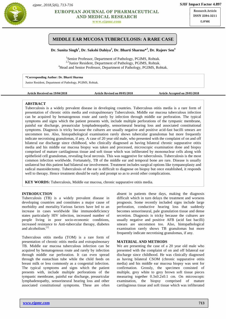
by mononuclear cell infilterate alongwith epithelioid cell
granulomas, revealing focal necrosis. This was
suggestive for tuberculosis. Though, Ziehl neelson
staining for acid fast bacilli using 20% H2SO4 was non-
contributory.
Fig 1: 100X photomicrograph of biopsy of middle ear TB showing necrosis and inflammation.
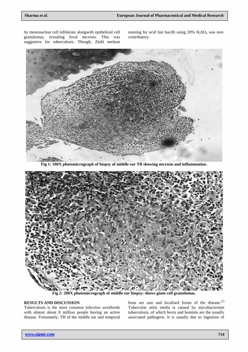
Fig 2: 200X photomicrograph of middle ear biopsy: shows giant cell granulomas.
RESULTS AND DISCUSSION
Tuberculosis is the most common infection worldwide
with almost about 8 million people having an active
disease. Fortunately, TB of the middle ear and temporal
bone are rare and localised forms of the disease.[2]
Tubercular otitis media is caused by mycobacterium
tuberculosis, of which bovis and hominis are the usually
associated pathogens. It is usually due to ingestion of
Sharma et al. European Journal of Pharmaceutical and Medical Research
www.ejpmr.com
715
infected cow’s milk if it occurs as a primary source
infection. Many say that it’s most common route of
infection is via the pharyngo-tympanic tube but others
insist on the fact that it’s mostly a hematogenous spread,
if there is a primary focus elsewhere. A congenital form
of the disease has also been found where infection occurs
via aspiration of amniotic fluid or by contact with
infected genital mucosa.
Disease is usually unilateral but this patient had bilateral
ear involvement. On otoscopy, initially the tympanic
membrane looks dull with subsequent thickening and
obliteration landmarks of the middle ear get
obliterated.[3]
A lot of complications like facial nerve
palsy, retroauricular fistulae, labyrinthitis, meningitis.etc
can occur. Pure tone audiogram shows deafness out of
proportion to the disease. Radiologic findings are non-
specific and include well pneumatised bone with some
soft tissue. Bony erosion is uncommon. Histopathology
of granulation tissue when abundant is most reliable
diagnostic method. However, biopsies need to be
frequently repeated for confirmation.[4]
Biopsy report
shows granulation tissue with epithelioid cell granulomas
and multinucleated giant cells (Langhans giant cells),
areas of central necrosis, lymphocytic infiltration,
ulceration and signs of bone resorption. Polymerase
chain reaction of the ear discharge can also be done.[3]
It
is very difficult to demonstrate AFB by staining and to
culture the discharge. Also superadded infection can
obscure our findings. Treatment includes surgical options
like tympanoplasty and radical mastoidectomy according
to the degree of damage. Medically, concomitant anti
tubercular therapy is also advised.
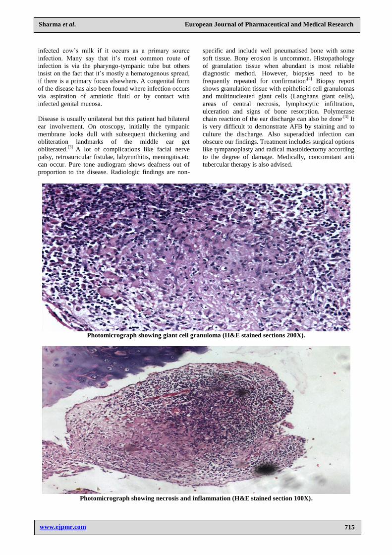
Photomicrograph showing giant cell granuloma (H&E stained sections 200X).
Photomicrograph showing necrosis and inflammation (H&E stained section 100X).
Sharma et al. European Journal of Pharmaceutical and Medical Research
www.ejpmr.com
716
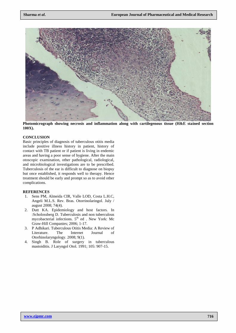
Photomicrograph showing necrosis and inflammation along with cartilegenous tissue (H&E stained section
100X).
CONCLUSION
Basic principles of diagnosis of tuberculous otitis media
include positive illness history in patient, history of
contact with TB patient or if patient is living in endemic
areas and having a poor sense of hygiene. After the main
otoscopic examination, other pathological, radiological,
and microbiological investigations are to be prescribed.
Tuberculosis of the ear is difficult to diagnose on biopsy
but once established, it responds well to therapy. Hence
treatment should be early and prompt so as to avoid other
complications.
REFERENCES
1. Sens PM, Almeida CIR, Valle LOD, Costa L.H.C,
Angeli M.L.S. Rev. Bras. Otorrinolaringol. July /
august 2008; 74(4).
2. Dutt KA. Epidemiology and host factors. In
:Scholossberg D. Tuberculosis and non tuberculous
mycobacterial infections. 5th
ed . New York: Mc
Graw-Hill Companies; 2006; 1-17.
3. P Adhikari. Tuberculous Otitis Media: A Review of
Literature. The Internet Journal of
Otorhinolaryngology. 2008; 9(1).
4. Singh B. Role of surgery in tuberculous
mastoiditis. J Laryngol Otol. 1991; 105: 907-15.