Embed Size (px)
Citation preview
Insect Molecular Biology (2005)
14
(2), 121–136
© 2005 The Royal Entomological Society
121
µBlackwell Publishing, Ltd.
Midgut and salivary gland transcriptomes of the arbovirus vector
Culicoides sonorensis
(Diptera: Ceratopogonidae)
C. L. Campbell*, K. A. Vandyke†, G. J. Letchworth*, B. S. Drolet*, T. Hanekamp‡ and W. C. Wilson*
*
USDA, ARS, Arthropod-Borne Animal Diseases Research Laboratory, College of Agriculture, Department 3354, 1000 E. University, Laramie, WY, USA; and
†
Department of Zoology and Physiology, and
‡
Department of Molecular Biology, University of Wyoming, Laramie, WY, USA
Abstract
Numerous
Culicoides
spp. are important vectors oflivestock or human disease pathogens. Transcriptomeinformation from midguts and salivary glands of adultfemale
Culicoides sonorensis
provides new insight intovector biology. Of 1719 expressed sequence tags (ESTs)from adult serum-fed female midguts harvested within5 h of feeding, twenty-eight clusters of serine proteaseswere derived. Four clusters encode putative iron bindingproteins (FER1, FERL, PXDL1, PXDL2), and two clustersencode metalloendopeptidases (MDP6C, MDP6D) thatprobably function in bloodmeal catabolism. In addition,a diverse variety of housekeeping cDNAs were identified.Selected midgut protease transcripts were analysedby quantitative real-time PCR (q-PCR): TRY1_115 andMDP6C mRNAs were induced in adult female midgutsupon feeding, whereas TRY1_156 and CHYM1 wereabundant in midguts both before and immediately afterfeeding. Of 708 salivary gland ESTs analysed, clustersrepresenting two new classes of protein families wereidentified: a new class of D7 proteins and a new classof Kunitz-type protease inhibitors. Additional cDNAsrepresenting putative immunomodulatory proteins werealso identified: 5′′′′
nucleotidases, antigen 5-relatedproteins, a hyaluronidase, a platelet-activating factoracetylhydrolase, mucins and several immune responsecDNAs. Analysis by q-PCR showed that all D7 andKunitz domain transcripts tested were highly enrichedin female heads compared with other tissues and
were generally absent from males. The mRNAs of twoadditional protease inhibitors, TFPI1 and TFPI2, weredetected in salivary glands of paraffin-embeddedfemales by
in situ
hybridization.
Keywords: expressed sequence tag, EST, haemato-phagous, gut, vector biology.
Introduction
Of about 1800
Culicoides
species world-wide, a small subsetare economically important vectors of livestock arthropod-borne viruses (arboviruses) or medically important vectorsof filarial or viral pathogens (reviewed in Mellor
et al
., 2000).The midgut and salivary gland tissues are of keen interestto vector biologists for study of the propagation, disseminationand transmission of arboviruses by these biting midges.According to dogma, the midgut milieu either restricts oractuates arbovirus proliferation in the insect vector.Subsequent virus dissemination to the salivary glands isa prerequisite for bite transmission to a naïve mammalianhost.
As pool feeders,
Culicoides
secrete pharmacologicallyimportant compounds into the bite site during blood-feedingto prevent coagulation and encourage vasodilation (Perezde Leon & Tabachnick, 1996; Mckeever
et al
., 1997; Perezde Leon
et al
., 1997, 1998). These factors may also alter thehost’s innate and acquired immune responses (Perez deLeon & Tabachnick, 1996; Perez de Leon
et al
., 1997, 1998;Limesand
et al
., 2000, 2003). Furthermore, saliva pharma-cological factors of other disease vectors potentiate infectionof a wide variety of pathogens, including arboviruses (Jones
et al
., 1992), protozoa (Titus & Ribeiro, 1988; Norsworthy
et al
., 2004) and bacteria (Gillespie
et al
., 2001).The growing dataset of genomic information and web-
based toolkits, such as FlyBase, for well-described dipterans(Adams
et al
., 2000; Myers
et al
., 2000; Holt
et al
., 2002;Zdobnov
et al
., 2002; Ribeiro, 2003) has greatly facilitatedstudy of lesser known vector insects, such as
Culicoidessonorensis
. Furthermore, the growing transcriptome data-sets from specific tissues of blood-sucking dipterans suchas mosquitoes, sand flies, and tsetse flies (Charlab
et al
.,1999; Lehane
et al
., 2003; Valenzuela
et al
., 2003; Calvo
et al
., 2004) allow more accurate sequence annotation for
doi: 10.1111/j.1365-2583.2004.00537.x
Received 15 June 2004; accepted after revision 1 October 2004. Correspond-ence: Dr Corey L. Campbell, USDA, ARS, ABADRL, College of Agriculture,Department 3354, 1000 E. University Ave., Laramie, WY 82071, USA. Tel.:+1 307 766 3626; fax: +1 307 766 3500; e-mail: [email protected]
122
C. L. Campbell
et al.
© 2005 The Royal Entomological Society,
Insect Molecular Biology
,
14
, 121–136
other families of disease vectors. In turn, analysis of the
C. sonorensis
transcriptome should provide insight into theconvergent evolution of haematophagous insects andfacilitate functional genomics studies of vector–pathogen–host relationships.
Until now, very little genomic information has been avail-able for
Culicoides
biting midges. Here, we report analysisof expressed sequence tags (ESTs) from salivary glandsand midguts of
C. sonorensis
. These cDNA datasets revealthe complexity of blood-feeding and digestive processes inthis vector insect.
Results and discussion
Midgut and salivary gland transcriptomes
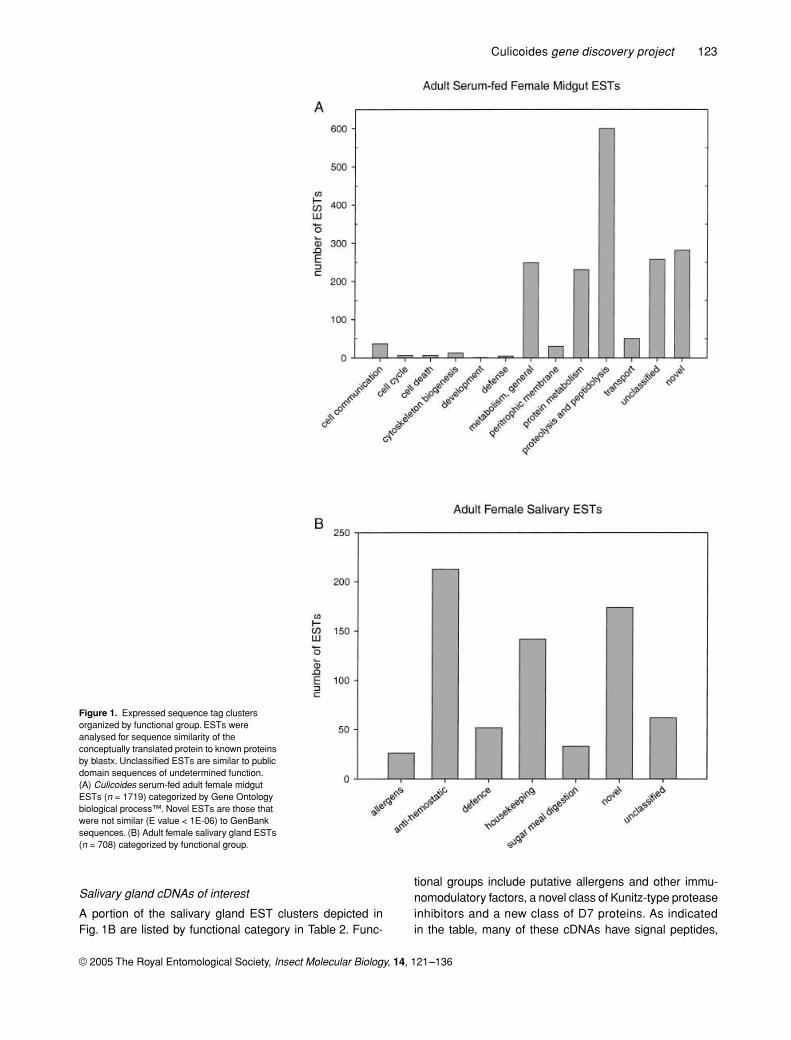
In this study, 1719 ESTs from a serum-fed adult femalemidge midgut cDNA library and 708 ESTs from an adultfemale salivary gland cDNA library were analysed. Forputative functional assignments, ESTs were comparedwith those in the public domain by sequence similaritysearch, tblastx or blastx (Altschul
et al
., 1990). MidgutESTs were grouped by Gene Ontology biological process(Fig. 1A) (Ashburner
et al
., 2000), and salivary gland ESTswere categorized by functional group (Fig. 1B). Becausefew public sequences were available for the family Cerat-opogonidae, nucleotide comparisons were largely unin-formative. This resulted in translated sequence similarityscores and corresponding E values for many
Culicoides
cDNAs to be lower than those reported for other dipteranfamilies.
The midgut and salivary gland EST collections correspondto two unique metabolic milieus. The midgut EST collectionwas prepared from poly A
+
RNA of female midges 2–5 hfollowing a serum meal. Therefore, most ESTs representdigestive enzymes and conserved metabolic factors. Theadult female salivary gland cDNA library was preparedfrom midges that had been provided with sugar water as afood source. Library construction required the use of totalRNA and a PCR-based cloning strategy, and an initialassessment of the EST collection revealed a large propor-tion of novel genes. In order to prevent reporting ofsequences resulting from PCR errors or chimeras gener-ated during cDNA library construction, novel EST single-tons with no identifiable Pfam protein domains (Bateman
et al
., 2004) and no apparent open reading frame (ORF)were excluded from further analysis. Therefore, of 1259 sal-ivary gland ESTs generated, 708 are reported here. Evenso, relaxation of analytical parameters was required to cat-egorize many salivary gene clusters, as salivary factors in
Culicoides
may have evolved independently of those ofother haematophagous arthropods. Because of the inher-ent differences between these two EST collections and thetissues they represent, each dataset is presented in sucha way as to reflect its unique characteristics.
Midgut cDNAs of interest
As indicated in Fig. 1A, a variety of diverse biological proc-esses are represented in the midgut EST collection; how-ever, in this report we have focused on selected functionalgroups. Of 1719 serum-fed adult female midgut ESTs ana-lysed, the most abundant functional group contains 600ESTs and encodes for putative proteins of proteolytic andpeptidolytic functions. Of these, 371 encode about twenty-eight clusters of serine proteases, mostly trypsin or chymo-trypsin. It is not yet clear whether any of these proteolyticenzymes might interact with ingested viruses, as has beenreported for other arthropods (Nakazawa
et al
., 2004).The 197 clusters in the protein metabolism category of
Fig. 1A account for about 11% of the total number of midgutESTs analysed. Components of the protein translationmachinery are represented by seventy-eight EST clustersin this category. These include major translation elongationfactors and a variety of ribosomal protein subunits, as wellas tRNA synthetases and transaminases. In contrast, thesalivary gland EST collection contains just forty-sevenclusters in the protein metabolism category, about 6% ofthe 708 sequences reported here. Of these, twenty-tworibosomal protein subunits are represented (data notshown).
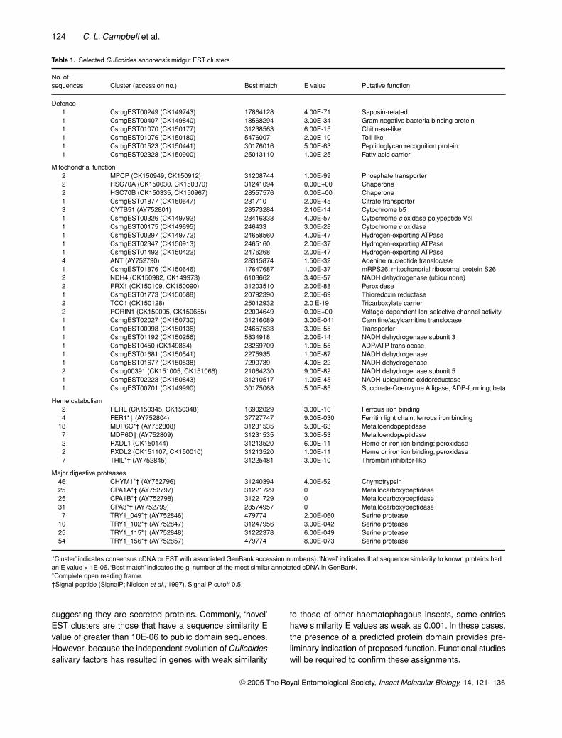
Table 1 depicts EST clusters for selected functional groups:defence, mitochondrial function, heme catabolism and majordigestive proteases. The EST clusters representing putativeimmune response genes are expected to act in antibacterialimmunity. A group of thirty-nine ESTs, representing twenty-sixclusters, code for proteins localized to the mitochondrialcompartment (Adams
et al
., 2000; Ashburner
et al
., 2000).These include a full range of components associated withmitochondrial function, including respiratory chain com-plex components and transporters, such as those thatshuttle ATP/ADP, phosphate or citrate across the mitochon-drial membrane. In contrast, just eleven clusters of thesalivary cDNA collection code for proteins localized tomitochondria. Most were constituents of the hydrogen-transporting ATPase complex (data not shown), perhapscontributing to cell membrane potentials required forhigh levels of secretory activity (Berridge
et al
., 1984;Zimmermann, 2000).
Seven midgut EST clusters were identified that mayprovide protection against the toxic effects of heme duringbloodmeal digestion. These include two clusters of metal-dependent proteases (MDP6C, MDP6D), as well as ferrousor heme binding proteins: FER1, FERL, PXDL1 and PXDL2(Table 1). In addition, cytochrome b5 in the mitochondrialcategory may also participate in heme catabolism (Wang
et al
., 2003). Interestingly, an additional cluster, THIL, wasalso identified that shows weak sequence similarity tothrombin inhibitors. It may help to prevent coagulation ofthe bloodmeal within the midgut during blood catabolism.
Culicoides
gene discovery project
123
© 2005 The Royal Entomological Society,
Insect Molecular Biology
,
14
, 121–136
Salivary gland cDNAs of interest
A portion of the salivary gland EST clusters depicted inFig. 1B are listed by functional category in Table 2. Func-
tional groups include putative allergens and other immu-nomodulatory factors, a novel class of Kunitz-type proteaseinhibitors and a new class of D7 proteins. As indicatedin the table, many of these cDNAs have signal peptides,
Figure 1. Expressed sequence tag clusters organized by functional group. ESTs were analysed for sequence similarity of the conceptually translated protein to known proteins by blastx. Unclassified ESTs are similar to public domain sequences of undetermined function. (A) Culicoides serum-fed adult female midgut ESTs (n = 1719) categorized by Gene Ontology biological process™. Novel ESTs are those that were not similar (E value < 1E-06) to GenBank sequences. (B) Adult female salivary gland ESTs (n = 708) categorized by functional group.
124
C. L. Campbell
et al.
© 2005 The Royal Entomological Society,
Insect Molecular Biology
,
14
, 121–136
suggesting they are secreted proteins. Commonly, ‘novel’EST clusters are those that have a sequence similarity Evalue of greater than 10E-06 to public domain sequences.However, because the independent evolution of
Culicoides
salivary factors has resulted in genes with weak similarity
to those of other haematophagous insects, some entrieshave similarity E values as weak as 0.001. In these cases,the presence of a predicted protein domain provides pre-liminary indication of proposed function. Functional studieswill be required to confirm these assignments.
Table 1. Selected Culicoides sonorensis midgut EST clusters
No. of sequences Cluster (accession no.) Best match E value Putative function
Defence1 CsmgEST00249 (CK149743) 17864128 4.00E-71 Saposin-related1 CsmgEST00407 (CK149840) 18568294 3.00E-34 Gram negative bacteria binding protein1 CsmgEST01070 (CK150177) 31238563 6.00E-15 Chitinase-like1 CsmgEST01076 (CK150180) 5476007 2.00E-10 Toll-like1 CsmgEST01523 (CK150441) 30176016 5.00E-63 Peptidoglycan recognition protein1 CsmgEST02328 (CK150900) 25013110 1.00E-25 Fatty acid carrier
Mitochondrial function2 MPCP (CK150949, CK150912) 31208744 1.00E-99 Phosphate transporter2 HSC70A (CK150030, CK150370) 31241094 0.00E+00 Chaperone2 HSC70B (CK150335, CK150967) 28557576 0.00E+00 Chaperone1 CsmgEST01877 (CK150647) 231710 2.00E-45 Citrate transporter3 CYTB51 (AY752801) 28573284 2.10E-14 Cytochrome b51 CsmgEST00326 (CK149792) 28416333 4.00E-57 Cytochrome c oxidase polypeptide VbI1 CsmgEST00175 (CK149695) 246433 3.00E-28 Cytochrome c oxidase1 CsmgEST00297 (CK149772) 24658560 4.00E-47 Hydrogen-exporting ATPase1 CsmgEST02347 (CK150913) 2465160 2.00E-37 Hydrogen-exporting ATPase1 CsmgEST01492 (CK150422) 2476268 2.00E-47 Hydrogen-exporting ATPase4 ANT (AY752790) 28315874 1.50E-32 Adenine nucleotide translocase1 CsmgEST01876 (CK150646) 17647687 1.00E-37 mRPS26: mitochondrial ribosomal protein S262 NDH4 (CK150982, CK149973) 6103662 3.40E-57 NADH dehydrogenase (ubiquinone)2 PRX1 (CK150109, CK150090) 31203510 2.00E-88 Peroxidase1 CsmgEST01773 (CK150588) 20792390 2.00E-69 Thioredoxin reductase2 TCC1 (CK150128) 25012932 2.0 E-19 Tricarboxylate carrier2 PORIN1 (CK150095, CK150655) 22004649 0.00E+00 Voltage-dependent Ion-selective channel activity1 CsmgEST02027 (CK150730) 31216089 3.00E-041 Carnitine/acylcarnitine translocase1 CsmgEST00998 (CK150136) 24657533 3.00E-55 Transporter1 CsmgEST01192 (CK150256) 5834918 2.00E-14 NADH dehydrogenase subunit 31 CsmgEST0450 (CK149864) 28269709 1.00E-55 ADP/ATP translocase1 CsmgEST01681 (CK150541) 2275935 1.00E-87 NADH dehydrogenase1 CsmgEST01677 (CK150538) 7290739 4.00E-22 NADH dehydrogenase2 Csmg00391 (CK151005, CK151066) 21064230 9.00E-82 NADH dehydrogenase subunit 51 CsmgEST02223 (CK150843) 31210517 1.00E-45 NADH-ubiquinone oxidoreductase1 CsmgEST00701 (CK149990) 30175068 5.00E-85 Succinate-Coenzyme A ligase, ADP-forming, beta
Heme catabolism2 FERL (CK150345, CK150348) 16902029 3.00E-16 Ferrous iron binding4 FER1*† (AY752804) 37727747 9.00E-030 Ferritin light chain, ferrous iron binding
18 MDP6C*† (AY752808) 31231535 5.00E-63 Metalloendopeptidase7 MDP6D† (AY752809) 31231535 3.00E-53 Metalloendopeptidase2 PXDL1 (CK150144) 31213520 6.00E-11 Heme or iron ion binding; peroxidase2 PXDL2 (CK151107, CK150010) 31213520 1.00E-11 Heme or iron ion binding; peroxidase7 THIL*† (AY752845) 31225481 3.00E-10 Thrombin inhibitor-like
Major digestive proteases46 CHYM1*† (AY752796) 31240394 4.00E-52 Chymotrypsin25 CPA1A*† (AY752797) 31221729 0 Metallocarboxypeptidase25 CPA1B*† (AY752798) 31221729 0 Metallocarboxypeptidase31 CPA3*† (AY752799) 28574957 0 Metallocarboxypeptidase7 TRY1_049*† (AY752846) 479774 2.00E-060 Serine protease
10 TRY1_102*† (AY752847) 31247956 3.00E-042 Serine protease25 TRY1_115*† (AY752848) 31222378 6.00E-049 Serine protease54 TRY1_156*† (AY752857) 479774 8.00E-073 Serine protease
‘Cluster’ indicates consensus cDNA or EST with associated GenBank accession number(s). ‘Novel’ indicates that sequence similarity to known proteins had an E value > 1E-06. ‘Best match’ indicates the gi number of the most similar annotated cDNA in GenBank.*Complete open reading frame.†Signal peptide (SignalP; Nielsen et al., 1997). Signal P cutoff 0.5.
Culicoides
gene discovery project
125
© 2005 The Royal Entomological Society,
Insect Molecular Biology
,
14
, 121–136
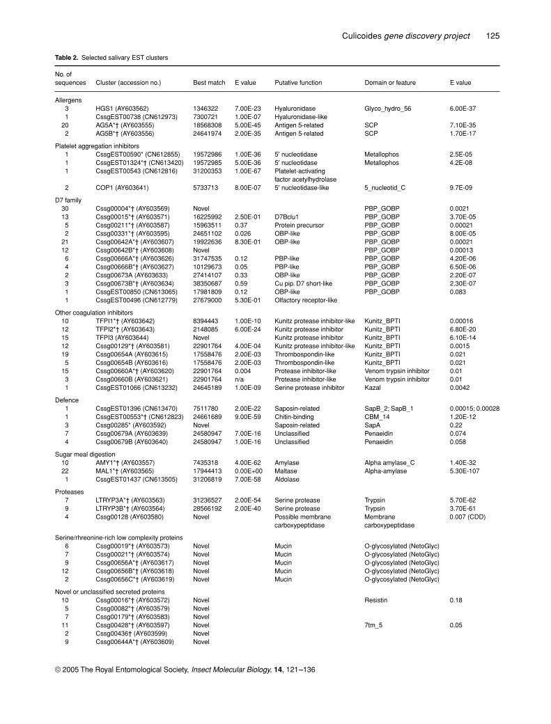
Table 2.
Selected salivary EST clusters
No. of sequences Cluster (accession no.) Best match E value Putative function Domain or feature E value
Allergens3 HGS1 (AY603562) 1346322 7.00E-23 Hyaluronidase Glyco_hydro_56 6.00E-371 CssgEST00738 (CN612973) 7300721 1.00E-07 Hyaluronidase-like
20 AG5A*† (AY603555) 18568308 5.00E-45 Antigen 5-related SCP 7.10E-352 AG5B*† (AY603556) 24641974 2.00E-35 Antigen 5-related SCP 1.70E-17
Platelet aggregation inhibitors1 CssgEST00590* (CN612855) 19572986 1.00E-36 5
′
nucleotidase Metallophos 2.5E-051 CssgEST01324*† (CN613420) 19572985 5.00E-36 5
′
nucleotidase Metallophos 4.2E-081 CssgEST00543 (CN612816) 31200353 1.00E-67 Platelet-activating
factor acetylhydrolase2 COP1 (AY603641) 5733713 8.00E-07 5
′
nucleotidase-like 5_nucleotid_C 9.7E-09
D7 family30 Cssg00004*† (AY603569) Novel PBP_GOBP 0.002113 Cssg00015*† (AY603571) 16225992 2.50E-01 D7Bclu1 PBP_GOBP 3.70E-055 Cssg00211*† (AY603587) 15963511 0.37 Protein precursor PBP_GOBP 0.000212 Cssg00331*† (AY603595) 24651102 0.026 OBP-like PBP_GOBP 8.00E-05
21 Cssg00642A*† (AY603607) 19922636 8.30E-01 OBP-like PBP_GOBP 0.0002112 Cssg00642B*† (AY603608) Novel PBP_GOBP 0.000136 Cssg00666A*† (AY603626) 31747535 0.12 PBP-like PBP_GOBP 4.20E-064 Cssg00666B*† (AY603627) 10129673 0.05 PBP-like PBP_GOBP 6.50E-062 Cssg00673A (AY603633) 27414107 0.33 OBP-like PBP_GOBP 2.20E-073 Cssg00673B*† (AY603634) 38350687 0.59 Cu pip. D7 short-like PBP_GOBP 2.30E-071 CssgEST00850 (CN613065) 17981809 0.12 OBP-like PBP_GOBP 0.0831 CssgEST00496 (CN612779) 27679000 5.30E-01 Olfactory receptor-like
Other coagulation inhibitors10 TFPI1*† (AY603642) 8394443 1.00E-10 Kunitz protease inhibitor-like Kunitz_BPTI 0.0001612 TFPI2*† (AY603643) 2148085 6.00E-24 Kunitz protease inhibitor Kunitz_BPTI 6.80E-2015 TFPI3 (AY603644) Novel Kunitz protease inhibitor Kunitz_BPTI 6.10E-1412 Cssg00129*† (AY603581) 22901764 4.00E-04 Kunitz protease inhibitor-like Kunitz_BPTI 0.001519 Cssg00654A (AY603615) 17558476 2.00E-03 Thrombospondin-like Kunitz_BPTI 0.0215 Cssg00654B (AY603616) 17558476 2.00E-03 Thrombospondin-like Kunitz_BPTI 0.021
15 Cssg00660A*† (AY603620) 22901764 0.004 Protease inhibitor-like Venom trypsin inhibitor 0.013 Cssg00660B (AY603621) 22901764 n/a Protease inhibitor-like Venom trypsin inhibitor 0.011 CssgEST01066 (CN613232) 24645189 1.00E-09 Serine protease inhibitor Kazal 0.0042
Defence1 CssgEST01396 (CN613470) 7511780 2.00E-22 Saposin-related SapB_2; SapB_1 0.00015; 0.000281 CssgEST00553*† (CN612823) 24661689 9.00E-59 Chitin-binding CBM_14 1.20E-123 Cssg00285* (AY603592) Novel Saposin-related SapA 0.227 Cssg00679A (AY603639) 24580947 7.00E-16 Unclassified Penaeidin 0.0744 Cssg00679B (AY603640) 24580947 1.00E-16 Unclassified Penaeidin 0.058
Sugar meal digestion10 AMY1*† (AY603557) 7435318 4.00E-62 Amylase Alpha amylase_C 1.40E-3222 MAL1*† (AY603565) 17944413 0.00E+00 Maltase Alpha-amylase 5.30E-1071 CssgEST01437 (CN613505) 31206819 7.00E-58 Aldolase
Proteases7 LTRYP3A*† (AY603563) 31236527 2.00E-54 Serine protease Trypsin 5.70E-629 LTRYP3B*† (AY603564) 28566192 2.00E-40 Serine protease Trypsin 3.70E-614 Cssg00128 (AY603580) Novel Possible membrane
carboxypeptidaseMembrane carboxypeptidase
0.007 (CDD)
Serine/rhreonine-rich low complexity proteins6 Cssg00019*† (AY603573) Novel Mucin O-glycosylated (NetoGlyc)7 Cssg00021*† (AY603574) Novel Mucin O-glycosylated (NetoGlyc)9 Cssg00656A*† (AY603617) Novel Mucin O-glycosylated (NetoGlyc)
12 Cssg00656B*† (AY603618) Novel Mucin O-glycosylated (NetoGlyc)2 Cssg00656C*† (AY603619) Novel Mucin O-glycosylated (NetoGlyc)
Novel or unclassified secreted proteins10 Cssg00016*† (AY603572) Novel Resistin 0.185 Cssg00082*† (AY603579) Novel7 Cssg00179*† (AY603583) Novel
11 Cssg00428*† (AY603597) Novel 7tm_5 0.052 Cssg00436† (AY603599) Novel9 Cssg00644A*† (AY603609) Novel
126
C. L. Campbell
et al.
© 2005 The Royal Entomological Society,
Insect Molecular Biology
,
14
, 121–136
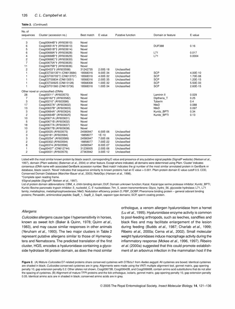
3 Cssg00644B*† (AY603610) Novel6 Cssg00651A*† (AY603613) Novel DUF388 0.165 Cssg00651B*† (AY603614) Novel6 Cssg00668A*† (AY603628) Novel L71 0.0174 Cssg00668B*† (AY603629) Novel L71 0.00042 Cssg00668C*† (AY603630) Novel7 Cssg00675A*† (AY603635) Novel
11 Cssg00675B*† (AY603636) Novel2 Cssg00433*† (AY603598) 31242735 2.00E-18 Unclassified1 CssgEST00135*† (CN613684) 18568316 9.00E-34 Unclassified SCP 4.00E-101 CssgEST00705*† (CN613707) 18568316 4.00E-32 Unclassified SCP 1.70E-081 CssgEST00834 (CN613051) 18568316 2.00E-35 Unclassified SCP 1.20E-151 CssgEST00925 (CN613128) 18568308 1.00E-32 Unclassified SCP 5.50E-331 CssgEST01560 (CN613736) 18568316 1.00E-34 Unclassified SCP 2.60E-15
Other novel or unclassified cDNAs28 Cssg00011* (AY603570) Novel Laminin II 0.0297 Cssg00162*† (AY603582) Novel Diptheria_T 0.253 Cssg00210* (AY603586) Novel Tuberin 0.4
17 Cssg00637A* (AY603602) Novel NfeD 0.08813 Cssg00637B* (AY603603) Novel NfeD 0.0973 Cssg00664A* (AY603624) Novel Kunitz_BPTI 0.342 Cssg00664B* (AY603625) Novel Kunitz_BPTI 0.13
10 Cssg00671A (AY603631) Novel5 Cssg00671B (AY603632) Novel
18 Cssg00677A (AY603637) Novel10 Cssg00677B (AY603638) Novel2 Cssg00025 (AY603575) 24580947 6.00E-06 Unclassified3 Cssg00181 (AY603584) 18858077 1E-10 Unclassified3 Cssg00254* (AY603589) 24580947 7.00E-06 Unclassified2 Cssg00302 (AY603594) 18858077 7.00E-22 Unclassified8 Cssg00374 (AY603596) 24580947 8.00E-07 Unclassified1 Cssg00437* (CN612744) 31239005 2.00E-06 Unclassified2 Cssg00031 (AY603576) 27363942 3.00E-12 Unclassified
Listed with the most similar known protein by blastx search, corresponding E value and presence of any putative signal peptide (SignalP website) (Nielsen
et al
., 1997), domain (Pfam website) (Bateman
et al
., 2004) or other feature. Except where indicated, all domains were determined using Pfam. ‘Cluster’ indicates consensus cDNA name with associated GenBank accession number. ‘Best match’ indicates the gi number of the most similar annotated protein in GenBank nr database, blastx search. ‘Novel’ indicates that sequence similarity to known proteins had an E value > 0.001. Pfam protein domain E value cutoff 0.5. CDD, Conserved Domain Database (Marchler-Bauer
et al
., 2003); NetoGlyc (Hansen
et al
., 1998).*Complete open reading frame.†Signal peptide (SignalP; Nielsen
et al.
, 1997).List of protein domain abbreviations: CBM_4, chitin binding domain; DUF, Domain unknown function; Kazal, Kazal-type serine protease inhibitor; Kunitz_BPTI, Kunitz/Bovine pancreatic trypsin inhibitor; 5_nucleotid_C, 5
′
nucleotidase; 7tm_5, seven transmembrane; Glyco_hydro_56, glycoside hydrolase; L71, L71 family; metallophos, metallophosphoesterase; NfeD, Nodulation efficiency protein D; PBP_GOBP, Pheromone binding protein – general odorant binding proteins; Penaeidin, antimicrobial peptide; SapB_1, SapB_2, SapA, saposin type domains; SCP, sperm coating protein.
No. of sequences Cluster (accession no.) Best match E value Putative function Domain or feature E value
Table 2.
(
Continued
)
Allergens
Culicoides
allergens cause type I hypersensitivity in horses,known as sweet itch (Baker & Quinn, 1978; Quinn
et al
.,1983), and may cause similar responses in other animals(Yeruham
et al
., 1993). The two major clusters in Table 2represent putative allergens similar to those of Hymenop-tera and Nematocera. The predicted translation of the firstcluster, HGS, encodes a hyaluronidase containing a glyco-side hydrolase 56 protein domain, as does the most similar
orthologue, a venom allergen hyaluronidase from a hornet(Lu
et al
., 1995). Hyaluronidase enzyme activity is commonto pool-feeding arthropods, such as leeches, sandflies andblack flies and may facilitate enlargement of the lesionduring feeding (Budds
et al
., 1987; Charlab
et al
., 1999;Ribeiro
et al
., 2000a; Cerna
et al
., 2002). Small molecularweight hyaluronidases induce macrophage activity during theinflammatory response (Mckee
et al
., 1996, 1997). Ribeiro
et al
. (2000a) suggested that this could promote establish-ment of an arbovirus infection in the mammalian host if the
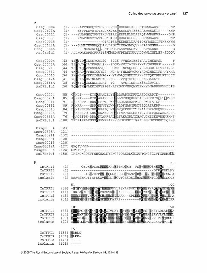
Figure 2.
(A)
Mature
Culicoides
D7-related proteins share conserved cysteines with D7Bclu1 from
Aedes aegypti
. All cysteines are boxed. Identical cysteines are shaded in black;
Culicoides
conserved cysteines are in grey. Alignments were made using the VNTI multiple alignment tool, gonnet matrix, gap opening penalty 10, gap extension penalty 0.2. Other alleles not shown, Cssg00673B, Cssg00642B, and Cssg00666B, contain amino acid substitutions that do not alter the spacing of cysteines. (B)
Alignment of mature TFPI proteins and the tick orthologue, ixolaris, gonnet matrix, gap opening penalty 10, gap extension penalty 0.05. Identical amino acis are in shaded in black; conserved amino acids are in grey.
Culicoides
gene discovery project
127
© 2005 The Royal Entomological Society,
Insect Molecular Biology
,
14
, 121–136
128
C. L. Campbell
et al.
© 2005 The Royal Entomological Society,
Insect Molecular Biology
,
14
, 121–136
pathogen in question infects macrophages, thus aidingdissemination.
A second major salivary EST cluster in this categoryencodes a predicted protein similar to antigen 5-relatedprotein of
Aedes aegypti
. As with hyaluronidases, antigen 5proteins are common to venom allergens of Hymenopteraand salivary factors of Nematocera. In addition, antigen 5proteins have been proposed to belong to the pathogenesis-related protein superfamily (Henriksen
et al
., 2001). Althoughthey have been identified in the salivary glands of severaldisease vector species, any clues to function remain unknown(Li
et al
., 2001; Francischetti
et al
., 2002b; Valenzuela
et al.,2002c; Ribeiro et al., 2004).
Platelet aggregation inhibitors
Apyrases are found in a variety of haematophagous insectsof the suborder Nematocera (Champagne et al., 1995;Charlab et al., 1999); they facilitate blood-feeding throughthe inhibition of ADP-mediated platelet aggregation. Divalentcation-dependent apyrase activity was previously foundin C. sonorensis salivary gland homogenates (Perez deLeon & Tabachnick, 1996). In the Culicoides EST collectionreported here, several putative 5′ nucleotidase apyraseswere identified: two partial cDNAs with a metallophosphoe-sterase protein domain and one with a 5′ nucleotidasedomain. Both of these domains may be found in a single full-length apyrase (Ribeiro et al., 2000b; Thomasova et al., 2002).All three Culicoides ESTs are most similar to Anophelesgambiae proteins.
A second putative platelet aggregation inhibitor wasalso identified. A conceptual translation of CssgEST00543showed 60% amino acid (a.a.) identity and 71% similarityto Drosophila melanogaster platelet activating factor (PAF)acetylhydrolase alpha subunit (O9644), as well as 38% a.a.identity and 55% similarity to a rat isoform (NP_446106)(data not shown). PAF acetylhydrolase degrades PAF,a pro-inflammatory phospholipid that is activated duringinflammatory injuries or infection (reviewed in Zimmermanet al., 2002). Although PAF hydrolysing activity has beenfound in salivary homogenates of Culex quinquefasciatus(Ribeiro & Francischetti, 2001), this is the first report of aPAF acetylhydrolase in the salivary gland transcriptome ofa haematophagous disease vector.
New D7 family
The odorant/pheromone binding protein superfamilycontains a class of proteins of unknown function referred toas the D7 family. This protein family was first identified asthe most abundant class of secreted proteins expressedexclusively in salivary glands of female mosquitoes (Jameset al., 1991). The distinguishing feature among the generallydissimilar D7 and D7-related family members is a set of sixconserved cysteines (Valenzuela et al., 2002a). These pro-teins have been proposed to function as small hydrophobic
ligand carriers and/or in binding of host haemostatic factors(Valenzuela et al., 2002a). Table 2 shows a number ofclusters that are proposed to encode new members of theD7 protein family. This group contains the most ESTs of thedataset, comprising 100 of the 708 ESTs analysed. Withthe exception of one, each of the corresponding proteinscontains a signature protein domain that substantiatesits place in the odorant/pheromone binding superfamily(Graham & Davies, 2002).
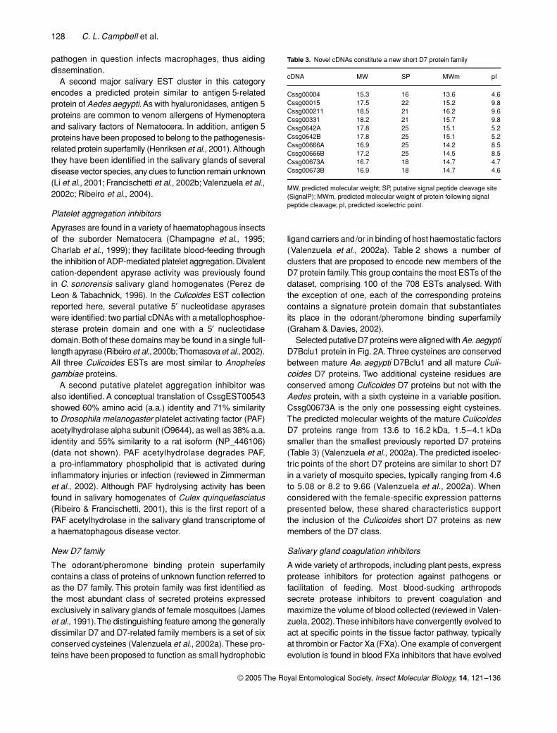
Selected putative D7 proteins were aligned with Ae. aegyptiD7Bclu1 protein in Fig. 2A. Three cysteines are conservedbetween mature Ae. aegypti D7Bclu1 and all mature Culi-coides D7 proteins. Two additional cysteine residues areconserved among Culicoides D7 proteins but not with theAedes protein, with a sixth cysteine in a variable position.Cssg00673A is the only one possessing eight cysteines.The predicted molecular weights of the mature CulicoidesD7 proteins range from 13.6 to 16.2 kDa, 1.5–4.1 kDasmaller than the smallest previously reported D7 proteins(Table 3) (Valenzuela et al., 2002a). The predicted isoelec-tric points of the short D7 proteins are similar to short D7in a variety of mosquito species, typically ranging from 4.6to 5.08 or 8.2 to 9.66 (Valenzuela et al., 2002a). Whenconsidered with the female-specific expression patternspresented below, these shared characteristics supportthe inclusion of the Culicoides short D7 proteins as newmembers of the D7 class.
Salivary gland coagulation inhibitors
A wide variety of arthropods, including plant pests, expressprotease inhibitors for protection against pathogens orfacilitation of feeding. Most blood-sucking arthropodssecrete protease inhibitors to prevent coagulation andmaximize the volume of blood collected (reviewed in Valen-zuela, 2002). These inhibitors have convergently evolved toact at specific points in the tissue factor pathway, typicallyat thrombin or Factor Xa (FXa). One example of convergentevolution is found in blood FXa inhibitors that have evolved
Table 3. Novel cDNAs constitute a new short D7 protein family
cDNA MW SP MWm pI
Cssg00004 15.3 16 13.6 4.6Cssg00015 17.5 22 15.2 9.8Cssg000211 18.5 21 16.2 9.6Cssg00331 18.2 21 15.7 9.8Cssg0642A 17.8 25 15.1 5.2Cssg0642B 17.8 25 15.1 5.2Cssg00666A 16.9 25 14.2 8.5Cssg00666B 17.2 25 14.5 8.5Cssg00673A 16.7 18 14.7 4.7Cssg00673B 16.9 18 14.7 4.6
MW, predicted molecular weight; SP, putative signal peptide cleavage site (SignalP); MWm, predicted molecular weight of protein following signal peptide cleavage; pI, predicted isoelectric point.
Culicoides gene discovery project 129
© 2005 The Royal Entomological Society, Insect Molecular Biology, 14, 121–136
independently in ticks, mosquitoes and midges. This typeof protease inhibitor activity has been identified in Culi-coides midges (Perez de Leon et al., 1998), black flies(Jacobs et al., 1990), ticks (Francischetti et al., 2002a) andculicine mosquitoes (Stark & James, 1996, 1998).
The midge salivary gland EST collection revealed avariety of abundant clusters with similarity to coagulationinhibitors. Among these, two clusters in Table 2 contain sig-nificant similarity to Kunitz-type protease inhibitors. Severaladditional abundant clusters represent novel proteins withdomains less similar to typical Kunitz-type features. Threeclusters bearing strong Kunitz-type domains are similar totissue factor pathway inhibitor proteins (TFPI). Therefore,one or more of these cDNAs may be responsible for the FXainhibitor activity previously identified in Culicoides salivarygland homogenates (Perez de Leon et al., 1998). Interest-ingly, the predicted translation product of Culicoides TFPI2
is most similar, with 32% identity and 38% similarity, to thetick Kunitz-type FXa inhibitor ixolaris (Francischetti et al.,2002a). The FXa inhibitor found in culicine mosquitoes is ahighly diverged member of the serpin superfamily (Stark &James, 1998), and the black fly sequence has not beenpublished. In contrast, anopheline mosquitoes secrete anti-coagulants that act on thrombin (Stark & James, 1996).
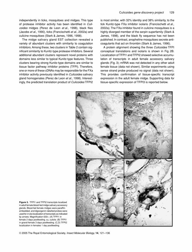
A protein alignment showing the three Culicoides TFPIconceptual translations and ixolaris is shown in Fig. 2B.Localization of TFPI1 and TFPI2 showed selective accumu-lation of transcripts in adult female accessory salivaryglands (Fig. 3); mRNA was not detected in any other adultfemale tissue (data not shown). Similar experiments usingsense strand probe produced no signal (data not shown).This provides confirmation of tissue-specific transcriptexpression in the adult female midge. Supporting data fortissue-specific expression of TFPI3 is reported below.
Figure 3. TFPI1 and TFPI2 transcripts localized in adult female blood-fed midge salivary accessory glands. Blood-fed female midges were paraffin-embedded, and digoxigenin-labelled probes were used for in situ localization of transcripts as indicated by arrows. Magnification 630×. (A) TFPI1 in female 2 days postfeeding. cu, cuticle. (B) TFPI1 in gravid female 3 days postfeeding. (C,D) TFPI2 localization in females 1 day postfeeding.
130 C. L. Campbell et al.
© 2005 The Royal Entomological Society, Insect Molecular Biology, 14, 121–136
Salivary secreted enzymes
Three clusters of salivary factors responsible for sugar mealdigestion were identified: amylase, maltase and aldolase(Table 2). In addition to these, sixteen ESTs code for twoclusters of secreted serine proteases (Table 2). The distribu-tion of one of these cDNAs, LTRYP3A, among differentmidge tissues as determined by quantitative PCR is describedbelow.
Putative mucins
Recent reports of other disease vector salivary transcrip-tomes have taken note of the possible significance of secretedsalivary mucins in the defence response (Francischettiet al., 2002b; Ribeiro et al., 2004). Therefore, all abundantCulicoides salivary EST clusters were checked for putativeO-glycosylation sites using the NetOGlyc server (Hansen
et al., 1998). Five consensus cDNAs with complete ORFsand poly adenylation signals code for predicted translationproducts with regions of low complexity (Table 2). All carryboth a signal peptide and clustered serines or threoninesindicative of putative mucins (Hansen et al., 1998). Fourcode for short proteins: Cssg00019, 63 a.a.; Cssg00656A,81 a.a.; Cssg00656B, 80 a.a.; and Cssg00656C, 77 a.a.Cssg00021 is the longest with 168 a.a.
Quantitative real-time PCR of selected cDNAs
To corroborate the putative functional assignments ofpredicted proteins presented in Tables 1 and 2, quantitativereal-time PCR (q-PCR) was performed for selected tran-scripts. Target tissues include heads with salivary glands,midguts, and remaining carcasses of both unfed andserum-fed adult female midges, as well as female salivaryglands and whole males and females. This sample set was
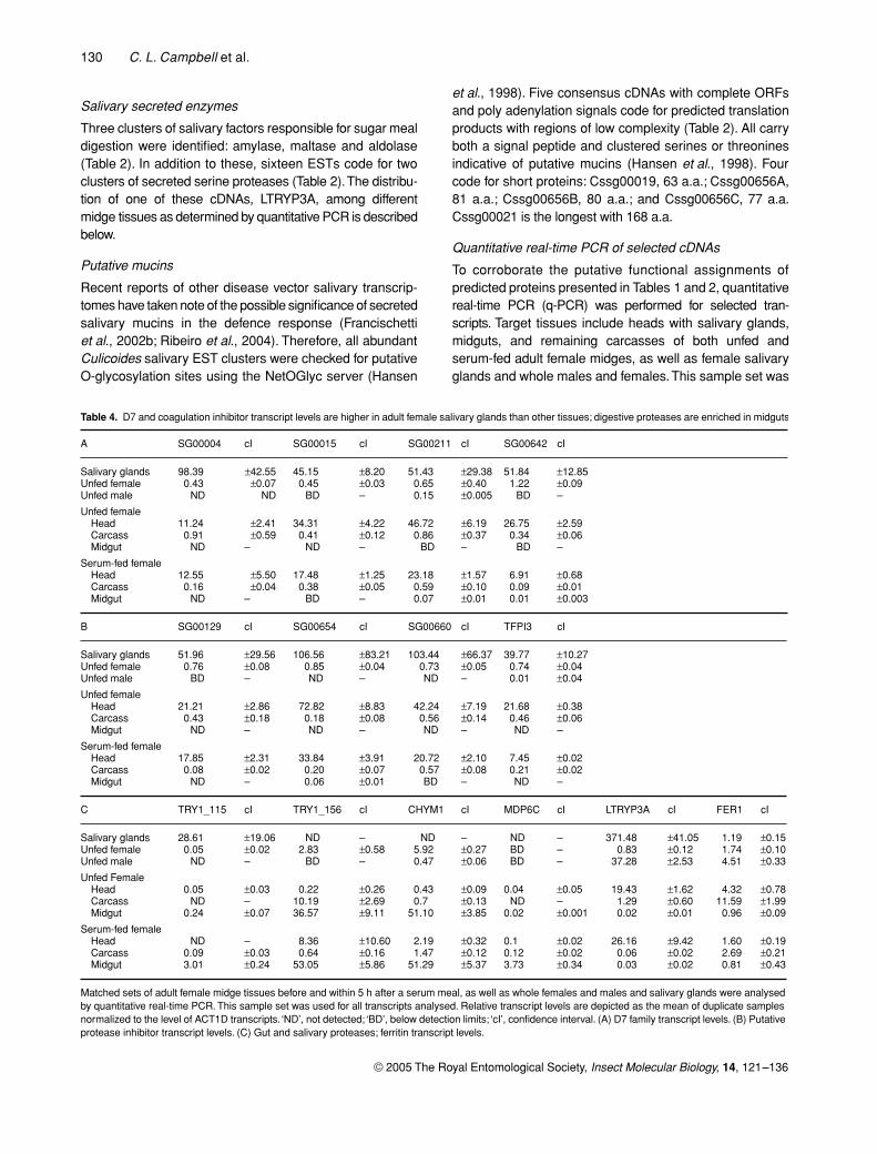
Table 4. D7 and coagulation inhibitor transcript levels are higher in adult female salivary glands than other tissues; digestive proteases are enriched in midguts
A SG00004 cI SG00015 cI SG00211 cI SG00642 cI
Salivary glands 98.39 ±42.55 45.15 ±8.20 51.43 ±29.38 51.84 ±12.85Unfed female 0.43 ±0.07 0.45 ±0.03 0.65 ±0.40 1.22 ±0.09Unfed male ND ND BD – 0.15 ±0.005 BD –
Unfed femaleHead 11.24 ±2.41 34.31 ±4.22 46.72 ±6.19 26.75 ±2.59Carcass 0.91 ±0.59 0.41 ±0.12 0.86 ±0.37 0.34 ±0.06Midgut ND – ND – BD – BD –
Serum-fed femaleHead 12.55 ±5.50 17.48 ±1.25 23.18 ±1.57 6.91 ±0.68Carcass 0.16 ±0.04 0.38 ±0.05 0.59 ±0.10 0.09 ±0.01Midgut ND – BD – 0.07 ±0.01 0.01 ±0.003
B SG00129 cI SG00654 cI SG00660 cI TFPI3 cI
Salivary glands 51.96 ±29.56 106.56 ±83.21 103.44 ±66.37 39.77 ±10.27Unfed female 0.76 ±0.08 0.85 ±0.04 0.73 ±0.05 0.74 ±0.04Unfed male BD – ND – ND – 0.01 ±0.04
Unfed femaleHead 21.21 ±2.86 72.82 ±8.83 42.24 ±7.19 21.68 ±0.38Carcass 0.43 ±0.18 0.18 ±0.08 0.56 ±0.14 0.46 ±0.06Midgut ND – ND – ND – ND –
Serum-fed femaleHead 17.85 ±2.31 33.84 ±3.91 20.72 ±2.10 7.45 ±0.02Carcass 0.08 ±0.02 0.20 ±0.07 0.57 ±0.08 0.21 ±0.02Midgut ND – 0.06 ±0.01 BD – ND –
C TRY1_115 cI TRY1_156 cI CHYM1 cI MDP6C cI LTRYP3A cI FER1 cI
Salivary glands 28.61 ±19.06 ND – ND – ND – 371.48 ±41.05 1.19 ±0.15Unfed female 0.05 ±0.02 2.83 ±0.58 5.92 ±0.27 BD – 0.83 ±0.12 1.74 ±0.10Unfed male ND – BD – 0.47 ±0.06 BD – 37.28 ±2.53 4.51 ±0.33
Unfed FemaleHead 0.05 ±0.03 0.22 ±0.26 0.43 ±0.09 0.04 ±0.05 19.43 ±1.62 4.32 ±0.78Carcass ND – 10.19 ±2.69 0.7 ±0.13 ND – 1.29 ±0.60 11.59 ±1.99Midgut 0.24 ±0.07 36.57 ±9.11 51.10 ±3.85 0.02 ±0.001 0.02 ±0.01 0.96 ±0.09
Serum-fed femaleHead ND – 8.36 ±10.60 2.19 ±0.32 0.1 ±0.02 26.16 ±9.42 1.60 ±0.19Carcass 0.09 ±0.03 0.64 ±0.16 1.47 ±0.12 0.12 ±0.02 0.06 ±0.02 2.69 ±0.21Midgut 3.01 ±0.24 53.05 ±5.86 51.29 ±5.37 3.73 ±0.34 0.03 ±0.02 0.81 ±0.43
Matched sets of adult female midge tissues before and within 5 h after a serum meal, as well as whole females and males and salivary glands were analysed by quantitative real-time PCR. This sample set was used for all transcripts analysed. Relative transcript levels are depicted as the mean of duplicate samples normalized to the level of ACT1D transcripts. ‘ND’, not detected; ‘BD’, below detection limits; ‘cI’, confidence interval. (A) D7 family transcript levels. (B) Putative protease inhibitor transcript levels. (C) Gut and salivary proteases; ferritin transcript levels.
Culicoides gene discovery project 131
© 2005 The Royal Entomological Society, Insect Molecular Biology, 14, 121–136
assessed using an unfed adult female sample as a stand-ard curve source for all q-PCR cDNA targets presented inTable 4, and thus allows comparison of relative transcriptlevels between transcripts, as well as between various tis-sues (except for MDP6C and TRY1_115: see Experimentalprocedures).
The hallmark of D7 protein family members is femalesalivary gland-specific protein expression (James et al.,1991; Arca et al., 1999). Here, we chose to assess transcriptabundance of selected Culicoides D7 family cDNAs toprovide corroborative evidence of their status in the D7family. Relative transcript levels of Cssg00004, Cssg00015,Cssg00211 and Cssg00642 are presented in Table 4A. Allcandidates tested were most highly enriched in femalesalivary glands. Considered together, D7 family transcriptlevels were near or below detection limits in midguts, at lowto moderate levels in all female carcasses (SD 0.46, range0.04–1.53) and high levels in heads (SD 17.04, range 5.04–66.64). The presence of D7 transcripts in the carcass isprobably due to the retention of residual salivary glandtissue remaining in the thorax upon head removal. Withthe exception of Cssg00004, all types of Culicoides D7transcripts in Table 4A are depleted following feeding(P < 0.05, t-test).
When comparing relative D7 transcript levels to eachother (Table 4A), Cssg00004 seemed to be the mostabundant in purified salivary glands; this cDNA was alsothe single most abundant EST cluster in the salivary glandcollection. Cssg00004, Cssg00015 and Cssg00642 levelswere below detection limits in males (see Experimentalprocedures). Cssg00211 was present in low levels in males.Cssg00211 and Cssg00642 were present in low levels inserum-fed female midguts, suggesting possible roles in mealcatabolism or defence. An additional D7 cDNA, Cssg00666A,was determined to be unsuitable for quantitative analysisdue to the presence of multiple amplification fragments;however, the absence of detection in males supports itsinclusion in the D7 family (data not shown).
Table 4B depicts the relative abundance of selected puta-tive blood coagulation inhibitor cDNAs, either containingKunitz-type protease inhibitor domains or weak homologyto a venom-type trypsin inhibitor domain (Table 2).Considered together, all transcripts tested in Table 4Bwere depleted from female heads following a serum meal(P < 0.05, t-test). Cssg00129, Cssg00654, Cssg00660 andCsTFPI3 were highly enriched in adult female salivaryglands and absent or below detection limits in males.Similar to the D7 protein family, one of the four cDNAs waspresent in low levels in adult female midguts following aserum meal, and all four were present in low levels in thecarcass. As for other salivary factors, this may reflect residualgland retained in the carcass.
Transcript levels of the most abundant midgut serineprotease EST clusters were assessed: TRY1_115, TRY1_156
and CHYM1. Together, these consensus transcriptsrepresent about one-third of the serine protease clustersidentified. The q-PCR analyses demonstrated that trypsinTRY1_115 is enriched in salivary glands (Table 4C) andincreased in midguts significantly within a few hours of aserum meal (P ≤ 0.01, t-test). In contrast, there were nosignificant differences in midgut levels of trypsin TRY1_156and chymotrypsin CHYM1 transcripts following a serummeal, and both transcripts were absent from salivary glands.Whether meal-induced or constitutively expressed, all threeof these transcripts were enriched in midguts over theremainder of the insect in the matched tissue sets, sub-stantiating their roles as digestive proteases.
A serine protease identified in adult female salivary glands,LTRYP3A, presented a markedly different tissue distribu-tion in that it was least abundant in midguts and mostabundant in purified female salivary glands and adult males(Table 4C), suggesting a possible role in defence or plantfeeding behaviour. Salivary gland secreted serine proteaseshave been described in both Ae. aeygpti and Ixodesscapularis (Valenzuela et al., 2002b,c). Further analysiswill be required to determine whether Culicoides salivaryserine proteases serve a role in post-translational modifi-cation of other proteins, non-haematophagous feeding orthe defence response.
Two cDNA clusters were assessed for possible rolesin meal catabolism, a metal-dependent protease (MDP6C)and a putative ferritin (FER1), an iron storage molecule.The predicted translation product of MDP6C has an astacindomain (1.9E-75) (Pfam website; Bateman et al., 2004)and shows 57% similarity and 43% identity to GmZmp, amidgut-specific metalloprotease of tsetse flies (Yan et al.,2002) (data not shown). When q-PCR Ct values were com-pared, MDP6C transcript levels are significantly differentbetween serum-fed midguts and unfed midguts (P ≤ 0.01,t-test), supporting its purported role in meal digestion.
The predicted translation product of Culicoides FER1cDNA is most similar to Ae. aegypti ferritin light chain (LCH)with 49% a.a. similarity and 35% identity. As with otherinsect ferritins, Culicoides FER1 has a signal peptide (SignalP,Nielsen et al., 1997). Like the Aedes LCH sequence (Geiseret al., 2003), the Culicoides FER1 5′-untranslated regiondoes not contain an iron responsive element (IRE) foundin other ferritins (reviewed in Nichol et al., 2002); however,unlike Aedes LCH, Culicoides FER1 does not containan N-glycosylation site. Table 4C shows that the FER1transcript was present in all female midge tissues tested, aswell as in males. Interestingly, transcript levels did not differsignificantly between unfed and serum-fed midguts, aswould be expected if FER1 was regulated at the transla-tional level as occurs with other insect ferritins that con-tain an IRE (reviewed in Nichol et al., 2002). Consideredtogether, the lack of an IRE in CsFER1 and the lack ofFER1 transcript induction in the midge midgut following
132 C. L. Campbell et al.
© 2005 The Royal Entomological Society, Insect Molecular Biology, 14, 121–136
a heme-deficient serum meal, along with the evidencethat Ae. aegypti ferritin light and heavy chains are bothregulated at the transcriptional level (Geiser et al., 2003),suggests that haematophagous insect midgut ferritingenes lacking an IRE are regulated at the transcriptionallevel by the presence of iron in the bloodmeal ratherthan at the translational level, as occurs with most non-haematophagous insect ferritins.
Interestingly, FER1 levels are higher in the unfed malethan in the unfed female, and in female tissues, comparisonof Ct values showed that levels of FER1 in the unfed femalecarcass are reduced within 5 h of feeding (P ≤ 0.01, t-test).Therefore, Culicoides FER1 might also play a role in gen-eral iron homeostasis rather than merely protecting themidgut from the toxic effects of heme during bloodmealcatabolism. If so, additional regulatory elements probablycontrol FER1 transcription in tissues other than the midgut.However, further characterization will be required fully tounderstand iron homeostasis in this insect.
Summary
This is the first report of substantial cDNA sequence infor-mation for the important disease vector C. sonorensis. Themidgut cDNA library allowed identification of importantconserved metabolic transcripts. The salivary gland cDNAlibrary revealed a number of important antihaemostatic andputative immunomodulatory factors that may be used infuture characterization of salivary potentiation of pathogentransmission by this insect. Two new classes of proteinswere identified: a new class of D7 proteins and a new classof Kunitz-type protease inhibitors. The addition of theseproteins to the growing dataset of dipteran salivary factorswill help define the convergent evolution of blood-feedinginsects.
Prior to this work, only fourteen nuclear-encodedmRNAs from Culicoides were in the public database, andeight of those were previously reported by this group(Campbell & Wilson, 2002). The cDNAs described hereprovide tools for future vector biology studies of bitingmidges and should facilitate future studies of arbovirusinfection and replication. This collection may be used toelucidate possible genetic determinants of arbovirusinfection and to devise potential strategies to control vectorinfection. In addition, this information should proveinvaluable in developing novel control strategies formidge-transmitted diseases.
Experimental procedures
Insects
Culicoides sonorensis were reared at the colony maintained atthe US Department of Agriculture Arthropod-Borne Animal Dis-eases Research Laboratory (ABADRL), Laramie, Wyoming. Adult
females (AK colony), 2–3 days old, provided de-ionized water,were allowed to feed on a meal of fetal bovine serum, 100 mg/mlphenol red sodium salt (Sigma, St Louis, MO, USA), and phagos-timulants ATP sodium salt, 50 mM, pH 7.0, and sodium bicarbonate,0.37 mg/ml (Nunamaker et al., 2000). Midguts were removed intoRNALater (Ambion, Austin, TX, USA) within 5 h postfeeding andstored at −80 °C. Salivary glands were removed from 2- to 5-dayold females that had been provided sugar water since emergence;glands were stored in RNAlater at −20 °C.
cDNA libraries
Serum-fed midges were used to prevent isolation of blood cellRNA. Total RNA was extracted from approximately 218 midgutsusing the Micro Poly (A) Pure isolation (Ambion) and purified togenerate poly A+ RNA according to the manufacturer’s recommen-dations. RNA was inserted into the plasmid pSPORT1, using theGateway Technology plasmid system according to the manufac-turer’s recommendations (Invitrogen, Carlsbad, CA, USA).
For the salivary gland library, total RNA was extracted from approx-imately 300 glands using the RNAqueous-4-PCR (Ambion) extrac-tion with DNase I treatment. Reverse transcription and cloning intothe pDNR-LIB plasmid was performed using the CREATOR SMARTsystem according to the manufacturer’s recommendations (BDBiosciences, Palo Alto, CA, USA).
Single pass 5′ DNA sequencing was performed with T7sequencing primers (GTAATACGACTCACTATAGGG) using themethods of Smith et al. (2000) on an ABI PRISM 3100 DNA ana-lyser (Applied Biosystems, Foster City, CA, USA). Sequenceswere trimmed of vector and low-quality sequence using VNTI(Informax, Frederick, MD, USA) or Seqman software (DNASTAR,Madison, WI, USA). Consensus transcripts were determined usingSeqman (DNASTAR).
Data analysis
In general, EST clusters were assigned putative function accord-ing to similarity to the most similar previously named cDNA or bythe first letter of each syllable or word of the gene ontology molec-ular function term assigned to the most closely related cDNA bytblastx or blastx search. ORFs were identified using VNTI software(Informax) and confirmed by alignment to the most similar knowncDNA by BLAST search (Altschul et al., 1990). Putative signalpeptides were determined using SignalP (Nielsen et al., 1997),cutoff 0.7. Protein domains were identified using the Pfam website(Bateman et al., 2004) or the Conserved Domain Database (CDD;Marchler-Bauer et al., 2003). Putative O-glycosylation sites weredetermined using the NetOGlyc website (Hansen et al., 1998).
In situ hybridization
Colony midges (2–3 days old) were fed a blood meal with an arti-ficial feeder according to established procedures (Hunt, 1994).Midges were held for 1–3 days, fixed and embedded in paraffin.cDNA fragments were amplified from a plasmid clone containingthe insert of interest. PCR primers: TFPI1 forward CCGGGGAT-GATAACTTTT, TFPI1 reverse CCTCATTTCCACTTCCTT. Theresulting cDNA insert was gel-purified, and in vitro transcriptionwas performed to produce an antisense TFPI1 probe using aprimer with 5′ extension bearing a T7 promoter (underlined):TAATACGACTCACTATAGGGAGACAAGGAAGTGGAAATG. ATFPI1 sense primer generated a negative control probe from the
Culicoides gene discovery project 133
© 2005 The Royal Entomological Society, Insect Molecular Biology, 14, 121–136
same cDNA insert using a primer with a T3 promoter (underlined):AATTAACCCTCACTAAAGGGCTTGGGTCAAATCTCAATG. TFPI2probes were prepared using similar methods. PCR primers: TFPI2forward CGAGAGTCGTGCATTG, TFPI2 reverse TCCTCAC-AGCGACGAT. Antisense probe primer: TAATACGACTCACTAT-AGGGAATATTGCACGACCAACACC. Sense probe primer:AATTAACCCTCACTAAAGGGCCTTCAATTTGTCAGGAGGA.
Digoxigenin probe preparation and in situ hybridization wereperformed according to the manufacturer’s instructions (RocheApplied Science, Indianapolis, IN, USA) with the followingmodifications: prehybridization, 1 h, 42 °C; hybridization overnight,42 °C. Probe was detected using NBT-BCIP substrate (VectorLaboratories, Burlingame, CA, USA) after 4 h of incubation.Sections were otherwise unstained.
Real-time quantitative PCR
RNA extractions. Adult female midges were fed a serum meal asdescribed above. Pools of ten heads, midguts and remaining car-casses were dissected in RNAlater and transferred to tubes con-taining RNAlater. Similar dissections were performed for femalesthat had only been provided water since eclosion. Whole insects werealso processed in pools of ten. RNA extractions for all sampleswere prepared using RNAqueous-4-PCR (Ambion) extraction withDNase I treatment, according to the manufacturer’s instructions. PCRwas performed in the absence of reverse transcription to confirm theabsence of detectable levels of contaminating genomic DNA. Reversetranscription reactions (RT rxns) were performed as describedpreviously using 100 ng total RNA for each sample (Campbell &Wilson, 2002). RT rxns were diluted 1–4 for use in q-PCR.
q-PCR. Relative gene expression levels were determined for allcDNA targets using SYBR Green 2X Master Mix (Applied Biosystems,Foster City, CA) on an ABI PRISM 7000 (Applied Biosystems)according to the manufacturer’s instructions. All samples were per-formed in quadruplicate. Dissociation curves were analysed for allcDNA targets to confirm a single peak, as multiple ampliconsnegate the accuracy of quantitative analysis. Cycling parameters:one cycle: 50 °C, 2 min, 95 °C, 10 min; forty cycles: 95 °C 15 s,60 °C 1 min. A ten-fold serial dilution series was prepared from anRT rxn of unfed female midges to generate a standard curve for eachsample run. This sample set was used for all cDNA targets analysedexcept for MDP6 and TRY1_115. For these two cDNAs, a similarstandard dilution series was prepared from serum-fed midguts.
Relative gene expression analysis. ABI PRISM 7000 analyticalsettings were a 0.2 threshold value and automatic baseline callingfor most targets in this study. Threshold settings and baseline set-tings were maintained across all independent runs for a given tar-get cDNA. The standard curve method was used to determinerelative transcript levels according to standard methods outlined inUser Bulletin #2 (Applied Biosystems). Briefly, for each cDNA tar-get, the standard curve slope and y-intercept were used to convertsample Ct values to estimated amounts of RNA. These valueswere normalized by a similar output value for actin generated fromthe same samples.
Statistical analysis. Reported values are the mean of duplicatesamples. When an amplification signal was undetected, a value of‘zero’ was assigned. When no amplification signal was detected inall four replicates, a value of ‘ND’ was assigned. Because SYBRGreen detection of double-stranded amplicons is prone to false
positive signals, rare targets that produced normalized values of≤ 0.01 were designated as below detection levels (BD). Con-fidence intervals were calculated from the standard deviation ofthe normalized means using an alpha value of 0.05. Except whereindicated, all t-tests were performed on normalized relative geneexpression values. In some cases, proof of statistical significancerequired the comparison of raw Ct values to increase the apparentnumber of samples analysed, as normalized values representcompressed sample sets of multiple replicates.
Primers for q-PCR
These are as follows: CHYM1f55, TATATTCGCCATGTTGGCTTTG;CHYM1r113, TCCTTGGACTGATTTGTTTACC; Ltryp3f192, TGCCC-GCTTGCATCAATTTC; Ltryp3r247, ACAACTTCCTCCTTCGCAT-GAc; MDP6Cf387, CATCAACTGTCGGAAGGAAAG; MDP6Cr470,TGGCAAGTCTGAAACAACc; SG0004f223, ATGGAAAGTGCC-ATCCAAc; SG0004r306, GATCCACCTGAACCAGATAAC;SG00015f249, ATGGATGCCCAAATTGGTAAC; SG00015r324,AAAGTGTGCGTAAATGAGg; SG00129f256, CAATGCGTC-TATAAGTGTTGG; SG00129r384, CAGATCAGGAATTGGGATAGG;SG00211f371, GTCCAACACGAGACAACTG; SG00211r465,CCGCCATTGAAGTGAATCC; SG00642Af377, CATGAAAGTC-GATGCCGATAG; SG00642Ar456, TGCCCAATTCAGCAGTTAC;SG00654Af122, GAGCCACGCCATAATCAAG; SG00654Ar211,GTCAGTTACCGGAACACAG; SG00660f286, TGCTGGGATTA-CAGGAAATG; SG00660r407, TTATCAGAGCGACTTGGAAC;SG00666Af344, GCCCAAGAAACACCTTCG; SG00666Ar407,CCGCTTCGTCAGTCAATC; CSTFPI3f147, TCCGAAGGATCG-CAAATAAATG; CSTFPI3r249, CCTCCGACAACTTTCTTGTAAC;CSTRY1_115f98, GACAATTCAACGAGGATCTTTC; CSTRY1_115R152, TACGATACGTTCACCAAGAG; CSTRY1_156F763,GTTGTGCAGAGAAGAATTTCC; CSTRY1_156R825, TGATC-CAATCACGAACGTAAG; CsFGS1F649, CTGGTGTTGCTCAC-TTTG; CsFGS1R763, CATACGGCTTGTCCTTTC; CSACT1DF686,TATGCCTTACCACATGCTATC; CCSACT1DF805, AATTTCACG-TTCGGCAGTTG.
Mention of trade names or commercial products in this publica-tion is solely for the purpose of providing specific information anddoes not imply recommendation or endorsement by the USDepartment of Agriculture.
Acknowledgements
This work is dedicated to the memory of Theodor Hane-kamp, a researcher whose enthusiasm for science hasoutlived him in the hearts and minds of his students andcolleagues. We thank J. Kempert and F. Stanek for providingcolony-reared midges, M. Larson for dissecting midgemidguts, and M. McNulty for dissecting salivary glands. Wealso thank M. West for helpful advice regarding statisticalanalysis. This work was performed with funding from the USDepartment of Agriculture, Agricultural Research serviceCRIS Project Number: 5410-32000-010-00D.
References
Adams, M.D., Celniker, S.E., Holt, R.A., Evans, C.A., Gocayne,J.D., Amanatides, P.G., Scherer, S.E., Li, P.W., Hoskins,
134 C. L. Campbell et al.
© 2005 The Royal Entomological Society, Insect Molecular Biology, 14, 121–136
R.A., Galle R.F., Zdobnov, E.M., von Mering, C., Letunic, I., Tor-rents, D., Suyama, M., Copley, R.R., Christophides, G.K., Tho-masova, D., Holt, R.A., Subramanian, G.M., Mueller, H.M.,Dimopoulos, G., Law, J.H., Wells, M.A., Birney, E., Charlab, R.,Halpern, A.L., Kokoza, E., Kraft, C.L., Lai, Z., Lewis, S., Louis, C.,Barillas-Mury, C., Nusskern, D., Rubin, G.M., Salzberg, S.L.,Sutton, G.G., Topalis, P., Wides, R., Wincker, P., Yandell, M.,Collins, F.H., Ribeiro, J., Gelbart, W.M., Kafatos, F.C. and Bork,P. (2000) The genome sequence of Drosophila melanogaster.Science 287: 2185–2195.
Altschul, S.F., Gish, W., Miller, W., Myers, E.W. and Lipman, D.J. (1990)Basic local alignment search tool. J Mol Biol 215: 403–410.
Arca, B., Lombardo, F., Capurro, M., Della Torre, A., Spanos, L.,Dimopoulos, G., Louis, C., James, A.A. and Coluzzi, M. (1999)Salivary gland-specific gene expression in the malaria vectorAnopheles gambiae. Parassitologia 41: 483–487.
Ashburner, M., Ball, C.A., Blake, J.A., Botstein, D., Butler, H.,Cherry, J.M., Davis, A.P., Dolinski, K., Dwight, S.S., Eppig, J.T.,et al. (2000) Gene ontology: tool for the unification of biology.The Gene Ontology Consortium. Nat Genet 25: 25–29.
Baker, K.P. and Quinn, P.J. (1978) A report on clinical aspects andhistopathology of sweet itch. Equine Vet J 10: 243–248.
Bateman, A., Coin, L., Durbin, R., Finn, R.D., Hollich, V., Griffiths-Jones, S., Khanna, A., Marshall, M., Moxon, S., Sonnhammer,E.L., Studholme, D.J., Yeats, C. and Eddy, S.R. (2004) ThePfam protein families database. Nucleic Acids Res 32 Data-base issue: D138–141.
Berridge, M.J., Buchan, P.B. and Heslop, J.P. (1984) Relationshipof polyphosphoinositide metabolism to the hormonal activationof the insect salivary gland by 5-hydroxytryptamine. Mol CellEndocrinol 36: 37–42.
Budds, M., Edwards, J., Olaveson, A. and Gacesa, P. (1987) Acomparison of the properties of the hyaluronidase from atemperate and a tropical species of leech. ComparativeBiochem Physiol 87B: 497–500.
Calvo, E., Andersen, J., De Francischetti, I.M.L.C.M., Debianchi,A.G., James, A.A., Ribeiro, J.M. and Marinotti, O. (2004) Thetranscriptome of adult female Anopheles darlingi salivaryglands. Insect Mol Biol 13: 73–88.
Campbell, C.L. and Wilson, W.C. (2002) Differentially expressedmidgut transcripts in Culicoides sonorensis (Diptera: ceratopo-gonidae) following Orbivirus (reoviridae) oral feeding. InsectMol Biol 11: 595–604.
Cerna, P., Mikes, L. and Volf, P. (2002) Salivary gland hyaluroni-dase in various species of phlebotomine sand flies (Diptera:psychodidae). Insect Biochem Mol Biol 32: 1691–1697.
Champagne, D.E., Smartt, C.T., Ribeiro, J.M. and James, A.A.(1995) The salivary gland-specific apyrase of the mosquitoAedes aegypti is a member of the 5′-nucleotidase family. ProcNatl Acad Sci USA 92: 694–698.
Charlab, R., Valenzuela, J.G., Rowton, E.D. and Ribeiro, J.M.(1999) Toward an understanding of the biochemical andpharmacological complexity of the saliva of a hematophagoussand fly Lutzomyia longipalpis. Proc Natl Acad Sci USA 96:15155–15160.
Francischetti, I.M., Valenzuela, J.G., Andersen, J.F., Mather, T.N.and Ribeiro, J.M. (2002a) Ixolaris, a novel recombinant tissuefactor pathway inhibitor (TFPI) from the salivary gland of thetick, Ixodes scapularis: identification of factor X and factor Xaas scaffolds for the inhibition of factor VIIa/tissue factor com-plex. Blood 99: 3602–3612.
Francischetti, I.M., Valenzuela, J.G., Pham, V.M., Garfield, M.K.and Ribeiro, J.M. (2002b) Toward a catalog for the transcriptsand proteins (sialome) from the salivary gland of the malariavector Anopheles gambiae. J Exp Biol 205: 2429–2451.
Geiser, D.L., Chavez, C.A., Flores-Munguia, R., Winzerling, J.J.and Pham, D.Q. (2003) Aedes aegypti ferritin. Eur J Biochem270: 3667–3674.
Gillespie, R.D., Dolan, M.C., Piesman, J. and Titus, R.G. (2001)Identification of an IL-2 binding protein in the saliva of the Lymedisease vector tick, Ixodes scapularis. J Immunol 166: 4319–4326.
Graham, L.A. and Davies, P.L. (2002) The odorant-binding pro-teins of Drosophila melanogaster: annotation and characteri-zation of a divergent gene family. Gene 292: 43–55.
Hansen, J.E., Lund, O., Tolstrup, N., Gooley, A.A., Williams, K.L.and Brunak, S. (1998) NetOglyc: prediction of mucin type O-glycosylation sites based on sequence context and surfaceaccessibility. Glycoconj J 15: 115–130.
Henriksen, A., King, T.P., Mirza, O., Monsalve, R.I., Meno, K.,Ipsen, H., Larsen, J.N., Gajhede, M. and Spangfort, M.D.(2001) Major venom allergen of yellow jackets, Ves v, 5: struc-tural characterization of a pathogenesis-related protein super-family. Proteins 45: 438–448.
Holt, R., Subramanian, A., Halpern, G.M., Sutton, A., Charlab,G.G., Nusskern, R., Wincker, D.R., Clark, P., Ribeiro, A.G.,Wides, J.M., et al. (2002) The genome sequence of the malariamosquito Anopheles gambiae. Science 298: 129–149.
Hunt, G. (1994) A Procedural Manual for the Large-Scale Rearingof the Biting Midge, Culicoides variipennis (Diptera: Ceratopo-gonidae). US Department of Agriculture, Agricultural ResearchService, Laramie, Wyoming, USA.
Jacobs, J.W., Cupp, E.W., Sardana, M. and Friedman, P.A. (1990)Isolation and characterization of a coagulation factor Xa inhibitorfrom black fly salivary glands. Thromb Haemost 64: 235–238.
James, A.A., Blackmer, K., Marinotti, O., Ghosn, C.R. andRacioppi, J.V. (1991) Isolation and characterization of the geneexpressing the major salivary gland protein of the femalemosquito, Aedes aegypti. Mol Biochem Parasitol 44: 245–253.
Jones, L.D., Matthewson, M. and Nuttall, P.A. (1992) Saliva-activated transmission (SAT) of Thogoto virus: dynamics of SATfactor activity in the salivary glands of Rhipicephalus appendic-ulatus, Amblyomma variegatum, and Boophilus microplus ticks.Exp Appl Acarol 13: 241–248.
Lehane, M.J., Aksoy, S., Gibson, W., Kerhornou, A., Berriman, M.,Hamilton, J., Soares, M.B., Bonaldo, M.F., Lehane, S. and Hall,N. (2003) Adult midgut expressed sequence tags from thetsetse fly Glossina morsitans morsitans and expressionanalysis of putative immune response genes. Genome Biol 4:R63.
Li, S., Kwon, J. and Aksoy, S. (2001) Characterization of genesexpressed in the salivary glands of the tsetse fly, Glossinamorsitans morsitans. Insect Mol Biol 10: 69–76.
Limesand, K.H., Higgs, S., Pearson, L.D. and Beaty, B.J. (2000)Potentiation of vesicular stomatitis New Jersey virus infection inmice by mosquito saliva. Parasite Immunol 22: 461–467.
Limesand, K.H., Higgs, S., Pearson, L.D. and Beaty, B.J. (2003)Effect of mosquito salivary gland treatment on vesicular stoma-titis New Jersey virus replication and interferon alpha/betaexpression in vitro. J Med Entomol 40: 199–205.
Lu, G., Kochoumian, L. and King, T.P. (1995) Sequence identityand antigenic cross-reactivity of white face hornet venom
Culicoides gene discovery project 135
© 2005 The Royal Entomological Society, Insect Molecular Biology, 14, 121–136
allergen, also a hyaluronidase, with other proteins. J Biol Chem270: 4457–4465.
Marchler-Bauer, A., Anderson, J.B., Deweese-Scott, C., Fedorova,N.D., Geer, L.Y., He, S., Hurwitz, D.I., Jackson, J.D., Jacobs,A.R., Lanczycki, C.J., et al. (2003) CDD: a curated Entrez data-base of conserved domain alignments. Nucleic Acids Res 31:383–387.
Mckee, C.M., Penno, M.B., Cowman, M., Burdick, M.D., Strieter, R.M.,Bao, C. and Noble, P.W. (1996) Hyaluronan (HA) fragmentsinduce chemokine gene expression in alveolar macrophages.The role of HA size and CD44. J Clin Invest 98: 2403–2413.
Mckee, C.M., Lowenstein, C.J., Horton, M.R., Wu, J., Bao, C., Chin,B.Y., Choi, A.M. and Nobie, P.W. (1997) Hyaluronan fragmentsinduce nitric-oxide synthase in murine macrophages through anuclear factor kappaB-dependent mechanism. J Biol Chem272: 8013–8018.
Mckeever, S., Brickle, D.S. and Hagan, D.V. (1997) Mouthparts,antennae and genitalia of intersex Culicoides stellifer parasit-ized by mermithid nematodes. Med Vet Entomol 11: 217–222.
Mellor, P.S., Boorman, J. and Baylis, M. (2000) Culicoides bitingmidges: their role as arbovirus vectors. Annu Rev Entomol 45:307–340.
Myers, E.W., Sutton, G.G., Delcher, A.L., Dew, I.M., Fasulo, D.P.,Flanigan, M.J., Kravitz, S.A., Mobarry, C.M., Reinert, K.H.,Remington, K.A., et al. (2000) A whole-genome assembly ofDrosophila. Science 287: 2196–2204.
Nakazawa, H., Tsuneishi, E., Ponnuvel, K.M., Furukawa, S.,Asaoka, A., Tanaka, H., Ishibashi, J. and Yamakawa, M. (2004)Antiviral activity of a serine protease from the digestive juice ofBombyx mori larvae against nucleopolyhedrovirus. Virology321: 154–162.
Nichol, H., Law, J.H. and Winzerling, J.J. (2002) Iron metabolismin insects. Annu Rev Entomol 47: 535–559.
Nielsen, H., Engelbrecht, J., Brunak, S. and Von Heijne, G. (1997)A neural network method for identification of prokaryotic andeukaryotic signal peptides and prediction of their cleavagesites. Int J Neural Syst 8: 581–599.
Norsworthy, N.B., Sun, J., Elnaiem, D., Lanzaro, G. and Soong, L.(2004) Sand fly saliva enhances Leishmania amazonensisinfection by modulating interleukin-10 production. Infect Immun72: 1240–1247.
Nunamaker, R.A., Perez De Leon, A.A., Campbell, C.L. andLonning, S.M. (2000) Oral infection of Culicoides sonorensis(Diptera: Ceratopogonidae) by vesicular stomatitis virus. J MedEntomol 37: 784–786.
Perez de Leon, A.A. and Tabachnick, W.J. (1996) Apyrase activityand adenosine diphosphate induced platelet aggregation inhi-bition by the salivary gland proteins of Culicoides variipennis,the North American vector of bluetongue viruses. Vet Parasitol61: 327–338.
Perez de Leon, A.A., Ribeiro, J.M., Tabachnick, W.J. andValenzuela, J.G. (1997) Identification of a salivary vasodi-lator in the primary North American vector of bluetongueviruses, Culicoides variipennis. Am J Trop Med Hyg 57: 375–381.
Perez de Leon, A.A., Valenzuela, J.G. and Tabachnick, W.J. (1998)Anticoagulant activity in salivary glands of the insect vectorCulicoides variipennis sonorensis by an inhibitor of factor Xa.Exp Parasitol 88: 121–130.
Quinn, P.J., Baker, K.P. and Morrow, A.N. (1983) Sweet itch:responses of clinically normal and affected horses to intra-
dermal challenge with extracts of biting insects. Equine Vet J15: 266–272.
Ribeiro, J.M. (2003) A catalogue of Anopheles gambiae transcriptssignificantly more or less expressed following a blood meal.Insect Biochem Mol Biol 33: 865–882.
Ribeiro, J.M. and Francischetti, I.M. (2001) Platelet-activating-factor-hydrolyzing phospholipase C in the salivary glands andsaliva of the mosquito Culex quinquefasciatus. J Exp Biol 204:3887–3894.
Ribeiro, J.M., Charlab, R., Rowton, E.D. and Cupp, E.W. (2000a)Simulium vittatum (Diptera: Simuliidae) and Lutzomyia longi-palpis (Diptera: Psychodidae) salivary gland hyaluronidaseactivity. J Med Entomol 37: 743–747.
Ribeiro, J.M., Rowton, E.D. and Charlab, R. (2000b) The salivary5′-nucleotidase/phosphodiesterase of the hematophagus sandfly, Lutzomyia longipalpis [corrected]. Insect Biochem Mol Biol30: 279–285.
Ribeiro, J.M., Charlab, R., Pham, V.M., Garfield, M. and Valen-zuela, J.G. (2004) An insight into the salivary transcriptomeand proteome of the adult female mosquito Culex pipiensquinquefasciatus. Insect Biochem Mol Biol 34: 543–563.
Smith, T.P., Godtel, R.A. and Lee, R.T. (2000) PCR-based setup forhigh-throughput cDNA library sequencing on the ABI 3700automated DNA sequencer. Biotechniques 29: 698–700.
Stark, K.R. and James, A.A. (1996) Salivary gland anticoagulantsin culicine and anopheline mosquitoes (Diptera: Culicidae). JMed Entomol 33: 645–650.
Stark, K.R. and James, A.A. (1998) Isolation and characterizationof the gene encoding a novel factor Xa-directed anticoagulantfrom the yellow fever mosquito, Aedes aegypti. J Biol Chem273: 20802–20809.
Thomasova, D., Ton, L.Q., Copley, R.R., Zdobnov, E.M., Wang, X.,Hong, Y.S., Sim, C., Bork, P., Kafatos, F.C. and Collins, F.H.(2002) Comparative genomic analysis in the region of a majorPlasmodium-refractoriness locus of Anopheles gambiae. ProcNatl Acad Sci USA 99: 8179–8184.
Titus, R.G. and Ribeiro, J.M. (1988) Salivary gland lysates fromthe sand fly Lutzomyia longipalpis enhance Leishmania infec-tivity. Science 239: 1306–1308.
Valenzuela, J.G. (2002) High-throughput approaches to studysalivary proteins and genes from vectors of disease. InsectBiochem Mol Biol 32: 1199–1209.
Valenzuela, J.G., Charlab, R., Gonzalez, E.C., De Miranda-Santos,I.K., Marinotti, O., Francischetti, I.M. and Ribeiro, J.M. (2002a)The D7 family of salivary proteins in blood sucking diptera.Insect Mol Biol 11: 149–155.
Valenzuela, J.G., Francischetti, I.M., Pham, V.M., Garfield, M.K.,Mather, T.N. and Ribeiro, J.M. (2002b) Exploring the sialome ofthe tick Ixodes scapularis. J Exp Biol 205: 2843–2864.
Valenzuela, J.G., Pham, V.M., Garfield, M.K., Francischetti, I.M.and Ribeiro, J.M. (2002c) Toward a description of the sialomeof the adult female mosquito Aedes aegypti. Insect BiochemMol Biol 32: 1101–1122.
Valenzuela, J.G., Francischetti, I.M., Pham, V.M., Garfield, M.K.and Ribeiro, J.M. (2003) Exploring the salivary gland transcrip-tome and proteome of the Anopheles stephensi mosquito.Insect Biochem Mol Biol 33: 717–732.
Wang, W.H., Lu, J.X., Yao, P., Xie, Y. and Huang, Z.X. (2003) Thedistinct heme coordination environments and heme-bindingstabilities of His39Ser and His39Cys mutants of cytochromeb5. Protein Eng 16: 1047–1054.
136 C. L. Campbell et al.
© 2005 The Royal Entomological Society, Insect Molecular Biology, 14, 121–136
Yan, J., Cheng, Q., Li, C.B. and Aksoy, S. (2002) Molecularcharacterization of three gut genes from Glossina morsitansmorsitans: cathepsin B, zinc-metalloprotease and zinc-carboxypeptidase. Insect Mol Biol 11: 57–65.
Yeruham, I., Braverman, Y. and Orgad, U. (1993) Field obser-vations in Israel on hypersensitivity in cattle, sheep and donkeyscaused by Culicoides. Aust Vet J 70: 348–352.
Zdobnov, E.M., von Mering, C., Letunic, I., Torrents, D., Suyama, M.,Copley, R.R., Christophides, G.K., Thomasova, D., Holt,R.A., Subramanian, G.M., Mueller, H.M., Dimopoulos, G., Law,J.H., Wells, M.A., Birney, E., Charlab, R., Halpern, A.L., Kokoza,E., Kraft, C.L., Lai, Z., Lewis, S., Louis, C., Barillas-Mury, C.,
Nusskern, D., Rubin, G.M., Salzberg, S.L., Sutton, G.G., Topalis,P., Wides, R., Wincker, P., Yandell, M., Collins, F.H., Ribeiro, J.,Gelbart, W.M., Kafatos, F.C. and Bork, P. (2002) Comparativegenome and proteome analysis of Anopheles gambiae andDrosophila melanogaster. Science 298: 149–159.
Zimmermann, B. (2000) Control of InsP3-induced Ca2+ oscillationsin permeabilized blowfly salivary gland cells: contribution ofmitochondria. J Physiol 525: 707–719.
Zimmerman, G.A., Mcintyre, T.M., Prescott, S.M. and Stafforini,D.M. (2002) The platelet-activating factor signaling system andits regulators in syndromes of inflammation and thrombosis.Crit Care Med 30: S294–S301.