Embed Size (px)
Citation preview
Grand Rounds Vol 7 pages 67–69
Speciality: General surgery; Laparoscopic surgery
Article Type: Case Report
DOI: 10.1102/1470-5206.2007.0020
� 2007 e-MED Ltd
Midgut malrotation as a rare cause of
chronic abdominal pain: a case report
and review of literature
A. Bajwa, H. Sheth and F. Hughes
Department of Surgery, Royal London Hospital Whitechapel, London, UK
Corresponding address: Adeel Bajwa, BSc, MBBS, MRCS, Surgical & Anaesthesia Directorate,
The Royal London Hospital, 2nd Floor West Wing, London, E1 1BB, UK.
E-mail: [email protected]
Date accepted for publication 11 September 2007
Abstract
Abnormalities in midgut rotation occur during the physiological herniation of the midgut between
the 5th and 10th week of gestation. The most significant abnormality is a narrow small bowel
mesentery which is prone to volvulus. This occurs most frequently in the neonatal period.
Less commonly, midgut malrotation presents in adulthood with either acute volvulus or
chronic abdominal symptoms. It is the latter group that represents a diagnostic challenge.
We report a case of a 31-year-old female patient who presented with a 6-year history of
non-specific gastro-intestinal symptoms. After extensive investigation the patient was diagnosed
with midgut malrotation following an upper gastro-intestinal series. The patient was treated with
a laparoscopic Ladd’s procedure and at 3 months was gaining weight and had stopped vomiting.
A laparoscopic Ladd’s procedure is an acceptable alternative to the open technique in treating
symptomatic malrotation in adults.
Keywords
Malrotation; adult; Ladd’s; laparoscopic; chronic; classification.
Case report
A 31-year-old woman was seen in the outpatient department with a 6-year history of abdominal
colic and bloating relieved by defecation. She had a long history of constipation, opening her
bowels 2–3 times a week with laxatives. Over the preceding few weeks, the patient had been
vomiting on most mornings with nausea persisting throughout the day. The patient had lost 14 lb
in weight over the previous 6 months and had a BMI of 19. Past medical history included
therapeutic laparoscopy for endometriosis and an appendicectomy at initial presentation.
Physical examination was normal except for scars from previous laparoscopy and
appendicectomy.
The patient had been investigated extensively. Normal investigations included liver function
tests, abdominal ultrasound, endoscopy of the upper and lower gastro-intestinal tract, jejunal
biopsy and oesophageal monometry. Electrogastrography revealed bradygastria but the patient
This paper is available online at http://www.grandrounds-e-med.com. In the event of a change in the URL
address, please use the DOI provided to locate the paper.
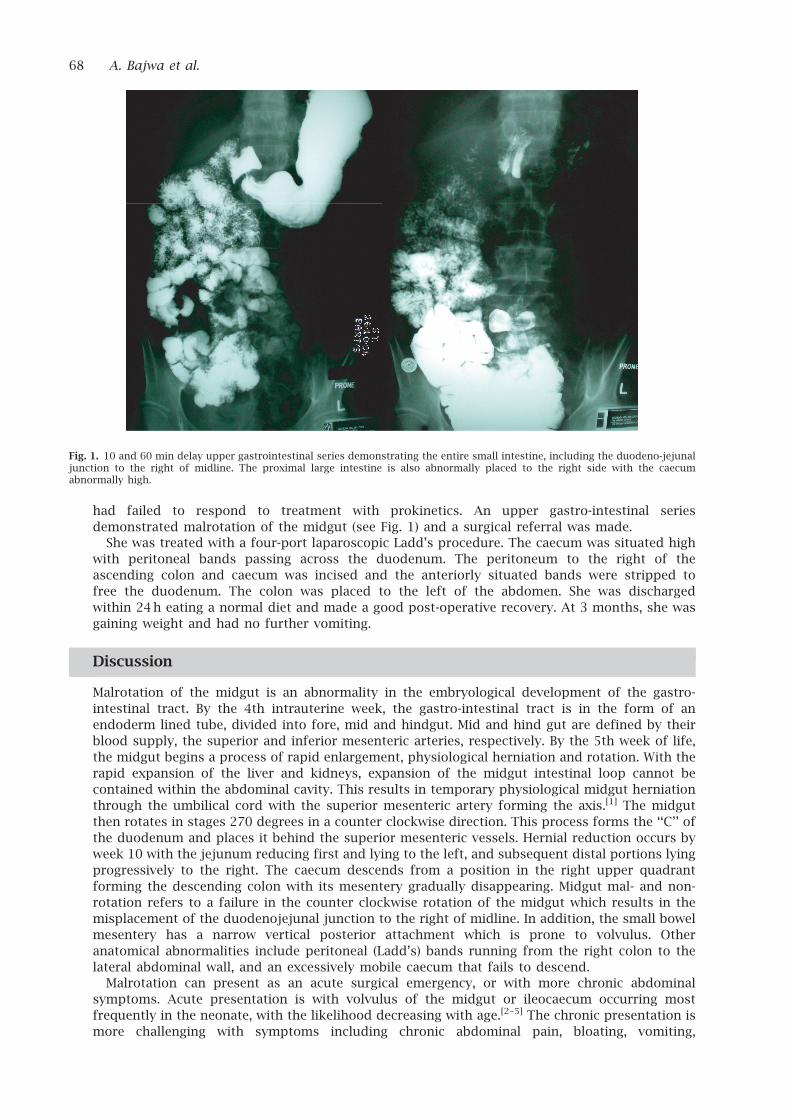
had failed to respond to treatment with prokinetics. An upper gastro-intestinal series
demonstrated malrotation of the midgut (see Fig. 1) and a surgical referral was made.
She was treated with a four-port laparoscopic Ladd’s procedure. The caecum was situated high
with peritoneal bands passing across the duodenum. The peritoneum to the right of the
ascending colon and caecum was incised and the anteriorly situated bands were stripped to
free the duodenum. The colon was placed to the left of the abdomen. She was discharged
within 24h eating a normal diet and made a good post-operative recovery. At 3 months, she was
gaining weight and had no further vomiting.
Discussion
Malrotation of the midgut is an abnormality in the embryological development of the gastro-
intestinal tract. By the 4th intrauterine week, the gastro-intestinal tract is in the form of an
endoderm lined tube, divided into fore, mid and hindgut. Mid and hind gut are defined by their
blood supply, the superior and inferior mesenteric arteries, respectively. By the 5th week of life,
the midgut begins a process of rapid enlargement, physiological herniation and rotation. With the
rapid expansion of the liver and kidneys, expansion of the midgut intestinal loop cannot be
contained within the abdominal cavity. This results in temporary physiological midgut herniation
through the umbilical cord with the superior mesenteric artery forming the axis.[1] The midgut
then rotates in stages 270 degrees in a counter clockwise direction. This process forms the ‘‘C’’ of
the duodenum and places it behind the superior mesenteric vessels. Hernial reduction occurs by
week 10 with the jejunum reducing first and lying to the left, and subsequent distal portions lying
progressively to the right. The caecum descends from a position in the right upper quadrant
forming the descending colon with its mesentery gradually disappearing. Midgut mal- and non-
rotation refers to a failure in the counter clockwise rotation of the midgut which results in the
misplacement of the duodenojejunal junction to the right of midline. In addition, the small bowel
mesentery has a narrow vertical posterior attachment which is prone to volvulus. Other
anatomical abnormalities include peritoneal (Ladd’s) bands running from the right colon to the
lateral abdominal wall, and an excessively mobile caecum that fails to descend.
Malrotation can present as an acute surgical emergency, or with more chronic abdominal
symptoms. Acute presentation is with volvulus of the midgut or ileocaecum occurring most
frequently in the neonate, with the likelihood decreasing with age.[2–5] The chronic presentation is
more challenging with symptoms including chronic abdominal pain, bloating, vomiting,
Fig. 1. 10 and 60 min delay upper gastrointestinal series demonstrating the entire small intestine, including the duodeno-jejunaljunction to the right of midline. The proximal large intestine is also abnormally placed to the right side with the caecumabnormally high.
68 A. Bajwa et al.
constipation and diarrhoea all being reported.[3,5,6] The pathophysiology of these chronic
symptoms may relate to the compressive effects of peritoneal bands running from the caecum
and ascending colon to the right lateral wall. Diagnosis is by imaging. Computed tomography scan
and ultrasound may reveal the superior mesenteric vein to lie abnormally to the left of the artery,
but these signs are frequently missed. Upper gastro-intestinal series remains the most likely
investigation to result in diagnosis as occurred in this report. These series will show the
duodenojejunal junction to the right of midline with an abnormally high caecum (Fig. 1). The
surgical management of intestinal malrotation was first described by William Ladd in 1936[4,7] and
remains the mainstay of management today. It involves reduction of volvulus if present, division
of abnormal peritoneal bands (duodeno-colic, duodenojejunal-ileocolic), and placement of the
small bowel to the right of the abdomen and the caecum to the left. Appendicectomy is also
performed, as patients may otherwise present with left sided appendicitis.[8] Increasingly,
laparoscopic Ladd’s procedures are being performed and have been shown to be effective
where there is no acute volvulus.[6,9,10] This minimally invasive approach allows for earlier oral
intake and discharge from hospital.
We report a case of a 31-year-old female patient with progressing chronic symptoms who
was eventually diagnosed after exhaustive investigation. She was successfully managed with a
laparoscopic Ladd’s procedure.
Teaching point
Symptomatic adult midgut malrotation is rare. Even in this age group, acute presentation with
volvulus can occur requiring emergency surgery.[4] The chronic presentation represents a
diagnostic challenge and may only be diagnosed incidentally following an upper gastro-intestinal
series. We report a 31-year-old patient with chronic non-specific abdominal symptoms who was
investigated thoroughly. six years later an upper gastro-intestinal series resulted in the diagnosis
of midgut malrotation. A laparoscopic Ladd’s procedure was carried out with resolution of
her symptoms.
References
1. Sadler TW, Langman J. Langman’s medical embryology. 9th ed. Philadelphia, PA: Lippincott
Williams & Wilkins.
2. Prasil P, Flageole H, Shaw KS, Nguyen LT, Youssef S, Laberge JM. Should malrotation in
children be treated differently according to age?. J Pediatr Surg 2000; 35: 756–8.
3. Cohen Z, Kleiner O, Finaly R, et al. How much of a misnomer is ‘‘asymptomatic’’ intestinal
malrotation?. Isr Med Assoc J 2003; 5: 172–4.
4. von Flue M, Herzog U, Ackermann C, Tondelli P, Harder F. Acute and chronic presentation
of intestinal nonrotation in adults. Dis Colon Rectum 1994; 37: 192–8.
5. Spigland N, Brandt ML, Yazbeck S. Malrotation presenting beyond the neonatal period.
J Pediatr Surg 1990; 25: 1139–42.
6. Seymour NE, Andersen DK. Laparoscopic treatment of intestinal malrotation in adults. JSLS
2005; 9: 298–301.
7. Ladd WE. Congenital obstruction of the duodenum in children. N Engl J Med 1932; 206:
273–283.
8. Kamiyama T, Fujiyoshi F, Hamada H, Nakajo M, Harada O, Haraguchi Y. Left-sided acute
appendicitis with intestinal malrotation. Radiat Med 2005; 23: 125–7.
9. Frantzides CT, Cziperle DJ, Soergel K, Stewart E. Laparoscopic Ladd procedure and cecopexy
in the treatment of malrotation beyond the neonatal period. Suenebrg Laparosc Endosc 1996;
6: 73–5.
10. Matzke GM, Dozois EJ, Larson DW, Moir CR. Surgical management of intestinal malrotation in
adults: comparative results for open and laparoscopic Ladd procedures. Surg Endosc 2005;
19: 1416–9.
Midgut malrotation 69


![Disorders of intestinal rotation and fixation (‘‘malrotation’’)deepblue.lib.umich.edu/bitstream/handle/2027.42/46708/... · 2020. 2. 13. · consequences [4]. ‘‘Malrotation’’](https://img.pdfslide.net/doc/110x75/60afb5330f88520c4e13c968/disorders-of-intestinal-rotation-and-ixation-aamalrotationaa-2020-2.jpg)