Embed Size (px)
Citation preview
The risk of unsuspected surgical emergencies during the neonatal period has decreased over the past several years because of widespread use of ultrasonography for fetal screening. However, surgical emergencies unique to the neonatal period still continue to plague the unsuspecting emergency physician. Most surgically correctable disorders in the neonate will present with vomiting, gastrointestinal bleeding, or respiratory distress. This article will provide the emergency phvsician with the typical presentation, diagnostic work- up, and treatment options for surgically correctable problems in the neonate. Clin Ped Emerg Med 3:3-13. Copyright 2002, Elsevier Science (USA). All rights reserved.
From the Department of Pediatrics, Division of Emergency Medicine, University of Texas, Southwestern Medical Center at Dallas, Dallas, TX; and the Department of Surgery, Division of Pediatric Surgery, University of Texas, Southwestern Medical Center at Dallas, Dallas, TX.
Address reprint requests to Pamela J. Okada, MD, Department of Pediatrics, University of Texas, Southwestern Medical Center, 5323 Harry Hines Boulevard, Dallas, TX 75390.
Copyright 2002, Elsevier Science (USA). All rights reserved.
1522-8401/02/0301-0002835.00/O doi: 10.1053/cpem.2002.30779
Neonatal Surgical Emergencies By Pamela J. Okada, MD and Barry Hicks, MD
DALLAS, TEXAS
T HE SCOPE OF PEDIATRIC SURGERY is broad and encom- passes a wide variety of diseases. Many surgical disease processes are age related. There are several common surgi- cal emergencies that physicians caring for neonates will
encounter in the emergency department. The most common neonatal surgical emergencies of the gastrointestinal tract are outlined in Table 1. This article will review common neonatal surgical emergencies that present with vomiting, bleeding, or respiratory distress.
Neonatal Surpical Disorders Associated with Vomiting
Surgical disorders associated with vomiting include intestinal obstruction, malrotation and midgut vo1vulus, Hirschsprung’s disease (HD), meconium ileus (MI), and abdominal wall defects. Other common emergencies, such as incarcerated inguinal her- nia, hypertrophic pyloric stenosis, and trauma, have been dis- cussed elsewhere in this issue.
Intestinal Obstruction-Atresias
Intestinal atresias involving the duodenum, jejunum, ileum, and colon together are the most common causes of intestinal obstruction in the newly born, affecting between 1 in 2,000 and 1 in 5,000 live births.1 There is equal representation of the sexes. Duodenal obstruction is postulated to occur as a result of incom- plete recanalization of the duodenal lumen during the tenth week of gestation. Jejunoileal atresia is believed to be the result of late mesenteric vascular accidents in utero.
Bilious vomiting is the most common presenting symptom in a newborn with intestinal obstruction. Although abdominal disten- sion is typical of distal atresias, the abdomen may be flat or even scaphoid in duodenal lesions. Abdominal distention is most prominent in ileal atresia, whereas polyhydramnios and jaundice
NEONATAL SURGICAL EMERGENCIES / OKADA AND HICKS 3
4 NEONATAL SURGICAL EMERGENCIES / OKADA AND HICKS
TABLE I. Common Neonatal Surgical Emergencies of the Gastrointestinal Tract
Intestinal obstruction
Duodenum, jejunum, ileum, colon
Atresia, web, stenosis
Malrotation and midgut volvulus
Hirschsprung’s disease
Meconium ileus
Abdominal wall defects
Omphalocele
Gastroschisis
Necrotizing enterocolitis
Duplications, Meckel’s diverticulum, mesenteric cysts
Incarcerated inguinal hernia
Hypertrophic pyloric stenosis
Trauma (consider nonaccidental trauma)
are more frequently associated with jejunal atresia. Approximately one third of newly born infants with duodenal atresia have associated Down’s syndrome.
Other subtle signs of intestinal obstruction in- clude respiratory difficulty, excessive salivation, presence of an abdominal mass, jaundice (within 24 hours of birth), and lethargy.2
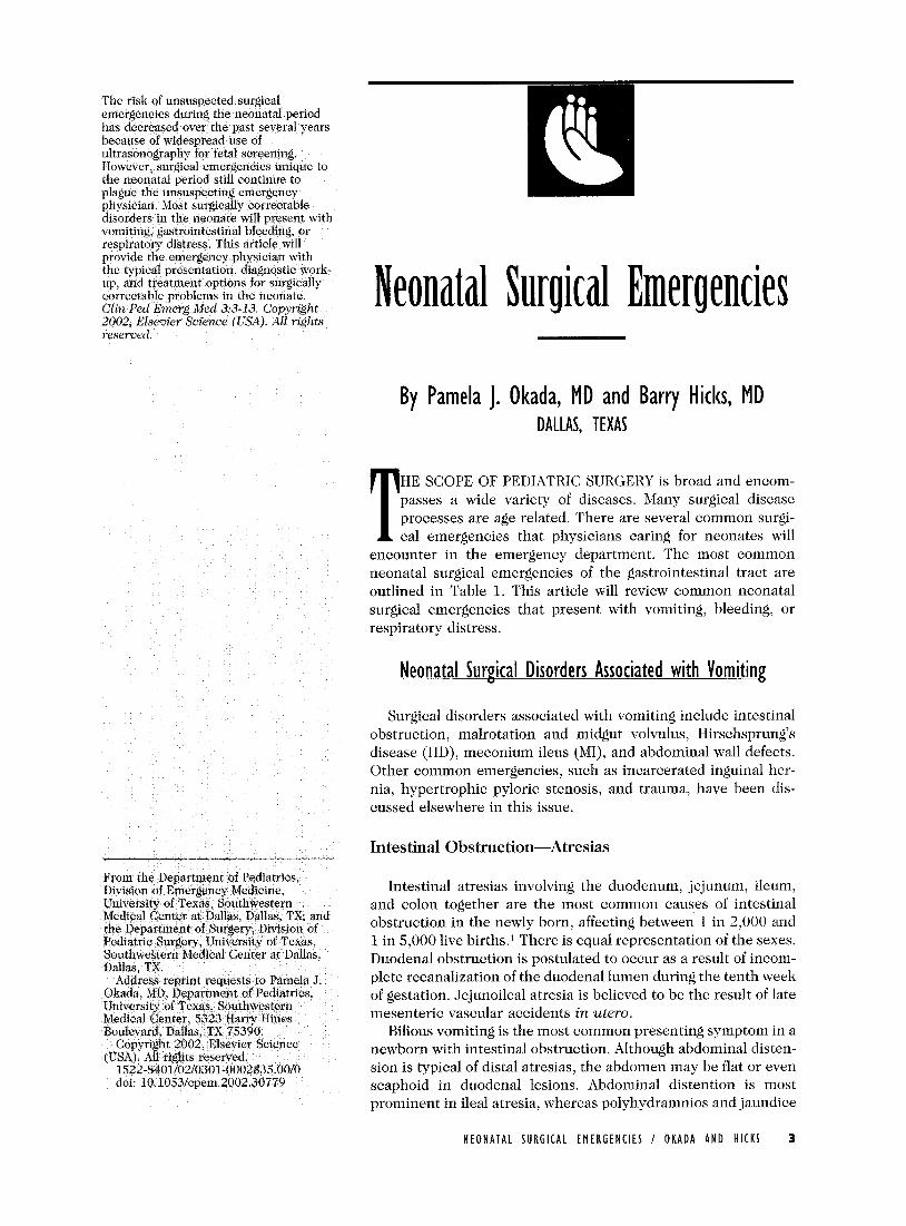
Abdominal plain films show a characteristic dou- ble bubble sign, showing air bubbles in the stomach and a dilated proximal duodenum in duodenal atre- sia (Fig 1). In jejunoileal atresia, abdominal films show air fluid levels proximal to the lesion. The more distal the obstruction, the greater the number of air fluid levels and distended loops of bowel. A contrast enema will show a small unused “micro- colon” in most infants with jejunoileal atresia.
A nasogastric or orogastric tube should be passed to decompress the stomach and duodenum. Intra- venous fluids are given to maintain adequate urine output and normoglycemia. Surgery is not urgent. The infant must be evaluated thoroughly for asso- ciated malformations, especially cardiac anomalies. Duodenal atresia has been associated with abnor- mal development of the pancreas (annular pan- creas) and Down’s syndrome (30% to 50%). There is also an association with malrotation, congenital heart disease, and esophageal atresia.
For duodenal malformations, most surgeons will perform a diamond-shaped duodenoduodenostomy using a standard side-to-side or proximal trans- verse-to-distal longitudinal anastomosis. Depending
on the type of intestinal atresia, different surgical strategies are indicated. In general, bowel recon- struction is achieved by an end-to-end (or end-to- side) anastomosis.2
Survival rates exceed 90%. Late complications from repair of duodenal atresia occur in approxi- mately 12% to 15% of patients and include me- gaduodenum, intestinal motility disorders, duodenogastric reflux, gastritis, peptic ulcer dis- ease, gastroesophageal reflux, and choledochal cysts.
Malrotation and Midgut Volv~~lus
The incidence of malrotation is 1 in 500 live births.3 Twenty-five percent to 40% of patients with symptomatic malrotation present within the first week of life, 50% present within the first month, 75% present before 1 year of age, and there is a 2:l male predominance in cases presenting in the neo- natal period. Malrotation of the midgut occurs when
Figure I. Double bubble sign in duodenal atresia.
NEONATAL SURGICAL EMERGENCIES ! OKADA AND HICKS 5
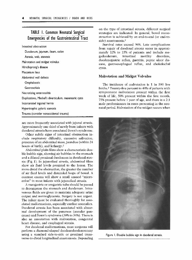
Figure 2. Malrotation with midgut voIvuIus.
the normal rotational process and fixation of the intestine fail to occur during the seventh to twelfth week of gestation.
Malrotation typically presents in the first month of life with bilious vomiting and sudden onset of abdominal pain (crying). In older infants and chil- dren, symptoms may be vague and may include chronic, intermittent vomiting and cramping ab- dominal pain, failure to thrive, constipation, bloody diarrhea, and hematemesis. Physical examination may exhibit a normal abdominal examination in 50% of patients. One third of patients present with abdominal distension without tenderness. Bowel distension, abdominal pain, and evidence of perito- nitis occurs as intestinal ischemia progresses to necrosis. As ischemia progresses to infarction, fe- ver, peritonitis, abdominal distension, profound de- hydration and vascular collapse worsen.
In the newborn, volvulus can rapidly result in significant bowel compromise with abdominal dis- tention, bloody stools, and eventually hypovolemic shock and peritonitis. An upper gastrointestinal (UGI) contrast study is the most reliable method to diagnose malrotation. The malrotated duodenum is often “coiled” to the right of the midline giving a corkscrew appearance (Fig 2). A cutoff appearance of the contrast (“beak”) suggests obstruction from volvulus
Evaluation, resuscitation, and preoperative prep- aration proceed simultaneously in patients with suspected malrotation with volvulus. In the emer- gency department, volume resuscitation, gastric de- compression, and broad-spectrum antibiotics are administered. The operative procedure for the cor- rection of malrotation, called the Ladd procedure, includes derotation of the volvu1us, division of mes- enteric bands, separation of the duodenojejunal mesentery from the cecocolic mesentery, and ap- pendectomy. An appendectomy is performed be- cause the colon is positioned on the left side of the abdomen during a Ladd procedure. Subsequent de- velopment of appendicitis could be confusing. A gastrostomy is rarely indicated.
With viable intestine, postoperative care is straightforward, and prognosis is excellent. The life- time risk of adhesive small bowel obstruction is 1% to 1O%.3 Volvulus should not recur. With extensive ischemic bowel and intestinal necrosis, the bowel is untwisted and reduced into the abdominal cavity. Twelve to 24 hours later, a “second look” procedure may be performed to assess bowel viability. This allows the surgeon to resect the necrotic bowel and to create an enterostomy at the distal end of the normal bowel. Bowel reconstruction is performed in a later operation. These patients may develop short gut syndrome and may become dependent on total parenteral nutrition (TPN).
The most serious complication of malrotation is midgut volvulus with necrosis of the entire midgut. Mortality in infants with malrotation ranges from 2.5% to 24% and is influenced by the presence of necrotic bowel at laparotomy, the presence of as- sociated anomalies, and younger patient age.4
Bilious vomiting in an otherwise healthy infant must be considered a surgical emergency and should be considered to be malrotation with midgut volvulus until proven otherwise. An emergency UGI must be performed as the patient is being evalu- ated, not on an elective basis.
Hirschsprung’s Disease
HD is a common cause of intestinal obstruction in newborns. It results from absence of ganglion cells in the myenteric plexus of the intestine. Inci- dence varies from 1 in 4,000 to 1 in 7,000 live births with a male to female ratio of 4:l.s It is usually an isolated disorder; however, Down’s syndrome is as- sociated with 4% to 12% of the cases.
HD results from the migratory arrest of vagally derived neuroblasts, which normally reach the rec- tum by the twelfth week of gestation. Any length of the intestine may be involved; however, 80% to 90%
6 NEONATAL SURGICAL EMERGENCIES / OKADA AND HICKS
of HD involves only the rectosigmoid colon.5 There are no “skip lesions” in the intestine, as HD is a failure of normal neuroblasts to complete their mi- gration from the proximal GI tract to the anus. A mutation in the (RET)-proto-oncogene appears to be a major factor in the development of the dis- ease.6 Also, molecules in the endothelin signaling pathway and the SOXlO transcriptional activator have been implicated.6
The typical presentation of a neonate with HD includes failure to pass meconium in the first 24 hours of life, constipation and abdominal disten- tion, bilious emesis, and refusal to feed. In some cases, failure to thrive may be the only initial sign.
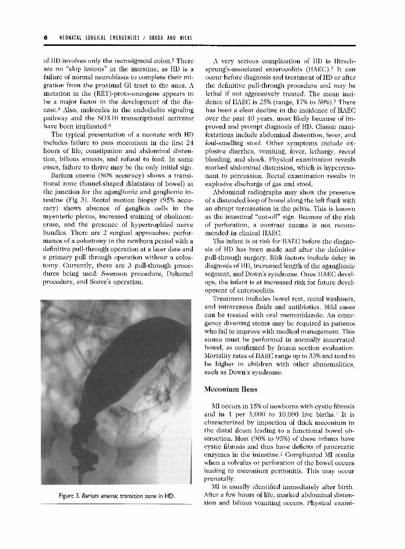
Barium enema (80% accuracy) shows a transi- tional zone (funnel-shaped dilatation of bowel) at the junction for the aganglionic and ganglionic in- testine (Fig 3). Rectal suction biopsy (95% accu- racy) shows absence of ganglion cells in the myenteric plexus, increased staining of cholinest- erase, and the presence of hypertrophied nerve bundles. There are 2 surgical approaches: perfor- mance of a colostomy in the newborn period with a definitive pull-through operation at a later date and a primary pull through operation without a colos- tomy. Currently, there are 3 pull-through proce- dures being used: Swenson procedure, Duhamel procedure, and Soave’s operation.
Figure 3. Barium enema; transition zone in HD.
A very serious complication of HD is Hirsch- sprung’s-associated enterocolitis (HAEC).’ It can occur before diagnosis and treatment of HD or after the definitive pull-through procedure and may be lethal if not aggressively treated. The mean inci- dence of HAEC is 25% (range, 17% to SO%)? There has been a clear decline in the incidence of HAEC over the past 40 years, most likely because of im- proved and prompt diagnosis of HD. Classic mani- festations include abdominal distention, fever, and foul-smelling stool. Other symptoms include ex- plosive diarrhea, vomiting, fever, lethargy, rectal bleeding, and shock. Physical examination reveals marked abdominal distension, which is hyperreso- nant to percussion. Rectal examination results in explosive discharge of gas and stool.
Abdominal radiographs may show the presence of a distended loop of bowel along the left flank with an abrupt termination in the pelvis. This is known as the intestinal “cut-off” sign. Because of the risk of perforation, a contrast enema is not recom- mended in clinical HAEC.
The infant is at risk for HAEC before the diagno- sis of HD has been made and after the definitive pull-through surgery. Risk factors include delay in diagnosis of HD, increased length of the aganglionic segment, and Down’s syndrome. Once HAEC devel- ops, the infant is at increased risk for future devel- opment of enterocolitis.
Treatment includes bowel rest, rectal washouts, and intravenous fluids and antibiotics. Mild cases can be treated with oral metronidazole. An emer- gency diverting stoma may be required in patients who fail to improve with medical management. This stoma must be performed in normally innervated bowel, as confirmed by frozen section evaluation. Mortality rates of HAEC range up to 33% and tend to be higher in children with other abnormalities, such as Down’s syndrome.
Meconium Ileus
MI occurs in 15% of newborns with cystic fibrosis and in 1 per 5,000 to 10,000 live births.1 It is characterized by impaction of thick meconium in the distal ileum leading to a functional bowel ob- struction. Most (90% to 95%) of these infants have cystic fibrosis and thus have deficits of pancreatic enzymes in the intestine.l Complicated MI results when a volvulus or perforation of the bowel occurs leading to meconium peritonitis. This may occur prenatally.
MI is usually identified immediately after birth. After a few hours of life, marked abdominal disten- sion and bilious vomiting occurs. Physical exami-
NEONATAL SURGICAL EMERGENCIES I OKADA AND HICKS 7
nation shows thickened bowel loops that are often visible and palpable through the abdominal wall. Rectal examination is difficult because of the small caliber of the rectum.
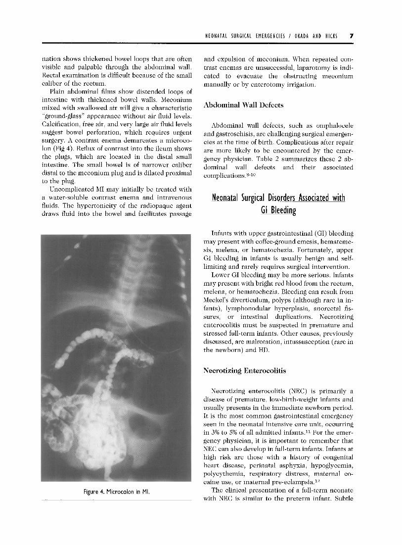
Plain abdominal films show distended loops of intestine with thickened bowel walls. Meconium mixed with swallowed air will give a characteristic “ground-glass” appearance without air fluid levels. Calcification, free air, and very large air fluid levels suggest bowel perforation, which requires urgent surgery. A contrast enema demarcates a microco- lon (Fig 4). Reflux of contrast into the ileum shows the plugs, which are located in the distal small intestine. The small bowel is of narrower caliber distal to the meconium plug and is dilated proximal to the plug.
Uncomplicated MI may initially be treated with a water-soluble contrast enema and intravenous fluids. The hypertonicity of the radiopaque agent draws fluid into the bowel and facilitates passage
Figure 4. Microcolon in Ml.
and expulsion of meconium. When repeated con- trast enemas are unsuccessful, laparotomy is indi- cated to evacuate the obstructing meconium manually or by enterotomy irrigation.
Abdominal Wall Defects
Abdominal wall defects, such as omphalocele and gastroschisis, are challenging surgical emergen- cies at the time of birth. Complications after repair are more likely to be encountered by the emer- gency physician. Table 2 summarizes these 2 ab- dominal wall defects and their associated complications.8-10
Neonatal Surgical Disorders Associated with Bleeding GI
Infants with upper gastrointestinal (GI) bleeding may present with coffee-ground emesis, hemateme- sis, melena, or hematochezia. Fortunately, upper GI bleeding in infants is usually benign and self- limiting and rarely requires surgical intervention.
Lower GI bleeding may be more serious. Infants may present with bright red blood from the rectum, melena, or hematochezia. Bleeding can result from Meckel’s diverticulum, polyps (although rare in in- fants), lymphonodular hyperplasia, anorectal fis- sures, or intestinal duplications. Necrotizing enterocolitis must be suspected in premature and stressed full-term infants. Other causes, previously discussed, are malrotation, intussusception (rare in the newborn) and HD.
Necrotising Enterocolitis
Necrotizing enterocolitis (NEC) is primarily a disease of premature, low-birth-weight infants and usually presents in the immediate newborn period. It is the most common gastrointestinal emergency seen in the neonatal intensive care unit, occurring in 3% to 5% of all admitted infants.11 For the emer- gency physician, it is important to remember that NEC can also develop in full-term infants. Infants at high risk are those with a history of congenital heart disease, perinatal asphyxia, hypoglycemia, polycythemia, respiratory distress, maternal co- caine use, or maternal pre-eclampsia.l*
The clinical presentation of a full-term neonate with NEC is similar to the preterm infant. Subtle
8 NEONATAL SURGICAL EMERGENCIES I OKADA AND HICKS
TABLE 2. Comparison of Gastroschisis and Omphalocele
Gastroschisis Omphalocele
Epidemiology
Embryology
Clinical
Location Right of intact umbilicus
Diagnosis Antenatal ultrasound
Treatment Primary repair versus staged repair with silastic silo
Other anomalies Rare; intestinal atresias
Complications Malrotation, midgut volvulus, hypoperistalsis, intestinal atresiaslstenoses, undescended testicles
I in 20,000 births
The anomaly probably results from a rupture at the base of the umbilical cord in an area weakened by the involution of the right umbilical vein.
Characterized by an intact umbilical cord with loops of intestine herniated through a small defect to the right side of the cord. There is no sac covering the intestinal defect. Bowel is matted and edematous in appearance.
I in 3,000 to I in 10,000 births
Failure of development or migration of the folds that form at the umbilical ring results in an anterior wall defect. The umbilical cord inserts into the amniotic sac.
Characterized by intestinal loops covered by amniotic membrane and peritoneum. Giant omphaloceles may contain liver as well as loops of intestine. Infants have a high incidence of associated anomalies-chromosomal, cardiac, genitourinary and/or craniofacial anomalies.
Proximal part of umbilical cord
Antenatal ultrasound
Small: One-stage surgical repair Large: Gradual reduction with silastic silo
Numerous; cardiac, chromosomal, Beckwith- Wiedemann, prune belly syndrome
Malrotation, midgut volvulus
signs may include feeding intolerance, abdominal distension, jaundice, and a change in stooling pat- tern. More ominous signs and symptoms include abdominal tenderness, bilious emesis, grossly bloody stools, lethargy, temperature instability, ap- neic episodes, or respiratory distress.
The pathologic hallmarks of NEC are coagulation necrosis, inflammation, and hemorrhage in the in- volved segment of intestine. The origin of NEC is multifactorial. Risk factors include bacterial coloni- zation, intestinal ischemia, hypoxia, and formula feeding. All of these stimulate proinflammatory me- diators that lead to bowel necrosis. Abdominal ra- diograph findings include ileus, persistent loops, pneumatosis intestinalis (the hallmark of NEC), portal venous gas, gasless abdomen, and pneumo- peritoneum (Fig 5). The treatment for uncompli- cated NEC (no stricture or perforation) is medical. Current treatment recommendations are to place an orogastric tube to low intermittent suction, ad- minister broad-spectrum antibiotics, and to give nothing by mouth for 10 to 14 days. Nutritional support is provided by TPN. This nonoperative management is successful in 75% of patients.14
Surgical intervention is indicated if there is evi- dence of perforation or intestinal necrosis. Relative indications for surgery include clinical deteri-
oration, refractory acidosis, oliguria, hypotension, thrombocytopenia, ventilatory failure, portal ve- nous gas, fixed dilated loop of bowel, or erythema of the abdominal wall. Surgical interventions include peritoneal drainage, laparotomy with resection and proximal end ostomy, or laparotomy with resection and primary anastomosis.14Jj Survival rates range from 50% to 70% depending on level of prematurity, severity of disease, and institution. Long-term com- plications include intestinal strictures and adhe- sions (10% to 35%) which may result in bowel obstruction, short bowel syndrome (malabsorption syndrome), bacterial overgrowth and life-threaten- ing sepsis, electrolyte and water loss from the ileos- tomy, or cholestasis secondary to prolonged TPN administration.16
Intestinal Duplications
Duplications are rare congenital anomalies that develop along the intestinal tract; occurring any- where from the mouth to the anus, but most com- monly in the ileum (50%). They may be cystic or tubular and possess 3 major characteristics:
1. Each is contiguous and adherrent to some part of the alimentary tract.
NEONATAL SURGICAL EMERGENCIES / OKADA AND HICKS 9
Figure 5. Pneumatosis intestinalis and portal venous gas in NEC.
2. Each has a smooth muscle coat. 3. Each is lined with mucosa or epithelium similar
to that of the stomach, small intestine, or co- 1oll.l’
Abdominal pain and melena are the most common presenting symptoms. An abdominal mass may be palpated in up to half of infants. Heterotropic gas- tric mucosa may be present (33%) and may result in ulceration, bleeding, and perforation. Other symp- toms, such as vomiting, hematemesis, abdominal distension, melena, abdominal pain, and peritoni- tis, may occur. Although ultrasonography or bar- ium enema studies may suggest the diagnosis, laparotomy is usually the only definitive method. Sixty percent are diagnosed by 6 months and 80% within the first 2 years of life.17
Small duplications are simply resected followed
by a primary anastomosis. Resection of long tubular duplications may not be possible without impairing the vasculature of the normal intestinal tract. Sev- eral complicated procedures are advocated; one of which involves mucosal stripping of the duplica- tion.18
Juvenile Polyps or Retention Polyps
Polyps are benign hemartomas typically found proximal to the transverse colon. They are uncom- mon in the newborn period. Bleeding occurs with sloughing of polyps and is painless and rarely mas- sive.
Anorectal Fissures
Anorectal bleeding, perhaps the most common cause of lower GI bleeding, is characterized by bright red blood on the outside of the stool and is typically from posterior-midline anal fissures. Fis- sures are treated with stool softeners and warm sitz baths. Excision is rarely needed.
Neonatal Surgical Emereencies That Present With Respiratory Distress
Congenital Diaphragmatic Hernia
Congenital diaphragmatic hernia (CDH) is a con- dition in which a defect in the diaphragm allows abdominal viscera to herniate into the thorax. The incidence of CDH is estimated to be 1 per 2,000 to 5,000 births. Approximately 30% of fetuses that have CDH die before birth, usually from chro- mosomal or lethal nonpulmonary malformations. Eighty-five percent of defects are left-sided, 13% are right-sided, and 2% are bilateral.19 Most studies have found an equal representation of genders. Up to 50% of newborn infants with CDH will have other anomalies, such as cardiac, genitourinary, gastroin- testinal, and chromosomal defects.
Although infants with CDH can have multiple anomalies, they all typically have some component of pulmonary hypoplasia and pulmonary artery hy- pertension. It is hypothesized that lung develop- ment proceeds normally until 9 to 10 weeks of development. This is when the midgut returns to the abdomen from the umbilical cord. If the pleu- roperitoneal folds fail to close or muscularize at 8 weeks, the intestine is able to pass into the thorax. Intestine in the thorax causes mediastinal shift, compression of the thoracic contents, and impair- ment of subsequent pulmonary growth resulting in
IO NEONATAL SURGICAL EMERGENCIES / OKADA AND HICKS
pulmonary hypoplasia, not only in the involved thorax, but also the contralateral lung as well.*O
CDH is usually diagnosed prenatally by routine obstetric ultrasound screening. Postnatal diagnosis of CDH is usually made within the first 24 hours of life. Classically, affected infants are born with a scaphoid abdomen and develop progressive respi- ratory distress as swallowed air causes intestinal distension and worsening lung compression. Medi- astinal compression may occur causing decreased venous return, poor perfusion, and hypotension. Although most patients who have CDH present within the first day of life, 10% to 20% present later with recurrent respiratory distress, chronic pulmo- nary infection, or acute GI symptoms caused by volvulus or intestinal obstruction. These late-pre- senting infants typically do very well, as they clearly have sufficient pulmonary function to sur- vive.21
Infants who present to the emergency room with CDH are typically not as ill as those diagnosed prenatally or at birth. Infants presenting within the first day of life are gravely ill and require endotra- cheal intubation, nasogastric tube decompression, intravenous fluids, and inotropic support. They should be cared for in a facility capable of providing advanced pulmonary support, including high-fre- quency ventilation, nitric oxide, and extracorporeal membrane oxygenation (ECMO). Infants who pre- sent to the emergency room outside of the newborn period are managed expectantly. Management in- cludes supplemental oxygen, support of respira- tions (ABCs), nasogastric tube decompression of abdominal contents, and intravenous fluids. Defin- itive surgery includes decompression of the lungs by reduction of the abdominal viscera and primary closure of the diaphragmatic defect. If the dia- phragm is inadequate, reconstruction can be done with use of nearby musculature or prosthetic material (Gortex, W.L. Gore & Associates, Flag- staff, AZ).21
Congenital Lung Malformations
Congenital cystic adenomatoid malformations (CCAM), pulmonary sequestrations, congenital lo- bar emphysema (CLE) and bronchogenic cysts are rare congenital lung malformations that can present with respiratory distress,
Most neonates with CCAM will present with re- spiratory distress, tachypnea, retractions, and cya- nosis at delivery. Other infants who do not ex- perience respiratory distress at birth present with infectious complications (recurrent pneumonias) or incidentally when a chest radiograph is obtained
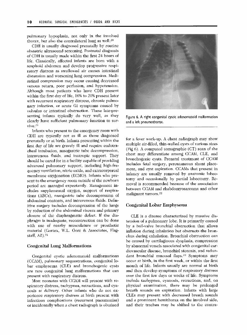
Figure 6. A right congenital cystic adenomatoid malformation and a left pneumothorax.
for a fever work-up. A chest radiograph may show multiple air-filled, thin-walled cysts of various sizes (Fig 6). A computed tomographic (CT) scan of the chest may differentiate among CCAM, CLE, and bronchogenic cysts. Prenatal treatment of CCAM includes fetal surgery, percutaneous shunt place- ment, and cyst aspiration. CCAMs that present in infancy are usually removed by anatomic lobec- tomy and occasionally by partial lobectomy. Re- moval is recommended because of the association between CCAM and rhabdomyosarcoma and other malignant tumorsz2
Congenital Lobar Emphysema
CLE is a disease characterized by massive dis- tension of a pulmonary lobe. It is primarily caused by a ball-valve bronchial obstruction that allows inflation during inhalation but obstructs the bron- chus during exhalation. Bronchial obstruction can be caused by cartilaginous dysplasia, compression by abnormal vessels associated with congenital car- diovascular disease, bronchial stenosis, and redun- dant bronchial mucosal flaps.22 Symptoms may occur at birth, in the first week, or within the first month of life. Infants usually are normal at birth and then develop symptoms of respiratory distress over the first few days or weeks of life. Symptoms include tachypnea, cyanosis, retractions, and, on physical examination, there may be prolonged breath sounds on expiration. Infants with large CLEs may present with decreased breath sounds and a prominent hemithorax on the involved side, and their trachea may be shifted to the contra-
NEONATAL SURGICAL EMERGENCIES / OKADA AND HICKS I I
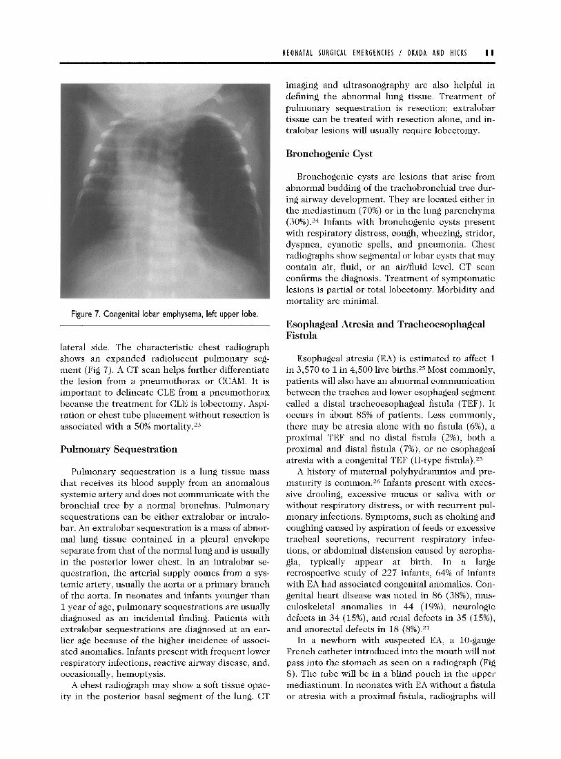
Figure 7. Congenital lobar emphysema, left upper lobe.
lateral side. The characteristic chest radiograph shows an expanded radiolucent pulmonary seg- ment (Fig 7). A CT scan helps further differentiate the lesion from a pneumothorax or CCAM. It is important to delineate CLE from a pneumothorax because the treatment for CLE is lobectomy. Aspi- ration or chest tube placement without resection is associated with a 50% mortality.23
Pulmonary Sequestration
Pulmonary sequestration is a lung tissue mass that receives its blood supply from an anomalous systemic artery and does not communicate with the bronchial tree by a normal bronchus. Pulmonary sequestrations can be either extralobar or intralo- bar. An extralobar sequestration is a mass of abnor- mal lung tissue contained in a pleural envelope separate from that of the normal lung and is usually in the posterior lower chest. In an intralobar se- questration, the arterial supply comes from a sys- temic artery, usually the aorta or a primary branch of the aorta. In neonates and infants younger than 1 year of age, pulmonary sequestrations are usually diagnosed as an incidental finding. Patients with extralobar sequestrations are diagnosed at an ear- lier age because of the higher incidence of associ- ated anomalies. Infants present with frequent lower respiratory infections, reactive airway disease, and, occasionally, hemoptysis.
A chest radiograph may show a soft tissue opac- ity in the posterior basal segment of the lung. CT
imaging and ultrasonography are also helpful in defining the abnormal lung tissue. Treatment of pulmonary sequestration is resection; extralobar tissue can be treated with resection alone, and in- tralobar lesions will usually require lobectomy.
Bronchogenic Cyst
Bronchogenic cysts are lesions that arise from abnormal budding of the trachobronchial tree dur- ing airway development. They are located either in the mediastinum (70%) or in the lung parenchyma (30%).24 Infants with bronchogenic cysts present with respiratory distress, cough, wheezing, stridor, dyspnea, cyanotic spells, and pneumonia. Chest radiographs show segmental or lobar cysts that may contain air, fluid, or an air/fluid level. CT scan confirms the diagnosis. Treatment of symptomatic lesions is partial or total lobectomy. Morbidity and mortality are minimal.
Esophageal Atresia and Tracheoesophageal Fis tula
Esophageal atresia (EA) is estimated to affect 1 in 3,570 to 1 in 4,500 live births.25 Most commonly, patients will also have an abnormal communication between the trachea and lower esophageal segment called a distal tracheoesophageal fistula (TEF). It occurs in about 85% of patients. Less commonly, there may be atresia alone with no fistula (6%), a proximal TEF and no distal fistula (2%), both a proximal and distal fistula (7%), or no esophageal atresia with a congenital TEF (H-type fistula).25
A history of maternal polyhydramnios and pre- maturity is common.26 Infants present with exces- sive drooling, excessive mucus or saliva with or without respiratory distress, or with recurrent pul- monary infections. Symptoms, such as choking and coughing caused by aspiration of feeds or excessive tracheal secretions, recurrent respiratory infec- tions, or abdominal distension caused by aeropha- &a, typically appear at birth. In a large retrospective study of 227 infants, 64% of infants with EA had associated congenital anomalies. Con- genital heart disease was noted in 86 (38%), mus- culoskeletal anomalies in 44 (19%), neurologic defects in 34 (15%), and renal defects in 35 (15%), and anorectal defects in 18 (8%).27
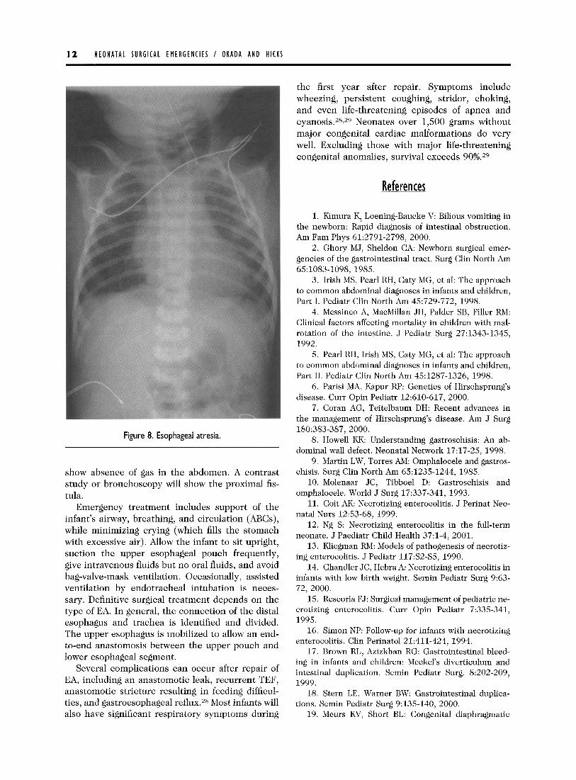
In a newborn with suspected EA, a lo-gauge French catheter introduced into the mouth will not pass into the stomach as seen on a radiograph (Fig 8). The tube will be in a blind pouch in the upper mediastinum. In neonates with EA without a fistula or atresia with a proximal fistula, radiographs will
I2 NEONATAL SURGICAL EMERGENCIES / OKADA AND HICKS
Figure 8. Esophageal atresia.
show absence of gas in the abdomen. A contrast study or bronchoscopy will show the proximal fis- tula.
Emergency treatment includes support of the infant’s airway, breathing, and circulation (ABCs), while minimizing crying (which fills the stomach with excessive air). Allow the infant to sit upright, suction the upper esophageal pouch frequently, give intravenous fluids but no oral fluids, and avoid bag-valve-mask ventilation. Occasionally, assisted ventilation by endotracheal intubation is neces- sary. Definitive surgical treatment depends on the type of EA. In general, the connection of the distal esophagus and trachea is identified and divided. The upper esophagus is mobilized to allow an end- to-end anastomosis between the upper pouch and lower esophageal segment.
Several complications can occur after repair of EA, including an anastomotic leak, recurrent TEF, anastomotic stricture resulting in feeding difficul- ties, and gastroesophageal reflux.2s Most infants will also have significant respiratory symptoms during
the first year after repair. Symptoms include wheezing, persistent coughing, stridor, choking, and even life-threatening episodes of apnea and cyanosis.28%29 Neonates over 1,500 grams without major congenital cardiac malformations do very well. Excluding those with major life-threatening congenital anomalies, survival exceeds 90%.29
References
1. Kimura K,, Loening-Baucke V: Bilious vomiting in the newborn: Rapid diagnosis of intestinal obstruction. Am Fam Phys 61:2791-2798, 2000.
2. Ghory MJ, Sheldon CA: Newborn surgical emer- gencies of the gastrointestinal tract. Surg Clin North Am 651083-1098, 1985.
3. Irish MS, Pearl RH, Caty MG, et al: The approach to common abdominal diagnoses in infants and children, Part I. Pediatr Clin North Am 45:729-772, 1998.
4. Messineo A, MacMillan JH, Palder SB, Filler RM: Clinical factors affecting mortality in children with mal- rotation of the intestine. J Pediatr Surg 27:1343-1345, 1992.
5. Pearl RH, Irish MS, Caty MG, et al: The approach to common abdominal diagnoses in infants and children, Part II. Pediatr Clin North Am 451287-1326, 1998.
6. Parisi MA, Kapur RI?: Genetics of Hirschsprung’s disease. Curr Opin Pediatr 12:610-617, 2000.
7. Coran AG, Teitelbaum DH: Recent advances in the management of Hirschsprung’s disease. Am J Surg 180:383-387, 2000.
8. Howell KK: Understanding gastroschisis: An ab- dominal wall defect. Neonatal Network 17:17-25, 1998.
9. Martin LW, Torres AM: Omphalocele and gastros- chisis. Surg Clin North Am 65:1235-1244, 1985.
10. Molenaar JC, Tibboel D: Gastroschisis and omphalocele. World J Surg 17:337-341, 1993.
11. Coit AK: Necrotizing enterocolitis. J Perinat Neo- natal Nurs 1253-68, 1999.
12. Ng S: Necrotizing enterocolitis in the full-term neonate. J Paediatr Child Health 37:1-4, 2001.
13. Kliegman RM: Models of pathogenesis of necrotiz- ing enterocolitis. J Pediatr 117:S2-S5, 1990.
14. Chandler JC, Hebra A: Necrotizing enterocolitis in infants with low birth weight. Semin Pediatr Surg 9:63- 72, 2000.
15. Rescoria FJ: Surgical management of pediatric ne- crotizing enterocolitis. Curr Opin Pediatr 7:335-341, 1995.
16. Simon NP: Follow-up for infants with necrotizing enterocolitis. Clin Perinatol 21:411-424, 1994.
17. Brown RL, Azizkhan RG: Gastrointestinal bleed- ing in infants and children: Meckel’s diverticulum and intestinal duplication. Semin Pediatr Surg. 8:202-209, 1999.
18. Stern LE, Warner BW: Gastrointestinal duplica- tions. Semin Pediatr Surg 9:135-140, 2000.
19. Meurs KV, Short BL: Congenital diaphragmatic
NEONATAL SURGICAL EMERGENCIES / OKADA AND HICKS 13
hernia: The neonatologist’s perspective. Pediatr Rev 20: E79-E87, 1999.
20. Harrison MR, deLorimier AA: Congenital dia- phragmatic hernia. Surg Clin North Am 61:1023-1035, 1981.
21. Skarsgard ED, Harrison MR: Congenital diaphrag- matic hernia: The surgeon’s perspective. Pediatr Rev 20: E71-E78, 1999.
22. Nuchtem JD, Harberg FJ: Congenital lung cysts. Semin Pediatr Surg 3:233-243, 1994.
23. Murray GF: Congenital lobar emphysema: Collec- tive review. Surg Gynecol Obstet 124:611-625, 1967.
24. Dilorenzo M, Collin Vaillancourt R: Bronchogenic cysts. J Pediatr Surg 24:988-991, 1989.
25. Beasley SW Esophageal atresia and tracheo- esophageal fistula, in Oldham KT, Colombani PM, Foglia
RP (eds): Surgery of Infants and Children: Scientific Prin- ciples and Practice. Philadelphia, PA, Lippincott-Raven, pp. 1021-1034, 1997.
26. Spitz L: Esophageal atresia: Past, present, and future. J Pediatr Surg 31:19-25, 1996.
27. Engum SA, Grosfeld JL, West KW, et al: Analysis of morbidity and mortality in 227 cases of esophageal atresia and/or tracheoesophageal fistula over two de- cades Arch Surg 130502-508, 1995.
28. Tsai JY, Berkery BS, Wesson DE, et al: Esophageal atresia and tracheoesophageal fistula: Surgical experi- ence over two decades. Ann Thorac Surg 64:778-784, 1997.
29. Spitz L, Kiely EM, Morecroft JA, et al: Oesopha- geal atresia: At-risk groups for the 1990s. J Pediatr Surg 29:723-725, 1994.