Embed Size (px)
Citation preview

ORIGINAL RESEARCH ARTICLEpublished: 24 July 2013
doi: 10.3389/fphys.2013.00181
Migraine in gulf war illness and chronic fatigue syndrome:prevalence, potential mechanisms, and evaluationRakib U. Rayhan*, Murugan K. Ravindran and James N. Baraniuk
Division of Rheumatology, Immunology and Allergy, Department of Medicine, Georgetown University, Washington, DC, USA
Edited by:
Julian M. Stewart, New YorkMedical College, USA
Reviewed by:
Kathleen S. Curtis, Oklahoma StateUniversity, USAAbdu Adem, United Arab EmiratesUniversity, UAE
*Correspondence:
Rakib U. Rayhan, Division ofRheumatology, Immunology andAllergy, Department of Medicine,Room 3004F, 3rd Floor PHCBuilding, Georgetown University,3800 Reservoir Road, NW,Washington, DC 20007-2197, USAe-mail: [email protected]
Objective: To assess the prevalence of headache subtypes in Gulf War Illness (GWI) andChronic Fatigue Syndrome (CFS) compared to controls.
Background: Approximately, 25% of the military personnel who served in the 1990–1991Persian Gulf War have developed GWI. Symptoms of GWI and CFS have considerableoverlap, including headache complaints. Migraines are reported in CFS. The type andprevalence of headaches in GWI have not been adequately assessed.
Methods: 50 GWI, 39 CFS and 45 controls had structured headache evaluations basedon the 2004 International Headache Society criteria. All subjects had history and physicalexaminations, fatigue and symptom related questionnaires, measurements of systemichyperalgesia (dolorimetry), and assessments for exclusionary conditions.
Results: Migraines were detected in 64% of GWI (odds ratio = 11.6 [4.1–32.5]) (mean[±95% CI]) and 82% of CFS subjects (odds ratio = 22.5 [7.8–64.8]) compared to only13% of controls. There was a predominance of females in the CFS compared to GWIand controls. However, migraine status was independent of gender in GWI and CFSgroups (x2 = 2.7; P = 0.101). Measures of fatigue, pain, and other ancillary criteria werecomparable between GWI and CFS subjects with and without headache.
Conclusion: The high prevalence of migraine in CFS was confirmed and extended to GWIsubjects. GWI and CFS may share dysfunctional central pathophysiological pathways thatcontribute to migraine and subjective symptoms. The high migraine prevalence warrantsthe inclusion of a structured headache evaluation in GWI and CFS subjects, and treatmentwhen present.
Keywords: migraine, gulf war illness, chronic fatigue syndrome, fibromyalgia, central sensitization, chronic pain,
fatigue, neurolimbic pathway
INTRODUCTIONGulf War Illness (GWI), also known as Chronic MultisymptomIllness (CMI), affects 25–30% of the 697,000 veterans deployed tothe 1990–1991 Persian Gulf War (Fukuda et al., 1998; Gray et al.,2002; RAC-GWVI, 2008; Li et al., 2011). Veterans with GWI sufferfrom a wide array of symptoms that include headaches, cognitivedysfunction, chronic pain, fatigue, and other complaints (Fukudaet al., 1998; Gray et al., 2002; Li et al., 2011). Gulf War veter-ans often meet the criteria for chronic fatigue syndrome (CFS)(Fukuda et al., 1994) and fibromyalgia (FM) strongly suggestingoverlap in symptoms for GWI and CFS (Wolfe et al., 1990; Pereset al., 2002; Latremoliere and Woolf, 2009; Baraniuk and Zheng,2010; Wolfe et al., 2010; Ravindran et al., 2011; de Tommaso et al.,2011).
Headache is one of the eight ancillary criteria in the CFScase definition (Fukuda et al., 1994; Baraniuk and Zheng,2010; Ravindran et al., 2011). Complaints of headaches are alsoreported in Gulf War veterans and over 50% of FM patients (Grayet al., 2002; de Tommaso et al., 2011; Li et al., 2011). However,the type and severity of headaches are not evaluated as a part ofthe GWI, CFS, or FM case designation criteria (Wolfe et al., 1990;Fukuda et al., 1994, 1998; RAC-GWVI, 2008; Wolfe et al., 2010;
Ravindran et al., 2011). We recently determined that migrainesare the primary type type of headaches in CFS (Ravindran et al.,2011). Studies in FM have reported similar migraine predomi-nance (de Tommaso et al., 2011). The exact relationships betweenheadache subtypes, fatigue, pain, hyperalgesia, and other systemiccomplaints has been difficult to establish because of heteroge-neous symptom presentation and/or lack of biomarkers thatidentify a distinct pathophysiological process in GWI.
One mechanism that has been proposed to drive the com-plaints of GWI and related illnesses is central sensitization(Latremoliere and Woolf, 2009; Baraniuk and Zheng, 2010). Thisprocess is defined as an amplification of responsiveness of cen-tral pain-signaling neurons that clinically manifest as hyperalgesiaand/or allodynia (Latremoliere and Woolf, 2009). This normaladaptive neuronal process is essential for learning, but has beenproposed to become dysregulated in each of these disorders and ineven in migraineurs (Gebhart, 2004; DaSilva et al., 2007; Tietjenet al., 2009).
Neuroimaging studies report underlying dysfunction inbrain energetics, the brainstem, and thalamocortical tracts inmigraineurs (DaSilva et al., 2007). Alterations in these regionsmay lead to the loss of descending anti-nociceptive processes
www.frontiersin.org July 2013 | Volume 4 | Article 181 | 1

Rayhan et al. Migraines in GWI and CFS
coupled with changes in cerebrovascular dynamics. Specifically,blood flow alterations in migraine headache are associated withdysfunction in the trigeminovascular system, which innervatecerebral blood vessels in the dura (Karatas et al., 2013). As aresult of trigeminal activation, migraineurs demonstrate hyper-algesia, allodynia, and cognitive dysfunction during and betweenepisodes (Gebhart, 2004; DaSilva et al., 2007; Tietjen et al., 2009;Baraniuk and Zheng, 2010; Karatas et al., 2013). These findingsmay lend credence to the theory of central sensitization.
Similar patterns of gray and white matter abnormalities andaltered brain energetics in GWI, CFS, FM, and migraine suggestthat common central mechanisms may contribute to the type ofheadaches and the cognitive impairments perceived as “brain fog”(Lutz et al., 2008; Barnden et al., 2011; Puri et al., 2012; Rayhanet al., 2013a,b). Understanding the pathophysiological mecha-nisms underlying migraine may identify the primary dysfunctionthat leads to the constellation of complaints in GWI, CFS, andother overlapping disorders (Figure 1).
Although GWI patients complain of headache symptoms thatoverlap with CFS, FM, and migraineurs, there is currently nostudy that specifically assesses this in GWI. We hypothesizedthat due to extensive symptom overlap, GWI subjects will havesimilar rates of headache complaints subtypes. As a first steptoward testing this hypothesis, we focused on identifying clinical
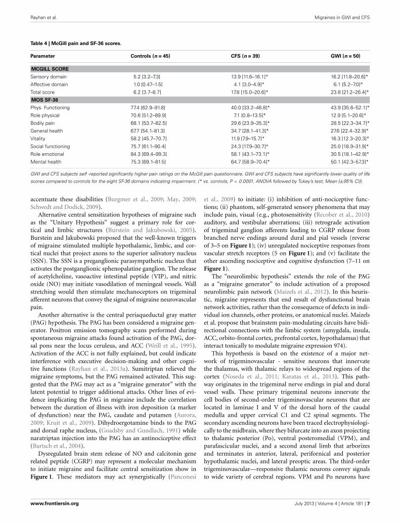
FIGURE 1 | Central sensitization using migraine as a model. (1) Corticalspreading depression (CSD) depolarizes cortical neurons and glia. (2) Theyrelease glutamate, K+, H+, metalloproteases, and other agents that dilatepial vessels and activate trigeminal nociceptive nerves. (3) The bifurcatedneurons release calcitonin gene related peptide (CGRP) and othervasodilators near dural vessels by the axon response mechanism. Vascularwall stretching activates additional trigeminal nociceptive neurons (4) thathave their primary synapse (5) in the upper cervical dorsal horn. (6)Ascending secondary afferents activate the thalamus. (7) Other afferentssignal the periaqueductal gray matter. (8) Descending relays to the magnusraphae nucleus activate descending serotonergic neurons to inhibit theprimary trigeminal (5 and 6) synapses. (9) Thalamocortical projectionsstimulate the hypothalamus, somatosensory cortex, amygdala, limbicsystem, and frontal cortex. (10) Pain, emotion, memory, frontal processingand other inputs converge on the anterior cingulate gyrus (ACC) and (11)interfere with its executive decision-making functions.
relationships between headache status, hyperalgesia (tenderness),pain, and fatigue ratings in GWI. In addition, we compared suchcomplaints to CFS subjects and controls to further identify sim-ilarities and/or differences between groups. The cross-sectionaldesign permitted estimation of the prevalences of headache, painand fatigue in GWI. Comparisons of GWI to CFS subjects allowedvalidation and extension of previous findings. Migraine status,GWI, CFS, FM co-morbidity and other conditions were diag-nosed based on history and physical examination rather thanmail-in and/or online questionnaires. Detailed history and physi-cal examination has proved to be highly reliable in previous stud-ies (Ravindran et al., 2011). To supplement the research study,we review the current literature into migraines and potentialmechanisms.
MATERIALS AND METHODSSUBJECTSGWI, CFS, and sedentary control subjects were recruited in 2 con-secutive cohorts and combined for this analysis. Both cohorts hadpopulations of all three groups (GWI, CFS, and controls). Cohort1 was recruited under Georgetown University InstitutionalReview Board protocol #2006-481 (clinicaltrials.gov identifica-tion number NCT00810329) between 2007 and 2009. Cohort 2participated in a separate protocol between 2009 and 2011 thatwas approved by the Georgetown University Institutional ReviewBoard (#2009-229; clinicaltrials.gov NCT01291758) and UnitedStates Army Medical Research Materiel Command HumanResearch Protection Office (USAMRMC HRPO #A-15547.0).
Approximately 400 GWI, CFS, and sedentary control subjectsresponded to print and on-line advertising by contacting ourlaboratory by telephone or e-mail. Each volunteer had an ini-tial telephone screening during which they gave verbal consent.Cohort 1 had 124 total eligible subjects identified after telephonescreening. Of the 124, 83 particpants completed the overall study.Cohort 2 had 167 total eligible subjects identified after telephonescreening. Of the 167, 83 participants completed the overall study.The 41 and 116 of the eligible participants from Cohort 1 and2, respectively, who were identified to be eligible after telephonescreening o did not participate due to lack of response to follow-up emails, phone calls, or did not show up to the scheduled clinicvisit.
Upon arrival in the Clinical Research Unit, all participants whodecided to complete the study reviewed and signed their informedconsent forms. All subjects had history and physical examinationswith scripted interviews to assess migraine, tension headache,CMI, CFS, and FM criteria.
INCLUSION AND CASE DESIGNATION CRITERIAInclusion criteria for veterans included military service for atleast 30 days between August 1, 1990 and July 31, 1991. GWIwas designated using the 1998 Center for Disease Control(CDC) criteria for CMI (Fukuda et al., 1998) with deploy-ment for ≥30 consecutive days to the Persian Gulf Warregion (RAC-GWVI, 2008). CMI criteria required >6 monthsof complaints from at least two of the following categories:(i) fatigue; (ii) musculoskeletal pain (muscle pain, joint pain,stiffness of joints); and (iii) cognition and mood (feeling
Frontiers in Physiology | Integrative Physiology July 2013 | Volume 4 | Article 181 | 2

Rayhan et al. Migraines in GWI and CFS
depressed, trouble remembering or focusing, mood changes, anx-ious feelings, difficulty finding words, and difficulty sleeping)(Fukuda et al., 1998).
CFS was defined by the 1994 CDC criteria (Fukuda et al.,1994). Fatigue lasting >6 months with no medical, psychiatric,or other attributable cause was required along with at least 4of the 8 ancillary criteria: (i) cognitive problems with mem-ory and concentrating, (ii) sore throat, (iii) sore lymph noderegions, (iv) myalgia, (v) arthralgia, (vi) headache with onset afterthe fatigue, (vii) sleep disturbances with un-refreshing sleep, or(viii) significant exacerbations of the severe fatigue and the othersymptoms immediately or as long as 24 h following exercise, cog-nitive, or another activity that was more strenuous than usual.All GWI subjects were also screened for CFS status during historyand physical examination to assess criteria overlap in conditions(Fukuda et al., 1994).
To assess comorbidity in CFS and GWI, FM was also identi-fied using the 1990 criteria (Wolfe et al., 1990) since the studystarted before the 2010 criteria were introduced (Wolfe et al.,2010). Subjects were assessed for widespread pain in 4 quadrantsabove and below the waist, to the left and right of the midlineand involving the axial skeleton that had been present 3 monthsand had no explanation. Manual thumb pressure of about 4 kgwas applied to 18 traditional tender points. FM required bothwidespread pain and tenderness at ≥11 of 18 points (Wolfe et al.,1990).
Migraine was defined by International Headache Society cri-teria (Headache Classification Subcommittee of the InternationalHeadache Society, 2004) using a structured interview to deter-mine if a subject had ≥5 episodes lasting 4–72 h with at least2 of (a) unilateral cephalgia, (b) pulsatile quality, (c) moderateto severe pain severity, and (d) aggravation of the headache byusual activities leading to disinclination to perform usual workor other activities of daily living. Sensitivity to light or sound,or nausea with or without emesis were required. Auras wereassessed for their prescient ability. For CFS diagnostic purposes,migraines had to be of new onset coinciding with symptomsof CFS, and so subjects with perimenstrual and progression ofheadaches with childhood onset were excluded from considera-tion. Information was collected about the duration of headachesand will be discussed in an alternate manuscript.
Exclusion criteria included positive pregnancy test, lacta-tion, active duty military personnel, claustrophobia, cardiovas-cular restrictions, intolerance of needles, ferrous based implants,major psychiatric illness, infectious status (e.g., HIV), neoplasm,untreated endocrine and other chronic disease that may haveaccounted for GWI or CFS associated symptoms (Reeves et al.,2003, 2005; Sullivan et al., 2005; RAC-GWVI, 2008; Jones et al.,2009).
QUESTIONNAIRESSubjects from cohort 1 completed paper questionnaires. Subjectsfrom cohort 2 received a confidential website log-in identifier,password and identification code so they could complete thepre-study symptom and psychometric questionnaires using ouronline confidential data collection procedure (Rayhan et al.,2013c). No personal identifying information was collected using
either system. Locked facilities were under 24 h surveillance.Workstations were only accessible by approved personnel andprotected by 128-bit encrypted password.
Subjects completed the CFS symptom severity questionnaireto self-report their symptoms for the past 6 months (Baraniuket al., 2013). The severity of fatigue and the 8 ancillary symp-toms was assessed using an anchored ordinal scale with 0 = nocomplaint, 1 = trivial, 2 = mild, 3 = moderate, or 4 = severeintensity. The scores of the 8 ancillary criteria were calculated andthe summed and are reported as “Sum8” (Baraniuk et al., 2013).
Subjective pain perceptions were quantified using the McGillshort form with its sensory, affective and total scores (Melzack,1987) and relative disability and quality of life using the MedicalOutcomes Survey Short Form 36 (SF-36) (Ware and Sherbourne,1995).
PROTOCOLHyperalgesia in FM has traditionally been ascertained by tender-ness to manual thumb pressure of about 4 kg at ≥11 of 18 tenderpoints (Wolfe et al., 1990). We adapted this concept by pressing apressure strain gauge dolorimeter at a rate of 1 kg/s at the 18 sitesand over 5 paranasal sinus regions on all subjects (Naranch et al.,2002; Rayhan et al., 2013a). Subjects were told that they were incomplete control of the pressure, and to report the point whenthe pressure sensation switched to become painful. The mean ofthe dolorimetry pressures has been previously used as a measureof systemic and sinus hyperalgesia (Naranch et al., 2002; Rayhanet al., 2013a).
STATISTICAL ANALYSISData were tabulated in Microsoft Excel 2010 (Redmond,Washington) for analysis with SPSS for Windows version 20(IBM, Armonk, NY). Means were reported with [±95% confi-dence intervals (CI)]. Significant differences in migraine statusand gender between the three groups were identified using the2 × 3 Fisher’s exact probability test with the Freeman-Haltonextension and two-tailed P-values (Freeman and Halton, 1951).Migraine headaches and CFS are more common in females thanmales (Lea et al., 2004; Ravindran et al., 2011; Critchley andHarrison, 2013). Due to this, gender was assessed as a controlledcomparison to test its influence on migraine headache and GWIand/or CFS status. All variables were entered as dichotomousvalues (0 or 1) and results from this analysis are reported withcontinuity correction (Sweeting et al., 2004).
To protect against inflating the type 1 error rate, one waymultivariate analysis of variance (MANOVA) was used across all15 non-dichotomous variables which included the ordinal rank-ings in the CFS severity score, McGill total pain score and itssubscales, and the continuous scale systemic and sinus dolorime-try values. This was followed by One-Way analysis of variance(ANOVA) on each variable with post hoc testing using Tukey’sHonest Significant Difference (HSD) test.
RESULTSDEMOGRAPHICS50 GWI, 39 CFS, and 45 control subjects were recruited. All ofthe GWI subjects also met criteria for CFS. More than half of
www.frontiersin.org July 2013 | Volume 4 | Article 181 | 3

Rayhan et al. Migraines in GWI and CFS
the GWI and control subjects were male, with a female predomi-nance of 4:1 in CFS (P = 0.00001 by Fisher’s Exact test), with nosignificant differences in age (Table 1).
HEADACHE STATUSMigraines were detected in 64% of GWI (odds ratio = 11.6[4.1–32.5]) and 82% of CFS (odds ratio = 22.5 [7.8–64.8]) sub-jects compared to 13% of controls (P = 5.5 × 10⁻¹¹; Table 2).Migraines status was characterized with aura (MA) and with-out aura (MO) with results showing the anticipated 3:1 ratio(Table 2). Co-morbid migraine and tension headaches were fre-quent. Tension headaches without a migraine diagnosis werepresent in ∼20% of GWI, 7% of CFS, and 26% of control sub-jects (Table 2). Gender did not influence CFS, GWI, and migrainestatus [χ2
(1, n = 89)= 2.69, P = 0.10].
During history and physical examinations, many patientsdescribed that they had complained of having frequent to dailyheadaches to their regular physicians. Despite this, new diagnoseswere made during the clinic visit in this protocol. This may sug-gest under evaluation of migraines in the normal clinical setting.Treatment was initiated in about half of the GWI and CFS sub-jects. Subjects who developed migraines during their visit herewere given trials of sumatriptan. The drug was uniformly bene-ficial lending additional support for migraine pathology. Resultsspecific to these findings are reported elsewhere.
MANOVA RESULTSOne-Way MANOVA confirmed the assumption that there wouldbe one or more significant differences between patient groups(control, CFS, or GWI; independent variables) using the multi-ple study outcomes [Pillai’s Trace = 0.904, F(26, 212) = 6.73, P =1.5 × 10⁻¹⁶]. The multivariate effect size (partial eta squared)was estimated at η2 = 0.452 which implies that 45.2% of thevariance in the dependent variable was accounted for by patientgrouping.
Table 1 | Demographics.
Controls (n = 45) CFS (n = 39) GWI (n = 50) P-value
Age 43.7 [39.9–47.5] 46.3 [43.1–49.5] 46.7 [44.3–49.1]
% Male 53.3% 20.5% 68.0% 0.00001*
*2 × 3 Fisher’s exact test; Mean [±95% CI].
Table 2 | Headache status.
Parameter Controls CFS GWI
(n = 45) (n = 39) (n = 50)
Migraine headache 6/45 (13.3%) 32/39 (82.1%) 32/50 (64%) 5.5 × 10⁻¹¹*
With Aura (MA) 1/6 22/32 24/32
Without Aura (MO) 5/6 10/32 8/32
Comorbid migraineand tensionheadaches
3/6 24/32 20/32
Tension headachealone
12/45 (26.6%) 3/39 (7.6%) 10/50 (20%)
∗2 × 3 Fisher’s exact test; Mean [±95% CI].
FIBROMYALGIA AND DOLORIMETRYSystemic hyperalgesia was noted as widespread pain in 62% ofGWI and 70% of CFS subjects. Manual thumb-pressure caus-ing pain at ≥11/18 tender points were more common in GWI(64%) and CFS (89%) than controls (18%; P = 7.7 × 10⁻⁸ byFisher’s Exact test). FM was diagnosed in 38 and 56% of CFSand GWI, respectively, compared to none of the control sub-jects. Dolorimetry indicated that GWI and CFS had significantlygreater hyperalgesia over systemic regions [F(2, 128) = 18.8, P =7.5 × 10⁻⁸; ANOVA followed by Tukey’s HSD test] and the 5paranasal sinus regions [F(2, 128) = 15.5, P = 9.6 × 10⁻⁷] thancontrol groups (Table 3).
SELF-REPORTED COMPLAINTSThe CFS severity score results were equivalent for GWI and CFS,and significantly greater than controls for fatigue, the 8 ancil-lary criteria, and the Sum8 score [Figures 2, 3A; F(2, 128) > 14.6,P < 1.8 × 10⁻⁶]. Sore throat and lymph nodes were the crite-ria with the lowest average scores in GWI and CFS. Controlshad significantly lower total and subscale scores on the McGillpain questionnaire compared to CFS and GWI subjects (Table 4;F(2, 128) > 38.1, P < 1.2 × 10⁻¹³]. Controls also had higher SF-36 domain scores compared to CFS and GWI subjects [Table 4;F(2, 128) > 40.1, P < 1.2 × 10⁻¹⁹].
COMPARISON OF CFS AND GWI PATIENTS WITH AND WITHOUTMIGRAINEBecause there was no significant association between genderand migraines, we combined the CFS and GWI cohorts tocompare groups with (n = 64) and without (n = 25) migrainediagnosis. All self-reported and demographic variables were sim-ilar. The Sum8 scores were also similar with the exception ofthe ordinal headache rating, systemic and sinus pain thresh-olds (Figures 3B–D). As expected, CFS and GWI subjects withmigraines had significantly higher headache ratings compared topatients without migraines and controls [Figure 3B; F(2, 128) =44.4, P = 2.2 × 10⁻¹⁵]. In addition, migraineurs had lower sys-temic [Figure 3C; F(2, 128 ) = 20.9, P = 1.5 × 10⁻⁸] and sinuspain thresholds [Figure 3D; F(2, 128) = 15.3, P = 1.1 × 10⁻⁶]indicating tenderness and potentially central sensitization.
DISCUSSIONAll of the GWI veterans recruited met CMI and CFS criteria(Fukuda et al., 1994, 1998). Overlap in case designation criteriais consistent with previous findings (Ravindran et al., 2011;Rayhan et al., 2013a,b). Scores for self-reported questionnaires,systemic and sinus pain thresholds, and migraine for GWI and
Table 3 | Systemic hyperalgesia.
Parameter Controls (n = 45) CFS (n = 39) GWI (n = 50)
Systemic dolorimetry (kg) 5.5 [4.7–6.3] 2.7 [2.2–3.0]* 3.5 [2.9–4.1]*
Sinus dolorimetry (kg) 2.2 [1.8–2.6] 0.96 [0.80–1.1]* 1.4 [1.2–1.7]*
GWI and CFS subjects had significantly lower systemic and sinus pain thresholds
compared to controls. ∗P < 0.0001 vs. controls, ANOVA followed by Tukey’s test;
mean [95% CI].
Frontiers in Physiology | Integrative Physiology July 2013 | Volume 4 | Article 181 | 4

Rayhan et al. Migraines in GWI and CFS
FIGURE 2 | CFS symptom severity scores. The severity of fatigue andthe 8 ancillary criteria were scored on an anchored ordinal scales from0 to 4. Controls (blue columns) had significantly lower scores for each
item compared to GWI (yellow columns) and CFS (red columns).∗P < 0.0000018; ANOVA followed by Tukey’s HSD test; error bars depictthe mean [±95% CI].
CFS subjects were comparable. The high prevalence of migrainein CFS was verified and extended to include GWI (Peres et al.,2002; Ravindran et al., 2011). GWI had significantly lower SF-36scores than controls indicating severe impairment of quality oflife (Buchwald et al., 1996). This indicates a large unmet need todiagnose and treat migraine in GWI and CFS, and to expand theoptions for therapy of these conditions.
GWI subjects tended to be male due to the higher ratio ofmen in the military. In contrast, CFS and FM is predominantlydiagnosed in the civilian female population (Reiffenberger andAmundson, 1996; Okifuji et al., 1999; Eisen et al., 2005; Stephen,2005; RAC-GWVI, 2008; Li et al., 2011). The high prevalencesof migraine in GWI and CFS were independent of gender. Thisfinding may be the result of exclusion of menstrual and chronicchildhood onset migraine from our study population, but is alsoconsistent with the lack of association of migraine with estrogen,progesterone and other sex hormone receptor polymorphisms(Schürks et al., 2010).
The causes of GWI and CFS have been hotly contested withlittle consensus. However, recent work suggests that toxic expo-sures (sarin gas, pesticides, particulates from oil fires, etc.) and/oracetylcholinesterase inhibitor use in the Persian Gulf may be tem-porally associated with GWI onset (RAC-GWVI, 2008; Steeleet al., 2012). Diverse infectious and stressor triggers have beenproposed, but not proven, to underlie CFS and FM in thecivilian population (Wolfe et al., 1990; Fukuda et al., 1994;Reiffenberger and Amundson, 1996; Peres et al., 2002; Ravindranet al., 2011).
The seemingly confusing symptomatology of these disor-ders requires a fresh, systems biology conceptual approach thatintegrates systems involved in assessing afferent inputs, spinal
cord and brain processing, and the conscious perceptions thatare conveyed as subjective complaints. Sensory afferent infor-mation from mucosal organs such as the nose (neurologicalnon-allergic rhinitis of CFS (Baraniuk et al., 2005), esophagus(“nutcracker esophagus”), bowel (irritable bowel syndrome) (Leaet al., 2004), bladder (irritable bladder syndrome and interstitialcystitis) is poorly localized in the insular somatosensory cortex.Interoceptive visceral sensations may enter the consciousness ofsome subjects and be perceived as serious illnesses (Critchley andHarrison, 2013). Chronic irritation, inflammation, or centrallyreleased mediators may lead to excessive glutamate release thatactivates neuronal AMPA receptors to promote the transmissionof nociceptive messages (Rowbottom et al., 1998; Goadsby et al.,2002; Fukui et al., 2008) The resulting perceptual state may be anexample of neural plasticity gone awry.
Dysregulation of neuropathic sensory relays to the dorsal ponsmay lead to excessive pain and tenderness (hyperalgesia, allo-dynia), fearful memories as in posttraumatic stress disorder,impaired executive decision-making (“brain fog”), and char-acteristic features of GWI and CFS. The synaptic molecularchanges lead to neural hypersensitivity and hyperresponsivenessto extrinsic and central, intrinsic activation that is termed “centralsensitization” (Latremoliere and Woolf, 2009). A range of genepolymorphisms such as ion channelopathies may modify the riskfor illnesses development in the population. For example, certainvariants of Panx1 ion channels may be dysfunctional and morevulnerable to stressors that promote widespread neurotransmitterrelease (Rowbottom et al., 1998; Goadsby et al., 2002; Fukui et al.,2008). In a rat model of migraine, activation of Panx1 channelson peripheral trigeminal neurons caused cerebrovascular dysreg-ulation with changes suggestive of aura followed by headache
www.frontiersin.org July 2013 | Volume 4 | Article 181 | 5

Rayhan et al. Migraines in GWI and CFS
FIGURE 3 | Sum8 and comparison between CFS and GWI subjects based
on migraine status. (A) Sum of 8 scores which total the 8 ancillary criteriafrom the CFS severity score. (B) CFS and GWI subjects were combined toshow that migraineurs (n = 64) had higher ratings of headache severity (2.9[2.7–3.2]) compared to CFS and GWI subjects with no migraines (2.1[1.6–2.5]; n = 25) and controls (0.88 [0.51–1.3]; n = 45). (C) CFS and GWI
subjects with migraines had lower systemic pain thresholds (2.8 [2.4–3.2])compared to those with no migraines (4.2 [3.4–5.1]) and controls (5.5[1.6–2.5]). (D) CFS and GWI migraineurs had lower sinus pain thresholds (1.1[0.9–1.3]) compared to CFS and GWI with no migraines (1.6 [1.3–1.9]) andcontrols (2.2 [1.8–2.6]). ∗P ≤ 0.05; ANOVA followed by Tukey’s HSD test;error bars depict the mean [±95% CI]. ∗∗P ≤ 0.001.
(Karatas et al., 2013). Evidence supporting central sensitizationhas been found in each of these chronic nociceptive, interocep-tive, and fatiguing illnesses (Malick and Burstein, 2000; Maizelsand Burchette, 2004; Meeus and Nijs, 2007).
Acute episodes of migraine and its consequences may providea model for central sensitization (Figure 1). The triggering eventmay be an acute, extreme, and prolonged depolarization calledcortical spreading depression (CSD) (Pietrobon, 2005; Smithet al., 2006; Eikermann-Haerter and Ayata, 2010). The molecularand electrical events initiating CSD involve glial and neuron cellbody depolarization with an extreme efflux of K+ out of cells, andinflux of Na+ and Ca2+ (Smith et al., 2006; Ayata, 2010; Charles,2010).
Returning the intracellular K+ concentration to the high levelrequired to repolarize these cells can take several minutes insteadof milliseconds. The prolonged depolarization leads to oligemiathat may cause transient hypoxia, depletion of oxygenated neu-roglobin reserves, (Casado et al., 2005) and anaerobic glucosemetabolism with lactic acid production. Release of autoregula-tory vasodilators leads to profound reactive cortical vessel dilationand hyperemia. In animal models, the CSD spreads outwards in
concentric fashion at the rate of 3–5 mm per minute as a wave ofintense depolarization with oligemia followed by autoregulatoryhyperemia. The change in blood flow can be detected by arterialspin labeling using 3 Tesla fMRI (T2 weighted perfusion changes)(Swartz and Kern, 2004). Peri-infarct depolarizations may be asimilar phenomena in strokes, brain trauma, and other headinjuries (Strong and Dardis, 2005; Fabricius et al., 2006; Stronget al., 2007). CSD may be analogous to the chaotic, stochasticcellular depolarization of atrial fibrillation, premature ventricularcontractions (PVC’s), esophageal spasm, or epilepsy.
Chronic CSD-like depolarization in migraine, GWI and CFSmay promote central sensitization and progressive dysfunctionof the anterior cingulate gyrus (ACC) and other neuroanatom-ical loci (Jensen et al., 2009; Obermann et al., 2009), “Neuralplasticity” (Pietrobon, 2005) may reinforce conditioned mem-ories and contribute to affective dysfunction, anxiety, fear andposttraumatic stress disorder (PTSD), (Woodward et al., 2006)fatigue, pain, hyperalgesia, allodynia and cognitive dysfunction(“brain fog”) (Meeus and Nijs, 2007). Neurovascular dysfunctionmay cause gray matter thinning and white matter abnormalities(prevalence = 16–40%; OR = 3.9, 95% CI = 2.26–6.72) that
Frontiers in Physiology | Integrative Physiology July 2013 | Volume 4 | Article 181 | 6
Rayhan et al. Migraines in GWI and CFS
Table 4 | McGill pain and SF-36 scores.
Parameter Controls (n = 45) CFS (n = 39) GWI (n = 50)
MCGILL SCORE
Sensory domain 5.2 [3.2–7.3] 13.9 [11.6–16.1]* 16.2 [11.8–20.6]*
Affective domain 1.0 [0.47–1.5] 4.1 [3.0–4.9]* 6.1 [5.2–7.0]*
Total score 6.2 [3.7–8.7] 17.8 [15.0–20.6]* 23.8 [21.2–26.4]*
MOS SF-36
Phys. Functioning 77.4 [62.9–91.8] 40.0 [33.2–46.8]* 43.9 [35.6–52.1]*
Role physical 70.6 [51.2–89.9] 7.1 [0.8–13.5]* 12.9 [5.1–20.6]*
Bodily pain 68.1 [53.7–82.5] 29.6 [23.9–35.3]* 28.5 [22.3–34.7]*
General health 67.7 [54.1–81.3] 34.7 [28.1–41.3]* 27.6 [22.4–32.9]*
Vitality 58.2 [45.7–70.7] 11.9 [7.9–15.7]* 16.3 [12.3–20.3]*
Social functioning 75.7 [61.1–90.4] 24.3 [17.9–30.7]* 25.0 [18.9–31.9]*
Role emotional 84.3 [69.4–99.3] 58.1 [43.1–73.1]* 30.5 [18.1–42.9]*
Mental health 75.3 [69.1–81.5] 64.7 [58.9–70.4]* 50.1 [42.3–57.3]*
GWI and CFS subjects self -reported significantly higher pain ratings on the McGill pain questionnaire. GWI and CFS subjects have significantly lower quality of life
scores compared to controls for the eight SF-36 domains indicating impairment. (* vs. controls, P < 0.0001; ANOVA followed by Tukey’s test; Mean [±95% CI]).
accentuate these disabilities (Burgmer et al., 2009; May, 2009;Schwedt and Dodick, 2009).
Alternative central sensitization hypotheses of migraine suchas the “Unitary Hypothesis” suggest a primary role for cor-tical and limbic structures (Burstein and Jakubowski, 2005).Burstein and Jakubowski proposed that the well-known triggersof migraine stimulated multiple hypothalamic, limbic, and cor-tical nuclei that project axons to the superior salivatory nucleus(SSN). The SSN is a preganglionic parasympathetic nucleus thatactivates the postganglionic sphenopalatine ganglion. The releaseof acetylcholine, vasoactive intestinal peptide (VIP), and nitricoxide (NO) may initiate vasodilation of meningeal vessels. Wallstretching would then stimulate mechanoceptors on trigeminalafferent neurons that convey the signal of migraine neurovascularpain.
Another alternative is the central periaqueductal gray matter(PAG) hypothesis. The PAG has been considered a migraine gen-erator. Positron emission tomography scans performed duringspontaneous migraine attacks found activation of the PAG, dor-sal pons near the locus ceruleus, and ACC (Weill et al., 1995).Activation of the ACC is not fully explained, but could indicateinterference with executive decision-making and other cogni-tive functions (Rayhan et al., 2013a). Sumitriptan relieved themigraine symptoms, but the PAG remained activated. This sug-gested that the PAG may act as a “migraine generator” with thelatent potential to trigger additional attacks. Other lines of evi-dence implicating the PAG in migraine include the correlationbetween the duration of illness with iron deposition (a markerof dysfunction) near the PAG, caudate and putamen (Aurora,2009; Kruit et al., 2009). Dihydroergotamine binds to the PAGand dorsal raphe nucleus, (Goadsby and Gundlach, 1991) whilenaratriptan injection into the PAG has an antinociceptive effect(Bartsch et al., 2004).
Dysregulated brain stem release of NO and calcitonin generelated peptide (CGRP) may represent a molecular mechanismto initiate migraine and facilitate central sensitization show inFigure 1. These mediators may act synergistically (Panconesi
et al., 2009) to initiate: (i) inhibition of anti-nociceptive func-tions; (ii) phantom, self-generated sensory phenomena that mayinclude pain, visual (e.g., photosensitivity (Recober et al., 2010)auditory, and vestibular aberrations; (iii) retrograde activationof trigeminal ganglion afferents leading to CGRP release frombranched nerve endings around dural and pial vessels (reverseof 3–5 on Figure 1); (iv) unregulated nociceptive responses fromvascular stretch receptors (5 on Figure 1); and (v) facilitate theother ascending nociceptive and cognitive dysfunction (7–11 onFigure 1).
The “neurolimbic hypothesis” extends the role of the PAGas a “migraine generator” to include activation of a proposedneurolimbic pain network (Maizels et al., 2012). In this heuris-tic, migraine represents that end result of dysfunctional brainnetwork activities, rather than the consequence of defects in indi-vidual ion channels, other proteins, or anatomical nuclei. Maizelset al. propose that brainstem pain-modulating circuits have bidi-rectional connections with the limbic system (amygdala, insula,ACC, orbito-frontal cortex, prefrontal cortex, hypothalamus) thatinteract tonically to modulate migraine expression 974).
This hypothesis is based on the existence of a major net-work of trigeminovascular - sensitive neurons that innervatethe thalamus, with thalamic relays to widespread regions of thecortex (Noseda et al., 2011; Karatas et al., 2013). This path-way originates in the trigeminal nerve endings in pial and duralvessel walls. These primary trigeminal neurons innervate thecell bodies of second-order trigeminovascular neurons that arelocated in laminae I and V of the dorsal horn of the caudalmedulla and upper cervical C1 and C2 spinal segments. Thesecondary ascending neurons have been traced electrophysiologi-cally to the midbrain, where they bifurcate into an axon projectingto thalamic posterior (Po), ventral posteromedial (VPM), andparafascicular nuclei, and a second axonal limb that arborizesand terminates in anterior, lateral, perifornical and posteriorhypothalamic nuclei, and lateral preoptic areas. The third-ordertrigeminovascular—responsive thalamic neurons convey signalsto wide variety of cerebral regions. VPM and Po neurons have
www.frontiersin.org July 2013 | Volume 4 | Article 181 | 7
Rayhan et al. Migraines in GWI and CFS
axons that bifurcate and issue dense terminal arborations inthe trigeminal barrel-field region of the primary somatosen-sory cortex (S1BF) and secondary somatosensory cortex (S2).Additional Po neurons project to extra-trigeminal regions ofS1, the primary auditory (AuD/Au1), retrosplenial (RSA), pari-etal association (PtA), primary and secondary visual (V1/V2)cortices, and primary and secondary motor cortices (M1/M2).Neurons from the laterodorsal (LD) and lateroposterior (LP) tha-lamic nucleus project to M1 and M2 regions, trigeminal andextratrigeminal S1 regions, secondary visual cortex mediome-dial (V2MM), and the ectorhinal cortex (Aurora, 2009; Bursteinet al., 2010; Noseda et al., 2011). This pathway connects theprimary meningeal nociceptive signals to the functionally dis-tinct and anatomically remote cortical areas that are involved inmigraine—related dysregulation of affect, motor function, visualand auditory perception, spatial orientation, memory retrieval,olfaction, and allodynia (Burstein et al., 2010).
Resting state fMRI functional connectivity studies of interic-tal migraine subjects provide additional support for abnormalbrainstem—limbic system interactions (Mainero et al., 2011).Functional connectivity is the property of brain regions toharmoniously signal to each other, and indicates their capability
to act in cooperative fashion. Migraineurs had increasedfunctional connectivity between the PAG and thalamus, posteriorparietal cortex, anterior insula, somatosensory cortex that areinvolved in somatosensory, interoceptive, and nociceptive pro-cessing. Those subjects with a high frequency of migraineshad increased connectivity between the PAG, anterior insula,nucleus cuneiformis, and hypothalamus, but reduced connec-tivity of PAG with the prefrontal cortex, ACC, amygdala, andmedial thalamus. Decreased connectivity between the PAG andanterior and posterior cingulate cortex, hippocampus, putamen,and posterior insula has been confirmed in a separate groupwho had frequent migraines (Maleki et al., 2011a). Women hadmore significantly reduced functional connectivity of the PAG tothe posterior cingulate cortex and amygdala than men (Malekiet al., 2011b). Functional connectivity alterations were morepronounced in adults than children (Lebel et al., 2011). Whenallodynia was present, migraineurs had decreased connectivitybetween the PAG, prefrontal cortex, ACC, and anterior insula.Therefore, magnetic imaging studies in GWI and CFS shouldtake into account potential changes in baseline cerebrovascularhemodynamic responses (Rayhan et al., 2013b), gray matter inter-connectivity, and white matter integrity (Rayhan et al., 2013a).
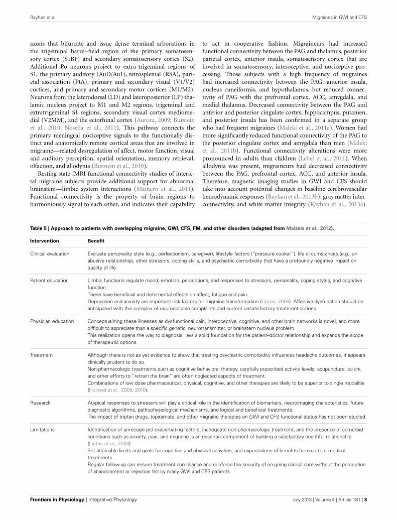
Table 5 | Approach to patients with overlapping migraine, GWI, CFS, FM, and other disorders (adapted from Maizels et al., 2012).
Intervention Benefit
Clinical evaluation Evaluate personality style (e.g., perfectionism, caregiver), lifestyle factors (“pressure cooker”), life circumstances (e.g., anabusive relationship), other stressors, coping skills, and psychiatric comorbidity that have a profoundly negative impact onquality of life.
Patient education Limbic functions regulate mood, emotion, perceptions, and responses to stressors, personality, coping styles, and cognitivefunction.These have beneficial and detrimental effects on affect, fatigue and pain.Depression and anxiety are important risk factors for migraine transformation (Lipton, 2009). Affective dysfunction should beanticipated with this complex of unpredictable complaints and current unsatisfactory treatment options.
Physician education Conceptualizing these illnesses as dysfunctional pain, interoceptive, cognitive, and other brain networks is novel, and moredifficult to appreciate than a specific genetic, neurotransmitter, or brainstem nucleus problem.This realization opens the way to diagnosis, lays a solid foundation for the patient–doctor relationship and expands the scopeof therapeutic options.
Treatment Although there is not as yet evidence to show that treating psychiatric comorbidity influences headache outcomes, it appearsclinically prudent to do so.Non-pharmacologic treatments such as cognitive behavioral therapy, carefully prescribed activity levels, acupuncture, tai chi,and other efforts to “retrain the brain” are often neglected aspects of treatment.Combinations of low dose pharmaceutical, physical, cognitive, and other therapies are likely to be superior to single modalitie(Holroyd et al., 2009, 2010).
Research Atypical responses to stressors will play a critical role in the identification of biomarkers, neuroimaging characteristics, futurediagnostic algorithms, pathophysiological mechanisms, and logical and beneficial treatments.The impact of triptan drugs, topiramate, and other migraine therapies on GWI and CFS functional status has not been studied.
Limitations Identification of unrecognized exacerbating factors, inadequate non-pharmacologic treatment, and the presence of comorbidconditions such as anxiety, pain, and migraine is an essential component of building a satisfactory healthful relationship(Lipton et al., 2003).Set attainable limits and goals for cognitive and physical activities, and expectations of benefits from current medicaltreatments.Regular follow-up can ensure treatment compliance and reinforce the security of on-going clinical care without the perceptionof abandonment or rejection felt by many GWI and CFS patients.
Frontiers in Physiology | Integrative Physiology July 2013 | Volume 4 | Article 181 | 8

Rayhan et al. Migraines in GWI and CFS
Disruption of these intricate neural pathways may represent a newparadigm for understanding complex neurological diseases.
These objective findings have important clinical implicationsfor the subjective presentation of migraine, GWI and CFS sub-jects. A common presentation is:
“. . . a middle-aged woman with chronic migraine and medicationoveruse, as well as fibromyalgia. In addition, there is anxiety anddepression, fatigue and insomnia, and the familiar exhaustive listof psychotropics and antiepileptic drugs tried and failed” (Maizelsand Burchette, 2004).
This type of patient may seem exceedingly frustrating, sincemany physicians find the panoply of symptoms daunting andtheir armamentarium bare. The physician’s quandary can bedeepened as they investigate the history further. Regardless ofwhether they consider themselves “lumpers” or “splitters,” thepresence of one functional syndrome increases the probability ofothers (Bradley, 2008). The situation is all the more exasperat-ing since current medical dogma does not provide a satisfactoryexplanation for the underlying pathophysiology. Maizels et al.(2012) provide a logical approach based on their neurolimbicmodel, knowledge of the dysfunctional of serotonergic neuralpathways, and central nature of the nociceptive, interoceptive,somatosensory, and fatigue complaints (Table 5).
This cross-sectional investigation had several limitations.Although presence and absence of aura was identified, dur-ing subgroup analysis of migraines subjects were not groupedbased upon the presence or absence of aura due to the smallsample size. It was not designed to correlate migraine phe-notypes in GWI with triggers such as fasting, loss of sleep,odors, hormonal changes, and stress (Fukui et al., 2008).Longitudinal changes in migraine severity, systemic hyperalge-sia, fatigue, and functional status await larger epidemiologicaland treatment studies. Analysis of magnetic imaging correlatesare pending.
CONCLUSIONGWI and CFS subjects have a high prevalence of migraineheadaches, comparable patterns of systemic and cognitive
complaints, and share overlapping phenomenological casedesignation criteria. CFS and GWI had lower systemic painthresholds indicating systemic hyperalgesia and central sensitiza-tion. The loss of descending inhibitory antinociceptive aminergicsignals may contribute to spinal sensitization, but cannot explainthe many other cerebral, brainstem, and autonomic abnormal-ities. It is tempting to speculate that the parallel findings ofGWI, CFS, and migraine indicate a shared underlying patho-physiological mechanism of central nervous system neural path-ways that may account for chronic pain, fatigue, and cognitivedysfunction (“brain fog”). Interdisciplinary studies that addressthese diverse components of illness are required to identify rel-evant dysfunctional pathways and molecular mechanisms thatmay lead to reductionist approaches for evaluation of futuredrug treatments.
AUTHORS’ CONTRIBUTIONSJames N. Baraniuk and Murugan K. Ravindran designed theprotocol. Rakib U. Rayhan, Murugan K. Ravindran, and JamesN. Baraniuk administered and conducted the study. RakibU. Rayhan and James N. Baraniuk interpreted and com-pleted the statistical analysis. Rakib U. Rayhan and JamesN. Baraniuk wrote, edited, and gave final approval for themanuscript.
ACKNOWLEDGMENTSThis project has been funded in whole or in partwith Federal funds (Grant # UL1TR000101, previouslyUL1RR031975) from the National Centre for AdvancingTranslational Sciences (NCATS), National Institutes of Health(NIH), through the Clinical and Translational ScienceAwards Program (CTSA), a trademark of DHHS, partof the Roadmap Initiative, “Re-Engineering the ClinicalResearch Enterprise.”; U.S. Public Health Service Award R01ES015382 from the National Institute of Environmental andHealth Science; Support was provided by Department ofDefence Congressionally Directed Medical Research Program(CDMRP) awards W81-XWH-07-1-0618 and W81-XWH-09-1-0526.
REFERENCESAurora, S. K. (2009). Spectrum of
illness: understanding biolog-ical patterns and relationshipsin chronic migraine. Neurology72(Suppl. 5), S8–S13. doi:10.1212/WNL.0b013e31819749fd
Ayata, C. (2010). Corticalspreading depression triggersmigraine attack. Headache 50,725–730. doi: 10.1111/j.1526-4610.2010.01647.x
Baraniuk, J. N., Adewuyi, O., Merck,S. J., Ali, M., Ravindran, M. K.,Timbol, C. R., et al. (2013). AChronic Fatigue Syndrome (CFS)severity score based on case desig-nation criteria. Am. J. Transl. Res. 1,53–68.
Baraniuk, J. N., Petrie, K. N., Le,U., Tai, C.-F., Park, Y.-J., Yuta,A., et al. (2005). Neuropathologyin rhinosinusitis. Am. J. Respir.Crit. Care Med. 171, 5–11. doi:10.1164/rccm.200403-357OC
Baraniuk, J. N., and Zheng, Y. (2010).Relationships among rhinitis,fibromyalgia, and chronic fatigue.Allergy Asthma Proc. 3, 169–178.doi: 10.2500/aap.2010.31.3311
Barnden, L. R., Crouch, B., Kwiatek,R., Burnet, R., Mernone, A.,Chryssidis, S., et al. (2011). Abrain MRI study of chronic fatiguesyndrome: evidence of brainstemdysfunction and altered homeosta-sis. NMR Biomed. 10, 1302–1312.doi: 10.1002/nbm.1692
Bartsch, T., Knight, Y. E., andGoadsby, P. J. (2004). Activation of5-HT1B/1D receptors in the peri-aqueductal grey inhibits meningealnociception. Ann. Neurol. 56,371–381. doi: 10.1002/ana.20193
Bradley, L. A. (2008). Pathophysiologicmechanisms of fibromyalgia and itsrelated disorders. J. Clin. Psychiatry69(Suppl. 2), S6–S13.
Buchwald, D., Pearlman, T., Umali,J., Schmaling, K., and Katon, W.(1996). Functional status in patientswith chronic fatigue syndrome,other fatiguing illnesses, and healthyindividuals. Am. J. Med. 4, 364–370.doi: 10.1016/S0002-934300234-3
Burgmer, M., Gaubitz, M., Konrad, C.,Wrenger, M., Hilgart, S., Heuft, G.,
et al. (2009). Decreased gray mat-ter volumes in the cingulo-frontalcortex and the amygdala in patientswith fibromyalgia. Psychosom. Med.5, 192–199.
Burstein, R., and Jakubowski, M.(2005). Unitary hypothesis formultiple triggers of the pain andstrain of migraine. J. Comp. Neurol.493, 9–14.
Burstein, R., Jakubowski, M., Garcia-Nicas, E., Kainz, V., Bajwa, Z.,Hargreaves, R., et al. (2010).Thalamic sensitization transformslocalized pain into widespreadallodynia. Ann. Neurol. 68, 81–91.doi: 10.1002/ana.21994
Casado, B., Pannell, L. K., Whalen,G., Clauw, D. J., and Baraniuk, J.
www.frontiersin.org July 2013 | Volume 4 | Article 181 | 9

Rayhan et al. Migraines in GWI and CFS
N. (2005). Human neuroglobinprotein in cerebrospinal fluid.BMC Proteome Sci. 3:2. doi:10.1186/1477-5956-3-2
Charles, A. (2010). Does corticalspreading depression initiatea migraine attack. Maybe not. . . Headache 50, 731–733. doi:10.1111/j.1526-4610.2010.01646.x
Critchley, H. D., and Harrison, N.A. (2013). Visceral influences onbrain and behavior. Neuron 77,624–638. doi: 10.1016/j.neuron.2013. 02.008
DaSilva, A. F., Granziera, C., Tuch,D. S., Snyder, J., Vincent, M., andHadjikhani, N. (2007). Interictalalterations of the trigeminalsomatosensory pathway and peri-aqueductal gray matter in migraine.Neuroreport 4, 301–305. doi:10.1097/WNR.0b013e32801776bb
de Tommaso, M., Federici, A., Serpino,C., Vecchio, E., Franco, G., Sardaro,M., et al. (2011). Clinical features ofheadache patients with fibromyal-gia comorbidity. J. Headache Pain 6,629–638. doi: 10.1007/s10194-011-0377-6
Eikermann-Haerter, K., and Ayata, C.(2010). Cortical spreading depres-sion and migraine. Curr. Neurol.Neurosci. Rep. 10, 167–173. doi:10.1007/s11910-010-0099-1
Eisen, S. A., Kang, H. K., Murphy, F.M., Blanchard, M. S., and Reda, D.J. (2005). Gulf war study partici-pating investigators. Gulf War vet-erans’ health: medical evaluation ofa US cohort. Ann. Intern. Med. 11,881–890. doi: 10.7326/0003-4819-142-11-200506070-00005
Fabricius, M., Fuhr, S., Bhatia, R.,Boutelle, M., Hashemi, P., Strong, A.J., et al. (2006). Cortical spreadingdepression and peri-infarct depo-larization in acutely injured humancerebral cortex. Brain 129, 778–790.doi: 10.1093/brain/awh716
Freeman, G. H., and Halton, J. H.(1951). Note on exact treatmentof contingency, goodness of fitand other problems of significance.Biometrika 38, 141–149.
Fukuda, K., Nisenbaum, R., Stewart,G., Thompson, W. W., Robin,L., Washko, R. M., et al. (1998).Chronic multisymptom illnessaffecting air force veterans of thegulf war. JAMA 280, 981–988. doi:10.1001/jama.280.11.981
Fukuda, K., Straus, S. E., Hickie, I.,Sharpe, M. C., Dobbins, J. G., andKomaroff, A. (1994). The chronicfatigue syndrome: a comprehensiveapproach to its definition andstudy. The International ChronicFatigue Syndrome Study Group.Ann. Intern. Med. 121, 953–959.
doi: 10.7326/0003-4819-121-12-199412150-199400009
Fukui, P. T., Goncalves, T. R., Strabelli,C. G., Lucchino, N. M., Matos,F. C., Santos, J. P., et al. (2008).Trigger factors in migrainepatients. Arq. Neuropsiquatr.66, 494–499. doi: 10.1590/S0004-282X2008000400011
Gebhart, G. F. (2004). Descendingmodulation of pain. Neurosci.Biobehav. Rev. 27, 729–737. doi:10.1016/j.neubiorev.2003.11.008
Goadsby, P. J., and Gundlach, A.L. (1991). Localization of [3H]-dihydroergotamine binding sites inthe cat central nervous system: rele-vance to migraine. Ann. Neurol. 29,91–94.
Goadsby, P. J., Lipton, R. B., andFerrari, M. D. (2002). Migraine-current understanding and treat-ment. N. Engl. J. Med. 346, 257–270.doi: 10.1056/NEJMra010917
Gray, G. C., Reed, R. J., Kaiser, K. S.,Smith, T. C., and Gastañaga, V. M.(2002). Self-reported symptomsand medical conditions among11, 868 Gulf War-era veterans:the Seabee Health Study. Am. J.Epidemiol. 11, 1033–1044.
Headache Classification Subcommitteeof the International HeadacheSociety. (2004). The internationalclassification of headache disorders:2nd edition. Cephalalgia 24(Suppl.1), 9–160.
Holroyd, K. A., Cottrell, C. K.,O’Donnell, F. J., Cordingley, G. E.,Drew, J. B., Carlson, B. W., et al.(2010). Effect of preventive (betablocker) treatment, behaviouralmigraine management, or theircombination on outcomes of opti-mized acute treatment in frequentmigraine: randomised controlledtrial. BMJ 341:c4871. doi: 10.1136/bmj.c4871
Holroyd, K. A., Labus, J. S., andCarlson, B. (2009). Moderation andmediation in the psychological anddrug treatment of chronic tension-type headache: the role of disor-der severity and psychiatric comor-bidity. Pain 143, 213–222. doi:10.1016/j.pain.2009.02.019
Jensen, K. B., Kosek, E., Petzke,F., Carville, S., Fransson, P.,Marcus, H., et al. (2009). Evidenceof dysfunctional pain inhibi-tion in fibromyalgia reflectedin rACC during provokedpain. Pain 144, 95–100. doi:10.1016/j.pain.2009.03.018
Jones, J. F., Lin, J. M., Maloney, E. M.,Boneva, R. S., Nater, U. M., Unger,E. R., et al. (2009). An evaluationof exclusionary medical/psychiatricconditions in the definition of
chronic fatigue syndrome. BMCMed. 7:57. doi: 10.1186/1741-7015-7-57
Karatas, H., Erdener, S. E., Gursoy-Ozdemir, Y., Lule, S., Eren-Koçak,E., Sen, Z. D., et al. (2013).Spreading depression trig-gers headache by activatingneuronal Panx1 channels.Science 6123, 1092–1095. doi:10.1126/science.1231897
Kruit, M. C., Launer, L. J., Overbosch,J., van Buchem, M. A., and Ferrari,M. D. (2009). Iron accumulationin deep brain nuclei in migraine:a population-based magnetic res-onance imaging study. Cephalalgia29, 351–359. doi: 10.1111/j.1468-2982.2008.01723.x
Latremoliere, A., and Woolf, C. J.(2009). Central sensitization:a generator of pain hyper-sensitivity by central neuralplasticity. J. Pain 10, 895–926. doi:10.1016/j.jpain.2009.06.012
Lea, R., Hopkins, V., Hastleton, J.,Houghton, L. A., and Whorwell,P. J. (2004). Diagnostic criteria forirritable bowel syndrome: utilityand applicability in clinical prac-tice. Digestion 70, 210–213. doi:10.1159/000082891
Lebel, A., Maleki, N., Drosos, A.,Guydon, A., Mattell, D., andMartonee, G. (2011). Alterationswith PAG connectivity in childrenvs adults with episodic migraine.(Abstract). Headache 51(Suppl. 1),46–47.
Li, B., Mahan, C. M., Kang, H. K.,Eisen, S. A., and Engel, C. C. (2011).Longitudinal health study of US(1991). Gulf War veterans: changesin health status at 10-year follow-up. Am. J. Epidemiol. 761–768. doi:10.1093/aje/kwr154
Lipton, R. B. (2009). Tracing trans-formation: chronic migraineclassification, progression,and epidemiology. Neurology72(Suppl. 5), S3–S7. doi:10.1212/WNL.0b013e3181974b19
Lipton, R. B., Silberstein, S. D., Saper,J. R., Bigal, M. E., and Goadsby, P.J. (2003). Why headache treatmentsfail. Neurology 60, 1064–1070.doi: 10.1212/01.WNL.0000052687.03646.74
Lutz, J., Jäger, L., de Quervain,D., Krauseneck, T., Padberg, F.,Wichnalek, M., et al. (2008). Whiteand gray matter abnormalities in thebrain of patients with fibromyalgia:a diffusion-tensor and volumetricimaging study. Arthritis Rheum. 12,3960–3969. doi: 10.1002/art.24070
Mainero, C., Boshyan, J., andHadjikhani, N. (2011). Alteredfunctional magnetic resonance
imaging resting-state connectivityin periaqueductal gray networks inmigraine. Ann. Neurol. 70, 838–845.doi: 10.1002/ana.22537
Maizels, M., Aurora, S., and Heinricher,M. (2012). Beyond Neurovascular:migraine as a dysfunctional neu-rolimbic pain network. Headache52, 1553–1565. doi: 10.1111/j.1526-4610.2012.02209.x
Maizels, M., and Burchette, R. (2004).Somatic symptoms in headachepatients: the influence of headachediagnosis, frequency, and comor-bidity. Headache 44, 983–993. doi:10.1111/j.1526-4610.2004.04192.x
Maleki, N., Nutile, L., Pendee, G.,Lebel, A., Drosos, A., and Deniel,R. G. (2011a). Novel findingswithin basal ganglia in episodicmigraine (Abstract). Headache51(Suppl. 1), 5.
Maleki, N., Linnman, C., Burstein,R., Becerra, L., and Borsook, D.(2011b). Gender-related differencesin periaqueductal gray functioningin episodic migraineurs. (Abstract).Headache 51(Suppl. 1), 40–41.
Malick, A., and Burstein, R. (2000).Peripheral and central sensitiza-tion during migraine. Funct. Neurol.15(Suppl. 3), 28–35.
May, A. (2009). New insights intoheadache: an update on functionaland structural imaging findings.Nat. Rev. Neurol. 5, 199–209. doi:10.1038/nrneurol.2009.28
Meeus, M., and Nijs, J. (2007). Centralsensitization: a biopsychosocialexplanation for chronic widespreadpain in patients with fibromyalgiaand chronic fatigue syndrome.Clin. Rheumatol. 26, 465–473. doi:10.1007/s10067-006-0433-9
Melzack, R. (1987). The short-formMcGill pain questionnaire. Pain30, 191–197. doi: 10.1016/0304-395991074-8
Naranch, K., Park, Y. J., Repka-Ramirez,M. S., Velarde, A., and Clauw,D. (2002). A tender sinus doesnot always mean rhinosinusitis.Otolaryngol. Head Neck Surg. 127,387–397.
Noseda, R., Jakubowski, M., Kainz,V., Borsook, D., and Burstein, R.(2011). Cortical projections of func-tionally identified thalamic trigemi-novascular neurons: implicationsfor migraine headache and its asso-ciated symptoms. J. Neurosci. 4,14204–14217.
Obermann, M., Pleger, B., de Greiff,A., Stude, P., Kaube, H., Diener,H. C., et al. (2009). Temporalsummation of trigeminal pain inhuman anterior cingulate cortex.Neuroimage 46, 193–200. doi:10.1016/j.neuroimage.2009.01.038
Frontiers in Physiology | Integrative Physiology July 2013 | Volume 4 | Article 181 | 10

Rayhan et al. Migraines in GWI and CFS
Okifuji, A., Turk, D. C., and Marcus,D. A. (1999). Comparison of gen-eralized and localized hyperalgesiain patients with recurrent headacheand fibromyalgia. Psychosom. Med.61, 771–780.
Panconesi, A., Bartolozzi, M. L.,and Guidi, L. (2009). Migrainepain: reflections against vasodi-latation. J. Headache Pain 10,317–325. doi: 10.1007/s10194-009-0130-6
Peres, M. F. P., Zukerman, E.,Young, W. B., and Silberstein,S. D. (2002). Fatigue inchronic migraine patients.Cephalagia 22, 720–724. doi:10.1046/j.1468-2982.2002.00426.x
Pietrobon, D. (2005). Migraine:new molecular mechanisms.Neuroscientist 11, 373–386. doi:10.1177/1073858405275554
Puri, B. K., Jakeman, P. M., Agour,M., Gunatilake, K. D., Fernando,K. A., Gurusinghe, A. I., et al.(2012). Regional grey and whitematter volumetric changes inmyalgic encephalomyelitis (chronicfatigue syndrome): a voxel-basedmorphometry 3 T MRI study. Br. J.Radiol. 85, e270–e273. doi: 10.1259/bjr/93889091
RAC-GWVI. (2008). Gulf War Illnessand the Health of Gulf War Veterans.Washington, DC: U.S. GovernmentPrinting Office.
Ravindran, M. K., Zheng, Y.,Timbol, C., Merck, S. J., andBaraniuk, J. N. (2011). Migraineheadaches in chronic fatiguesyndrome (CFS): comparison oftwo prospective cross-sectionalstudies. BMC Neurol. 11:30. doi:10.1186/1471-2377-11-30
Rayhan, R. U., Stevens, B., Adewuyi, O.,Timbol, C., VanMeter, J. W., Walitt,B., et al. (2013a). Increased brainwhite matter axial diffusivity is asso-ciated with pain, fatigue and hyper-algesia in Gulf War Illness. PLoSONE 8:e58493. doi: 10.1371/jour-nal.pone.0058493
Rayhan, R. U., Stevens, B. W., Raksit,M., Adewuyi, O., Ripple, J. A.,Timbol, C. R., et al. (2013b).Exercise challenge in Gulf WarIllness reveals two subgroupswith altered brain structure and
function. PLoS ONE 8:e63903. doi:10.1371/journal.pone.0063903
Rayhan, R. U., Zheng, Y., Uddin, E.,Timbol, C. R., Adewuyi, O., andBaraniuk, J. N. (2013c). Administerand collect medical questionnaireswith Google documents: a simple,safe, and free system. Appl. Med.Inform. 32, (in press).
Recober, A., Kaiser, E. A., Kuburas,A., and Russo, A. F. (2010).Induction of multiple photopho-bic behaviors in a transgenicmouse sensitized to CGRP.Neuropharmacology 1, 156–165. doi:10.1016/j.neuropharm.2009.07.009
Reeves, W. C., Lloyd, A., Vernon, S. D.,Klimas, N., Jason, L. A., Bleijenberg,G., et al. (2003). Internationalchronic fatigue syndrome studygroup. Identification of ambiguitiesin the 1994 chronic fatigue syn-drome research case definition andrecommendations for resolution.BMC Health Serv. Res. 3:25. doi:10.1186/1472-6963-3-25
Reeves, W. C., Wagner, D., Nisenbaum,R., Jones, J. F., Gurbaxani, B.,Solomon, L., et al. (2005). Chronicfatigue syndrome–a clinicallyempirical approach to its definitionand study. BMC Med. 3:19. doi:10.1186/1741-7015-3-19
Reiffenberger, D. H., and Amundson, L.H. (1996). Fibromyalgia syndrome:a review. Am. Fam. Physician 53,1698–1712.
Rowbottom, D., Keast, D., Pervan,Z., Goodman, C., Bhagat, C.,Kakulas, B., et al. (1998). The roleof Glutamine in the aetiology ofthe Chronic Fatigue Syndrome: aprospective study. J. Chron. FatigueSyndr. 2, 3–22.
Schürks, M., Rist, P. M., and Kurth, T.(2010). Sex hormone receptor genepolymorphisms and migraine: a sys-tematic review and meta-analysis.Cephalalgia 11, 1306–1328. doi:10.1177/0333102410364155
Schwedt, T. J., and Dodick, D. W.(2009). Advanced neuroimaging ofmigraine. Lancet Neurol. 8, 560–568.doi: 10.1016/S1474-442270107-3
Smith, J. M., Bradley, D. P., James,M. F., and Huang, C. L. (2006).Physiological studies of corticalspreading depression. Biol. Rev.
Camb. Philos. Soc. 81, 457–481. doi:10.1017/S1464793106007081
Steele, L., Sastre, A., Gerkovich, M. M.,and Cook, M. R. (2012). Complexfactors in the etiology of GulfWar illness: wartime exposuresand risk factors in veteran sub-groups. Environ. Health Perspect. 1,112–118.
Stephen, D. (2005). Atlas of Migraineand Other Headaches. London:Taylor and Francis Group.
Strong, A. J., and Dardis, R. (2005).Depolarisation phenomenain traumatic and ischaemicbrain injury. Adv. Tech. Stand.Neurosurg. 30, 3–49. doi:10.1007/3-211-27208-9_1
Strong, A. J., Hartings, J. A., andDreier, J. P. (2007). Cortical spread-ing depression: an adverse but treat-able factor in intensive care. Curr.Opin. Crit. Care 13, 126–133. doi:10.1097/MCC.0b013e32807faffb
Sullivan, P. F., Pedersen, N. L., Jacks, A.,and Evengard, B. (2005). Chronicfatigue in a population sample:definitions and heterogeneity.Psychol. Med. 35, 1337–1348. doi:10.1017/S0033291705005210
Swartz, R. H., and Kern, R. Z. (2004).Migraine is associated with mag-netic resonance imaging white mat-ter abnormalities: a meta-analysis.Arch. Neurol. 61, 1366–1368. doi:10.1001/archneur.61.9.1366
Sweeting, M. J., Sutton, A. J., andLambert, P. C. (2004). What toadd to nothing. Use and avoidanceof continuity corrections in meta-analysis of sparse data. Stat. Med. 9,1351–1375. doi: 10.1002/sim.1761
Tietjen, G. E., Brandes, J. L., Peterlin,B. L., Eloff, A., Dafer, R. M., Stein,M. R., et al. (2009). Allodyniain migraine: association withcomorbid pain conditions.Headache 9, 1333–1344. doi:10.1111/j.1526-4610.2009.01521.x
Ware, J. E., and Sherbourne, C. D.(1995). The MOS 36-item short-form health survey (SF-36): I.Conceptual framework and itemselection. Med. Care 30, 473–483.doi: 10.1097/00005650–199206000-199200002
Weill, C., May, A., and Limmroth,V. (1995). Brainstem activation
in spontaneous human migraineattacks. Nat. Med. 1, 658–660. doi:10.1038/nm0795-658.
Wolfe, F., Clauw, D. J., Fitzcharles,M. A., Goldenberg, D. L., Katz, R.S., Mease, P., et al. (2010). TheAmerican College of Rheumatologypreliminary diagnostic criteria forfibromyalgia and measurement ofsymptom severity. Arthritis CareRes. (Hoboken) 62, 600–610. doi:10.1002/acr.20140
Wolfe, F., Smythe, H., Yunus, M.,Bennett, R., Bombardier, C.,and Goldenberg, D. (1990). TheAmerican College of Rheumatology1990 criteria for the classificationof fibromyalgia. Report of themulticenter criteria committee.Arthritis Rheum. 33, 160–172. doi:10.1002/art.1780330203
Woodward, S. H., Kaloupek, D. G.,Streeter, C. C., Martinez, C.,Schaer, M., and Eliez, S. (2006).Decreased anterior cingulate vol-ume in combat-related PTSD.Biol. Psychiatry 59, 582–587. doi:10.1016/j.biopsych.2005.07.033
Conflict of Interest Statement: Theauthors declare that the researchwas conducted in the absence of anycommercial or financial relationshipsthat could be construed as a potentialconflict of interest.
Received: 26 March 2013; accepted: 25June 2013; published online: 24 July2013.Citation: Rayhan RU, Ravindran MKand Baraniuk JN (2013) Migraine in gulfwar illness and chronic fatigue syndrome:prevalence, potential mechanisms, andevaluation. Front. Physiol. 4:181. doi:10.3389/fphys.2013.00181This article was submitted to Frontiersin Integrative Physiology, a specialty ofFrontiers in Physiology.Copyright © 2013 Rayhan, Ravindranand Baraniuk. This is an open-accessarticle distributed under the terms of theCreative Commons Attribution License,which permits use, distribution andreproduction in other forums, providedthe original authors and source are cred-ited and subject to any copyright noticesconcerning any third-party graphics etc.
www.frontiersin.org July 2013 | Volume 4 | Article 181 | 11