Embed Size (px)
Citation preview

Mini-PROTEAN®
Precast Gels
Instruction Manual and
Application Guide
For technical support, call your local Bio-Rad office, or in the U.S., call 1-800-4BIORAD (1-800-424-6723)


Table of Contents
Section 1 General Information ...................................................................11.1 Introduction .............................................................................................11.2 Mini-PROTEAN Precast Gel Specifications.............................................21.3 Important Notes ......................................................................................31.4 Mini-PROTEAN Comb Configurations.....................................................3
Section 2 Setup and Basic Operation ........................................................32.1 Required Materials ..................................................................................32.2 Mini-PROTEAN Precast Gel Set-Up Overview........................................42.3 Assembling the Mini-PROTEAN Tetra Cell Electrophoresis Module ........5
Section 3 SDS-PAGE ...................................................................................63.1 Introduction .............................................................................................63.2 Mini-PROTEAN TGX Gel Composition....................................................73.3 Mini-PROTEAN TGX Gel Selection Guide ..............................................73.4 SDS-PAGE Buffers .................................................................................83.5 Sample Preparation ................................................................................83.6 Running Conditions.................................................................................8
Section 4 Native PAGE ................................................................................84.1 Introduction .............................................................................................84.2 Native PAGE Buffers...............................................................................84.3 Sample Preparation ................................................................................84.4 Running Conditions.................................................................................8
Section 5 Buffers.........................................................................................9
Section 6 Total Protein Gel Stains for SDS-PAGE and Native PAGEDetection ...................................................................................10
Section 7 Troubleshooting........................................................................11
Appendix A Stock Solutions.........................................................................13
Appendix B Total Protein Blot Stains...........................................................14
Appendix C Related Literature .....................................................................14
Appendix D Ordering Information ................................................................15D.1 Mini-PROTEAN TGX Precast Gels .................................................15D.2 Premixed Running and Sample Buffers ..........................................15D.3 Individual Reagents ........................................................................15D.4 Total Protein Gel and Blot Stains ....................................................16D.5 Immunoblot Detection.....................................................................17D.6 Immunoblot Detection Reagents.....................................................17D.7 Blotting Membranes........................................................................18D.8 Protein Standards...........................................................................18D.9 Equipment ......................................................................................18


Section 1General Information
1.1 IntroductionMini-PROTEAN® precast gels greatly simplify polyacrylamide gel electrophoresis. They are
specifically designed for use with the Mini-PROTEAN Systems (Mini-PROTEAN Tetra, Mini-PROTEAN 3, and Mini-PROTEAN Dodeca™ Cells).
Mini-PROTEAN precast gels come ready to use with pre-formed sample wells and a stackinglayer. Each Mini-PROTEAN cassette is 8.5 cm x 10 cm (H x W) and 4.0 mm thick. Gel dimensionis 7.2 cm x 8.6 cm (H x W) and 1.0 mm thick. Each gel is individually packaged in a leak proof storage pouch with gel buffer containing 0.02% sodium azide.
The migration pattern of proteins on Mini-PROTEAN TGX™ precast gels is similar to thatobserved with standard Laemmli Tris-HCl gels. Mini-PROTEAN TGX precast gels are run usingstandard Laemmli sample buffer and Tris-Glycine-SDS running buffer. The precast gels containno sodium dodecyl sulfate (SDS) and can therefore be used for either sodium dodecyl sulfatepolyacrylamide gel electrophoresis (SDS-PAGE) or native gel electrophoresis depending uponthe sample buffer and the running buffer used.
Advantages of Mini-PROTEAN TGX precast gels:
• Increased stability and long shelf life up to 12 months
• Laemmli-like separation pattern
• Exceptionally straight lanes and sharp bands
• No need for special, expensive buffers
• Superior staining quality
• No gel foot to remove prior to blotting
• Bottom open cassette that unlocks with four easy clicks
The Mini-PROTEAN Tetra cell runs both hand cast gels and Mini-PROTEAN TGX precastgels interchangeably. The cell can run from one to four gels, and the mini tank is compatiblewith other Bio-Rad electrode modules for tank blotting.
The Mini-PROTEAN 3 cell runs both hand-cast and Mini-PROTEAN TGX precast gels.The cell can run one or two gels, and the mini tank is compatible with other Bio-Rad electrodemodules for tank blotting, 2-D electrophoresis and electroelution.
The Mini-PROTEAN 3 Dodeca cell is a multi-cell for high through-put system gel electrophoresis. It can run up to 12 identical polyacrylamide gels simultaneously. The Dodecacell includes clamping frames, buffer dams, and a drain line.
1

1.2 Mini-PROTEAN Precast Gel Specifications
Gel material Polyacrylamide Gel dimensions 7.2 x 8.6 cm (H x W) Gel thickness 1.0 mm Resolving gel height 6.2 cm Cassette dimensions 8.5 x 10 cm (H x W)Cassette material Styrene copolymerComb material Polycarbonate
Total running buffer volume 700 ml for 2 gels, 1,000 ml for 4 gels (Mini-PROTEAN Tetra Cell & Mini-PROTEAN 3)
Storage conditions Store flat between 2°C and 8°C; DO NOT FREEZE
Mini-PROTEAN Tetra Cell Specifications
Casting stand PolycarbonatePin, retaining ring and spring Stainless steelCasting frames PolysulfoneGray gaskets Thermoplastic rubber (gray)Electrode assembly Glass filled polybutylene terephthalateElectrodes Platinum wire, 0.010” diameterGasket, electrode inner core Silicone rubber (green)Tank and lid PolycarbonateSample loading guides Delrin
Combs Polycarbonate
Mini-PROTEAN 3 Cell SpecificationsElectrode assembly Glass-filled liquid crystal polymerElectrodes Platinum wire, 0.010” diameterGasket, electrode inner core Silicone rubber (green)Tank and lid Molded polycarbonateSample loading guides Delrin
Combs Polycarbonate
Mini-PROTEAN 3 Dodeca Cell SpecificationsTank and lid AcrylicClamping frame Polycarbonate and liquid crystal polymerUpper electrode holder Polycarbonate with 109 mm (4.3”) platinum wireLower electrode assembly Polycarbonate with 89 mm (3.5”) platinum wireDrain line Tygon tubingDrain line connectors DelrinCooling coil AcrylicCooling coil connector tubing TygonMaximum buffer volume 4.4 LMinimum buffer volume 3.4 LOverall size 41.5 x 15 x 16.2 cm (L x W x H)Safety limits 300 V, 150 WWeight 5 kg (11 lb)
2

1.3 Important Notes (See Appendix C for Related Literature)• Mini-PROTEAN Tetra and Dodeca cell components are not compatible with acetone or
ethanol. Use of organic solvents voids all warranties.
• Each Mini-PROTEAN precast gel should be used shortly after it is removed from the storage pouch.
• It is not advisable to run more than one gel type in the same apparatus at the sametime. The different gel percentages will have different conductivity and therefore differentrun rates.
• When running 1 or 2 gels in the Tetra cell, use the electrode assembly (with the bananaplugs), not the companion running module (without the banana plugs). When running 3 or 4 gels, both the electrode assembly and the companion running module must beused.
• When running 1 or 2 gels only, DO NOT place the companion running module in thetank. Doing so will cause excessive heat generation and degrade the quality of the electrophoretic separation.
• Improper storage of Mini-PROTEAN precast gels can produce numerous artifacts. Gelsshould be stored flat between 2°C and 8°C. Avoid freezing or prolonged storage above8°C. If you suspect your gels have been stored improperly, THEY SHOULD BE DISCARDED.
• Do not attempt to lock the green arms of the electrode assembly without first ensuringthat the gel cassettes are correctly aligned against the notches on the green gaskets ofthe module. To prevent the gels from shifting during the locking step, firmly and evenlygrip them in place against the core of the module (see Figure 2c and 2e).
1.4 Mini-PROTEAN Comb Configurations
Mini-PROTEAN TGX Gel
Comb type Well volume
10 well 30 µl
15 well 15 µl
IPG 7 cm ReadyStrip™ IPG strip
Section 2 Setup and Basic Operation
2.1 Required Materials • Clean Mini-PROTEAN® Tetra cell tank • Electrophoresis module: to run 1 or 2 gels, use the electrode module. To run 3 or 4
gels, use the electrode module and companion module • PowerPac™ power supply or equivalent• Sample buffer• Running buffer (700 ml for 2 gels; 1,000 ml for 4 gels) • Mini-PROTEAN precast gels
3

Fig. 1. Mini-PROTEAN Precast Gel Cassette.
2.2 Mini-PROTEAN Precast Gel Set Up Overview1. Remove Comb: Position both thumbs on the ridges of the comb. Remove the comb by
pushing upward in one smooth continuous motion.
2. Remove Tape: Pull gently to remove the green tape from the bottom of the cassette.
3. Rinse Wells: Use a syringe wash bottle or a disposable transfer pipette to rinse the wellswith running buffer. Straighten the sides of the wells, if necessary.
4. Run Gel: Assemble the cassette into the running module of the Mini-PROTEAN system.Add running buffer to the inner and outer chambers. Prepare the samples in sample bufferand load the samples into the wells. Run the gel at 200 V until the dye front reaches theline on the bottom of the gel cassette (approximately 30–40 min). At the completion of therun, disconnect the cell and remove the cassette.
5. Open Cassette: Align the arrow on the opening key with the arrows marked on the cassette. Insert the key between the cassette plates at all 4 locations and apply downwardpressure to break each seal. Do not twist the lever. Gently pull apart the two plates beginning from the top of the cassette.
6. Remove Gel: Gently remove the gel from the cassette.
4

Fig. 2. Assembling the Mini-PROTEAN Tetra Cell Electrphoresis Module.
2.3 Assembling the Mini-PROTEAN Tetra Cell Electrophoresis Module 1. Set the electrode assembly to the open position on a clean flat surface (see Figure 2a)
2. Place the first gel cassette (with the short plate facing inward) onto the gel supports; gelsupports are molded into the bottom of the electrode assembly. There are two supportson each side of the electrode assembly. Note that the gel will now rest at a 30° angle,tilting away from the center of the electrode assembly. Use caution when placing thefirst gel, making sure that the electrode assembly remains balanced and does not tipover. Place the second gel or buffer dam on the other side of the electrode assembly,again by resting the gel on the supports. At this point there will be two gels resting at a30° angle, one on either side of the electrode assembly, tilting away from the center ofthe frame (see Figure 2b). It is critical that gel cassettes be placed into the electrodeassembly with the short plate facing inward to form the inner buffer chamber. The elec-trode assembly requires two gels to create a functioning assembly; if an odd number ofgels (1 or 3) is being run, you must use the buffer dam to complete the assembly (seeFigure 2b).
3. Using one hand, gently push both gels toward each other, making sure that they restfirmly and squarely against the green gasket that is built into the electrode assembly.Align the short plates to ensure the edge sits just below the notch at the top of thegreen gasket (Figure 2e).
4. While gently squeezing the gel cassettes or a gel cassette and a buffer dam against thegreen gaskets with one hand (keeping constant pressure and both gels firmly held inplace), slide the green arms of the clamping frame over the gels, locking them intoplace (see Figure 2c).
5
2a 2b 2c
2d 2f
2e
Notch
GelCassette
Short Plate
Long Plate
Gel Support
Gasket
ClampingFrame

5. The wing clamps of the electrode assembly lift each gel cassette up against the notchin the green gasket, forming a seal (Figure 2d). Check again to make certain that theshort plates sit just below the notch at the top of the green gasket (Figure 2e). Place theassembled electrophoresis module into the tank (Figure 2f) and fill the buffer chambers.At this point, the sample wells can be washed out with running buffer, if this was notdone earlier, and the sample can be loaded. If running more than 2 gels, repeat steps2a–2d with the companion running module.
Section 3 SDS-PAGE
3.1 IntroductionMini-PROTEAN® TGX™ precast gels provide a versatile system for sodium dodecyl
sulfate polyacrylamide gel electrophoresis (SDS-PAGE), a gel electrophoretic techniquethat separates proteins according to their molecular weight.
SDS-PAGE relies on a discontinuous buffer system. Two ions of differing electrophoreticmobility (glycinate and chloride) form a moving boundary when voltage is applied. Proteinshave an intermediate mobility, causing them to concentrate, or stack, into a narrow zone atthe beginning of electrophoresis. As the boundary moves through the gel, the sieving effectof the polyacrylamide gel matrix causes different proteins to move at different rates. Thestacking effect is responsible for the high resolving power of SDS-PAGE. The sample isloaded in a relatively broad zone, and the moving boundary concentrates the proteins intosharp bands prior to separation.
Protein samples for SDS-PAGE are prepared using SDS and a thiol reductant, usually2-mercaptoethanol or dithiothreitol (DTT). SDS forms complexes with proteins giving thema rod like shape and similar charge to mass ratio. The reductant cleaves disulfide bondsbetween and within proteins allowing complete denaturation and dissociation. Heat treatment in the presence of SDS and reductant effectively eliminates the effects of 2° and 3°protein structure and native charge on electrophoretic mobility, so the migration distancedepends primarily on molecular weight. Molecular mass is determined by plotting the logarithm of protein molecular mass vs. the relative mobility (Rf) of the protein (Rf = dis-tance migrated by protein/distance relative to the dye front of the protein). Refer to technotes 3133 and 3144.
Mini-PROTEAN TGX precast gels are prepared without SDS. Although gels for SDS-PAGE have historically been cast with SDS in the gel, high quality SDS-PAGE separations are obtained in gels lacking SDS, provided that the sample buffer and runningbuffer contain sufficient SDS to maintain SDS saturation during electrophoresis. The recommended concentrations of SDS are >1% in the sample buffer and 0.1% in the runningbuffer. The absence of SDS in the gel itself provides additional flexibility, as the gels mayalso be used for native electrophoresis (see Section 4).
Mini-PROTEAN TGX precast gels differ from the standard Laemmli system (Tris-HClSDS-PAGE) gels due to a proprietary modification to their formulations that provides thegels with extended shelf life and improved separation characteristics. They are designed tobe run using standard Laemmli sample and running buffers. No additional special buffers orreagents are required.
6

3.2 Mini-PROTEAN TGX Precast Gel CompositionMini-PROTEAN TGX gels are comprised of polyacrylamide with a bisacrylamide cross
linker. Each gel has a 4% polyacrylamide stacking layer extending approximately 5 mmfrom the bottom of the loading well to the top of the resolving gel. The proprietary gel formulation provides a shelf life of 12 months and improved separation characteristics.
The gel is packaged with storage buffer of the same composition with additional 0.02%sodium azide as a preservative.
3.3 Mini-PROTEAN TGX Precast Gel Selection Guide
Mini-PROTEAN TGX gels are available in a wide selection of single percentages andgradients for the separation of proteins by SDS-PAGE.
Gel Selection Guide
Gel % Optimal Sample Running Run Conditions* Run Type Separation Range Buffer Buffer Voltage/Current** Time***
TGX 7.5 40–200 kD SDS-PAGE SDS-PAGE 200 V constant 38 minsample buffer running Starting current
buffer (per gel): 37 mAFinal current (per gel): 23 mA
TGX 10 30–150 kD SDS-PAGE SDS-PAGE 200 V constant 38 minsample running Starting current buffer buffer (per gel): 37 mA
Final current (per gel): 23 mA
TGX 12 20–120 kD SDS-PAGE SDS-PAGE 200 V constant 38 minsample running Starting current buffer buffer (per gel): 37 mA
Final current (per gel): 23 mA
TGX 4–15 20–250 kD SDS-PAGE SDS-PAGE 200 V constant 30 minsample running Starting current buffer buffer (per gel): 50 mA
Final current (per gel): 33 mA
TGX 4–20 10–200 kD SDS-PAGE SDS-PAGE 200 V constant 30 minsample running Starting current buffer buffer (per gel): 50 mA
Final current (per gel): 33 mA
TGX Any kD****10–200 kD SDS-PAGE SDS-PAGE 200 V constant 28 minsample running Starting current buffer buffer (per gel): 50 mA
Final current (per gel): 33 mA
*This may vary depending on water and buffer conductivity, which may vary from one lab setting to the next.
**Current should be multiplied by the number of gels being run.
***Approximate time required for dye front to reach the line at the bottom of the cassette.
****Any kD is a unique single percentage formulation that provides a broad separation range and short running time.
7

3.4 SDS-PAGE BuffersSee Section 5 for buffer recipes.
3.5 Sample PreparationThe appropriate concentration of sample depends on the load volume and the detection
method used. (See Section 6 for approximate stain sensitivities). Add 50 µl 2-mercaptoethanolper 950 µl of sample buffer for a final concentration of 5% 2-mercaptoethanol, or 710 mM.As an alternative, DTT may be used at a final concentration of 350 mM (54 mg/ml). Dilute 1 part sample with at least 1 part sample buffer with added reductant. Heat the mixture at95°C for 5 min.
3.6 Running Conditions Run gels at 200 V constant voltage until the dye front reaches the line near the bottom
edge of the gel cassette. Approximate run times will vary between 28 and 38 min dependingon the gel type (see Section 3.3).
Section 4 Native PAGE
4.1 IntroductionMini-PROTEAN® TGX™ gels are made without SDS, allowing native separations using
SDS- and reductant-free sample and running buffers. The nonreducing and nondenaturing environment of native PAGE allows protein separation with retention of biological activity.Native PAGE can also be used to resolve multiple protein bands when molecular mass separation by SDS-PAGE would reveal only one.
Native PAGE uses the same moving boundary described in Section 3.1. Proteins are prepared in nonreducing nondenaturing sample buffer, which maintains the proteins’ secondary structure and native charge density. Protein mobility depends on the size andshape of the protein as well as its molecular weight and net charge. Native PAGE is thereforenot suitable for molecular weight determination.
4.2 Native PAGE BuffersSee Section 5 for buffer recipes.
4.3 Sample PreparationDetermine the desired protein concentration and load volume of your sample based on
the detection method used. (See Section 6 for approximate stain sensitivities). Proteins canbe separated using a standard protocol, following dilution of the sample with an equal volume of Native Sample Buffer (see Section 5, DO NOT HEAT SAMPLES). Strongly basicproteins (pl >8.5) will have a net positive charge and will not enter a native PAGE TGX gel.
4.4 Running ConditionsSee Section 3.3.
8

Section 5 Buffers (see Appendix A for Stock Solutions)
Name Working Concentration Notes Pre-Mixed Alternative SDS-Page running buffer
1X 25 mM Tris base 192 mM glycine 0.1% (w/v) SDS
Running buffer should be ~ pH 8.3. Do not adjust the pH
10x Tris/Glycine/SDS, 1 L, 161-0732 10x Tris/Glycine/SDS, 5 L cube, 161-0772
SDS-PAGE sample buffer
2X 62.5 mM Tris HCl, pH 6.8 2% (w/v) SDS 25% (v/v) glycerol 0.01% (w/v) Bromophenol Blue 5% (v/v) 2-mercaptoethanol or 350 mM DTT (added fresh)
Dilute 1 part sample with 1 part sample buffer. More sample buffer can be added if necessary. 1 part sample to 2 parts sample buffer dilution also works. Dry samples can be dissolved directly into the sample buffer
Laemmli sample buffer, 30 ml, 161-0737
Native PAGE running buffer working concentration
25 mM Tris Base 192 mM glycine
Running buffer should be ~ pH 8.3. Do not adjust the pH
10x Tris/Glycine, 1 L,
10x Tris/Glycine, 5 L cube,
161-0771
Native PAGE sample buffer
62.5 mM Tris-HCl, pH 6.8 40% glycerol 0.01% Bromophenol Blue
Native sample buffer, 30 ml, 161-0738
-
-
-
161-0734
Dilute 1 part sample with 1 part sample buffer. More sample buffer can be added if necessary. 1 part sample to 2 parts sample buffer dilution also works. Dry samples can be dissolved directly into the sample buffer
9

Section 6 Total Protein Gel Stains for SDS-PAGE and NativePAGE Detection
Method Sensitivity Optimal Protein Load
Advantages Disadvantages Imaging Instruction Manual Number
Coomassie Blue R-250
36–47 ng ~0.5 µg/band
Laboratory standard
Requires MeOH
Photography with white light or transmission densitometry (Gel Doc™ or GS-800™)
Consult scientific literature
Bio-Safe™ Coomassie stain
8–28 ng ~0.5 µg/band
Nonhazardous Photography with white light or transmission densitometry (Gel Doc or GS-800)
4307051
Zinc stain 6–12 ng ~0.2 µg/band
High-contrast, fast, reversible stain
Negative stain, must be photographed, SDS-PAGE only
Photography with white light or transmission densitometry (Gel Doc or GS-800)
4006082
Silver Stain™ Plus kit
0.6–1.2 ng ~0.01 µg/band
Simple, robust, mass spectrometry compatible
Does not stain Glycoproteins well
Photography with white light or transmission densitometry (Gel Doc or GS-800)
LIT442
Silver stain 0.6–1.2 ng ~0.01 µg/band
Stains complex proteins, i.e., glycoproteins, and lipoproteins
Not mass spectrometry compatible
Photography with white light or transmission densitometry (Gel Doc or GS-800)
LIT34
Dodeca™ Silver Stain Kit
0.5–1.2 ng ~0.1 µg/band
Convenient staining for a large number of gels
Photography with white light or transmission densitometry (Gel Doc or GS-800)
4110150
SYPRO™ Ruby protein gel stain
1–10 ng ~0.2 µg/band
Broad dynamic range, simple robust protocol
Requires imaging instrument for maximum sensitivity
Fluorescence visualization with UV trans-illumination or laser scanning
4006173
Flamingo™ Fluorescent
0.25–0.5 ng ~0.01 µg/band
Broad dynamic range, mass spec compatible
Requires imaging instrument for maximum sensitivity
Fluorescence visualization with UV trans-illumination or laser scanning (best option)
10003321
Oriole Fluorescent protein gel stain
0.5 ng ~0.2 µg/band
High sensitivity Broad dynamic range
Will not work with visible excitation
Fluorescence visualization with UV transil-lumination (Gel Doc, Chemi Doc)
10017295
10

Section 7 TroubleshootingProblem Cause Solution
Current is zero or less than • Tape at the bottom of • Remove tapeexpected and samples do not the cassette not migrate into gel removed
• Insufficient buffer in inner • Fill buffer chamber withbuffer chamber 700 ml running buffer
• Insufficient buffer • Make sure the innerin outer buffer and outer buffer chamber chambers are
sufficiently filled toensure that the wells ofthe gel are completely covered
• Electrical disconnection • Check electrodes and connections
Bands “smile” across gel, • Excess heating of gel • Check buffer compositionband pattern curves upward • Do not exceed at both sides of the gel recommended running
conditions
• Insufficient buffer • Make sure the innerand outer bufferchambers aresufficiently filled toensure that the wells ofthe gel are completelycovered
Smiling or frowning bands • Overloaded proteins • Load less proteinwithin the gel lane • Sample • Consider minimizing
preparation/buffer salts, detergents and issues solvents in sample
preparation and samplebuffer
• Running speed • Check to make sure thecorrect voltage hasbeen set
Skewed or distorted bands, • Excess salt in samples • Remove salts from lateral band spreading sample by dialysis or
desalting column prior to sample preparation
• Insufficient sample • Check buffer buffer or wrong composition and formulation dilution instructions
Vertical streaking • Overloaded samples • Dilute sample
11

Problem Cause SolutionVertical streaking • Sample precipitation • Selectively remove
predominant protein in the sample
• Dilute sample in moresample buffer
• Insoluble materials • Centrifuge samples toin the samples remove particulates(e.g., membranes) prior to sample loading
Gels run faster than • Running buffer is too • Check buffer expected concentrated and gel composition
temperature is too high• Incorrect running buffer
type is used
Artifact bands at ~60–70 kD • Possible skin keratin • Thoroughly clean all contamination dishware and wear
gloves while handling and loading gel
• Filter all solutionsthrough 0.2 µm or 0.45 µm filter
Leaking from inner • Incomplete gasket seal • Wet the gasket with buffer chamber running buffer before
use
• Improper assembly of • Check that the top edgethe gel into the of the short plate fits electrode/companion under the notch at the assembly top of the gasket
• Make sure that the top of the short plate istouching the green gasket
Poor resolution • High sample volume • If possible, load a moreor fuzzy bands concentrated sample
in a lower volume ofsample buffer
• Diffuse sample loading • Load sample with zone syringe or gel loading
pipette tips
• Sample diffusion during • Fix gel with 40% staining with Coomassie methanol, 10% aceticstain acid for 80 min prior to
staining
• Incompatible sample • Consider minimizingcomponents salts, detergents, and
solvents in samplepreparation and samplebuffer
Bands are not present • Proteins have • Use a smaller poreor are missing from the transferred through the size membraneblotting membrane* membrane • Decrease the transfer
time• Decrease the voltage
*For more Western blot troubleshooting suggestions, see the Mini Trans-blot® Electrophoretic Transfer Cell Instruction Manual(1703930) or the Trans-Blot® SD Semi-Dry Electrophoretic Transfer Cell Instruction Manual (1703940).
12

Appendix AStock SolutionsBuffer NotesSDS-PAGE running 10x Stock Running buffer should buffer Tris base 15.0 g be ~pH 8.3. Do not adjust
Glycine 72.0 g the pHSDS 5.0 gTo 500 ml with DI H2O
SDS-PAGE sample 2x Stockbuffer 0.5M Tris-HCl, pH 6.8 1.0 ml
10% (w/v) SDS 1.6 mlGlycerol 2.0 ml1.0% Bromophenol Blue 0.08 ml2-Mercaptoethanol 0.4 mlDI H2O 2.92 mlTotal Volume 8.0 ml
Native PAGE running 10x Stock Running buffer should buffer Tris base 15.0 g be ~pH 8.3. Do not adjust
Glycine 72.0 g the pH
To 500 ml with DI H2O
Native PAGE sample 2x Stockbuffer 0.5M Tris-HCl, pH 6.8 1.0 ml
Glycerol 2.2 ml1% Bromophenol Blue 0.08 mlDI H2O 3.72 mlTotal Volume 8.0 ml
0.5 M Tris-HCl Tris base 6.06 g Adjust to pH 6.8 with HCl.DI H2O ~60 ml Make to 100 ml with Total Volume 100 ml DI H2O. Store at 4°C
10% SDS SDS 1.0 g Stir gentlyTo 10 ml with DI H2O
1% Bromophenol Blue Bromophenol Blue 100 mg Stir gentlyTo 10 ml with DI H2O
Coomassie Blue R-250 Methanol (40%) 400 ml Dissolve Coomassie R-250 staining solution (0.1%) Acetic Acid (10%) 100 ml in methanol/acetic acid.
Coomassie Blue R-250 (0.1%) 1.0 g Add DI H2O to a final To 1,000 ml with DI H2O volume of 1,000 ml
Coomassie Blue R-250 Methanol 400 mldestaining solution Acetic acid 100 ml
DI H2O 500 ml
13
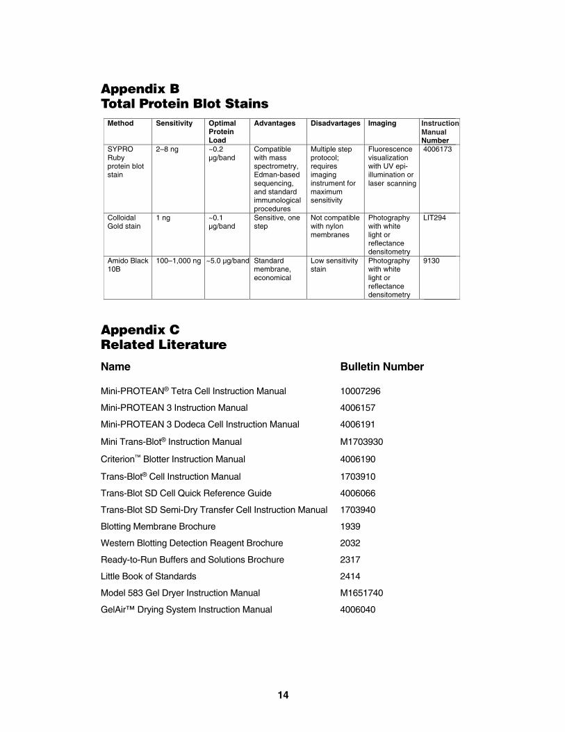
Appendix BTotal Protein Blot Stains
Appendix CRelated Literature
Name Bulletin Number
Mini-PROTEAN® Tetra Cell Instruction Manual 10007296
Mini-PROTEAN 3 Instruction Manual 4006157
Mini-PROTEAN 3 Dodeca Cell Instruction Manual 4006191
Mini Trans-Blot® Instruction Manual M1703930
Criterion™ Blotter Instruction Manual 4006190
Trans-Blot® Cell Instruction Manual 1703910
Trans-Blot SD Cell Quick Reference Guide 4006066
Trans-Blot SD Semi-Dry Transfer Cell Instruction Manual 1703940
Blotting Membrane Brochure 1939
Western Blotting Detection Reagent Brochure 2032
Ready-to-Run Buffers and Solutions Brochure 2317
Little Book of Standards 2414
Model 583 Gel Dryer Instruction Manual M1651740
GelAir™ Drying System Instruction Manual 4006040
Method Sensitivity Optimal Protein Load
Advantages Disadvantages Imaging
Number
SYPRO Ruby protein blot stain
2–8 ng ~0.2 µg/band
Compatible with mass spectrometry, Edman-based sequencing, and standard immunological procedures
Multiple step protocol; requires imaging instrument for maximum sensitivity
Fluorescence visualization with UV epi-illumination or laser scanning
4006173
Colloidal Gold stain
1 ng ~0.1 µg/band
Sensitive, one step
Not compatible with nylon membranes
Photography with white light or reflectance densitometry
LIT294
Amido Black 10B
100–1,000 ng ~5.0 µg/band
Standard membrane, economical
Low sensitivity stain
Photography with white light or reflectance densitometry
9130
InstructionManual
14

Appendix DOrdering Information
D.1 Mini-PROTEAN® TGX™ Precast Gels
10 Gels per box 2 Gels per box
10-Well 15-Well IPG Comb 10-Well30 µl/well 15 µl/well 7 cm IPG Strip 30 µl
7.5% 456-1023 456-1026 456-1021 456-1023S10% 456-1033 456-1036 456-1031 456-1033S12% 456-1043 456-1046 456-1041 456-1043S4–15% 456-1083 456-1086 456-1081 456-1083S4–20% 456-1093 456-1096 456-1091 456-1093SAny kD ™ 456-9033 456-9036 456-9031 456-9033S
D.2 Premixed Running and Sample BuffersCatalogNumber Product Description
161-0732 10x Tris/Glycine/SDS, 1 L161-0772 10x Tris/Glycine/SDS, 5 L cube161-0737 Laemmli Sample Buffer, 30 ml161-0738 Native Sample Buffer, 30 ml161-0734 10x Tris/Glycine, 1 L161-0771 10x Tris/Glycine, 5 L cube161-0778 10x Tris/CAPS, 1 L 161-0780 10x Phosphate Buffered Saline, 1 L170-6435 10x Tris Buffered Saline, 1 L161-0783 1x Phosphate Buffered Saline With 1% Casein, 1 L161-0782 1x Tris Buffered Saline With 1% Casein, 1 L
D.3 Individual ReagentsCatalogNumber Product Description
161-0719 Tris, 1 kg 161-0716 Tris, 500 g 161-0717 Glycine, 250 g 161-0718 Glycine, 1 kg 161-0724 Glycine, 2 kg 161-0301 SDS, 100 g 161-0302 SDS, 1 kg161-0416 SDS Solution, 10% (w/v), 250 ml161-0418 SDS Solution, 20% (w/v), 1 L170-6404 Blotting-Grade Blocker, 300 g161-0710 2-Mercaptoethanol, 25 ml 161-0610 Dithiothreitol, 1 g 161-0611 Dithiothreitol, 5 g 161-0404 Bromophenol Blue, 10 g
15
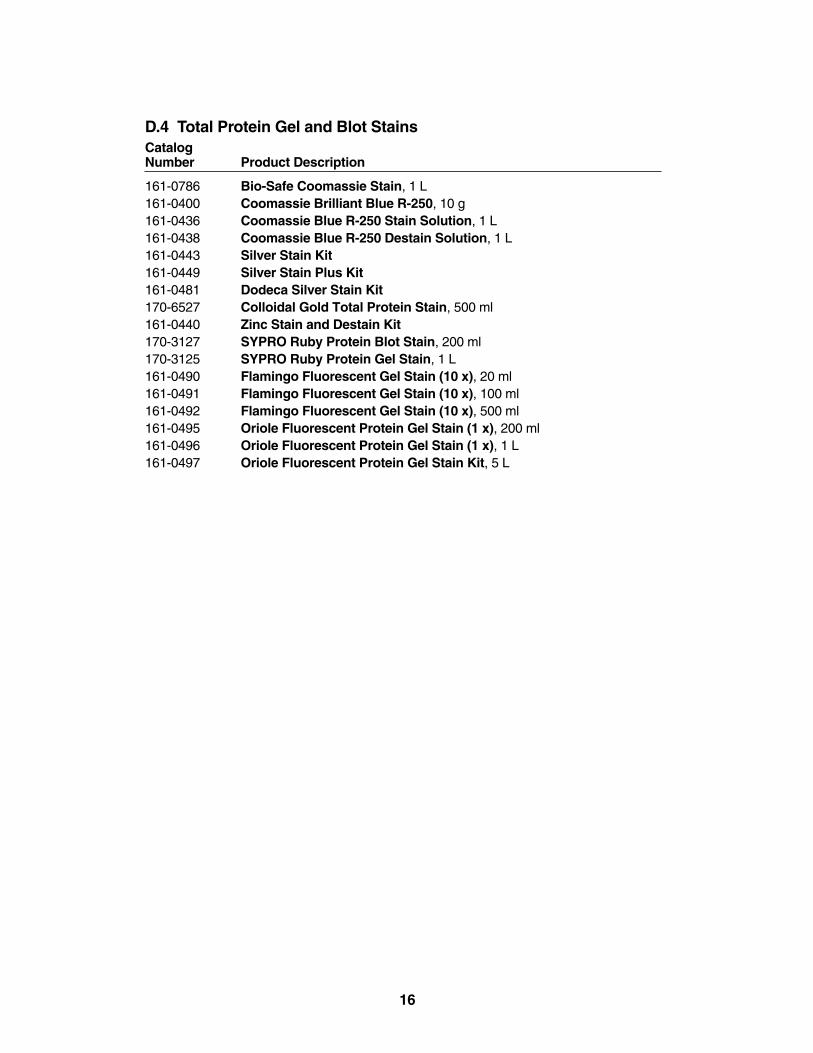
D.4 Total Protein Gel and Blot StainsCatalogNumber Product Description
161-0786 Bio-Safe Coomassie Stain, 1 L 161-0400 Coomassie Brilliant Blue R-250, 10 g 161-0436 Coomassie Blue R-250 Stain Solution, 1 L 161-0438 Coomassie Blue R-250 Destain Solution, 1 L 161-0443 Silver Stain Kit161-0449 Silver Stain Plus Kit 161-0481 Dodeca Silver Stain Kit170-6527 Colloidal Gold Total Protein Stain, 500 ml 161-0440 Zinc Stain and Destain Kit170-3127 SYPRO Ruby Protein Blot Stain, 200 ml 170-3125 SYPRO Ruby Protein Gel Stain, 1 L 161-0490 Flamingo Fluorescent Gel Stain (10 x), 20 ml 161-0491 Flamingo Fluorescent Gel Stain (10 x), 100 ml 161-0492 Flamingo Fluorescent Gel Stain (10 x), 500 ml 161-0495 Oriole Fluorescent Protein Gel Stain (1 x), 200 ml161-0496 Oriole Fluorescent Protein Gel Stain (1 x), 1 L161-0497 Oriole Fluorescent Protein Gel Stain Kit, 5 L
16

D.5 Immunoblot Detection
See related literature in Appendix C for information on Western blotting and gel drying.
D.6 Immunoblot Detection ReagentsCatalogNumber Product Description
170-5070 Immun-Star WesternC Chemiluminescent Kit, 100 ml170-6431 HRP Conjugate Substrate Kit, 4CN 170-6535 HRP Color Development Reagent, DAB, 5 g170-8238 Amplified Opti-4CN Substrate Kit 170-8235 Opti-4CN Substrate Kit170-6432 AP Conjugate Substrate Kit170-5012 Immun-Star Substrate Pack
Method Sensitivity Optimal Protein Load
Advantages Disadvantages Imaging
4CN colorimetric (HRP)
500 pg ~0.25 µg/band
Fast detection
Results may fade
Photography with white light or reflectance densitometry
DAB colorimetric (HRP)
500 pg ~0.25 µg/band
Fast detection Contains toxic chemicals
Photography with white light or reflectance densitometry
Opti-4CN™ colorimetric (HRP)
100 pg ~0.05 µg/band
Color does not fade
More expensive than 4CN
Photography with white light or reflectance densitometry
Amplified Opti-4CN colorimetric (HRP)
10 pg ~0.005 µg/band
High sensitivity, low background
Amplification requires additional steps
Photography with white light or reflectance densitometry
BCIP/NBT colorimetric
100 pg ~0.05 µg/band
Sensitive, multiple antigen
May detect endogenous (AP) enzyme activity
Photography with white light or reflectance densitometry
Immun-Star™chemiluminescent (AP)
10 pg
~0.005 µg/band
Long-lasting signal, short and multiple exposures possible
Requires visualization
on film or instrumentation
Chemiluminescent visualization with film or imager
Immun-Star chemiluminescent HRP
1–3 pg
~0.005 µg/band
Intensifies signal output, very sensitive
Requires visualization on
film or instrumentation
Chemiluminescent visualization with film or imager
Immun-Star WesternC™ (HRP)
10 fg
~0.005 µg/band
Long-lasting signal, short and multiple exposures possible
Requires visualzation on film or instrumentation
Chemiluminescent visualization with film or imager
17

D.7 Blotting MembranesCatalogNumber Product Description
162-0232 0.2 µm Nitrocellulose/Filter Paper Sandwich, 8.5 x 13.5 cm, 20 pack 162-0233 0.2 µm Nitrocellulose/Filter Paper Sandwich, 8.5 x 13.5 cm, 50 pack 162-0234 0.45 µm Nitrocellulose/Filter Paper Sandwich, 8.5 x 13.5 cm, 20 pack 162-0235 0.45 µm Nitrocellulose/Filter Paper Sandwich, 8.5 x 13.5 cm, 50 pack 162-0236 Sequi-Blot™ PVDF/Filter Paper Sandwich, 8.5 x 13.5 cm, 20 pack 162-0237 Sequi-Blot PVDF/Filter Paper Sandwich, 8.5 x 13.5 cm, 50 pack
D.8 Protein StandardsCatalogNumber Product Description
161-0363 Precision Plus Protein™ Unstained Standards (10–250 kD), 1,500 µl,150 applications
161-0373 Precision Plus Protein All Blue Prestained Standards (10–250 kD),500 µl, 50 applications
161-0374 Precision Plus Protein Dual Color Standards (10–250kD), 500 µl, 50 applications
161-0375 Precision Plus Protein Kaleidoscope™ Standards (10–250 kD), 500 µl, 50 applications
161-0376 Precision Plus Protein WesternC™ Standards (10–250kD), 250 µl, 50 applications
161-0385 Precision Plus Protein WesternC Pack (10–250kD), 50 applicationseach of standard and StrepTactin-HRP
161-0317 SDS-PAGE Standards, broad range, 200 µl161-0320 2-D SDS-PAGE Standards, Unstained, 500 µl
D.9 EquipmentCatalogNumber Product Description
165-8004 Mini-PROTEAN Tetra Cell165-4100 Mini-PROTEAN 3 Dodeca™ Cell170-3930 Mini Trans-Blot® Electrophoretic Transfer Cell 170-3940 Trans-Blot SD Semi-Dry Electrophoretic Transfer Cell164-5050 PowerPac™ Basic Power Supply164-5052 PowerPac HC High-Current Power Supply164-5070 PowerPac Universal Power Supply164-5056 PowerPac HV Power Supply165-1789 Hydrotech™ Gel Drying System, 100/120V165-1790 Hydrotech Gel Drying System, 220/240V165-1771 GelAir™ Drying System, 115V, 60Hz165-1772 GelAir Drying System, 230V, 50Hz
SYPRO is a trademark of Molecular Probes, Inc. Bio-Rad is licensed to sell SYPRO products for researchuse only, under US Patent 5,616,502.
18


Life ScienceGroup
00-0000 0000 Sig 0308Bulletin 0000 Rev A US/EG
Bio-Rad Laboratories, Inc.
Web site www.bio-rad.com USA 800 4BIORAD Australia 61 02 9914 2800 Austria 01 877 89 01 Belgium 09 385 55 11 Brazil 55 21 3237 9400 Canada 905 364 3435 China 86 21 6426 0808 Czech Republic 420 241 430 532 Denmark 44 52 10 00 Finland 09 804 22 00 France 01 47 95 69 65 Germany 089 318 84 0 Greece 30 210 777 4396 Hong Kong 852 2789 3300 Hungary 36 1 455 8800 India 91 124 4029300 Israel 03 963 6050 Italy 39 02 216091 Japan 03 6361 7000 Korea 82 2 3473 4460 Mexico 52 555 488 7670 The Netherlands 0318 540666 New Zealand 0508 805 500 Norway 23 38 41 30 Poland 48 22 331 99 99 Portugal 351 21 472 7700 Russia 7 495 721 14 04 Singapore 65 6415 3188 South Africa 27 861 246 723 Spain 34 91 590 5200 Sweden 08 555 12700 Switzerland 061 717 95 55 Taiwan 886 2 2578 7189 United Kingdom 020 8328 2000
1658100 Rev A