Embed Size (px)
Citation preview
Biochemical Pharmacology, Vol. 30, No. 14. pp. 1925-1929, 1981. 00062952/81/141925J.l5 $02.0010
Printed in Great Britain. 0 1981 Pergamon Press Ltd.
MISONIDAZOLE-INDUCED THYMIDINE RELEASE FROM DNA
R. J. KNOX, R. C. KNIGHT and D. I. EDWARDS*
Chemotherapy Research Unit, Department of Paramedical Sciences, North East London Polytechnic, Romford Road, London El5 4LZ, U.K.
(Receiued 18 December 1980; accepted 17 February 1981)
Abstract-Misonidazole reduced electrolyrically at constant potential in the presence of DNA of varying base composition has been shown to release specifically a mixture of thymidine mono- and diphosphates. Release is not dependent upon helix integrity nor upon base sequences. About 5-6 per cent of the total thymidine is released irrespective of the total A + T content of DNA. Since uridine phosphates are released from RNA the target in DNA does not involve the sugars, nor the methyl group of the thymidine residue but presumably involves the uracil ring system. Binding of reduced misonidazole to DNA is of a very low order, is proportiOna to the G + C content and is a secondary event unrelated to the cytotoxic effect of the drug.
Misonidazole (2-nitro-1-imidazolyl-3-methoxy-2- propanol) is a radiosensitizer of hypoxic tumours at present undergoing clinical trials [l, 21. Its clinical potential is enhanced by a cytotoxic effect in hypoxic or anaerobic cells [3-61 the mechanism of which appears to differ in mammalian and bacterial cells although both require reduction of the nitro group and both result in DNA damage. In order to study the cytotoxic mechanism we have developed an elec- trolytic model of the drug-target interaction in which the nitro group of misonidazole is selectively reduced at constant potential in the presence of DNA and damage analysed subsequently. Such studies have shown that misonidazole and other reduced nitro- imidazole drugs induce strand breakage and desta- bilization of DNA leading to the formation of single stranded regions [7-lo]. Such damage is not random but depends upon the base composition of DNA- more particularly the adenine plus thymine (A + T) content [ 1 l] which suggests a specific target in DNA. Preliminary results [12] indicate the target to be thymidine and we now report that misonidazole specifically cleaves thymidine phosphates from DNA.
MATERIALS AND METHODS
DNA from calf thymus (type I), Escherichia coli (type VIII), Micrococcus lysodeikticus (type XI), Clostridium perfringens (type XII) and yeast RNA (type III) were obtained from the Sigma Chemical Co (Poole, U.K.). Single stranded DNA from E. coli was prepared by heating double stranded DNA in 15 mM NaCl, 1.5 mM trisodium citrate pH 7.1 at 100” for 8min and cooling rapidly to -12” in a salt-ice mixture. E. coli DNA labelled with 14C in the carbon-2 group of thymine was obtained from the Radiochemical Centre, (Amersham, U.K.). Poly [d(A-T)] and poly [d(G-C)] were obtained from the Boehringer Corporation, (London, U.K.). Misoni- dazole (2-nitro-1-imidazolyl-3-methoxy-2-propanol)
* Author to whom correspondence should be addressed.
and 2-14C-misonidazole were obtained from Roche Products Ltd (Welwyn Garden City, U.K.). Chro- matographic standards of thymine, thymidine, thymidine-3-phosphate and thymidine-5-phosphate were obtained from Sigma Chemical Co. and thymidine-2,5-diphosphate was obtained from CP Laboratories Ltd (Bishop Stortford, U.K.).
Electrochemical reduction of misonidazole was carried out with various DNA sources at a drug nucleotide ratio of 1: 1 as previously described [9, lo] at a constant potential of -800 mV. More negative potentials were avoided because of degradative interactions between the electrodes and DNA 113,141.
After reduction samples were dialysed against three times their volume of water and the dialysate examined spectrophotometrically, then reduced in volume in uacuo to 1 ml before chromatographic analysis.
The dialysate obtained after drug reduction was chromatographed on a Sephadex G-10 column (150 x 0.9 cm) and eluted with 0.1 M ammonium for- mate, HCl buffer pH 4.0 at a flow rate of 5 ml/hr. The eluate was monitored at 260 nm through a 0.5 ml flow cell and fractions of 2.5 ml collected for further analysis. ‘%-DNA samples before and after dialysis were chromatographed on Sephadex G-50 columns (60 X 0.9 cm) using an elution buffer of 8 M urea, 0.5 M ammonium bicarbonate buffer pH 8.6 at a flow rate of 5 ml/hr and 0.5 ml fractions collected and analysed for radioactivity.
Apurinic and apyrimidinic acid derivatives of calf thymus DNA were fractionated on Sephadex G-100 columns (60 x 0.9 cm) with a urea-bicarbonate buffer pH 8.6 as detailed above and the eluate moni- tored continuously at 260 nm using a flow rate of 20 ml/hr. The column was calibrated using protein standards (Sigma Chemical Co.) which consisted of lysozyme (14,300); /?-lactoglobulin (18,400)) trypsi- nogen (24,000), pepsin (34,700), egg albumin (45,000) and bovine plasma albumin (66,000).
Spectrophotometry of samples before and after
1925
1926 R. J. KNOX, R. C. KNIGHT and D. I. EDWARDS
A xc
A 26C
CPM
A 260
c
L 0
:d) i
I 123 4 5
I!, I 20 40 60 80 100
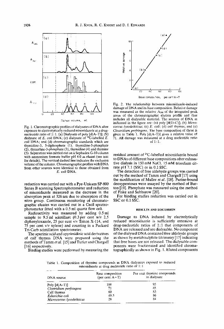
Fig. 1. Chromatographic profiles of dialysates of DNA after exposure to electrolytically reduced misonidazole at a drug- nucleotide ratio of 1: 1. (a) Dialysate of poly [d(A-T)]; (b) dialysate of E. coli DNA; (c) dialysate of “C-labelled E. cofi DNA; and (d) chromatographic standards which are thymidine-3, 5-diphosphate (l), thymidine-5-phosphate (2), thymidine-3-phosphate (3), thymidine (4) and thymine (5). Separation was carried out on a Sephadex G-10 column with ammonium formate buffer pH 4.0 as eluant (see text for details). The vertical dashed line indicates the exclusion volume of the column. Chromatographic profiles with DNA from other sources were identical to those obtained from
E. coli DNA.
reduction was carried out with a Pye-Unicam SP-800 Series B scanning Spectrophotometer and reduction of misonidazole measured as the decrease in the absorption peak at 326 nm due to absorption of the nitro group. Continuous monitoring of chromato- graphic eluates was carried out in a Cecil spectro- photometer fitted with a 0.5 ml quartz flow cell.
Radioactivity was measured by adding 0.5 ml sample to 9.5 ml scintillant (0.3 per cent w/v 2,5 diphenyloxazole, 25 per cent v/v Triton X-114, and 75 per cent v/v xylene) and counting in a Packard Tri-Carb scintillation spectrometer.
The apurinic acid and apyrimidinic acid derivatives of calf thymus DNA were prepared using the methods of Tamm et al. [15] and Turler and Chargaff [ 161 respectively.
Binding studies were performed by measuring the
Bose composltlon, per cent A +T
Fig. 2. The relationship between misonidazole-induced damage of DNA and its base composition. Relative damage was measured as the relative A260 of the integrated peak areas of the chromatographic elution profile and thus includes all dialysable material. The sources of DNA as indicated in the figure are: (a) poly [d(G-C)]; (b) Micro- coccus 1ysodeikticus; (c) E. coli; (d) calf thymus; and (e) Clostridium perfringens. The base composition of these is given in Table 1. Poly [d(A-T)] gives a relative value of 71. All damage was measured at a drug nucleotide ratio
of 1:l.
residual amount of 14C-labelled misonidazole bound to DNAs of different base compositions after exhaus- tive dialysis in 150 mM NaCl, 15 mM trisodium cit- rate pH 7.1 @SC) or in 0.1 SSC.
The detection of free aldehyde groups was carried out by the method of Tamm and Chargaff [17] using the modification of Muller et al. [18]. Purine-bound deoxypentoses were assayed by the method of Bur- ton [19]. Phosphate was measured using the method of Fiske and Subbarow [20].
For binding studies reduction was carried out in SSC or 0.1 SSC.
RESULTS AND DISCUSSION
Damage to DNA induced by electrolytically reduced misonidazole is sufficiently extensive at drug-nucleotide ratios of 1: 1 that components of DNA are released and are dialysable. No component of the dialysed DNA contained free aldehyde groups as shown by metabisulphite titrimetry [17] indicating that free bases are not released. The dialysable com- ponents were fractionated and identified chroma- tographically as shown in Fig. 1. Eluted components
Table 1. Composition of thymine compounds in DNA dialysates exposed to reduced misonidazole at drug nucleotide ratio of 1: 1
DNA source
Poly [d(A-T)] Clostridium perfringens Calf thymus Esherichia coli Micrococcus lysodeikticus
Base composition (per cent A+T)
100 71 60 49.5 28
Per cent thymine compounds in dialysate
95 85 83 86 84
Misonidazole and DNA 1927
(e) (b) Cc) Cd) .
-•-.-•- .
I I I I I I 20 40 60 80 100
Base composhon, per cent A + T
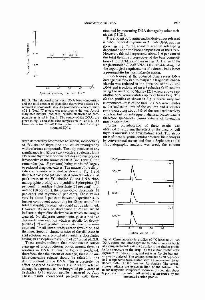
Fig. 3. The relationship between DNA base composition and the total amount of thymidine derivatives released by reduced misonidazole at a drug-nucleotide concentration of 1: 1. Total ‘T’ release was measured as the total AZ~ of dialysable material and thus includes all thymidine com- ponents as listed in Fig. 1. The source of the DNAs are given in Fig. 2 and their base composition in Table 1. The lower value for E. coli DNA (point c) is that for single
stranded DNA.
were detected by absorbance at 260 nm, radioactivity of 14C-labelled thymidine and co-chromatography with reference compounds. The only products of any significance (cu. 85 per cent) which are released from DNA are thymine mononucleotides and nucleosides irrespective of the source of DNA (see Table l), the remainder (cu. 15 per cent) being attributed largely to reduced drug derivatives. The nature of the dialy- sate components separated as shown in Fig. 1 and their relative yield (as calculated from the integrated peak areas of the 14C-labelled E. coli DNA chro- matographic profile) are thymidine-3-phosphate (32 per cent), thymidine-5-phosphate (22 per cent), thy- midine (18 per cent), thymidine-3$diphosphate (15 per cent) and thymine (3 per cent). These values vary by about 5 per cent between experiments. A further component accounting for 10 per cent of the total dialysable radioactivity could not be identified. However, its lack of absorbance at 260 nm would indicate a thymidine derivative in which the ring is cleaved. No dialysate components gave a positive diphenylamine reaction which is specific for deoxy- purines [19] and positive phosphate reactions were obtained for all compounds except thymidine and thymine. Spectral characteristics of the dialysate in acid solution were typical of thymidine phosphates having an absorption maximum of 267 nm at pH 2.0.
These results indicate that misonidazole causes cleavage of phosphodiester bonds around thymine residues in DNA. It may be logically predicted, therefore, that the extent of damage, that is, thym- idine-derivative release should be related to the A + T content of the DNA. This is precisely the effect observed as shown in Fig. 2 where relative damage is expressed as the integrated peak areas of Sephadex G-10 elution profile measured by AZm, These results correspond precisely with those
obtained by measuring DNA damage by other tech- niques [ll, 211.
The amount of thymine and its derivatives released is 5-6% of total thymine in E. coli DNA and, as shown in Fig. 2, the absolute amount released is dependent upon the base composition of the DNA. However, this still represents about 5-6 per cent of the total thymine irrespective of the base composi- tion of the DNA as shown in Fig. 3. The yield for single stranded E. coli DNA is similar indicating that the topological requirements of a double helix is not a prerequisite for misonidazole action.
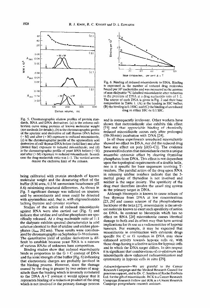
To determine if the reduced drug causes DNA damage resulting in non-dialysable fragments mison- idazole was reduced in the presence of 14C E. coli DNA and fractionated on a Sephadex G-50 column using the method of Stanley [22] which allows sep- aration of oligonucleotides up to 25 bases long. The elution profiles as shown in Fig. 4 reveal only two components-that of the bulk of DNA which elutes at the exclusion limit of the column and a smaller peak containing about 6% of the total radioactivity which is lost on subsequent dialysis. Misonidazole therefore specifically causes release of thymidine mononucleotides.
Further corroboration of these results was obtained by studying the effect of the drug on calf thymus apurinic and apyrimidinic acid. The struc- tures of these oligonucleotides preclude their analysis by conventional means and thus a Sephadex G-100 chromatographic analysis was used, the column
(O)l
Oki== E 8
1000 1 A -
0 ‘1 \
(cl
1000 c A ,\
IO 20 30 40
Elutlon volume, ml
Fig. 4. Chromatographic profiles of 14C-labelled E. coli DNA before and after exposure to reduced misonidazole at a drug-nucleotide ratio-of 1: 1. (a) is the elution profile before exposure to the drug, (b) the elution profile after exposure to reduced drug and (c) is as for (b) but sub- sequently dialysed. The column contained G-50 Seohadex L and components were eluted with an ammonium bicar- bonate bufferpH 8.6 (see text for details). The vertical arrows indicate the exclusion limit of thd column. The minor dialysable component shown in (b) contains about 6 per cent of the total radioactivity as measured by the
integrated elution profile.
1928 R. J. KNOX, R. C. KNIGHT and D. I. EDWARDS
“*, (a )
l \ l -._
0 06 -M+M
A 260 004 I) 0 0 02
006
0 06 I- (dl
Elutlon volume, ml
Fig. 5. Chromatographic elution profiles of protein stan- dards, RNA and DNA derivatives. (a) is the column cali- bration curve using proteins of known molecular weight (see methods for details); (b) is the chromatographic profile of the apurinic acid derivative of calf thymus DNA before (- M} and after (i- M) exposure to reduced misonidazole, (c) is the chromatographic profile of the apyrimidinic acid derivative of calf thymus DNA before (solid line) and after (dotted line) exposure to reduced misonid~ole; and (d) is the chromatographic profile of yeast RNA before (-M) and after (+M) exposure to reduced misonidazole. In each case the drug-nucleotide ratio was 1: 1. The vertical arrows
denote the exclusion limit of the column.
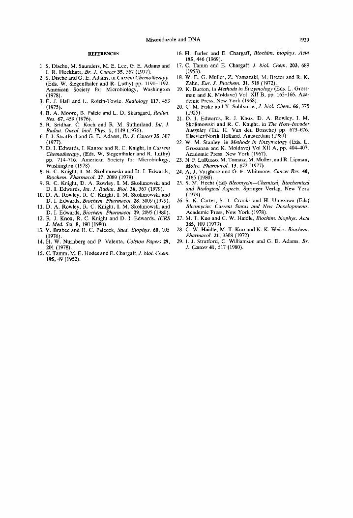
being calibrated with protein standards of known molecular weight and the denaturing effect of the buffer (8 M urea, 0.5 M ammonium bicarbonate pH 8.6) minimising structural differences. As shown in Fig. 5 signific~t damage was inflicted on apurinic acid by misonidazole and none could be detected with apy~midinic acid, that is, with oligonucleotides lacking thymine and cytosine residues.
Studies of the action of reduced misonidazole against RNA were also carried out {Fig. 5) and indicate that uridine and uridine phosphates are spe- cifically released. At a drug nucleatide ratio of 1: 1 the dialysate exhibits spectral characteristics in acid solution identical to that of uridine and uridine phos- phates (&,,, 262 nm). These results were corrobor- ated by chromatography on Sephadex G-10 columns. The quantitative yield of uridine derivatives is dif- ficult to establish because yeast RNA is a mixture of various RNAs of unknown base composition.
Binding studies show that reduced misonidazole binds in proportion to the G + C content of DNA and the ionic strength of the buffer (Fig. 6) indicating that electrostatic charges are probably involved in the binding process. However, since the damage caused by the drug is greater by two orders of mag- nitude than the binding which is inversely correlated to the DNA A+T content it would appear that it represents binding of a reduction product of the drug which is not involved in the primary damage process
&tse comp=ttMn, per cent A + T
Fig. 6. Binding of reduced misonidazole to DNA. Binding is expressed as the number of reduced drug molecules bound per 103 nucleotides and was measured as the amount of non-dialysable “C-iabetled misonidazole after reduction in the presence of DNA at a drug-nucleotide ratio of 1: 1, The source of each DNA is given in Fig. 2 and their base composition in Table 1. (A) is the binding in SSC buffer; (B) the binding in O.lSSC; and (C) the binding of unreduced
drug in either SSC or 0.1 SSC.
and is consequently irrelevant. Other workers have shown that metronidazole also exhibits this effect [23] and that appreciable binding of chemically reduced misonidazole occurs only after prolonged (10-50 min) incubation with DNA (241.
In all these experiments unreduced misonidazole showed no effect on DNA, nor did the reduced drug have any effect on poly [d(G-C)]. The evidence presented indicates that misonidazole exerts a unique anaerobic cytotoxic effect by cleaving thymidine phosphates from DNA. This effect is not dependant upon the topological requirements of a double helix, nor is it specific for base sequences involving T- residues. The parallel action of the drug upon RNA in releasing uridine residues indicate that the 5 methyl group of thymidine is not involved and neither is the sugar moiety. The specificity of the drug must therefore involve the uracii ring system as the primary target in DNA.
Although bleomycin is known to cause release of free thymine from DNA at low concentrations 123,267 and causes scission of the phosphodiester backbone of the helix [27], misonidazole is the small- est molecule known lo exert such specificity of action on DNA. In contrast to bieomycin which has no effect on RNA [ZS] misonidazole causes identical damage to both and its effect may thus have sevetal implications for its use as a radiosensitizer of hypoxic tumours. For example, it may be expected that misonidazole in combination with cytotoxic drugs specific for C or G residues in DNA may show enhanced activity towards hypoxic cells or, with those drugs having a selective action for hypoxic celis and in which the DNA target differs. In this respect it is significant that combinations of &-platinum and misonidazole show enhanced radiosensitization and cytotoxicity in hypoxic cells in vitro [29].
Acknowledgements-We are grateful to the Cancer Research Campaign and the Medical Research Council for generous support, and to Dr. C. Smithen of Roche Products Ltd. for the gift of misonidazole. RCK is a Cancer Research Campaign Research Fellow and RJK is a Cancer Research Campaign postgraduate research assistant.
Misonidazole and DNA 1929
REFERENCES
1. S. Dische, M. Saunders, M. E. Lee, G. E. Adams and I. R. Flockhart, Br. J. Cancer 35, 567 (1977).
2. S. Dische and G. E. Adams, in Current Chemotherapy. (Eds. W. Siegenthaler and R. Luthy) pp. 1191-1192. American Society for Microbiology, Washington (1978).
3. E. J. Hall and L. Roizin-Towle, Radiology 117, 453 (1975).
4. B. A. Moore, B. Palcic and L. D. Skarsgard, Radial. Res. 67, 459 (1976).
5. R. Sridhar, C. Koch and R. M. Sutherland, ht. J. Radiat. Oncol. biol. Phys. 1, 1149 (1976).
6. I. J. Stratford and G. E. Adams, Et-. J. Cancer 3.5, 307 (1977).
7. D. I. Edwards, I. Kantor and R. C. Knight, in Current Chemotherapy, (Eds. W. Siegenthaler and R. Luthy) pp. 714-716. American Society for Microbiology, Washington (1978).
8. R. C. Knight, I. M. Skolimowski and D. I. Edwards, Biochem. Pharmacol. 27. 2089 (1978).
9. R. C. Knight, D. A. Rowley, I: M. Skohmowski and D. I. Edwards, Int. J. Radiat. Biol. 36, 367 (1979).
10. D. A. Rowley, R. C. Knight, I. M. Skolimowski and D. I. Edwards, Biochem. Pharmacol. 28, 3009 (1979).
11. D. A. Rowley, R. C. Knight, I. M. Skolimowski and D. I. Edwards. Biochem. Pharmacol. 29.2095 (1980).
12. R. J. Knox, R. C. Knight and D. I. Edwards: ICR.Y J. Med. Sci. 8, 190 (1980).
13. V. Brabec and E. C. Palecek. Stud. Biophvs. 60. 105 .s
(1976). 14. H. W. Nurnberg and P. Valenta, Colston Papers 29,
201 (1978). 15. C. Tamm, M. E. Hodes and E. Chargaff, J. biol. Chem.
195, 49 (1952).
16. H. Turler and E. Chargaff, Biochim. biophys. Acta 195, 446 (1969).
17. C. Tamm and E. Chargaff, J. biol. Chem. 203, 689 (1953).
18. W. El G. Muller, 2. Yamazaki, M. Breter and R. K. Zahn. Eur. J. Biochem. 31. 518 (1972).
19. K. Burton, in Methods in Enzym&ogy’(Eds. L. Gross- man and K. Moldave) Vol. XII B, pp. 163-166. Aca- demic Press, New York (1968).
20. C. M. Fiske and Y. Subbarow, J. biol. Chem. 66, 375 (1925).
21. D. I. Edwards, R. J. Knox, D. A. Rowley, I. M. Skolimowski and R. C. Knight, in The Host-Znuader Interplay (Ed. H. Van den Bossche) pp. 673-676. ElsevieriNorth Holland, Amsterdam (1980).
22. W. M. Stanley, in Methods in Enzymology (Eds. L. Grossman and K. Moldave) Vol XII A, pp. 404-407. Academic Press, New York (1967).
23. N. F. LaRusso, M. Tomasz, M. Muller, and R. Lipman, Molec. Pharmacol. 13, 872 (1977).
24. A. J. Varghese and G. F. Whitmore, Cancer Res. 40, 2165 (1980).
25. S. M. Hecht (Ed) Bleomycin-Chemical, Biochemical and Biological Aspects. Springer Verlag, New York (1979).
26. S. K. Carter, S. T. Crooks and H. Umezawa (Eds) Bleomycin: Current Status and New Developments. Academic Press, New York (1978).
27. M. T. Kuo and C. W. Haidle, Biochim. biophys. Acta 385, 109 (1973).
28. C. W. Haidle, M. T. Kuo and K. K. Weiss. Biochem. Pharmacol. 21, 3308 (1972).
29. I. J. Stratford, C. Williamson and G. E. Adams, Br. J. Cancer 41, 517 (1980).


![CO6-1 Characterization of Clustered DNA Damage Induced by ......tered DNA damage is a unique radiation damage [1], and estimate quantity and quality of clustered DNA damage induced](https://img.pdfslide.net/doc/110x75/5fe67e48b2da127c1835f903/co6-1-characterization-of-clustered-dna-damage-induced-by-tered-dna-damage.jpg)