Embed Size (px)
Citation preview
Effect of fertility restorer gene, Rf2, onmitochondrial RNA and proteins in LeadRice-type cytoplasmic male sterile rice cells
Disa Bäckström
Degree project in biology, Bachelor of science, 2012Examensarbete i biologi 15 hp till kandidatexamen, 2012Biology Education Centre, Uppsala University, and Laboratory of Environmental Plant Biotechnologyat Tohoku UniversitySupervisors: Kinya Toriyama and Lars Liljas
1
Contents
Contents………………………………………………………………………………………….1
Summary ………………………………………………………………………………………..2
Introduction……………………………………………………………………………………….3
Cytoplasmic Male Sterility in Rice………………………………………………….3
Infertility-Causing Open Reading Frames………………………………………....4
Fertility Restorer Genes……………………………………………………………..4
Aims…...........…………………………………………………………………………5
Results…………………………………………………………………………………………….6
Detection of the transferred Rf2 gene and Rf2 expression ………………………6
Detection of ORF79 protein ………………………………………………………….7
Detection of atp6 and orf79 transcripts …………………….…………..…………..7
Discussion and conclusion………………………………………………………………….......9
Materials and methods………………………………………………….………………………12
Samples………………………………………………………………………….……12
Total RNA and DNA analysis…………………………………………………………12
Total RNA extraction from callus cells……………………………………12
RT-PCR……………………………………………………………………..13
Total DNA extraction from callus cells…………………………………...13
Mitochondrial protein analysis trough western blotting……………………..…….14
Extraction of mitochondria ……..………………………………………..14
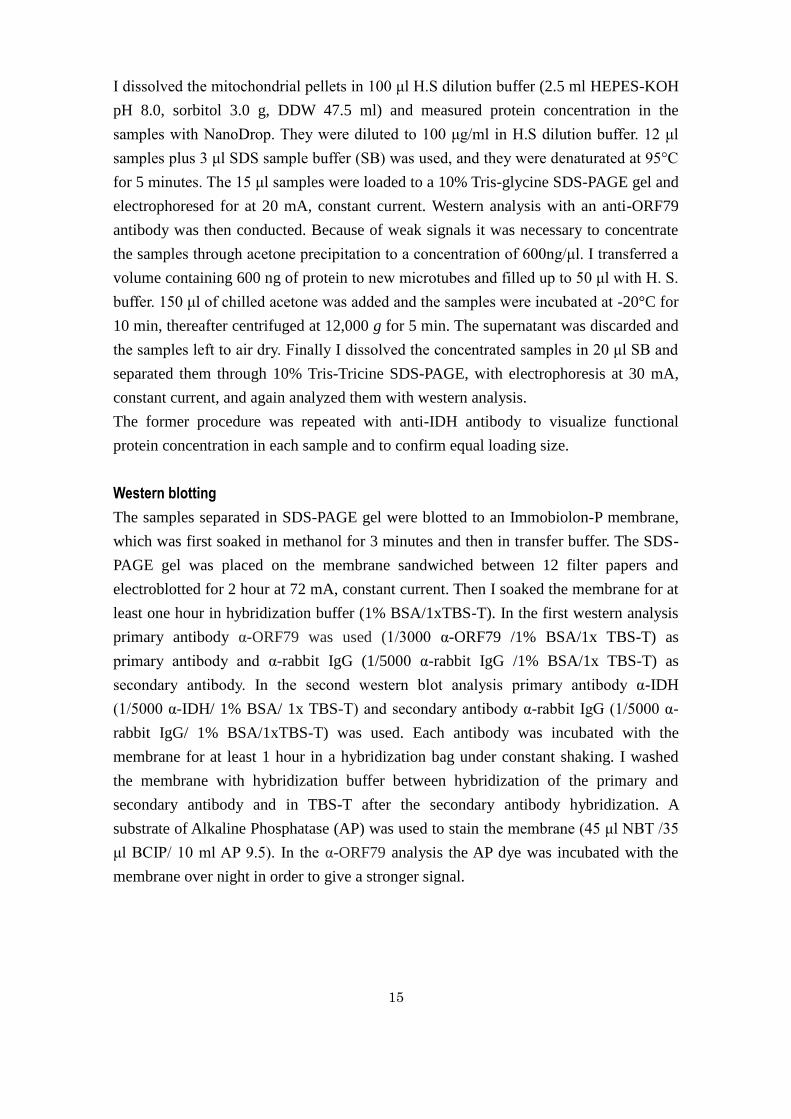
Protein separation trough SDS-PAGE electrophoresis……………....15
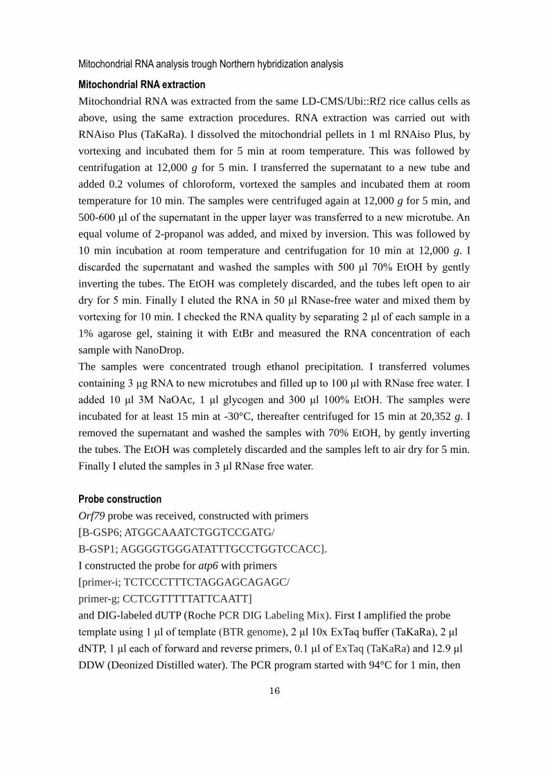
Western blotting………………………………………………………......15
Mitochondrial RNA analysis trough Northern hybridization analysis…………....16
Mitochondrial RNA extraction………………………………………….....16
Probe construction………………………………………………………...16
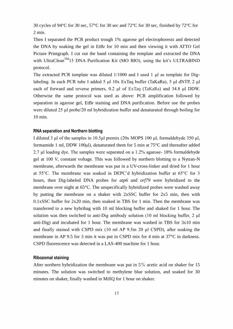
RNA separation and Northern blotting…………………………………..17
Ribozomal staining ………………………………………………………..17
Acknowledgements…..……………………………………………………………………….…..18
References…………………………………………………………………………………………19
2
Summary
Cytoplasmic male sterility (CMS) is a maternally inherited trait in plants leading to
dysfunctional pollen production and infertility. It is both useful in the breeding of
hybrids and is an example of nucleus-mitochondrion cross talk in plants.
In Lead Rice-type (Oryza sativa L.) CMS is caused by an atp6-orf79 gene, with
an unusual, cytotoxic open reading frame, orf79, coupled to the mitochondrial atp6
gene. Fertility is restored by a nuclear fertility restorer gene, Rf2. The mechanism of the
protein product RF2 is however not fully understood. In this experiment I analyzed the
protein and RNA content in Rf2 over-expressing LD-CMS rice cells and observed that
in lines with high expression of Rf2, atp6-orf79 RNA was processed before any ORF79
was produced, indicating that RF2 acts by degrading the atp6-orf79 RNA before
translation.
3
Introduction
Cytoplasmic Male Sterility in rice
Genetic regulation between the nucleus and organelle genomes, so called anterograde
regulation, is very important in eukaryotes. Likewise, retrograde signaling from
organelles plays a big role in regulating nuclear gene expression. In plants, since they
are prone to cross pollination, genome conflicts between the nucleus and a foreign
cytoplasm can easily happen, leading to developmental disorders (Fujii & Toriyama
2008). Cytoplasmic Male Sterility (CMS) is such a trait in plants, caused by
mitochondrion-nucleus incompatibility which leads to faulty gene regulation and finally
a failure for the plant to produce functional pollen. Studies of the CMS system have
contributed to revealing the importance of retrograde signaling since it was found that
expression of a large number of nuclear genes differ between CMS and wild type lines
(Carlsson et al. 2007). CMS is considered to have originally arisen in wild type plants
through mutations, and the alleles have been conserved since it might acts as a
population regulator. Restorer systems have thus co-evolved with the infertility-causing
genes. (Hanson & Bentolila 2004). CMS can also be induced by crossing plants of
different lines, so that the nucleus and mitochondrion regulation systems are
mismatched. Because the CMS line has sterile pollen, so self-pollination can be
avoided, this has commercial applications for example in the production of hybrid
varieties (Mackenzie 2004). In many crops it is not necessary to restore the fertility of
the hybrid line because seed production does not affect the harvested product, and it is
more profitable for the seed companies if the farmers need to buy new seeds every
season. In rice and other cereals however, it is necessary to restore fertility in the hybrid
because a stable yield relies on the plants ability to produce seeds through self-
pollination.
Understanding the fertility restorer systems of rice is especially of big importance in
Asia, where the main cereal is rice, and the mechanisms in different rice varieties are
currently under research. One of the most well studied fertility restorer systems in
Oryza sativa japonica rice is the Chinsurah Boro II type BT-CMS/Rf1 system. A fertility
restorer gene has been identified and named Rf1 (restorer of fertility) and the
mechanism of action has been outlined. The restorer gene is a nuclear gene coding for a
mitochondrion targeting protein involved in post-transcriptional regulation of the CMS
causing gene (Kazama et al. 2008). A rice variety that has been shown to have a similar
CMS causing gene and fertility restorer system as BT-CMS is the Lead Rice- type
(LD)/Rf2 type (Itabashi et al. 2011).
4
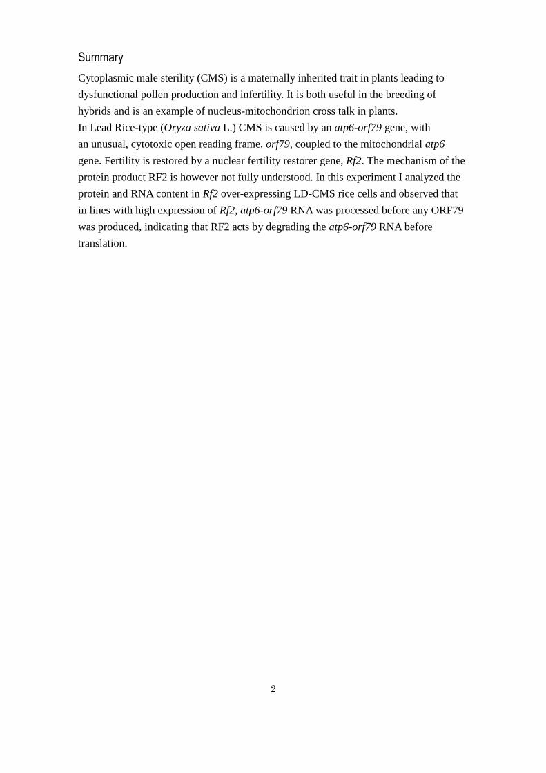
Infertility-causing open reading frames
In BT-CMS plants infertility is caused
by the expression of a chimeric atp6-
orf79 gene in mitochondria. The open
reading frame orf79 is homologous in
it’s 5’ region to cox1, the mitochondrial
gene encoding cytochrome oxidase
subunit 1 (Akagi et al. 1994). It’s 3’
region is of unknown origin and
encodes a cytotoxic transmembrane
protein which causes sterility by
impeding pollen development (Wang et
al. 2006). Infertility in LD-CMS plants
is caused by a similar atp6-orf79 gene
to that of BT-CMS, differing only in a
single nucleotide polymorphism in
orf79 and a 4bp insertion between atp6
and orf79. Another difference between
the two lines is that the BT-CMS
mitochondria have two copies of the atp6 gene, also possessing a normal copy of the
atp6 without the orf79. LD-CMS mitochondria however, only carry the atp6-orf79 locus
(Itabashi et al. 2009). A model comparing the infertility-causing genes in LD-CMS and
BT-CMS lines can be seen in Figure 1.
Fertility restorer genes
In CMS plants fertility can be restored either gametophytically or sporophytically. In
most cases the restorer gene acts gametophytically, including BT and LD-CMS plants
(Itabashi et al. 2009). Gametophytic restoration means that the individual pollen grain
will determine their own fertility, depending on if they contain a functional restorer gene
or not. Thus, in a restored line 50% of the T1 generation pollen will be infertile while
fertility has been totally restored in the T2 generation (Fujii et al. 2008). The general
principle of fertility restoration is that the nuclear restorer gene processes the RNA of
infertility-causing ORF by regulation of mitochondrial gene expression. In BT-CMS the
restorer of fertility, Rf1, encodes a pentatricopeptide repeat (PPR) containing protein,
which locates to mitochondria and binds to RNA. Most fertility restorer systems consist
of a PPR protein, exhibiting RNA-binding properties (Hu et al. 2012). It has been
shown that RF1 binds to B-atp6-orf79 RNA, in the region between atp6 and orf79, and
Figure 1. Schematic diagram of the atp6-orf79 locus in
normal Oryza sativa Japonica mitochondrial genome,
compared with BT-CMS and LD-CMS varieties. N-atp6 is
the normal atp6 gene and B-atp6 is the duplicate with
orf79 present in BT-type cytoplasm. L-atp6 is the locus
present in LD-type cytoplasm. E, Eco RI site. (Adapted
from Itabashi et al. 2009)
5
processes it before translation. Wang et al. (2006), outlined the mechanism of two
different fertility restorer genes, RF1A and RF1B. They showed that RF1A acts by
endonucleolytic cleaving of the B-atp6-orf79 transcript, and RF1B acts by degrading
the B-atp6-orf79 transcript. Kazama et al. (2008) showed that only the cleaving RF1 is
required for fertility restoration, even though ORF79 accumulation was only reduced to
50%. These results show that reducing the amount of the infertility-causing ORF79 is
the critical function of the fertility restorer protein.
In LD-CMS plants, restorer of fertility Rf2 has been identified, but the mechanism of its
protein product is not yet fully known. RF2 is a glycine-rich protein (GRP) containing a
mitochondrial targeting sequence, but no known RNA-binding motif. It consists of 152
amino acids, with a glycine-rich region (Itabashi et al. 2011), which is hypothesized to
be the functional region of the protein. GRPs are proteins containing more than 60%
glycine, including plant cell wall proteins and RNA-binding proteins. Glycine-rich
regions are often associated with other proteins in multi-protein complexes (Mousavi &
Hotta 2005). It was recently discovered that a GRP, GRP162, in complex with a PPR-
containing fertility restorer protein, RF5, restores fertility in Hong-Lian CMS rice as a
fertility restoration complex (Hu et al. 2012). The GRP protein in that case contains the
RNA-binding motif, but no mitochondrial targeting sequence. It is possible that RF2
functions in a similar way, but it is still not established at which step it prevents the
production of the infertility-causing ORF79. Itabashi et al. (2009) could not detect
ORF79 accumulation in the LD-CMS line, and concluded that the sterility induction and
fertility restoration system in LD-CMS is different from that of BT-CMS. In later
studies, with other anti-ORF79 antibodies, accumulation of the protein could be
detected in LD-CMS. This result is still unpublished, but the same antibody is used in
this project. Because of the similarity of the atp6-orf79 transcripts of LD-CMS and BT-
CMS lines it is likely that they are processed by a similar mechanism, even though the
RF proteins differ.
Aims
The question I tried to answer in this project is: What is the function of RF2? Does it act
by preventing transcription of orf79, by processing of the atp6-orf79 transcript, by
preventing translation of orf79 or by destabilization of ORF79? If atp6-orf79 RNA
could be detected in samples where ORF79 was absent it would mean that RF2 acts by
destabilizing the protein. I analyzed protein and RNA content in LD-CMS rice callus
cells trough western and northern blotting and concluded that RF2 acts by RNA
degradation.
6
Results
I used 5 lines of Oryza sativa japonica callus cells with LD-CMS type overexpressing
Rf2 under the control of ubiquitin promoter (Ubi::Rf2), including a sample of wildtype
LD-CMS cells.
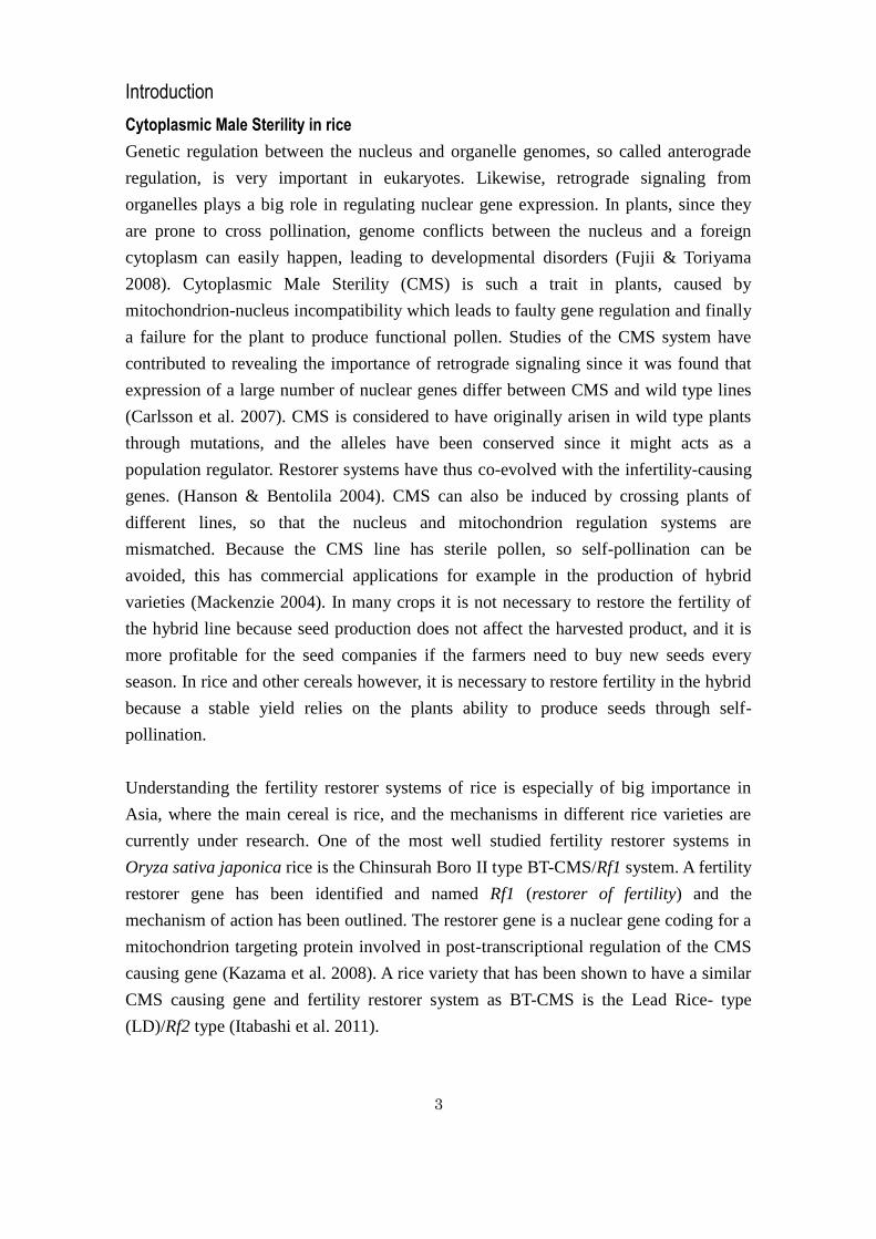
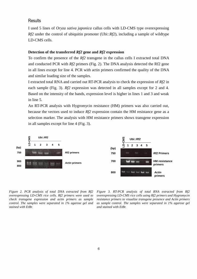
Detection of the transferred Rf2 gene and Rf2 expression
To confirm the presence of the Rf2 transgene in the callus cells I extracted total DNA
and conducted PCR with Rf2 primers (Fig. 2). The DNA analysis detected the Rf2 gene
in all lines except for line 4. PCR with actin primers confirmed the quality of the DNA
and similar loading size of the samples.
I extracted total RNA and carried out RT-PCR analysis to check the expression of Rf2 in
each sample (Fig. 3). Rf2 expression was detected in all samples except for 2 and 4.
Based on the intensity of the bands, expression level is higher in lines 1 and 3 and weak
in line 5.
An RT-PCR analysis with Hygromycin resistance (HM) primers was also carried out,
because the vectors used to induce Rf2 expression contain the HM resistance gene as a
selection marker. The analysis with HM resistance primers shows transgene expression
in all samples except for line 4 (Fig. 3).
Actin primers
1 2 3 4 5
Ubi::Rf2
LD
-CM
S
Rf2 primers
900
800
750
(bp)
LD
-CM
S
1 2 3 4 5
750
700
800
Rf2 Primers
HM resistance
primers
Actin
primers
Ubi::Rf2
(bp)
Figure 2. PCR analysis of total DNA extracted from Rf2
overexpressing LD-CMS rice cells. Rf2 primers were used to
check transgene expression and actin primers as sample
control. The samples were separated in 1% agarose gel and
stained with EtBr.
Figure 3. RT-PCR analysis of total RNA extracted from Rf2
overexpressing LD-CMS rice cells using Rf2 primers and Hygromycin
resistance primers to visualize transgene presence and Actin primers
as sample control. The samples were separated in 1% agarose gel
and stained with EtBr.
7
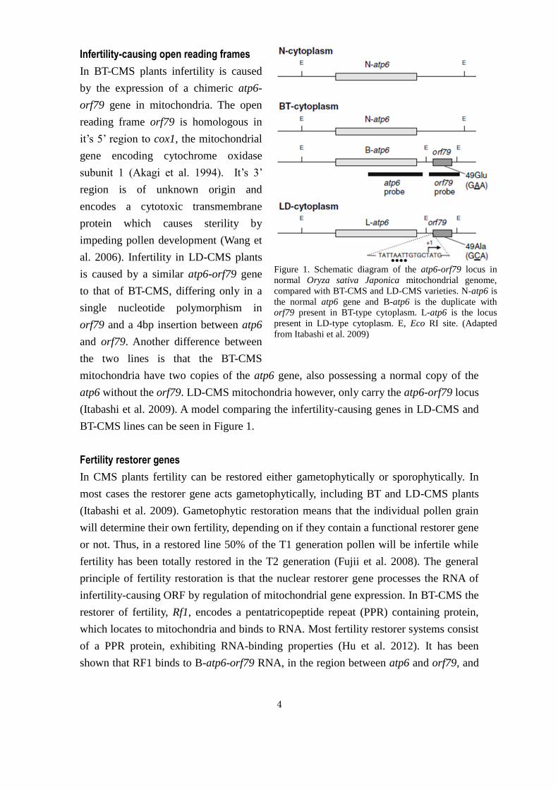
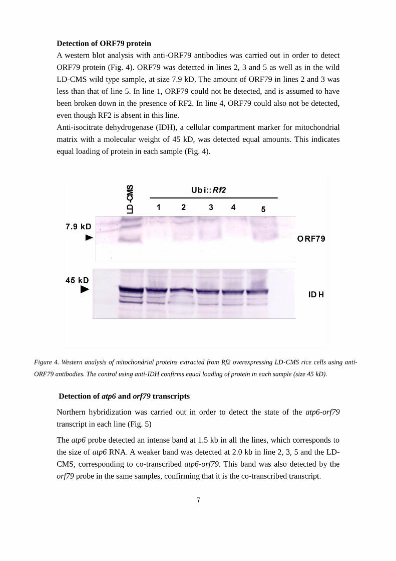
Detection of ORF79 protein
A western blot analysis with anti-ORF79 antibodies was carried out in order to detect
ORF79 protein (Fig. 4). ORF79 was detected in lines 2, 3 and 5 as well as in the wild
LD-CMS wild type sample, at size 7.9 kD. The amount of ORF79 in lines 2 and 3 was
less than that of line 5. In line 1, ORF79 could not be detected, and is assumed to have
been broken down in the presence of RF2. In line 4, ORF79 could also not be detected,
even though RF2 is absent in this line.
Anti-isocitrate dehydrogenase (IDH), a cellular compartment marker for mitochondrial
matrix with a molecular weight of 45 kD, was detected equal amounts. This indicates
equal loading of protein in each sample (Fig. 4).
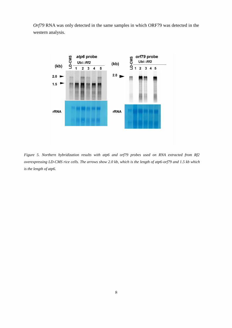
Detection of atp6 and orf79 transcripts
Northern hybridization was carried out in order to detect the state of the atp6-orf79
transcript in each line (Fig. 5)
The atp6 probe detected an intense band at 1.5 kb in all the lines, which corresponds to
the size of atp6 RNA. A weaker band was detected at 2.0 kb in line 2, 3, 5 and the LD-
CMS, corresponding to co-transcribed atp6-orf79. This band was also detected by the
orf79 probe in the same samples, confirming that it is the co-transcribed transcript.
Figure 4. Western analysis of mitochondrial proteins extracted from Rf2 overexpressing LD-CMS rice cells using anti-
ORF79 antibodies. The control using anti-IDH confirms equal loading of protein in each sample (size 45 kD).
8
Figure 5. Northern hybridization results with atp6 and orf79 probes used on RNA extracted from Rf2
overexpressing LD-CMS rice cells. The arrows show 2.0 kb, which is the length of atp6-orf79 and 1.5 kb which
is the length of atp6.
Orf79 RNA was only detected in the same samples in which ORF79 was detected in the
western analysis.
9
Discussion and conclusion
The question that I wanted to answer with this study was how RF2 restores fertility in
LD-CMS lines. The Rf2 expression levels and amount of ORF79 protein and atp6-orf79
transcript was very varied between the lines. This is because the lines used in this study
might contain different copy number of the Rf2 transgene. If the T1 line carries a single
transgene, then 50% of the the T2 lines might carry Rf2 homozygously (Rf2, Rf2) and
50% might carry the gene heterogously (Rf2, -). Because Rf2 gametophytically restores
fertility, 100% of the T2 generation will be fertile even though the expression level of
Rf2 might be different between the lines. In line 2, Rf2 expression was not detected by
RT-PCR although the Rf2 transgene was present. In this line the orf79 transcript does
not seem to have been processed. It is possible that the transgene lost its function over
time during the callus development. In line 4, the atp6-orf79 transcript and ORF79 were
not detected even though the transgene was not detected. Since genomic PCR and RT-
PCR was carried out with a time delay after the western and northern analysis it is
possible that the transgene could have fallen away over time, as usually happens in
transgenic cell cultures. In the other samples varying levels of Rf2 expression was
observed. I assume that the atp6-orf79 transcript was totally degraded in line 1, under
high expression of Rf2. In line 3 the Rf2 expression was weaker, and the atp6-orf79
transcript is weakly degraded. Expression of Rf2 in line 5 is likely too weak to degrade
the atp6-orf79 transcript. Tables 1 summarizes the results of my study. Line 4 is not
included, because of the absence of the Rf2 transgene.
Table 1. Summary of Rf2 expression, degradation of atp6-orf79 transcripts and accumulation of ORF79
protein.
Line LD-CMS 1 2 3 5
Rf2 expression level - +++ - ++ +
Degradation of atp-orf79 - +++ - ++ +
ORF79 accumulation ++++ - + ++ +++
My results indicate that two types of RNAs are transcribed from the single atp6-orf79
locus in LD type cytoplasm. One is the 2.0 kb transcript of atp6-orf79 and the second
one is the 1.5 kb transcript of atp6. Because atp6 was detected in all samples, RF2 does
not affect the atp6 transcript. Selective transcription of atp6 already takes place, so it is
most likely that the added effect of RF2 happens after RNA transcription. I conclude
that RF2 play a role in degrading the atp6-orf79 transcripts before translation.
10
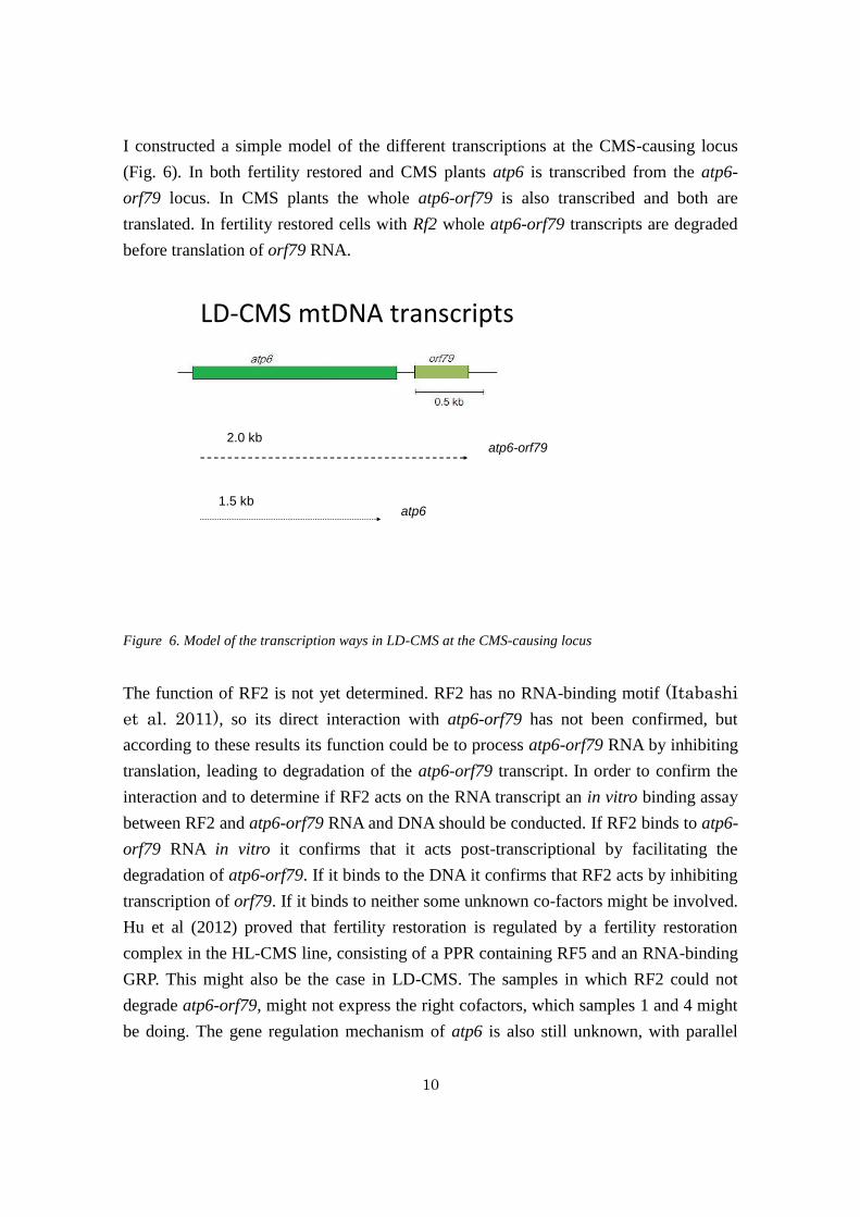
I constructed a simple model of the different transcriptions at the CMS-causing locus
(Fig. 6). In both fertility restored and CMS plants atp6 is transcribed from the atp6-
orf79 locus. In CMS plants the whole atp6-orf79 is also transcribed and both are
translated. In fertility restored cells with Rf2 whole atp6-orf79 transcripts are degraded
before translation of orf79 RNA.
Figure 6. Model of the transcription ways in LD-CMS at the CMS-causing locus
The function of RF2 is not yet determined. RF2 has no RNA-binding motif (Itabashi
et al. 2011), so its direct interaction with atp6-orf79 has not been confirmed, but
according to these results its function could be to process atp6-orf79 RNA by inhibiting
translation, leading to degradation of the atp6-orf79 transcript. In order to confirm the
interaction and to determine if RF2 acts on the RNA transcript an in vitro binding assay
between RF2 and atp6-orf79 RNA and DNA should be conducted. If RF2 binds to atp6-
orf79 RNA in vitro it confirms that it acts post-transcriptional by facilitating the
degradation of atp6-orf79. If it binds to the DNA it confirms that RF2 acts by inhibiting
transcription of orf79. If it binds to neither some unknown co-factors might be involved.
Hu et al (2012) proved that fertility restoration is regulated by a fertility restoration
complex in the HL-CMS line, consisting of a PPR containing RF5 and an RNA-binding
GRP. This might also be the case in LD-CMS. The samples in which RF2 could not
degrade atp6-orf79, might not express the right cofactors, which samples 1 and 4 might
be doing. The gene regulation mechanism of atp6 is also still unknown, with parallel
LD-CMS mtDNA transcripts
2.0 kb
1.5 kb
atp6-orf79
atp6

11
transcription of atp6 and atp6-orf79. Further studies should therefore focus on
answering the question of how atp6 transcription is regulated and to confirm if RF2 acts
after transcription.
12
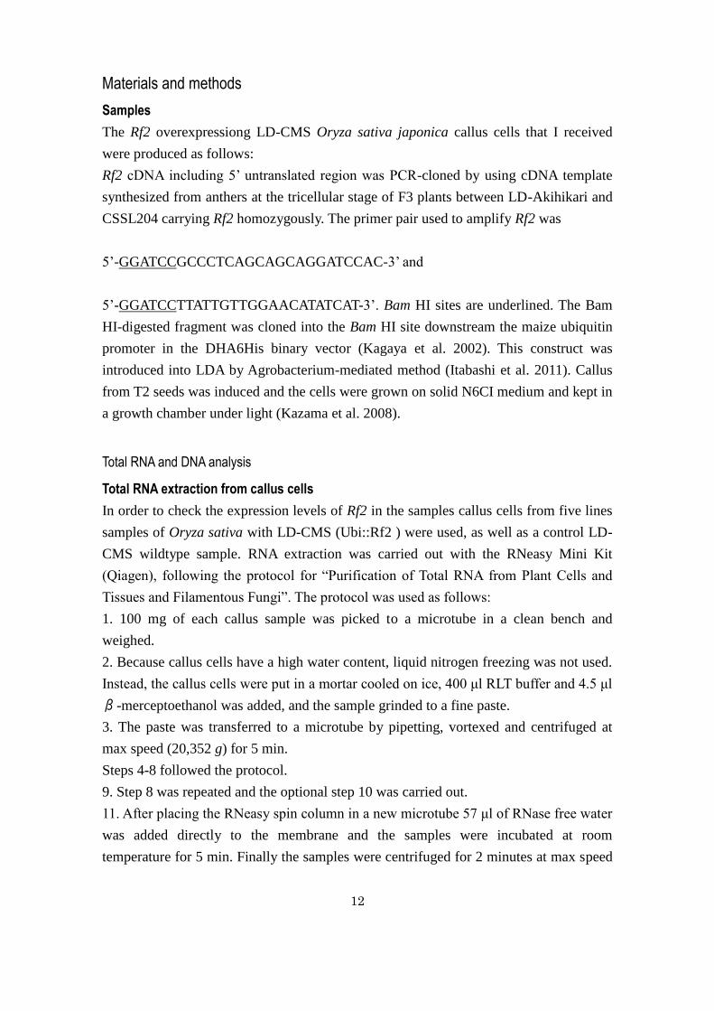
Materials and methods
Samples
The Rf2 overexpressiong LD-CMS Oryza sativa japonica callus cells that I received
were produced as follows:
Rf2 cDNA including 5’ untranslated region was PCR-cloned by using cDNA template
synthesized from anthers at the tricellular stage of F3 plants between LD-Akihikari and
CSSL204 carrying Rf2 homozygously. The primer pair used to amplify Rf2 was
5’-GGATCCGCCCTCAGCAGCAGGATCCAC-3’ and
5’-GGATCCTTATTGTTGGAACATATCAT-3’. Bam HI sites are underlined. The Bam
HI-digested fragment was cloned into the Bam HI site downstream the maize ubiquitin
promoter in the DHA6His binary vector (Kagaya et al. 2002). This construct was
introduced into LDA by Agrobacterium-mediated method (Itabashi et al. 2011). Callus
from T2 seeds was induced and the cells were grown on solid N6CI medium and kept in
a growth chamber under light (Kazama et al. 2008).
Total RNA and DNA analysis
Total RNA extraction from callus cells
In order to check the expression levels of Rf2 in the samples callus cells from five lines
samples of Oryza sativa with LD-CMS (Ubi::Rf2 ) were used, as well as a control LD-
CMS wildtype sample. RNA extraction was carried out with the RNeasy Mini Kit
(Qiagen), following the protocol for “Purification of Total RNA from Plant Cells and
Tissues and Filamentous Fungi”. The protocol was used as follows:
1. 100 mg of each callus sample was picked to a microtube in a clean bench and
weighed.
2. Because callus cells have a high water content, liquid nitrogen freezing was not used.
Instead, the callus cells were put in a mortar cooled on ice, 400 μl RLT buffer and 4.5 μl
β-merceptoethanol was added, and the sample grinded to a fine paste.
3. The paste was transferred to a microtube by pipetting, vortexed and centrifuged at
max speed (20,352 g) for 5 min.
Steps 4-8 followed the protocol.
9. Step 8 was repeated and the optional step 10 was carried out.
11. After placing the RNeasy spin column in a new microtube 57 μl of RNase free water
was added directly to the membrane and the samples were incubated at room
temperature for 5 min. Finally the samples were centrifuged for 2 minutes at max speed
13
and the flow through containing the extracted RNA was kept in freezer.
To check the quality of extracted RNA a 1% agarose gel electrophoresis was carried out
and RNA detected with EtBr staining. Contaminating DNA was removed by incubating
the samples with DNase I (50 μl RNA sample, 10 μl 10xDNase I buffer, 5 μl DNase I
(TaKaRa), 35 μl RNase free DDW) at 37°C for 2 hours. Then 100 μl phenol-chloroform
was added to remove the DNase, the samples centrifuged for 5 min at 20,352 g and the
upper layer transferred to new microtubes. Finally the samples were concentrated by
ethanol precipitation. The extracted RNA was used for RT-PCR to confirm expression
of Rf2.
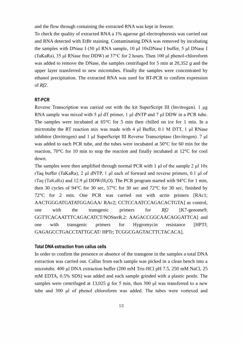
RT-PCR
Reverse Transcription was carried out with the kit SuperScript III (Invitrogen). 1 μg
RNA sample was mixed with 5 μl dT primer, 1 μl dNTP and 7 μl DDW in a PCR tube.
The samples were incubated at 65°C for 5 min then chilled on ice for 1 min. In a
mictrotube the RT reaction mix was made with 4 μl Buffer, 0.1 M DTT, 1 μl RNase
inhibitor (Invitrogen) and 1 μl SuperScript III Reverse Transcriptase (Invitrogen). 7 μl
was added to each PCR tube, and the tubes were incubated at 50°C for 60 min for the
reaction, 70°C for 10 min to stop the reaction and finally incubated at 12°C for cool
down.
The samples were then amplified through normal PCR with 1 μl of the sample 2 μl 10x
rTaq buffer (TaKaRa), 2 μl dNTP, 1 μl each of forward and reverse primers, 0.1 μl of
rTaq (TaKaRa) and 12.9 μl DDW(H2O). The PCR program started with 94°C for 1 min,
then 30 cycles of 94°C for 30 sec, 57°C for 30 sec and 72°C for 30 sec, finished by
72°C for 2 min. One PCR was carried out with actin primers [RAc1;
AACTGGGATGATATGGAGAA/ RAc2; CCTCCAATCCAGACACTGTA] as control,
one with the transgenic primers for Rf2 [K7-genome9;
GGTTCACAATTTCAGACATCT/NOSterR.2: AAGACCGGCAACAGGATTCA] and
one with transgenic primers for Hygromycin resistance [HPTf;
GAGAGCCTGACCTATTGCAT/ HPTr; TCGGCGAGTACTTCTACACA].
Total DNA extraction from callus cells
In order to confirm the presence or absence of the transgene in the samples a total DNA
extraction was carried out. Callus from each sample was picked in a clean bench into a
microtube. 400 μl DNA extraction buffer (200 mM Tris-HCl pH 7.5, 250 mM NaCl, 25
mM EDTA, 0.5% SDS) was added and each sample grinded with a plastic pestle. The
samples were centrifuged at 13,025 g for 5 min, then 300 μl was transferred to a new
tube and 300 μl of phenol chloroform was added. The tubes were vortexed and
14
centrifuged again under the same conditions. 150 μl was transferred from the upper
layer into a new mictrotube and to concentrate the samples 150 μl of 2-propanol was
added. The samples were mixed by inverting the tube a few times and centrifuged again.
The liquid was discarded, then the samples were washed by adding 500 μl of 70%
ethanol and gently inverting the tube. The ethanol was discarded and the samples
flashed at 0.8 g for a few seconds, then the remaining ethanol was discarded by
pipetting and the tubes left open for 5 min to air dry the samples. Finally 100 μl of
DDW was added and the samples mixed by tapping and flashing.
The presence of the transgene was controlled by conducting a PCR with the K7/Nos
primer pair and Actin primers as control. The PCR product was electrophoresed in 1%
agar gel and stained with EtBr.
Mitochondrial protein analysis through western blotting analysis
Extraction of mitochondria
I extracted the mitochondria according to standard procedures with A. buffer and G.
buffer mixed as follows. MillQ is ultrapure water (Millipore corporation).
A. Buffer G.Buffer (Lysis buffer)
0.35 M sorbitol 0.3 M sucrose
50 mM Tris-HCl (pH 8.0) 0.05 M Tris
5 mM EDTA (pH 8.0) 0.001 M EDTA
0.1% BSA Adjust pH to 7.5 with HCl
1.25 ml/L β-merceptoethanol 0.05% BSA
MillQ H2O 0.001 M β-merceptoethanol
MillQ H2O
Each sample was weighed and put in a mortar with 4.0 ml/g A. buffer and grinded to a
fine paste. The paste was squeezed through a filter made up of 4 layers of gauze and 1
layer Miracloth, thereafter centrifuged at 565 g (at 4°C) for 10 min. The supernatant was
transferred to a new plastic tube and centrifuged again at 5789 g, for 10 min. The
supernatant was discarded and the remaining pellet dissolved in 1 ml G. buffer by
vortexing and tapping, thereafter centrifuged at 565 g for 10 min. The supernatant was
transferred to a new microtube and centrifuged at 5789 g for 20 min. Finally the
supernatant was discarded with an aspirator to completely remove all liquid. The
resulting pellets containing the mitochondria were stored in the freezer and used for
either protein or RNA analysis.
Protein separation trough SDS-PAGE electrophoresis
I conducted three SDS-PAGE electrophoreses, as follows.
15
I dissolved the mitochondrial pellets in 100 μl H.S dilution buffer (2.5 ml HEPES-KOH
pH 8.0, sorbitol 3.0 g, DDW 47.5 ml) and measured protein concentration in the
samples with NanoDrop. They were diluted to 100 μg/ml in H.S dilution buffer. 12 μl
samples plus 3 μl SDS sample buffer (SB) was used, and they were denaturated at 95°C
for 5 minutes. The 15 μl samples were loaded to a 10% Tris-glycine SDS-PAGE gel and
electrophoresed for at 20 mA, constant current. Western analysis with an anti-ORF79
antibody was then conducted. Because of weak signals it was necessary to concentrate
the samples through acetone precipitation to a concentration of 600ng/μl. I transferred a
volume containing 600 ng of protein to new microtubes and filled up to 50 μl with H. S.
buffer. 150 μl of chilled acetone was added and the samples were incubated at -20°C for
10 min, thereafter centrifuged at 12,000 g for 5 min. The supernatant was discarded and
the samples left to air dry. Finally I dissolved the concentrated samples in 20 μl SB and
separated them through 10% Tris-Tricine SDS-PAGE, with electrophoresis at 30 mA,
constant current, and again analyzed them with western analysis.
The former procedure was repeated with anti-IDH antibody to visualize functional
protein concentration in each sample and to confirm equal loading size.
Western blotting
The samples separated in SDS-PAGE gel were blotted to an Immobiolon-P membrane,
which was first soaked in methanol for 3 minutes and then in transfer buffer. The SDS-
PAGE gel was placed on the membrane sandwiched between 12 filter papers and
electroblotted for 2 hour at 72 mA, constant current. Then I soaked the membrane for at
least one hour in hybridization buffer (1% BSA/1xTBS-T). In the first western analysis
primary antibody α-ORF79 was used (1/3000 α-ORF79 /1% BSA/1x TBS-T) as
primary antibody and α-rabbit IgG (1/5000 α-rabbit IgG /1% BSA/1x TBS-T) as
secondary antibody. In the second western blot analysis primary antibody α-IDH
(1/5000 α-IDH/ 1% BSA/ 1x TBS-T) and secondary antibody α-rabbit IgG (1/5000 α-
rabbit IgG/ 1% BSA/1xTBS-T) was used. Each antibody was incubated with the
membrane for at least 1 hour in a hybridization bag under constant shaking. I washed
the membrane with hybridization buffer between hybridization of the primary and
secondary antibody and in TBS-T after the secondary antibody hybridization. A
substrate of Alkaline Phosphatase (AP) was used to stain the membrane (45 μl NBT /35
μl BCIP/ 10 ml AP 9.5). In the α-ORF79 analysis the AP dye was incubated with the
membrane over night in order to give a stronger signal.
16
Mitochondrial RNA analysis trough Northern hybridization analysis
Mitochondrial RNA extraction
Mitochondrial RNA was extracted from the same LD-CMS/Ubi::Rf2 rice callus cells as
above, using the same extraction procedures. RNA extraction was carried out with
RNAiso Plus (TaKaRa). I dissolved the mitochondrial pellets in 1 ml RNAiso Plus, by
vortexing and incubated them for 5 min at room temperature. This was followed by
centrifugation at 12,000 g for 5 min. I transferred the supernatant to a new tube and
added 0.2 volumes of chloroform, vortexed the samples and incubated them at room
temperature for 10 min. The samples were centrifuged again at 12,000 g for 5 min, and
500-600 μl of the supernatant in the upper layer was transferred to a new microtube. An
equal volume of 2-propanol was added, and mixed by inversion. This was followed by
10 min incubation at room temperature and centrifugation for 10 min at 12,000 g. I
discarded the supernatant and washed the samples with 500 μl 70% EtOH by gently
inverting the tubes. The EtOH was completely discarded, and the tubes left open to air
dry for 5 min. Finally I eluted the RNA in 50 μl RNase-free water and mixed them by
vortexing for 10 min. I checked the RNA quality by separating 2 μl of each sample in a
1% agarose gel, staining it with EtBr and measured the RNA concentration of each
sample with NanoDrop.
The samples were concentrated trough ethanol precipitation. I transferred volumes
containing 3 μg RNA to new microtubes and filled up to 100 μl with RNase free water. I
added 10 μl 3M NaOAc, 1 μl glycogen and 300 μl 100% EtOH. The samples were
incubated for at least 15 min at -30°C, thereafter centrifuged for 15 min at 20,352 g. I
removed the supernatant and washed the samples with 70% EtOH, by gently inverting
the tubes. The EtOH was completely discarded and the samples left to air dry for 5 min.
Finally I eluted the samples in 3 μl RNase free water.
Probe construction
Orf79 probe was received, constructed with primers
[B-GSP6; ATGGCAAATCTGGTCCGATG/
B-GSP1; AGGGGTGGGATATTTGCCTGGTCCACC].
I constructed the probe for atp6 with primers
[primer-i; TCTCCCTTTCTAGGAGCAGAGC/
primer-g; CCTCGTTTTTATTCAATT]
and DIG-labeled dUTP (Roche PCR DIG Labeling Mix). First I amplified the probe
template using 1 μl of template (BTR genome), 2 μl 10x ExTaq buffer (TaKaRa), 2 μl
dNTP, 1 μl each of forward and reverse primers, 0.1 μl of ExTaq (TaKaRa) and 12.9 μl
DDW (Deonized Distilled water). The PCR program started with 94°C for 1 min, then
17
30 cycles of 94°C for 30 sec, 57°C for 30 sec and 72°C for 30 sec, finished by 72°C for
2 min.
Then I separated the PCR product trough 1% agarose gel electrophoresis and detected
the DNA by soaking the gel in EtBr for 10 min and then viewing it with ATTO Gel
Picture Printgraph. I cut out the band containing the template and extracted the DNA
with UltraCleanTM
15 DNA Purification Kit (MO BIO), using the kit’s ULTRABIND
protocol.
The extracted PCR template was diluted 1/1000 and I used 1 μl as template for Dig-
labeling. In each PCR tube I added 5 μl 10x ExTaq buffer (TaKaRa), 5 μl dNTP, 2 μl
each of forward and reverse primers, 0.2 μl of ExTaq (TaKaRa) and 34.8 μl DDW.
Otherwise the same protocol was used as above: PCR amplification followed by
separation in agarose gel, EtBr staining and DNA purification. Before use the probes
were diluted 25 μl probe/20 ml hybridization buffer and denaturated through boiling for
10 min.
RNA separation and Northern blotting
I diluted 3 μl of the samples in 10.5μl premix (20x MOPS 100 μl, formaldehyde 350 μl,
formamide 1 ml, DDW 100μl), denaturated them for 5 min at 75°C and thereafter added
2.7 μl loading dye. The samples were separated on a 1.2% agarose- 18% formaldehyde
gel at 100 V, constant voltage. This was followed by northern blotting to a Nytran-N
membrane, afterwards the membrane was put in a UV-cross-linker and dried for 1 hour
at 55°C. The membrane was soaked in DEPC’d hybridization buffer at 65°C for 3
hours, then Dig-labeled DNA probes for atp6 and orf79 were hybridized to the
membrane over night at 65°C. The unspecifically hybridized probes were washed away
by putting the membrane on a shaker with 2xSSC buffer for 2x5 min, then with
0.1xSSC buffer for 2x20 min, then soaked in TBS for 1 min. Then the membrane was
transferred to a new hybribag with 10 ml blocking buffer and shaked for 1 hour. The
solution was then switched to anti-Dig antibody solution (10 ml blocking buffer, 2 μl
anti-Dig) and incubated for 1 hour. The membrane was washed in TBS for 3x10 min
and finally stained with CSPD mix (10 ml AP 9.5m 20 μl CSPD), after soaking the
membrane in AP 9.5 for 3 min it was put in CSPD mix for 4 min at 37°C in darkness.
CSPD fluorescence was detected in a LAS-400 machine for 1 hour.
Ribozomal staining
After northern hybridization the membrane was put in 5% acetic acid on shaker for 15
minutes. The solution was switched to methylene blue solution, and soaked for 30
minutes on shaker, finally washed in MillQ for 1 hour on shaker.

18
Acknowledgements
I would like to express my gratitude to Professor Kinya Toriyama, for accepting my
application to join his lab and for giving me such an interesting project.
It would not have been possible for me to complete this project without the kind
supervision, patience and encouragement from Professor Tomohiko Kazama.
Lastly I thank all members of the Environmental Plant Biotechnology Lab for their
warm welcome.
19
References
Akagi, H., Sakamoto, M., Shinjyo, C., Shimada, H., and Fujimura, T. (1994). A unique
sequence located downstream from the rice mitochondrial apt6 may cause male
sterility. Current Genetics. 25, 52–58.
Carlsson J., Lagercrantz U., Sundstrom J., Teixeira R., Wellmer F., Meyerowitz E. M.,
Glimelius K. (2007). Microarray analysis reaveals altered expression of a
large number of nuclear genes in developing Cytoplasmic male sterile Brassica
napus flowers. The Plant Journal, 49, 452-462.
Fujii S., Kazama T., Toriyama K., (2008). Molecular Studies on Cytoplasmic Male
Sterility-associated Genes and Restorer Genes in Rice. Rice Biology in the
Genomics Era, Biotechnology in Agriculture and Forestry, 62, II.7, 205-215.
Fujii S., Toriyama K. (2008). Genome Barriers between Nuclei and Mitochondria
Exemplified by Cytoplasmic Male Sterility. Plant Cell Physiology, 49, 1484-
1494.
Hanson M.R., Bentolila S. (2004). Interactions of Mitochondrial and Nuclear Genes
That Affect Male Gametophyte Development. The Plant Cell, 16, S154–S169.
Hu J., Wang K., Wenchao H., Gai L., Ya G., Jianming W.,, Qi H., Yanxiao J., Xiaojian
Q., Lei W., Renshan Z., Shaoqing L., Daichang Y., Yingguo Z. (2012). The Rice
Pentatricopeptide Repeat Protein RF5 Restores Fertility in Hong-Lian
Cytoplasmic Male-Sterile Lines via a Complex with the Glycine-Rich Protein
GRP162. The Plant Cell, 24, 109–122.
Kagaya Y., Hobo T., Murata M., Ban A., Hattori T. (2002). Abscisic acid-induced
transcription is mediated by phosphorylation of an abscisic acid response
element binding factor, TRAB1. The Plant Cell, 14, 3177-3189.
Kazama T., Nakamura T., Watanabe M., Sugita M., Toriyama K. (2008). Suppression
mechanism of mitochondrial ORF79 accumulation by Rf1 protein in BT-type
cytoplasmic male sterile rice. The Plant Journal, 55, 619–628.
Itabashi E., Kazama T., Toriyama K. (2009) Characterization of cytoplasmic male
sterility of rice with Lead Rice Cytoplasm in comparison with that with
Chinsurah Boro II cytoplasm. Plant Cell Rep. 28. 233-239.
Itabashi E., Iwata N., Fuji S., Kazama T., Toriyama K. (2011) The fertility restorer gene,
Rf2, for Lead Rice-type cytoplasmic male sterility of rice encodes a
mitochondrial glycine rich protein.. The Plant Journal. 65, 359-367.
Mackenzie S. A. (2010). The Influence of Mitochondrial Genetics on Crop Breeding
Strategies. Plant Breeding Reviews, 25, Ch5.
Mousavi A., Hotta Y. (2005). Glycine-rich Proteins. A Class of Novel Proteins.
Applied Biochemistry and Biotechnology, 120, 169-174.

20
Wang Z., Zou Y., Li X., Zhang Q., Chen L., Wu H., Su D., Chen Y., Guo J., Luo D.,
Long Y., Zhong Y., Liu Y. (2006). Cytoplasmic Male Sterility of Rice with Boro
II Cytoplasm Is Caused by a Cytotoxic Peptide and Is Restored by Two Related
PPR Motif Genes via Distinct Modes of mRNA Silencing. The Plant Cell,
18, 676–687.


![Zipcode RNA-Binding Proteins and Membrane Trafficking ... · Zipcode RNA-Binding Proteins and Membrane Trafficking Proteins Cooperate to Transport Glutelin mRNAs in Rice Endosperm[OPEN]](https://img.pdfslide.net/doc/110x75/5fedaa08e6ee6243c45b24a5/zipcode-rna-binding-proteins-and-membrane-trafficking-zipcode-rna-binding-proteins.jpg)