Embed Size (px)
Citation preview
O R I G I N A L A R T I C L E
Modeling neuro-immune interactions during Zikavirus infectionPinar Mesci1,2,†, Angela Macia1,2,†, Christopher N. LaRock3, Leon Tejwani1,2,Isabella R. Fernandes1,2, Nicole A. Suarez1,2, Paolo M. de A. Zanotto4,Patricia C.B. Beltr~ao-Braga5,6,7, Victor Nizet3 and Alysson R. Muotri1,2,*1Department of Pediatrics/Rady Children’s Hospital San Diego, 2Department of Cellular & Molecular Medicine,Stem Cell Program, School of Medicine, University of California San Diego, La Jolla, CA 92093-0695, USA,3Department of Pediatrics and Skaggs School of Pharmacy and Pharmaceutical Sciences, School of Medicine,University of California San Diego, La Jolla, CA 92093-0760, USA, 4Laboratory of Molecular Evolution andBioinformatics, Department of Microbiology, Institute of Microbiology Sciences, University of Sao Paulo, S~aoPaulo, SP 05508-000, Brazil, 5Laboratory of Stem Cell and Disease Modeling, Department of Microbiology,Institute of Biomedical Science, University of S~ao Paulo, S~ao Paulo, SP, 05508-000, Brazil, 6Department ofObstetrics, School of Arts Sciences and Humanities, University of S~ao Paulo, S~ao Paulo, SP, 03828-000, Braziland 7Center for Cellular and Molecular Therapy (NETCEM), School of Medicine, University of S~ao Paulo, S~aoPaulo, SP, 01246-903, Brazil
*To whom correspondence should be addressed at: 2880 Torrey Pines Scenic Drive, La Jolla, CA 92093-0695, USA. Tel: þ1 8585349320; Fax: þ1 8582461579;Email: [email protected]
AbstractAlthough Zika virus (ZIKV) infection is often asymptomatic, in some cases, it can lead to birth defects in newborns or seriousneurologic complications in adults. However, little is known about the interplay between immune and neural cells that couldcontribute to the ZIKV pathology. To understand the mechanisms at play during infection and the antiviral immuneresponse, we focused on neural precursor cells (NPCs)-microglia interactions. Our data indicate that human microgliainfected with the current circulating Brazilian ZIKV induces a similar pro-inflammatory response found in ZIKV-infectedhuman tissues. Importantly, using our model, we show that microglia interact with ZIKV-infected NPCs and further spreadthe virus. Finally, we show that Sofosbuvir, an FDA-approved drug for Hepatitis C, blocked viral infection in NPCs and there-fore the transmission of the virus from microglia to NPCs. Thus, our model provides a new tool for studying neuro-immuneinteractions and a platform to test new therapeutic drugs.
Introduction
Zika virus (ZIKV) is an arbovirus belonging to the genus Flavivirusfirst described in 1947 in Uganda during blood analyses of senti-nel Rhesus monkeys (1). Until the 21st century, African and Asian
lineages of ZIKV did not cause substantial human infections.However, in 2007, vectored by Aedes aegypti mosquitoes, the firstnoteworthy epidemic of ZIKV occurred on the island of Yap inMicronesia (2). The recent dramatic increase in newborns withmicrocephaly and other congenital malformations in Brazil has
†The authors wish it to be known that, in their opinion, the first two authors should be regarded as Co-First Authors.Received: September 21, 2017. Revised: September 21, 2017. Accepted: October 15, 2017
VC The Author 2017. Published by Oxford University Press. All rights reserved. For Permissions, please email: [email protected]
1
Human Molecular Genetics, 2017, Vol. 0, No. 0 1–12
doi: 10.1093/hmg/ddx382Advance Access Publication Date: 17 October 2017Original Article
Downloaded from https://academic.oup.com/hmg/article-abstract/doi/10.1093/hmg/ddx382/4557143by University of California, San Diego Libraries useron 09 November 2017
been associated with an outbreak of ZIKV (3,4). The outbreak hasfurther been linked to the autoimmune neurological disorderGuillain-Barre syndrome (GBS) in older children and adults (5).The circulating Asian-lineage strain of ZIKV in Brazil (ZIKVBR) hasbeen detected in the placenta and amniotic fluid of women withmicrocephalic fetuses (6–8) and in the blood of microcephalicnewborns. Curiously, although the African MR-766 strain of ZIKV(ZIKVMR766, strain MR-766) was first identified in the 1950s, nobirth defects in newborns or neurological complications in adultshave been associated with this strain. Thus, recent publicationsfocused on the mechanisms of transmission and spreading ofZIKV in the Americas (9–12).
Earlier this year, Liu and colleagues reported that ZIKVevolved to acquire a spontaneous mutation in its NS1 (A188Vmutation) domain leading to increased antigenaemia (12). Thisenhancement of NS1 antigenaemia promoted ZIKV infectivityand prevalence in mosquitoes that could ease the transmissionand possibly explain the recent ZIKV outbreaks. Following thispublication, another group suggested that the A188V mutationon NS1 domain arose in Southeast Asia at the early 2000s andcirculated in that region several years before spreading to SouthPacific Islands and the Americas (13). Moreover, there is nowconverging evidence that the African strains lead to higherinfection rate and viral production as well as stronger celldeath in cellular and immunocompetent adult mouse modelsleading to the increased mortality while the Asian strains didnot (14–16). This feature of the Asian strain might be responsiblefor causing chronic infections observed in congenital microce-phaly cases and explain why no other diseases were observeduntil recently. Hence, there is an urgent need to develop modelsystems to examine the complex relationship between ZIKVBR
infection and brain malformations.In order to model ZIKVBR infectivity, we previously showed
that ZIKVBR crosses the placenta to infect the fetus in vivo, causingmicrocephaly and birth defects (17). In addition, we showed thatZIKVBR targeted human neural precursor cells (NPCs), growing asneurospheres or organoids, induced cell death and led to a reduc-tion of proliferative zones and a disruption of cortical layers aspreviously observed in vivo (17). Despite advances regarding thecausal relationship between the ZIKVBR and birth defects, little isknown about the pathogenic interactions of this virus with differ-ent cell types and its vertical transmission to fetal brain.
Since the onset of the current Brazilian outbreak in 2015,most ZIKV studies have focused on neurons and glial cells(17,18). However, further examination of ZIKV interaction withfetal circulating monocytes and the interplay between microgliaand neural cells are important steps for understanding ZIKVpathophysiology. A recent publication, using the contemporaryZIKV strain PRVABC59 (PR2015), showed that the virus caninfect and replicate in Hofbauer cells (HCs), a type of primaryhuman placental macrophage (19). Hence, one possibility forintrauterine transmission allowing ZIKV to gain access to thefetal compartment is by direct infection of cells comprising theplacental barrier (19). Yolk sac macrophages invade the brainparenchyma through the blood vessels during embryogenesisto become resident macrophages of the central nervous system(CNS), microglia (20). Considering the timing during embryogen-esis of the entry of myeloid precursors, we hypothesized thatmicroglia could also actively participate during ZIKV infection,perhaps acting as a Trojan horse by transporting the ZIKV dur-ing CNS invasion (21). Thus, in this study, using a ZIKV strainisolated from a clinical case in northeast of Brazil (17), ZIKVBR,we aimed to mimic those early interactions between NPCs andmacrophage/microglia.
Here, we used human-induced pluripotent stem cells(hiPSCs) to model immune interactions in the developing centralnervous system of the fetal brain during ZIKV infection by gener-ating human macrophages/microglia and NPCs from the samehealthy donor. Then, we established a co-culture system tomodel the cellular interplay that naturally occurs in the develop-ing human brain. Our data reveal that a similar immuneresponse was elicited in developing human microglia uponinfection with the ZIKVBR as was found in the humoral immuneresponse present in ZIKV-infected human tissues (22–25). Uponco-culture, microglia interacted with ZIKVBR-infected NPCs. Ourdata further show that ZIKV-infected macrophages/microgliatransmit the virus to naıve NPCs, increasing the apoptosis ofNPCs. These results imply that during embryogenesis, myeloidprecursors could be responsible for ZIKV transmission to theCNS during their normal process of brain invasion. Finally, wetested Sofosbuvir (SOF), an FDA-approved drug against HepatitisC infection, previously tested against ZIKV (26–28), and showedthat SOF was able to decrease the cell death of ZIKV-infectedNPCs. Our findings reveal that human microglia are profoundlyinfluenced by their microenvironment and can sense ZIKV-infected cells in their immediate surroundings, providinginsights on the mechanism of infection. Thus, our co-culturesystem could be used for studying neuro-immune interactionsin vitro and testing new therapeutic candidates against ZIKV.
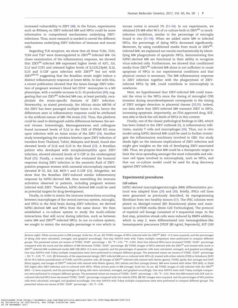
ResultsCharacterization of hiPSC-derived macrophage/microglia (MU)
To understand the immune component of neurologic abnor-malities observed in ZIKV-infected newborns, we first generatedMU from hiPSCs using an edited version by Douvarasand colleagues of the previously published protocol byYanagimachi et al. (29,30). This serum-free, feeder-free methodgave rise to CD14þmonocytes, precursors of MU, in suspensionstarting at 15 days in vitro. CD14þ cells were then sorted usingmagnetic-activated cell sorting (MACS) system, treated with M-CSF and IL-34 for a week as previously published (29), and usedin the subsequent experiments (Supplementary Material, Fig.S1A). These monocytes expressed classical MU markers such asIba1 and CD68 (Fig. 1A). The analysis of the cells in suspension,prior to CD14 sorting, revealed that they expressed monocyte,macrophage and myeloid cell markers such as CD45/CD14,CD14/HLA-DRDPDQ, CD14/CD11b and CD14/CD68, (Fig. 1C,Supplementary Material, Fig. S1B). One week after sorting,CD14þ cells established a homogeneous macrophage popula-tion (Supplementary Material, Fig. S1C) and had similar cellularmorphology and expression of cell markers such as CD68 andIba1 to blood monocyte-derived macrophages (SupplementaryMaterial, Fig. S1C and D). We next assessed whether the MUwere functional by measuring their capacity for phagocytosis(Fig. 1B). In a classical assay (31–33), hiPSC-derived MU engulfedthe yeast particle zymosan (Fig. 1B). The MU were also able tophagocytose living cells of the leading neonatal bacterial patho-gen group B Streptococcus (GBS) whereas hiPSC-derived NPCs or4-week-old neurons did not internalize the bacterium (Fig. 1B);primary human neutrophils were used as positive control ofphagocytosis. In addition, we measured cytokine (IL-6, IL-1b,IL12/IL23p40), chemokine (MIP1a, MIP1b, RANTES, Fractalkine,IL8 and IP10) and the growth factor G-CSF release by hiPSC-derived MU after activating them with the classical pro-inflammatory TLR-4 ligand lipopolysaccharide (LPS) using
2 | Human Molecular Genetics, 2017, Vol. 00, No. 00
Downloaded from https://academic.oup.com/hmg/article-abstract/doi/10.1093/hmg/ddx382/4557143by University of California, San Diego Libraries useron 09 November 2017
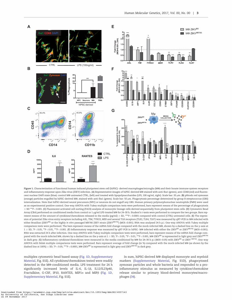
multiplex cytometric bead based-assay (Fig. 1D, SupplementaryMaterial, Fig. S1E). All cytokines/chemokines tested were readilydetected in the MU-conditioned media. LPS treatment for 24 hsignificantly increased levels of IL-6, IL-1b, IL12/IL23p40,Fractalkine, G-CSF, IP10, RANTES, MIP1a and MIPb (Fig. 1D,Supplementary Material, Fig. S1E).
In sum, hiPSC-derived MU displayed monocyte and myeloidmarkers (Supplementary Material, Fig. S1D), phagocytosedzymosan particles and whole bacteria and responded to a pro-inflammatory stimulus as measured by cytokine/chemokinerelease similar to primary blood-derived monocytes/macro-phages (34).
5 b 1 6 R 2 8 B 1 M 9 A 6
A
C
D
B LPS (100ng/ml) CTRL
Iba1 CD68 DAPI
45.4%
CD45
CD14
Iba1 Zymosan DAPI
NPC Neurons MΦ PMN0
5
10203040
hiPSC-derived
***
% P
hago
cyto
sis
45.8%
CD14
CD11b
0
100
200
300
400
500
RANTES
pg/m
l
****
0
2000
4000
6000
8000
10000
IL6
pg/m
l
****
AXL TIM TYRO3 MER TLR3 TLR4 TLR70.0
0.5
1.0
1.5
2.0246
Fold
Cha
nge
(MO
I=0.
001,
24h
)
****
**** ***** ***
MΦ ZIKVBR
MΦ ZIKVMR766
E
CC
R5
IL1b
AIF
1
IL6
IL4R
CC
L2
CD
68
CYB
B
IGF1
ITG
AM
CD
209
CLE
C5A
CD
206
0.0
0.5
1.0
1.5
2.02468
10
Fold
Cha
nge
(MO
I=0.
001,
24h
)
***
***
* ***
* *** ** **
****
** *
ZIKVAF
MEDIA COLLECTION ZIKVBR
+
Mφ
ZIKVMR766
ZIKVBR
0
100
200
300
400%
com
pare
d to
moc
k(M
OI=
0.05
, 24h
)
MIP
1β
RANT
ES
G-C
SF
IP10 IL
6
MIP
α
IL1β
Frac
talk
ine
IL8
sCD1
4
MCP
1
IL10
****
*
****
*
IL12
/IL23
p40
*
F
G
CTRLLPS
CCR5
IL1b
AIF1
IL6
IL4R
CCL2
CD68
CYBB
IGF1
ITG
AM
CD20
9
CLEC
5A
CD20
6
AXL TIM TYRO3 MER TLR3 TLR4 TLR7
IL12
/IL23
P40
MIP
1β
RANT
ES
G-C
SF
IP10
IL6
MIP
α
IL1β
Frac
talk
ine
IL8
sCD1
4
MCP
1
IL10
Figure 1. Characterization of functional human induced pluripotent stem cell (hiPSC)- derived macrophages/microglia (MU) and their Innate immune system receptorsand inflammatory response upon Zika virus (ZIKV) infection. (A) Representative images of hiPSC-derived MU stained with anti-Iba1 (green), anti-CD68 (red) and fluores-cent nuclear DAPI stain (blue): control MU untreated CTRL, (left) and treated with lipopolysaccharides (LPS, 100 ng/ml, right). Scale bar: 20 lm. (B) pHrodo red zymosan(orange) particles engulfed by hiPSC-derived MU, stained with anti-Iba1 (green). Scale bar: 50 lm. Phagocytosis percentage determined by group B streptococcus (GBS)internalization. Note that hiPSC-derived neural precursors (NPC) or neurons do not engulf any GBS. Human primary polymorphonuclear neutrophils (PMN) were usedas an experimental positive control. One-way ANOVA with Tukey multiple comparison tests were performed, bars represent means of the percentage of phagocytosis6 SD ***P<0.001. (C) Fluorescent activated cell-sorting (FACS) analysis of monocytic lineage cells derived sequentially from pluripotent stem cells. (D) Cytometric BeadArray (CBA) performed on conditioned media from control or 1 lg/ml LPS-treated MU for 24–30 h. Student’s t-tests were performed to compare the two groups. Bars rep-resent means of the amount of cytokines/chemokines released in the media (pg/ml) 6 SD, ****P< 0.0001 compared with control (CTRL) untreated cells. (E) The expres-sion of potential Zika virus entry receptors including AXL, TIM, TYRO3, MER and several TLR receptors (TLR3, TLR4, TLR7) was measured by qRT-PCR in MU infected witheither Brazilian (ZIKVBR) or the highly in vitro passaged MR766 ZIKV strain (ZIKVMR766) (MOI¼0.001). RNA was analysed 24 h p.i. One-way ANOVA with Tukey multiplecomparison tests were performed. The bars represent means of the mRNA fold change compared with the mock-infected MU, shown by a dashed line on the y-axis at1 6 SD, *P<0.05, **P<0.01, ***P<0.001. (F) Inflammatory response was measured by qRT-PCR in hiPSC- MU infected with either the ZIKVBR or ZIKVMR766 (MOI¼0.001).RNA was extracted 24 h after infection. One-way ANOVA with Tukey multiple comparison tests were performed, bars represent means of the mRNA fold change com-pared with the mock-infected MU, shown by a dashed line on the y-axis at 1 6 SD, *P<0.05, **P<0.01, ***P<0.001, MU ZIKVBR is represented in light grey and ZIKVMR766
in dark grey. (G) Inflammatory cytokines/chemokines were measured in the media conditioned by MU for 24–30 h p.i (MOI¼0.05) with ZIKVBR or ZIKV MR766. One-wayANOVA with Sidak multiple comparisons tests were performed. Bars represent average of fold change (in %) compared with the mock-infected MU (as shown by thedashed line at 100%) 6 SD, *P<0.05, ****P<0.0001, MU ZIKVBR is represented in light grey and ZIKVMR766 in dark grey.
3Human Molecular Genetics, 2017, Vol. 00, No. 00 |
Downloaded from https://academic.oup.com/hmg/article-abstract/doi/10.1093/hmg/ddx382/4557143by University of California, San Diego Libraries useron 09 November 2017
Immune response of macrophages/microglia (MU) uponinfection with the ZIKV
Next, we studied the immune response of MU upon infectionwith ZIKVBR as well as with ZIKVMR766, a lab-adapted, highlyin vitro passaged African MR766 strain, presumably differentfrom the circulating African strain at 12 and 24 h post-infection(p.i) (Supplementary Material, Fig S2A and B). We selected apanel of potential virus entry receptor candidates based onrecent publications including AXL, TIM, TYRO3, MER and severalTLR receptors previously linked to ZIKV and arboviruses such asDENV (32,33,39); TLR3, TLR4, TLR7, to measure the expressionlevels by qRT-PCR (17,18,35,36). At 12h p.i., we did not observeany significant changes between the strains when comparedwith mock-infected samples (Supplementary Material, Fig. S2A).At 24 h p.i., both strains of the virus triggered a TLR7 up-regulation and only ZIKVMR766 -infected MU expressed higherlevels of the MER receptor when compared with mock-infectedconditions (Supplementary Material, Fig. S2B). Next, we used alower MOI aiming to mimic the ZIKV infection in vivo and tounveil the most sensitive changes without inducing a dramaticinflammatory response that could mask small and strain-specific changes (37–41). Thus, these ranges of MOIs and 24h p.i.time point were used in the subsequent experiments (Fig. 1Eand F).
TYRO3 and AXL were upregulated upon the ZIKVBR infection,with AXL gene expression increased by three-fold comparedwith the mock-infected MU (Fig. 1E). In contrast, expression ofMER and TIM did not differ between ZIKVBR-infected and mock-infected MU (Fig. 1E). As for the TLR receptors expression, wefocused on three relevant for our study: TLR3, a sensor of RNAviruses, which was recently linked to ZIKV infection (35); TLR4,which plays a central role in recognition of LPS from Gram-negative bacteria, which was previously shown to be upregu-lated in ZIKV-infected human neurospheres (36); and TLR7, asensor of ssRNA involved in the detection of other arbovirusessuch as DENV (42). Surprisingly, all were downregulated whencompared with mock-infected controls (Fig. 1E). Interestingly,the corresponding changes seen in AXL, TYRO3, and TLR7 geneexpression upon infection with the ZIKVBR were absent in paral-lel infections with the ZIKVMR766, which did not differ frommock-infected MU in expression of all three receptor genes(Fig. 1E).
The geographic and symptomatology overlap between ZIKVand other Flaviviruses such as Dengue (DENV) initially compli-cated diagnosis (42). The vector by which it is propagated (Aedesaegypti) is shared by both viruses and clinically presented sero-logical cross-reactivity. In addition, DENV is known to target pri-mary blood cells such as monocytes and macrophages andinduces an inflammatory response (42). Thus, we next meas-ured the expression of genes encoding three receptors describedfor DENV infection: CLEC5A, CD209/DC-SIGN, and CD206/MRC1(Fig. 1F). All three viral receptors, CLEC5A, CD209/DC-SIGN, andCD206/MRC1, were downregulated in ZIKVBR or ZIKVMR766-infected MU compared with mock-infected MU, suggesting thatZIKV does not implicate these DENV-related receptors in iPSCs-derived MU (Fig. 1F).
Next, we studied the expression of the MU markers such asAIF1/Iba1, CD68, and ITGAM/CD11b by qRT-PCR upon infectionwith the ZIKV strains. AIF1/Iba1 expression was increased inthe MU infected with ZIKVBR compared with the mock-infectedor ZIKVMR766-infected cells, whereas similar changes in CD68expression did not achieve statistic significance (Fig. 1F).Notably, pro-inflammatory genes encoding IL6, IL1B, and CCR5
were markedly induced upon infection by the ZIKVBR, but notZIKVMR766. In contrast, the MU marker ITGAM/CD11b and theanti-inflammatory factor IGF1 were decreased by infectionswith either ZIKV strain, whereas expression of CYBB/NOX2remained unchanged upon infection with the viruses (Fig. 1F).These results indicate that the inflammatory response of MUinfected with two different ZIKV strains at the same viral MOIelicited different inflammatory responses.
We next compared cytokine/chemokine release by thehiPSC-derived MU 24 h p.i. with the ZIKVBR or ZIKVMR766, (Fig.1G, Supplementary Material, Fig. S4) using the same multiplexcytometric bead array that was used previously (Fig. 1D,Supplementary Material, Fig. S1E). Using several inocula of virus(MOI¼ 0.05, MOI¼ 0.01 and MOI¼ 0.001), we detected changes inrelease of several pro-inflammatory cytokines/chemokines (Fig.1G, Supplementary Material, Fig. S4B and C). MU release of pro-inflammatory cytokines IL12/IL23p40, IL1b, IL10 and the growthfactor G-CSF was significantly higher in response to ZIKVBR
compared with ZIKVMR766 infection; whereas MIP1b, RANTES,IP-10, IL6, and MIP1a release from ZIKV-infected MU were com-parable between the two strains (Fig. 1G, SupplementaryMaterial, Fig. S4B and C), arguing for a ZIKVBR-specific responseby the hiPSC-derived MU.
Differential expression of TAM/TIM and TLR receptors inhuman NPCs
Human NPCs were shown to be targeted by ZIKVBR (17). Similarto MU, we first infected NPCs at an MOI of 0.1 with either strainsand studied the expression of different viral entry receptors at12 and 24 h p.i. by qRT-PCR (Supplementary Material, Fig. S2Cand D), however, we did not detect any differences between thetwo different strains at 12h p.i. (Supplementary Material, Fig.S2C). At 24h p.i., TYRO3 and TLR4 were both upregulated in NPCsupon infection with both ZIKVBR and ZIKVMR766 strains(Supplementary Material, Fig. S2D). Given that we did not seemajor differences in gene expression in both of the strainstested, we focused on the same MOI (0.001) determined fromthe MU expression analysis and the time points (24 and 96 hp.i.) (Fig. 1A and B, Supplementary Material, Fig. S2A and B) forthe NPCs (Supplementary Material, Fig. S3A and B). Of fourreceptors analysed, only the MER was increased in the ZIKVBR
24 h p.i. compared with mock but returned to mock levels by 96h p.i. (Supplementary Material, Fig. S3A). Expression of TYRO3was downregulated in both mock- and ZIKVBR-infected NPCs at96 h p.i. compared with 24 h p.i. (Supplementary Material, Fig.S3A). TLR4 was also decreased as in MU, while TLR7 expressionwas increased compared with the mock-infected controls at24 h p.i. (Supplementary Material, Fig. S3B). The expression ofTAM/TIM receptors and TLRs observed in NPCs at 96 h p.i. wereaccompanied by a decreased expression of cell markers such asNES/Nestin and PAX6 in both mock- and ZIKVBR-infected NPCs(Supplementary Material, Fig. S3C).
To summarize, we did not observe great changes in ZIKVBR-infected NPCs except for MER and TLR7, which were significantlyupregulated compared with the mock-infected NPCs at 24-hpost-infection.
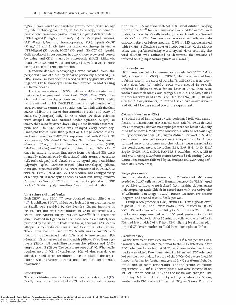
The interplay between NPCs with macrophages/microglia (MU) during ZIKV infection
To investigate interactions of hiPSC-derived MU with ZIKVBR-infected NPCs we established a co-culture experimental
4 | Human Molecular Genetics, 2017, Vol. 00, No. 00
Downloaded from https://academic.oup.com/hmg/article-abstract/doi/10.1093/hmg/ddx382/4557143by University of California, San Diego Libraries useron 09 November 2017
platform. NPCs were infected (MOI¼ 0.1) for 2 h with the ZIKVBR
or ZIKVMR766, media replaced, and MU overlaid 2 h after themedia change. MU and NPCs were maintained in co-culture for96 h, and the fraction of cells undergoing cell death examinedby DNA fragmentation using terminal deoxynucleotidyl trans-ferase dUTP nick end labeling (TUNEL) (Fig. 2A and B,Supplementary Material, Fig. S5). We initially seeded the sameamount of MU and NPCs, however, the proliferation rate of theNPCs was higher and thus the percentage of MU in the co-culture with NPCs was around 2% at 96 h p.i. (SupplementaryMaterial, Fig. S5C). In the absence of infection, addition of MU inco-culture atop NPCs did not influence the number of TUNELþ
cells (#10%, see Fig. 2C). However, as previously shown (17),ZIKVBR infection at MOI¼ 0.1 doubled the percentage of TUNELþ
NPCs (Fig. 2B and C). No increase in TUNELþ was detectedin NPCs infected with ZIKVBR at MOI¼ 0.01 for 96h (Supplementary Material, Fig. S5A). Infection with ZIKVMR766
had similar effect by increasing the amount of TUNELþNPCs(Supplementary Material, Fig. S5B). In addition, the amount ofMU undergoing apoptosis (TUNELþ) remained under #5% inpresence or absence of ZIKVBR indicating that the ZIKVBR wasnot cytotoxic to MU under our experimental conditions (Fig. 2Band C, Supplementary Material, Fig. S5D). Interestingly, addingMU to ZIKV-infected NPCs reduced the percentage of TUNELþ
cell levels similar to mock-infection (Fig. 2B and C,Supplementary Material, Fig. S5B). Thus, while adding naıve MUto uninfected NPCs did not increase cell death, infection ofNPCs with the ZIKVBR increased cell death, and the presence ofMU reduced cell death in ZIKVBR-infected NPCs (Fig. 2B and C).To elucidate the mechanism by which MU presence coulddecrease TUNELþ ZIKVBR-infected NPCs, whether it is throughphagocytosis or through protection by release of neurotrophicfactors by MU, we used conditioned media (CM) from mock orZIKVBR- infected MU and added on top of NPCs (Fig. 2D). MockMU CM did not have any effect on the basal level of apoptosis inmock-infected NPCs whereas ZIKVBR MU CM addition tended toincrease the level of TUNELþ NPCs (Fig. 2D). Although the addi-tion of mock or ZIKVBR MU CM on top of ZIKVBR-infected NPCshad a small tendency to decrease the amount of TUNELþ cells,there was no statistically significant change compared withZIKVBR-infected NPCs in the absence of any CM (Fig. 2D). Thus,MU most likely need to be in contact with NPCs to decrease theamount of TUNELþ NPCs, most likely by phagocytosing theapoptotic cells.
Finally, to assess whether ZIKV-infected MU could transmitthe virus to the NPCs, we first infected MU with ZIKV (ZIKVBR orZIKVMR766, Supplementary Material, Fig. S5), washed and addedon top of NPCs the next day (Fig. 2E). We co-cultured ZIKV-infected MU with NPCs for 96 h and analysed the amount ofTUNELþ cells (Fig. 2E and F, Supplementary Material, Fig. S5E).Importantly, upon addition of ZIKV-infected MU on top of NPCs,the amount of TUNELþ cells increased significantly (Fig. 2F andG, Supplementary Material, Fig. S5E). Keeping in mind that MUrepresent around 2% of the cells in total (SupplementaryMaterial, Fig. S5C) and that ZIKV infection did not increase theMU in apoptosis (Supplementary Material, Fig. S5D), theincrease in TUNEL can be attributed to the increase of cell deathof NPCs (Fig. 2G, Supplementary Material, Fig. S5E). More impor-tantly, we tested an FDA-approved drug, SOF, which waspreviously shown efficient against ZIKV infection in our model(26–28).
Next, in a following experiment, we added 20 lM SOF at thesame time as the addition of ZIKV-infected MU on top of unin-fected NPCs (Fig. 2E, G and H, Supplementary Material, Fig. S5E
and F). Importantly, addition of SOF decreased significantly theamount of TUNELþ cells in the case of ZIKVBR-infected MU addi-tion (Fig. 2F and G) and showed a tendency to decrease uponinfection with ZIKVMR766-infected MU (Supplementary Material,Fig. S5E). In addition, this decrease was accompanied by adecrease the amount of the flavivirus NS1þ domain specificstaining, suggesting that SOF was able to limit the ZIKVBR repli-cation (Fig. 2F–H). As for the MR766, SOF had only a tendency todecrease the amount of NS1þ cells (Supplementary Material,Fig. S5F). Thus, SOF was able to block the increased cell death ofNPCs due to the infection by ZIKV-infected MU.
Altogether, our model was able to mimic neuro-immuneinteractions, which is likely to occur during human neurodevel-opment and was proven to be useful for testing new therapeuticdrugs to block ZIKV-associated phenotypes.
DiscussionIn this study, we modeled immune interactions between NPCsand macrophages/microglia (MU) that would occur in the devel-oping brains of the ZIKVBR-infected fetuses/newborns, using aninduced pluripotent stem cell experimental platform. A funda-mental strength of induced pluripotent stem cells is the possi-bility of deriving different cell types from individuals in vitro.Generation of MU using this technology will circumvent theneed for MHC-matched bone marrow-derived MU from ahealthy donor. In addition, by generating NPCs and MU from thesame donor, we established an autologous approach to thestudy of immune interactions between different cell typesderived from the same genetic background. The novelty of ourstudy relies on the establishment of an in vitro platform, able tomimic human neuro-immune interactions. In particular, theseinteractions in human cells were studied by co-culturing hiPSC-derived NPCs together with hiPSC-derived MU. To our knowl-edge this is the first study enabling the analysis of humanneuro-immune interactions, unlike the studies using 3D orga-noids, where the mesoderm-derived immune cells such as MUof the central nervous system are lacking, which is a major limi-tation of this technology (17,35,43–45). Thus, we strongly believethat our platform could be further used in other studies impli-cating MU to model the interplay among the different cell typesand their impact on neurodevelopmental diseases.
In this study, using low MOI infections to mimic the ZIKVinfection in vivo, we analysed the cytokine and immune recep-tor responses to infections with the ZIKVBR or ZIKVMR766 strains.We then investigated cellular receptors implicated in ZIKV cellentry in developing human MU, beginning with C-type lectinreceptors known to bind DENV on the surface of macrophagesand dendritic cells and modulate inflammatory responses(42,46). Unlike in DENV infections, CLEC5A, CD209/DC-SIGN andCD206/MRC1 all had reduced expression in ZIKVBR andZIKVMR766-infected MU compared with controls (42,46). TAM/TIM receptors and TLRs have been recently implicated in ZIKVinfection (18,35). We found that MU and NPCs had differentialexpressions of the genes encoding these receptors upon infec-tion with ZIKVBR: TYRO3 and AXL were upregulated in MUinfected with ZIKVBR unlike NPC, which had only an upregula-tion of MER, suggesting that AXL does not have a central role inZIKV infections in NPCs. Indeed, several publications focusedon AXL receptors over the last year since it was suggested as apotential viral entry receptor (18), however, two independentstudies showed that blocking AXL does not prevent ZIKV infec-tion of NPCs (46,47). But a recent publication claims that selec-tive expression of Musashi in NPCs could explain their
5Human Molecular Genetics, 2017, Vol. 00, No. 00 |
Downloaded from https://academic.oup.com/hmg/article-abstract/doi/10.1093/hmg/ddx382/4557143by University of California, San Diego Libraries useron 09 November 2017
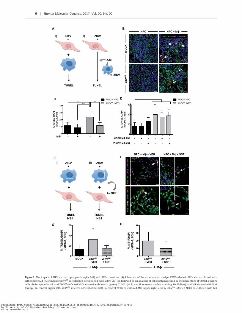
Figure 2. The impact of ZIKV on macrophage/microglia (MU) and NPCs co-culture. (A) Schematic of the experimental design. ZIKV-infected NPCs are co-cultured witheither naıve MU (I), or mock or ZIKVBR-infected MU-conditioned media (MU CM) (II), followed by an analysis of cell death measured by the percentage of TUNEL positivecells. (B) Images of mock and ZIKVBR-infected NPCs stained with Nestin (green), TUNEL (pink) and fluorescent nuclear staining, DAPI (blue), and MU stained with Iba1(orange) in control (upper left), ZIKVBR-infected NPCs (bottom left), in control NPCs co-cultured MU (upper right) and in ZIKVBR-infected NPCs co-cultured with MU
6 | Human Molecular Genetics, 2017, Vol. 00, No. 00
Downloaded from https://academic.oup.com/hmg/article-abstract/doi/10.1093/hmg/ddx382/4557143by University of California, San Diego Libraries useron 09 November 2017
increased vulnerability to ZIKV (48). In the future, experimentssuch as RNAseq on ZIKV-infected MU and NPCs could be moreinformative to comprehend mechanisms underlying ZIKV-infections. Thus, more research is needed to unveil the differentmechanisms underlying ZIKV infection of immune and neuralcells.
Regarding TLR receptors, we show that all three TLRs, TLR3,TLR4 and TLR7 were downregulated in ZIKVBR-infected MU. Oncloser examination of the inflammatory response, we showedthat ZIKVBR-infected MU expressed higher levels of AIF1, IL6,IL1b and CCR5 and released higher levels of IL12/IL23p40, IL1b,IL10 and G-CSF upon infection with ZIKVBR but not withZIKVMR766 suggesting that the Brazilian strain might induce adistinct inflammatory response at lower MOIs. In line with this,a recent publication showed that the Asian-lineage ZIKV infec-tion of pregnant women‘s blood led CD14þ monocytes to a M2phenotype, with a notable increase in IL-10 production (49), sug-gesting that our ZIKVBR-infected MU can, at least partially, reca-pitulate the strain-specific features of ZIKV infection.Noteworthy, as stated previously, the African strain MR766 ofthe ZIKV has been passaged multiple times in vitro, hence thedifferences seen in inflammatory response could be attributedto the artificial nature of MR-766 strain (50). Thus, this platformcould be used to distinguish subtle differences between the sev-eral viruses. Interestingly, Manangeeswaran and colleaguesfound increased levels of IL1b in the CNS of IFNAR KO miceupon infection with an Asian strain of the ZIKV (24). Anotherstudy investigating the cytokine kinetics upon ZIKV infection oftravelers from Thailand, Tahiti, Malaysia and Brazil, found alsoelevated levels of IL1b and IL10 in the blood (23). A Brazilianpatient who developed with encephalomyelitis upon ZIKVinfection, showed elevated levels of G-CSF in the cerebrospinalfluid (25). Finally, a recent study that evaluated the humoralresponse during ZIKV infection in the amniotic fluid of ZIKV-positive pregnant women with neonatal microcephaly reportedelevated IP-10, IL6, IL8, MCP-1 and G-CSF (22). Altogether, weshow that the Brazilian ZIKV-induced similar inflammatoryresponse by hiPSC-derived MU, thus resembling the immuneactivation observed in patients, including pregnant womeninfected with ZIKV. Therefore, hiPSC-derived MU could be usedas potential targets for drug development.
Finally, in order to mimic the immune interactions occurringbetween macrophages of the central nervous system, microglia,and NPCs in the fetal brain during ZIKV infection, we derivedhiPSC-derived MU and NPCs from the same donor. We thenestablished a co-culture system to study the multi-cellularinteractions that will occur during infection, such as betweennaıve MU and ZIKVBR-infected NPCs. In our co-culture system,we sought to mimic the microglia percentage in vivo which in
mouse cortex is around 5% (51–54). In our experiments, weobtained 2% MU after 96 h of co-culture both in ZIKVBR or mock-infection conditions, similar to the percentage of microgliafound in vivo (51–54). When we added naıve MU to infectedNPCs, the percentage of dying NPCs decreased significantly.Moreover, by using conditioned media from mock or ZIKVBR-infected MU, we explained our results mechanistically by identi-fying MU phagocytosis of apoptotic NPCs, demonstrating thathiPSC-derived MU are functional in their ability to recognizevirus-infected cells. Furthermore, we showed that conditionedmedia from ZIKVBR-infected MU alone is not sufficient to causeapoptosis of NPCs in our experimental conditions and thatphysical contact is necessary. The MU inflammatory responseto ZIKV infection together with the phagocytosis of ZIKV-infected NPCs by MU, could contribute to microcephaly innewborns.
Next, we hypothesized that ZIKV-infected MU could trans-mit the virus to the NPCs since the timing of microglial CNSinvasion during neurodevelopment corresponds to the timingof ZIKV antigen detection in placental tissues (20,55). Indeed,our data show that ZIKV-infected MU transmit ZIKV to NPCs,increasing apoptosis. Importantly, an FDA-approved drug, SOF,was able to block the cell death of NPCs in this context.
Finally, one of the classic pathological findings in GBS, whichhas been linked to the ZIKV outbreak (5), is inflammatory infil-trates, mainly T cells and macrophages (56). Thus, our in vitromodel using hiPSC-derived MU could be used to further investi-gate the inflammatory machinery involved in GBS. Our studysheds light on the immune implications of ZIKV infection andmight give insights on the risk of developing ZIKV-associatedGBS. Thus, we propose that MU could be a therapeutic target tolimit the virus spreading amongst the most vulnerable and rele-vant cell types involved in microcephaly, such as NPCs, andthat our co-culture model could be used for drug discoveryagainst ZIKV infection.
Experimental procedures
Cell culturehiPSC-derived macrophages/microglia (MU) differentiation pro-tocol was adapted from (29) and (30). Briefly, iPSCs cell lineswere generated as previously described, by reprogrammingfibroblast from two healthy donors (57). The iPSC colonies wereplated on Matrigel-coated (BD Biosciences) plates and main-tained in mTESR media (Stem Cell Technologies). The protocolof myeloid cell lineage consisted of 4 sequential steps. In thefirst step, primitive streak cells were induced by BMP4 addition,which in step 2, were differentiated into hemangioblast-likehematopoietic precursors [VEGF (80 ng/ml, Peprotech), SCF (100
(bottom right). White arrows point to MU (orange). Scale bar: 20 lm. (C) TUNEL images of NPCs infected with the ZIKVBR (MOI ¼0.1) were acquired, and the percentagesof dying cells were calculated, averaged, and graphed accordingly. One-way ANOVA tests with Tukey multiple comparison were performed to compare differentgroups. The presented values are means of TUNELþ/DAPIþ percentage 6 SD, **P<0.01, ***P<0.001. Note that infected NPCs have increased TUNELþ/DAPIþ percentagecompared with the mock and the addition of MU decreases TUNELþ/DAPIþ percentage. (D) TUNEL images of NPCs infected with the ZIKVBR and treated with mock orZIKVBR-infected MU conditioned media (MU CM) (MOI¼0.1) were acquired, and the percentages of apoptotic cells were calculated, averaged, and graphed accordingly.One-way ANOVA with Tukey multiple comparisons tests were performed to compare different groups. The presented values are means of TUNELþ/DAPIþ percentage6 SD, *P<0.05, **P< 0.01. (E) Schematic of the experimental design: ZIKV-infected MU are co-cultured with NPCs (I), treated with either vehicle (VEH) or Sofosbuvir (SOF)(II) for 96 h before quantification of TUNEL and NS1 positive cells. (F) Images of ZIKVBR-infected cells stained with Nestin (green), TUNEL (pink), Iba1 (orange) and DAPI(blue) (upper), and images of ZIKVBR-infected cells stained with Nestin (green), NS1 (white) and Iba1 (orange) (lower). MU were treated with either VEH (left), or 20lMSOF (right) after ZIKV-infection and co-cultured with NPCs. White arrows point to MU (orange). Scale bar: 20 lm. (G) TUNEL images of cells infected with the ZIKVBR
(MOI ¼1) were acquired, and the percentages of dying cells were calculated, averaged, and graphed accordingly. One-way ANOVA tests with Tukey multiple compari-son were performed to compare different groups. The presented values are means of TUNELþ/DAPIþ percentage 6 SD, **P<0.01. Note that MU treated with SOF and co-cultured infected NPCs have decreased TUNELþ/DAPIþ percentage compared with the vehicle (VEH). (H) NS1 images were acquired, and the percentages of NS1 positivecells were calculated, averaged, and graphed accordingly. One-way ANOVA with Tukey multiple comparisons tests were performed to compare different groups. Thepresented values are means of NS1þ/DAPIþ percentage 6 SD, *P<0.05.
7Human Molecular Genetics, 2017, Vol. 00, No. 00 |
Downloaded from https://academic.oup.com/hmg/article-abstract/doi/10.1093/hmg/ddx382/4557143by University of California, San Diego Libraries useron 09 November 2017
ng/ml, Gemini) and basic fibroblast growth factor (bFGF), (25 ng/ml, Life Technologies)]. Then, in the third step, the hemato-poietic precursors were pushed towards myeloid differentiation[FLT-3 ligand (50 ng/ml, HumanZyme), IL-3 (50 ng/ml, Gemini),SCF (50 ng/ml, Gemini), Thrombopoietin, TPO (5 ng/ml), M-CSF(50 ng/ml)] and finally into the monocytic lineage in step 4[FLT3-ligand (50 ng/ml), M-CSF (50ng/ml), GM-CSF (25 ng/ml)].Cells produced in suspension in step 4 were recovered, sortedby using anti-CD14 magnetic microbeads (MACS, Miltenyi),treated with 50ng/ml M-CSF and 50ng/ml IL-34 for a week beforebeing used in different experiments.
Monocyte-derived macrophages were isolated from theperipheral blood of a healthy donor as previously described (34).PBMCs were isolated from the blood by density gradient centri-fugation. CD14þ monocytes were then purified by MACS usingCD14 microbeads.
For the generation of NPCs, cell were differentiated andmaintained as previously described (57–59). Two iPSCs linesobtained from healthy patients maintained in mTSER mediawere switched to N2 [DMEM/F12 media supplemented with1xN2 NeuroPlex Serum-Free Supplement (Gemini) with the dualSMAD inhibitors 1 lM of dorsomorphin (Tocris) and 10uM ofSB431542 (Stemgent) daily, for 48 h. After two days, colonieswere scraped off and cultured under agitation (95rpm) asembryoid bodies for seven days using N2 media with dorsomor-phin and SB431542. Media was changed every other day.Embryoid bodies were then plated on Matrigel-coated dishes,and maintained in DMEM/F12 supplemented with 0.5x of N2supplement, 0.5x Gem21 NeuroPlex Serum-Free Supplement(Gemini), 20 ng/ml basic fibroblast growth factor (bFGF,LifeTechnologies) and 1% penicillin/streptomycin (P/S). After 7days in culture, rosettes that arose from the plated EBs weremanually selected, gently dissociated with StemPro Accutase(LifeTechnologies) and plated onto 10 lg/ml poly-L-ornithine(Sigma)/5 lg/ml Laminin-coated (LifeTechnologies) plates.Neuronal precursor cells (NPCs) were maintained in DMEM/F12with N2, Gem21, bFGF and P/S. The medium was changed everyother day. NPCs were split as soon as confluent, using StemProAccutase for 5min at 37 $C, centrifuged and replated with NGFwith a 1: 3 ratio in poly-L-ornithine/Laminin-coated plates.
Virus culture and amplificationBoth ZIKVBR and ZIKVMR766 were obtained and amplified as in(17): lyophilized ZIKVBR, which was isolated from a clinical casein Brazil, was provided by the Evandro Chagas Institute inBelem, Para and was reconstituted in 0.5 ml of sterile DEPCwater. The African-lineage MR-766 (ZIKVMR766), a referencestrain isolated in Uganda in 1947, used here as a control, wasprovided by the Institute Pasteur in Dakar, Senegal. C6/36 Aedesalbopictus mosquito cells were used to culture both viruses.The culture medium used for C6/36 cells was Leibovitz‘s L-15medium supplemented with 10% fetal bovine serum (FBS)(Gemini), 1% non-essential amino acids (Gibco), 1% sodium pyr-uvate (Gibco), 1% penicillin/streptomycine (Gibco) and 0.05%amphotericin B (Gibco). The cells were kept at 27 $C. When cellsreached around 70% of confluency, 50ll of viral sample wasadded. The cells were subcultured three times before the super-natant was harvested, titrated and used for experimentalinoculation.
Virus titrationThe virus titration was performed as previously described (17).Briefly, porcine kidney epithelial (PS) cells were used for virus
titration in L15 medium with 5% FBS. Serial dilution rangingfrom 10%1 to 10%11 for each virus stock were added onto 24-wellplates, followed by PS cells seeding into each well of a 24-wellplate for 3 h at 37 $C. Next, each well was overlaid with completecarboxymethyl cellulose medium (0.6% in L15 supplementedwith 3% FBS). Following 5 days of incubation in 37 $C, the plaqueassay was performed using 0.05% crystal violet solution. Theviral dilution was estimated to determine the amount ofinfected cells (plaque forming units or PFU.ml%1).
In vitro infectionNPCs were infected with commercially available ZIKVMR766 (MR-766, obtained from ATCC) and ZIKVBR, which was isolated froma febrile case in the state of Paraiba (Brazil-ZKV2015) as previ-ously described (17). Briefly, NPCs were seeded in 24-well,infected at different MOIs for an hour at 37 $C, then werewashed and their media was changed. For NPC and MU, both ofthe viruses were used at MOIs of 0.001 for RNA, 0.001, 0.01 and0.05 for CBA experiments, 0.1 for the first co-culture experimentand MOI of 1 for the second co-culture experiment.
Cytometric bead array (CBA)The bead-based immunoassay was performed following manu-facturer‘s instructions (BD Biosciences). Briefly, iPSCs-derivedMU or monocyte-derived macrophages were seeded at a densityof 5x10⁴ cells/well. Media was conditioned with or without 1lg/ml lipopolysaccharides (LPS, Sigma Aldrich) for 24–30h. 50ll ofconditioned media per sample were analysed by CBA. A cus-tomized array of cytokines and chemokines were measured inthe conditioned media, including IL1b, IL-6, IL-8, IL-10, IL12/23p40, G-CSF, IP10, sCD14, RANTES, Fractalkine, MIP1a, MIP1band MCP1, using a BD fluorescence activated cell sorting (FACS)Canto II instrument followed by an analysis on FCAP Array soft-ware (BD Biosciences).
Phagocytosis assayFor internalization experiments, hiPSCs-derived MU wereseeded to 2 x10⁴ cells per well. Human neutrophils (PMNs), usedas positive controls, were isolated from healthy donors usingPolyMorphPrep (Axis-Shield) in accordance with the Universityof California, San Diego, (UCSD) Human Research ProtectionsProgram, and seeded to 2 x10⁴ cells per well.
Group B Streptococcus (GBS) strain COH1 was grown over-night at 37 $C in Todd-Hewitt broth (Difco), diluted in PBS toMOI¼ 10, and spun onto cell 167 g for 3 min. After 90 min, themedia was supplemented with 100mg/ml gentamicin to killextracellular bacteria. After 30 min, the cells were washed 1x inPBS and lysed with 0.05% Triton-X100 (Sigma) for dilution plat-ing and CFU enumeration on Todd-Hewitt agar plates (Difco).
Co-culture assayFor the first co-culture experiment, 2 & 104 NPCs per well of a96-well plate were plated 24 h prior to the ZIKV infection. AfterZIKV infection for an hour at 37 $C, cells were washed and freshmedia was added. Two hours later, 2 & 104 naıve hiPSCs-derivedMU per well were plated on top of the NPCs. Cells were fixed 96h post-infection for further analysis with 4% paraformaldehyde,for 20 min at room temperature. For the second co-cultureexperiment, 2 & 104 NPCs were plated. MU were infected at anMOI of 1 for an hour at 37 $C and the media was changed. Thenext day, MU were lifted off by adding accutase for 5 min,washed with PBS and centrifuged at 300g for 5 min. The cells
8 | Human Molecular Genetics, 2017, Vol. 00, No. 00
Downloaded from https://academic.oup.com/hmg/article-abstract/doi/10.1093/hmg/ddx382/4557143by University of California, San Diego Libraries useron 09 November 2017
were counted and 2 & 104 cells/well of MU were added ontoNPCs with 20 lM of SOF (Acme Bioscience AB3793) or vehicle(DMSO). The dose of 20lM of SOF used on NPCs was optimizedin our laboratory (Mesci et al. manuscript in revision). The cellswere fixed after 96 h of incubation. For mock controls, the samevolume of supernatant was added to each experiment.
Imaging analysesCells were fixed with 4% paraformaldehyde for 20 min at roomtemperature. Next, samples were permeabilized in 1x-PBS con-taining 0.1% (v/v) Triton X-100 for 10 min. Cells were next incu-bated with blocking solution [1% fetal bovine serum, (LifeTechnologies) in 1xPBS]. After 1 h, the primary antibody wasadded (diluted in blocking solution) and samples were incu-bated overnight at 4 $C. Slides were then washed two times with1x-PBS, and incubated with the secondary antibody for 30 minat 4 $C. Secondary antibodies (all conjugated to Alexa Fluor 488,555 and 647) were purchased from Life Technologies and usedat a 1: 1000 dilution. After the 30 min incubation, samples werewashed twice (1x-PBS), incubated for 5 min with and fluorescentnuclear DAPI stain, and mounted with Prolong gold anti-fadereagent (Life Technologies). Samples were imaged using an AxioObserver Z1 Microscope with ApoTome (Zeiss). Captured imageswere analysed with Zen software from Zeiss. Antibodies anddilutions used: Monoclonal Mouse Anti-Human CD68, (1: 500,Dako); Polyclonal Rabbit Anti-Iba-1, (1: 500, Wako). For TUNELanalysis, NPCs were plated, infected after 24 h, and fixed 96 h p.iwith 4% paraformaldehyde for 20 min. Samples were permeabi-lized with 0.25% Triton X-100 for 15 min, and stained for frag-mented DNA using TUNEL (Click-iT TUNEL 647 assay kit fromLife Technologies). The cells were then blocked 1% bovineserum albumin for 60 min. Cells were incubated in primary anti-bodies: Monoclonal Mouse Nestin, Polyclonal anti-Chicken orPolyclonal Rabbit Nestin (1: 500, Abcam); Polyclonal Rabbit Anti-Iba-1, (1: 500, Wako) and monoclonal mouse NS1 (1: 250,Millipore) overnight at 4 $C and stained with secondary antibod-ies conjugated to Alexa Fluor and DAPI the following day priorto mounting. Images were blindly collected using an AxioObserver Z1 Microscope with ApoTome (Zeiss) and blindly ana-lysed with ImageJ software.
Fluorescence-activated cell sorting analysesUsing previously mentioned protocol to generate monocytes/macrophages (29), we have collected the cells in the superna-tant at the step 4 and labeled them with the appropriateantibodies (CD45 BV786, CD14 PE, CD11b APC, CD68 PE-Cy7,HLA-DRDPDQ FITC all from BD Biosciences). Briefly, cells wereharvested and counted, then washed twice with the stain buf-fer, containing PBS and 1% heat inactivated FBS, at 300g for 5min. The antibodies were added in the cell mixture and incu-bated in RT in the dark for 20–30 min. The cells were thenwashed twice with the stain buffer. The cells were resuspendedin 500 ll stain buffer and the viability dye, 7-AAD was added.The data were acquired in BD LSRFORTESSA instrument and theplots were generated using the software FlowJo.
RNA extraction and expression analyses by qPCRTotal RNA was extracted from cells using RNeasy Micro andMini kit for iPSC-derived MU and NPCs respectively (Qiagen), fol-lowing manufacturer’s instructions. Next, 900 ng of total RNAwas treated RNase-free DNaseI (Qiagen), and was reverse tran-scribed using QuantiTect Reverse Transcription Kit (Qiagen).Approximately, 15 ng of cDNAs was used per reaction and PCRs
were carried out in a final volume of 10 ll. Triplicate sampleswere analysed in a CFX384 Touch Real-Time PCR DetectionSystem (Bio-rad) using iQTM SYBRVR Green Supermix (Bio-rad).GADPH was used as internal normalization control. For thecomplete list of primers, please refer to Table 1. The run methodwas as follows: 3min at 95 $C, 40 cycles of 10s at 95 $C followedby 30s at 58 $C, and a melting curve was performed to confirmthe identity of the amplified product.
Statistical analysisOne-way ANOVA or two-way ANOVA (if two or more variables)tests followed by a Tukey or Sidak multiple comparison testswere used to compare groups and Student’s t-test to comparemeans of two groups.
Supplementary MaterialSupplementary Material is available at HMG online.
Table 1. List of primers
AXL-F GTTTGGAGCTGTGATGGAAGGCAXL-R CGCTTCACTCAGGAAATCCTCCCCR5-F CCTGCTGCTTTGCCTACATTGCCCR5-R ACACACTTGGCGGTTCTTTCGGCD11b-F GGAACGCCATTGTCTGCTTTCGCD11b-R ATGCTGAGGTCATCCTGGCAGACD206-F AGCCAACACCAGCTCCTCAAGACD206-R CAAAACGCTCGCGCATTGTCCACD209-F GCAGTCTTCCAGAAGTAACCGCCD209-R GCTCTCCTCTGTTCCAATACTGCCD68-F CGAGCATCATTCTTTCACCAGCTCD68-R ATGAGAGGCAGCAAGATGGACCCLEC5A-F TTGTCAACACGCCAGAGAAACTGCLEC5A-R CAACGCCACCTTTTCTCTTCACGIBA1-F CCCTCCAAACTGGAAGGCTTCAIBA1-R CTTTAGCTCTAGGTGAGTCTTGGIGF1-F CTCTTCAGTTCGTGTGTGGAGACIGF1-R CAGCCTCCTTAGATCACAGCTCIL1B-F CCACAGACCTTCCAGGAGAATGIL1B-R GTGCAGTTCAGTGATCGTACAGGIL6-F AGACAGCCACTCACCTCTTCAGIL6-R TTCTGCCAGTGCCTCTTTGCTGMCP1-F AGAATCACCAGCAGCAAGTGTCCMCP1-R TCCTGAACCCACTTCTGCTTGGMERTK-F CAGGAAGATGGGACCTCTCTGAMERTK-R GGCTGAAGTCTTTCATGCACGCNOX2-F CTCTGAACTTGGAGACAGGCAAANOX2-R CACAGCGTGATGACAACTCCAGTIM-F CTTCACCTCAGCCAGCAGAAACTIM-R GCCATCTGAAGACTCTGTCACGTLR3-F GCGCTAAAAAGTGAAGAACTGGATTLR3-R GCTGGACATTGTTCAGAAAGAGGTLR4-F CCCTGAGGCATTTAGGCAGCTATLR4-R AGGTAGAGAGGTGGCTTAGGCTTLR7-F CTTTGGACCTCAGCCACAACCATLR7-R CGCAACTGGAAGGCATCTTGTAGTYRO3-F GCAAGCCTTTGACAGTGTCATGGTYRO3-R GTTCATCGCTGATGCCCAAGCTNES-F CCATAGAGGGCAAAGTGGTAANES-R TTCTTCCCATATTTCCTGCTGCPAX-F TGTCCAACGGATGTGTGAGTAPAX-R CAGTCTCGTAATACCTGCCCAGAPDH-F TGCACCACCAACTGCTTAGCGAPDH-R GGCATGGACTGTGGTCATGAG
9Human Molecular Genetics, 2017, Vol. 00, No. 00 |
Downloaded from https://academic.oup.com/hmg/article-abstract/doi/10.1093/hmg/ddx382/4557143by University of California, San Diego Libraries useron 09 November 2017
Acknowledgements
We would like to thank Dr. Edison Durigon and his group for theZIKVBR aliquots, the UCSD stem cell core and Dr. ChristopherAlfonso from BD Biosciences. We would like to thank SpencerM. Moore for critical reading of the manuscript.
Conflict of Interest statement. None declared.
FundingThis work was supported by grants from the California Institutefor Regenerative Medicine (DISC2–09649), the NationalInstitutes of Health through the U19MH107367, Zika NetworkFAPESP projects 2011/18703–2 and 2014/17766–9, the NGO “thetooth fairy project”, a fellowship from the A.P. GianniniFoundation to C.N.L., and an NARSAD Independent InvestigatorGrant to A.R.M. Dr. Mesci has an International Rett syndromefoundation (IRSF) mentored training fellowship. Dr. Macia has aNARSAD Young Investigator grant.
References1. Dick, G.W.A., kitchen, S.F. and haddow, A.J. (1952) Zika virus.
I. Isolations and serological specificity. Trans. R. Soc. Trop.Med. Hyg., 46, 509–520.
2. Lanciotti, R.S., Kosoy, O.L., Laven, J.J., Velez, J.O., Lambert,A.J., Johnson, A.J., Stanfield, S.M. and Duffy, M.R. (2008)Genetic and serologic properties of Zika virus associatedwith an epidemic, Yap State, Micronesia, 2007. Emerg. Infect.Dis., 14, 1232–1239.
3. Campos, G.S., Bandeira, A.C. and Sardi, S.I. (2015) Zika virusoutbreak, Bahia, Brazil. Emerg. Infect. Dis., 21, 1885–1886.
4. Mlakar, J., Korva, M., Tul, N., Popovi!c, M., Polj"sak-Prijatelj, M.,Mraz, J., Kolenc, M., Resman Rus, K., Vesnaver Vipotnik, T.,Fabjan Vodu"sek, V. et al. (2016) Zika virus associated withmicrocephaly. N. Engl. J. Med., 374, 951–958.
5. Beckham, J.D., Pastula, D.M., Massey, A. and Tyler, K.L. (2016)Zika virus as an emerging global pathogen: neurologicalcomplications of Zika virus. JAMA Neurol.,10.1001/jamaneurol.2016.0800.
6. Calvet, G., Aguiar, R.S., Melo, A.S.O., Sampaio, S.A., deFilippis, I., Fabri, A., Araujo, E.S.M., de Sequeira, P.C., deMendonca, M.C.L., de Oliveira, L. et al. (2016) Detection andsequencing of Zika virus from amniotic fluid of fetuses withmicrocephaly in Brazil: a case study. Lancet. Infect. Dis., 16,653–660.
7. Martines, R.B., Bhatnagar, J., Keating, M.K., Silva-Flannery,L., Muehlenbachs, A., Gary, J., Goldsmith, C., Hale, G., Ritter,J., Rollin, D. et al. (2016) Notes from the field : evidence of Zikavirus infection in brain and placental tissues from two con-genitally infected newborns and two fetal losses—Brazil,2015. MMWR. Morb. Mortal. Wkly. Rep., 65, 1–2.
8. Sarno, M., Sacramento, G.A., Khouri, R., do Rosario, M.S.,Costa, F., Archanjo, G., Santos, L.A., Nery, N., Vasilakis, N.,Ko, A.I. et al. (2016) Zika virus infection and stillbirths: a caseof hydrops fetalis, hydranencephaly and fetal demise. PLoSNegl. Trop. Dis., 10, e0004517.
9. Metsky, H.C., Matranga, C.B., Wohl, S., Schaffner, S.F., Freije,C.A., Winnicki, S.M., West, K., Qu, J., Baniecki, M.L., Gladden-Young, A. et al. (2017) Zika virus evolution and spread in theAmericas. Nature, 546, 411–415.
10. Faria, N.R., Quick, J., Claro, I.M., Theze, J., de Jesus, J.G.,Giovanetti, M., Kraemer, M.U.G., Hill, S.C., Black, A., da Costa,
A.C. et al. (2017) Establishment and cryptic transmission ofZika virus in Brazil and the Americas. Nature, 546, 406–410.
11. Grubaugh, N.D., Ladner, J.T., Kraemer, M.U.G., Dudas, G.,Tan, A.L., Gangavarapu, K., Wiley, M.R., White, S., Theze, J.,Magnani, D.M. et al. (2017) Genomic epidemiology revealsmultiple introductions of Zika virus into the United States.Nature, 546, 401–405.
12. Liu, Y., Liu, J., Du, S., Shan, C., Nie, K., Zhang, R., Li, X.-F.,Zhang, R., Wang, T., Qin, C.-F. et al. (2017) Evolutionaryenhancement of Zika virus infectivity in Aedes aegypti mos-quitoes. Nature, 545, 482–486.
13. Delatorre, E., Mir, D. and Bello, G. (2017) Tracing the origin ofthe NS1 A188V substitution responsible for recent enhance-ment of Zika virus Asian genotype infectivity. Mem. Inst.Oswaldo Cruz., 10.1590/0074-02760170299.
14. Davis, B.S., Duggal, N.K., Chang, G.-J.J., Ritter, J.M., McDonald,E.M., Romo, H., Brault, A.C. and Guirakhoo, F. (2017)Differential neurovirulence of African and Asian genotypeZika virus isolates in outbred immunocompetent mice. Am.J. Trop. Med. Hyg., 10.4269/ajtmh.17-0263.
15. Anfasa, F., Siegers, J.Y., van der Kroeg, M., Mumtaz, N., StalinRaj, V., de Vrij, F.M.S., Widagdo, W., Gabriel, G., Salinas, S.,Simonin, Y. et al. (2017) Phenotypic differences betweenAsian and African lineage Zika viruses in human neural pro-genitor cells. mSphere, 2, e00292-17.
16. Simonin, Y., Loustalot, F., Desmetz, C., Foulongne, V.,Constant, O., Fournier-Wirth, C., Leon, F., Moles, J.-P.,Goubaud, A., Lemaitre, J.-M. et al. (2016) Zika virus strainspotentially display different infectious profiles in humanneural cells. EBioMedicine, 12, 161–169.
17. Cugola, F.R., Fernandes, I.R., Russo, F.B., Freitas, B.C., Dias,J.L.M., Guimar~aes, K.P., Benazzato, C., Almeida, N., Pignatari,G.C., Romero, S. et al. (2016) The Brazilian Zika virus straincauses birth defects in experimental models. Nature, 534,267–271.
18. Nowakowski, T.J., Pollen, A.A., Di Lullo, E., Sandoval-Espinosa, C., Bershteyn, M. and Kriegstein, A.R. (2016)Expression analysis highlights AXL as a candidate Zika virusentry receptor in neural stem cells. Cell Stem Cell, 18, 591–596.
19. Quicke, K.M., Bowen, J.R., Johnson, E.L., McDonald, C.E., Ma,H., O’Neal, J.T., Rajakumar, A., Wrammert, J., Rimawi, B.H.,Pulendran, B. et al. (2016) Zika virus infects human placentalmacrophages. Cell Host Microbe, 20, 83–90.
20. Ginhoux, F., Greter, M., Leboeuf, M., Nandi, S., See, P.,Gokhan, S., Mehler, M.F., Conway, S.J., Ng, L.G., Stanley, E.R.et al. (2010) Fate mapping analysis reveals that adult micro-glia derive from primitive macrophages. Science, 330,841–845.
21. Lum, F.-M., Low, D.K.S., Fan, Y., Tan, J.J.L., Lee, B., Chan,J.K.Y., Renia, L., Ginhoux, F. and Ng, L.F.P. (2017) Zika virusinfects human fetal brain microglia and induces inflamma-tion. Clin. Infect. Dis., 64, 914–920.
22. Ornelas, A.M.M., Pezzuto, P., Silveira, P.P., Melo, F.O.,Ferreira, T.A., Oliveira-Szejnfeld, P.S., Leal, J.I., Amorim,M.M.R., Hamilton, S., Rawlinson, W.D. et al. (2017) Immuneactivation in amniotic fluid from Zika virus-associatedmicrocephaly. Ann. Neurol., 81, 152–156.
23. Tappe, D., Perez-Giron, J.V., Zammarchi, L., Rissland, J.,Ferreira, D.F., Jaenisch, T., Gomez-Medina, S., Gunther, S.,Bartoloni, A., Mu~noz-Fontela, C. et al. (2016) Cytokinekinetics of Zika virus-infected patients from acute to recon-valescent phase. Med. Microbiol. Immunol., 205, 269–273.
24. Manangeeswaran, M., Ireland, D.D.C. and Verthelyi, D. (2016)Zika (PRVABC59) infection is associated with T cell
10 | Human Molecular Genetics, 2017, Vol. 00, No. 00
Downloaded from https://academic.oup.com/hmg/article-abstract/doi/10.1093/hmg/ddx382/4557143by University of California, San Diego Libraries useron 09 November 2017
infiltration and neurodegeneration in CNS of immunocom-petent neonatal C57Bl/6 mice. PLoS Pathog., 12, e1006004.
25. Galliez, R.M., Spitz, M., Rafful, P.P., Cagy, M., Escosteguy, C.,Germano, C.S.B., Sasse, E., Goncalves, A.L., Silveira, P.P.,Pezzuto, P. et al. (2016) Zika virus causing encephalomyelitisassociated with immunoactivation. Open Forum Infect. Dis., 3,ofw203.
26. Sacramento, C.Q., de Melo, G.R., de Freitas, C.S., Rocha, N.,Hoelz, L.V.B., Miranda, M., Fintelman-Rodrigues, N.,Marttorelli, A., Ferreira, A.C., Barbosa-Lima, G. et al. (2017)The clinically approved antiviral drug sofosbuvir inhibitsZika virus replication. Sci. Rep., 7, 40920.
27. Bullard-Feibelman, K.M., Govero, J., Zhu, Z., Salazar, V.,Veselinovic, M., Diamond, M.S. and Geiss, B.J. (2017) TheFDA-approved drug sofosbuvir inhibits Zika virus infection.Antiviral Res., 137, 134–140.
28. Onorati, M., Li, Z., Liu, F., Sousa, A.M.M., Nakagawa, N., Li, M.,Dell’Anno, M.T., Gulden, F.O., Pochareddy, S., Tebbenkamp,A.T.N. et al. (2016) Zika virus disrupts phospho-tbk1 localiza-tion and mitosis in human neuroepithelial stem cells andradial glia. Cell Rep., 16, 2576–2592.
29. Yanagimachi, M.D., Niwa, A., Tanaka, T., Honda-Ozaki, F.,Nishimoto, S., Murata, Y., Yasumi, T., Ito, J., Tomida, S.,Oshima, K. et al. (2013) Robust and highly-efficient differen-tiation of functional monocytic cells from human pluripo-tent stem cells under serum- and feeder cell-free conditions.PLoS One, 8, e59243.
30. Douvaras, P., Sun, B., Wang, M., Kruglikov, I., Lallos, G.,Zimmer, M., Terrenoire, C., Zhang, B., Gandy, S., Schadt, E.et al. (2017) Directed differentiation of human pluripotentstem cells to microglia. Stem Cell Reports,10.1016/j.stemcr.2017.04.023.
31. Grozdanov, V., Bliederhaeuser, C., Ruf, W.P., Roth, V.,Fundel-Clemens, K., Zondler, L., Brenner, D., Martin-Villalba,A., Hengerer, B., Kassubek, J. et al. (2014) Inflammatory dysre-gulation of blood monocytes in Parkinson‘s disease patients.Acta Neuropathol., 128, 651–663.
32. Underhill, D.M. (2003) Macrophage recognition of zymosanparticles. J. Endotoxin Res., 9, 176–180.
33. Speert, D.P. and Silverstein, S.C. (1985) Phagocytosis ofunopsonized zymosan by human monocyte-derived macro-phages: maturation and inhibition by mannan. J. Leukoc.Biol., 38, 655–658.
34. Ohradanova-Repic, A., Machacek, C., Fischer, M.B. andStockinger, H. (2016) Differentiation of human monocytesand derived subsets of macrophages and dendritic cells bythe HLDA10 monoclonal antibody panel. Clin. Transl.Immunol., 5, e55.
35. Dang, J., Tiwari, S.K., Lichinchi, G., Qin, Y., Patil, V.S.,Eroshkin, A.M. and Rana, T.M. (2016) Zika virus depletes neu-ral progenitors in human cerebral organoids through activa-tion of the innate immune receptor TLR3. Cell Stem Cell, 19,258–265.
36. Garcez, P.P., Nascimento, J.M., de Vasconcelos, J.M., Madeiroda Costa, R., Delvecchio, R., Trindade, P., Loiola, E.C., Higa,L.M., Cassoli, J.S., Vitoria, G. et al. (2017) Zika virus disruptsmolecular fingerprinting of human neurospheres. Sci. Rep., 7,40780.
37. Rosenberger, C.M., Podyminogin, R.L., Askovich, P.S.,Navarro, G., Kaiser, S.M., Sanders, C.J., McClaren, J.L., Tam,V.C., Dash, P., Noonan, J.G. et al. (2014) Characterization ofinnate responses to influenza virus infection in a novel lungtype I epithelial cell model. J. Gen. Virol., 95, 350–362.
38. Biacchesi, S., Skiadopoulos, M.H., Yang, L., Lamirande, E.W.,Tran, K.C., Murphy, B.R., Collins, P.L. and Buchholz, U.J.(2004) Recombinant human metapneumovirus lacking thesmall hydrophobic SH and/or attachment G glycoprotein:deletion of G yields a promising vaccine candidate. J. Virol.,78, 12877–12887.
39. Tan, M.C., Battini, L., Tuyama, A.C., Macip, S., Melendi, G.A.,Horga, M.A. and Luca Gusella, G. (2007) Characterization ofhuman metapneumovirus infection of myeloid dendriticcells. Virology, 357, 1–9.
40. Bayless, N.L., Greenberg, R.S., Swigut, T., Wysocka, J. andBlish, C.A. (2016) Zika virus infection induces cranial neuralcrest cells to produce cytokines at levels detrimental forneurogenesis. Cell Host Microbe, 20, 423–428.
41. Tang, H., Hammack, C., Ogden, S.C., Wen, Z., Qian, X., Li, Y.,Yao, B., Shin, J., Zhang, F., Lee, E.M. et al. (2016) Zika virusinfects human cortical neural progenitors and attenuatestheir growth. Cell Stem Cell, 18, 587–590.
42. Chen, S., Lin, Y., Huang, M., Wu, M., Cheng, S., Lei, H., Lee, C.,Chiou, T., Wong, C. and Hsieh, S. (2008) CLEC5A is critical fordengue-virus-induced lethal disease. 453, 672–676.
43. Garcez, P.P., Loiola, E.C., Madeiro da Costa, R., Higa, L.M.,Trindade, P., Delvecchio, R., Nascimento, J.M., Brindeiro, R.,Tanuri, A. and Rehen, S.K. (2016) Zika virus impairs growthin human neurospheres and brain organoids. Science,10.1126/science.aaf6116.
44. Qian, X., Nguyen, H.N., Song, M.M., Hadiono, C., Ogden, S.C.,Hammack, C., Yao, B., Hamersky, G.R., Jacob, F., Zhong, C.et al. (2016) Brain-region-specific organoids usingmini-bioreactors for modeling ZIKV exposure. Cell, 165,1238–1254.
45. Wells, M.F., Salick, M.R., Wiskow, O., Ho, D.J., Worringer,K.A., Ihry, R.J., Kommineni, S., Bilican, B., Klim, J.R., Hill, E.J.et al. (2016) Genetic ablation of AXL does not protect humanneural progenitor cells and cerebral organoids from Zikavirus infection. Cell Stem Cell, 19, 703–708.
46. Miller, J.L., deWet, B.J.M., Martinez-Pomares, L., Radcliffe,C.M., Dwek, R. a., Rudd, P.M. and Gordon, S. (2008) The man-nose receptor mediates dengue virus infection of macro-phages. PLoS Pathog, 4, 11.
47. Meertens, L., Labeau, A., Dejarnac, O., Cipriani, S., Sinigaglia,L., Bonnet-Madin, L., Le Charpentier, T., Hafirassou, M.L.,Zamborlini, A., Cao-Lormeau, V.-M. et al. (2017) Axl mediatesZIKA virus entry in human glial cells and modulates innateimmune responses. Cell Rep., 18, 324–333.
48. Chavali, P.L., Stojic, L., Meredith, L.W., Joseph, N., Nahorski,M.S., Sanford, T.J., Sweeney, T.R., Krishna, B.A., Hosmillo, M.,Firth, A.E. et al. (2017) Neurodevelopmental protein Musashi1 interacts with the Zika genome and promotes viral replica-tion. Science (80-.).
49. Foo, S.-S., Chen, W., Chan, Y., Bowman, J.W., Chang, L.-C.,Choi, Y., Yoo, J.S., Ge, J., Cheng, G., Bonnin, A. et al. (2017)Asian Zika virus strains target CD14(þ) blood monocytes andinduce M2-skewed immunosuppression during pregnancy.Nat. Microbiol., 10.1038/s41564-017-0016-3.
50. Haddow, A.D., Schuh, A.J., Yasuda, C.Y., Kasper, M.R., Heang,V., Huy, R., Guzman, H., Tesh, R.B., Weaver, S.C. and Olson,K.E. (2012) Genetic characterization of Zika virus strains:geographic expansion of the Asian lineage. PLoS Negl. Trop.Dis., 6, e1477.
51. Mandrekar-Colucci, S. and Landreth, G.E. (2010) Microgliaand inflammation in Alzheimer‘s disease. CNS Neurol.Disord. Drug Targets, 9, 156–167.
11Human Molecular Genetics, 2017, Vol. 00, No. 00 |
Downloaded from https://academic.oup.com/hmg/article-abstract/doi/10.1093/hmg/ddx382/4557143by University of California, San Diego Libraries useron 09 November 2017
52. Rezaie, P., Patel, K. and Male, D.K. (1999) Microglia in the humanfetal spinal cord–patterns of distribution, morphology and phe-notype. Brain Res. Dev. Brain Res., 115, 71–81.
53. Aloisi, F. (2001) Immune function of microglia. Glia, 36,165–179.
54. Lawson, L.J., Perry, V.H., Dri, P. and Gordon, S. (1990)Heterogeneity in the distribution and morphology of microgliain the normal adult mouse brain. Neuroscience, 39, 151–170.
55. Martines, R.B., Bhatnagar, J., de Oliveira Ramos, A.M., Davi,H.P.F., Iglezias, S. DAndretta., Kanamura, C.T., Keating, M.K.,Hale, G., Silva-Flannery, L., Muehlenbachs, A. et al. (2016)Pathology of congenital Zika syndrome in Brazil: a case ser-ies. Lancet (London, England), 388, 898–904.
56. Yuki, N. and Hartung, H.-P. (2012) Guillain-Barre syndrome.N. Engl. J. Med., 366, 2294–2304.
57. Marchetto, M.C.N., Carromeu, C., Acab, A., Yu, D., Yeo, G.W.,Mu, Y., Chen, G., Gage, F.H. and Muotri, A.R. (2010) A modelfor neural development and treatment of Rett syndromeusing human induced pluripotent stem cells. Cell, 143,527–539.
58. Nageshappa, S., Carromeu, C., Trujillo, C.A., Mesci, P.,Espuny-Camacho, I., Pasciuto, E., Vanderhaeghen, P.,Verfaillie, C.M., Raitano, S., Kumar, A. et al. (2015) Alteredneuronal network and rescue in a human MECP2 duplicationmodel. Mol. Psychiatry, 21, 178–188.
59. Griesi-Oliveira, K., Acab, A., Gupta, A.R., Sunaga, D.Y.,Chailangkarn, T., Nicol, X., Nunez, Y., Walker, M.F., Murdoch,J.D., Sanders, S.J. et al. (2014) Modeling non-syndromic autismand the impact of TRPC6 disruption in human neurons. Mol.Psychiatry, 20, 1350–1365.
12 | Human Molecular Genetics, 2017, Vol. 00, No. 00
Downloaded from https://academic.oup.com/hmg/article-abstract/doi/10.1093/hmg/ddx382/4557143by University of California, San Diego Libraries useron 09 November 2017