Embed Size (px)
Citation preview
1345
Models of Glucagon Secretion, Their Application to the Analysis of the Defects in Glucagon Counterregulation and Potential Extension
to Approximate Glucagon Action
Leon S. Farhy, Ph.D. and Anthony L. McCall, M.D., Ph.D.
Author Affiliations: Department of Medicine, Center for Biomathematical Technology, University of Virginia, Charlottesville, Virginia
Abbreviations: (BG) blood glucose, (GABA) γ-aminobutyric acid, (GCR) glucagon counterregulation, (GLP-1) glucagon-like peptide-1, (HGO) hepatic glucose output, (ID50) median infective dose, (MCN) minimal control network, (STZ) streptozotocin, (TM) transfer model
Keywords: counterregulation, diabetes mellitus, feedback, glucagon, hypoglycemia, intrapancreatic network, mathematical model
Corresponding Author: Leon S. Farhy, Ph.D., Departments of Medicine, Center for Biomathematical Technology, Box 800735, University of Virginia, Charlottesville, VA 22908; e-mail address [email protected]
Journal of Diabetes Science and Technology Volume 4, Issue 6, November 2010 © Diabetes Technology Society
AbstractThis review analyzes an interdisciplinary approach to the pancreatic endocrine network-like relationships that control glucagon secretion and glucagon counterregulation (GCR). Using in silico studies, we show that a pancreatic feedback network that brings together several explicit interactions between islet peptides and blood glucose reproduces the normal GCR axis and explains its impairment in diabetes. An α-cell auto-feedback loop drives glucagon pulsatility and mediates triggering of GCR by hypoglycemia by a rapid switch-off of β-cell signals. The auto-feedback explains the enhancement of defective GCR in β-cell deficiency by a switch-off of signals in the pancreas that suppress α cells. Our models also predict that reduced β-cell activity decreases and delays the GCR. A key application of our models is the in silico simulation and testing of possible scenarios to repair defective GCR in β-cell deficiency. In particular, we predict that partial suppression of hyperglucagonemia may repair the impaired GCR. We also outline how the models can be extended and tested using human data to become a part of a larger construct including the regulation of the hepatic glucose output by the pancreas, circulating glucose, and incretins. In conclusion, a model of the normal GCR control mechanisms and their dysregulation in insulin-deficient diabetes is proposed and partially validated. The model components are clinically measurable, which permits its application to the study of the abnormalities of the human endocrine pancreas and their role in the progression of many diseases, including diabetes, metabolic syndrome, polycystic ovary syndrome, and others. It may also be used to examine therapeutic responses.
J Diabetes Sci Technol 2010;4(6):1345-1356
SYMPOSIUM
Introduction
Blood glucose (BG) homeostasis is maintained by a complex system involving coordinated interplay between various hormone and metabolite signals. One critical component, the endocrine pancreas, regulates glucose production and metabolism by a synchronized reciprocal
release of insulin and glucagon in response to changes in BG, incretins, and other signals. Abnormal secretion and action of the pancreatic peptides play a role in the progression of many diseases, including diabetes, metabolic syndrome, polycystic ovary syndrome, and
1346
Models of Glucagon Secretion, Their Application to the Analysis of the Defects in Glucagon Counterregulation and Potential Extension to Approximate Glucagon Action Farhy
www.journalofdst.orgJ Diabetes Sci Technol Vol 4, Issue 6, November 2010
others. Diminished or complete loss of endogenous insulin secretion in diabetes is associated with failure of the pancreas to respond properly with glucagon secretion not only to hyper- but also to hypoglycemia. The latter is not caused by loss of glucagon secreting α cells, but by defects in glucagon counterregulation (GCR) signaling, through an unknown mechanism. Defective GCR is a major barrier to safe treatment of diabetes1,2 since unopposed hypoglycemia can cause coma, seizures, or even death.3,4 Our experimental5 and mathematical-modeling5–7 results show that a novel understanding of the defects in the GCR control mechanisms can be gained if the network of intrapancreatic interactions that control glucagon secretion is described and analyzed by a mathematical model.
We first developed5,6 a model of the endocrine pancreas suitable for the study of rodent physiology. Later, a new simplified construct7 (in which δ-cell somatostatin was not explicitly involved) was also shown to closely approximate the GCR control mechanisms. This construct was applied to study the abnormalities in glucagon secretion and counterregulation and to explore in silico ways for their repair. Here, we review these efforts and highlight possible applications and extension of our models.
Construct DevelopmentTo describe the glucagon axis, we simplified the system by clustering all known and unknown factors into a small number of explicitly recognized physiological relationships chosen initially to explain key experimental results (e.g., the in vivo enhancement of GCR by switch-off of insulin).8 The postulated network5 included relationships between the α cells, δ cells, BG, and switch-off signals (intrapancreatically infused α-cell inhibitors that are terminated during hypoglycemia). This network explained the repair of GCR in diabetic rats by switch-off signals by interpreting the GCR as a rebound or disinhibition effect. It also predicted that (1) in β-cell deficiency, multiple α-cell suppressors should enhance GCR if they are terminated during hypoglycemia, and (2) the switch-off-triggered glucagon response must be pulsatile. We confirmed these predictions in vivo, in STZ-treated rats.5 The construct was further extended6 to reflect the assumption that the α-cell activity can be regulated differently by different α-cell inhibitors as suggested by earlier experiments.5 However, the explicit involvement of somatostatin in the model limits the potential for clinical applications as pancreatic somatostatin cannot be measured reliably in humans
in vivo and the ability of the model to describe the human glucagon axis cannot be verified. To address this limitation, we reduced our initial construct into a minimal control network (MCN) of the GCR control axis in which the δ cells are no longer explicitly involved but their effects are implicitly incorporated.7 Our analyses (reviewed later) show that the MCN is an excellent model of the glucagon axis and can replace the earlier, more complex structure. It can be also tested clinically and used to predict the behavior of the human GCR axis both in β-cell sufficiency and deficiency.
Our models are based on published studies of the pancreatic peptides that as a whole suggest that a network of interacting pathways modulates the secretion of glucagon. These key relationships are summarized here and are described extensively elsewhere.5–7
β-Cell Inhibition of α Cells1. Blood within the islets flows from β to α to δ
cells.9-12
2. β cells secrete signals known to inhibit the α cells: insulin, zinc, GABA and amylin.9,13–21
δ-Cell Inhibition of α Cells1. Exogenous somatostatin inhibits glucagon (and
insulin).22–33
2. The α and β cells express somatostatin receptors25,31,32 that may mediate the inhibition of glucagon by endogenous δ-cell somatostatin.22,23,32
3. The δ cells are in close proximity to α cells, and δ cell processes extend into α-cell clusters.30,34
α-Cell Stimulation of δ Cells1. Blood within the islets flows from α to δ cells
(mentioned earlier) and administration of glucagon antibodies in the perfused human pancreas inhibits somatostatin.11,12,29
2. The glucagon receptor has been colocalized with immunoreactive somatostatin cells.35
3. Exogenous glucagon stimulates somatostatin release.29,34,36,37
4. Glutamate stimulates somatostatin release from diencephalic neurons38 suggesting that a similar relation may exist in the pancreas where glutamate is cosecreted with glucagon.
1347
Models of Glucagon Secretion, Their Application to the Analysis of the Defects in Glucagon Counterregulation and Potential Extension to Approximate Glucagon Action Farhy
www.journalofdst.orgJ Diabetes Sci Technol Vol 4, Issue 6, November 2010
Glucose Stimulation of β and δ Cells1. It is well established that hyperglycemia directly
stimulates β cells.39–42
2. δ cells have a glucose-sensing mechanism similar to those in β cells.28,43
3. δ-cell somatostatin release is stimulated by glucose in vitro.44,45
Glucose Inhibition of α CellsHyperglycemia inhibits glucagon even though hypo-glycemia alone may be insufficient to stimulate GCR.16,46–51
Indirect evidence also supports the concept of network control of GCR: pulsatility of the pancreatic hormones;52–54 release of insulin and somatostatin pulses in phase;55,56 insulin and glucagon pulses and somatostatin and glucagon pulses with a phase shift;53 and entrainment of α- and δ-cell oscillations by insulin pulses.57
We have shown that a network based on the relationships mentioned earlier explains key experimental findings and have presented experimental evidence to support the proposed construct.5,6 In particular, we have used differential equations-based modeling to show that the proposed network explains each of the following findings in diabetic rats:
1. Glucagon pulsatility during hypoglycemia after a switch-off of α-cell inhibiting signals, with pulses recurring at 15 to 20 minutes;5
2. Pronounced pulsatile glucagon response following a switch-off of either insulin or somatosatin during hypoglycemia;5
3. Restriction of the GCR enhancement by insulin switch-off by high glucose;8
4. Lack of a GCR response to hypoglycemia when there is no switch-off signal;5
5. Suppression of GCR when insulin is infused into the pancreas but not switched off during hypoglycemia;8
6. Higher GCR response to insulin vs somatostatin switch-off and stronger glucagon suppression by somatostatin than by insulin before a switch-off.5
Next, we have simplified the network in such a way that somatostatin is no longer explicitly involved but
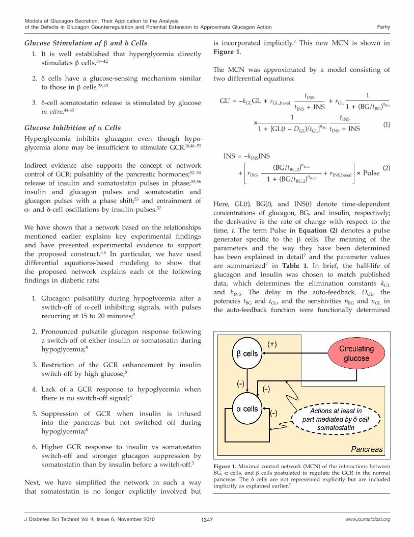
is incorporated implicitly.7 This new MCN is shown in Figure 1.
The MCN was approximated by a model consisting of two differential equations:
GL’ = –kGLGL + rGL,basal tINS
tINS + INS + rGL
11 + (BG/tBG)nBG
11 + [GL(t – DGL)/tGL]nBG
tINS
tINS + INS× (1)
INS = –kINSINS
+ rINS (BG/tBG,2)
nBG,2
1 + (BG/tBG,2)nBG,2
+ rINS,basal × Pulse (2)
Here, GL(t), BG(t), and INS(t) denote time-dependent concentrations of glucagon, BG, and insulin, respectively; the derivative is the rate of change with respect to the time, t. The term Pulse in Equation (2) denotes a pulse generator specific to the β cells. The meaning of the parameters and the way they have been determined has been explained in detail7 and the parameter values are summarized7 in Table 1. In brief, the half-life of glucagon and insulin was chosen to match published data, which determines the elimination constants kGL and kINS. The delay in the auto-feedback, DGL, the potencies tBG and tGL, and the sensitivities nBG and nGL in the auto-feedback function were functionally determined
Figure 1. Minimal control network (MCN) of the interactions between BG, α cells, and β cells postulated to regulate the GCR in the normal pancreas. The δ cells are not represented explicitly but are included implicitly as explained earlier.7
1348
Models of Glucagon Secretion, Their Application to the Analysis of the Defects in Glucagon Counterregulation and Potential Extension to Approximate Glucagon Action Farhy
www.journalofdst.orgJ Diabetes Sci Technol Vol 4, Issue 6, November 2010
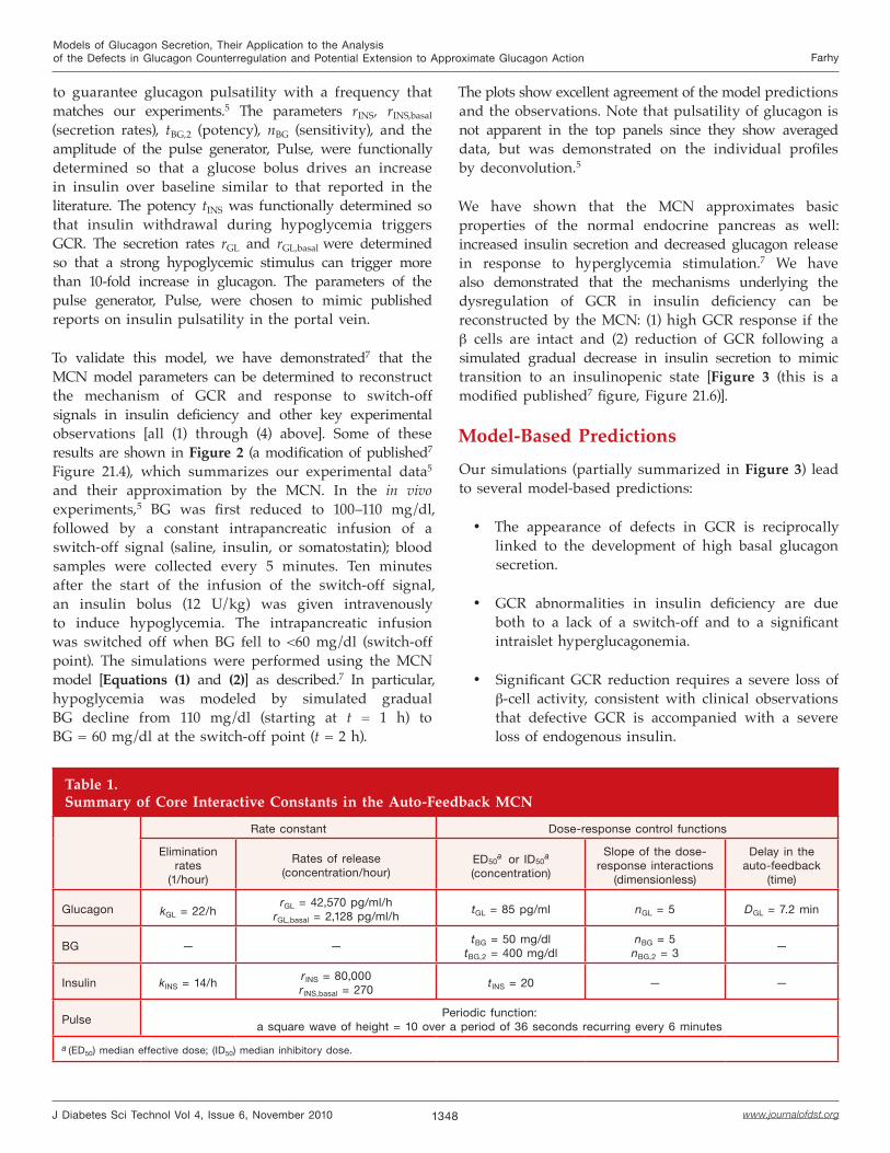
Table 1.Summary of Core Interactive Constants in the Auto-Feedback MCN
Rate constant Dose-response control functions
Elimination rates
(1/hour)
Rates of release(concentration/hour)
ED50a or ID50
a
(concentration)
Slope of the dose-response interactions
(dimensionless)
Delay in the auto-feedback
(time)
Glucagon kGL = 22/hrGL = 42,570 pg/ml/h
rGL,basal = 2,128 pg/ml/htGL = 85 pg/ml nGL = 5 DGL = 7.2 min
BG — —tBG = 50 mg/dl
tBG,2 = 400 mg/dlnBG = 5nBG,2 = 3
—
Insulin kINS = 14/hrINS = 80,000rINS,basal = 270
tINS = 20 — —
PulsePeriodic function:
a square wave of height = 10 over a period of 36 seconds recurring every 6 minutes
a (ED50) median effective dose; (ID50) median inhibitory dose.
to guarantee glucagon pulsatility with a frequency that matches our experiments.5 The parameters rINS, rINS,basal (secretion rates), tBG,2 (potency), nBG (sensitivity), and the amplitude of the pulse generator, Pulse, were functionally determined so that a glucose bolus drives an increase in insulin over baseline similar to that reported in the literature. The potency tINS was functionally determined so that insulin withdrawal during hypoglycemia triggers GCR. The secretion rates rGL and rGL,basal were determined so that a strong hypoglycemic stimulus can trigger more than 10-fold increase in glucagon. The parameters of the pulse generator, Pulse, were chosen to mimic published reports on insulin pulsatility in the portal vein.
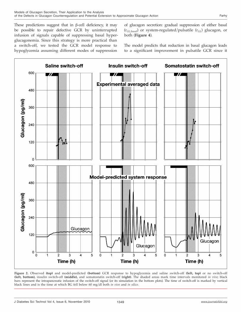
To validate this model, we have demonstrated7 that theMCN model parameters can be determined to reconstruct the mechanism of GCR and response to switch-off signals in insulin deficiency and other key experimental observations [all (1) through (4) above]. Some of these results are shown in Figure 2 (a modification of published7 Figure 21.4), which summarizes our experimental data5 and their approximation by the MCN. In the in vivo experiments,5 BG was first reduced to 100–110 mg/dl, followed by a constant intrapancreatic infusion of a switch-off signal (saline, insulin, or somatostatin); blood samples were collected every 5 minutes. Ten minutes after the start of the infusion of the switch-off signal, an insulin bolus (12 U/kg) was given intravenously to induce hypoglycemia. The intrapancreatic infusion was switched off when BG fell to <60 mg/dl (switch-off point). The simulations were performed using the MCN model [Equations (1) and (2)] as described.7 In particular, hypoglycemia was modeled by simulated gradual BG decline from 110 mg/dl (starting at t = 1 h) to BG = 60 mg/dl at the switch-off point (t = 2 h).
The plots show excellent agreement of the model predictions and the observations. Note that pulsatility of glucagon is not apparent in the top panels since they show averaged data, but was demonstrated on the individual profiles by deconvolution.5
We have shown that the MCN approximates basic properties of the normal endocrine pancreas as well: increased insulin secretion and decreased glucagon release in response to hyperglycemia stimulation.7 We have also demonstrated that the mechanisms underlying the dysregulation of GCR in insulin deficiency can be reconstructed by the MCN: (1) high GCR response if the β cells are intact and (2) reduction of GCR following a simulated gradual decrease in insulin secretion to mimic transition to an insulinopenic state [Figure 3 (this is a modified published7 figure, Figure 21.6)].
Model-Based PredictionsOur simulations (partially summarized in Figure 3) lead to several model-based predictions:
• The appearance of defects in GCR is reciprocally linked to the development of high basal glucagon secretion.
• GCR abnormalities in insulin deficiency are due both to a lack of a switch-off and to a significant intraislet hyperglucagonemia.
• Significant GCR reduction requires a severe loss of β-cell activity, consistent with clinical observations that defective GCR is accompanied with a severe loss of endogenous insulin.
1349
Models of Glucagon Secretion, Their Application to the Analysis of the Defects in Glucagon Counterregulation and Potential Extension to Approximate Glucagon Action Farhy
www.journalofdst.orgJ Diabetes Sci Technol Vol 4, Issue 6, November 2010
Figure 2. Observed (top) and model-predicted (bottom) GCR response to hypoglycemia and saline switch-off (left, top) or no switch-off (left, bottom), insulin switch-off (middle), and somatostatin switch-off (right). The shaded areas mark time intervals monitored in vivo; black bars represent the intrapancreatic infusion of the switch-off signal (or its simulation in the bottom plots). The time of switch-off is marked by vertical black lines and is the time at which BG fell below 60 mg/dl both in vivo and in silico.
These predictions suggest that in β-cell deficiency, it may be possible to repair defective GCR by uninterrupted infusion of signals capable of suppressing basal hyper-glucagonemia. Since this strategy is more practical than a switch-off, we tested the GCR model response to hypoglycemia assuming different modes of suppression
of glucagon secretion: gradual suppression of either basal (rGL,basal) or system-regulated/pulsatile (rGL) glucagon, or both (Figure 4).
The model predicts that reduction in basal glucagon leads to a significant improvement in pulsatile GCR since it

1350
Models of Glucagon Secretion, Their Application to the Analysis of the Defects in Glucagon Counterregulation and Potential Extension to Approximate Glucagon Action Farhy
www.journalofdst.orgJ Diabetes Sci Technol Vol 4, Issue 6, November 2010
removes a system repression mediated by auto-feedback (Figure 4, top blue curve). On the other hand, reduction
Figure 3. Model-derived GCR response to hypoglycemia (stepwise BG decline) in normal physiology with intact insulin release (top). Predicted decrease and delay in GCR and increase in basal glucagon with loss of insulin secretion [2nd panel, 50%; 3rd panel, 75%; 4th panel, 100% (complete absence)] gradually lost to mimic a transition from a normal to an insulin-deficient state.
Figure 4. Changes in the maximal GCR response to a stepwise BG decline (as in Figure 3) in the 1-hour interval after BG reaches 60 mg/dl caused by gradual suppression of basal glucagon (blue, filled triangles), system-regulated (pulsatile) glucagon (pink, filled squares), or both (black, open diamonds).
in the pulsatile glucagon secretion entails consistent reduction in the GCR (Figure 4, bottom pink curve). However, if inhibition of total (both basal and pulsatile) glucagon secretion is assumed (as one might expect during in vivo infusions of α-cell suppressors), the model predicts initial enhancement of the GCR response, followed by its gradual decline (Figure 4, middle black curve). Maximal amplification is predicted at about 45% reduction of glucagon. Figure 5 exemplifies this concept by depicting some minor GCR improvement (Figure 5, top vs Figure 3, bottom) by 10% reduction of total glucagon, complete GCR repair by 40% reduction (Figure 5, middle vs Figure 3, top), and reversal of this improvement by further suppression (Figure 5, bottom). This leads to two key model-based predictions:
• If an α-cell suppressing signal is administered at high doses and is not switched off, the GCR response to hypoglycemia will be suppressed.
• Lower, carefully selected infusion rates of α-cell inhibitors to partially reduce glucagon secretion may repair defective GCR in insulin-deficient diabetes.
These predictions are supported by some clinical observations58–61 and suggest strategies to manipulate the in vivo MCN to repair defective GCR in β-cell deficiency. For example, if some α-cell suppressors (GLP-1, amylin, GABA, etc.) or GLP-1 stimulators (vildagliptin) are continuously infused at clinically appropriate rates,

1351
Models of Glucagon Secretion, Their Application to the Analysis of the Defects in Glucagon Counterregulation and Potential Extension to Approximate Glucagon Action Farhy
www.journalofdst.orgJ Diabetes Sci Technol Vol 4, Issue 6, November 2010
they may restore the GCR and decrease glucagon during eu- and hyperglycemia. We also have some pilot data suggesting that uninterrupted intrapancreatic infusion of GABA can enhance GCR in STZ-treated rats (unpublished).
Further Model Verification
Additional In Vivo Verification in Rodent ModelsThe model-based simulations are consistent with key experimental outcomes and support the postulated MCN model. However, these simulations reconstruct only general “averaged” behavior of the in vivo system and approximate portal rather than circulating concentrations. Therefore, new experimental data are required to demonstrate further that the model approximates well the glucagon axis. These should involve interventional studies to infuse hormones and signals into the pancreas and analyze frequently glucagon and other peptide responses in portal vein blood samples. Analysis of such data by the mathematical model will evaluate whether the MCN provides an objectively good description of the action of the complex GCR control mechanism.
Relating Portal and Circulating Glucagon Concentrations: Use of a Transfer ModelThe mathematical methods for analyzing the GCR control mechanisms use portal rather than peripheral venous hormone concentrations and can be easily tested in rats but not humans by experiments in which the pancreatic hormones are sampled in the portal vein. If blood samples from the portal vein cannot be collected, the methodology could be extended to reconstruct the GCR axis from peripheral venous concentrations only. Such capabilities are thus critical for the analysis of clinical data since it is not feasible to sample insulin and glucagon in the portal vein in humans.
One simple empirical way to relate the concentration of a hormone in the portal vein to the concentration of the same hormone in the general circulation is to use the following equation to describe how the rate of change of circulating glucagon (Glucagoncirc) is affected by the concentrations seen into the portal vein (Glucagonportal).
Glucagoncirc’ = –kclGlucagoncirc + VdistGlucagonportal (3)
This equation accounts for delay, spread, and partial clearance of glucagon by the liver and circulation (modeled by the compound elimination parameter kcl). The difference between the portal and general circulation distribution volumes and flow rates is accounted for by the compound parameter Vdist. A similar equation
Figure 5. Changes in GCR response to hypoglycemia in (complete) insulin deficiency in response to three levels (10%, top; 40%, middle; and 80%, bottom) of α-cell suppression.
[Equation (4)], or, alternatively, one of the existing C-peptide models (e.g., see Tura and colleagues)62 could be used to relate portal to circulating insulin. Thus, a model that can be used in analyzing data collected in the circulation consists of the original pancreatic model, Equations (1) and (2), and an additional transfer model (TM), Equations (3) and (4). In rodents, one can determine precisely the TM parameters by measuring both portal and circulating glucagon and insulin. In humans, they have to be inferred from circulating data or extrapolated from the rodent model.

1352
Models of Glucagon Secretion, Their Application to the Analysis of the Defects in Glucagon Counterregulation and Potential Extension to Approximate Glucagon Action Farhy
www.journalofdst.orgJ Diabetes Sci Technol Vol 4, Issue 6, November 2010
To illustrate that the TM can be validly combined with the original construct, we utilize data from pilot experiments where uninterrupted intrapancreatic infusion of GABA appear to enhance the pulsatile GCR response to hypoglycemia in STZ-treated rats (unpublished). The original model of the insulin-deficient pancreas [Equation (1) with INS = 0] was used to approximate the release of glucagon into the portal vein in combination with Equation (3) to relate portal vein to circulating glucagon. We determined the system parameters such that the model response to the experimentally observed decline of BG into hypoglycemia provides the best fit to the observed circulating glucagon response. Accordingly, several parameters were individualized. In Equation (1), we adjusted DGL (delay in the glucagon auto-feedback), tGL [median infective dose (ID50) for glucagon as it suppresses its own release], kGL (rate of elimination of glucagon in the portal circulation), and nGL (Hill coefficient/slope of the glucagon auto-feedback action). The effect of GABA on basal glucagon secretion was estimated by fitting rGL,basal. In Equation (3) we fitted kcl and Vdist. The other parameters were fixed to the previously determined values (Table 1). The parameters of the GCR control mechanisms under reference conditions (GABA not infused) were determined by finding a value of rGL,basal such that the GCR response to hypoglycemia is suppressed. Figure 6 shows that the reconstructed GCR control mechanism provides an excellent description of the data: 99.07% of the variance in the experimentally observed circulating glucagon data was explained.
The figure also depicts the reconstructed portal vein glucagon (dotted green line) reaching ~13-fold higher concentrations than the levels observed in the circulation. We found that a 4-fold increase of rGL,basal blocked the GCR (Figure 6, blue dashed line), which suggests that in this particular experiment, GABA may have exerted its GCR amplifying action by inhibiting basal glucagon
~4-fold.
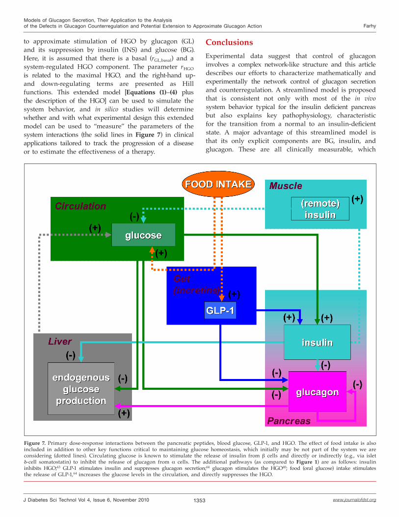
Perspectives: Extension of the Model to Approximate Glucagon ActionIn vivo, the interactions between pulsatile insulin and glucagon are under the regulation not only of BG but of incretins as well and their combined effects exert a network control on the hepatic glucose output (HGO) (Figure 7).
Abnormalities in this larger network underlie the progression of many diseases, including diabetes, metabolic syndrome, and polycystic ovary syndrome. Defects of GCR
Figure 6. Circulating glucagon (open blue diamonds), BG (connected pink squares), predicted circulating (black line), and portal vein (green dotted line) glucagon response to hypoglycemia. The blue dashed line shows the model-predicted circulating GCR response if GABA is not administered. The arrow indicates the insulin bolus given to cause hypoglycemia.
might be also mediated in part by defects in the way the HGO is regulated. Hyperinsulinemia, found in many metabolic disorders, may originate from several individual or a combination of different mechanisms. Therefore, a natural future extension of our models is to approximate the network shown in Figure 7. First, to include GLP-1, one can multiply the right-hand side terms in Equations (1) and (2) by {1 + [GLP(t)/tGLP,1]nGLP,1}-1
and (GLP/tGLP,2)nGLP,2[1 + (GLP/tGLP,2)nGLP,2]-1, respectively, to describe negative regulation of glucagon and stimulation of insulin by GLP-1. The HGO rate of change can be described as
rHGO,basal + rHGO
11 + (BG/tBG,3)
nBG,3
[GL/tGL,2]nBG,2
1 + [GL/tGL,2]nBG,2
11 + [INS/tINS,2]
nINS,2×
1353
Models of Glucagon Secretion, Their Application to the Analysis of the Defects in Glucagon Counterregulation and Potential Extension to Approximate Glucagon Action Farhy
www.journalofdst.orgJ Diabetes Sci Technol Vol 4, Issue 6, November 2010
Figure 7. Primary dose-response interactions between the pancreatic peptides, blood glucose, GLP-1, and HGO. The effect of food intake is also included in addition to other key functions critical to maintaining glucose homeostasis, which initially may be not part of the system we are considering (dotted lines). Circulating glucose is known to stimulate the release of insulin from β cells and directly or indirectly (e.g., via islet δ-cell somatostatin) to inhibit the release of glucagon from α cells. The additional pathways (as compared to Figure 1) are as follows: insulin inhibits HGO;63 GLP-1 stimulates insulin and suppresses glucagon secretion;64 glucagon stimulates the HGO40; food (oral glucose) intake stimulates the release of GLP-1,64 increases the glucose levels in the circulation, and directly suppresses the HGO.
to approximate stimulation of HGO by glucagon (GL) and its suppression by insulin (INS) and glucose (BG). Here, it is assumed that there is a basal (rGL,basal) and a system-regulated HGO component. The parameter rHGO is related to the maximal HGO, and the right-hand up- and down-regulating terms are presented as Hill functions. This extended model [Equations (1)–(4) plus the description of the HGO] can be used to simulate the system behavior, and in silico studies will determine whether and with what experimental design this extended model can be used to “measure” the parameters of the system interactions (the solid lines in Figure 7) in clinical applications tailored to track the progression of a disease or to estimate the effectiveness of a therapy.
ConclusionsExperimental data suggest that control of glucagon involves a complex network-like structure and this article describes our efforts to characterize mathematically and experimentally the network control of glucagon secretion and counterregulation. A streamlined model is proposed that is consistent not only with most of the in vivo system behavior typical for the insulin deficient pancreas but also explains key pathophysiology, characteristic for the transition from a normal to an insulin-deficient state. A major advantage of this streamlined model is that its only explicit components are BG, insulin, and glucagon. These are all clinically measurable, which

1354
Models of Glucagon Secretion, Their Application to the Analysis of the Defects in Glucagon Counterregulation and Potential Extension to Approximate Glucagon Action Farhy
www.journalofdst.orgJ Diabetes Sci Technol Vol 4, Issue 6, November 2010
should allow the application of the new construct to the study of the control, function, and abnormalities of the human glucagon axis. A key model-based prediction is that careful partial suppression of hyperglucagonemia may repair defective GCR in insulin-deficient diabetes. If proven correct, such an outcome will have important clinical implications.
The selected few MCN components cannot recreate (nor should they need to) all signals that control the glucagon axis, which is influenced by various extrapancreatic factors with important impact on glucagon secretion and GCR, including autonomic input, catecholamines, growth hormone, ghrelin, and incretins.13,58,65-69 These other signals are not omitted but are unified in the MCN based on the assumption that the primary physiological relationships that are explicit in the model are influenced by these factors.
The primary application of our models is to simulate system behavior, and the reviewed in silico results suggest that they are consistent with the existing experimental data. New experiments, however, are needed to further validate the model. These should include infusions manipulating the signaling input to the pancreas combined with frequent sampling of the portal vein to better capture the corresponding changes in the pancreatic output.
Finally, our models can be modified so that concentrations of pancreatic peptides can be reconstructed from their circulating levels. This is important in the analysis of clinical data where portal sampling is impossible. They can also be part of a larger construct, showing their regulation of the hepatic glucose output by the pancreas, glucose, and incretins. It is possible that this larger model can be used in clinical applications to track the progression of a disease or of a specific therapy.
Funding:
The study was supported by the National Institutes of Health/National Institute of Diabetes and Digestive and Kidney Diseases Grants R21 DK072095 and R01 DK082805.
References:
1. Cryer PE, Gerich JE. Relevance of glucose counterregulatory systems to patients with diabetes: critical roles of glucagon and epinephrine. Diabetes Care. 1983;6(1):95-9.
2. Gerich JE. Lilly lecture 1988. Glucose counterregulation and its impact on diabetes mellitus. Diabetes. 1988;37(12):1608-17.
3. Cryer PE. Hypoglycemia is the limiting factor in the management of diabetes. Diabetes Metab Res Rev. 1999;15(1):42-6.
4. Cryer PE. Hypoglycemia: the limiting factor in the glycaemic management of type I and type II diabetes. Diabetologia. 2002;45(7):937-48.
5. Farhy LS, Du Z, Zeng Q, Veldhuis PP, Johnson ML, Brayman KL, McCall AL. Amplification of pulsatile glucagon secretion by switch-off of alpha-cell-suppressing signals in streptozotocin-treated rats. Am J Physiol Endocrinol Metab. 2008;295(3):E575-85.
6. Farhy LS, McCall AL. System-level control to optimize glucagon counterregulation by switch-off of alpha-cell suppressing signals in beta-cell deficiency. J Diabetes Sci Technol. 2009;3(1):21-33.
7. Farhy LS, McCall AL. Pancreatic network control of glucagon secretion and counterregulation. Methods Enzymol. 2009;467:547-81.
8. Zhou H, Tran PO, Yang S, Zhang T, LeRoy E, Oseid E, Robertson RP. Regulation of alpha-cell function by the beta-cell during hypoglycemia in Wistar rats: the “switch-off” hypothesis. Diabetes. 2004;53(6):1482-7.
9. Samols E, Stagner JI. Intra-islet regulation. Am J Med. 1988;85(5A):31-5.
10. Samols E, Stagner JI. Islet somatostatin--microvascular, paracrine, and pulsatile regulation. Metabolism. 1990;39(9 Suppl 2):55-60.
11. Stagner JI, Samols E, Bonner-Weir S. beta-alpha-delta pancreatic islet cellular perfusion in dogs. Diabetes. 1988;37(12):1715-21.
12. Stagner JI, Samols E, Marks V. The anterograde and retrograde infusion of glucagon antibodies suggests that A cells are vascularly perfused before D cells within the rat islet. Diabetologia. 1989;32(3):203-6.
13. Gromada J, Franklin I, Wollheim CB. Alpha-cells of the endocrine pancreas: 35 years of research but the enigma remains. Endocr Rev. 2007;28(1):84-116.
14. Gedulin BR, Rink TJ, Young AA. Dose-response for glucagonostatic effect of amylin in rats. Metabolism. 1997;46(1):67-70.
15. Ito K, Maruyama H, Hirose H, Kido K, Koyama K, Kataoka K, Saruta T. Exogenous insulin dose-dependently suppresses glucopenia-induced glucagon secretion from perfused rat pancreas. Metabolism. 1995;44(3):358-62.
16. Rorsman P, Hellman B. Voltage-activated currents in guinea pig pancreatic alpha 2 cells. Evidence for Ca2+-dependent action potentials. J Gen Physiol. 1988;91(2):223-42.
17. Rorsman P, Berggren PO, Bokvist K, Ericson H, Mohler H, Ostenson CG, Smith PA. Glucose-inhibition of glucagon secretion involves activation of GABAA-receptor chloride channels. Nature. 1989;341(6239):233-6.
18. Wendt A, Birnir B, Buschard K, Gromada J, Salehi A, Sewing S, Rorsman P, Braun M. Glucose inhibition of glucagon secretion from rat alpha-cells is mediated by GABA released from neighboring beta-cells. Diabetes. 2004;53(4):1038-45.
19. Xu E, Kumar M, Zhang Y, Ju W, Obata T, Zhang N, Liu S, Wendt A, Deng S, Ebina Y, Wheeler MB, Braun M, Wang Q. Intraislet insulin suppresses glucagon release via GABA-GABAA receptor system. Cell Metab. 2006;3(1):47-58.
20. Ishihara H, Maechler P, Gjinovci A, Herrera PL, Wollheim CB. Islet beta-cell secretion determines glucagon release from neighboring α-cells. Nat Cell Biol. 2003;5(4):330-5.
1355
Models of Glucagon Secretion, Their Application to the Analysis of the Defects in Glucagon Counterregulation and Potential Extension to Approximate Glucagon Action Farhy
www.journalofdst.orgJ Diabetes Sci Technol Vol 4, Issue 6, November 2010
21. Maruyama H, Hisatomi A, Orci L, Grodsky GM, Unger RH. Insulin within islets is a physiologic glucagon release inhibitor. J Clin Invest. 1984;74(6):2296-9.
22. Brunicardi FC, Kleinman R, Moldovan S, Nguyen TH, Watt PC, Walsh J, Gingerich R. Immunoneutralization of somatostatin, insulin, and glucagon causes alterations in islet cell secretion in the isolated per fused human pancreas. Pancreas. 2001;23(3):302-8.
23. Cejvan K, Coy DH, Efendic S. Intra-islet somatostatin regulates glucagon release via type 2 somatostatin receptors in rats. Diabetes. 2003;52(5):1176-81.
24. Klaff LJ, Taborsky GJ Jr. Pancreatic somatostatin is a mediator of glucagon inhibition by hyperglycemia. Diabetes. 1987;36(5):592-6.
25. Ludvigsen E, Olsson R, Stridsberg M, Janson ET, Sandler S. Expression and distribution of somatostatin receptor subtypes in the pancreatic islets of mice and rats. J Histochem Cytochem. 2004;52(3):391-400.
26. Sumida Y, Shima T, Shirayama K, Misaki M, Miyaji K. Effects of hexoses and their derivatives on glucagon secretion from isolated perfused rat pancreas. Hormon Metab Res. 1994;26(5):222-5.
27. Tirone TA, Norman MA, Moldovan S, DeMayo FJ, Wang XP, Brunicardi FC. Pancreatic somatostatin inhibits insulin secretion via SSTR-5 in the isolated perfused mouse pancreas model. Pancreas. 2003;26(3):e67-73.
28. Gopel SO, Kanno T, Barg S, Rorsman P. Patch-clamp characteri-sation of somatostatin-secreting -cells in intact mouse pancreatic islets. J Physiol. 2000;528(Pt 3):497-507.
29. Brunicardi FC, Atiya A, Moldovan S, Lee TC, Fagan SP, Kleinman RM, Adrian TE, Coy DH, Walsh JH, Fisher WE. Activation of somatostatin receptor subtype 2 inhibits insulin secretion in the isolated perfused human pancreas. Pancreas. 2003;27(4):e84-9.
30. Kleinman R, Gingerich R, Wong H, Walsh J, Lloyd K, Ohning G, De Giorgio R, Sternini C, Brunicardi FC. Use of the Fab fragment for immunoneutralization of somatostatin in the isolated perfused human pancreas. Am J Surg. 1994;167(1):114-9.
31. Portela-Gomes GM, Stridsberg M, Grimelius L, Oberg K, Janson ET. Expression of the five different somatostatin receptor subtypes in endocrine cells of the pancreas. Appl Immunohistochem Mol Morphol. 2000;8(2):126-32.
32. Strowski MZ, Parmar RM, Blake AD, Schaeffer JM. Somatostatin inhibits insulin and glucagon secretion via two receptors subtypes: an in vitro study of pancreatic islets from somatostatin receptor 2 knockout mice. Endocrinology. 2000;141(1):111-7.
33. Schuit FC, Derde MP, Pipeleers DG. Sensitivity of rat pancreatic A and B cells to somatostatin. Diabetologia. 1989;32(3):207-12.
34. Kleinman R, Gingerich R, Ohning G, Wong H, Olthoff K, Walsh J, Brunicardi FC. The influence of somatostatin on glucagon and pancreatic polypeptide secretion in the isolated perfused human pancreas. Int J Pancreatol. 1995;18(1):51-7.
35. Kieffer TJ, Heller RS, Unson CG, Weir GC, Habener JF. Distribution of glucagon receptors on hormone-specific endocrine cells of rat pancreatic islets. Endocrinology. 1996;137(11):5119-25.
36. Epstein S, Berelowitz M, Bell NH. Pentagastrin and glucagon stimulate serum somatostatin-like immunoreactivity in man. J Clin Endocrinol Metab. 1980;51(6):1227-31.
37. Utsumi M, Makimura H, Ishihara K, Morita S, Baba S. Determination of immunoreactive somatostatin in rat plasma and responses to arginine, glucose and glucagon infusion. Diabetologia. 1979;17(5):319-23.
38. Tapia-Arancibia L, Astier H. Glutamate stimulates somatostatin release from diencephalic neurons in primary culture. Endocrinology. 1988;123(5):2360-6.
39. Bell GI, Pilkis SJ, Weber IT, Polonsky KS. Glucokinase mutations, insulin secretion, and diabetes mellitus. Annu Rev Physiol. 1996;58:171-86.
40. Schuit FC, Huypens P, Heimberg H, Pipeleers DG. Glucose sensing in pancreatic beta-cells: a model for the study of other glucose-regulated cells in gut, pancreas, and hypothalamus. Diabetes. 2001;50(1):1-11.
41. Ashcroft FM, Proks P, Smith PA, Ammala C, Bokvist K, Rorsman P. Stimulus-secretion coupling in pancreatic beta cells. J Cell Biochem. 1994;55(Suppl):54-65.
42. Dunne MJ, Harding EA, Jaggar JH, Squires PE. Ion channels and the molecular control of insulin secretion. Biochem Soc Trans. 1994;22(1):6-12.
43. Fujitani S, Ikenoue T, Akiyoshi M, Maki T, Yada T. Somatostatin and insulin secretion due to common mechanisms by a new hypoglycemic agent, A-4166, in perfused rat pancreas. Metabolism. 1996;45(2):184-9.
44. Hermansen K, Christensen SE, Orskov H. Characterization of somatostatin release from the pancreas: the role of potassium. Scand J Clin Lab Invest. 1979;39(8):717-22.
45. Efendić S, Nylén A, Roovete A, Uvnäs-Wallenstein K. Effects of glucose and arginine on the release of immunoreactive somatostatin from the isolated perfused rat pancreas. FEBS Lett. 1978;92(1):33-5.
46. Heimberg H, De Vos A, Pipeleers D, Thorens B, Schuit F. Differences in glucose transporter gene expression between rat pancreatic alpha- and beta-cells are correlated to differences in glucose transport but not in glucose utilization. J Biol Chem. 1995;270(15):8971-5.
47. Heimberg H, De Vos A, Moens K, Quartier E, Bouwens L, Pipeleers D, Van Schaftingen E, Madsen O, Schuit F. The glucose sensor protein glucokinase is expressed in glucagon-producing alpha-cells. Proc Natl Acad Sci USA. 1996;93(14):7036-41.
48. Reaven GM, Chen YD, Golay A, Swislocki AL, Jaspan JB. Documentation of hyperglucagonemia throughout the day in nonobese and obese patients with noninsulin-dependent diabetes mellitus. J Clin Endocrinol Metab. 1987;64(1):106-10.
49. Schuit F, De Vos A, Farfari S, Moens K, Pipeleers D, Brun T, Prentki M. Metabolic fate of glucose in purified islet cells. Glucose-regulated anaplerosis in beta cells. J Biol Chem. 1997;272(30):18572-9.
50. Gopel SO, Kanno T, Barg S, Weng XG, Gromada J, Rorsman P. Regulation of glucagon release in mouse -cells by KATP channels and inactivation of TTX-sensitive Na+ channels. J Physiol. 2000;528(Pt 3):509-20.
51. Unger RH. Glucagon physiology and pathophysiology in the light of new advances. Diabetologia. 1985;28(8):574-8.
52. Genter P, Berman N, Jacob M, Ipp E. Counterregulatory hormones oscillate during steady-state hypoglycemia. Am J Physiol. 1998;275(5 Pt 1):E821-9.
53. Grapengiesser E, Salehi A, Quader SS, Hellman B. Glucose induces glucagon release pulses antisynchronous with insulin and sensitive to purinoceptor inhibition. Endocrinology. 2006;147(7):3472-7.
54. Grimmichová T, Vrbíková J, Matucha P, Vondra K, Veldhuis PP, Johnson ML. Fasting insulin pulsatile secretion in lean women with polycystic ovary syndrome. Physiol Res. 2008;57(Suppl 1):S91-8.
55. Jaspan JB, Lever E, Polonsky KS, Van Cauter E. In vivo pulsatility of pancreatic islet peptides. Am J Physiol. 1986;251(2 Pt 1):E215-26.
56. Matthews DR, Hermansen K, Connolly AA, Gray D, Schmitz O, Clark A, Orskov H, Turner RC. Greater in vivo than in vitro pulsatility of insulin secretion with synchronized insulin and somatostatin secretory pulses. Endocrinology. 1987;120(6):2272-8.
1356
Models of Glucagon Secretion, Their Application to the Analysis of the Defects in Glucagon Counterregulation and Potential Extension to Approximate Glucagon Action Farhy
www.journalofdst.orgJ Diabetes Sci Technol Vol 4, Issue 6, November 2010
57. Salehi A, Quader SS, Grapengiesser E, Hellman B. Pulses of somatostatin release are slightly delayed compared with insulin and antisynchronous to glucagon. Regul Pept. 2007;144(1-3):43-9.
58. Heise T, Heinemann L, Heller S, Weyer C, Wang Y, Strobel S, Kolterman O, Maggs D. Effect of pramlintide on symptom, catecholamine, and glucagon responses to hypoglycemia in healthy subjects. Metabolism. 2004;53(9):1227-32.
59. Nauck MA, Heimesaat MM, Behle K, Holst JJ, Nauck MS, Ritzel R, Hüfner M, Schmiegel WH. Effects of glucagon-like peptide 1 on counterregulatory hormone responses, cognitive functions, and insulin secretion during hyperinsulinemic, stepped hypoglycemic clamp experiments in healthy volunteers. J Clin Endocrinol Metab. 2002;87(3):1239-46.
60. Degn KB, Brock B, Juhl CB, Djurhuus CB, Grubert J, Kim D, Han J, Taylor K, Fineman M, Schmitz O. Effect of intravenous infusion of exenatide (synthetic exendin-4) on glucose-dependent insulin secretion and counterregulation during hypoglycemia. Diabetes. 2004;53(9):2397-403.
61. Ahrén B, Schweizer A, Dejager S, Dunning BE, Nilsson PM, Persson M, Foley JE. Vildagliptin enhances islet responsiveness to both hyper- and hypoglycemia in patients with type 2 diabetes. J Clin Endocrinol Metab. 2009;94(4):1236-43.
62. Tura A, Ludvik B, Nolan JJ, Pacini G, Thomaseth K. Insulin and C-peptide secretion and kinetics in humans: direct and model-based measurements during OGTT. Am J Physiol Endocrinol Metab. 2001;281(5):E966-74.
63. Girard J. The inhibitory effects of insulin on hepatic glucose production are both direct and indirect. Diabetes. 2006;55(Suppl 2):S65-9.
64. Schirra J, Göke B. The physiological role of GLP-1 in human: incretin, ileal brake or more? Regul Pept. 2005;128(2):109-15.
65. Havel PJ, Ahren B. Activation of autonomic nerves and the adrenal medulla contributes to increased glucagon secretion during moderate insulin-induced hypoglycemia in women. Diabetes. 1997;46(5):801-7.
66. Havel PJ, Taborsky GJ Jr. The contribution of the autonomic nervous system to changes of glucagon and insulin secretion during hypoglycemic stress. Endocr Rev. 1989;10(3):332-50.
67. Brelje TC, Scharp DW, Sorenson RL. Three-dimensional imaging of intact isolated islets of Langerhans with confocal microscopy. Diabetes. 1989;38(6):808-14.
68. Bolli GB, Fanelli CG. Physiology of glucose counterregulation to hypoglycemia. Endocrinol Metab Clin North Am. 1999;28(3):467-93, v.
69. Taborsky GJ Jr, Ahren B, Havel PJ. Autonomic mediation of glucagon secretion during hypoglycemia: implications for impaired alpha-cell responses in type 1 diabetes. Diabetes. 1998;47(7):995-1005.