Embed Size (px)
Citation preview

Moderator : Dr. Mammohan Singh
Dr. G.D. Satyarthee
Presenter : Dr. Anand Gupta
Dr. Mammohan Singh
Dr. G.D. Satyarthee
Dr. Anand Gupta

Primary brain tumor – 6 persons/100000/year
Metastatic brain tumor – 6 persons/100000/year
1 in 15 primary brain tumors occur in children under 15 years
Introduction
In adults, the
commonest tumors
are gliomas,
metastases and
meniongiomas; most
lie in the
supratentorial
compartment
6 persons/100000/year
6 persons/100000/year
1 in 15 primary brain tumors occur in children under 15 years
Introduction

Intra-axial Post Fossa Tumors
Adult
� Metastasis 16%
� Hemangioblastoma 7-12%
� Pilocytic astrocytoma (2nd
decade)decade)
� Brain stem glioma (1% of adult tumor)
� Choroid plexus tumor (<1% of primary brain tumor)
� Cerebellar liponeurocytoma
axial Post Fossa Tumors
Paediatric
• PNET (including medulloblastoma) 27%
• Cerebellar (Pilocyticastrocytoma) 27%astrocytoma) 27%
• Brain stem glioma 27%
• Ependymoma 15%
• Choroid plexus papilloma (<1% of primary brain tumor)
• Dermoid cyst (<0.5% of primary intraaxial tumor)
• Atypical teratoid/ rhabdoidtumor

Extra-axial Lesion
� Vestibular schwannoma
� Meningioma
� Epidermoid� Epidermoid
� Metastases
� Trigeminal neuroma
� Facial nerve neuroma
� Arachnoid cyst
axial Lesion
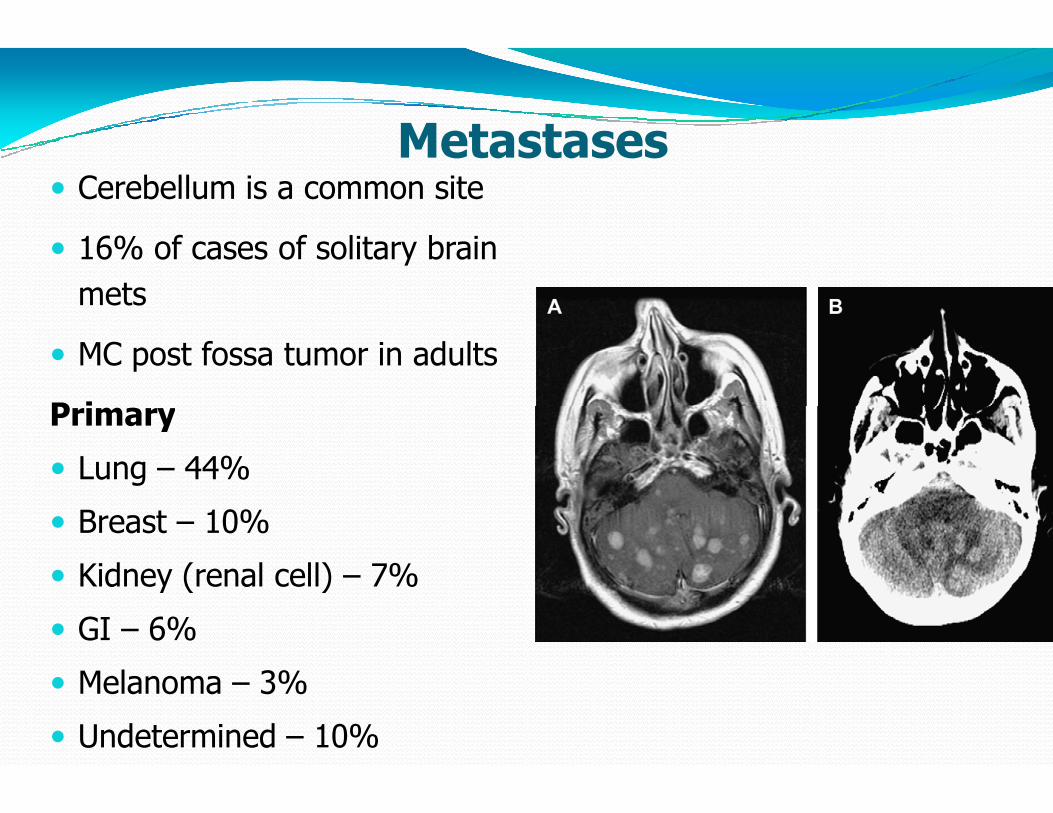
Metastases� Cerebellum is a common site
� 16% of cases of solitary brain
mets
� MC post fossa tumor in adults
PrimaryPrimary
� Lung – 44%
� Breast – 10%
� Kidney (renal cell) – 7%
� GI – 6%
� Melanoma – 3%
� Undetermined – 10%
Metastases

Pathology� Rounded solid partially cystic mass Age
� Rare in children, most common in older adults
(> 40 years)
LocationLocation
� Anywhere: grey white junction most common site
Imaging
� NECT: Iso / hyperdense; Ca++ rare in untreated metastases
� CECT: Strong solid/ring enhancement
� MR: Most hypointense on T1, hyperintense on T
enhance moderately intensely following contrast administration
Rounded solid partially cystic mass ± edema
Rare in children, most common in older adults
Anywhere: grey white junction most common site
rare in untreated metastases
CECT: Strong solid/ring enhancement
, hyperintense on T2W1, most
enhance moderately intensely following contrast administration

Management
� Mostly palliative
� Median survival of patient 26
MedicalMedical
� Corticosteroids
� Anticonvulsants.
Management
Median survival of patient 26-32 weeks

Surgical Management
Solitary lesion
Surgical excision of solitary lesion:
� Primary disease quiescent or radioresistant
� Lesion accessible, symptomatic or life threatening
� For recurrent small cell lung carcinoma following XRT
� Diagnosis unknown
Surgical Management
Surgical excision of solitary lesion:
Primary disease quiescent or radioresistant
Lesion accessible, symptomatic or life threatening
For recurrent small cell lung carcinoma following XRT

Multiple Lesions
� Worse prognosis than solitary lesion
� Usually treated with XRT without surgery
Situations where surgery is done:
� One particular and accessible lesion symptomatic � One particular and accessible lesion symptomatic and/or life threatening
� Multiple lesions that can all be completely removed
Stereotactic Biopsy
� Lesions not appropriate for surgery
� Not candidates for surgical resection
� To ascertain a diagnosis
Multiple Lesions
Worse prognosis than solitary lesion
Usually treated with XRT without surgery
Situations where surgery is done:
One particular and accessible lesion symptomatic One particular and accessible lesion symptomatic
Multiple lesions that can all be completely removed
Lesions not appropriate for surgery
Not candidates for surgical resection

Stereotactic Radiosurgery
� No mass effect, no hydrocephalus
� Advantage: No risk of hemorrhage, infection or
mechanical spread of tumor cells, Can be used for 3 or mechanical spread of tumor cells, Can be used for 3 or
fewer mets
� Disadvantage: Histological proof not obtained, Cannot be
used for lesion > 3 cm
Stereotactic Radiosurgery
No mass effect, no hydrocephalus
No risk of hemorrhage, infection or
mechanical spread of tumor cells, Can be used for 3 or mechanical spread of tumor cells, Can be used for 3 or
Histological proof not obtained, Cannot be

Median survival following craniotomy
Lung
Breast
Colon
KidneyKidney
Melanoma
Miscellaneous
Sarcoma
Urologic (testis, Bladder, Prostate)
Unknown
Esophagus
Median survival even with best treatment is only
Median survival following craniotomy
Month
11
11
8
1212
6.5
11
6
10
10
4
Median survival even with best treatment is only – 8 months

Hemangioblastoma (HGB)
� Most common primary intra
in adults (7-12% of post fossa tumors)
� Highly vascular well circumscribed solid or cystic � Highly vascular well circumscribed solid or cystic
neoplasm of CNS or retina
� May occur sporadically (4th
Hippel Lindau disease (3rd decade)
� 30% of patients with cerebellar HGB have VHL
Hemangioblastoma (HGB)
Most common primary intra-axial posterior fossa tumor
12% of post fossa tumors)
Highly vascular well circumscribed solid or cystic Highly vascular well circumscribed solid or cystic
Decade) or as part of Von
decade)
30% of patients with cerebellar HGB have VHL

Pathology
� 60% cystic with nodule – 40% solid
� Gross hemorrhage, calcification necrosis rare
Age
� Adults with peak during 40 to 60 years, rare in children� Adults with peak during 40 to 60 years, rare in children
Location
� 80% to 85% cerebellum
� 3% to 13% spinal cord
� 2% to 3% Medulla
Supratentorial lesions occur but are uncommon
� 60% of patients with VHL have retinal lesions
Pathology
40% solid
Gross hemorrhage, calcification necrosis rare
Age
Adults with peak during 40 to 60 years, rare in childrenAdults with peak during 40 to 60 years, rare in children
Location
Supratentorial lesions occur but are uncommon
60% of patients with VHL have retinal lesions

Imaging
� Vertebral Angiography: Vascular nodule with intense, prolonged stain ± avascular cyst
� CT: Low density cyst with strongly enhancing mural nodule that abuts a pial surface
MR: Cyst slightly hyperintense to CSF on T� MR: Cyst slightly hyperintense to CSF on Thyperintense to brain on T2Wenhances strongly
Labs
• Polycythemia • Catecholamine production from pheochromocytoma
Imaging
Vertebral Angiography: Vascular nodule with intense, avascular cyst
CT: Low density cyst with strongly enhancing mural nodule
MR: Cyst slightly hyperintense to CSF on T W ; MR: Cyst slightly hyperintense to CSF on T1W1; W1; mural nodule variable but
Labs
Catecholamine production from pheochromocytoma

Axial CECT scan showing a solid cerebellar
hemangioblastoma with a uniformly enhancing
mass (arrows) without associated cyst
Axial CECT scan showing a solid cerebellar
hemangioblastoma with a uniformly enhancing
mass (arrows) without associated cyst

Typical angiographic finding of a cystic cerebellar
hemangioblastoma
Typical angiographic finding of a cystic cerebellar
hemangioblastoma

Classic MR findings of cystic cerebellar hemangioblastomaClassic MR findings of cystic cerebellar hemangioblastoma

Treatment
� May be curative in cases of HGB, not in VHL
� Preop embolisation reduces the vascularity
� Cystic hemagloblastoma require removal of mural nodule.
Stereotactic Radiosurgery
� For asymptomatic HGB > 5 mm diameter if they are cystic
or progressing in size during surveillance
Treatment
May be curative in cases of HGB, not in VHL
Preop embolisation reduces the vascularity
Cystic hemagloblastoma require removal of mural nodule.
Radiosurgery
For asymptomatic HGB > 5 mm diameter if they are cystic
or progressing in size during surveillance

Radiation Treatment
� Effectiveness dubious
� May be useful to reduce tumor size or to retard growth in
patients who are not surgical candidates for multiple
brainstem HGBbrainstem HGB
Chemotherapy
� Ongoing phase II trial with Sunitnib, an inhibitor of
vascular endothelial growth factor and platelet derived
growth factor
Radiation Treatment
May be useful to reduce tumor size or to retard growth in
patients who are not surgical candidates for multiple
Chemotherapy
Ongoing phase II trial with Sunitnib, an inhibitor of
vascular endothelial growth factor and platelet derived

Origin of cells (WHO- PNET)
Static- external granular layer
Origin from remnant of cells of the external
granular layer of the cerebellum.
MEDULLOBLASTOMAMEDULLOBLASTOMA
granular layer of the cerebellum.
Dynamic – neural progenitor cells
Transformation of normal undifferentiated
progenitor cells of superior medullary velum
which migrate to the fourth ventricle
PNET)
external granular layer
Origin from remnant of cells of the external
granular layer of the cerebellum.
MEDULLOBLASTOMAMEDULLOBLASTOMA
granular layer of the cerebellum.
neural progenitor cells
Transformation of normal undifferentiated
progenitor cells of superior medullary velum
which migrate to the fourth ventricle

Histology
� Medulloblastoma (Grade 4)
� Desmoplastic/nodular medulloblastoma
Medulloblastoma with extensive nodularity
MEDULLOBLASTOMAMEDULLOBLASTOMA
� Medulloblastoma with extensive nodularity
� Anaplastic medulloblastoma
� Large cell medulloblastoma
Desmoplastic/nodular medulloblastoma
Medulloblastoma with extensive nodularity
MEDULLOBLASTOMAMEDULLOBLASTOMA
Medulloblastoma with extensive nodularity
Anaplastic medulloblastoma
Large cell medulloblastoma

MEDULLOBLASTOMAMEDULLOBLASTOMA
� Histology
Cellular, small cells, scant cytoplasm, Homer
Immuno histochemistryImmuno histochemistry
GFAP +
EMA –
MEDULLOBLASTOMAMEDULLOBLASTOMA
Cellular, small cells, scant cytoplasm, Homer-Wright rosettes

CLINICAL FEATURES
HYDROCEPHALUS : RAISED ICP
� BEHAVIORAL CHANGE, LISTLESSNESS, IRRITABILITY, VOMITING, AND
DECREASED SOCIAL INTERACTIONS.
MEDULLOBLASTOMAMEDULLOBLASTOMA
DECREASED SOCIAL INTERACTIONS.
� HEADACHE
� DOUBLE VISION.
� HEAD TILT : TONSILLAR HERNIATIONMAGNUM
� CEREBELLAR SYMPTOMS
� BRAIN STEM INVOLVEMENT
� LEPTOMENINGEAL DISSEMINATION
BEHAVIORAL CHANGE, LISTLESSNESS, IRRITABILITY,
DECREASED SOCIAL INTERACTIONS.
MEDULLOBLASTOMAMEDULLOBLASTOMA
DECREASED SOCIAL INTERACTIONS.
HERNIATION BELOW THE FORAMEN
DISSEMINATION

Examination
• Increasing head circumference , full anterior fontanelle with widely split cranial sutures.
• Papilledema 90% of patients
MEDULLOBLASTOMAMEDULLOBLASTOMA
• Diplopia and lateral gaze paresis
• Fourth cranial nerve palsy ( should be considered in any patient with a head tilt )
• Nystagmus
• Cerebellar signs ( ataxia > unilateral dysmetria )
Increasing head circumference , full anterior fontanelle with
MEDULLOBLASTOMAMEDULLOBLASTOMA
Diplopia and lateral gaze paresis
Fourth cranial nerve palsy ( should be considered in any patient
Cerebellar signs ( ataxia > unilateral dysmetria )

Imaging
CT HEAD
MEDULLOBLASTOMAMEDULLOBLASTOMAMEDULLOBLASTOMAMEDULLOBLASTOMA

� MRI- T1- low to isointense T2-
� Homogenous contrast enhancement
(may be absent in about 15 –20 % )
� DWI shows restricted diffusion with increased ADC.
MEDULLOBLASTOMAMEDULLOBLASTOMA
� DWI shows restricted diffusion with increased ADC.
Spinal imaging –
� At diagnosis (11-71% show dissemination)
� Within 24 hrs after surgery or
� Surveillance imaging at 3-6 months
hyperintense
Homogenous contrast enhancement
20 % )
DWI shows restricted diffusion with increased ADC.
MEDULLOBLASTOMAMEDULLOBLASTOMA
DWI shows restricted diffusion with increased ADC.
71% show dissemination)
after surgery or 2 weeks post surgery
6 months

MEDULLOBLASTOMAMEDULLOBLASTOMA
T1 Post Gd DWI
MEDULLOBLASTOMAMEDULLOBLASTOMA
DWI ADC

MEDULLOBLASTOMAMEDULLOBLASTOMA
Leptomeningeal Dissemination
MEDULLOBLASTOMAMEDULLOBLASTOMA
Leptomeningeal Dissemination
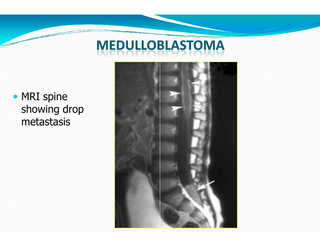
� MRI spine showing drop metastasis
MEDULLOBLASTOMAMEDULLOBLASTOMA
metastasis
MEDULLOBLASTOMAMEDULLOBLASTOMA

� Management
Steroids
CSF cytology- LP, EVD, Cisternamagna
CSF diversion
MEDULLOBLASTOMAMEDULLOBLASTOMA
CSF diversion
Definitive surgery
Adjuvant therapy
LP, EVD, Cisternamagna
MEDULLOBLASTOMAMEDULLOBLASTOMA

CHANG CLASSIFICATION
MEDULLOBLASTOMAMEDULLOBLASTOMA
CHANG CLASSIFICATION
MEDULLOBLASTOMAMEDULLOBLASTOMA
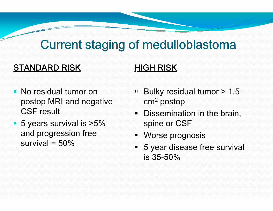
Current staging of medulloblastomaCurrent staging of medulloblastomaCurrent staging of medulloblastomaCurrent staging of medulloblastomaSTANDARD RISKSTANDARD RISKSTANDARD RISKSTANDARD RISK
� No residual tumor on postop MRI and negative CSF result
HIGH RISK HIGH RISK HIGH RISK HIGH RISK
�
postop MRI and negative CSF result
� 5 years survival is >5% and progression free survival = 50%
�
�
�
Current staging of medulloblastomaCurrent staging of medulloblastomaCurrent staging of medulloblastomaCurrent staging of medulloblastomaHIGH RISK HIGH RISK HIGH RISK HIGH RISK
� Bulky residual tumor > 1.5 cm2 postopcm postop
� Dissemination in the brain, spine or CSF
� Worse prognosis� 5 year disease free survival
is 35-50%

�
Presenation : MRI Brain and spine
Surgical resection
Management of hydrocephalus
> 3 years
Standard risk Poor risk
Craniospinal radiation
OR Reduced dose radiation with
CT on reasarch protocol
Cranispinal radiation + adjunct CT
( CCNU, cisplatin vincristine
or CT on research protocol
Management algorithm for medulloblastoma
Presenation : MRI Brain and spine
Surgical resection
Management of hydrocephalus
< 3 years
Cranispinal radiation + adjunct CT
( CCNU, cisplatin vincristine
or CT on research protocol
Chemotherapy (No standard regimen)
Follow OR
Delayed RT till 3 years old
Management algorithm for medulloblastoma

Management…….. � Gross Total Resection, if possible (arises from roof of fourth
ventricle- soft reddish vascular with some times sugar
coating).
� Brainstem damage should be avoided.
� Resolution of natural CSF pathways.� SURGERY alone : NOT CURATIVE
� RADIOTHERAPY : Cornerstone of adjuvant therapy.
� 54 to 58 Gy - primary site � 35Gy - craniospinal axis
.. SurgerySurgerySurgerySurgeryGross Total Resection, if possible (arises from roof of fourth
soft reddish vascular with some times sugar
Brainstem damage should be avoided.
Resolution of natural CSF pathways.NOT CURATIVE
RADIOTHERAPY : Cornerstone of adjuvant therapy.

ManagementManagementManagementManagement…….. Recurrent Medulloblastoma.. Recurrent Medulloblastoma.. Recurrent Medulloblastoma.. Recurrent Medulloblastoma
� Chemotherapy : limited due to chemo resistance in those
patients who have previously undergone CT
� Redosing with RT avoided due to radiation necrosis� Redosing with RT avoided due to radiation necrosis
� High-dose chemotherapy with
autologous BMR: subject of intense investigation
Prognosis
• 5 - year recurrence-free survival rates : 55%
• Most common site : PRIMARY TUMOR SITE
.. Recurrent Medulloblastoma.. Recurrent Medulloblastoma.. Recurrent Medulloblastoma.. Recurrent Medulloblastoma
Chemotherapy : limited due to chemo resistance in those
patients who have previously undergone CT
with RT avoided due to radiation necrosiswith RT avoided due to radiation necrosis
dose chemotherapy with autologous SCR or
BMR: subject of intense investigation
free survival rates : 55% - 67%.
PRIMARY TUMOR SITE

Ependymoma
� 10% of brain tumors in children
� Peak age - 0-4yrs
� Male preponderance
� Children 90% in cranium
� Adults in spinal
10% of brain tumors in children

EPENDYMOMA� MYXOPAPILLARY (WHO Grade 1)
� SUBEPENDYMOMA (WHO Grade 1)
� Ependymoma (WHO Grade 2)
Cellular� Cellular
� Papillary
� Clear cell
� Tanycytic
� Anaplastic ependymoma (WHO Grade 3)
MYXOPAPILLARY (WHO Grade 1)
SUBEPENDYMOMA (WHO Grade 1)
Ependymoma (WHO Grade 2)
Anaplastic ependymoma (WHO Grade 3)

Ependymoma ……
CT : Typically isodense with heterogenous enhancement
Calcification : common ( can be seen in one half ofcases)
…….. Imaging

Ependymoma…..MRI• On MRI, heterogeneous secondary to necrosis,
hemorrhage and calcification.
• Heterogenous contrast enhancement• Heterogenous contrast enhancement
• Plasticity
• Extension to the cerebellopontine
ependymomas
..MRIOn MRI, heterogeneous secondary to necrosis,
hemorrhage and calcification.
Heterogenous contrast enhancementHeterogenous contrast enhancement
cerebellopontine angle is characteristic of

EPENDYMOMAEPENDYMOMAEPENDYMOMAEPENDYMOMAEPENDYMOMAEPENDYMOMAEPENDYMOMAEPENDYMOMA…..MRI..MRI..MRI..MRI

EPENDYMOMAEPENDYMOMAEPENDYMOMAEPENDYMOMA…..MRI..MRI..MRI..MRI..MRI..MRI..MRI..MRI

Ependymoma…..� INTRA OPINTRA OPINTRA OPINTRA OP---- Tumor arises from the floor and is
lobulated gritty and firmlobulated gritty and firm
� Staging: Staging: Staging: Staging: No conventional staging criteria.
� Postoperative MRI is recommended within 48 hours
..Tumor arises from the floor and is greyish
No conventional staging criteria.
Postoperative MRI is recommended within 48 hours

EpendymomaEpendymomaEpendymomaEpendymoma…Role of RadiotherapyRole of RadiotherapyRole of RadiotherapyRole of Radiotherapy� Post-operative radiation recommended for patients
older than 3 years.
� Stereotactic radiosurgery : Therapeutic option in patients with residual, unresectablepatients with residual, unresectable
Role of Chemotherapy
� May be useful < 3 years : Delay cranial radiation
� Childhood intracranial ependymomaschemo-resistant
Role of RadiotherapyRole of RadiotherapyRole of RadiotherapyRole of Radiotherapyoperative radiation recommended for patients
: Therapeutic option in unresectable or recurrent tumorunresectable or recurrent tumor
May be useful < 3 years : Delay cranial radiation
ependymomas : in general

AIIMS Protocol
•
Low Grade
CSF -VE
Surgery
Radiotherapy
56Gy / 28# / 5.5 wks
(50 Gy followed by a boost of 6 Gy)
AIIMS Protocol
High grade
CSF + VE
Surgery
Surgery followed by
CSI and 6 cycles
chemotherapy.
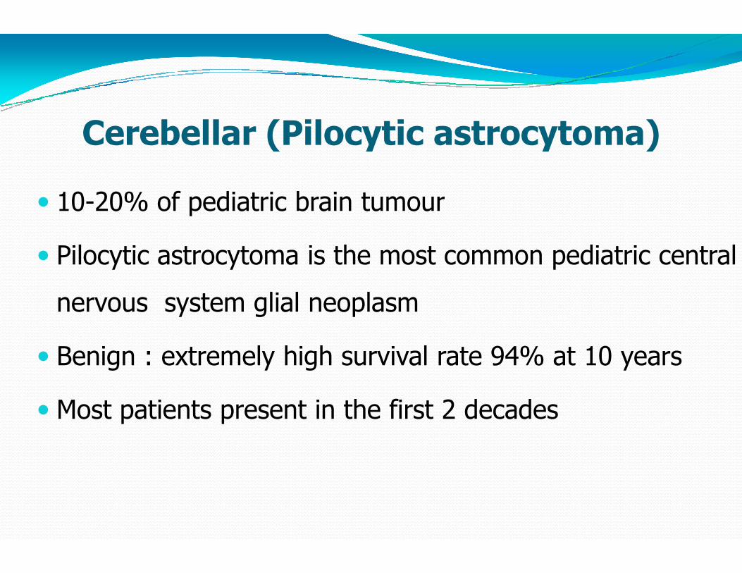
Cerebellar (Pilocytic astrocytoma)
� 10-20% of pediatric brain tumour
� Pilocytic astrocytoma is the most common pediatric central
nervous system glial neoplasmnervous system glial neoplasm
� Benign : extremely high survival rate 94% at 10 years
� Most patients present in the first 2 decades
Cerebellar (Pilocytic astrocytoma)
tumour
is the most common pediatric central
neoplasmneoplasm
Benign : extremely high survival rate 94% at 10 years
Most patients present in the first 2 decades

Pilocytic astrocytoma….NCCT + CECTPilocytic astrocytoma….NCCT + CECT

Pilocytic astrocytomaFour predominant imaging patterns :
Mass with a nonenhancing cyst and an intensely enhancing
mural nodule (21%)
Mass with an enhancing cyst wall and an intensely enhancing
mural nodule (46%)
Necrotic mass with a central nonenhancing
Predominantly solid mass with minimal to no cyst like
component (17%)
Pilocytic astrocytoma….MRI
cyst and an intensely enhancing
Mass with an enhancing cyst wall and an intensely enhancing
nonenhancing zone (16%), and
Predominantly solid mass with minimal to no cyst like

Pilocytic astrocytoma….MRIPilocytic astrocytoma….MRIPilocytic astrocytoma….MRIPilocytic astrocytoma….MRIPilocytic astrocytoma….MRIPilocytic astrocytoma….MRIPilocytic astrocytoma….MRIPilocytic astrocytoma….MRI

PilocyticPilocyticPilocyticPilocytic astrocytomaastrocytomaastrocytomaastrocytoma� Surgical resection of cerebellar pilocytic astrocytomas
is considered the treatment of choice
� Resection of mural nodule –� Resection of mural nodule –
� Resection of cyst wall – controversial ??
� Radiation therapy is strictly avoided, given its risk of
causing significant morbidity in children younger than
5 years of age
astrocytomaastrocytomaastrocytomaastrocytoma…....Surgical resection of cerebellar pilocytic astrocytomas
is considered the treatment of choice
– key surgical objective– key surgical objective
controversial ??
Radiation therapy is strictly avoided, given its risk of
causing significant morbidity in children younger than

Brainstem gliomas (BSG)
� 75% in children, 25 % in adults
� Median-6.5years
� 1 % of pediatric brain tumors and 25 % of pediatric post
tumors
� 75% diffuse variety
� Either very benign or malignant
Brainstem gliomas (BSG)
1 % of pediatric brain tumors and 25 % of pediatric post fossa
Either very benign or malignant
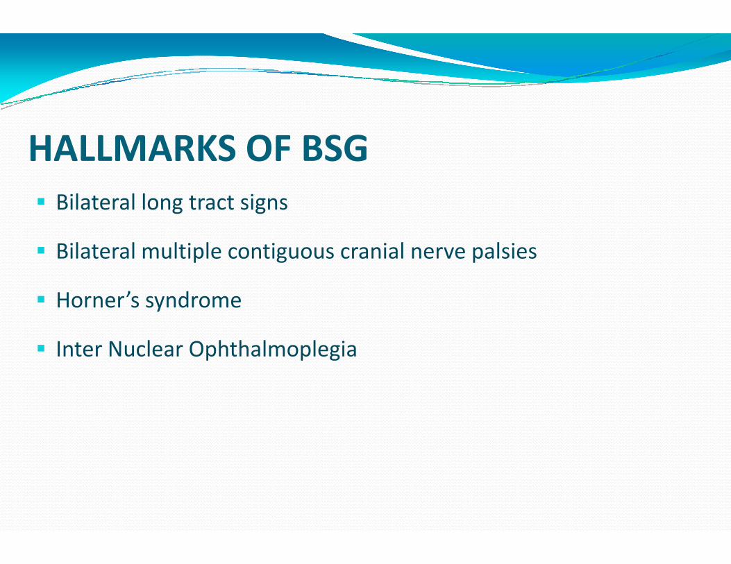
HALLMARKS OF BSG
� Bilateral long tract signs
� Bilateral multiple contiguous cranial nerve palsies
� Horner’s syndrome
� Inter Nuclear Ophthalmoplegia
HALLMARKS OF BSG
Bilateral multiple contiguous cranial nerve palsies

BSGBSGBSGBSG……ClassificationClassificationClassificationClassification� The most recent classification system by
Choux et al based on both CT and MRI imaging
� Type I – Diffuse� Type II – Intrinsic, focal� Type III – Exophytic, focal� Type IV – Cervicomedullary
� Pediatric Neurosurgery. New York, Churchill Livingstone, 2000, pp 471Pediatric Neurosurgery. New York, Churchill Livingstone, 2000, pp 471Pediatric Neurosurgery. New York, Churchill Livingstone, 2000, pp 471Pediatric Neurosurgery. New York, Churchill Livingstone, 2000, pp 471
ClassificationClassificationClassificationClassificationThe most recent classification system by Choux et al based on both CT and MRI
Exophytic, focalCervicomedullary
Pediatric Neurosurgery. New York, Churchill Livingstone, 2000, pp 471Pediatric Neurosurgery. New York, Churchill Livingstone, 2000, pp 471Pediatric Neurosurgery. New York, Churchill Livingstone, 2000, pp 471Pediatric Neurosurgery. New York, Churchill Livingstone, 2000, pp 471–491.491.491.491.

BSG……� Type I : Diffuse brainstem gliomas
� 75% of all BSG
� Hypointense on CT
� No significant enhancement on MRI.
� Characterized by diffuse infiltration and
� swelling of the brainstem.
� Typically, are malignant fibrillary
� astrocytomas (WHO grade III or IV).
gliomas
No significant enhancement on MRI.
Characterized by diffuse infiltration and
fibrillary
(WHO grade III or IV).

Diffuse Brainstem GliomaDiffuse Brainstem Glioma
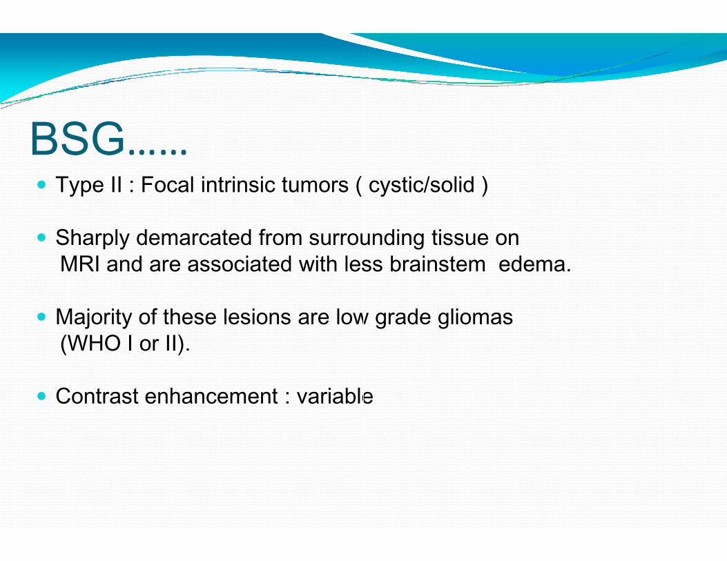
BSG……� Type II : Focal intrinsic tumors ( cystic/solid )
� Sharply demarcated from surrounding tissue on MRI and are associated with less brainstem edema.
� Majority of these lesions are low grade gliomas (WHO I or II).
� Contrast enhancement : variable
intrinsic tumors ( cystic/solid )
Sharply demarcated from surrounding tissue on MRI and are associated with less brainstem edema.
Majority of these lesions are low grade gliomas
Contrast enhancement : variable

Focal Medullary BSG
T1 CONTRAST
Focal Medullary BSG
CONTRAST T2

BSG……
� Type III : Exophytic tumors that arise from the subependymal glial tissue of the fourth ventricle and mostly grow dorsally or laterally.
� MRI characteristics similar to type II lesions,and histologically, these lesions are usually low-grade lesions (WHO I or II) like type II lesions.
Type III : Exophytic tumors that arise from the subependymal glial tissue of the fourth ventricle and mostly grow dorsally or laterally.
MRI characteristics similar to type II lesions,and histologically, these lesions are usually
grade lesions (WHO I or II) like type II
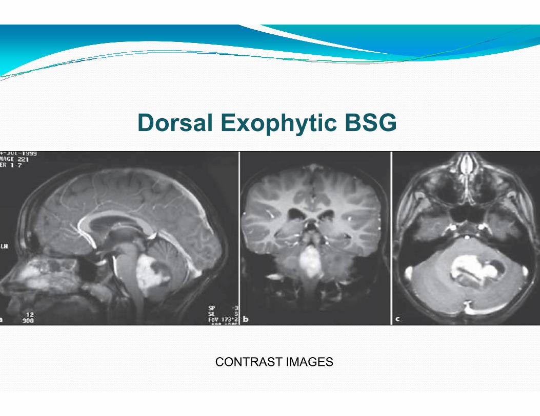
Dorsal Exophytic
CONTRAST IMAGES
Exophytic BSG
CONTRAST IMAGES

BSG……� Type IV lesions are cervicomedullary
brainstem gliomas.
� Imaging, histology and behavior : similar to intramedullary spinal cord gliomas.
� Majority are low-grade, nontumors.
Type IV lesions are cervicomedullary
Imaging, histology and behavior : similar to intramedullary spinal cord gliomas.
grade, non-infiltrative

BSG…..Management� Biopsy : only for indeterminate lesions
� Stereotactic biopsy: can provide diagnostic tissue.
� Stereotactic radiosurgery� Stereotactic radiosurgery
� Not without risk:
Damage to the cranial nerves and long tracts Damage to the cranial nerves and long tracts Damage to the cranial nerves and long tracts Damage to the cranial nerves and long tracts
Tissue heterogeneity Tissue heterogeneity Tissue heterogeneity Tissue heterogeneity
..ManagementBiopsy : only for indeterminate lesions
Stereotactic biopsy: can provide diagnostic tissue.
Damage to the cranial nerves and long tracts Damage to the cranial nerves and long tracts Damage to the cranial nerves and long tracts Damage to the cranial nerves and long tracts

� Focal cystic tumors- SX+RT
� Focal solid tumors- SX
MANAGEMENT
� Dorsal Exophytic tumors- SX + Focal RT
� Dorsal Exophytic malignant tumor
� Diffuse infiltrating – RT + steroids
SX + Focal RT
Dorsal Exophytic malignant tumor- RT+CT
RT + steroids

Choroid Plexus TumorsChoroid Plexus TumorsChoroid Plexus TumorsChoroid Plexus Tumors� Neoplasms of the choroid plexus.
� Lateral ventricles : most common location in children.
� 4th ventricle : most common location in adults.� 4 ventricle : most common location in adults.
� 4-6% of the intracranial neoplasms in children younger than 2
years.
� Choroid plexus tumors
� Choroid plexus papilloma (WHO Grade 1)
� Atypical choroid plexus papilloma (WHO Grade 2)
� Choroid plexus carcinoma (WHO Grade 3)
Neoplasms of the choroid plexus.
Lateral ventricles : most common location in children.
ventricle : most common location in adults.ventricle : most common location in adults.
6% of the intracranial neoplasms in children younger than 2
Choroid plexus papilloma (WHO Grade 1)
Atypical choroid plexus papilloma (WHO Grade 2)
Choroid plexus carcinoma (WHO Grade 3)
Choroid Plexus TUMORSChoroid Plexus TUMORSChoroid Plexus TUMORSChoroid Plexus TUMORS� Hydrocephalus and raised ICT
� The tumor itself can cause mass effect.� The tumor itself can cause mass effect.
� Surgery may not resolve HCP (derangement of reabsorption mechanisms or blockage at
other sites in the ventricular system
Choroid Plexus TUMORSChoroid Plexus TUMORSChoroid Plexus TUMORSChoroid Plexus TUMORS…..Clinical..Clinical..Clinical..ClinicalHydrocephalus and raised ICT
The tumor itself can cause mass effect.The tumor itself can cause mass effect.
Surgery may not resolve HCP derangement of reabsorption mechanisms or blockage at
other sites in the ventricular system)

Choroid Plexus Papilloma…..Radiology
T2W Post Gd
Choroid Plexus Papilloma…..Radiology
Post Gd Post Gd

Choroid Plexus PapillomaChoroid Plexus PapillomaChoroid Plexus PapillomaChoroid Plexus Papilloma…ManagementManagementManagementManagement� Treatment of hydrocephalus must be considered both
before and after any surgical procedures.
An acute increase in ICP : V P Shunt. � An acute increase in ICP : V P Shunt.
� Hydrocephalus often resolves following removal of the mass.
ManagementManagementManagementManagementTreatment of hydrocephalus must be considered both before and after any surgical procedures.
An acute increase in ICP : V P Shunt. An acute increase in ICP : V P Shunt.
Hydrocephalus often resolves following removal of the
Choroid Plexus PapillomaChoroid Plexus PapillomaChoroid Plexus PapillomaChoroid Plexus Papilloma…ManagementManagementManagementManagement� Total surgical resection is the goal.
� Complete removal: generally curative in papilloma
� Choroid plexus carcinoma -total resection leads to the best possible outcome.
� Adjuvant CT and RT have been demonstrated to increase survival
ManagementManagementManagementManagementTotal surgical resection is the goal.
Complete removal: generally curative in papilloma
total resection leads to the best
Adjuvant CT and RT have been demonstrated to increase

Dermoid cyst� Congenital ectodermal inclusion cysts.
� Extremely rare < 0.5% of primary intracranial tumors
� Midline sellar, parasellar, or frontonasal regions : most � Midline sellar, parasellar, or frontonasal regions : most
common sites.
� Posterior fossa ( vermis or within the 4
� Growth can lead to rupture of the cyst contents, causing
a chemical meningitis that may lead to vasospasm,
infarction, and even death
Congenital ectodermal inclusion cysts.
Extremely rare < 0.5% of primary intracranial tumors
Midline sellar, parasellar, or frontonasal regions : most Midline sellar, parasellar, or frontonasal regions : most
Posterior fossa ( vermis or within the 4th ventricle)
Growth can lead to rupture of the cyst contents, causing
a chemical meningitis that may lead to vasospasm,

Dermoid cyst� Well - defined, lobulated, “
variable size.
� Characteristically - cyst contains thick, disagreeable, foul smelling, yellow material due to the secretion of sebaceous glands and desquamated epithelium
� The cysts may also contain hair and/or teeth
“pearly” mass of
cyst contains thick, disagreeable, foul smelling, yellow material due to the secretion of sebaceous glands and
The cysts may also contain hair and/or teeth

Dermoid cyst…..MRISame imaging characteristics as fat
Hyperintense on T1WI and do not
enhanceenhance
Heterogeneous signal intensity on
T2WI
..MRI

Salient steps in surgery� Midline incision
� V shaped fascia opening
� Craniotomy
Dura opened in y shaped� Dura opened in y shaped
� Arachnoid opened
� Cottonoid placed over cisterna magna and floor of fourth ventricle
Salient steps in surgery
Cottonoid placed over cisterna magna and floor of fourth

Cerebellar tumors
� Hemispheric tumor approached via thinnest portion through horizontal incision
� Midline tumor via vermis splitting or Telovelar approach
Hemispheric tumor approached via thinnest portion through
Midline tumor via vermis splitting or Telovelar approach

IVth ventricular tumors
� Telovelar approach or vermian splitting
� Dorsal portion debulked, shave off the floor
� Aqueduct , roof floor , lateral recess and obex inspection
ventricular tumors
Telovelar approach or vermian splitting
Dorsal portion debulked, shave off the floor
Aqueduct , roof floor , lateral recess and obex inspection

Brainstem tumor
Dorsal exophytic tumor-
Identify superiorly and inferiorly normal brain stem
Start superior pole till iv ventricular floor , tumor slowly Start superior pole till iv ventricular floor , tumor slowly
separated till it is completely removed.
Focal brainstem tumor-
safe passage through brainstem using EMG and tumor
bulking from core to periphery.
Identify superiorly and inferiorly normal brain stem
Start superior pole till iv ventricular floor , tumor slowly Start superior pole till iv ventricular floor , tumor slowly
separated till it is completely removed.
safe passage through brainstem using EMG and tumor

Complications
� Pseudomeningocoele
� Cranial nerve paresis
� Mutism
� Subdural hygroma
� Aseptic meningitis
� Cerebellar cognitive affect syndrome Cerebellar cognitive affect syndrome

CONCLUSION
� Pilocytic astrocytoma bears� Management of hydrocephalus
controversial.� Though surgery and RT remains
treatment of choice for medulloblastomatreatment of choice for medulloblastomaoptimal craniospinal radiationdebatable.
� Outcome for brainstem gliomasdismal.
bears the best outcome.hydrocephalus still remains
remains themedulloblastoma;medulloblastoma;
radiation dose remains
gliomas remains

Thank YouThank YouThank YouThank You