Embed Size (px)
Citation preview
MODIFICATION OF CLASSIC RECONSTRUCTIVE TECHNIQUES AND
EVALUATION OF NEW ESTHETIC METHODS IN TERMS OF THE
EXPECTATIONS OF MODERN OTO-RHINO-LARYNGOLOGY AND HEAD-NECK
SURGERY
PHD THESIS
GÁBOR VASS MD
Department of Oto-Rhino-Laryngology and Head-Neck Surgery
University of Szeged
University of Szeged, Faculty of Medicine
Doctoral School of Clinical Medicine
Ph.D. Program:
The role of tissue reconstruction in case of advanced and recurrent laryngo-pharyngeal
tumors considering function-preservation and quality of life
Program Director: Prof. Dr. Lajos Kemeny D.Sc.
Supervisor: Dr. Laszlo Ivan Ph.D.
Szeged
2017
PUBLICATIONS RELATED TO THE PHD THESIS
I. László I, Gábor V, Zsolt B, László T, József J. Synchronous myeloproliferative
and inflammatory disease of the nasal cavity and paranasal sinuses: an
interesting differential diagnostic problem. Rhinology. 2009 Sep;47(3):323-6.
Impact Factor: 2.182
II. Vass G, Torkos A, Altmayer A, Czigner J, Jóri J, Rovó L, Iván L. Epicutaneous
patch test--a new diagnostic option to prevent the rejection of silicone-covered
cochlear implants in children. Int J Pediatr Otorhinolaryngol. 2013
Oct;77(10):1635-8. doi: 10.1016/j.ijporl.2013.06.023. Epub 2013 Aug 12.
Impact Factor:1.319
III. Vass G; Mohos G; Paczona R; Varga J; Iván L; Rovó L. Ajtószárny lebenyek
speciális felhasználási lehetőségei fej-nyaki tumoros beteganyagunkon. Magyar
Traumatológia, Ortopédia, Kézsebészet és Plasztikai Sebészet. - ISSN 1217-3231. -
2015. 58. évf. 4. sz., p. 257-265.
IV. Vass G, Bella Zs, Tóbiás Z, Nagy A, Iván L, Rovó L. Esztétikai és funkcionális
szempontból is kedvező sebészi alternatíva a maxilloethmoidális daganatok
eltávolítására: a módosított „facial degloving” technika Fül-Orr-
Gégegyógyászat 2016. 62:(3) p. 118.
V. Vass G, Mohos G, Bere Z, Ivan L, Varga J, Piffko J, Rovo L. Secondary
correction of nasal deformities in cleft lip and palate patients: surgical
technique and outcome evaluation. Head Face Med. 2016 Dec 1;12(1):34.
Impact Factor: 0.916
ABBREVIATIONS
ACD Allergic Contact Dermatitis
CI Cochlear Implant
CLP Cleft Lip and Palate
CT Computer Tomography
ELISA Enzyme-Linked Immunosorbent Assay
ELTMF Extended Lower Trapezius Musculocutaneous Flap
EPT Epicutaneous Patch Test
FNA Fine Needle Aspiration
LDMF Latissimus Dorsi Musculocutaneous Flap
MFD Modified Facial Degloving
MRI Magnetic Resonance Imaging
PET Positron Emission Tomography
PM Pectoralis Major
ROEQ Rhinoplasty Outcome Evaluation Questionnaire
TNM Tumor - Lymph Node - Metastasis
TOF Turnover Flap
VAC Vacuum Assisted Closure
LIST OF CONTENTS
1. INTRODUCTION 1
1.1. ALTERNATIVE SURGICAL METHODS FOR MALIGNANT TUMORS
OF THE HEAD AND NECK 1
1.1.1. The Modified Facial Degloving technique 1
1.1.2. Application of alternative reconstructive surgical methods in
special cases of head-neck cancer (turnover flap and
extended lower trapezius musculocutaneous flap) 3
1.2. COMPLICATIONS OF WOUND HEALING AND REJECTION
RELATED TO COCHLEAR IMPLANTS – SURGICAL SOLUTIONS
AND PREVENTION OPTION 3
1.3. SECONDARY CORRECTION OF NASAL DEFORMITIES IN
CLEFT LIP AND PALATE PATIENTS: SURGICAL TECHNIQUE
AND OUTCOME EVALUATION 4
2. AIMS OF THE THESIS 6
2.1. ALTERNATIVE SURGICAL METHODS FOR MALIGNANT TUMORS
OF THE HEAD AND NECK 6
2.2. COMPLICATIONS OF WOUND HEALING AND REJECTION
RELATED TO COCHLEAR IMPLANTS – SURGICAL SOLUTIONS
AND PREVENTION OPTION 6
2.3. SECONDARY CORRECTION OF NASAL DEFORMITIES IN
CLEFT LIP AND PALATE PATIENTS: SURGICAL TECHNIQUE
AND OUTCOME EVALUATION 7
3. METHODS AND SUBJECTS 8
3.1. ALTERNATIVE SURGICAL METHODS FOR MALIGNANT TUMORS
OF THE HEAD AND NECK 8
3.1.1. The Modified Facial Degloving technique 8
3.1.2. Application of alternative reconstructive surgical methods in
special cases of head-neck cancer 14
3.1.2.1. Turnover flap 14
3.1.2.2. Extended lower trapezius musculocutaneous flap 17
3.2. COMPLICATIONS OF WOUND HEALING AND REJECTION
RELATED TO COCHLEAR IMPLANTS – SURGICAL SOLUTIONS
AND PREVENTION OPTION 19
3.3. SECONDARY CORRECTION OF NASAL DEFORMITIES IN
CLEFT LIP AND PALATE PATIENTS: SURGICAL TECHNIQUE
AND OUTCOME EVALUATION 22
4. RESULTS 24
4.1. ALTERNATIVE SURGICAL METHODS FOR MALIGNANT TUMORS
OF THE HEAD AND NECK 24
4.1.1. The Modified Facial Degloving technique 24
4.1.2. Application of alternative reconstructive surgical methods in
special cases of head-neck cancer 26
4.1.2.1. Turnover flap 26
4.1.2.2. Extended lower trapezius musculocutaneous flap 29
4.2. COMPLICATIONS OF WOUND HEALING AND REJECTION
RELATED TO COCHLEAR IMPLANTS – SURGICAL SOLUTIONS
AND PREVENTION OPTION 29
4.3. SECONDARY CORRECTION OF NASAL DEFORMITIES IN
CLEFT LIP AND PALATE PATIENTS: SURGICAL TECHNIQUE
AND OUTCOME EVALUATION 30
5. DISCUSSION 33
5.1. ALTERNATIVE SURGICAL METHODS FOR MALIGNANT TUMORS
OF THE HEAD AND NECK 33
5.1.1. The Modified Facial Degloving technique 33
5.1.2. Application of alternative reconstructive surgical methods in
special cases of head-neck cancer 35
5.1.2.1. Turnover flap 35
5.1.2.2. Extended lower trapezius musculocutaneous flap 36
5.2. COMPLICATIONS OF WOUND HEALING AND REJECTION
RELATED TO COCHLEAR IMPLANTS – SURGICAL SOLUTIONS
AND PREVENTION OPTION 37
5.3. SECONDARY CORRECTION OF NASAL DEFORMITIES IN
CLEFT LIP AND PALATE PATIENTS: SURGICAL TECHNIQUE
AND OUTCOME EVALUATION 41
6. CONCLUSIONS 42
6.1. ALTERNATIVE SURGICAL METHODS FOR MALIGNANT TUMORS
OF THE HEAD AND NECK 42
6.2. COMPLICATIONS OF WOUND HEALING AND REJECTION
RELATED TO COCHLEAR IMPLANTS – SURGICAL SOLUTIONS
AND PREVENTION OPTION 43
6.3. SECONDARY CORRECTION OF NASAL DEFORMITIES IN
CLEFT LIP AND PALATE PATIENTS: SURGICAL TECHNIQUE
AND OUTCOME EVALUATION 43
7. ACKNOWLEDGEMENT 44
8. REFERENCES 45
9. APPENDIX 51

1
1. INTRODUCTION
1.1. ALTERNATIVE SURGICAL METHODS FOR MALIGNANT TUMORS OF
THE HEAD AND NECK
Benign and malignant tumors of the head and neck region imply a great surgical
challenge in virtue of their localization, as operative scars are nearly always visible in these
regions. Patients have to face not only the tumor itself, but the stigma caused by the surgeon’s
knife. Oncosurgical principles, however, should not be abandoned for aesthetics, because
radicality and ablasticity are priority in case of malignant tumors. Continuous development of
the surgical instruments and operative techniques have set the focus of surgical treatment on
one hand onto microsurgery and free flap reconstruction also in the field of
otorhinolaryngology and head-neck surgery nowadays. However, these operations are rather
time-consuming, mean a great intra- and postoperative burden for the patient, and require
highly specialized, skilled surgical team.
1.1.1. The Modified Facial Degloving technique
Endoscopic surgery has become the cutting-edge therapeutical approach nowadays, as
it is minimal-invasive, scarless and can be carried out mostly on one-day-surgery basis.
However, the endoscopic approach - especially in case of malignant sino-nasal tumors - has
its limitations (orbital involvement, infiltration of the premaxillary soft tissues or skin, lateral
extension/infiltration of the tumor in the internal carotid artery’s region, etc.), not to mention
the long learning curve of becoming an expert in endoscopic surgery, the time-consuming
operations and high cost of the endoscopic sets with navigation system (1). In such cases,
when microsurgical reconstruction or endoscopic surgery are inadequate or limited, or the
personal and financial resources are insufficient, simpler reconstructive methods or classic
external approaches may come forth, together with increasingly raised expectations of
patients towards the head and neck surgeon to elaborate and apply new surgical methods with
concealable or hidden scars.
2
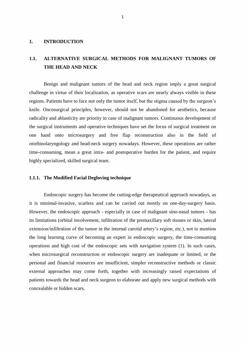
Maxillo-ethmoidal malignant and aggressively growing benign tumors are removed
routinely through the widely-used Weber-Ferguson’s incision (2). This surgical approach
provides a very good sight of the operative field but may cause postoperative aesthetic
deformities (Fig. 1).
Figure 1: Weber-Ferguson’s approach – late postoperative pictures
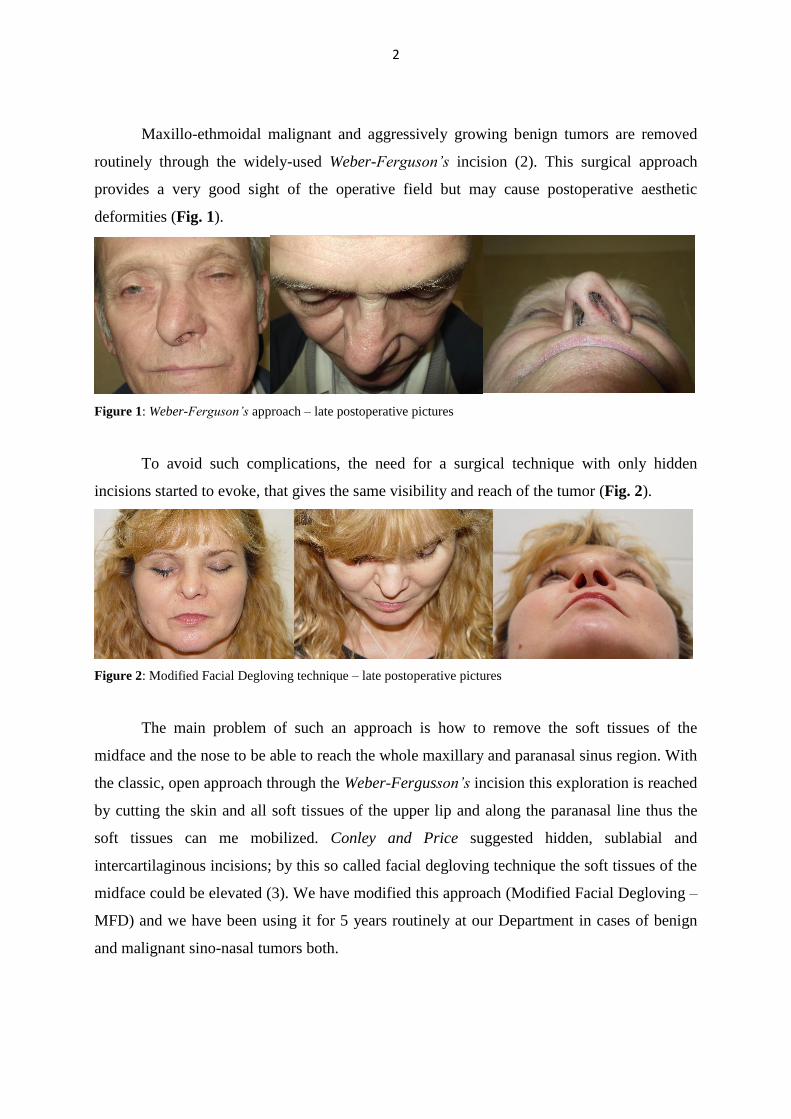
To avoid such complications, the need for a surgical technique with only hidden
incisions started to evoke, that gives the same visibility and reach of the tumor (Fig. 2).
Figure 2: Modified Facial Degloving technique – late postoperative pictures
The main problem of such an approach is how to remove the soft tissues of the
midface and the nose to be able to reach the whole maxillary and paranasal sinus region. With
the classic, open approach through the Weber-Fergusson’s incision this exploration is reached
by cutting the skin and all soft tissues of the upper lip and along the paranasal line thus the
soft tissues can me mobilized. Conley and Price suggested hidden, sublabial and
intercartilaginous incisions; by this so called facial degloving technique the soft tissues of the
midface could be elevated (3). We have modified this approach (Modified Facial Degloving –
MFD) and we have been using it for 5 years routinely at our Department in cases of benign
and malignant sino-nasal tumors both.
3
1.1.2. Application of alternative reconstructive surgical methods in special cases of
head-neck cancer
From the surgeon’s point of view, the spreading of oncotherapy against his first
treatment choice of oncosurgery is another problem, as wound healing disorders may rise
after radio-chemotherapy, and additional reconstructive operations could become necessary.
Tissue environment alterations due to oncotherapy and/or multiple scars caused by previous
operations require elaboration of alternative tissue restoration methods, which are uniquely
planned for each case and are often redesigns of a previously used and published flap
technique. Turnover Flaps (TOF) are flaps, that are turned by 180 degrees into the defect over
their fulcrum, which provides their blood supply. Flaps containing skin and subcutaneous fat
have random blood supply, while pedicled TOFs have muscle and fascia components.
Comprehensive usage of these flaps have been published in the international literature (4-7).
Most frequent application of these flaps are: defects of the lower limbs and hands, sacral
sores, myelomeningocele, chest wall defect, tracheostomy, fistulas of the pharynx and oral
cavity and nasal defects (8-11).
In reconstruction of defects of the skin and underlying soft tissue of the neck it is the
surgeon’s main objective to achieve proper long-term coverage of the exposed vital organs
with well-vascularised tissue harvested from a distant donor site (12, 13). A precise
evaluation of the extent of the defect, the patient’s physiological data, and the area to be
covered will guide the surgeon in choosing the best option (12, 14, 15). The extended lower
trapezius musculocutaneous flap (ELTMF) and latissimus dorsi musculocutaneous flaps
(LDMF) are the two available muscle compartments that can be transferred on a reliable
vascular pedicle to the dorsal, suprascapular, and neck regions. The selection of flap depends
on a thorough understanding of their anatomy, their way rotation, and an analysis of the size,
extension, and site of the defect (12, 14).
4
1.2. COMPLICATIONS OF WOUND HEALING AND REJECTION RELATED TO
COCHLEAR IMPLANTS – SURGICAL SOLUTIONS AND PREVENTION
Cochlear Implant (CI) revolutionized hearing restoration in patients with bilateral,
severe or profound sensorineural hearing loss. A CI’s function is to substitute the inner ear, to
code and to transfer the sound stimuli to the acoustic nerve. The receiver-stimulator unit is
implanted under the scalp and the electrode array is installed into the cochlea during a surgical
procedure (16). In the past decades, the increasing number of cochlear implantations
worldwide, also in Hungary, has led to an elevated number of possible reoperations due to
certain complications (17, 18). One of these complications might be the skin necrosis above
the transmitter coil and the concomitant exposure of the receiver-stimulator unit – this is a
major complication of the operation (19-21). In some of our cases, the skin necrosis occurred
repeatedly notwithstanding the various surgical solutions to re-cover the implant. As all
implants have been operated by the same team with standard methods, and no infective or
histopathological anomaly has been revealed in our patients with implant rejection, and
furthermore all of our adopted reconstructive methods, that have been published previously as
definitive surgical solutions, we started to seek for non-surgical cause. According to literature
data the silicone housing of any implanted medical device may pay a role in this rejection
process via immune system modulation, by inducing foreign-body type and local and
systemic nonspecific (nonallergic) inflammatory reactions (22). However, evidence for true
allergic reactions to silicone-coated medical devices has also been reported in some cases
(22). In general, we can state, that any kind of silicone implant may induce the production of
auto-antibodies in genetically susceptible patients causing immune system disorders (23).
This draw our attention towards the possibility of individual silicone hypersensitivity against
the covering of the CI, because all four patients with skin necrosis had the same silicone-
covered device implanted. In the dermatology-allergology practice routinely used, non-
invasive Epicutaneous Patch Testing (EPT) seemed to be a proper method to reveal the
possibility of individual silicone hypersensitivity.
5
1.3. SECONDARY CORRECTION OF NASAL DEFORMITIES IN CLEFT LIP
AND PALATE PATIENTS: SURGICAL TECHNIQUE AND OUTCOME
EVALUATION
Cleft lip and palate (CLP) deformities are among the most common congenital
malformations (24). In Hungary the incidence of combined oro-facial clefts is 2 in 1000 live
births (25). Although CLP together occur more commonly in males, isolated cleft palate is
more common in females (24, 25). Surgical correction of CLP should be performed before the
first year of age, usually between 3-6 months-of-age, prior to speech development (24, 25).
The aim of the operation is to reunite all tissue layers of the lip, to reposition the nasal septum
and to separate the oral and nasal cavities; and restore the valve function of the soft palate.
This functional repair helps also with the preservation of facial growth and the development
of proper dentition (24, 25). If this adequate primary surgical correction of CLP fails, the
consequentially developing nasal deformity associated with CLP is one of the most
challenging reconstructive problems in rhinoplasty. The characteristic CLP nose represents a
stigma for the patient. This results from a combination of altered anatomy, surgical scarring
from previous reconstructive operations and includes deformities of the septum, nasal
pyramid, malformation of the nasal tip and malposition of alar cartilages. The indication for
surgery is on one hand the difficult nasal breathing and altered nasal function (tendency for
chronic rhinosinusitis) and on the other hand the aesthetic look of the nose, both of which
may affect the patient’s quality of life negatively and can cause heavy psycho-social burden
for them. Accompanying nasal deformities are mainly characterized by a shortened columella,
a depressed nasal tip, bilateral dislocation of the alar cartilage, eversion of the alar bases and
nasal obstruction (26-28). Although numerous secondary rhinoplasty methods have been
described in the literature for the lengthening of the columella, or for grafting techniques, no
standardized technique exists. Statistical analysis or comparison of the surgical methods and
their results are hardly comparable this way.
6
2. AIMS OF THE THESIS
2.1. ALTERNATIVE SURGICAL METHODS FOR MALIGNANT TUMORS OF
THE HEAD AND NECK
To work-out and demonstrate a novel surgical method, which gives the opportunity to
resect malignant sino-nasal tumors according to the oncosurgical principles without visible
skin scars and aesthetic deformities. Furthermore, to present the alternative, modified
application of two well-known and previously published flaps (TOF, ELTMF) for the
reconstruction of special tissue defects in the head and neck region after malignant tumor
resection and oncotherapy.
2.2. COMPLICATIONS OF WOUND HEALING AND REJECTION RELATED TO
COCHLEAR IMPLANTS – SURGICAL SOLUTIONS AND PREVENTION
OPTION
To establish a diagnostic method, which could shift the focus from reconstruction
towards prevention in CI with complications. As three of the four implantees with implant
rejection have had a positive skin reaction with the EPT for the silicone sample and the
repeated, different reconstructive operations have been unsuccessful, possible individual
silicone hypersensitivity has arisen. This, in our opinion, plays a very important role in the
process of wound healing disorder and the rejection of the implant, because no other cause
could have been proved by histopathology, bacteriology or methodology analysis. EPT, as a
non-invasive method, might be useful in childhood before the planned cochlear implantation
to reveal optional silicone allergy. Thus rejection tendency, skin necrosis above the implant
and stressful reconstructive surgeries with stigmatizing scars on the scalp could be avoided.

7
2.3. SECONDARY CORRECTION OF NASAL DEFORMITIES IN CLEFT LIP
AND PALATE PATIENTS: SURGICAL TECHNIQUE AND OUTCOME
EVALUATION
To standardize the surgical method according to the experience gained during the
secondary rhinoplasty operations of CLP patients at our University, and to evaluate our results
by the adaptation of a previously published patient satisfaction questionnaire (ROEQ –
Rhinoplasty Outcome Evaluation Questionnaire).
8
3. METHODS AND SUBJECTS
3.1. ALTERNATIVE SURGICAL METHODS FOR MALIGNANT TUMORS OF
THE HEAD AND NECK
3.1.1. The Modified Facial Degloving technique
In case of head and neck tumors, especially in sino-nasal localization, radiological
imaging is inevitable for the evaluation of the tumor’s extent, the infiltration of bony
structures or soft tissues and the presence of metastatic lymph nodes. Beside the clinical
picture, these images help to predestinate the chosen surgical method, which is mainly the
endoscopic approach for benign lesions (chronic rhinosinusitis, nasal polys or papilloma) and
early-stage malignant tumors of the maxillo-ethmoidal region (1). Considering malignant
tumors, endoscopic surgery has its well-defined limitations, when an open approach becomes
necessary (1). Open surgery is advised for benign lesions with complications (e.g. frontal
sinusitis with intracranial spreading), for malignant tumors and other indications such as
mucormycosis, foreign body in the sinuses or certain maxillary fractures (29-32). First line
imaging method for the above listed cases is the Computer Tomography (CT), which gives a
proper status of the bony structures and the extent of the disease; however, its specificity is
rather low: inflammation and tumor can hardly be distinguished. In order to differentiate
between a malignant tumor and chronic inflammation, as we have published before, Magnetic
Resonance (MR) examination should be performed (neoplastic tissues often have higher
proton density and relaxation time than the healthy tissues), i.e. on T2 flares the chronic
inflammation is hyperdense, while the tumor remains hypodense.
Twenty-three consecutive patients have been operated with this technique at our
Department between 2012-2016. Patients had a mean age of 44,4 years (varying between 19
and 67 years); gender ratio was 14 (61%) female and 9 (39%) male. All patients had been
diagnosed with histologically proven malignant sino-nasal tumors and had preoperative
CT/MRI imaging. Inclusion criteria for surgery have been: limitations of endoscopic surgery
(orbital involvement, infiltration of the premaxillary soft tissues or skin, lateral
extension/infiltration of the tumor in the internal carotid artery’s region, etc.), tumor TNM
stage (T2-T4a, tumor mass reduction in cases of T4b before oncotherapy) and informed
9
consent from the patients for the MFD surgery. Exclusion criteria: contraindication for
general anesthesia.
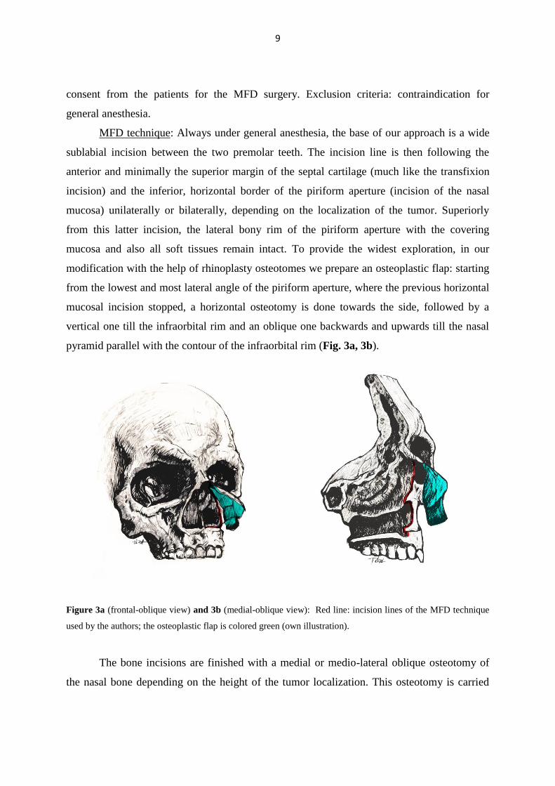
MFD technique: Always under general anesthesia, the base of our approach is a wide
sublabial incision between the two premolar teeth. The incision line is then following the
anterior and minimally the superior margin of the septal cartilage (much like the transfixion
incision) and the inferior, horizontal border of the piriform aperture (incision of the nasal
mucosa) unilaterally or bilaterally, depending on the localization of the tumor. Superiorly
from this latter incision, the lateral bony rim of the piriform aperture with the covering
mucosa and also all soft tissues remain intact. To provide the widest exploration, in our
modification with the help of rhinoplasty osteotomes we prepare an osteoplastic flap: starting
from the lowest and most lateral angle of the piriform aperture, where the previous horizontal
mucosal incision stopped, a horizontal osteotomy is done towards the side, followed by a
vertical one till the infraorbital rim and an oblique one backwards and upwards till the nasal
pyramid parallel with the contour of the infraorbital rim (Fig. 3a, 3b).
Figure 3a (frontal-oblique view) and 3b (medial-oblique view): Red line: incision lines of the MFD technique
used by the authors; the osteoplastic flap is colored green (own illustration).
The bone incisions are finished with a medial or medio-lateral oblique osteotomy of
the nasal bone depending on the height of the tumor localization. This osteotomy is carried
10
out through a narrow tunnel prepared by a sharp Cottle elevator in the supraseptal-
subperiosteal plane - just like in closed technique rhinoplasty - above and medial from the
nasal cartilages; thus no incision in the nasal vestibule or in the region of the nasal cartilages
is needed. The lateral nasal wall is then separated - like in Denker’s operation - and later on
resected together with the tumor mass. This way the cartilaginous framework, the nasal bone
and the anterior bony wall of the maxillary sinus could be elevated together with the soft
tissues of the nose and the midface. In the classic approach the nasal skin is reached and lifted
through the intercartilaginous incisions used in rhinoplasty (31-34). In our modification so as
to be more simple, and not to disrupt the integrity of the cartilage framework of the nose thus
minimizing the postoperative and aesthetic complications, the soft tissues above the bony
nasal pyramid and the anterior wall of the maxillary sinus are not dissected at all, but these are
elevated and mobilized together with the nasal bone and the anterior bony wall of the
maxillary sinus after performing the above detailed osteotomies. This approach makes it
possible to perform endonasal resection and to explore the whole nasal and paranasal region
according to the localization and extent of the tumor. If necessary, the effect of radicality can
be controlled with 30-45-70 degree rigid endoscopes; furthermore, endoscopes can be used
intraoperatively to dissect and coagulate the sphenopalatine and ethmoid arteries in order to
have a better intraoperative bleeding control (30). The nasal septum can be incised parallel
with the palate at the level of the nasal floor, and a swinging-door technique can be used to
gain more space for tumor resection, or the septum can be partially resected in case of
tumorous infiltration. As the final step of operation the osteoplastic flap and the soft tissues
are repositioned; care is taken to have the nasolacrimal duct open freely into the nasal cavity
(35). If partial resection of the infraorbital rim, or the anterior bony wall of the maxillary sinus
or the nasal bone is necessary due to tumorous infiltration, reconstruction with rib cartilage or
mini-plate osteosynthesis can be an option (29). Because the integrity of attached tissues is
intact within the osteoplastic flap, usually there is no need for fixation of the elevated and
repositioned bones. The sublabial incision is sutured with absorbable stitches. The operative
common nasal-paranasal cavity is filled with tamponage so as the nasal vestibule; no sutures
are applied in this region. We routinely use iodoform gauze as packing for 3-5 days, which in
our opinion has two important advantages: firstly, the used material is antiseptic, and could be
left within the surgical cavity for several days, which minimizes crust formation. Secondly it
11
provides a continuous compression for the clipped vessels and mucosa, thus providing
bleeding control during the early and mid-term postoperative days. Our patients generally
complain about only minimal postoperative pain; they have no discomfort related to the
tamponage. Of course other methods, packing materials and shorter tamponage intervals are
also available, depending on the individual case and the extent of the surgical resection.
We illustrate the main points of our technique and its advantages versus the
endoscopic approach below in different tumor localizations and types.
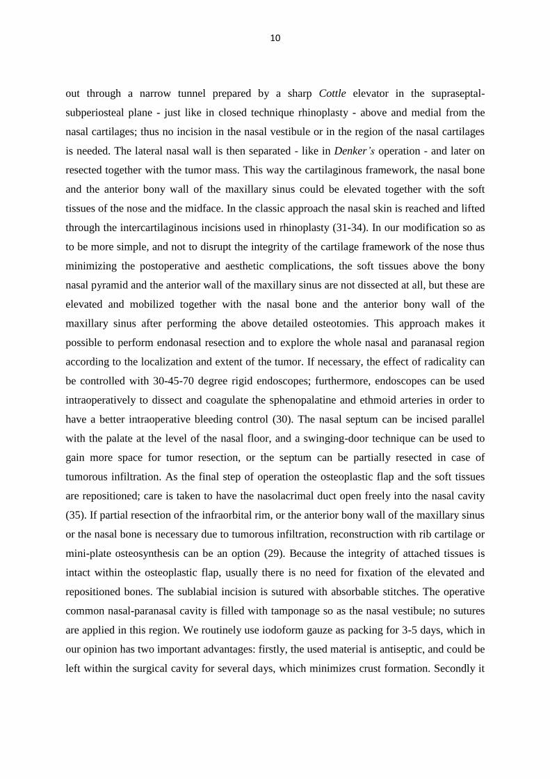
In case of a basal cell adenocarcinoma of the mesostructure and the orbit (37-year-old
female) due to the limitations of endoscopic surgery (i.e. orbital involvement) we decided on
the MFD approach, during which we carried out medial maxilla resection, removed all
turbinates and the entire ethmoid region, the medial orbital wall, the medial segment of
Tenon’s capsule within the orbit and also the inferior wall of the frontal sinus, which had not
been infiltrated yet, but we wanted to gain a better endoscopic visibility of the affected
regions and their surroundings during the control examinations (Fig. 4a). The osteoplastic
flap was repositioned (Fig. 4b), the sublabial incision was sutured with absorbable thread; the
surgical cavity was filled with tamponage for four days. Radical tumor resection was achieved
with excellent cosmetic results; no complications were observed. She has been tumor-free for
one and a half years.
Figure 4a: preoperative CT and 3D reconstruction (blue triangle shows the field of view during operation, the
tumor mass is colored green), Figure 4b: postoperative 3D CT reconstruction with bone window, red oval line
shows the repositioned osteoplastic flap.
12
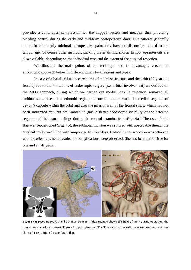
In case of a basaloid squamous cell carcinoma of the meso- and suprastructure (23-
year-old female) the tumor originated from the left nasal cavity, it destroyed the medial wall
of the maxillary sinus, the ethmoid cells and infiltrated also the frontal sinus, which latter
meant a limitation for ablative endoscopic resection (Fig. 5a). The epipharynx and the orbit
were intact according to the CT scan. Based on the age and gender of the patient and the
invasion of the whole frontal sinus, we decided on the MFD approach instead of open surgery
or endoscopic resection. During the operation in general anesthesia we removed the
remaining medial, and partially the anterior wall of the left maxillary sinus, all of the
turbinates and partially the left nasal bone. By putting the patient into Trendelenburg’s
position, we were able to reach the frontal sinus as well, since its frontal and lateral recess
were also infiltrated, so the tumor mass could be completely and radically removed (Fig. 5b).
The total gross resection was controlled in this case even with a 30-degree rigid endoscope in
the created common operative cavity. The sublabial incision was sutured with absorbable
thread; the surgical cavity was filled with tamponage for seven days. Radical tumor resection
was achieved with acceptable cosmetic results; no complications were observed. She has been
tumor-free for two years.
Figure 5a: preoperative CT and 3D reconstruction (blue triangle shows the field of view during operation, the
tumor mass is colored green), Figure 5b: intraoperative picture, surgical approach (the end of the metal suction
tube is on the upper wall of the frontal sinus).
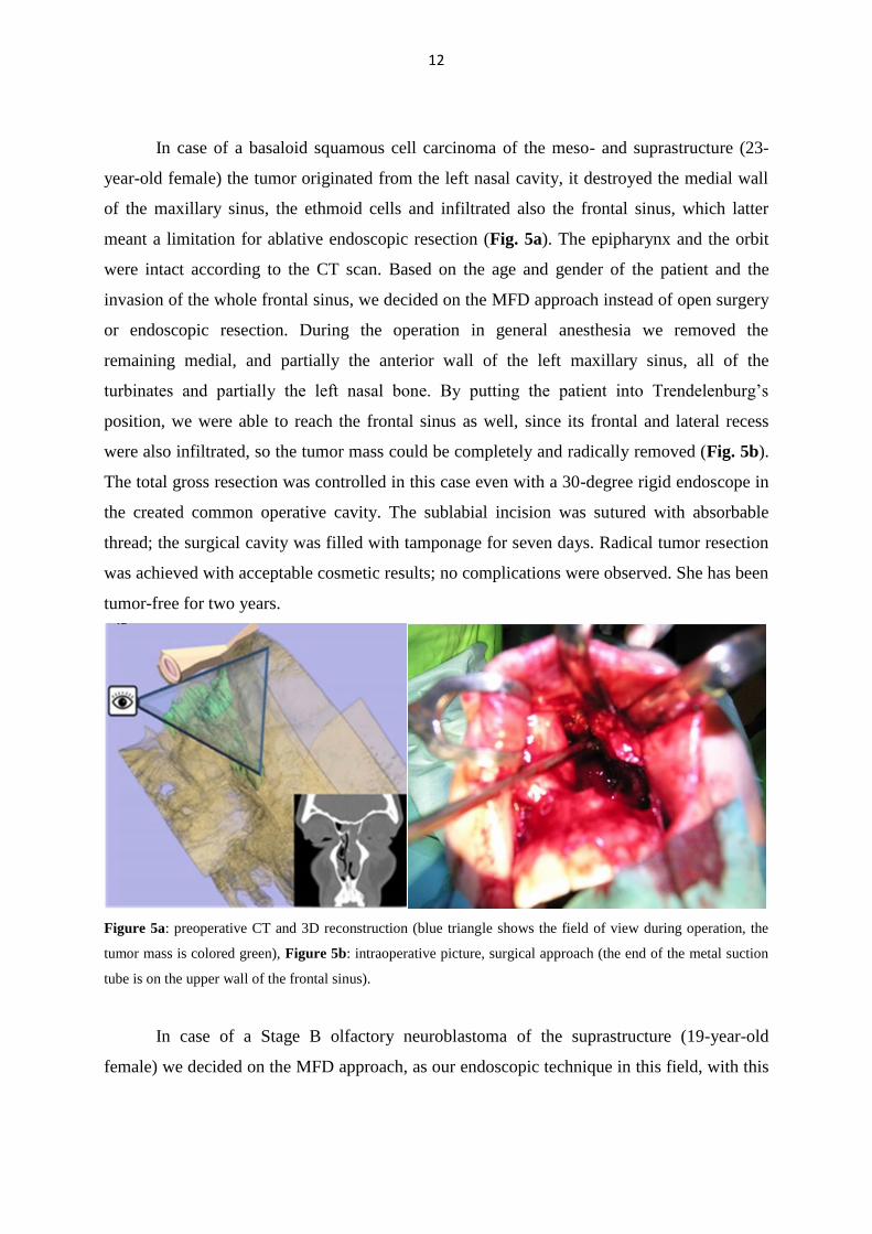
In case of a Stage B olfactory neuroblastoma of the suprastructure (19-year-old
female) we decided on the MFD approach, as our endoscopic technique in this field, with this
13
kind of tumor was still in the beginning phase of the learning curve. Fig. 6a is our schematic
illustration of the surgical approach, while Fig. 6b shows the intra-operative situation. The
medial wall of the maxillary sinus, all the turbinates and the whole ethmoid region was
resected up to the cribriform plate, which we found intact. In order to have a full endoscopic
overview during control examinations of the whole maxillo-ethmoidal region, both the frontal
and the sphenoid sinuses were opened; however, no abnormalities were found in them. The
sublabial incision was sutured with absorbable thread; the surgical cavity was filled with
tamponage for five days. Radical tumor resection was achieved with excellent cosmetic
results; no complications were observed. Postoperative full dose radiotherapy was
administered; she has been tumor-free for one year.
Figure 6a: our own illustration shows the operative approach – the elevated osteoplastic flap on the right side
(the incision lines are blue dotted) Figure 6b: intraoperative picture, (elevated osteoplastic flap on the right
side).
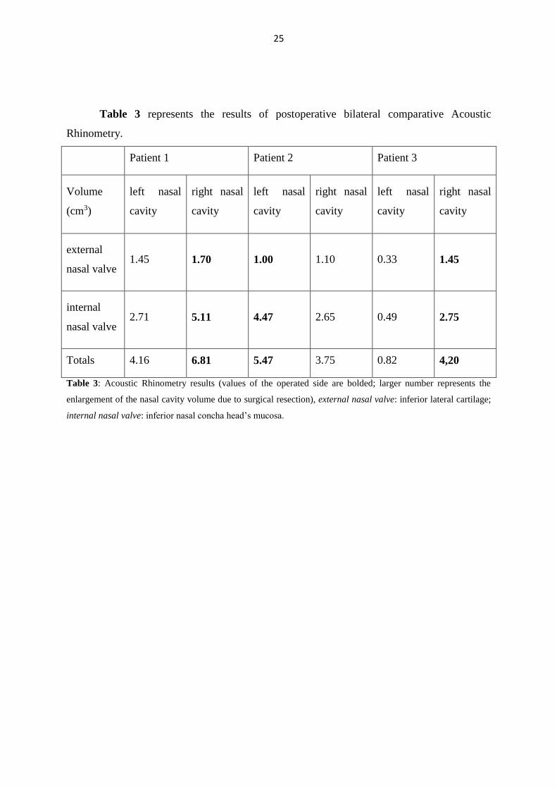
Our results were evaluated by Acoustic Rhinometry (Acoustic Rhinometer A1-209,
GM Instruments LTD, Ashgrove, UK) without decongestion to prove that the operative
technique does not result in narrowing of the nasal cavity and does not cause loss of nasal
breathing function.
14
3.1.2. Application of alternative reconstructive surgical methods in special cases of
head-neck cancer
3.1.2.1.Turnover flap
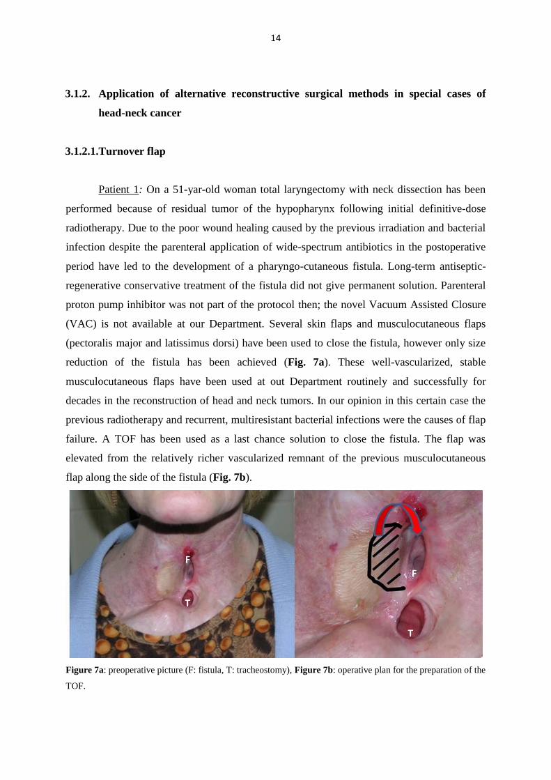
Patient 1: On a 51-yar-old woman total laryngectomy with neck dissection has been
performed because of residual tumor of the hypopharynx following initial definitive-dose
radiotherapy. Due to the poor wound healing caused by the previous irradiation and bacterial
infection despite the parenteral application of wide-spectrum antibiotics in the postoperative
period have led to the development of a pharyngo-cutaneous fistula. Long-term antiseptic-
regenerative conservative treatment of the fistula did not give permanent solution. Parenteral
proton pump inhibitor was not part of the protocol then; the novel Vacuum Assisted Closure
(VAC) is not available at our Department. Several skin flaps and musculocutaneous flaps
(pectoralis major and latissimus dorsi) have been used to close the fistula, however only size
reduction of the fistula has been achieved (Fig. 7a). These well-vascularized, stable
musculocutaneous flaps have been used at out Department routinely and successfully for
decades in the reconstruction of head and neck tumors. In our opinion in this certain case the
previous radiotherapy and recurrent, multiresistant bacterial infections were the causes of flap
failure. A TOF has been used as a last chance solution to close the fistula. The flap was
elevated from the relatively richer vascularized remnant of the previous musculocutaneous
flap along the side of the fistula (Fig. 7b).
Figure 7a: preoperative picture (F: fistula, T: tracheostomy), Figure 7b: operative plan for the preparation of the
TOF.
15
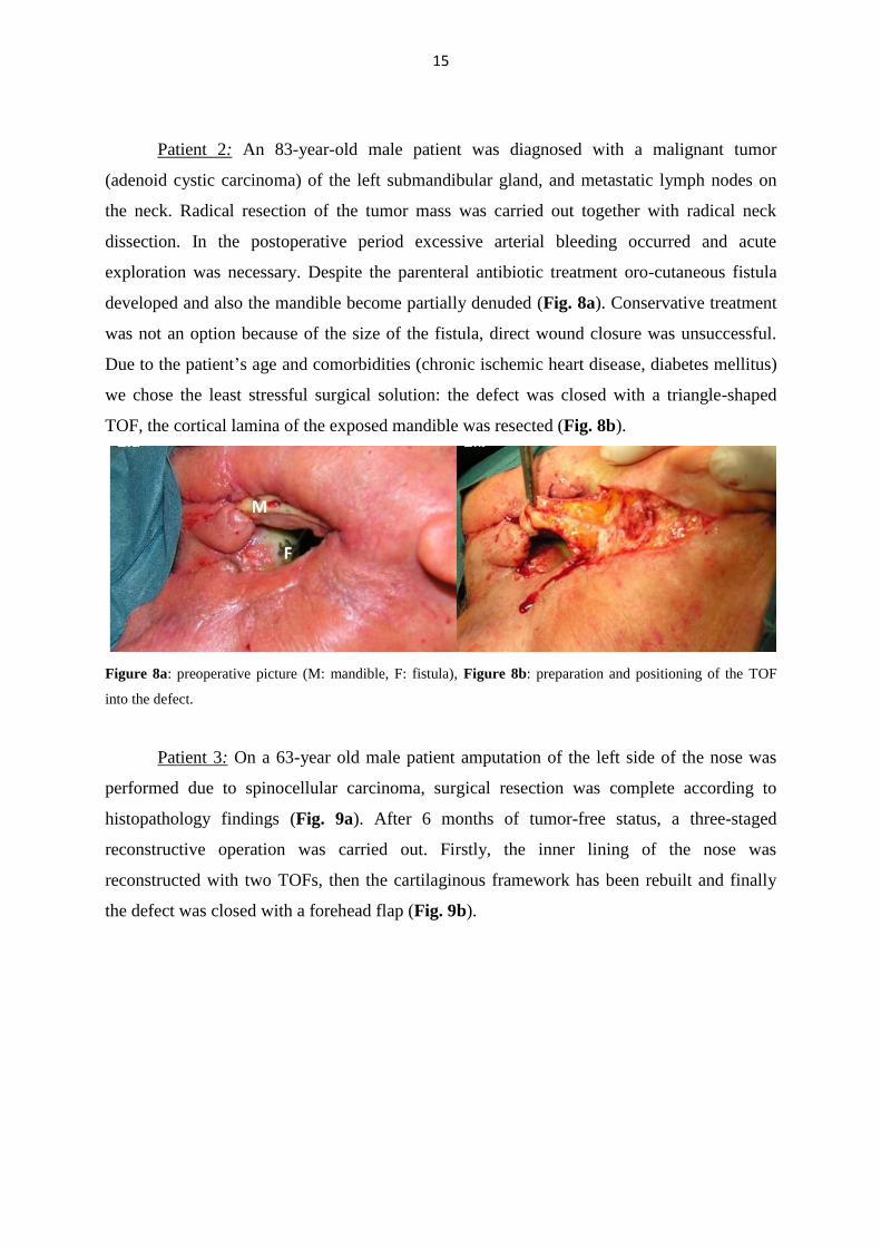
Patient 2: An 83-year-old male patient was diagnosed with a malignant tumor
(adenoid cystic carcinoma) of the left submandibular gland, and metastatic lymph nodes on
the neck. Radical resection of the tumor mass was carried out together with radical neck
dissection. In the postoperative period excessive arterial bleeding occurred and acute
exploration was necessary. Despite the parenteral antibiotic treatment oro-cutaneous fistula
developed and also the mandible become partially denuded (Fig. 8a). Conservative treatment
was not an option because of the size of the fistula, direct wound closure was unsuccessful.
Due to the patient’s age and comorbidities (chronic ischemic heart disease, diabetes mellitus)
we chose the least stressful surgical solution: the defect was closed with a triangle-shaped
TOF, the cortical lamina of the exposed mandible was resected (Fig. 8b).
Figure 8a: preoperative picture (M: mandible, F: fistula), Figure 8b: preparation and positioning of the TOF
into the defect.
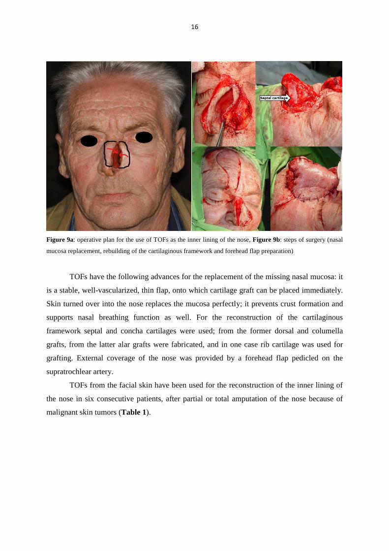
Patient 3: On a 63-year old male patient amputation of the left side of the nose was
performed due to spinocellular carcinoma, surgical resection was complete according to
histopathology findings (Fig. 9a). After 6 months of tumor-free status, a three-staged
reconstructive operation was carried out. Firstly, the inner lining of the nose was
reconstructed with two TOFs, then the cartilaginous framework has been rebuilt and finally
the defect was closed with a forehead flap (Fig. 9b).
16
Figure 9a: operative plan for the use of TOFs as the inner lining of the nose, Figure 9b: steps of surgery (nasal
mucosa replacement, rebuilding of the cartilaginous framework and forehead flap preparation)
TOFs have the following advances for the replacement of the missing nasal mucosa: it
is a stable, well-vascularized, thin flap, onto which cartilage graft can be placed immediately.
Skin turned over into the nose replaces the mucosa perfectly; it prevents crust formation and
supports nasal breathing function as well. For the reconstruction of the cartilaginous
framework septal and concha cartilages were used; from the former dorsal and columella
grafts, from the latter alar grafts were fabricated, and in one case rib cartilage was used for
grafting. External coverage of the nose was provided by a forehead flap pedicled on the
supratrochlear artery.
TOFs from the facial skin have been used for the reconstruction of the inner lining of
the nose in six consecutive patients, after partial or total amputation of the nose because of
malignant skin tumors (Table 1).
17
Extensive nasal defects
Gender Age (years) Localization and extent of the defect Histopathology
female 85 total right cartilaginous nose cc. basocellulare
female 72 total alar and tip region on the left side cc. basocellulare
female 57 nasal tip, columella, anterior part of septum cc. planocellulare
female 79 total right cartilaginous nose cc. planocellulare
male 63 total left cartilaginous nose cc. planocellulare
male 58 total cartilaginous nose and partial septum cc. basocellulare
Table 1: Summary of our patients with nasal skin tumor, who have been operated with TOFs (2005-2015.)
3.1.2.2.Extended Lower Trapezius Musculocutaneous Flap (ELTMF)
A 49-year-old patient was admitted to our head and neck surgery department in 2003
with a squamous cell carcinoma of the right tonsillar region and the soft palate (T2N0M0).
We performed a transoral carbon dioxide laser excision of the tumor, and histopathological
examination showed tumor-free resection margins. He was given postoperative radiotherapy
(total dose 66 Gy). Four years after the operation a late metastasis was found in the right
submandibular region and verified by fine needle aspiration (FNA). He was treated by
modified radical neck dissection and given four cycles of postoperative chemotherapy.
During the next two years we removed solitary metastases that were verified by both
Positron Emission Tomography (PET) scanning and FNA in two occasions from the deep
compartments of the neck. Histopathological examination showed free margins for each
specimen. He was also given irradiation (32 Gy) and cetuximab postoperatively. Despite the
complex surgical and oncological treatment, the tumor spread aggressively and in November
2009 another late metastasis appeared below the mastoid region and infiltrated the skin, the
subcutaneous tissue, the deep neck muscles, and the carotid artery itself. We resected it as
radically as we could by excising even the X cranial nerve and the external branch of the
carotid artery. Histopathological examination showed tumor-free margins and scar tissue. The
large and deep tissue defect (5 x 12 x 3 cm) needed extensive coverage, so we decided to use
a LDMF from the same side, which could fill the neck defect properly. Postoperatively we
noticed that the musculocutaneous flap was slowly becoming necrotic. Our musculocutaneous
18
flap had failed, so we had to recover the same defect (Fig. 10a). Our second choice was the
ELTMF, which is also safe, well-vascularized by the dorsal scapular artery, and voluminous
enough to be an alternative flap to cover a dorsocervical defect.
We marked the trapezius and the rhomboid muscles, and the contour of the scapula on
the skin. Next to the medial-superior edge of the scapula we marked the rotation point where
the supplying vessels enter at the muscle. Finally, above the end of the trapezius muscle we
marked a skin island equivalent in size to the defect (5 x 12 cm) (Fig. 10b).
Figure 10a: Large neck defect (5 x 12 x 3 cm) remaining after necrosis of the LDMF. Figure 10b: Operative
planning – the black arrow shows the rotation point of the flap and the white arrow shows the skin island.
The island flap was excised and its muscle pedicle dissected up to the rotation point at
the medial-superior edge of the scapula. The supplying vessels were identified on the lower
surface of the pedicle. We debrided the recipient site, freed the carotid artery of scar tissue,
and removed the remnants of the LDMF pedicle. The tunnel of the pedicle was drained. The
ELTMF was rotated laterally into the defect, and the donor site was closed free of tension
after mobilization of the wound edges (Fig. 11).
19
Figure 11: Preparation of the musculocutaneous flap with the skin island. The forceps on the lower-left picture
is pointing on to the supplying DSA branch. Viable skin island sutured into the defect (upper-right). Primary
closure of the donor site (lower-right).
3.2. COMPLICATIONS OF WOUND HEALING AND REJECTION RELATED TO
COCHLEAR IMPLANTS – SURGICAL SOLUTIONS AND PREVENTION
EPT according to our method was applied with different silicone samples provided by
the implant manufacturer, Cochlear AG, Basel. The test was applied at the Dermatology and
Allergology Department of our University, and the reading was done by a skilled
dermatologist so as to correctly identify the positive allergic versus irritant reaction. The
silicone samples were attached onto the back of the child for 48 h, the results were read after
20 min, 48 and 72h.
Patient 1: a three-year-old girl was implanted in March 1999 on the left ear due to
bilateral deafness, she received a Nucleus 24M device (silicone coverage). Antibiotic
prophylaxis was administered perioperatively. During the postoperative weeks granulation
developed in the incision line several times, which was removed by debridement. In
November 1999 skin necrosis of 2 cm - 3 cm in extension developed above the receiver-
stimulator unit of the implant (Fig. 12a). The defect was covered with a parieto-occipitally
20
pedicled skin flap following necrectomy. The postoperative period and wound healing was
undisturbed. Four months later repeated skin necrosis evolved in the same location,
surprisingly on the intact skin and not in the scar line. An occipitally pedicled skin flap was
used to cover the implant and we placed a Liodura sheet onto the receiver-stimulator unit in
order to avoid direct contact of the implant with the subcutaneous tissue. The donor site of the
flap was covered with split-thickness skin graft from the gluteal region. Wound healing was
normal again. Six months later the skin necrosis developed again above the implant body, so
we decided to remove the implant. In 2006 we performed a right-sided implantation, the Med-
El Pulsar implant (ceramic coverage) showed no signs of rejection. A remarkable fact is that
during the aesthetic correction of the alopecia in the area of the split-thickness skin graft we
used a croissant-shaped silicone tissue expander, one end of which also extruded because of
skin necrosis during the fill-up period. However, by removing the expander we were already
able to achieve an acceptable cosmetic result by removing the scary skin and wound healing
was undisturbed (Fig. 12b). According to the above mentioned details we presumed silicone
allergy and carried out a dermatological-allergological examination. EPT was applied and we
observed positive skin reactions with the silicone samples.
Figure 12a: large skin necrosis developed above the receiver-stimulator unit of the implant. Figure 12b:
occipitally pedicled rotation skin flap gained by tissue expander to cover the skin defect after the removal of
previous scars
Patient 2: a four-year-old boy’s right-sided cochlear implantation was performed in
June 2003, he received a Nucleus 24M device. Five months after the operation we detected
granulation and early-stage tissue necrosis in the incision line. The implant functioned
properly. Necrectomy, antiseptic-regenerative conservative treatment and antibiotic treatment
21
were administered. Following temporary improvement, discharge and the progressive
rejection of the implant was observed, thus the implant was removed in February 2004, the
electrode was left in its place to preserve the cochleostoma. The defect was closed with
parietally and temporally pedicled rotation skin flaps, wound healing was undisturbed. EPT
showed positive reaction. Cochlear implantation on the opposite side was done in February
2006, we used a Med-El Pulsar implant. Wound healing was normal, the device remained
fully functional.
Patient 3: a 4-year-old, mentally retarded girl was operated on in February 2006 due to
bilateral, profound sensorineural hearing loss. Perioperative antibiotic treatment was
administered. The implant was a Nucleus 24R type device. Two years after the surgery a skin
necrosis evolved above the rim of the receiver-stimulator coil, which was covered with a
perichondrium sheet harvested from the ear and a parietally pedicled rotation skin flap. Six
months following surgery granulation and skin necrosis developed in the location of the
previous defect. In November 2008 we elevated occipitally pedicled skin flaps to cover the
defect paying close attention to place the incision lines and scars as far as possible from the
implant while maintaining the blood supply of the flap (Fig. 13). The postoperative period
and wound healing was undisturbed. Half year after we observed the recurrence of the skin
defect with suppuration and granulation, so the implant had to be removed in June 2009. Re-
implantation is planned later on with a Med-El implant. EPT was positive.
Figure 13: skin necrosis evolved above the rim of the receiver-stimulator coil and its surgical solution.
Patient 4: a five-year-old boy was implanted on the left ear in January 2001 because of
bilateral profound sensorineural hearing loss and disturbed speech development. Nucleus
24M type device was implanted. Six months following the operation we saw suppuration,
abscess formation and insufficiency of the anchoring sutures. Antibiotic treatment,
necrectomy and re-suturing resulted in total recovery. One month later wound insufficiency,
22
granulation, rejection of the implant occurred and the device was removed. Skin testing with
the silicone samples was negative in this case. Re-implantation was cancelled for the parent’s
explicit request.
3.3. Secondary correction of nasal deformities in cleft lip and palate patients: surgical
technique and outcome evaluation
Between 2012 and 2014 twelve consecutive patients with combined CLP deformities
underwent nasal reconstructive surgery performed by the same operative team in cooperation
with other departments of our University. Every patient had already undergone dental and
maxillo-facial rehabilitation (orthodontia, oro-nasal fistula closure, bimaxillary orthognathic
surgery, etc.), no further surgical intervention was planned in connection with their congenital
malformation. Ten patients had unilateral and two patients had bilateral CLP deformity. They
included four males and eight females, their ages varied from 17 to 26 with a mean age of 21
years. There were no exclusion criteria and only two inclusion criteria were set: patients had
to have CLP and had to be older than 16. All patients signed the informed consent documents
of the operation. As all surgical methods have already been published in the literature; our
innovation was to combine the different techniques into a standard surgical protocol, thus no
ethical approval was necessary.
After analyzing the pathological anatomy of the nose the following surgical steps were
used generally: philtrum surgery, septal surgery, alar and nasal tip surgery and nasal pyramid
reposition. Surgery was always carried out under general anesthesia via an open rhinoplasty
approach. The columellar skin was in each case lengthened via a V-Y plasty of the philtrum
area. During the septal surgery part an interalar approach was used, followed by
subperichondral and subperiosteal tunneling. The deviated cartilaginous and bony parts were
resected, the remaining septal plates were then positioned back to the midline and, if
available, septal cartilage was harvested for grafting. If any severe deviation of the septal
dorsum was visible, dorsal grafts were used unilaterally or bilaterally on one hand to
straighten it, on the other hand to adjust the height of the dorsum. The anterior septal base was
then sutured to the anterior nasal spine, or if this was dislocated, to the midline (27, 36).
Autologous nasal septum cartilage grafts and, if necessary, autologous cartilage from
the concha, were used to rebuild the nasal framework in the second step. The lower lateral
23
cartilage on the cleft side was positioned into a more medial and prominent position and the
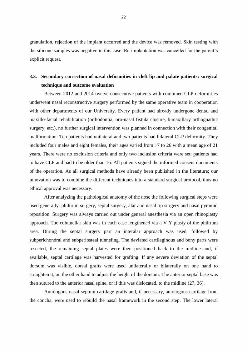
two medial crural cartilages were sutured together with the columella strut to set the tip
projection. If the lateral crus was buckled, strengthening was done with an onlay conchal
graft. Occasionally a shield graft was used to define the nasal tip. (Fig. 14) (27, 28, 36).
Figure 14: nasal grafting with septal cartilage; columella strut graft on the left and dorsal graft on the right
picture (A: alar cartilage, CS: columella strut, D: dorsal graft, S: nasal septum).
Bony pyramid surgery, if rarely necessary, consisted of hump resection, medial and
lateral osteotomies and repositioning of the nasal bone (27).
All 12 patients received both columella and dorsal grafts, harvested 11 times from the
nasal septum and once from the ears; shield graft or tip graft was used in three patients
fabricated from septal cartilage and an onlay alar graft, harvested from the concha, was
necessary in one case.
To measure the patient satisfaction, we adapted the ROEQ, which was first described
by Alsarraf et al to measure facial aesthetic surgery outcome (37). The questionnaire was
modified by Arima et al for patients having rhinoplasty (38). Our adapted ROEQ asks the
same four questions before and after surgery, the patient has to score each question with 0-4
points, where 0 represents the least satisfaction and 4 represents the highest one:
1. How much do you like the appearance of your nose?
2. How much can you breathe through your nose?
3. How much do you think your friends and those close to you like your nose?
4. Do you think the appearance of your nose limits your social or professional activities?
Scores for each individual question were compared using a t-test (IBM SPSS Statistics ver20),
p was considered significant at 0.005.
24
4. RESULTS
4.1. ALTERNATIVE SURGICAL METHODS FOR MALIGNANT TUMORS OF
THE HEAD AND NECK
4.1.1. The Modified Facial Degloving technique
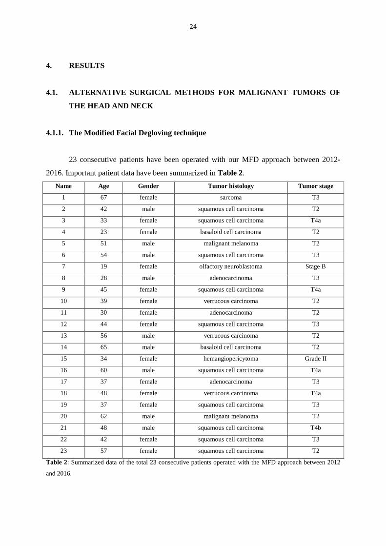
23 consecutive patients have been operated with our MFD approach between 2012-
2016. Important patient data have been summarized in Table 2.
Name Age Gender Tumor histology Tumor stage
1 67 female sarcoma T3
2 42 male squamous cell carcinoma T2
3 33 female squamous cell carcinoma T4a
4 23 female basaloid cell carcinoma T2
5 51 male malignant melanoma T2
6 54 male squamous cell carcinoma T3
7 19 female olfactory neuroblastoma Stage B
8 28 male adenocarcinoma T3
9 45 female squamous cell carcinoma T4a
10 39 female verrucous carcinoma T2
11 30 female adenocarcinoma T2
12 44 female squamous cell carcinoma T3
13 56 male verrucous carcinoma T2
14 65 male basaloid cell carcinoma T2
15 34 female hemangiopericytoma Grade II
16 60 male squamous cell carcinoma T4a
17 37 female adenocarcinoma T3
18 48 female verrucous carcinoma T4a
19 37 female squamous cell carcinoma T3
20 62 male malignant melanoma T2
21 48 male squamous cell carcinoma T4b
22 42 female squamous cell carcinoma T3
23 57 female squamous cell carcinoma T2
Table 2: Summarized data of the total 23 consecutive patients operated with the MFD approach between 2012
and 2016.
25
Table 3 represents the results of postoperative bilateral comparative Acoustic
Rhinometry.
Patient 1 Patient 2 Patient 3
Volume
(cm3)
left nasal
cavity
right nasal
cavity
left nasal
cavity
right nasal
cavity
left nasal
cavity
right nasal
cavity
external
nasal valve 1.45 1.70 1.00 1.10 0.33 1.45
internal
nasal valve 2.71 5.11 4.47 2.65 0.49 2.75
Totals 4.16 6.81 5.47 3.75 0.82 4,20
Table 3: Acoustic Rhinometry results (values of the operated side are bolded; larger number represents the
enlargement of the nasal cavity volume due to surgical resection), external nasal valve: inferior lateral cartilage;
internal nasal valve: inferior nasal concha head’s mucosa.
26
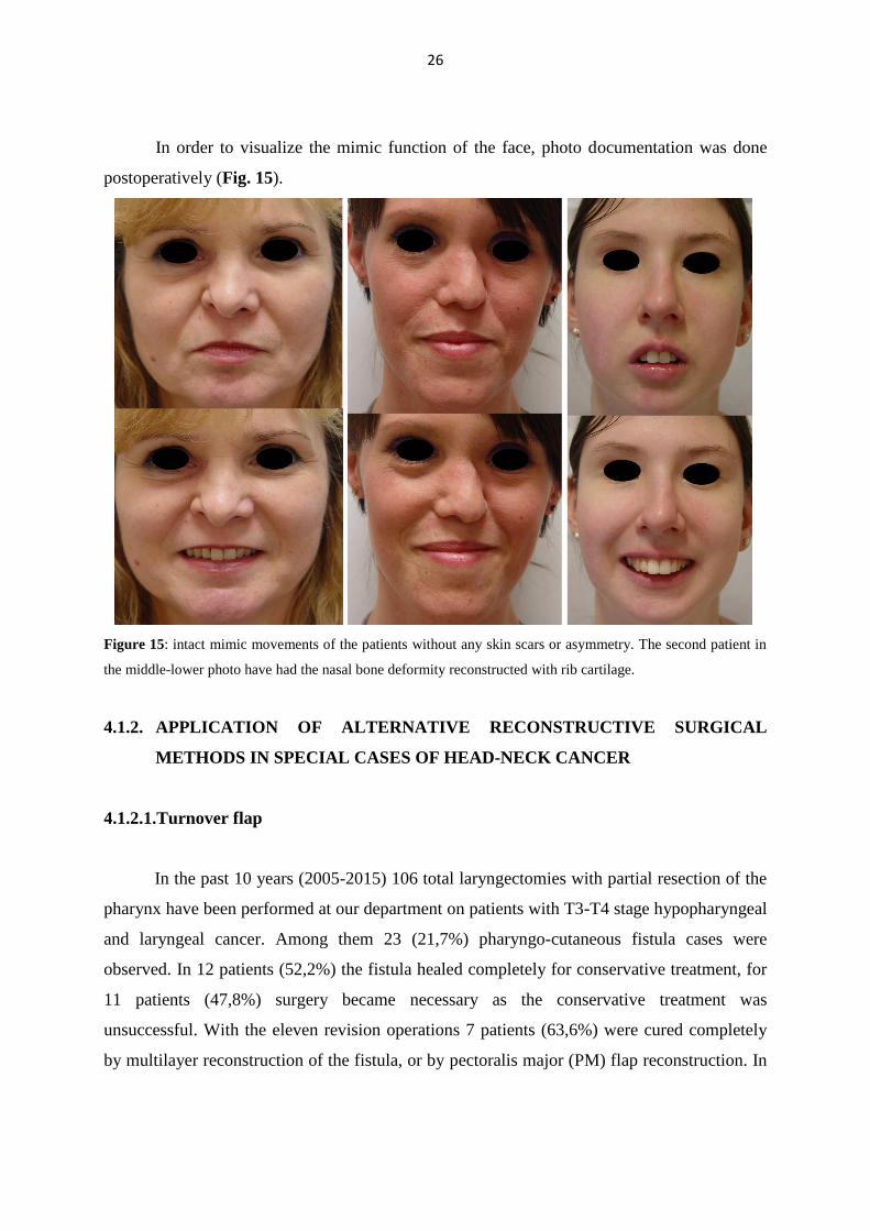
In order to visualize the mimic function of the face, photo documentation was done
postoperatively (Fig. 15).
Figure 15: intact mimic movements of the patients without any skin scars or asymmetry. The second patient in
the middle-lower photo have had the nasal bone deformity reconstructed with rib cartilage.
4.1.2. APPLICATION OF ALTERNATIVE RECONSTRUCTIVE SURGICAL
METHODS IN SPECIAL CASES OF HEAD-NECK CANCER
4.1.2.1.Turnover flap
In the past 10 years (2005-2015) 106 total laryngectomies with partial resection of the
pharynx have been performed at our department on patients with T3-T4 stage hypopharyngeal
and laryngeal cancer. Among them 23 (21,7%) pharyngo-cutaneous fistula cases were
observed. In 12 patients (52,2%) the fistula healed completely for conservative treatment, for
11 patients (47,8%) surgery became necessary as the conservative treatment was
unsuccessful. With the eleven revision operations 7 patients (63,6%) were cured completely
by multilayer reconstruction of the fistula, or by pectoralis major (PM) flap reconstruction. In
27
4 cases (36,4%) however, all the above listed methods failed, thus we decided on the
application of the simple TOF as the final solution to close the fistula. Three of these patients
received initial chemo-radiotherapy, one patient had postoperative oncotherapy. Oro-
cutaneous fistula developed in one case, without preliminary oncological treatment.
In the first presented case after the application of the TOF the defect was covered with
a split thickness skin graft. Complete healing has been achieved, with normal swallowing
function (Fig. 16).
Figure 16: two months’ postoperative picture
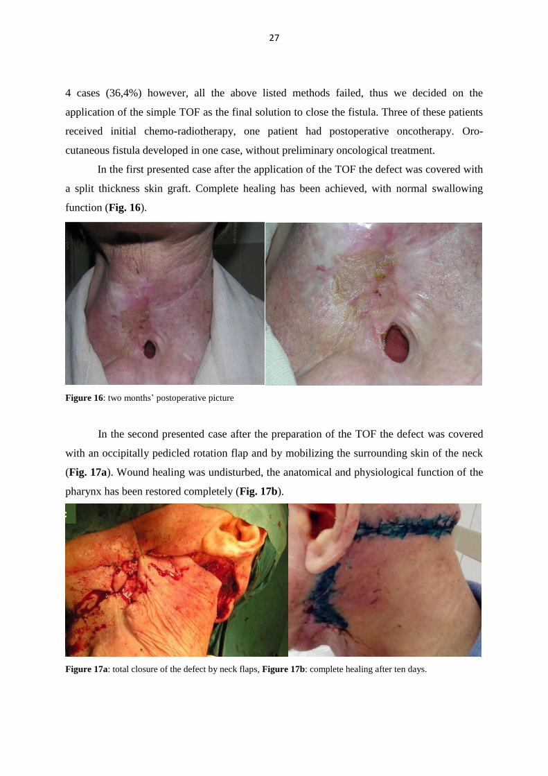
In the second presented case after the preparation of the TOF the defect was covered
with an occipitally pedicled rotation flap and by mobilizing the surrounding skin of the neck
(Fig. 17a). Wound healing was undisturbed, the anatomical and physiological function of the
pharynx has been restored completely (Fig. 17b).
Figure 17a: total closure of the defect by neck flaps, Figure 17b: complete healing after ten days.
28
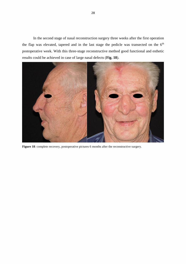
In the second stage of nasal reconstruction surgery three weeks after the first operation
the flap was elevated, tapered and in the last stage the pedicle was transected on the 6th
postoperative week. With this three-stage reconstructive method good functional and esthetic
results could be achieved in case of large nasal defects (Fig. 18).
Figure 18: complete recovery, postoperative pictures 6 months after the reconstructive surgery.

29
4.1.2.2.Extended lower trapezius musculocutaneous flap
With the novel application of the ELTMF the umpteen reconstructive operation was
successful, the flap remained viable, and the wounds healed primarily (Fig. 19).
Figure 19: Five weeks after the operation, the skin island is vital and wound healing undisturbed.
4.2. COMPLICATIONS OF WOUND HEALING AND REJECTION RELATED TO
COCHLEAR IMPLANTS – SURGICAL SOLUTIONS AND PREVENTIVE
OPTION
CI operations have been performed since 1995 at our Department. Until 31 December
2010 we carried out a total number of 223 CI surgeries, in detail 169 child and 54 adult
implantations. In 4 pediatric cases (2,37 % of child implantations, 1,79 % of all CI operations)
we faced skin necrosis above the receiver-stimulator unit and the concomitant exposure of the
implant. The epicutaneous patch test proved to be positive in 3 cases (75%) and negative in
one case (25%).
30
4.3. SECONDARY CORRECTION OF NASAL DEFORMITIES IN CLEFT LIP
AND PALATE PATIENTS: SURGICAL TECHNIQUE AND OUTCOME
EVALUATION
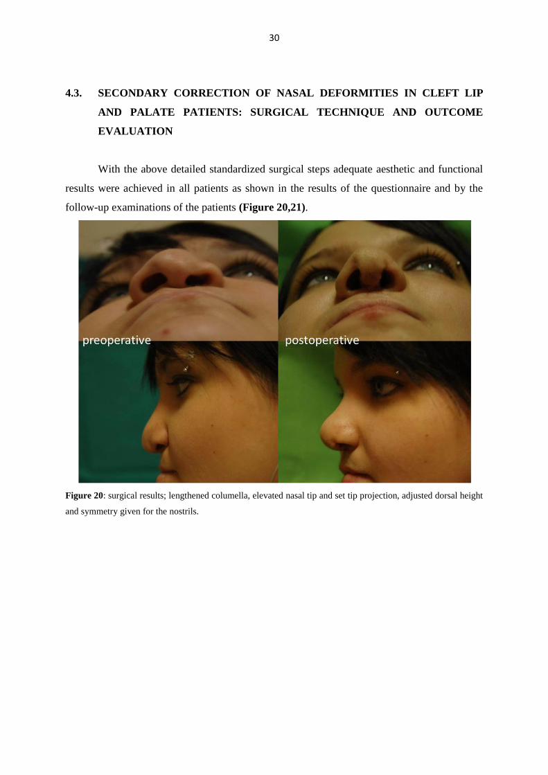
With the above detailed standardized surgical steps adequate aesthetic and functional
results were achieved in all patients as shown in the results of the questionnaire and by the
follow-up examinations of the patients (Figure 20,21).
Figure 20: surgical results; lengthened columella, elevated nasal tip and set tip projection, adjusted dorsal height
and symmetry given for the nostrils.
31
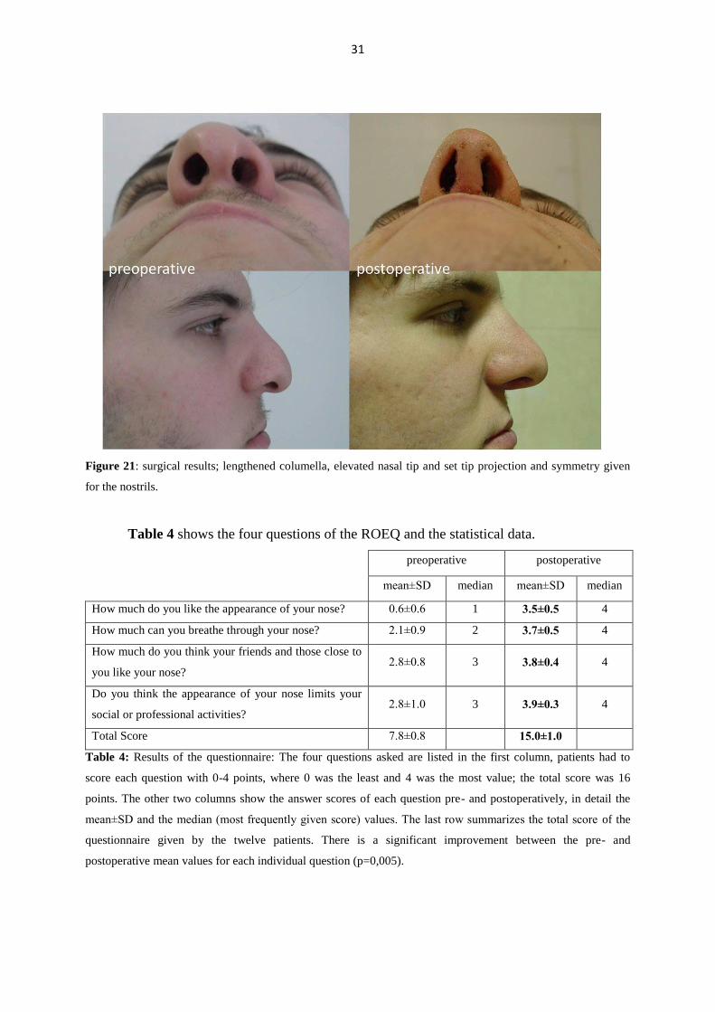
Figure 21: surgical results; lengthened columella, elevated nasal tip and set tip projection and symmetry given
for the nostrils.
Table 4 shows the four questions of the ROEQ and the statistical data.
preoperative postoperative
mean±SD median mean±SD median
How much do you like the appearance of your nose? 0.6±0.6 1 3.5±0.5 4
How much can you breathe through your nose? 2.1±0.9 2 3.7±0.5 4
How much do you think your friends and those close to
you like your nose? 2.8±0.8 3 3.8±0.4 4
Do you think the appearance of your nose limits your
social or professional activities? 2.8±1.0 3 3.9±0.3 4
Total Score 7.8±0.8 15.0±1.0
Table 4: Results of the questionnaire: The four questions asked are listed in the first column, patients had to
score each question with 0-4 points, where 0 was the least and 4 was the most value; the total score was 16
points. The other two columns show the answer scores of each question pre- and postoperatively, in detail the
mean±SD and the median (most frequently given score) values. The last row summarizes the total score of the
questionnaire given by the twelve patients. There is a significant improvement between the pre- and
postoperative mean values for each individual question (p=0,005).
32
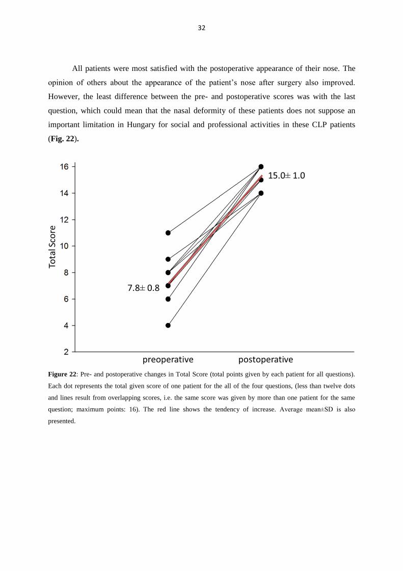
All patients were most satisfied with the postoperative appearance of their nose. The
opinion of others about the appearance of the patient’s nose after surgery also improved.
However, the least difference between the pre- and postoperative scores was with the last
question, which could mean that the nasal deformity of these patients does not suppose an
important limitation in Hungary for social and professional activities in these CLP patients
(Fig. 22).
Figure 22: Pre- and postoperative changes in Total Score (total points given by each patient for all questions).
Each dot represents the total given score of one patient for the all of the four questions, (less than twelve dots
and lines result from overlapping scores, i.e. the same score was given by more than one patient for the same
question; maximum points: 16). The red line shows the tendency of increase. Average mean±SD is also
presented.
33
5. DISCUSSION
5.1. ALTERNATIVE SURGICAL METHODS FOR MALIGNANT TUMORS OF
THE HEAD AND NECK
5.1.1. The Modified Facial Degloving technique
Lizars was the first to perform a maxillary resection, since then the surgical technique
has developed significantly (33, 34). The still widely-used open approach was first described
by Fergusson (2). The importance of functional and aesthetic integrity of the face was already
emphasized by Casson et al in 1974, however in 1979 Conley and Price were the ones who
used and published the facial degloving technique based on the elevation of the soft tissues of
the midface in the surgical treatment of sino-nasal malignant tumors (31-34). In the decades
passed since then several modifications of the method have been described, highlighting the
favorable exploration of the surgical field and the good cosmetic result provided by the
method (39, 40).
At our Department we have been using the method routinely since 2010 for malignant
and benign sino-nasal tumors. None of the possible side effects of the method have been
observed (temporary infraorbital anesthesia, nasal deformity, nasal valve stenosis and
epiphora requiring cannulation of the nasolacrimal duct) (35, 40). However, among the
patients operated with Weber-Ferguson’s technique all side effects occurred. Some of our
patients complained about dryness of the nasal mucosa, crusting and recurrent nasal bleeding;
however, all of these could be easily treated locally.
Almost all benign (e.g. inverted papilloma, pituitary adenoma) and some selected
cases of malignant tumors of the maxillo-ethmoidal region and even the skull base are treated
mainly endoscopically at our University in accordance with the international
recommendations and tendencies (1). However, the proper and adequate use of the
endoscopic approach in our opinion requires well trained experts with a lot of surgical
experience in this field and special set of expensive instruments, both of which is not
available everywhere by all means. Furthermore, the endoscopic approach also has some
limitations, which are highlighted by expert authors in cornerstone papers of the literature (1).
34
These limitations are: orbital infiltration, total maxillectomy, skin excision, involvement of
anterior/lateral wall of frontal sinus, dura or brain involvement, insertional beak on the
anterior maxillary wall, infratemporal fossa invasion, tumor lateral to internal carotid artery,
internal carotid artery or cavernous sinus invasion, etc.
We think our MFD technique represents a simple, relatively easy and ablative
alternative in between the two endpoints of surgical therapy for maxillo-ethmoidal lesions, the
minimally-invasive endoscopic approach and the open, distorting surgeries. Our method can
be easily combined with endoscopic surgery, and can even substitute it when any of the above
listed limitations are present; the postoperative results of the two approaches are absolutely
comparable. Moreover, if necessary, conversion to/combination with open surgeries is also
possible, even intraoperatively.
As it has been published before, there is no significant difference in the recurrence of
inverted papilloma between patients operated via the open approach (lateral rhinotomy with
Weber-Fergusson’s incision) or with the facial degloving technique (40). The same
perception was true for our patients according to our previous experience, however, nowadays
we would exclusively choose the scarless approach for this benign lesion - which still carries
the potential of malignant transformation - if the endoscopic technique fails for any reason.
In the demonstrated cases we have chosen the facial degloving versus the endoscopic
technique because of the involvement of the orbit in the first patient and invasion of the
frontal sinus in the second patient. Although there are recent publications showing that
endoscopic management of olfactory neuroblastoma provides higher overall and disease-free
survival rates compared with external approaches (41), mastering such endoscopic technique
requires long learning curves. We have been pursuing hard to reach and further extend our
limitations in endoscopic surgery, for our thirdly presented young female patient the facial
degloving approach meant a safer procedure with the same excellent cosmetic results.
Avoiding an external approach was essential in each case.
In order to prove that the nasal breathing function is not affected by the method itself
and the postoperative scar formation, we carried out acoustic rhinometry minimum one year
after the operation on the basis of literature data (42). The results, presented in Table 3 show
that no narrowing of the nasal cavity is observed, neither in the external, nor in the internal
35
nasal valve area in comparison with the contralateral side. Statistical analysis was not
performed due to the small number of patients.
Postoperative photo documentation of the face mimic shows no dysfunction on the
operated side; however, in our second patient the left lamina of the nasal bone was also
resected because of tumorous infiltration, so slight rotation of the nasal pyramid to the
operated left side is visible. Without any skin and underlying soft tissue scars secondary
correction with rib cartilage graft of this deformity was carried out with acceptable aesthetic
result (Figure 15).
5.1.2. Application of alternative reconstructive surgical methods in special cases of
head-neck cancer
5.1.2.1.Turnover flap
The incidence of oro-cutaneous and pharyngo-cutaneous fistulas with or without
preliminary radiotherapy is between 2-66%, with an average of 10-15% in most publications
(43). Although fistulas may close spontaneously occasionally, after long lasting conservative
treatment that help granulation and epithelization, this means a very sustained healing time
and depletive psycho-social burden (e.g. nasogastric feeding tube or percutaneous
gastrostomy). Direct relation between the preoperative radiotherapy and the development of
fistulas have been proved by several publications (8, 43); furthermore, age, nutritive status,
wound superinfection and tumor stage are additive predisposing factors (43). Despite the
relatively high frequency of fistula occurrence, its surgical treatment is a difficult and
challenging issue.
In head and neck tumor patients, who have disturbed wound healing (oro-cutaneous or
pharyngo-cutaneous fistula, skin necrosis, etc.), the affected skin and soft tissue area is
usually damaged by irradiation, scabby and has deteriorated circulation. In the literature
several methods have been described for the surgical treatment of fistulas: from
fasciocutaneous island flaps through local and distant skin- and musculocutaneous flaps up to
free microvascular flaps (4, 6, 8, 43). In spite of all these methods, the recurrence of the
36
fistula is rather high, in case of PM musculocutaneous flaps it can be 35% – which may
suspect even local tumor recurrence (43).
According to our experience and observations detailed above, we tried to find the
safest and simplest reconstructive method without much operative burden for these five
patients with pharyngo- and oro-cutaneous fistula, which proved to be the TOF.
In 1987 Spear et al were the first to describe the nasolabial TOF for alar
reconstruction. Since then several modifications of the method have been described
depending on the size and localization of the defect (44-46).
Surgical reconstruction of the defects of the cartilaginous framework and the perialar
region of the nose after radical resection of malignant tumors is a highly challenging issue for
the head and neck surgeon. Not only because of the complex spatial structure of the nose, but
also the consideration of the aesthetic subunits of it, without which the combined
reconstruction of function and appearance could not be fully performed. In order to be able to
achieve the latter, reconstruction of all tissue layers of the nose is inevitable (45).
For our patients we have used turnover skin flaps mainly from the nasolabial region to
substitute the nasal mucosa, the inner lining of the nose. We have found our method adequate
for functional reconstruction, as it replaces the mucosa perfectly, it is thin, incorporates
perfectly into the nasal cavity and prevents stenosis and crusting. The rich vascular supply of
the flaps provides good nutritive medium for immediate implantation of cartilage grafts.
Nasal skin has always been reconstructed with pedicled forehead flap.
5.1.2.2. Extended lower trapezius musculocutaneous flap
In 1979, Demergasso and Piazza described the trapezius musculocutaneous flap, in
which the transverse cervical artery and paraspinous attachment of the trapezius were left
intact (47). In 1980, Baek et al. first described the ELTMF for reconstructing cutaneous
defects or for subcutaneous augmentation of the face (48). In 2000, Tan and Tan reported the
vascular anatomy and clinical use of the ELTMF based solely on the dorsal scapular arterial
system (49). In 2004, Ugurlu et al. proposed the use of the extended vertical trapezius
musculocutaneous flap based solely on the transverse cervical artery in a salvage procedure
for failed previous flaps and recurrent tumors (50).
37
The superior trapezius flap is based on the occipital artery and its paraspinous
perforators, while the lateral and lower island trapezius musculocutaneous flaps are based on
the branches of the transverse cervical artery (12, 49, 51). The use of the lower trapezius
musculocutaneous flap is contraindicated in cases where there is suspicion of trauma to the
descending branch of the transverse cervical artery (12, 52). Tan and Tan incorporated an
extension of the flap that runs obliquely from the tip of the scapula towards the mid-axillary
line (49). Their technique was based on the vascular supply from the dorsal scapular artery,
which originates either directly from the subclavian artery as an independent branch, or from
the trunk of the transverse cervical artery (12, 49, 51, 52).
In comparison with the LDMF, we can say that the latissimus dorsi muscle offers a
limited axis of rotation because of the axillary origin of its pedicle and the frequent need for a
split thickness skin graft at the donor site because of the difficulty to achieve a tension-free
closure of the wound during the management of extensive defects (12, 14, 15, 52, 53).
The ELTMF flap has several advantages: the donor site can usually be closed easily,
resulting in a tension-free but rather long scar; the flap fills the defect created by the neck
dissection and covers the vessels of the neck, preventing damage to the vessels; and the long,
thin musculocutaneous pedicle allows for easy transfer of the island flap, which can even be
tunneled into a defect if necessary (12, 15, 50, 52, 53)
With our first solution, the LDMF, we found that the problem was the tunneling of the
flap. However, the supplying vessels of the trapezius muscle and the muscle itself remained
intact, and we were able to use this flap for the secondary reconstruction (12, 50).
5.2. COMPLICATIONS OF WOUND HEALING AND REJECTION RELATED TO
COCHLEAR IMPLANTS – SURGICAL SOLUTIONS AND PREVENTION
OPTION
CI nowadays has become a safe and widely used surgical method for the treatment of
either prelingual or postlingual deafness. The intervention, like every other operation, may
have complications. According to Cohen and Hoffman the so called major complications of
CI surgery are the following: dislocation of the electrode array, permanent facial nerve palsy,
cerebrospinal fluid leak and damage of the device (17, 19-21). The most frequent minor
38
complications are temporary facial nerve palsy, disturbed wound healing and transient
dizziness (17, 18, 20, 21, 54).
The incidence of skin necrosis following CI surgery, which is one of the possible
complications as presented by the authors, varies between 0-5.4% in the international
literature after Cohen and Hoffman (19).
The treatment of skin necrosis depends mainly on the extent of the defect. In case of
small, superficial lesions, necrectomy, skin regeneration therapy, usage of e.g. Epigard™ or
Allevyn™ might be satisfactory. Choosing the appropriate surgical method in case of larger
defects is sometimes difficult. One option is to relocate the implant to a “safer” location in the
surrounding of the original operation site, however this is technically difficult and results is
new scars and areas of alopecia (55-58). Local rotation flaps, which also produce scars, are
the most reasonable choice, if the skin incision line avoids the implant location. Occipital
flaps have the best vascularization and their other advantage is their proximity to the defect
and that the scar lines can be easily disguised by a longer hair style (55, 56, 58, 59). A special
entity is the Superficial Temporal Fascia Flap, which receives its blood supply from the fascia
of the temporal muscle, so a well vascularized layer is placed between the skin and the
implant. This way the danger of repeated skin necrosis decreases (56, 57). Following several
correction surgery and the formation of multiplex scars the use of a tissue expander may be
required in order to gain more skin, like in our first case, and to allow the removal of the
majority of scars and covering of the defect with hairy skin.
Possible causes of skin necrosis can be the inadequately planned and placed incision
line, over-narrowing of the flap, displacement of the implant, latent autoimmune diseases
(vasculitis e.g.) (18, 19, 54). In our opinion other factors, like surgical incision lines and
technique, too strong magnets, infection and individual silicone allergy may also play a role in
the pathogenesis. We discuss these as follows:
In all of our four cases Nucleus 24 type, silicone covered implant was used for the first
implantation. The surgical intervention was carried out by the same, highly trained and
experienced person with the same technique; this excludes the causative role of the surgeon in
connection with the skin necrosis.
The magnet is located in the transmitter coil and its strength is adjustable – four levels
of strength is available. In children we use the magnet strength 1 or 2. However the skin
39
defect never occurred under the magnet, where the skin is between the transmitter and the
receiver units, rather by the edge of the receiver-stimulator part which is covered with
silicone.
As we always met the patients after the development of the skin defect, only tissue
samples of necrectomy were histologically examined. Fibrosis, granulocytes and reparative
signs were mentioned in histopathology data.
Wound cultures were always negative, no bacterial infection was proved. Also
perioperative and postoperative parenteral antibiotic treatment is administered routinely in all
of our CI patients.
Therefore, we thought that individual silicone hypersensitivity can be in the
background of the skin flap necrosis in most of the cases. Several publications dealt with
silicone allergy, they mostly examined the blood level of auto-antibodies against collagen
type I and II by the Enzyme-Linked Immunosorbent Assay (ELISA) method in patients with
silicone breast implants and they compared the results with the level of the same auto-
antibodies in patients suffering from autoimmune connective tissue diseases and healthy
control patients. However, no significant relationship was observed (60). In general, we can
state, that any kind of silicone implant may induce the production of auto-antibodies in
genetically susceptible patients causing immune system disorders (23).
Epicutaneous Patch Testing (EPT) is the gold standard method for the diagnosis of
allergic contact dermatitis (ACD). The use of patch testing to diagnose ACD was first
developed by Jadassohn in 1895 (61). Sulzberger brought the technique to the United States
in the 1930s (61). Over the past 40 years, the pathophysiological understanding of ACD and
the technique of patch testing have been expanded and redefined. Since in all of our four cases
a Nucleus type, silicone-covered implant was used, the possibility of individual silicone
allergy was obvious; our goal was to prove the possibility of silicone allergy with the
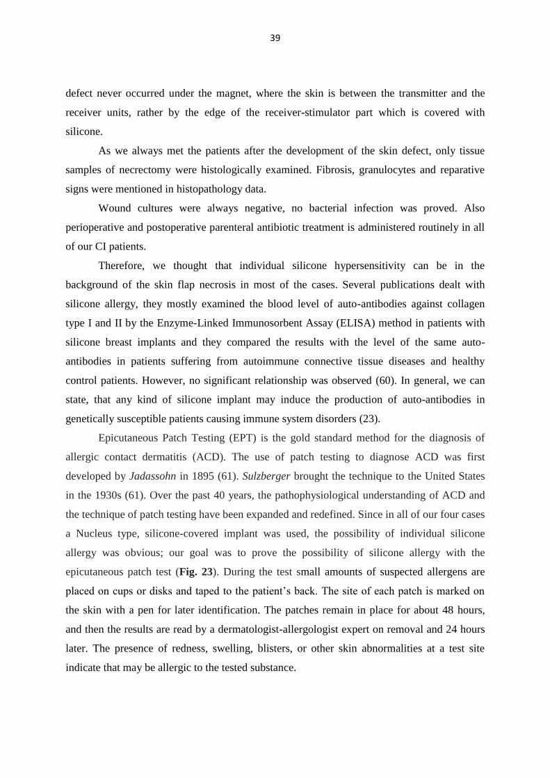
epicutaneous patch test (Fig. 23). During the test small amounts of suspected allergens are
placed on cups or disks and taped to the patient’s back. The site of each patch is marked on
the skin with a pen for later identification. The patches remain in place for about 48 hours,
and then the results are read by a dermatologist-allergologist expert on removal and 24 hours
later. The presence of redness, swelling, blisters, or other skin abnormalities at a test site
indicate that may be allergic to the tested substance.
40
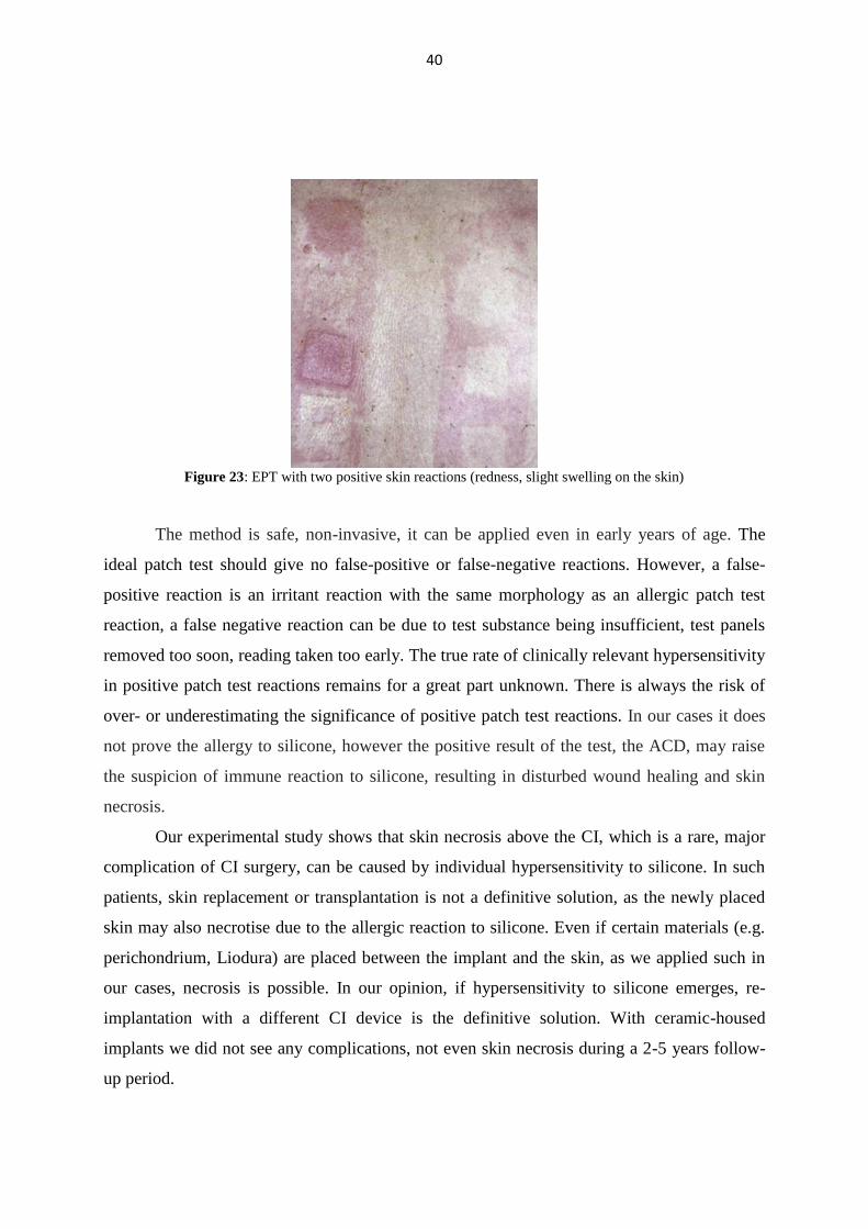
Figure 23: EPT with two positive skin reactions (redness, slight swelling on the skin)
The method is safe, non-invasive, it can be applied even in early years of age. The
ideal patch test should give no false-positive or false-negative reactions. However, a false-
positive reaction is an irritant reaction with the same morphology as an allergic patch test
reaction, a false negative reaction can be due to test substance being insufficient, test panels
removed too soon, reading taken too early. The true rate of clinically relevant hypersensitivity
in positive patch test reactions remains for a great part unknown. There is always the risk of
over- or underestimating the significance of positive patch test reactions. In our cases it does
not prove the allergy to silicone, however the positive result of the test, the ACD, may raise
the suspicion of immune reaction to silicone, resulting in disturbed wound healing and skin
necrosis.
Our experimental study shows that skin necrosis above the CI, which is a rare, major
complication of CI surgery, can be caused by individual hypersensitivity to silicone. In such
patients, skin replacement or transplantation is not a definitive solution, as the newly placed
skin may also necrotise due to the allergic reaction to silicone. Even if certain materials (e.g.
perichondrium, Liodura) are placed between the implant and the skin, as we applied such in
our cases, necrosis is possible. In our opinion, if hypersensitivity to silicone emerges, re-
implantation with a different CI device is the definitive solution. With ceramic-housed
implants we did not see any complications, not even skin necrosis during a 2-5 years follow-
up period.
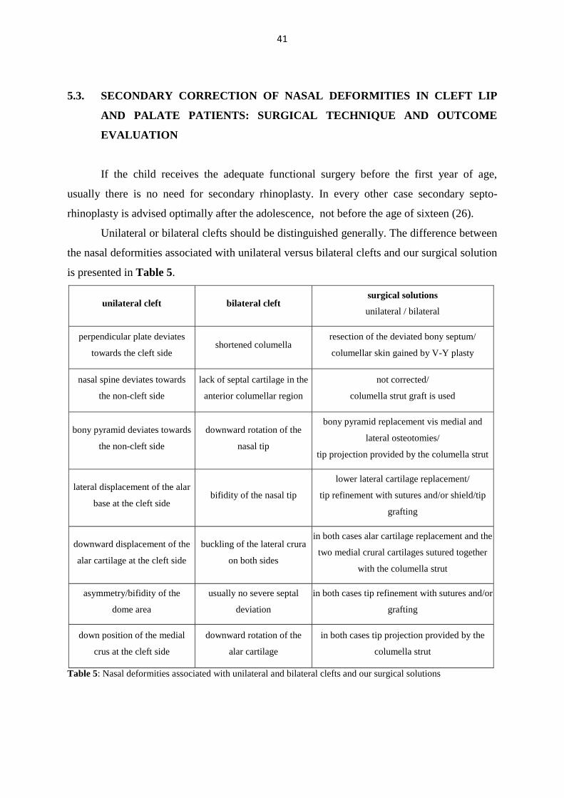
41
5.3. SECONDARY CORRECTION OF NASAL DEFORMITIES IN CLEFT LIP
AND PALATE PATIENTS: SURGICAL TECHNIQUE AND OUTCOME
EVALUATION
If the child receives the adequate functional surgery before the first year of age,
usually there is no need for secondary rhinoplasty. In every other case secondary septo-
rhinoplasty is advised optimally after the adolescence, not before the age of sixteen (26).
Unilateral or bilateral clefts should be distinguished generally. The difference between
the nasal deformities associated with unilateral versus bilateral clefts and our surgical solution
is presented in Table 5.
unilateral cleft bilateral cleft surgical solutions
unilateral / bilateral
perpendicular plate deviates
towards the cleft side shortened columella
resection of the deviated bony septum/
columellar skin gained by V-Y plasty
nasal spine deviates towards
the non-cleft side
lack of septal cartilage in the
anterior columellar region
not corrected/
columella strut graft is used
bony pyramid deviates towards
the non-cleft side
downward rotation of the
nasal tip
bony pyramid replacement vis medial and
lateral osteotomies/
tip projection provided by the columella strut
lateral displacement of the alar
base at the cleft side bifidity of the nasal tip
lower lateral cartilage replacement/
tip refinement with sutures and/or shield/tip
grafting
downward displacement of the
alar cartilage at the cleft side
buckling of the lateral crura
on both sides
in both cases alar cartilage replacement and the
two medial crural cartilages sutured together
with the columella strut
asymmetry/bifidity of the
dome area
usually no severe septal
deviation
in both cases tip refinement with sutures and/or
grafting
down position of the medial
crus at the cleft side
downward rotation of the
alar cartilage
in both cases tip projection provided by the
columella strut
Table 5: Nasal deformities associated with unilateral and bilateral clefts and our surgical solutions
42
6. CONCLUSIONS
6.1. ALTERNATIVE SURGICAL METHODS FOR MALIGNANT TUMORS OF
THE HEAD AND NECK
In our opinion the MFD technique might be a useful alternative or supplementary of
the widely used minimally invasive endoscopic approaches in sino-nasal surgery (30, 62).
Moreover, it might help the less skilled surgeons in the acquisition of the endoscopic
techniques for resecting tumors of the maxillo-ethmoidal region. By combining the two
methods, the required radicality can be achieved with good cosmetic results. If needed – in
certain severe cases, where e.g. orbital surgery (e.g. exenteration) is also necessary – the
method could also be combined with the open approach (63). The latter should possibly be
avoided nowadays as a first-line approach due to the stigmatizing skin scars and facial
deformities.
According to our experience TOFs might be safe tissue replacement alternatives even
in special cases without much operative burden, when e.g. the surrounding tissue is scabby
and irradiated or tissue harvesting is limited due to the anatomic localization. The presented
cases are fortunately not frequent; however, according to the international literature standard
deviation among irradiated patients is relatively high (2-66%). The demonstrated surgical
methods are able to restore anatomical and physiological functions through the reconstruction
of head-neck defects, which are result of radical tumor resection or develop as a complication.
Another advantage of these flaps is the relatively low operative burden and that they can be
harvested from the surroundings rather easily, however the over-irradiated skin can be the risk
factor of failure. In such cases musculocutaneous flaps are the gold standard of therapy, but in
our demonstrated patients no permanent success has been reached by them, no matter the
routine use of these flaps and our great operative experience. TOFs then are adequate
alternatives, as they can be easily prepared from the remnants of the musculocutaneous flap.
The ELTMF flap has several advantages: the donor site can usually be closed easily,
resulting in a tension-free but rather long scar; the flap fills the defect created by the neck
dissection and covers the vessels of the neck, preventing damage to the vessels; and the long,
43
thin musculocutaneous pedicle allows for easy transfer of the island flap, which can even be
tunneled into a defect if necessary (15, 50, 52, 53).
6.2. COMPLICATIONS OF WOUND HEALING AND REJECTION RELATED TO
COCHLEAR IMPLANTS – SURGICAL SOLUTIONS AND PREVENTION
OPTION
CI is one of the most advanced surgical interventions to restore hearing. Skin necrosis
and concomitant implant rejection seems to be one of the most difficult problems to handle
from among the possible complications. In our experience the closure of the skin defect over
the implant with loco-regional flaps or the numerous plastic surgical operations done in
accordance with international recommendations, does not always provide a permanent
solution. However, we think, that the method described above could be a safe, non-invasive,
fast method to test the possibility of skin necrosis and implant rejection in case of a silicone-
covered CI also in children. Final recovery of the surgical site and the skin defect – because of
the factors mentioned above – can only be expected after the removal of the implant.
6.3. SECONDARY CORRECTION OF NASAL DEFORMITIES IN CLEFT LIP
AND PALATE PATIENTS: SURGICAL TECHNIQUE AND OUTCOME
EVALUATION
In our opinion with the above mentioned operative protocol we were able to
standardize our surgical technique in the secondary septo-rhinoplasty of patients with CLP.
Skin incisions, cartilage harvesting and grafting, endonasal surgery and re-establishment of
the nasal framework were successfully unified thus providing a more predictable functional
and aesthetic outcome for the already psychosocially affected CLP patients.
Statistical comparative analysis of the pre- and postoperative data from our ROEQ
confirmed, that with our standardized surgical protocol improved aesthetic and functional
results and good patient satisfaction rates were achieved.
We think our modified ROEQ is an adequate and simple method for the evaluation of
the surgical results of secondary septo-rhinoplasty among patients with CLP.
44
7. ACKNOWLEDGEMENT
Without help, support and encouragement from many colleagues, I would have never
been able to complete this work.
First of all, I would like to express my gratitude and thanks to my supervisor, Dr. László
Iván and my professor László Rovó, for encouraging and supporting my work from the very
beginning.
I wish to express my gratitude to Professor József Jóri and Professor József Géza Kiss
who introduced me to clinical work and provided me with the opportunity to begin my
scientific career.
I would like to express my gratitude to Professor Lajos Kemény, Head of Department of
Dermatology and Allergology, and Dr. Klára Kapitány, the former Head of Plastic Surgery
Ward for providing me the opportunity to start my plastic surgery training.
I am deeply indebted to my colleagues, János Varga and Gábor Mohos, my teachers and
mentors in plastic surgery for their cooperative work.
Thanks to my colleagues at the Department of Oto-Rhino-Laryngology and Head-Neck
Surgery and the Department of Dermatology and Allergology – Plastic Surgery.
Last but not least, I wish to thank my family and friends for their constant support and
encouragement.
45
8. REFERENCES
1. Lund VJ, Stammberger H, Nicolai P, Castelnuovo P, Beal T, Beham A, et al. European
position paper on endoscopic management of tumours of the nose, paranasal sinuses and
skull base. Rhinology Supplement. 2010(22):1-143.
2. Fergusson W. Operations of the upper jaw. 2nd ed. Fergusson W, editor. Philadelphia:
Lea and Blanchard; 1845.
3. Conley J, Price JC. Sublabial approach to the nasal and nasopharyngeal cavities. Am J
Surg. 1979;138(4):615-8.
4. Caronia FP, Fiorelli A, Santini M, Castorina S. A persistent tracheocutaneous fistula
closed with two hinged skin flaps and rib cartilage interpositional grafting. Gen Thorac
Cardiovasc Surg. 2016;64(10):625-8.
5. Liang W, Zhou Z, Zhao Z. Application of split gluteus maximus muscle--adipofascial
turnover flap and subcutaneous tension-reducing suture technique in repair of decubitus
ulcers. Int Surg. 2014;99(4):447-51.
6. Tatekawa Y, Yamanaka H, Hasegawa T. Closure of a tracheocutaneous fistula by two
hinged turnover skin flaps and a muscle flap: A case report. Int J Surg Case Rep.
2013;4(2):170-4.
7. Zahiri HR, Lumpkins K, Kelishadi S, Stromberg JA, Silverman RP, Slezak S, et al.
Pectoralis major turnover versus advancement technique for sternal wound
reconstruction. Ann Plast Surg. 2013;70(2):211-5.
8. Neubauer P, Canadas K, Sasaki CT. Delayed platysma myocutaneous turnover flap for
repair of pharyngocutaneous fistula. Am J Otolaryngol. 2015;36(1):93-6.
9. Patel KB, Taghinia AH, Proctor MR, Warf BC, Greene AK. Extradural
myelomeningocele reconstruction using local turnover fascial flaps and midline linear
skin closure. Journal of plastic, reconstructive & aesthetic surgery : JPRAS.
2012;65(11):1569-72.
10. Sierakowski A, Niranjan N. Sternomastoid fascial turnover flap for Frey syndrome: An
unusual case. Journal of plastic, reconstructive & aesthetic surgery : JPRAS.
2011;64(2):e53-4.
46
11. Silva JB, Faloppa F, Albertoni W, Gazzalle A, Cunha GL. Adipofascial turnover flap
for the coverage of the dorsum of the thumb: an anatomic study and clinical application.
J Hand Surg Eur Vol. 2013;38(4):371-7.
12. Mohos G, Vass G, Kemeny L, Jori J, Ivan L. Extended lower trapezius myocutaneous
flap to cover a deep lateral neck defect on irradiated skin: a new application. Journal of
plastic surgery and hand surgery. 2013;47(1):70-2.
13. Ndayishimiye JM, Husson JL, Watier E. Reconstruction of extensive posterior mid-
thoracic soft-tissue defects after spinal surgery on irradiated skin. Orthop Traumatol
Surg Res. 2009;95(4 Suppl 1):S35-40.
14. Datta G, Boriani F, Degano K, Carlucci S, Ferrando PM, Verna G. Combination of two
long-pedicled myocutaneous flaps for closure of a complex contralateral dorsal defect.
Ann Thorac Surg. 2009;87(6):1930-3.
15. Papadas T, Goumas P, Alexopoulou MM, Papakyriakos I, Papavasiliou D,
Antonopoulos D. Cancer patients with large defects. Reconstructional options: a case
study. Braz J Otorhinolaryngol. 2005;71(1):87-90.
16. Ribari O, Kustel M. Overview of our 13-year-long experience with cochlear implants.
Acta chirurgica Hungarica. 1998;37(1-2):33-7.
17. Kempf HG, Johann K, Lenarz T. Complications in pediatric cochlear implant surgery.
Eur Arch Otorhinolaryngol. 1999;256(3):128-32.
18. Miyamoto RT, Young M, Myres WA, Kessler K, Wolfert K, Kirk KI. Complications of
pediatric cochlear implantation. Eur Arch Otorhinolaryngol. 1996;253(1-2):1-4.
19. Cohen NL, Hoffman RA. Complications of cochlear implant surgery in adults and
children. Ann Otol Rhinol Laryngol. 1991;100(9 Pt 1):708-11.
20. Kim CS, Oh SH, Chang SO, Kim HM, Hur DG. Management of complications in
cochlear implantation. Acta Otolaryngol. 2008;128(4):408-14.
21. Migirov L, Taitelbaum-Swead R, Hildesheimer M, Kronenberg J. Revision surgeries in
cochlear implant patients: a review of 45 cases. Eur Arch Otorhinolaryngol.
2007;264(1):3-7.
22. Rubio A, Ponvert C, Goulet O, Scheinmann P, de Blic J. Allergic and nonallergic
hypersensitivity reactions to silicone: a report of one case. Allergy. 2009;64(10):1555.
47
23. Teuber SS, Rowley MJ, Yoshida SH, Ansari AA, Gershwin ME. Anti-collagen
autoantibodies are found in women with silicone breast implants. Journal of
autoimmunity. 1993;6(3):367-77.
24. Fisher DM, Sommerlad BC. Cleft lip, cleft palate, and velopharyngeal insufficiency.
Plast Reconstr Surg. 2011;128(4):342e-60e.
25. Piffko J, Meyer U, Joos U. Possibilities and limitations in evaluating treatment concepts
in lip-jaw-palate clefts. Mund-, Kiefer- und Gesichtschirurgie : MKG. 2002;6(1):49-52.
26. Nakamura N, Sasaguri M, Okawachi T, Nishihara K, Nozoe E. Secondary correction of
bilateral cleft lip nose deformity - Clinical and three-dimensional observations on pre-
and postoperative outcome. J Craniomaxillofac Surg. 2011;39(5):305-12.
27. Nolst Trenité GJ. Secondary surgery of the cleft-lip nose. Nolst Trenité GJ, editor.
Amsterdam: Kugler Publications; 1993.
28. Pitak-Arnnop P, Hemprich A, Dhanuthai K, Yildirim V, Pausch NC. Panel and patient
perceptions of nasal aesthetics after secondary cleft rhinoplasty with versus without
columellar grafting. J Craniomaxillofac Surg. 2011;39(5):319-25.
29. Baumann A, Ewers R. Midfacial degloving: an alternative approach for traumatic
corrections in the midface. Int J Oral Maxillofac Surg. 2001;30(4):272-7.
30. El-Banhawy OA, Shehab El-Dien Ael H, Amer T. Endoscopic-assisted midfacial
degloving approach for type III juvenile angiofibroma. International journal of pediatric
otorhinolaryngology. 2004;68(1):21-8.
31. Ferreira LM, Rios AS, Gomes EF, Azevedo JF, Araujo Rde P, Moraes RB. Midfacial
degloving--access to nasal cavity and paranasal sinuses lesions. Braz J
Otorhinolaryngol. 2006;72(2):158-62.
32. Kitagawa Y, Baur D, King S, Helman JI. The role of midfacial degloving approach for
maxillary cysts and tumors. J Oral Maxillofac Surg. 2003;61(12):1418-22.
33. Jaber JJ RF, Zender CA. Facial degloving approach to the midface. Op Tech Otolar.
2010;21:171-4.
34. Zaghloul AS, Nouh MA, Fatah HA. Midfacial degloving approach for malignant
maxillary tumors. J Egypt Natl Canc Inst. 2004;16(2):69-75.
35. Har-El G. Medial maxillectomy via midfacial degloving approach. Op Tech Otolar.
1999;10(2):82-6.
48
36. Chaithanyaa N, Rai KK, Shivakumar HR, Upasi A. Evaluation of the outcome of
secondary rhinoplasty in cleft lip and palate patients. Journal of plastic, reconstructive
& aesthetic surgery : JPRAS. 2011;64(1):27-33.
37. Alsarraf R, Larrabee WF, Jr., Anderson S, Murakami CS, Johnson CM, Jr. Measuring
cosmetic facial plastic surgery outcomes: a pilot study. Arch Facial Plast Surg.
2001;3(3):198-201.
38. Arima LM, Velasco LC, Tiago RS. Crooked nose: outcome evaluations in rhinoplasty.
Braz J Otorhinolaryngol. 2011;77(4):510-5.
39. Jeon SY, Jeong JH, Kim HS, Ahn SK, Kim JP. Hemifacial degloving approach for
medial maxillectomy: a modification of midfacial degloving approach. Laryngoscope.
2003;113(4):754-6.
40. Kim HJ, Kim CH, Kang JW, Shin WC, Kim YS, Do YK, et al. A modified midfacial
degloving approach for the treatment of unilateral paranasal sinus tumours. J
Craniomaxillofac Surg. 2011;39(4):284-8.
41. Fu TS, Monteiro E, Muhanna N, Goldstein DP, de Almeida JR. Comparison of
outcomes for open versus endoscopic approaches for olfactory neuroblastoma: A
systematic review and individual participant data meta-analysis. Head Neck. 2016;38
Suppl 1:E2306-16.
42. Lindemann J, Leiacker R, Sikora T, Rettinger G, Keck T. Impact of unilateral sinus
surgery with resection of the turbinates by means of midfacial degloving on nasal air
conditioning. Laryngoscope. 2002;112(11):2062-6.
43. Sadigh PL, Wu CJ, Feng WJ, Hsieh CH, Jeng SF. New double-layer design for 1-stage
repair of orocutaneous and pharyngocutaneous fistulae in patients with postoperative
irradiated head and neck cancer. Head Neck. 2016;38 Suppl 1:E353-9.
44. Godoy-Gijon E, Palacio-Aller L, Gonzalez-Sabin M. Reconstruction of the ala nasi
using a malar turnover island pedicle flap. Actas dermo-sifiliograficas.
2015;106(3):233-5.
45. Jellinek NJ, Nguyen TH, Albertini JG. Paramedian forehead flap: advances, procedural
nuances, and variations in technique. Dermatologic surgery. 2014;40 Suppl 9:S30-42.
49
46. Shim HC, Kim G, Choi JH, Kim JH, Kim EJ, Park HJ, et al. The reverse nasolabial flap
with a cartilage graft for the repair of a full-thickness alar defect: a single-stage
procedure. Ann Dermatol. 2014;26(3):377-80.
47. Demergasso F, Piazza MV. Trapezius myocutaneous flap in reconstructive surgery for
head and neck cancer: an original technique. Am J Surg. 1979;138(4):533-6.
48. Baek SM, Biller HF, Krespi YP, Lawson W. The lower trapezius island myocutaneous
flap. Ann Plast Surg. 1980;5(2):108-14.
49. Tan KC, Tan BK. Extended lower trapezius island myocutaneous flap: a
fasciomyocutaneous flap based on the dorsal scapular artery. Plast Reconstr Surg.
2000;105(5):1758-63.
50. Ugurlu K, Ozcelik D, Huthut I, Yildiz K, Kilinc L, Bas L. Extended vertical trapezius
myocutaneous flap in head and neck reconstruction as a salvage procedure. Plast
Reconstr Surg. 2004;114(2):339-50.
51. Chen WL, Li JS, Yang ZH, Huang ZQ, Wang JQ. Extended vertical lower trapezius
island myocutaneous flap for repairing extensive oropharyngeal defects. J Oral
Maxillofac Surg. 2009;67(6):1349-53.
52. Stillaert FB, Van Landuyt K. Stable coverage of a cervico-thoracic defect with an
extended lower trapezius myocutaneous flap. Journal of plastic, reconstructive &
aesthetic surgery : JPRAS. 2009;62(5):e101-2.
53. Urken ML, Naidu RK, Lawson W, Biller HF. The lower trapezius island
musculocutaneous flap revisited. Report of 45 cases and a unifying concept of the
vascular supply. Arch Otolaryngol Head Neck Surg. 1991;117(5):502-11.
54. Ovesen T, Johansen LV. Post-operative problems and complications in 313 consecutive
cochlear implantations. J Laryngol Otol. 2009;123(5):492-6.
55. Arnoldner C, Baumgartner WD, Gstoettner W, Hamzavi J. Surgical considerations in
cochlear implantation in children and adults: a review of 342 cases in Vienna. Acta
Otolaryngol. 2005;125(3):228-34.
56. Beckenstein MS, Steenerson RL, Elliott LF, Hartrampf CR, Jr. Use of a superficial
temporal fascia flap for coverage of an exposed cochlear implant. Otolaryngology-Head
and Neck Surgery. 1999;120(6):940-2.
50
57. Harrison HC, Gibson WP, Thompson PG. Cochlear implant extrusion in a young child--
a preventive procedure. J Laryngol Otol. 1995;109(5):425-8.
58. Waldman EH NJ. The avoidance and treatment of scalp flap complications in cochlear
implant surgery. Oper Techn Otolaryngol. 2005;16:149-53.
59. Sorrentino T, Cote M, Eter E, Laborde ML, Cochard N, Deguine O, et al. Cochlear
reimplantations: technical and surgical failures. Acta Otolaryngol. 2009;129(4):380-4.
60. Rowley MJ, Cook AD, Mackay IR, Teuber SS, Gershwin ME. Comparative epitope
mapping of antibodies to collagen in women with silicone breast implants, systemic
lupus erythematosus and rheumatoid arthritis. Current topics in microbiology and
immunology. 1996;210:307-16.
61. Nelson JL, Mowad CM. Allergic Contact Dermatitis: Patch Testing Beyond the TRUE
Test. The Journal of clinical and aesthetic dermatology. 2010;3(10):36-41.
62. Ikeda K, Suzuki H, Oshima T, Nakatsuka S, Takasaka T. Midfacial degloving approach
facilitated by endoscope to the sinonasal malignancy. Auris Nasus Larynx.
1998;25(3):289-93.
63. Browne JD, Messner AH. Lateral orbital/anterior midfacial degloving approach for
nasopharyngeal angiofibromas with cavernous sinus extension. Skull Base Surg.
1994;4(4):232-8.
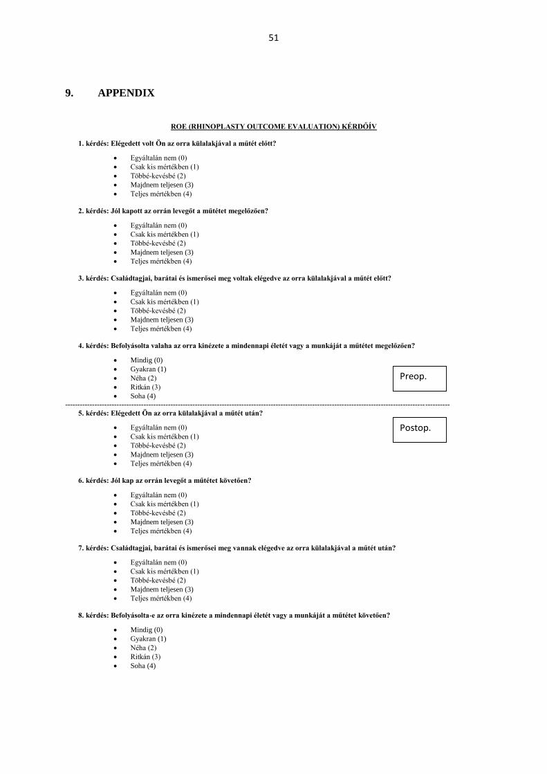
51
9. APPENDIX
ROE (RHINOPLASTY OUTCOME EVALUATION) KÉRDŐÍV
1. kérdés: Elégedett volt Ön az orra külalakjával a műtét előtt?
Egyáltalán nem (0)
Csak kis mértékben (1)
Többé-kevésbé (2)
Majdnem teljesen (3)
Teljes mértékben (4)
2. kérdés: Jól kapott az orrán levegőt a műtétet megelőzően?
Egyáltalán nem (0)
Csak kis mértékben (1)
Többé-kevésbé (2)
Majdnem teljesen (3)
Teljes mértékben (4)
3. kérdés: Családtagjai, barátai és ismerősei meg voltak elégedve az orra külalakjával a műtét előtt?
Egyáltalán nem (0)
Csak kis mértékben (1)
Többé-kevésbé (2)
Majdnem teljesen (3)
Teljes mértékben (4)
4. kérdés: Befolyásolta valaha az orra kinézete a mindennapi életét vagy a munkáját a műtétet megelőzően?
Mindig (0)
Gyakran (1)
Néha (2)
Ritkán (3)
Soha (4)
-------------------------------------------------------------------------------------------------------------------------------------------------------------
5. kérdés: Elégedett Ön az orra külalakjával a műtét után?
Egyáltalán nem (0)
Csak kis mértékben (1)
Többé-kevésbé (2)
Majdnem teljesen (3)
Teljes mértékben (4)
6. kérdés: Jól kap az orrán levegőt a műtétet követően?
Egyáltalán nem (0)
Csak kis mértékben (1)
Többé-kevésbé (2)
Majdnem teljesen (3)
Teljes mértékben (4)
7. kérdés: Családtagjai, barátai és ismerősei meg vannak elégedve az orra külalakjával a műtét után?
Egyáltalán nem (0)
Csak kis mértékben (1)
Többé-kevésbé (2)
Majdnem teljesen (3)
Teljes mértékben (4)
8. kérdés: Befolyásolta-e az orra kinézete a mindennapi életét vagy a munkáját a műtétet követően?
Mindig (0)
Gyakran (1)
Néha (2)
Ritkán (3)
Soha (4)
Postop.
Preop.