Embed Size (px)
Citation preview
Module FModule F
Computed Tomography Computed Tomography Physics, Instrumentation, Physics, Instrumentation,
and Imagingand Imaging

DisclaimerDisclaimer This workforce solution was funded by a grant awarded This workforce solution was funded by a grant awarded
under the President’s Community-Based Job Training Grants under the President’s Community-Based Job Training Grants as implemented by the U.S. Department of Labor’s as implemented by the U.S. Department of Labor’s Employment and Training Administration. The solution was Employment and Training Administration. The solution was created by the grantee and does not necessarily reflect the created by the grantee and does not necessarily reflect the official position of the U.S. Department of Labor. The official position of the U.S. Department of Labor. The Department of Labor makes no guarantees, warranties, or Department of Labor makes no guarantees, warranties, or assurances of any kind, express or implied, with respect to assurances of any kind, express or implied, with respect to such information, including any information on linked sites such information, including any information on linked sites and including, but not limited to, accuracy of the and including, but not limited to, accuracy of the information or its completeness, timeliness, usefulness, information or its completeness, timeliness, usefulness, adequacy, continued availability, or ownership. This adequacy, continued availability, or ownership. This solution is copyrighted by the institution that created it. solution is copyrighted by the institution that created it. Internal use by an organization and/or personal use by an Internal use by an organization and/or personal use by an individual for non-commercial purposes is permissible. All individual for non-commercial purposes is permissible. All other uses require the prior authorization of the copyright other uses require the prior authorization of the copyright owner.owner.

PixelsPixels
Represent segment of the anatomy Represent segment of the anatomy scannedscanned
Contain numerical information based on Contain numerical information based on tissue densitytissue density
Two-dimensional representationTwo-dimensional representation Slice thickness turns pixels into voxels Slice thickness turns pixels into voxels
(volume elements)(volume elements) Expressed in Hounsfield unitsExpressed in Hounsfield units Hounsfield units are directly proportional Hounsfield units are directly proportional
to the attenuation coefficient of the tissueto the attenuation coefficient of the tissue
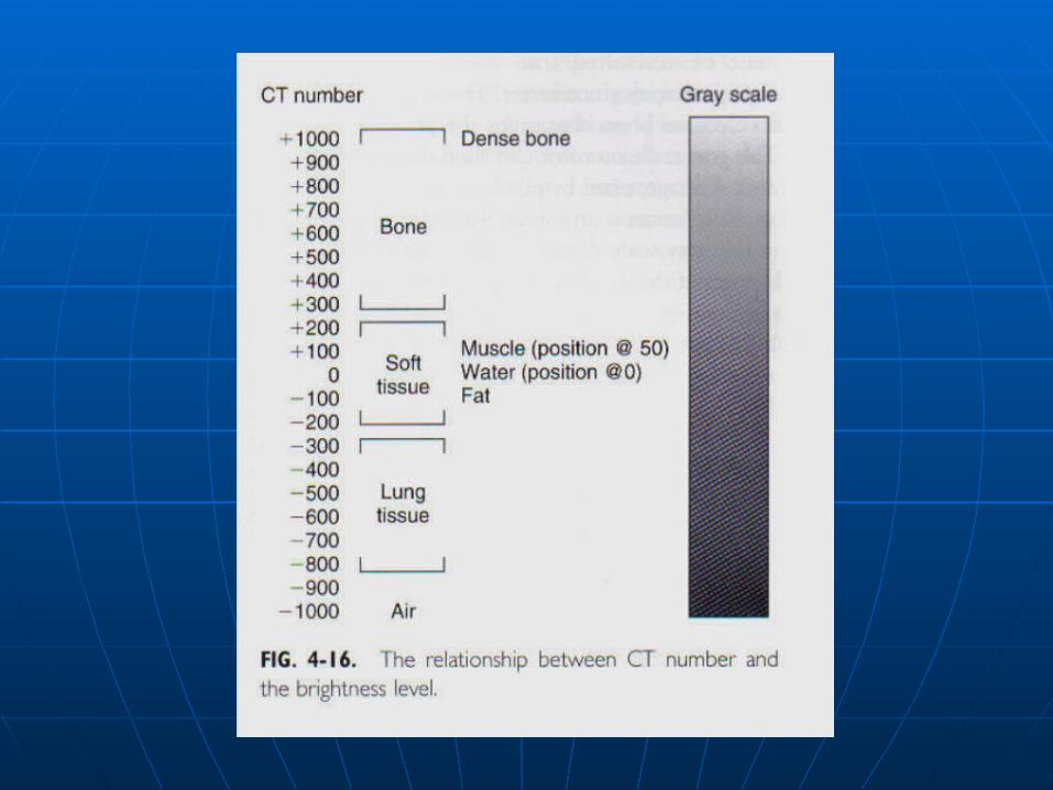
Windowing and LevelingWindowing and Leveling
The process of “windowing” in CT The process of “windowing” in CT Defines the shades of gray of Defines the shades of gray of interest.interest.

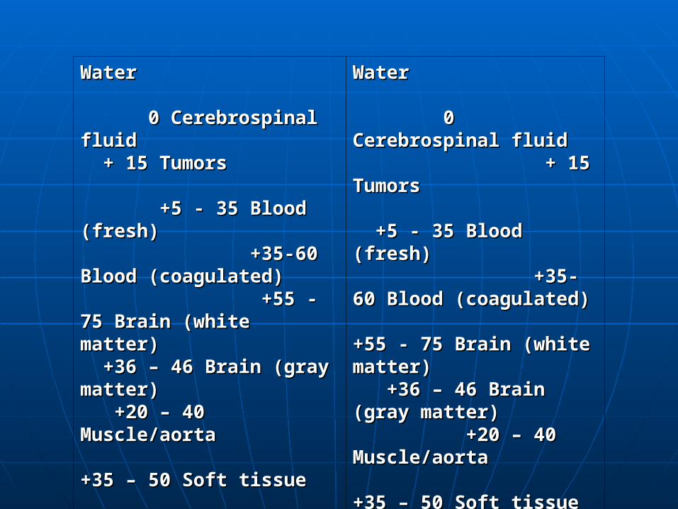
Water Water 0 Cerebrospinal fluid 0 Cerebrospinal fluid + 15 Tumors + 15 Tumors +5 - 35 Blood (fresh) +5 - 35 Blood (fresh) +35-60 Blood +35-60 Blood (coagulated) +55 - (coagulated) +55 - 75 Brain (white matter) 75 Brain (white matter) +36 – 46 Brain (gray matter) +36 – 46 Brain (gray matter) +20 – 40 Muscle/aorta +20 – 40 Muscle/aorta +35 – 50 Soft +35 – 50 Soft tissue +40 – 70 tissue +40 – 70 ( liver, spleen, kidney, pancreas, ( liver, spleen, kidney, pancreas, etc. ) etc. ) Bone (average) Bone (average) +100 – 1000 Petrous bone +100 – 1000 Petrous bone +3000 Air +3000 Air -1000 Lung tissue -1000 Lung tissue -150 – 400 Fat -150 – 400 Fat -100 -100
Water Water 0 Cerebrospinal fluid 0 Cerebrospinal fluid + 15 Tumors + 15 Tumors +5 - 35 Blood +5 - 35 Blood (fresh) +35-(fresh) +35-60 Blood (coagulated) 60 Blood (coagulated) +55 - 75 Brain (white +55 - 75 Brain (white matter) +36 – 46 matter) +36 – 46 Brain (gray matter) Brain (gray matter) +20 – 40 Muscle/aorta +20 – 40 Muscle/aorta +35 – 50 Soft tissue +35 – 50 Soft tissue +40 – 70 +40 – 70 ( liver, ( liver, spleen, kidney, pancreas, etc. ) spleen, kidney, pancreas, etc. ) Bone (average) Bone (average) +100 – 1000 Petrous bone +100 – 1000 Petrous bone +3000 Air +3000 Air -1000 Lung tissue -1000 Lung tissue -150 – 400 Fat -150 – 400 Fat -100 -100
Windowing and LevelingWindowing and Leveling
Window level determines the Window level determines the center of center of the gray scalethe gray scale and is generally set at and is generally set at the average tissue density of the the average tissue density of the structures within the anatomy being structures within the anatomy being scanned.scanned.
Window width is normally set to include Window width is normally set to include other structures or pathology that may other structures or pathology that may also be located in the scan plane.also be located in the scan plane.
Water Water 0 Cerebrospinal fluid 0 Cerebrospinal fluid + 15 Tumors + 15 Tumors +5 - 35 Blood (fresh) +5 - 35 Blood (fresh) +35-60 Blood +35-60 Blood (coagulated) +55 - (coagulated) +55 - 75 Brain (white matter) 75 Brain (white matter) +36 – 46 Brain (gray matter) +36 – 46 Brain (gray matter) +20 – 40 Muscle/aorta +20 – 40 Muscle/aorta +35 – 50 Soft +35 – 50 Soft tissue +40 – 70 tissue +40 – 70 ( liver, spleen, kidney, pancreas, ( liver, spleen, kidney, pancreas, etc. ) etc. ) Bone (average) Bone (average) +100 – 1000 Petrous bone +100 – 1000 Petrous bone +3000 Air +3000 Air -1000 Lung tissue -1000 Lung tissue -150 – 400 Fat -150 – 400 Fat -100 -100
Water Water 0 Cerebrospinal fluid 0 Cerebrospinal fluid + 15 Tumors + 15 Tumors +5 - 35 Blood +5 - 35 Blood (fresh) +35-(fresh) +35-60 Blood (coagulated) 60 Blood (coagulated) +55 - 75 Brain (white +55 - 75 Brain (white matter) +36 – 46 matter) +36 – 46 Brain (gray matter) Brain (gray matter) +20 – 40 Muscle/aorta +20 – 40 Muscle/aorta +35 – 50 Soft tissue +35 – 50 Soft tissue +40 – 70 +40 – 70 ( liver, ( liver, spleen, kidney, pancreas, etc. ) spleen, kidney, pancreas, etc. ) Bone (average) Bone (average) +100 – 1000 Petrous bone +100 – 1000 Petrous bone +3000 Air +3000 Air -1000 Lung tissue -1000 Lung tissue -150 – 400 Fat -150 – 400 Fat -100 -100

DetectorsDetectors
Receive the attenuated x-ray photonsReceive the attenuated x-ray photons For detection to occur the attenuated For detection to occur the attenuated
photon must:photon must: Be captured by the detector chamberBe captured by the detector chamber ““collide” with an atom of the detector materialcollide” with an atom of the detector material ““Collision” must produce an electromechanical Collision” must produce an electromechanical
conversion suitable for measurementconversion suitable for measurement Be able to be amplified and transmittedBe able to be amplified and transmitted
Types of DetectorsTypes of Detectors
Gas DetectorsGas Detectors
Solid-state DetectorsSolid-state Detectors
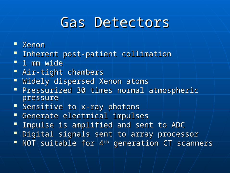
Gas DetectorsGas Detectors Xenon Xenon Inherent post-patient collimationInherent post-patient collimation 1 mm wide1 mm wide Air-tight chambersAir-tight chambers Widely dispersed Xenon atomsWidely dispersed Xenon atoms Pressurized 30 times normal atmospheric Pressurized 30 times normal atmospheric
pressurepressure Sensitive to x-ray photonsSensitive to x-ray photons Generate electrical impulsesGenerate electrical impulses Impulse is amplified and sent to ADCImpulse is amplified and sent to ADC Digital signals sent to array processorDigital signals sent to array processor NOT suitable for 4NOT suitable for 4thth generation CT scanners generation CT scanners
Solid-State DetectorsSolid-State Detectors
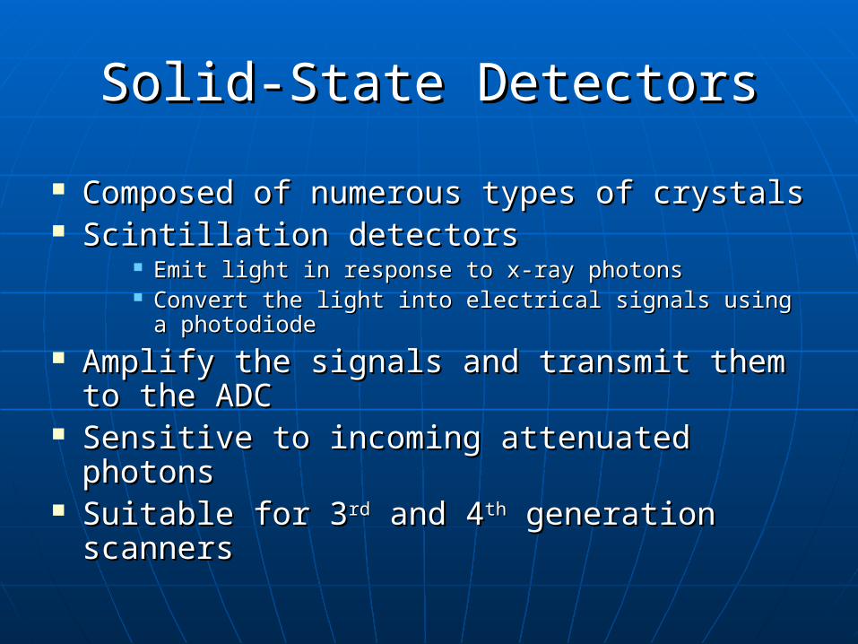
Composed of numerous types of crystalsComposed of numerous types of crystals Scintillation detectorsScintillation detectors
Emit light in response to x-ray photonsEmit light in response to x-ray photons Convert the light into electrical signals using a Convert the light into electrical signals using a
photodiodephotodiode Amplify the signals and transmit them to Amplify the signals and transmit them to
the ADC the ADC Sensitive to incoming attenuated photonsSensitive to incoming attenuated photons Suitable for 3Suitable for 3rdrd and 4 and 4thth generation scanners generation scanners
Hounsfield UnitsHounsfield Units
Also called CT number or an integerAlso called CT number or an integer
An integer is assigned to each An integer is assigned to each amplified electrical signal in the form amplified electrical signal in the form of a Positive or Negative Whole of a Positive or Negative Whole number.number.
The stronger the signal the greater the The stronger the signal the greater the value of the integervalue of the integer
ADCADC
ADC (Analog to Digital Converter)ADC (Analog to Digital Converter)
Signals reaching the ADC are in Signals reaching the ADC are in analog form.analog form.
Signals are converted to integers and Signals are converted to integers and sent to the Array Processorsent to the Array Processor
Array ProcessorArray Processor
special purpose logical processing unitspecial purpose logical processing unit used to perform rapid image used to perform rapid image
reconstructionsreconstructions Computer computationsComputer computations solves all of the complex mathematical solves all of the complex mathematical
problems (reconstructive algorithms) problems (reconstructive algorithms) Reconstructions, retrospectives, post Reconstructions, retrospectives, post
processing techniquesprocessing techniques

Reconstructive AlgorithmsReconstructive Algorithms
Filtered-back projectionFiltered-back projection Simple-back projectionSimple-back projection Convolution Convolution
Convolution and filtered back Convolution and filtered back projection are considered projection are considered analytical analytical reconstruction algorithmreconstruction algorithm
Convolution MethodConvolution Method
Projection profiles are obtainedProjection profiles are obtained Logarithm of data obtainedLogarithm of data obtained Logarithm values are multiplied by a Logarithm values are multiplied by a
digital or convolution filter (kernel)digital or convolution filter (kernel) Filtered profiles are then back-Filtered profiles are then back-
projectedprojected Filtered profiles are addedFiltered profiles are added Results in Results in “blur-free”“blur-free” images images
Raw DataRaw Data
Convolution filters can only Convolution filters can only be applied to raw data or be applied to raw data or (scan data).(scan data).
Convolution filters can NOT Convolution filters can NOT use image data. use image data.

Image DataImage Data
Image Data can be used for post-Image Data can be used for post-processing techniques:processing techniques:
3D reformations3D reformations MIP’sMIP’s Volume rendering etc.Volume rendering etc.
Spiral CTSpiral CT
The data is acquired in Volume rather than The data is acquired in Volume rather than slice by slice.slice by slice.
Filtered-back projection cannot be used Filtered-back projection cannot be used alone to generate images.alone to generate images.
Filtered-back-projection with linear Filtered-back-projection with linear interpolation is used as the reconstruction interpolation is used as the reconstruction algorithm in single-detector-row spiral.algorithm in single-detector-row spiral.
(the exact process depends on the (the exact process depends on the manufacturer)manufacturer)
InterpolationInterpolation
- defined as a mathematical technique used defined as a mathematical technique used to determine the value of a function from to determine the value of a function from known values on either side of it.known values on either side of it.
- This is a mathematical estimation This is a mathematical estimation
technique.technique.
- Linear interpolation is the simplest form of Linear interpolation is the simplest form of interpolation. interpolation.
Linear interpolation equationLinear interpolation equation
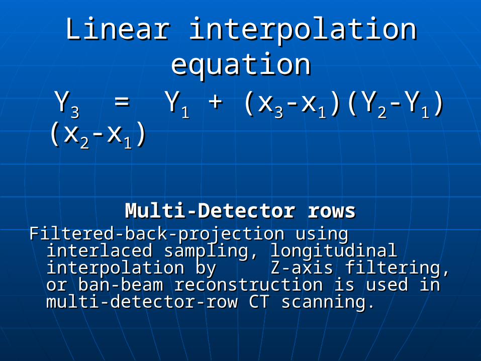
YY33 = Y = Y11 + (x + (x33-x-x11)(Y)(Y22-Y-Y11)(x)(x22--xx11))
Multi-Detector rowsMulti-Detector rowsFiltered-back-projection using interlaced Filtered-back-projection using interlaced
sampling, longitudinal interpolation by sampling, longitudinal interpolation by Z-axis filtering, or ban-beam Z-axis filtering, or ban-beam reconstruction is used in multi-detector-reconstruction is used in multi-detector-row CT scanning.row CT scanning.
Spatial ResolutionSpatial Resolution
-the degree of blurring in an image-the degree of blurring in an image
This is regarded as “the This is regarded as “the measuremeasure of of the the abilityability of a CT scanner to of a CT scanner to discriminatediscriminate objects of varying objects of varying densities located close together, densities located close together, against a uniform background”.against a uniform background”.
Spatial Resolution Spatial Resolution
Represented by:Represented by:• Point Spread Function (PSF)Point Spread Function (PSF)• Line Spread Function (LSF)Line Spread Function (LSF)• Modulation Transfer Function Modulation Transfer Function
(MTF)(MTF)
Geometric factors for Spatial Geometric factors for Spatial resolutionresolution
Focal spot sizeFocal spot size Detector response curveDetector response curve Slice thicknessSlice thickness Focal distanceFocal distance Iso-center (center of rotation of Iso-center (center of rotation of
the gantry)the gantry) Detector and sampling distanceDetector and sampling distance
Contrast resolutionContrast resolution
Contrast resolution, low-Contrast resolution, low-contrast resolution or tissue contrast resolution or tissue resolution is the ability of the resolution is the ability of the CT scanner to demonstrate CT scanner to demonstrate small changes in tissue small changes in tissue contrast.contrast.
Factors which affect low-Factors which affect low-contrast resolutioncontrast resolution
Photon fluxPhoton flux Slice thicknessSlice thickness Patient sizePatient size Detector sensitivityDetector sensitivity
Reconstruction Reconstruction algorithmalgorithm
Image displayImage display Image recordingImage recording Quantum noiseQuantum noise
Reconstruction LimitationsReconstruction Limitations
The ability of the scanner to The ability of the scanner to perform various reconstruction perform various reconstruction process is determined by the process is determined by the scanning parameters chosen by scanning parameters chosen by the Technologist as well as the Technologist as well as equipment characteristics!equipment characteristics!
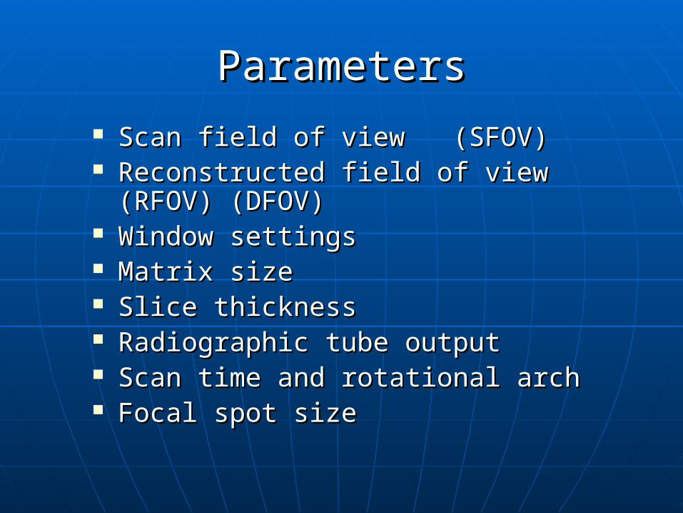
ParametersParameters
Scan field of view (SFOV)Scan field of view (SFOV) Reconstructed field of view (RFOV) Reconstructed field of view (RFOV)
(DFOV)(DFOV) Window settingsWindow settings Matrix sizeMatrix size Slice thicknessSlice thickness Radiographic tube outputRadiographic tube output Scan time and rotational archScan time and rotational arch Focal spot sizeFocal spot size